dynamic control of the yeast ampk/snf1 pathway in response ... · dynamic control of the yeast...
TRANSCRIPT
Lo
ub
na B
end
riou
a
Dyn
amic C
on
trol o
f the Y
east AM
PK
/SN
F1 P
athway in R
espo
nse to
Glu
cose S
ign
als
Loubna Bendrioua
Ph.D. thesisDepartment of Chemistry and Molecular Biology
University of Gothenburg
2014ISBN 978-91-628-9026-1Printed by Ale Tryckteam AB
Dynamic Control of the Yeast AMPK/SNF1 Pathway in
Response to Glucose Signals


THESIS FOR THE DEGREE OF DOCTOR OF PHILOSOPHY
Dynamic control of the yeast AMPK/SNF1 pathway in response to glucose signals
Loubna Bendrioua
Department of Chemistry and Molecular Biology
Gothenburg, Sweden 2014

Cover picture: Brightfield, GFP and mCherry images showing the localization of Mig1 and
Nrd1 localization. The graph represents the ratio of the fluorescence intensity of Mig1 in
the nucleus relative to the cytoplasm.
© Loubna Bendrioua 2014
ISBN: 978-91-628-9026-1
http://hdl.handle.net/2077/35487
Printed by Ale Tryckteam Bohus, Sweden

Abstract
The SNF1/AMP-activated protein kinase (AMPK) belongs to a family of energy sensors
that is conserved in all eukaryotes. Activated by ATP depletion, AMPK plays a vital role
in restoring the energy balance by enhancing energy-generating and damping energy-
requiring processes. Yeast SNF1 is activated by depletion of glucose in the growth
medium but is also affected by other environmental stresses such as salt, oxidative and
alkaline stresses. Currently the regulatory mechanism by which glucose controls the
activity of SNF1 is incompletely understood. The aim of this thesis was to achieve a better
understanding of the glucose regulation of the SNF1/AMPK pathway in the yeast
Saccharomyces cerevisiae. By employing time-lapse imaging of the nucleo-cytoplasmic
shuttling of the transcription factor Mig1, which is directly controlled by Snf1, we revealed
the ability of the Snf1-Mig1 system to monitor not only the changes in glucose
concentrations but also the absolute levels of glucose. It was also found that this system is
highly flexible and rapidly adapts to glucose changes. Monitoring Mig1 migration in cells
expressing different glucose uptake systems indicated that the profile of Snf1-Mig1
activity parallels the characteristics of the expressed hexose transporter, suggesting a firm
link between glucose uptake and the regulation of the SNF1 pathway. Single cell studies of
Mig1 nuclear/cytoplasmic shuttling revealed a significant cell-to-cell variability, which
was studied using nonlinear mixed effects modelling. Our model was able to quantify
characteristics of Mig1 translocation which cannot be directly measured experimentally
such as the time, amplitude and duration of Mig1 transient response. SNF1 shares a
number of structural and functional similarities with its mammalian ortholog AMPK. We
show that different AMPK isoforms confer growth of the snfΔ mutant on SNF1-dependend
carbon sources indicating functional complementation. Moreover, mammalian AMPK
expressed in yeast showed proper regulation by glucose suggesting a conserved mode of
regulation. Our data also showed that compound 991, an AMPK activating drug candidate,
was able to enhance the activity of yeast-expressed AMPK providing scope for employing
yeast for the screening of drugs affecting AMPK activity.
Keywords: AMPK, SNF1, Mig1, glucose signalling, dynamic control, mechanism,
Saccharomyces cerevisiae

Abbreviations
AMPK AMP-activated protein kinase
O2 Oxygen
MFS Major facilitator superfamily
ATP Adenosine triphosphate
ADP Adenosine diphosphate
AMP Adenosine monophosphate
cAMP Cyclic AMP
PP1 Protein phosphatase 1
AIS Autoinhibitory sequence
LKB1 Liver kinase B1
Calmodulin-dependant protein kinase kinase
STRAD Sterile-20-related adaptor
MO25 Mouse-protein 25
Transforming growth factor-beta- activated kinase 1
AMPKK AMPK kinase
Tumour necrosis factor (TNF)-related apoptosis-inducing ligand
PECK Phosphoenolpyruvate carboxykinase
Peroxisome proliferator-activated receptor-γ co-activator 1α
FOXO1 Forkhead box protein O1
5-aminoimidazole-4-carboxamide riboside
SnRKs SNF1-related protein kinases
FC Flow cytometry
FACS Fluorescence-activated flow sorting
CE Capillary electrophoresis
LSC Laser scanning cytometry
LOC Lab-on-chip
µTAS Micro-total analysis systems
LIC Laboratory-in-a-cell
PDMS Poly-dimethylsiloxane
Re Reynolds number
GFP Green fluorescent protein
FISH Fluorescence in situ hybridization
BFP Blue fluorescent protein
YFP Yellow fluorescent protein
CFP Cyan fluorescent protein
FRET Fluorescence resonance energy transfer
FRAP Fluorescence recovery after bleaching
FLIP Fluorescence loss in photo bleaching
NLS Nuclear localization signal
Species
S. cerevisiae Saccharomyces cerevisiae
D. melanogaster Drosophila melanogaster
E. coli Escherichia coli
Nomenclature
The term SNF1 refers to the heterotrimeric complex, whereas Snf1 denotes to the catalytic subunit of the complex
In S. cerevisiae it is common to refer to the gene as SNF1 and to the protein as Snf1.

List of appended papers
This thesis is based on the work conducted in five different papers:
I. Bendrioua L, Smedh M, Almquist J, Cvijovic M, Jirstrand M, Goksör M, Adiels CB, Hohmann S- (2014) Yeast AMP-Activated Protein Kinase Monitors Glucose Concentration Changes as well as Absolute Glucose Levels. J Biol Chem 289: 12863-12875 II. Bendrioua L, Welkenhuysen N, Adiels CB, Goksör M, Hohmann S- The response profile of the Snf1-Mig1 glucose derepression system is tightly coupled to the cell’s capacity for glucose uptake. Manuscript
III. Almquist J, Bendrioua L, Adiels CB, Goksör M, Hohmann S, Jirstrand M- A nonlinear mixed effects approach for modeling the cell-to-cell variability of Mig1 dynamics in yeast. Manuscript for PLOS Computational Biology
IV. Lubitz T, Welkenhuysen N, Bendrioua L, Shashkova S, Klipp E, Krantz M, Hohmann S- Network reconstruction of the yeast Snf1 pathway based on comprehensive literature mining and experimental evidence classification. Manuscript
V. Ye T, Bendrioua L, Carmena D, García-Salcedo R, Dahl P, Carling D and Hohmann S- The mammalian AMP-activated protein kinase complex mediates glucose regulation of gene expression in the yeast Saccharomyces cerevisiae. FEBS Letters- In press

Table of contents
1. Introduction……………………………………………………………... 1 1.1. Aim of this study……………………………………..................... 2
2. Saccharomyces cerevisiae as a model eukaryote…………………….. 4 3. Glucose metabolism: glucose uptake and phosphorylation………… 6
3.1. Yeast metabolism……………………………………................... 6 3.2. Glucose uptake………………………………………................... 6 3.3. Glucose phosphorylation……………………………………….... 8 3.4. Energy metabolism and ATP/ADP/AMP homeostasis…………. 10
4. Glucose signalling pathways…………………………………………… 12
4.1. The Ras-cAMP/ PKA pathway………………………………….. 13 4.2. The Snf3/Rgt2 glucose induction pathway…………………….... 16 4.3. Glucose repression……………………………….......................... 17
4.3.1. The Snf1/Mig1 glucose repression pathway…..………........ 18 4.3.1.1. The transcriptional repressor Mig1……………….... 20
4.4. The AMPK pathway, the mammalian Snf1 ortholog...................... 23 4.4.1. Significance of the AMPK/SNF1 pathway…………….…. 27
4.5. Snf1 orthologs in other eukaryotes…………………………….... 28 5. Single cell analysis……………………………………………………… 30
5.1. Why single cell analysis?............................................................... 30 5.2. Cell variability…………………………………………………... 30 5.3. Single cell dynamics-time resolution………………..................... 32
6. Methods for single cell analysis…………………………………..…… 34
6.1. Microfluidics……………………………………........................... 35 6.2. Optical tweezers……………………………………………….…. 36 6.3. Fluorescence………………………………………………...…… 37
6.3.1. Bleaching…………………………………….......................... 39 6.3.1.1. Bleaching techniques…………………….................. 39
6.4. Technical limitations and drawbacks…………………………….. 40 7. Summary of appended papers……………………………………........ 42 8. Conclusion and future perspectives…………………............................ 45 9. Acknowledgements……………………………………………………... 50 10. References………………………………………………………………. 52


1
1. Introduction
All living cells require energy in order to ensure their biological functions such as
viability, homeostasis, growth and proliferation. For an optimal cellular functioning, there
needs to be a balance between energy supply (usually via means of glycolysis and
respiration) and energy consumption (via e.g. biosynthesis, homeostasis and proliferation).
It is now well established that any disorder in energy balance would lead to serious
diseases such as obesity and obesity-related morbidities including type 2-diabetes,
hypertension, cardiovascular and pulmonary diseases (Hamilton, et al. 2007, Kahn, et al.
2005, Schols, et al. 1991). The major energy sensor in mammalian cells is the AMP-
activated protein kinase (AMPK). AMPK senses energy depletion and restores energy
homeostasis by switching on energy-generating pathways and down-regulating energy-
consuming pathways. Identified in 1981, the yeast Saccharomyces cerevisiae SNF1 was
later found to be similar to AMPK and was therefore referred to as the yeast AMPK
ortholog. It now appears that AMPK exists in all eukaryotes where this protein complex
shares the same fundamental function: maintaining the energy balance. In this thesis, we
employed Saccharomyces cerevisiae as a model eukaryote to investigate the dynamic
regulation of the SNF1 and AMPK signalling pathways.
The SNF1 glucose repression pathway has been intensively studied for nearly 40 years.
Although the core components of this pathway have apparently been identified, several
questions regarding its regulation remain unanswered. For instance, it is still unknown how
the glucose signal is transmitted to SNF1; it is unclear how glucose regulates SNF1
activity and how this pathway behaves in response to different glucose levels. In other
words, how does SNF1 differentiate between high and low glucose levels? Is there a
certain glucose concentration threshold for the response of the pathway? Is it the change in
glucose concentration or the absolute glucose levels or both that trigger SNF1 activation?
It is now believed that glucose generates an internal signal since altering SNF1 activity
requires glucose uptake and phosphorylation. However we still do not know the nature of
this stimulus. AMPK activity appears to be modulated by adenylate ligand binding but
whether this mechanism holds for SNF1 as well is controversial. Recent studies suggest

2
that the main activator for AMPK is indeed AMP while yeast SNF1 appears to be activated
rather by ADP (Chandrashekarappa, et al. 2013, Mayer, et al. 2011).
One way to deal with complex biological systems is to integrate different pieces of
information generated from biological experiments with mathematical modelling. Hence
the transition from molecular to systematic cell biology has enabled the contextualization
and the structuration of biological networks (Alon 2007, Hartwell, et al. 1999). The non-
linearity and interconnectivity of biological networks is not possible to study by basic
molecular biology techniques. Rather, this interconnectivity requires large scale ‘omics’
approaches e.g. genomics, proteomics, as well as quantitative time-resolved data collection
at cell population and single cell levels, combined with computational simulations. Maybe
only then, it would be possible to achieve a comprehensive picture of biological systems.
In this work, we aimed to elucidate the dynamic properties and design principles of the
Snf1 module in response to glucose from a systematic angle starting by addressing the
questions raised above. By combining a range of cutting-edge biological techniques such
as time-lapse fluorescence microscopy, confocal microscopy, microfluidics and optical
tweezers with quantitative mathematical modelling and simulation, we present novel
insights into the dynamics of yeast glucose signalling.
1.1. Aims of this study
The overall goal of this thesis was to achieve a better understanding of the control of the
yeast AMP-activated protein kinase, the Snf1-Mig1 pathway. The AMPK Snf1-Mig1
pathway controls energy homeostasis and, in yeast, glucose repression/derepression and
hence we surmised that this pathway responds rapidly to altered glucose levels. Therefore
we were interested in the underexplored dynamics of the control of this crucial regulatory
system.

3
The overall goal of this thesis can be broken down into five specific research objectives:
1. Employing Mig1 nucleo-cytoplasmic shuttling as a read-out in single cell analysis
we wished to determine the threshold concentrations of glucose that caused changes in
Snf1-Mig1 pathway activity. With this information at hand we wanted to determine the
dynamic characteristics of the pathway around those thresholds as we expected those to
reveal information of pathway control mechanisms.
2. Following up on aim (1) we further wished to determine the effects of altered
glucose uptake activity on the dynamics of the Snf1-Mig1 system. By employing yeast
strains expressing different uptake systems, we aimed at monitoring the effect of glucose
uptake on Mig1 migration in real time.
3. Pursuing aim (1) we also wished to understand the characteristic features and the
mechanisms associated with cell-to-cell variability.
4. Based on reliable information collected from the literature, we aimed to provide a
reconstruction of the yeast Snf1-Mig1 network by using the publicly available rxncon
software. In addition, we wanted to provide a detailed overview of our current
understanding of the yeast Snf1-Mig1 pathway.
5. We aimed to test whether AMPK would be able to substitute SNF1 and mediate
glucose derepression in yeast cells. To this end, we wished to functionally express the
AMPK complex in a yeast strain that is devoid of its SNF1 complex. Furthermore, we
wished to investigate the effect of anti-diabetic drug candidates on the activity of this
yeast-expressed AMPK.
4
2. Saccharomyces cerevisiae as a eukaryotic model organism
The introduction in experimental research of the yeast Saccharomyces cerevisiae (S.
cerevisiae)-baker’s yeast- in the 1930’s has attracted the attention of many biologists. This
unicellular fungus (Fig. 1) combines a number of advantages. Its simplicity, reasonable
growth time, ease of handling and disposal, and more importantly non-pathogenicity made
this organism suitable for the study of various cellular processes. S. cerevisiae was the first
eukaryote whose genome was fully sequenced (Botstein and Fink 2011). The wealth of
genetic information owing to the completion of the yeast genome sequencing project in
1996 (Goffeau, et al. 1996) has opened the possibility for functional analysis. Moreover,
the availability of sequence information has also allowed comparative studies between
yeast and higher eukaryotes e.g. animals and plants. In addition to genome sequencing,
genetic tractability and ease of manipulation has made yeast a versatile system for
investigation of the function and the regulation of genes from other eukaryotes by
expressing those in yeast. A number of the Nobel Prize winning-studies in physiology and
medicine have employed yeast as a model organism. This involves the discovery, by
Hartwell and Nurse, of the different yeast cell cycle, CDC, genes and the roles of
checkpoints in monitoring the different stages of the cell division cycle. Another example
is the striking findings of the role of telomeres and telomerase in the protection of
eukaryotic chromosomes and hence the maintenance of genome stability by Blackburn,
Greider and Szostak. Very recently, Schekman was also awarded for his seminal studies of
the genes that are involved in vesicular trafficking in yeast cells.

5
Figure 1. Bright field image showing the yeast S. cerevisiae

6
3. Glucose metabolism: glucose uptake and phosphorylation
3.1. Yeast metabolism
Metabolism refers to the overall biochemical reactions that take place within living cells
and includes energy-driven anabolic pathways and energy-generating catabolic pathways.
In S. cerevisiae, metabolism depends on two major factors: oxygen availability and the
quality of carbon source in the surrounding environment (Lagunas 1976). Depending on
oxygen levels, yeast can display a respiratory, a respiro-fermentative or a fermentative
lifestyle. However, even though O2 is present, alcoholic fermentation occurs in the
presence of high glucose (Postma, et al. 1989, Pronk, et al. 1996). This effect is referred to
as the Crabtree effect which is characteristic to S. cerevisiae and closely related yeasts. In
addition to glucose, S. cerevisiae is able to ferment other mono-saccharides such as
fructose, mannose and galactose. In its natural habitat, free glucose is rarely present and S.
cerevisiae obtains glucose mainly from the disaccharides sucrose and maltose. While the
hydrolysis of sucrose occurs outside the cell and requires the extracellular form of
invertase, the cleavage of maltose takes place inside the cell. Maltose is taken up via
maltose permease and is hydrolysed to glucose by maltase (α-glucosidase) (Carlson, et al.
1981b, Carlson, et al. 1983, Dubin, et al. 1985). The yeast S. cerevisiae is also able to
consume non-fermentable (respirative) carbon sources such as glycerol, ethanol, acetate
and lactate.
3.2. Glucose uptake
S. cerevisiae commonly lives on the surface of decaying fruits where sugar concentrations
can be extremely high. Since environmental conditions may vary and the sugar
concentration can drop drastically due to, for instance, sugar consumption, S. cerevisiae
has developed a wide range of hexose transporters with distinct glucose affinities.
Following its uptake, glucose is converted through glycolysis into pyruvate and through

7
the action of pyruvate decarboxylase and alcohol dehydrogenase into ethanol. Early studies
(Becker and Betz 1972), have defined glucose uptake as the rate-limiting step of
glycolysis. The yeast hexose transporters are integral plasma membrane proteins belonging
to the major monosaccharide facilitator super family (MFS) (Marger and Saier 1993). The
hexose transporter family includes 18 hexose transporters; Hxt1p-Hxt17p and Gal2p, and
two glucose sensors Snf3p and Rgt2 (reviewed in (Horak 2013). These sensors seem to
have lost their transport ability and serve as glucose sensors with distinct affinities toward
glucose. While Snf3 represents the high affinity glucose sensor and is required for the
induction of HXT2 and HXT4 genes in response to low glucose, Rgt2, its low affinity
counterpart, is necessary for the induction by high glucose concentrations of the HXT1
gene (Ozcan, et al. 1998, Ozcan, et al. 1996). As members of the major facilitator
superfamily (MFS), the yeast hexose transporters contain 12 transmembrane α-helical
domains and mediate transport of sugars by facilitated diffusion. Although these sugar
facilitators have similar structures, they present different affinities toward glucose and
distinct expression profiles (Marger and Saier 1993, Reifenberger, et al. 1997). Depending
on the affinity of their products to glucose the hexose transporters genes can be subdivided
into three different groups; those encoding low affinity transporters represented by HXT1
and HXT3, expression of which is upregulated in the presence of high glucose
concentrations, intermediate affinity transporters including HXT2 and HXT4 expressed at
low glucose concentrations around 0.1% (Ozcan, et al. 1996) and finally high affinity
transporters, HXT6 and HXT7, that are highly expressed in the absence of glucose or at
very low glucose conditions (Maier, et al. 2002, Reifenberger, et al. 1997). Several pieces
of evidence have indicated that the high affinity transport system is repressed by moderate
and high glucose, owing to the absent co-existence of the high and low affinity transport
and to the observed repression of HXT7 transcription at high glucose (Bisson 1988, Bisson,
et al. 1993, Fuhrmann and Völker 1992, Ye, et al. 2001). HXT5 on the other hand, is a
moderate affinity transporter highly expressed at slow growth conditions such as
sporulation, stationary phase and in the absence of non-fermentable carbon sources
(Diderich, et al. 2001). The function and the regulation of Hxt8-Hxt17 are poorly
documented. Although, when overexpressed, these transporters (except HXT12) were able
to mediate glucose transport, they are thought to be involved in processes other than
8
hexose transport (Horak 2013, Wieczorke, et al. 1999). Indeed, Hxt9 and Hxt11 were
found to be involved in pleiotropic drug resistance (Nourani, et al. 1997).
Construction of yeast strains expressing individual hexose transporters has enabled the
characterization of different aspects of hexose transporters (Wieczorke, et al. 1999).
Another approach based on the construction of chimeric hexose transporter proteins by
fusing the amino-terminus of Hxt1 and the carboxy-terminus of Hxt7 has resulted in a
strain with novel characteristics such that the mode of metabolism has been rewired from
fermentation to respiration (Elbing, et al. 2004a, Otterstedt, et al. 2004). We took
advantage of both approaches in order to investigate the effect of glucose uptake on the
behaviour of the Snf1/Mig1 module (Paper II). For this purpose, we employed three
strains expressing each, as a sole glucose uptake system, the low affinity glucose
transporter Hxt1, the high affinity glucose transporter Hxt7 and their respective chimera
TM6*. Furthermore, we followed the migration of GFP-tagged Mig1 under different
glucose up- and downshifts in single cells arrayed in microfluidic devices. We found that
in cells expressing HXT7 or TM6* transporters and growing in high glucose, Mig1
displayed a cytosolic localization and did not respond to any glucose downshift. However,
in HXT1-expressing cells, Mig1 exhibited a rapid and sustained response over a wide range
of concentrations following either glucose up- or downshifts. However, in this strain Mig1
did not translocate to the nucleus when glucose starved cells were shifted to low glucose
levels, consistent with the low affinity characteristic of Hxt1. These studies provide
evidence for a firm link between the Snf1/Mig1 pathway and hexose transporters.
3.3. Glucose phosphorylation
In contrast to hexokinase PI and hexokinase PII which phosphorylate glucose and fructose,
glucokinase appears to phosphorylate glucose and mannose. The fact that there is only 26
to 28% identity in the amino acid sequence between hexokinases and glucokinase might
explain this divergence toward sugar substrate specificity and indicate different
physiological roles (Albig and Entian 1988). Both hexokinases and glucokinase mediate
9
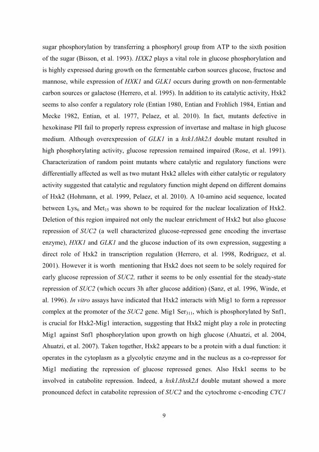
sugar phosphorylation by transferring a phosphoryl group from ATP to the sixth position
of the sugar (Bisson, et al. 1993). HXK2 plays a vital role in glucose phosphorylation and
is highly expressed during growth on the fermentable carbon sources glucose, fructose and
mannose, while expression of HXK1 and GLK1 occurs during growth on non-fermentable
carbon sources or galactose (Herrero, et al. 1995). In addition to its catalytic activity, Hxk2
seems to also confer a regulatory role (Entian 1980, Entian and Frohlich 1984, Entian and
Mecke 1982, Entian, et al. 1977, Pelaez, et al. 2010). In fact, mutants defective in
hexokinase PII fail to properly repress expression of invertase and maltase in high glucose
medium. Although overexpression of GLK1 in a hxk1Δhk2Δ double mutant resulted in
high phosphorylating activity, glucose repression remained impaired (Rose, et al. 1991).
Characterization of random point mutants where catalytic and regulatory functions were
differentially affected as well as two mutant Hxk2 alleles with either catalytic or regulatory
activity suggested that catalytic and regulatory function might depend on different domains
of Hxk2 (Hohmann, et al. 1999, Pelaez, et al. 2010). A 10-amino acid sequence, located
between Lys6 and Met15 was shown to be required for the nuclear localization of Hxk2.
Deletion of this region impaired not only the nuclear enrichment of Hxk2 but also glucose
repression of SUC2 (a well characterized glucose-repressed gene encoding the invertase
enzyme), HXK1 and GLK1 and the glucose induction of its own expression, suggesting a
direct role of Hxk2 in transcription regulation (Herrero, et al. 1998, Rodriguez, et al.
2001). However it is worth mentioning that Hxk2 does not seem to be solely required for
early glucose repression of SUC2, rather it seems to be only essential for the steady-state
repression of SUC2 (which occurs 3h after glucose addition) (Sanz, et al. 1996, Winde, et
al. 1996). In vitro assays have indicated that Hxk2 interacts with Mig1 to form a repressor
complex at the promoter of the SUC2 gene. Mig1 Ser311, which is phosphorylated by Snf1,
is crucial for Hxk2-Mig1 interaction, suggesting that Hxk2 might play a role in protecting
Mig1 against Snf1 phosphorylation upon growth on high glucose (Ahuatzi, et al. 2004,
Ahuatzi, et al. 2007). Taken together, Hxk2 appears to be a protein with a dual function: it
operates in the cytoplasm as a glycolytic enzyme and in the nucleus as a co-repressor for
Mig1 mediating the repression of glucose repressed genes. Also Hxk1 seems to be
involved in catabolite repression. Indeed, a hxk1Δhxk2Δ double mutant showed a more
pronounced defect in catabolite repression of SUC2 and the cytochrome c-encoding CYC1

10
genes as compared to the hxk2Δ single mutant (Ma and Botstein 1986). Moreover, when
overexpressed, Hxk1 seemed to partially restore glucose repression of maltase and
invertase (Rose, et al. 1991).
Hxk2 exists in a dimer-monomer equilibrium where only the monomeric form but not the
dimer appears to be phosphorylated (Randez-Gil, et al. 1998). Hxk2 is phosphorylated in
the absence of glucose at Ser14, possibly by Snf1 (Fernandez-Garcia, et al. 2012, Kriegel,
et al. 1994) and dephosphorylated in the presence of glucose by the Reg1-Glc7
phosphatase complex. It has been proposed that phosphorylation and dephosphorylation at
Ser14 are responsible for Hxk2 nuclear export and import, respectively, since
phosphorylated Hxk2 binds the exportin Xpo1 and dephosphorylated Hxk2 binds the α/β
importin Kap60 (Fernandez-Garcia, et al. 2012). This nucleo-cytoplasmic shuttling pattern
is analogous to other transcription factors such as the glucose repressor Mig1 (see section
on Mig1 below) and the calcineurin-responsive zinc finger Crz1 (Cyert 2003, De Vit, et al.
1997). Hence, these results suggest a model where Snf1 phosphorylation of Hxk2 mediates
its nuclear export under low levels of glucose, while Reg1-Glc7 dephosphorylates Hxk2
under high glucose levels promoting its nuclear localization, thereby participating in the
repression of Mig1 repressible-genes. This is contradictory to the previously reported
epistatic relationship of SNF1 to HXK2 suggesting that Hxk2 acts upstream of Snf1 in the
glucose-repression pathway (Neigeborn and Carlson 1984). In the same line, Prieto’s
group has reported that Snf1 is not the kinase responsible for Hxk2 phosphorylation since
the phosphorylation of the Hxk2 monomer upon shift from glucose to galactose or ethanol
was unaffected by deletion of SNF1 gene (Randez-Gil, et al. 1998). Altogether, the
physiological relationship between Hxk2, Snf1 and Reg1-Glc7 remains to be elucidated.
3.4. Energy metabolism and ATP/ADP/AMP homeostasis
All living cells require energy as a fuel to drive diverse biosynthetic functions such as cell
growth and proliferation. However, Lagunas has reported that as much as 60% of ATP
produced by yeast cells appears to be spent on processes that are not directly associated to
biomass production but rather to futile cycles and pH homeostasis in the cytoplasm and the

11
vacuole (Lagunas 1976, Verduyn, et al. 1991). S. cerevisiae utilizes sugars, most
preferably glucose, as a source of energy. During glycolysis, two molecules of the energy-
rich molecule ATP are consumed while four ATP molecules are generated from each
molecule of glucose. In addition to ATP, some of the intermediary metabolites are used for
biosynthesis. For an optimal functioning of the cell, there needs to be a balance between
energy production and energy consumption. This energy balance is owed to the ubiquitous
adenylate kinase which catalyses nucleotide interconversion reaction (2 ADP ATP
+ AMP) and hence plays an important role in the balance of cellular adenine nucleotide
pool in all organisms (Dzeja, et al. 2007, Dzeja and Terzic 2009, Noma 2005). In yeast, the
major form of the adenylate kinase, Aky2 is present in both mitochondria and cytoplasm
and is required for growth on non-fermentable carbon sources (Bandlow, et al. 1988).
According to the aforementioned reaction, any minor changes in the equilibrium between
ATP and ADP would result in large alterations of AMP levels. Hence, a drop in cellular
energy resulting in an increase in ADP, would cause adenylate kinase to shift the reaction
toward the production of ATP and AMP (reviewed in (Hardie, et al. 2012). Thus, it is not
surprising that the ratio of levels of AMP to ATP is an indicator of the energy status in the
cell. The involvement of adenosine nucleotides in the regulation of energy homeostasis
was proposed in 1964 when Atkinson et al., observed that ATP inhibits the activity of the
yeast phosphofructokinase and that this inhibition is overcome by AMP (Ramaiah, et al.
1964). In addition to phosphofructokinase, the glycogen-catalysing enzyme, glycogen
phosphorylase, and the gluconeogenic enzyme, fructose-1,6-biphosphatase are also
regulated by the AMP:ATP ratio (Hardie, et al. 2012). Likewise, the mammalian AMP-
activated protein kinase and its yeast counterpart SNF1 are regulated by AMP and ADP
respectively (Chandrashekarappa, et al. 2013, Hardie, et al. 2003, Mayer, et al. 2011).

12
4. Glucose signalling pathways
All living cells are able to sense and respond to different environmental stimuli by the
means of distinct signalling pathways leading thus to a meticulous regulation of gene
expression. In the yeast S. cerevisiae as in other microorganisms, nutrient availability is
one of the major factors affecting growth and differentiation. Being the most preferred
carbon and energy sources, glucose (or fructose) represents a global regulator of cell
growth and metabolism. Addition of glucose to yeast cells growing on non-fermentable
carbon sources induces not only the genes required for its uptake and metabolism, but also
the genes responsible for growth and proliferation. Moreover, glucose represses the
expression of the genes that are required for respiration and the utilization of alternative
carbon sources. This mechanism is referred to as glucose repression and is mediated by the
Snf1/Mig1 pathway. By a mechanism called glucose inactivation, glucose triggers the
inactivation and/or the degradation of a number of already existing enzymes such as
fructose-1,6-bisphosphatase (Holzer 1976). This transcriptional and metabolic effect of
glucose involves various signalling networks such as the Ras /PKA, Gpr1/PKA,
Snf3/Rgt2, Snf1/Mig1, Hap and Sch9 (Conrad, et al. 2014). Although the core
components of most of these pathways have presumably been identified, the mechanism
by which glucose activates some of these pathways is poorly understood. It is now thought
that the increase in intracellular pH generated by glucose is the stimulus of the Ras/PKA
pathway. Moreover, it has been speculated that the G-protein coupled receptor Gpr1
functions as a glucose receptor and that the binding of glucose to Gpr1 promotes the
activation of Gpa2 and subsequently the activation of the Gpr1/PKA pathway (Lemaire, et
al. 2004). The fact that Snf1 activity is altered by glucose phosphorylation implies that the
glucose signal occurs internally. Accordingly, a model that links metabolites such as
adenosine nucleotides to the glucose regulation of Snf1 activity has been proposed. In this
model, the low energy adenylate ligand seems to bind to the SNF1 heterotrimer protecting
it from dephosphorylation by the phosphatase (Chandrashekarappa, et al. 2013, Mayer, et
al. 2011, Townley and Shapiro 2007).

13
4.1. The Ras-cAMP/ PKA pathway
The Ras-cAMP pathway (Fig. 2) is a central glucose-regulated signalling pathway that is
required for growth on glucose. Indeed, activation of this pathway by glucose addition
affects 90% of glucose-induced transcriptional changes (Zaman, et al. 2009). In cells
growing on non-fermentable carbon sources or in stationary phase, the cAMP-dependent
protein kinase (PKA) is present as an inactive hetero-tetramer. This complex is composed
of two catalytic subunits, encoded by three redundant genes, TPK1, TPK2 and TPK3, and
two identical regulatory subunits encoded by BCY1 (Conrad, et al. 2014). Addition of
rapidly fermentable carbon sources, such as glucose, results in the activation of the
adenylate cyclase Cyr1 (Cdc35) which synthesizes cyclic AMP (cAMP) from ATP. This
results in a rapid but transient increase in the intracellular cAMP concentration.
Subsequently, cAMP binds to the Bcy1 subunits promoting its release from the tetramer
and resulting in the activation of PKA. Similar to the situation in mammalian cells, yeast
Cyr1 is activated by the small GTP-binding proteins Ras1 and Ras2 (Kataoka, et al. 1985).
Ras1/2 are plasma membrane-attached proteins that continuously cycle between the GDP-
bound inactive and the GTP-bound active states (Broek, et al. 1987, Toda, et al. 1985). Ras
activation is promoted by the guanine nucleotide exchange factor Cdc25 and its homolog
Sdc25 (Camonis, et al. 1986, Damak, et al. 1991), which exchange GDP for GTP. Ras is
negatively regulated by Ras-GTPase activating proteins (Gap) Ira1 and Ira2 (Tanaka, et al.
1989, Tanaka, et al. 1990). In addition to Ras protein, the G-protein coupled receptor Gpr1
and the α-subunit of the G-protein Gpa2 also participate in the activation of PKA through
the activation of adenylate cyclase (De Vries and Gist Farquhar 1999, Guan and Han 1999,
Kraakman, et al. 1999, Kubler, et al. 1997). Hence two signalling branches seem to
activate adenylate cyclase; the Ras system and the Gpa2/Gpr1 system. It has been argued
that intracellular acidification caused by glucose addition, but not glucose itself, triggers
the Ras system and that stimulation of the Gpa2/Gpr1 system specifically depends on
glucose (Arguelles, et al. 1990, Colombo, et al. 1998, Dechant, et al. 2010, Kraakman, et
al. 1999). A model where Gpr1 functions as a glucose receptor, which interacts with and
activates Gpa2, which in turn stimulates adenylate cyclase, has been proposed (Xue, et al.
1998, Yun, et al. 1997). However, several pieces of evidence are not consistent with this

14
model; the lack of pharmacological assays for Gpr1-ligand binding, the weak affinity of
Gpr1 for glucose and the absence of transcriptional effects after glucose addition in gpa2Δ
and gpr1Δ mutants craves for the need of further studies (Broach 2012, Lemaire, et al.
2004, Zaman, et al. 2009). Active PKA targets a number of effectors such as Msn2, Msn4,
Yak1 and Rim15 (Conrad, et al. 2014, Pedruzzi, et al. 2003, Thevelein and de Winde
1999) affecting the transcriptional regulation of a number of genes whose products are
involved in the stimulation of mass accumulation, stress resistance and cell cycle
progression. All these effects converge into one ultimate outcome, which is the regulation
of cell growth in response to the quality and quantity of the available carbon source.

15
Figure 2. The Ras-cAMP/PKA pathway. The uptake of glucose by hexose transporters (Hxt) and its phosphorylation by hexokinases (Hxk) generates an intracellular signal that activates the Ras-cAMP pathway. Ras1,2 are two plasma membrane-attached proteins that constantly cycle between the GDP and GTP states due to the activity of the guanine nucleotide exchange Cdc25 and its homolog Sdc25 (only Cdc25 is depicted) and the Ras-GTPase activating proteins (Gap) Ira1 and Ira2. The Ras-GTP-bound form binds adenylate cyclase (Cyr1), which catalyses the production of cyclic AMP (cAMP) from ATP. Extracellular glucose seems to be detected by the Gpr1-Gpa2 system. The synthesized cAMP activates PKA by binding to its inhibitory subunit, Bcy1. Active PKA targets the effectors, Rim15, Msn2 and Msn4 in order to stimulate cell cycle progression and proliferation and dampen the expression of stress genes.

16
4.2. The Snf3/Rgt2 glucose induction pathway To ensure an optimal production of energy, yeast cells have adapted a system (Fig. 3) for
the upregulation of glycolytic genes and, most importantly, for the induction of glucose
transporter encoding genes. Snf3 and Rgt2 are very similar sensor proteins (Ozcan, et al.
1996), which confer intracellular glucose signalling via their long cytoplasmic C-termini.
The ultimate target of the Snf3/Rgt2 signalling cascade is the transcription factor Rgt1.
Rgt1 is a Zn2Cys6 DNA-binding protein that negatively regulates the transcription of HXT
genes during glucose depletion (Kim, et al. 2003). In the absence of glucose, Rgt1 forms a
repressing complex at the promoter of HXT genes together with the co-repressors Mth1
and Std1 (Lakshmanan, et al. 2003) and the general repressors Tup1-Cyc8 (Ssn6).
However, the presence of glucose promotes the phosphorylation of Std1 and Mth1 by the
casein kinases I, Yck1 and Yck2, targeting them to degradation by ubiquitination (Flick, et
al. 2003b, Moriya and Johnston 2004, Spielewoy, et al. 2004). Consequently, Rgt1
becomes hyper-phosphorylated causing an intramolecular interaction between its zinc-
binding and central domains thus preventing DNA binding. Several pieces of evidence
(Flick, et al. 2003a, Jeong, et al. 2004, Jouandot, et al. 2011, Mosley, et al. 2003) suggest
that the hyper-phosphorylation of Rgt1 not only interferes with its DNA binding but it also
converts Rgt1 into an activator required for the maximal induction of HXT1. Thus, Rgt1
has a dual function where it acts as a repressor in the absence of glucose and an activator in
the presence of glucose, depending on its phosphorylation state (Jouandot, et al. 2011).
4.2. The Snf3/Rgt2 glucose induction pathway
17
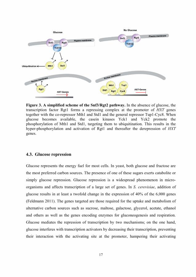
Figure 3. A simplified scheme of the Snf3/Rgt2 pathway. In the absence of glucose, the transcription factor Rgt1 forms a repressing complex at the promoter of HXT genes together with the co-repressor Mth1 and Std1 and the general repressor Tup1-Cyc8. When glucose becomes available, the casein kinases Yck1 and Yck2 promote the phosphorylation of Mth1 and Std1, targeting them to ubiquitination. This results in the hyper-phosphorylation and activation of Rgt1 and thereafter the derepression of HXT genes.
4.3. Glucose repression
Glucose represents the energy fuel for most cells. In yeast, both glucose and fructose are
the most preferred carbon sources. The presence of one of these sugars exerts catabolite or
simply glucose repression. Glucose repression is a widespread phenomenon in micro-
organisms and affects transcription of a large set of genes. In S. cerevisiae, addition of
glucose results in at least a twofold change in the expression of 40% of the 6,000 genes
(Feldmann 2011). The genes targeted are those required for the uptake and metabolism of
alternative carbon sources such as sucrose, maltose, galactose, glycerol, acetate, ethanol
and others as well as the genes encoding enzymes for gluconeogenesis and respiration.
Glucose mediates the repression of transcription by two mechanisms; on the one hand,
glucose interferes with transcription activators by decreasing their transcription, preventing
their interaction with the activating site at the promoter, hampering their activating

18
functions, or by inducing their nuclear exclusion. On the other hand glucose facilitates the
action of transcription repressors (Gancedo 1998). The central pathway for glucose
repression is the Snf1/Mig1 system.
4.3.1. The Snf1/Mig1 glucose repression pathway
The central regulator of glucose repression is the well-established Snf1/Mig1 pathway
(Fig. 4). Snf1 was identified in 1981 by screening for mutants that are unable to utilize
sucrose (Carlson, et al. 1981a) and was later on found to be allelic to cat1 and ccr1
mutants (Ciriacy 1977, Zimmermann, et al. 1977). The SNF1 gene encodes a Ser/Thr
kinase usually found in a complex (SNF1) together with one of the three β subunits (Sip1,
Sip2 and Gal83) and the γ subunit Snf4 (Celenza and Carlson 1986, Celenza, et al. 1989).
Activated by glucose depletion, Snf1 stimulates expression of genes that are required for
the uptake and the utilization of other carbon sources, gluconeogenic genes and genes
required for respiration (reviewed in (Carlson 1999, Gancedo 1998)). In addition to
nutrient limitation, SNF1 is also involved in the regulation of other cellular processes such
as meiosis and sporulation, aging, fatty acid metabolism, glycogen synthesis, fitness and
stress resistance (Ashrafi, et al. 2000, Hardy, et al. 1994, Honigberg and Lee 1998,
Navarro and Igual 1994, Zhang, et al. 2013). Activation of SNF1 requires phosphorylation
on Thr210 in the activation loop of the catalytic subunit, Snf1 (Estruch, et al. 1992, Nath, et
al. 2003). Any of the three partially redundant upstream kinases Sak1, Tos3 and Elm1 is
able to mediate this phosphorylation, with Sak1 being the major activating kinase
(Estruch, et al. 1992, Hong, et al. 2003a, Liu, et al. 2011, Nath, et al. 2003, Sutherland, et
al. 2003). In addition to Snf1 phosphorylation, Snf4 is also required for Snf1 activity. In
response to glucose limitation, Snf4 binds the C-terminal regulatory domain of Snf1
preventing the auto-inhibition of the kinase domain by the C-terminus (Jiang and Carlson
1996). This mechanism was confirmed by structural analyses of the yeast SNF1
heterotrimer indicating that the interaction between Snf1 and Snf4 occurs at the regulatory
region of Snf1 illustrating the role of this interaction in the regulation of Snf1 activity
(Amodeo, et al. 2007). The β-subunits interact with both Snf1 and Snf4 and serve not only

19
to stabilize the Snf1-Snf4 association in the complex but also to regulate the subcellular
localization of the SNF1 complex and substrate definition (Jiang and Carlson 1996,
Schmidt and McCartney 2000, Vincent, et al. 2001). Under glucose abundance, all the β-
subunits are distributed in the cytosol. However, upon glucose limitation each subunit is
differentially localized with Gal83 being the only subunits that localizes to the nucleus
(Hedbacker, et al. 2004). The β-subunits seem to also confer upstream kinase specificity
depending on growth conditions (Hedbacker, et al. 2004, McCartney, et al. 2005).
Recent structural studies (Chandrashekarappa, et al. 2013) suggested that Snf1
phosphorylation promotes the association of the kinase domain with the heterotrimer core
resulting in the remodelling of the activation loop and its exposure to both kinases and
phosphatases. SNF1 is negatively regulated mostly by the Reg1-Glc7 protein phosphatase1
(PP1). The catalytic subunit Glc7 is targeted to SNF1 by the Reg1 subunit resulting in Snf1
dephosphorylation and return to the auto-inhibitory status (Sanz, et al. 2000, Tu and
Carlson 1995). It has been proposed that the recruitment of Glc7 by Reg1 affects the
conformation of SNF1 complex (Sanz, et al. 2000). Under derepressing conditions, Snf1
phosphorylates Reg1, probably promoting its release from the kinase complex (Sanz, et al.
2000). Additional phosphatases including Sit4 and Ptc1 appear to also contribute to Snf1
dephosphorylation (Ruiz, et al. 2011, Ruiz, et al. 2013). Whether glucose regulates SNF1
activity through either phosphorylation or dephosphorylation or both is not clear.
Rubenstein and co-workers (Rubenstein, et al. 2008) have shown that glucose most likely
regulates the accessibility of the phosphatase to the Snf1 activation loop. This is consistent
with the recent structural analyses of both AMPK and SNF1 suggesting that ADP might
induce a reorganization of the complex into a phosphatase-resistant form
(Chandrashekarappa, et al. 2013, Gowans, et al. 2013, Mayer, et al. 2011).
It is still unclear how the presence of glucose affects Snf1 activity. Given the structural and
functional similarities between Snf1 and its mammalian ortholog AMPK it is thought that
the activity of SNF1 is also regulated by adenosine nucleotides. Indeed, in vivo studies
have indicated the existence of a positive correlation between SNF1 activity and nucleotide
ratios (e.g. AMP: ATP) (Wilson, et al. 1996). However in these, and in other studies
(Mitchelhill, et al. 1994, Woods, et al. 1994), SNF1 was not activated allosterically by

20
AMP. Comparative studies of the AMP-binding site between the AMPK γ-subunit and
Snf4 revealed that the latter contains a His151Gly substitution. This residue is critical for
AMP binding to the AMPK γ-subunit and may explain why AMP does not bind to the
Snf1-Snf4 complex. Recent studies (Mayer, et al. 2011) suggested that ADP, but not AMP,
might be the “long-sought” stimulator for Snf1 phosphorylation. Although, ADP did not
allosterically regulate SNF1 activity, it protected the enzyme from dephosphorylation.
Moreover, binding studies showed that Snf4 contains two nucleotide-binding sites and that
ADP binding to the weaker site confers protection of Thr210 against dephosphorylation.
ADP was also found to bind to the γ-subunit of the AMPK heterotrimer and participates in
AMPK activation albeit not to the same extent as AMP (Oakhill, et al. 2011). In addition it
appears that ADP-binding to the ATP-binding pocket of the Snf1 active site independently
of the β- and γ- subunits may also protect Snf1 from dephosphorylation
(Chandrashekarappa, et al. 2013). Taken together, although the regulation of AMPK by
adenylate ligands appears to be well established, the effect of these nucleotides on Snf1
regulation remains opaque and hence requires further investigations, in particular in vivo.
Active Snf1 promotes the phosphorylation of several effectors including the transcriptional
activators Sip4, Cat8, Adr1, Rds2, Hsf1 and the repressor Mig1 (Hahn and Thiele 2004,
Lesage, et al. 1996, Randez-Gil, et al. 1997, Ratnakumar, et al. 2009, Smith, et al. 1999,
Soontorngun, et al. 2007).
4.3.1.1. The Transcriptional repressor Mig1
The Multi-copy Inhibitor of GAL gene expression, Mig1, is a Cys2His2 zing finger DNA
binding protein that binds the promoter of several glucose-repressed genes including SUC,
GAL and MAL genes (Flick and Johnston 1992, Griggs and Johnston 1991, Hu, et al. 1995,
Nehlin and Ronne 1990). Active Snf1 phosphorylates Mig1 at at least four different sites
disrupting its interaction with the Tup1-Cyc8 complex and promoting its nuclear extrusion
(Papamichos-Chronakis, et al. 2004, Smith, et al. 1999, Treitel, et al. 1998, Östling and
Ronne 1998). The phosphorylation of Mig1 by Snf1 seems to be regulated by glucose

21
since phosphorylated active Snf1 does not mediate Mig1 phosphorylation in the presence
of glucose (García-Salcedo, et al. 2014, Ye, et al. 2008). A model was proposed where
Hxk2 interacts with both Mig1 and Snf1 in the presence of glucose thereby conferring
protection of the Ser311 residue of Mig1 from phosphorylation by Snf1 (Ahuatzi, et al.
2004, Ahuatzi, et al. 2007). DNA-binding of Mig1 requires a GC-box core and an adjacent
AT-rich region which facilitates Mig1 access to the DNA (Lundin, et al. 1994). Mig1
mediates repression by interaction with the Ssn6-Tup1 general co-repressor complex
(Papamichos-Chronakis, et al. 2004, Treitel and Carlson 1995). This interaction appears to
be disrupted by the conformational change of Mig1 following phosphorylation by Snf1,
leading to derepression of glucose-repressed genes (Papamichos-Chronakis, et al. 2004). It
is now well established that the nuclear-cytoplasmic localization of Mig1 is glucose
regulated. Mig1 enters to the nucleus within a few seconds after glucose addition and
quickly exits the nucleus after glucose removal (Bendrioua, et al. 2014, DeVit and
Johnston 1999). In addition to Snf1, the nuclear export of Mig1 is dependent on the
nuclear transporter, Msn5 (De Vit, et al. 1997, DeVit and Johnston 1999, Papamichos-
Chronakis, et al. 2004). Msn5 is a β-importin homolog that mediates the export of Mig1
from the nucleus after its phosphorylation by Snf1. In the absence of Msn5, Mig1 is
normally phosphorylated and properly represses GAL1 transcription but its ability to exit
the nucleus is impaired (DeVit and Johnston 1999). Although the export mechanism of
Mig1 has been established, the mechanism by which Mig1 is imported to the nucleus
remains elusive. Mig1 has a nuclear localization signal (NLS) in the C-terminal domain
which is required for its nuclear import after glucose addition (DeVit and Johnston 1999).
Snf1-dependent phosphorylation of Mig1 is antagonized by the Reg1-Glc7 phosphatase
complex although it is not entirely clear if Glc7-Reg1 directly dephosphorylates Mig1 or
mediates its effect by controlling Snf1. In glucose-grown cells bearing reg1Δ mutation,
Mig1 displayed a reduced mobility shift reflecting its partial phosphorylation (McCartney
and Schmidt 2001). Moreover, in the absence of Reg1 Mig1 displayed an inefficient
nuclear localization (De Vit, et al. 1997). Finally, Schmidt and co-workers have
demonstrated that deletion of REG1 increased Mig1 phosphorylation under both high and
low glucose suggesting a constitutive activity of Reg1-Glc7 toward Mig1 (Rubenstein, et
22
al. 2008). However, although the Snf1-Mig1 system has been studied extremely well in
terms of protein-protein interactions, a direct interaction between Mig1 and Glc7-Reg1 has
not been reported so far. Hxk2 seems to also participate, most likely indirectly, in Mig1
dephosphorylation. Genetic analyses from our group have indicated that Hxk2 appears to
facilitate the role of the phosphatase Reg1 in Mig1 dephosphorylation (Ye et al.,
unpublished results).
Mig2 and Mig3 are two other Cys2His2 zinc finger transcription factors that share similar
DNA-binding specificity with Mig1 (Lutfiyya, et al. 1998, Lutfiyya and Johnston 1996)
but their function and the mode of regulation are significantly different. Like Mig1, Mig2
binds to GC-rich sequences at the promoter DNA. However, in contrast to Mig1, Mig2
does not seem to be phosphorylated by Snf1 and its nuclear-cytoplasmic localization does
not seem to be regulated by glucose (Lutfiyya, et al. 1998). Instead, expression of MIG2 is
upregulated by glucose in a Snf3/Rgt2 dependent manner (Kaniak, et al. 2004). Mig3 on
the other hand is regulated by glucose at different levels; the expression of MIG3 gene is
upregulated by glucose and Mig3 is degraded after phosphorylation by Snf1 in the absence
of glucose (Dubacq, et al. 2004). Mig1 and Mig2 cooperate to mediate repression of
glucose-repressed genes where the individual contributions of Mig1 and Mig2 vary in a
gene-specific manner (Westholm, et al. 2008). Mig3 seems to marginally participate in
catabolite repression but appears to be involved in genotoxic stress and aging (Dubacq, et
al. 2004, Lutfiyya and Johnston 1996, Westholm, et al. 2008).
23
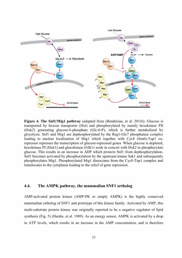
Figure 4. The Snf1/Mig1 pathway (adapted from (Bendrioua, et al. 2014)). Glucose is transported by hexose transporter (Hxt) and phosphorylated by mainly hexokinase PII (Hxk2) generating glucose-6-phosphate (Glc-6-P), which is further metabolized by glycolysis. Snf1 and Mig1 are dephosphorylated by the Reg1-Glc7 phosphatase complex leading to nuclear localization of Mig1 which together with Cyc8 (Ssn6)-Tup1 co-repressor represses the transcription of glucose-repressed genes. When glucose is depleted, hexokinase PI (Hxk1) and glucokinase (Glk1) work in concert with Hxk2 to phosphorylate glucose. This results in an increase in ADP which protects Snf1 from dephosphorylation. Snf1 becomes activated by phosphorylation by the upstream kinase Sak1 and subsequently phosphorylates Mig1. Phosphorylated Mig1 dissociates from the Cyc8-Tup1 complex and translocates to the cytoplasm leading to the relief of gene repression.
4.4. The AMPK pathway, the mammalian SNF1 ortholog
AMP-activated protein kinase (AMP-PK or simply AMPK) is the highly conserved
mammalian ortholog of SNF1 and prototype of this kinase family. Activated by AMP, this
multi-substrate protein kinase was originally reported to be a negative regulator of lipid
synthesis (Fig. 5) (Hardie, et al. 1989). As an energy sensor, AMPK is activated by a drop
in ATP levels, which results in an increase in the AMP concentration, and is therefore

24
activated by metabolic stresses including exercise, glucose depletion, hypoxia,
hyperosmotic and heat shock, a number of hormones and pharmacological drugs (reviewed
in (Hardie 2007, Hardie, et al. 2012)). Active AMPK operates by turning off ATP-
consuming anabolic pathways such as lipid, protein and glycogen synthesis and by turning
on ATP-generating catabolic pathways for instance glucose and fatty-acid metabolism. In
the brain, activation of AMPK by low glucose stimulates eating desire. In analogy to
SNF1, AMPK is an αβγ heterotrimeric complex. While yeast possesses only one isoform
of the α- and γ-subunits and three isoforms of the β-subunits, the AMPK trimer contains
two alterative α-catalytic subunits (α1 and α2), two scaffolding β-subunits (β1 and β2) and
three regulatory γ-subunits (γ1, γ2 and γ3). Sequencing of the mammalian AMPK α-subunit
kinase domain has revealed a strong amino acid sequence homology with that of the S.
cerevisiae Snf1 catalytic subunit indicating conserved structural and functional features
(Gao, et al. 1995, Mitchelhill, et al. 1994). Like Snf1, the AMPKα contains the auto-
inhibitory sequence (AIS) and binds to β- and γ-subunits through its C-terminus (Crute, et
al. 1998, Dyck, et al. 1996).
AMPK is activated by phosphorylation at the Thr172 in the activation loop of the catalytic
α-subunit (Hawley, et al. 1996). This phosphorylation is mediated by the upstream kinases,
the tumour suppressor LKB1 and the two calmodulin-dependent protein kinase kinases,
CaMKKα and CaMKKβ. LKB1 is the major activating kinase (Hawley, et al. 2003,
Hawley, et al. 2005). LKB1 functions conjointly with the pseudo-kinase STRAD (sterile-
20-related adaptor) and the scaffolding protein MO25 (mouse-protein 25) (Hawley, et al.
2003). The identification in a genetic screen of the transforming growth factor-beta-
activated kinase 1 (TAK1) as a Snf1-activating kinase in a triple mutant lacking all the
upstream kinases suggest that this mitogen-activated protein kinase kinase kinase might be
an additional AMPKK (Momcilovic, et al. 2006). Indeed, further genetic analyses
indicated that the cytokine tumour necrosis factor (TNF)-related apoptosis-inducing ligand
(TRAIL) induced activation of AMPK is dependent on TAK1 (Herrero‐Martín, et al.
2009). However, how TAK1 regulates the activity of AMPK in vivo needs to be further
investigated. While activation of AMPK by LKB1 appears to occur in response to a raise

25
in AMP, AMPK activation by CaMKKs is enhanced by an increase in the intracellular
concentration of Ca2+ (Mihaylova and Shaw 2011, Sanders, et al. 2007).
The mechanism of AMPK regulation by AMP has long been debated. It is now proposed
that AMP acts at three different levels: (1) AMP allosterically activates AMPK, (2)
promotes phosphorylation of the α-Thr172 following the myristoylation of the β-subunit and
(3) prevents its dephosphorylation by one or several protein phosphatases (Davies, et al.
1995, Gowans, et al. 2013, Oakhill, et al. 2011, Sanders, et al. 2007, Suter, et al. 2006).
Active AMPK targets metabolic enzymes and regulates gene expression by
phosphorylating a number of effectors such as the transcription factors HNF-4α, required
for the activation of glucose, cholesterol and fatty acid metabolism, and the nuclear
receptor co-activator p300 (Hong, et al. 2003b, Leclerc, et al. 2001, Leff 2003, Yang, et al.
2001).
In addition to their high structural and functional conservation, the mammalian AMPK
and the yeast SNF1 complexes also seem to be functionally interchangeable ((Woods, et
al. 1994) and Paper V). Based on this concept, we expressed entire mammalian complexes
in yeast and investigated the regulation of AMPK by the yeast regulatory machinery in
cells lacking the endogenous SNF1 complex (Paper V). Different combinations of the
AMPK isoforms were constructed. All combinations comprising the α1-, β1- or β2- and
γ1- or γ3- subunits conferred growth on alternative carbon sources such as raffinose,
ethanol and glycerol. Most importantly, the yeast-expressed AMPK was normally
regulated by glucose availability. We investigated the phosphorylation status of AMPK in
different mutants lacking, REG1, REG2 and SIT4 in order to elucidate if AMPK was
controlled by the same phosphatases as SNF1. While the yeast SNF1 phosphorylation state
is unresponsive to changes in glucose availability, AMPK was normally regulated in the
reg1∆ and reg1∆ reg2∆ double mutant. It appears that the type 2A-related protein
phosphatase Sit4 may contribute to some extent to the dephosphorylation of AMPK in
yeast (our unpublished data). Altogether, these studies revealed that it is possible to
express a functional AMPK that is properly regulated by glucose in yeast.
26
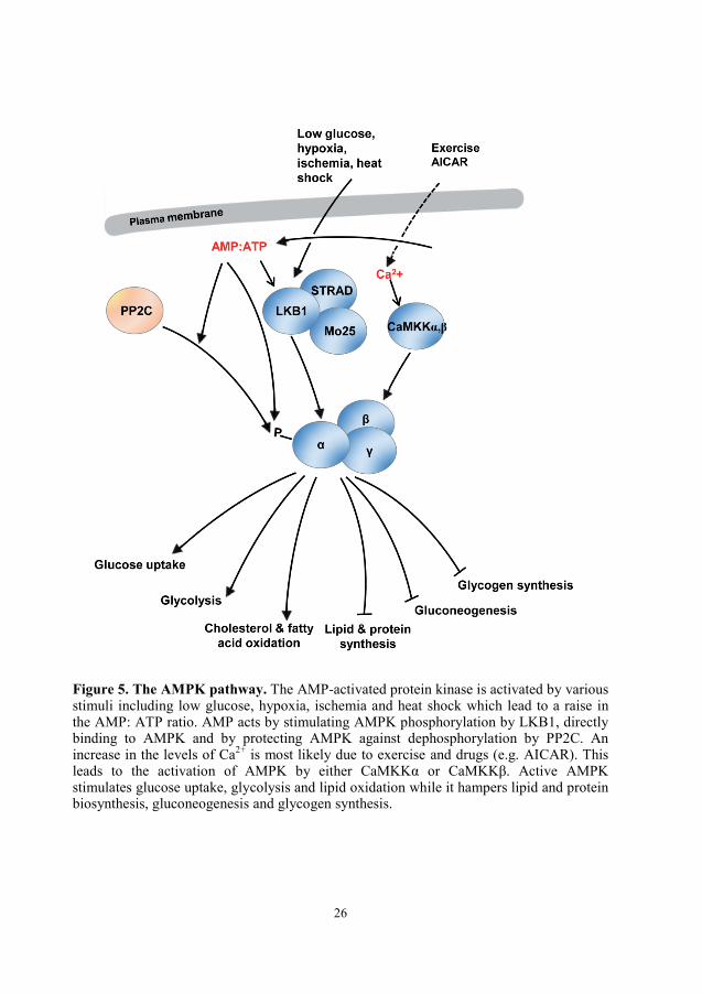
Figure 5. The AMPK pathway. The AMP-activated protein kinase is activated by various stimuli including low glucose, hypoxia, ischemia and heat shock which lead to a raise in the AMP: ATP ratio. AMP acts by stimulating AMPK phosphorylation by LKB1, directly binding to AMPK and by protecting AMPK against dephosphorylation by PP2C. An increase in the levels of Ca2+ is most likely due to exercise and drugs (e.g. AICAR). This leads to the activation of AMPK by either CaMKKα or CaMKKβ. Active AMPK stimulates glucose uptake, glycolysis and lipid oxidation while it hampers lipid and protein biosynthesis, gluconeogenesis and glycogen synthesis.

27
4.4.1. Significance of the AMPK/SNF1 pathway
Given its pleiotropic actions in the regulation of lipid and glucose metabolism, AMPK has
become a potential target for therapeutic interventions in diabetes and metabolic diseases.
AMPK is expressed in multiple tissues that are metabolically active such as pancreas,
liver, lung, skeletal muscle, adipose tissue and hypothalamus, where it exerts different
functions (reviewed in (Zhang, et al. 2009)). In liver, active AMPK functions to repress
hepatic gluconeogenesis by down-regulating the transcription of the genes encoding the
rate-controlling gluconeogenic steps phosphoenolpyruvate carboxykinase (PECK) and
glucose-6-phosphatase (Horike, et al. 2008, Lochhead, et al. 2000, Shaw, et al. 2005).
These results suggest a role of AMPK in counteracting hyperglycemia-related type 2-
diabetes. In muscle, AMPK activation by muscle contraction stimulates mitochondrial
fatty acid oxidation and upregulates mitochondrial and lipid utilization genes through the
activation of the transcriptional regulators PGC-1α and FOXO1 respectively (Canto, et al.
2010, Suwa, et al. 2003). In parallel, activated AMPK promotes muscle glucose uptake by
upregulating GLUT-4 transcription and stimulating its translocation to the cell surface in
the muscle tissue (Hayashi, et al. 1998, Merrill, et al. 1997, Russell, et al. 1999).
Altogether, activation of AMPK seems to be a potential approach for bypassing the defects
of insulin-resistance and metabolic syndrome-related diseases.
A number of chemical compounds and anti-diabetic drugs such as phenformin, metformin
and pioglitazone activate AMPK suggesting that at least some of their beneficial effects
are mediated through AMPK (Cool, et al. 2006, Saha, et al. 2004, Xiao, et al. 2013, Zhou,
et al. 2001). While metformin and phenformin activate AMPK indirectly and
independently on adenosine nucleotides, the 5-aminoimidazole-4-carboxamide riboside
(AICAR) mimics the allosteric effect of AMP and hence directly activate AMPK (Corton,
et al. 1995, Sakamoto, et al. 2004, Zhou, et al. 2001). Our studies (Paper V) indicated that
metformin, phenformin and AICAR did not affect the phosphorylation of yeast-expression
AMPK suggesting that these drugs are probably not imported into or quickly exported out
of the cells. Another possibility could be that the mechanism of AMPK activation by these
drugs (especially metformin and phenformin) in mammalian cells is distinct from that in

28
yeast. Moreover, these studies highlight the use of humanized yeast where mammalian
genes and proteins homologs can be functionally expressed and serve to complement yeast
deletion mutants (Botstein and Fink 2011). Studies involving the expression of
heterologous proteins in yeast have been successfully applied not only to the study of the
structural and functional regulation of these proteins but also for the pharmacological
purposes such as the screening of novel drugs.
4.5. Snf1 orthologs in other eukaryotes
In addition to yeast and mammals, the AMPK/SNF1 pathway is present in most, if not all,
eukaryotes. In plants, there are numerous SNF1-related protein kinases (SnRKs) with
SnRK1 being most similar to Snf1 (reviewed in (Ghillebert, et al. 2011, Halford and
Hardie 1998, Halford, et al. 2003, Hardie 2007). SnRK1 was originally isolated from rye
in 1991 and restored SNF1 function when expressed in the yeast snf1Δ mutant (Alderson,
et al. 1991). Arabidopsis SnRK1 displays 48% sequence similarity with SNF1 and AMPK
(Halford and Hardie 1998). Similar to SNF1 and AMPK1, SnRK1 is regulated by
phosphorylation and has a direct effect on metabolism by regulating metabolic enzymes
and an indirect role by controlling gene expression. SnRK1 plays an important role in
starch biosynthesis by enhancing the expression of the sucrose synthase gene Sus4
(Purcell, et al. 1998). In the moss Physcomitrella patens, two SNF1-related protein kinase
encoding genes, PpSNF1a and PpSNF1b, play an important role in coping with darkness
since the double knockout mutant snf1asnf1b was unable to grow in a normal day-night
light cycle (Thelander, et al. 2004). The nematode Caenorhabditis elegans has two
homologs of the AMPKα subunit, Aak1 and Aak2, which are activated by AMP and
involved in the extension of lifespan (Apfeld, et al. 2004). The fruitfly Drosophila
melanogaster contains single isoforms of the α-, β- and γ- subunits that are essential for the
activity of the AMPK complex. In analogy to mammalian AMPK, D. melanogaster
AMPK is sensitive to the AMP: ATP ratio and the α-subunit is activated by
phosphorylation at Thr184 within its activation loop (Pan and Hardie 2002). Deletion of
AMPK causes severe abnormalities in cell polarity and mitosis (Lee, et al. 2007). SNF1

29
has also been studied in other yeasts than S. cerevisiae. While Kluyveromyces lactis Snf1
(KlSnf1) is responsible for sugar (e.g. lactose) uptake and metabolism (Lodi, et al. 2001,
Wiedemuth and Breunig 2005), Candida tropicalis Snf1 (CtSnf1) was found to be
indispensable for cell growth and viability (Kanai, et al. 1999). Taken together, AMPK
belongs to an evolutionary conserved family of energy sensors which seem to exist in all
eukaryotes. These energy sensors are able to detect energy depletion issued by nutrient
limitation or environmental stresses and respond to these cues by readjusting energy
metabolism. Hence these AMPK orthologs share the same fundamental function, i.e.
control of energy homeostasis.
30
5. Single cell analysis
5.1. Why single cell analysis?
It is not until recently that the fields of molecular and cellular biology have started to
apprehend the existence of cell-to-cell variability and estimate the importance of single cell
analysis. Such variability can have a significant impact on the overall behaviour of the
population. Since a population does not always manifest a normal distribution but it can
display a bimodal or even a multimodal distribution, the average result obtained from this
population represents therefore an intermediate response that is false and misleading. In
addition to the widespread diversity of the population, sample concentration due to
filtering and centrifugation of a cell culture might be a major problem in functional
genomics. Hence the results obtained might reflect the response of the cells to the
concentration rather than to the condition under study (Lidstrom and Meldrum 2003).
Another major benefit using single cell analysis is the significantly reduced cell amount
enabling the study of cells that are hard or impossible to culture (Lidstrom and Meldrum
2003). Hence, now that we are aware of the limits of bulk studies, the use of single cells
becomes imperative in order to attain a complete picture of biological processes and cell
design.
5.2. Cell variability
Traditional microbiology has focused on the study of large populations where the obtained
information is mostly based on the inference from population-level data. Such studies are
subject to the averaging effects obtained from bulk measurements and mask important
parameters such as cellular discrepancies that exist within a clonal cell population (Irish, et
al. 2006). This data only reflects the overall behaviour of the population and is unable to
describe the behaviour of individual cells that constitute this population. A clear example
of the disconnection between population and single cell measurements is the recent study
undertaken by Mathies’ group where they examined siRNA knockout of GAPDH gene in

31
single cells. Their single cell measurements suggest the presence of two populations of
cells with partial (50%) and complete (0%) gene silencing. These results were clearly
different from the average of all the 50 cells where gene knockout corresponded to ~ 80%
(Toriello, et al. 2008). Early studies (Raser and O'Shea 2004) have demonstrated that cell
variability is a result of cellular noise which is the sum of extrinsic noise, defined by the
size, shape and the cycle stage of the cell and intrinsic noise which reflects the
stochasticity at gene expression level. Such stochasticity is often explained by genetic
noise resulting from transcriptional and translational fluctuations and was shown to largely
contribute to the heterogeneity in the cellular behaviour within an isogenic eukaryotic
population (Blake, et al. 2003). It has long been argued that in living cells, most regulatory
genes, mainly involved in cell growth and survival, are expressed in low copy numbers
(Guptasarma 1995). This low molecular copy number would give rise to large variances
(noise) in molecules’ concentrations and consequently in the rate of the reactions they
regulate (Thattai and van Oudenaarden 2001). Cell-to-cell variability does not necessarily
represent an obstacle in cellular biology but rather an evolutionary feature that enables
microorganisms to survive hostile conditions and adapt to novel milieus. Population
modelling of bacterial gene expression and growth has suggested that the emergence of a
heterogeneous population with broader phenotypes as a result to changing environments
might be a beneficial property that bacterial and yeast cells acquire in order to survive
sudden external changes (McAdams and Arkin 1999, Thattai and van Oudenaarden 2004).
Moreover, simulations of genetic regulation in single cells have shown that the production
of signal proteins occurs at random time intervals and results in a variable number of
proteins. This non-genetic diversity would account for diverse fates of cellular events
reflected by diverse phenotypes between cells (McAdams and Arkin 1997). However, cells
have also adapted other features such as gene redundancy and feedback loops in order to
reduce the noise when a more deterministic and precise cellular response is desired
(McAdams and Arkin 1999). Cell heterogeneity is inherent to all cells and especially
cancer cells. Hence, dissection of cell-to-cell variability is highly appreciated in order to
understand and survey tumour development steps and target therapeutic responses (Wang
and Bodovitz 2010).

32
5.3. Single cell dynamics-Time-resolution
The cell is able to sense and respond to external signals through the interplay of a complex
system. This includes a number of signal messengers and protein interactions, which
convey the signal to the nucleus leading subsequently to the regulation of target genes
expression. In order to completely understand the structural architecture and the function
of these signalling cascades, it is vital to accurately define not only the spatial but also the
temporal integration of their components. The importance of the spatiotemporal
relationships of these complex systems has become evident since it has been demonstrated
that it affects the biological response (Murphy, et al. 2002). Analysis of cell signalling at
this level of resolution is not possible to achieve by traditional experimental tools which
provide snapshots of averaged behaviours. Hence the use of real-time single cell
methodology has become imperative to unravel inter and intracellular dynamics.
Owing to the advances in optics and engineering and to the wealth of fluorescent probes,
light microscopy has revolutionized into fluorescence time-lapse microscopy. This
sophisticated technique has made it possible to follow the chronological incidence of
distinct biological events. For instance, fluorescence time-lapse studies were applied to
investigate the coordination of cell apoptosis events and cell cycle progression in single
cells (Chen, et al. 2006, Goldstein, et al. 2000, Munoz-Pinedo, et al. 2005). Combination
of multi-photon auto-fluorescence imaging with second harmonic generation has allowed
visualization of human skin with a significantly high temporal resolution in orders of
picoseconds (Konig and Riemann 2003). Fluorescent microscopy is widely applied to
examine protein localization. Because protein function is related to its subcellular
localization, it is fundamentally plausible to unravel the regulatory function of the protein
of interest by fluorescently tagging it and tracking it in real-time (Losick and Shapiro
1999). Such studies have become possible with the introduction of immunofluorescence
microscopy in 1993 for E. coli (Maddock and Shapiro 1993). Improvements in confocal
microscopy have enabled visualization of fluorescent proteins at high resolutions (Betzig,
et al. 2006, Hell 2003). We have combined fluorescence time-lapse microscopy, optical
tweezers and microfluidics in order to investigate the dynamic shuttling of Mig1 in

33
response to various glucose up- and downshifts in single yeast cells (Paper I and Paper
II). Nevertheless, time-lapse techniques generate a large set of data that is not possible to
be handled by the human eye and therefore necessitates the use of automated software
(reviewed in (Meijering, et al. 2008)).
34
6. Methods for single cell analysis
Studies at the level of single cell would not have been possible without the development of
new tools that allow isolation of single cells from a cell population. Techniques such as
flow cytometry (FC) and its extensions (e.g. fluorescence-activated flow sorting (FACS)
and capillary electrophoresis (CE)) have allowed the study of individual cells and their
chemical contents respectively (Krylov and Dovichi 2000). The principle of flow
cytometry relies on the measurements of flowing cells passing through the flow cytometer
(Shapiro 2005). This technique is applied for various purposes such as cell counting,
sorting, and determination of cell size, shape and other biochemical and physiological
aspects of single cells. The introduction of fluorescence to flow cytometry in the late
1960’s has allowed the application of FACS (Dean and Hoffman 2007). This technique
uses not only light scattering but also fluorescence properties of the single cells passing
through the flow cytometer in order to dissect cell populations into subpopulations.
Although FC techniques are able to simultaneously measure several parameters of several
thousands of cells and can generate high-throughput data within a considerably short time,
they have some limitations. Since these techniques are based on fluid stream where the
cells once measured are streamed away, they do not allow the examination of the same
cells over a certain time. That is to say, FC does not allow either cell manipulating or the
study of solid substrates-attached cells (Shapiro 2005). Techniques such as fluorescence
microscopy and laser scanning cytometry (LSC) are able, to some extent, to measure
similar features to flow cytometry but dispose of fewer limitations (Deptala, et al. 2001,
Golchin, et al. 2012). Instead of flowing cells, LSC is performed on cells attached to a
solid support (e.g. glass slides). In the LSC, cells are scanned by a moving laser beam and
the scattered fluorescence is then detected at multiple wavelengths by a microscope. This
technique provides information on cell morphology and allows sequential analyses on the
same cells (Darzynkiewicz, et al. 1999). However as any other technique, LSC has also
some restrictions such as its lack of cell sorting.
Advances in micro and nanotechnologies have contributed to the emergence of tools such
as the lab-on-chip (Lochhead, et al.) or micro-total analysis systems (µTAS). Introduced in

35
the 90’s, these portable micro-chambers are designed for various experimental purposes
including electronic, mechanical and fluidic functions (Abgrall and Gue 2007). Due to
their low cost, reduced scale and the significantly short experimental time these tools have
attracted many investigators (Hatch, et al. 2001). The success of this technology has
allowed the extension of this concept and hence the development of novel field-specific
technologies such as the laboratory-in-a-cell (LIC) where a single cell is considered as a
laboratory for detailed investigations of, for instance, intracellular processes (Andersson
and van den Berg 2004). A number of additional LOC-based techniques have emerged
within the past few years. Techniques such as counting low-copy number proteins, micro-
PCR, patch-clamp and single cell-mRNA purification and analysis have enabled studies of
single cells at different levels of details to be carried out (Bontoux, et al. 2008, Chen and
Folch 2006, Huang, et al. 2007, Marcus, et al. 2006).
6.1. Microfluidics
Microfluidics is the technology that handles small amounts of fluids running through
channels of micrometre scales. Microfluidics offers a number of advantages, namely, the
use of small volumes of samples and reagents, short reactions time, liquids separation and
the significantly low cost (Whitesides 2006). In addition, and most importantly,
microfluidics offers laminar flow.
The development of microfluidics technology has largely benefited from the application of
photo lithography. In fact, original works in the field of microfluidic systems have
combined the use of silicon and glass. However, due to their high cost and significant
opacity to visible and ultraviolet light, the use of silicon and glass became less desirable in
optics. Besides, both silicon and glass turned out to be inappropriate for the study of
biological systems and more specially for the study of mammalian cells (Whitesides 2006).
The introduction of elastomers namely the polymer poly-dimethylsiloxane (abbreviated as
PDMS) to microfluidics has been revolutionary. PDMS is a soft elastomer, relatively inert
and optically transparent, which favoured its use in optical methods. Its softness has

36
enabled the fabrication of different shapes of microstructures and its high permeability to
oxygen and carbon dioxide has allowed cell culturing inside microfluidic chambers.
Working with microfluidics requires the knowledge of a set of parameters. The
dimensionless Reynolds number (Re) defines the flow regime, which can be either
turbulent or laminar, and is considered one of the most important parameters when dealing
with fluids (Whitesides 2006). Re corresponds to the ratio between inertial and viscous
forces on fluids. At low Reynolds number, the viscous forces dominate over the inertial
forces resulting in a laminar flow. That is to say, when introduced into the microfluidics,
two fluid streams flow in parallel without disruption. A Re that is below 2,300 dictates that
the flow is laminar (Beebe, et al. 2002). In our experiments, employing microfluidics, the
Re is normally below 1, indicating that the flow inside the micro-channels is laminar and
that the only way for two fluid streams to mix is by diffusion.
6.2. Optical tweezers
The study of how single cells communicate with each other and with their environment
and how cells adapt and operate in different environmental conditions requires the ability
to manipulate cells. Techniques such as optical tweezers have been developed for such
purpose. The ability to use light for trapping and manipulating single particles such as
bacteria and viruses (Ashkin and Dziedzic 1987) has made a major breakthrough in many
scientific and engineering fields. Particle manipulation reflects acceleration, deceleration,
guidance and deflection (Ashkin 1997). Originally this technique was designed for atoms
trapping but extended later on in order to trap bacteria, yeast, mammalian and even plant
cells (Ashkin 1997). By using an infrared trapping, Ashkin et al., 1987 developed a
damage-free procedure, allowing bacterial cells to reproduce inside the optical trap. The
principle of this technique relies on the use of a single strong Gaussian-distributed laser
beam, which is a collection of rays with different intensities and with the highest intensity
being in the middle of the beam. The incidence of the laser beam on a spherical particle
results in a scattering force, in the direction of the incident light, and a gradient force in the
direction of the intensity gradient (Ashkin and Dziedzic 1987). In order to allow particle

37
trapping, the gradient force must be dominating over the scattering force. This can be
achieved by strongly focusing the light beam through a high numerical aperture objective
creating a three-dimensional stable trap.
6.3. Fluorescence
The fact that molecules are able to fluoresce has made a major breakthrough in the history
of single cell (Brehm-Stecher and Johnson 2004). Fluorescence occurs when a photon
from a light source is absorbed by a fluorophore, a molecule with fluorescence properties
(Lichtman and Conchello 2005). This leads to the transition of an electron in the
fluorophore to an excited energy state. Eventually, the excited molecule returns to a lower
energy state. This return is accompanied by the emission of light in form of fluorescence as
a result of energy loss. This separation in energy or rather wavelength is referred to as
Stokes shift, in other words, fluorescence shift (Davey and Kell 1996). Stokes shift
separates the emitted light by optical filters and hence quantify the amount of fluorescence.
When dealing with fluorescence measurements, certain parameters should be considered.
Hence two major fluorescence characteristics can be distinguished: the extinction
coefficient corresponding to the amount of light absorbed at a certain wavelength (Davey
and Kell 1996) and the quantum yield which describes the ratio between the absorbed and
the emitted photons (Lichtman and Conchello 2005). In addition to these two parameters,
the photo-stability of the fluorophore is another factor to consider. Photo-stability defines
the number of times a dye can be excited before decaying (Davey and Kell 1996). Hence
an excess of excitation results in a decrease of photo-stability and consequently in an
increase of photo-bleaching. The excited-state lifetime of a fluorophore is also of some
importance. It refers to the time a fluorophore remains excited before decaying to the
ground state (Lichtman and Conchello 2005). The lifetime is affected by the proximity of
an energy acceptor. Hence the nearer the acceptor is to the donor the shorter the lifetime.
The discovery of the green fluorescent protein (GFP) from the Aequorea victoria jellyfish
has revolutionized biosciences and especially live imaging. Irradiated by the near
38
ultraviolet or blue light, GFP is largely used as an indicator of gene expression and protein
localization (Chalfie, et al. 1994). This 238 amino-acid β-barrel-shaped protein contains
the tripeptide Ser-Tyr-Gly which after oxidation becomes a fluorophore (Heim, et al.
1994). Given its many advantages such as its fairly small size, non-toxicity and most
importantly its ability to fuse to almost any protein without altering its endogenous
function, GFP has become a versatile tool for the dynamic investigation of many cellular
events including protein abundance, motility and interactions (Chalfie, et al. 1994,
Lippincott, et al. 2001, Zimmer 2002). In addition to its role as a reporter, GFP can be
combined with fixation-based staining techniques namely fluorescence in situ
hybridization (FISH) (Brehm-Stecher and Johnson 2004). Further improvement of GFP
generated GFP proteins with distinct emission and absorbance spectra such as blue
fluorescent (BFP), yellow fluorescent (YFP) and cyan fluorescent (CFP) proteins, designed
to simultaneously look at different targets at the same time and within the same specimen
(Lippincott-Schwartz and Patterson 2003). This prosperity in fluorescent proteins has
allowed the development of techniques such as fluorescence resonance energy transfer
(FRET). The concept of this technique is to detect the interaction between two proteins
based on their intermolecular distance. Technically, a transfer of energy occurs from an
excited fluorophore called the donor to another molecule called the acceptor (Sekar and
Periasamy 2003). Moreover, the expansion of the large range of fluorophores and the rise
of diverse GFP variants has enabled multicolour microscopy where different cellular
compartments and/or components can be monitored simultaneously.
In parallel to the development of fluorescence molecules, fluorescence microscopy has
evolved. Given the Stokes shift, a fundamental characteristic of fluorescence microscopy,
it is possible to illuminate an object with a given wavelength and filter the resulting light in
order to solely see longer wavelength (Lichtman and Conchello 2005). A wide range of
fluorescence microscopes, extending from a conventional wide-field to a super-resolution
microscope, with different levels of resolution has been established. In our experiments
involving fluorescence microscopy, we employed an epi-fluorescence microscope where
the incident light which is used to excite the sample can be separated from the relatively
weaker light that is emitted from the specimen. This latter is then being detected (Webb
and Brown 2013). In addition to an EM-CCD camera, this inverted epi-fluorescence

39
microscope is equipped with a fluorescence light source EL 6000, a GFP filter cube
(472/30 nm exciter, 520/35 nm emitter and 495LP dichroic mirror) and an mCherry filter
cube (560/40x exciter, 630/75m emitter and 585LP dichroic mirror) (for a detailed
description of this step, see Materials and Methods section in Paper I and Paper II). This
set-up has allowed us to perform time-lapse imaging to monitor localization dynamics of
the glucose repressor Mig1.
6.3.1. Bleaching
The estimated number of excitation-emission cycles for a good fluorophore ranges
between 10,000 and 40,000 (Lichtman and Conchello 2005). After that the fluorescence
signal of the fluorophore irreversibly fades. This phenomenon is called bleaching (or
photobleaching). In contrast, when the fluorescence signal fades reversibly then it is the
quenching which takes place. Although the bleaching is a common issue in fluorescence
microscopy, the photochemistry of this phenomenon still remains obscure. One
explanation could be that photobleaching is related to the triplet states of the fluorophore
as it could be a result of an interaction between the fluorophore’s triplet state and the
molecular oxygen.
6.3.1.1. Bleaching techniques
Despite the undesirable effect of bleaching, techniques such as fluorescence recovery after
photobleaching (FRAP) and fluorescence loss in photobleaching (FLIP) take advantage of
bleaching. FRAP and FLIP techniques allow the study of the mobility of fluorescent
tagged molecules in living cells. In the case of FRAP, fluorescent molecules of a small
region of the cell are irreversibly photobleached using a laser beam. The recovery of non-
bleached fluorescent molecules from the neighbouring areas into the irradiated region can
be quantitatively monitored by time-lapse microscopy. Such recovery would reflect the
movement of the fluorescent particles between these two compartments. The recovered
fluorescence into the bleached region is plotted over time (Ishikawa-Ankerhold, et al.

40
2012). From this graph, many parameters can be distinguished namely the mobile and
immobile fractions which correspond respectively to the fraction of the proteins able or
unable to diffuse to the bleached region. Another important parameter is the half time, half
maximum or the half-life corresponding to the time it takes to reach the half recovery
plateau (Ishikawa-Ankerhold, et al. 2012). Thus, the shorter the half time the faster the
recovery hence the diffusion.
Complementary to FRAP, FLIP is used to investigate protein redistribution and the inter-
connectivity between two compartments (Ellenberg, et al. 1997). FLIP technique
corresponds to the repetitive photobleaching of a small region of the cell. This results in
the loss of fluorescence in the irradiated region which is, soon after, recovered by the
inflow of fluorescent molecules from the surrounding regions. The loss of fluorescence in
the unbleached area which represents the mobile fraction of the fluorescent particles is
monitored over time. Both FRAP and FLIP can be used in order to study the transit of
fluorescent tagged protein of interest between two cellular compartments. In Paper I, we
employed both FRAP and FLIP in order to explore the kinetics of the rate of Mig1 nuclear
cytoplasmic shuttling.
6.4. Technical limitations and drawbacks
As discussed above studies at the single cell level unveil the apparent phenotypic
distinction between individual cells within a clonal population. However, given the large
influence of biological and technical noises, single cell studies are technically challenging.
A number of factors might contribute to the complexity of single cell analysis. For
instance, the small size of microfluidic channels increases the risk of clogging of cells or
PDMS, which subsequently affect the laminar flow through the channel. Further,
photobleaching, which occurs during imaging, might also affect the results. In our case, we
have dealt with this issue by reducing the intensity of the fluorescence lamp. However, at
low intensities the lamp is not always stable and can show small irregular intensity
fluctuations over time. This could affect fluorophore excitation and therefore fluorescence

41
intensity. One approach to correct for the differences in the starting intensity between cells
is to perform data normalization. In principle, normalization would be performed by
dividing the intensity of all time points by that of the initial time point. Since we took only
one image at the start of the experiment (prior to changing the microfluidic flow),
normalizing to the intensity resulting from this image would be biased. A proper
normalization would be to divide by the average intensity acquired from several images
before the shift. However, data normalization is not always desirable since it might also
mask important aspects of biological variation.
In addition to the technical noise, the biological noise emerging from intrinsic and extrinsic
noises also adds to the complexity of single cell analysis. In fact, the low copy number of
proteins, inherent to components of signaling pathways and transcriptional regulators
(Huang, et al. 2007), enhances the challenges of single cell analysis considerably. It might
be possible to reduce the extrinsic noise by synchronizing the cell-cycle of the cells. We
presently do not know if the cell cycle stage affects Snf1-Mig1 signalling, a question that
could be addressed in our setting.
Another limitation arising from the small sample size and the various stochastic effects
described above, is the difficulty to interpret single cell data generated from one single run
and the possibility to compare data between different runs. Therefore experimental and
biological replicas are necessary to minimize the uncertainties and generate accurate
results. Statistics and quantitative mathematical modelling are two frameworks that might
allow discerning between technical and biological noises. Nevertheless, due to the sample
size, standard statistical methods such as standard deviation cannot be applied. Hence,
specific statistical approaches, such as e.g. mixed effects models, need to be developed for
the analysis of single cell data. This is an area of active research but still in its infant
stages.

42
7. Summary of appended papers
Since its emergence as an energy sensor and regulator in all eukaryotes, the AMPK/SNF1
has attained a considerable increase in attention. In this thesis, we examined different
aspects of the regulation of the AMPK Snf1-Mig1 pathway. We surmised that the analysis
of time-dependent signalling behaviour provide insights into the dynamic properties of this
system. By employing the real-time kinetics of Mig1 nucleo-cytoplasmic shuttling as a
read-out, we were able to elucidate characteristics of the Snf1/Mig1 pathway in response to
different glucose levels (Paper I, Paper II and Paper III). In order to fully control
glucose concentrations, we constructed arrays of individual yeast cells in a microfluidic
device allowing a shift in glucose concentration within 2 s time. The cell arrays were then
exposed to different glucose up- and downshift regimes (Paper I and Paper II).
In Paper I, we found that following glucose upshifts, Mig1 displayed a rapid and sustained
nuclear entry over a wide range of the tested concentrations including actually
derepressing concentrations. However, after a shift to glucose concentrations that are
below 0.01%, Mig1 migration was slower and not all the cells responded. On the other
hand, following glucose downshifts from 4% to concentrations ranging between 1.5% and
0.5%, we found that Mig1 migrated rapidly but transiently to the cytosol, before returning
to the nucleus. Below these concentrations, Mig1 remained in the cytosol. Our FLIP and
FRAP data indicated that Mig1 nucleo-cytoplasmic mobility is constant regardless of
glucose concentration. In summary, this data suggests that the Snf1/Mig1 signalling
pathway is highly sensitive to glucose changes. It is also able to monitor glucose change as
well as the absolute glucose level. Our single cell analyses displayed a significant cell-to-
cell variability throughout a wide range of the conditions tested. This variability may
indicate an evolutionary adaptive feature to assure an optimal utilization of the available
glucose in the surrounding environment of the population. We assumed that analysis of the
cell-to-cell variability might bear information about the regulatory mechanism of the
system. Hence this issue was addressed in Paper III.
Our data from Paper I showed that the Snf1/Mig1 system displayed different sensitivities
depending on glucose up- and downshifts suggesting a fast adaptation of metabolism to
43
glucose availability. This behaviour may be connected to glucose uptake whose
characteristics depend on glucose availability (Horak 2013, Ozcan and Johnston 1995). We
hypothesized that the behaviour of Mig1 migration would be affected by the characteristics
of the uptake system (Paper II). To this end, we monitored Mig1 translocation, using the
same experimental set up described above, in three different strains, expressing each as a
sole uptake system the low affinity hexose transporter Hxt1, the high affinity Hxt7 or their
chimeric, TM6* under different glucose up- and downshifts. We found that in cells
expressing either HXT7 or TM6* and growing in high glucose, Mig1 was constitutively
cytosolic and did not respond to any glucose downshift. However in the Hxt1-only strain,
the Mig1 migration profile was consistent with the affinity profile of this transporter.
When we shifted these cells from 4% to 1% glucose, we did not observe the transient
behaviour observed in the wild type strain. This data clearly indicates that Mig1 migration,
and hence the Snf1/Mig1 system is affected by glucose uptake.
It is now accepted that cell-to-cell variability is ubiquitous to any population and might
bear important information regarding the underlying regulatory mechanisms of biological
systems (Li and You 2013). By applying nonlinear mixed effects modelling, we aimed to
quantify the dynamic behaviour of Mig1 shuttling in response to glucose downshifts
(Paper III). Based on this approach, we were able to construct a dynamical mathematical
model successfully simulating the observed transient nuclear exit following glucose shift
from 4% to intermediary concentrations ranging between 1.5% and 0.5%. Interestingly, the
estimated parameter distributions showed a significant correlation between the two
parameters determining the initial time scale of Mig1 exit and the time scale of the
adaptation process. These findings suggest that Mig1 nuclear exit and re-entry might likely
be controlled by the same mechanism. By applying Monte Carlo simulation of the
established model we were able to determine various characteristics of the transient
behaviour of Mig1. We found that Mig1 exits the nucleus more quickly and re-enters it
more slowly as glucose concentrations decreases. Also at these low concentrations, there
was an enhanced cell-to-cell variability. These results elucidate biological features of the
Snf1/Mig1 module which could not be directly measured experimentally and represent a
step toward the understanding of the mechanism underlying Mig1 shuttling.

44
The aim of paper IV was to provide a reconstruction of the Snf1 network and a detailed
review about our current understanding of the Snf1/Mig1 pathway based on solid and
structured pieces of evidence collected from over 400 published articles and on the
publically available rxncon software. Moreover, we discuss various aspects of the pathway
including the mechanistic architecture, the environmental perturbations, and the functional
response. In addition, we review the cross-talk with other pathways and the conservation
among other species.
Given the remarkable conservation between yeast and human proteins, including those
involved in human diseases, it is now possible to study the biological function of such
orthologous proteins in yeast (Dolinski and Botstein 2007). This approach has been
adopted by Woods et al., who provided evidence for the functional interchangeability
between the yeast SNF1 and its mammalian ortholog AMPK (Woods, et al. 1994). In
Paper V, we tested whether mammalian AMPK could functionally substitute for the SNF1
complex. To this end, we expressed the mammalian AMPK heterotrimeric complex in
yeast cells lacking the entire endogenous SNF1 complex. Using twelve different
combinations of AMPK subunits, we found that combinations comprising α1-, β1- or β2-
and γ1- or γ3 subunits appeared to be functional since they conferred growth on alternative
carbon sources. Remarkably, heterologous AMPK is properly regulated by glucose.
AMPK regulation was not affected by deletion of REG1 and REG2 however the
phosphatase Sit4 seemed to contribute, to some extent, to the negative regulation of
AMPK. In contrast to the anti-diabetic drugs phenformin and metformin (Sakamoto, et al.
2004, Zhou, et al. 2001), which indirectly activate endogenous AMPK, the drug candidate
compound 991 seemed to moderately activate the yeast-expressed AMPK. Taken together,
this data highlights the conservation of the structural and functional AMPK and SNF1
regulatory systems and opens up possibilities for the screening of drugs affecting AMPK
activity in yeast.

45
8. Conclusions and future perspectives
The Snf1/Mig1 pathway plays a vital role in energy homeostasis. Although this pathway
has been extensively studied over the past decades, several questions remain yet to be
answered. Given that the overall structural and functional organization is conserved
between SNF1 and its mammalian ortholog AMPK, it is likely that also the mechanism of
their activation is related, while life style specific adaptation have probably evolved.
Indeed, in analogy to AMPK, SNF1 activity seems to be affected by adenosine
nucleotides, but still the mechanistic and physiological underpinnings of this regulation
remain to be elucidated. Thus, this area clearly requires more investigations. In this thesis,
we aimed to elucidate the mechanisms governing the dynamics of glucose signal
transmission in single cells and at population level.
We provide for the first time a detailed and time-resolved study describing different
characteristics of the Snf1/Mig1 pathway in response to various glucose levels such as a
fast response and a rapid adaptation to changes in glucose levels. We also demonstrate that
characterization of Mig1 translocation could be a proper approach for the study of the
regulation of the Snf1/Mig1 pathway. A number of open questions concerning Mig1
translocation remain yet to be addressed. For instance, it is still unclear whether the
regulation occurs at the level of Mig1 phosphorylation by Snf1 or at its nuclear transport,
or both. Recent studies have indicated that active Snf1 does not automatically
phosphorylate Mig1 but that there seems to be an additional glucose-regulated step that
controls Mig1 phosphorylation by Snf1 (García-Salcedo, et al. 2014, Ye, et al. 2008).
Perhaps access of active Snf1 to Mig1 is controlled by glucose. Evidence has been
reported that Hxk2 interacts with both Snf1 and Mig1 in order to prevent Mig1
phosphorylation under repressing conditions (Ahuatzi, et al. 2007). While details about
Mig1 nuclear export have been reported it is still unknown how Mig1 is imported to the
nucleus. Nuclear import of the related Mig2 is mediated by the importin-β Kap95 and
requires the presence of GTP-binding protein Gsp1 and the nuclear localization signal
(NLS) motif of Mig2 (Fernandez-Cid, et al. 2012). Also Mig1 has an NLS region between
the amino acid residues 359 to 373 and hence it is possible that the nuclear translocation of

46
Mig1 might also involve a similar importin (De Vit, et al. 1997, DeVit and Johnston 1999).
Screening for importins and importin-like proteins that physically interact with Mig1
might provide some clues about the regulation of Mig1 nuclear cytoplasmic translocation.
The finding that Mig1 constantly shuttles between the nucleus and the cytoplasm
regardless of glucose concentration raises the question of the physiological relevance of
the nuclear localization of Mig1 in absence of glucose. We hypothesize that
phosphorylated and unphosphorylated Mig1 coexist under all conditions as consequence of
a balance between the phosphorylation and dephosphorylation reactions, which could
allow a fast response when glucose levels change. Our data is consistent with the
previously observed constitutive presence of Mig1 on the promoter of glucose-repressible
genes under both repressing and derepressing conditions (Papamichos-Chronakis, et al.
2004). These observations raise the question whether Mig1 could act like Sko1. This
transcriptional repressor also mediates repression in complex with Cyc8 (Ssn6)-Tup1 co-
repressor but under hyperosmotic stress is converted to an activator following
phosphorylation by Hog1 (Proft and Serrano 1999, Proft and Struhl 2002). Mig1 was
previously found to act as an activator in the absence of Ssn6 suggesting that the Mig1-
Ssn6 interaction might prevent Mig1 to either act as an activator or bind to an activator
(Treitel and Carlson 1995). However, given the significant derepression of SUC2 in the
absence of MIG1 and the predominant cytoplasmic distribution of Mig1 under
derepressing conditions, it is unlikely that Mig1 might be an activator, at least not in a
wild-type background ((Bendrioua, et al. 2014, Bu and Schmidt 1998, De Vit, et al. 1997)
and our unpublished data). Instead, it is possible that the absence of Mig1 allows other
transcriptional activators to occupy Mig1-DNA binding site.
By monitoring Mig1 nucleo-cytoplasmic shuttling in strains with different glucose uptake
rates, we confirm a tight link between glucose metabolism and the activity of the
Snf1/Mig1 pathway. The Snf1/Mig1 pathway does not directly perceive the level of
glucose but rather the glycolytic flux and/or the level/turnover of AXPs in the cell. Our
data is in accordance with the previously observed correlation between glycolytic rates and
Mig1 phosphorylation status (Elbing, et al. 2004b). Strains expressing a high affinity
glucose transporter are in a derepressed state and Gustafsson and co-workers (Bosch, et al.
2008) have demonstrated that in the Tm6* strain the ratio of ATP: ADP and of ATP:

47
AMP is in fact higher than in wild type. This observation is in contradiction with the
paradigm stating that AMPK is activated by a drop in the ATP: ADP. Hence, it remains
still elusive how adenosine nucleotide levels are linked to SNF1 activity in vivo. Further
studies are clearly necessary to investigate the mechanism of regulation of the Snf1/Mig1
pathway by glycolytic metabolism. The newly emerging time-resolved technology
involving aptamer-based nanosensing could be an appropriate approach to elucidate the
link between AXP and the Snf1/Mig1 pathway in real-time. Technically, it would be
feasible to simultaneously monitor the levels of AXP-conjugated aptamers in the cell and
the shuttling of fluorescently-tagged Mig1. The strains used in this study, Hxt7 and Tm6*,
might provide valuable information about the governing mechanism but given their re-
wired metabolism (e.g. respirative glucose metabolism) also a control strain expressing all
hexose transporters needs to be studied.
It has proven difficult to functionally swap the Snf1 protein with the AMPK α-subunit.
Previously, Carling and his co-worker (Daniel and Carling 2002) reported that the yeast-
expressed chimeric AMPKα1-Snf1 was able to rescue growth of snf1Δ mutant only on
raffinose indicating limited activity of the chimaera. By replacing the entire SNF1 complex
by different combinations of the AMPK subunits, we were able, for the first time, to fully
complement SNF1 function by heterologous AMPK. Interestingly, AMPK was properly
regulated by glucose implying that it was phosphorylated by yeast kinases and
dephosphorylated by yeast phosphatases. While we did not test which yeast protein kinase
phosphorylates AMPK, it appears that Glc7-Reg1, which normally is the main Snf1
phosphatase, does not act on AMPK. The type 2A-like protein phosphatase Sit4 seems to
marginally contribute to AMPK dephosphorylation ((Ye, et al. 2014) and our unpublished
data). Sit4 was reported to participate in Snf1 dephosphorylation (Ruiz, et al. 2011)
indicating some analogy in the regulation between SNF1 and AMPK. Hence, screening for
the phosphatases affecting phosphorylation of yeast-expressed AMPK might provide
information on the regulation of endogenous AMPK.
It appears that AMPK recognizes and accurately regulates Snf1 targets in yeast. Active
Snf1 translocates to the nucleus in order to phosphorylate its effector Mig1 (Vincent, et al.
2001). Therefore, it would be intriguing to monitor the localization of AMPK under

48
derepressing conditions. This would probably help gain insight into the regulation of
AMPK not only in yeast but also in higher eukaryotes.
Altogether, the finding that AMPK was not affected by Glc7-Reg1 but still properly
regulated by glucose indicates that glucose control of the AMPK/SNF1 complex may not
be mediated by the phosphatase alone, but rather by the kinase complex.
The fact that compound 991 affected the activity of yeast-expressed AMPK is of a major
interest. This small molecule activator binds to the α-subunit of the AMPK complex (Xiao,
et al. 2013), therefore it would be tempting to investigate the mechanism of AMPK
activation by compound 991 in yeast. The modest activation of yeast-expressed AMPK
activation by compound 991 compared to that observed for the endogenous AMPK and the
lack of a dose-response may be due to a restricted uptake into the cell, an active efflux
from the cell or a decreased accessibility of the compound to the AMPK complex. It would
be plausible to distinguish between these scenarios by measuring the uptake and the efflux
rate of radiolabeled compound 991, as described for the analysis of vinorelbine transport in
lung cancer cells (Stuckler, et al. 2005). Moreover, it would be intriguing to determine
whether 991-activated AMPK regulates its targets. This could be done by performing
kinase activity and invertase assays. Unfortunately, due to the limited amount of the
compound, we were unable to perform such assays. Furthermore, the binding ability of
compound 991 to yeast expressed AMPK could be investigated by structural analyses such
as electron microscopy and X-ray crystallography.
Taken together, our studies with AMPK activating compounds will open up new
possibilities for the identification of drug candidates against metabolic disorders such as
insulin-resistance, obesity and type 2-diabetes, employing the humanized yeast as a
platform.
We have learned from dynamic quantitative modelling of cell-to-cell variability that the
initial time scale of Mig1 exit and the time scale required for the adaptation process
strongly correlate upon shift from high to intermediate glucose concentrations.
Surprisingly, we found that despite the dispersed profile between cells, it appears that the
49
mechanism underlying the behaviour of different cells is similar. Our single cell results
(Bendrioua, et al. 2014) indicated that upon a shift to a concentration ≤0.2% glucose, Mig1
rapidly localized to the cytoplasm and remained cytosolic under the entire experimental
running time. Thus, it seems that the glucose concentration threshold for Mig1 nuclear exit
is between 0.5% and 0.2% glucose. It would be appealing to further extend the current
model in order to predict the exact glucose threshold. Such information could, for instance,
serve to investigate experimentally the behaviour of Mig1 exit around the glucose
threshold concentration. Moreover, it would be useful to develop this model, which is
mainly descriptive so far, to a mechanistic model providing more insights into the
mechanism controlling Mig1 shuttling.
Taken together, the work in this thesis has provided a better understanding of the glucose
control of the Snf1/Mig1 pathway and more importantly a solid ground for further research
in this field.

50
9. Acknowledgements
This thesis is the fruit of over four years of hard work and the great support of a number of
people to whom I would like to address my acknowledgements.
First of all I would like to express my deep gratitude to my supervisor Stefan, for giving
me the opportunity to proceed in scientific research, for your instructive guidance and
constant support and assistance. Thank you for encouraging me to participate in national
and international courses and conferences and for making research at your lab much easier.
My special thanks go to my co-supervisor Mattias for welcoming me to your group and
for your encouragements and enthusiastic support and to my second co-supervisor
Caroline for your guidance, endless help and support (even when you were not supposed
to work) and for being so cheering and friendly.
Raúl: you have been such a great mentor, I learned a lot from you. Thank you for taking
care of me during my arrival to the lab, for your advices and generous help, all the way
from Spain.
Peter: The hero of the Lundberg lab, thank you for the nice discussions and for all your
help and advices.
I would like to also thank my collaborators: Maria S., Marcus, Timo and Joachim for a
fruitful collaboration and for creating such a nice teamwork.
My warmest thanks to the previous and current members of the Hohmann and Tamas lab:
Clara: thank you for being such a good friend and colleague. Roja: I enjoyed having you
around during our conference trips. Mikael: thank you for the funny stories and interesting
conversations and for being so cool. Cissi: are you still working working working?
Therese: my teaching partner, what would I have done without you? Thank you for your
help and also for the nice discussions. To Markus, Karin E., Tian, Niek, Dory, Maria

51
E., Kentaro, Takako, Sveta, Sebastian and Madde: Thank you for your help and for the
nice atmosphere!
I would like to also thank the administrative staff: Agneta, Inga, Ann, Susanne, Helena
and Desiree for helping me with the paper work and making my life easier. Lars, Bruno:
thank you for the technical help and for saving twice my computer. What a big relief!
I owe huge thanks to my wonderful friends Joanna, Claire, Lilly, Daniela, Sebastien,
Katja and Lejla. Thank you for cheering me and for always being there for me!
I am deeply grateful and indebted to my parents, my brother, Zakaraye and my sister
Ghizlane. This journey would not have been possible without your unfailing help and
support!
Also big thanks to the family Karlsson for your support and understanding and for being
who you are.
Last but not least, to my dear husband Henrik, tack för att du finns! Du har varit mitt
största stöd under den här resan. Jag kan inte tacka dig nog. Jag lovar jag kommer att
gottöra dig!

52
10. References
Abgrall P, Gue A (2007) Lab-on-chip technologies: making a microfluidic network and coupling it into a complete microsystem—a review. Journal of Micromechanics and Microengineering 17: R15
Ahuatzi D, Herrero P, de la Cera T, Moreno F (2004) The glucose-regulated nuclear localization of hexokinase 2 in Saccharomyces cerevisiae is Mig1-dependent. J Biol Chem 279: 14440-14446
Ahuatzi D, Riera A, Pelaez R, Herrero P, Moreno F (2007) Hxk2 regulates the phosphorylation state of Mig1 and therefore its nucleocytoplasmic distribution. J Biol Chem 282: 4485-4493
Albig W, Entian KD (1988) Structure of yeast glucokinase, a strongly diverged specific aldo-hexose-phosphorylating isoenzyme. Gene 73: 141-152
Alderson A, Sabelli PA, Dickinson JR, Cole D, Richardson M, Kreis M, Shewry PR, Halford NG (1991) Complementation of snf1, a mutation affecting global regulation of carbon metabolism in yeast, by a plant protein kinase cDNA. Proc Natl Acad Sci U S A 88: 8602-8605
Alon U (2007) Network motifs: theory and experimental approaches. Nat Rev Genet 8: 450-461
Amodeo GA, Rudolph MJ, Tong L (2007) Crystal structure of the heterotrimer core of Saccharomyces cerevisiae AMPK homologue SNF1. Nature 449: 492-495
Andersson H, van den Berg A (2004) Microtechnologies and nanotechnologies for single-cell analysis. Curr Opin Biotechnol 15: 44-49
Apfeld J, O'Connor G, McDonagh T, DiStefano PS, Curtis R (2004) The AMP-activated protein kinase AAK-2 links energy levels and insulin-like signals to lifespan in C. elegans. Genes Dev 18: 3004-3009
Arguelles JC, Mbonyi K, Van Aelst L, Vanhalewyn M, Jans AW, Thevelein JM (1990) Absence of glucose-induced cAMP signaling in the Saccharomyces cerevisiae mutants cat1 and cat3 which are deficient in derepression of glucose-repressible proteins. Arch Microbiol 154: 199-205
Ashkin A (1997) Optical trapping and manipulation of neutral particles using lasers. Proc Natl Acad Sci U S A 94: 4853-4860
Ashkin A, Dziedzic JM (1987) Optical trapping and manipulation of viruses and bacteria. Science 235: 1517-1520
Ashrafi K, Lin SS, Manchester JK, Gordon JI (2000) Sip2p and its partner snf1p kinase affect aging in S. cerevisiae. Genes Dev 14: 1872-1885
Bandlow W, Strobel G, Zoglowek C, Oechsner U, Magdolen V (1988) Yeast adenylate kinase is active simultaneously in mitochondria and cytoplasm and is required for non-fermentative growth. Eur J Biochem 178: 451-457
Becker JU, Betz A (1972) Membrane transport as controlling pacemaker of glycolysis in Saccharomyces carlsbergensis. Biochim Biophys Acta 274: 584-597
Beebe DJ, Mensing GA, Walker GM (2002) Physics and applications of microfluidics in biology. Annu Rev Biomed Eng 4: 261-286

53
Bendrioua L, Smedh M, Almquist J, Cvijovic M, Jirstrand M, Goksor M, Adiels CB, Hohmann S (2014) Yeast AMP-Activated Protein Kinase Monitors Glucose Concentration Changes as well as Absolute Glucose Levels. J Biol Chem
Betzig E, Patterson GH, Sougrat R, Lindwasser OW, Olenych S, Bonifacino JS, Davidson MW, Lippincott-Schwartz J, Hess HF (2006) Imaging intracellular fluorescent proteins at nanometer resolution. Science 313: 1642-1645
Bisson LF (1988) High-affinity glucose transport in Saccharomyces cerevisiae is under general glucose repression control. J Bacteriol 170: 4838-4845
Bisson LF, Coons DM, Kruckeberg AL, Lewis DA (1993) Yeast sugar transporters. Crit Rev Biochem Mol Biol 28: 259-308
Blake WJ, M KA, Cantor CR, Collins JJ (2003) Noise in eukaryotic gene expression. Nature 422: 633-637
Bontoux N, Dauphinot L, Vitalis T, Studer V, Chen Y, Rossier J, Potier MC (2008) Integrating whole transcriptome assays on a lab-on-a-chip for single cell gene profiling. Lab Chip 8: 443-450
Bosch D, Johansson M, Ferndahl C, Franzen CJ, Larsson C, Gustafsson L (2008) Characterization of glucose transport mutants of Saccharomyces cerevisiae during a nutritional upshift reveals a correlation between metabolite levels and glycolytic flux. FEMS Yeast Res 8: 10-25
Botstein D, Fink GR (2011) Yeast: an experimental organism for 21st century biology. Genetics 189: 695-704
Brehm-Stecher BF, Johnson EA (2004) Single-cell microbiology: tools, technologies, and applications. Microbiol Mol Biol Rev 68: 538-559
Broach JR (2012) Nutritional control of growth and development in yeast. Genetics 192: 73-105
Broek D, Toda T, Michaeli T, Levin L, Birchmeier C, Zoller M, Powers S, Wigler M (1987) The S. cerevisiae CDC25 gene product regulates the RAS/adenylate cyclase pathway. Cell 48: 789-799
Bu Y, Schmidt MC (1998) Identification of cis-acting elements in the SUC2 promoter of Saccharomyces cerevisiae required for activation of transcription. Nucleic Acids Res 26: 1002-1009
Camonis JH, Kalekine M, Gondre B, Garreau H, Boy-Marcotte E, Jacquet M (1986) Characterization, cloning and sequence analysis of the CDC25 gene which controls the cyclic AMP level of Saccharomyces cerevisiae. EMBO J 5: 375-380
Canto C, Jiang LQ, Deshmukh AS, Mataki C, Coste A, Lagouge M, Zierath JR, Auwerx J (2010) Interdependence of AMPK and SIRT1 for metabolic adaptation to fasting and exercise in skeletal muscle. Cell Metab 11: 213-219
Carlson M (1999) Glucose repression in yeast. Curr Opin Microbiol 2: 202-207 Carlson M, Osmond BC, Botstein D (1981a) Mutants of yeast defective in sucrose utilization.
Genetics 98: 25-40 Carlson M, Osmond BC, Botstein D (1981b) SUC genes of yeast: a dispersed gene family.
Cold Spring Harb Symp Quant Biol 45 Pt 2: 799-803 Carlson M, Taussig R, Kustu S, Botstein D (1983) The secreted form of invertase in
Saccharomyces cerevisiae is synthesized from mRNA encoding a signal sequence. Mol Cell Biol 3: 439-447

54
Celenza JL, Carlson M (1986) A yeast gene that is essential for release from glucose repression encodes a protein kinase. Science 233: 1175-1180
Celenza JL, Eng FJ, Carlson M (1989) Molecular analysis of the SNF4 gene of Saccharomyces cerevisiae: evidence for physical association of the SNF4 protein with the SNF1 protein kinase. Mol Cell Biol 9: 5045-5054
Chalfie M, Tu Y, Euskirchen G, Ward WW, Prasher DC (1994) Green fluorescent protein as a marker for gene expression. Science 263: 802-805
Chandrashekarappa DG, McCartney RR, Schmidt MC (2013) Ligand binding to the AMP-activated protein kinase active site mediates protection of the activation loop from dephosphorylation. J Biol Chem 288: 89-98
Chen C, Folch A (2006) A high-performance elastomeric patch clamp chip. Lab Chip 6: 1338-1345
Chen X, Zhou X, Wong ST (2006) Automated segmentation, classification, and tracking of cancer cell nuclei in time-lapse microscopy. IEEE Trans Biomed Eng 53: 762-766
Ciriacy M (1977) Isolation and characterization of yeast mutants defective in intermediary carbon metabolism and in carbon catabolite derepression. Mol Gen Genet 154: 213-220
Colombo S, Ma P, Cauwenberg L, Winderickx J, Crauwels M, Teunissen A, Nauwelaers D, de Winde JH, Gorwa MF, Colavizza D, Thevelein JM (1998) Involvement of distinct G-proteins, Gpa2 and Ras, in glucose- and intracellular acidification-induced cAMP signalling in the yeast Saccharomyces cerevisiae. EMBO J 17: 3326-3341
Conrad M, Schothorst J, Kankipati HN, Van Zeebroeck G, Rubio-Texeira M, Thevelein JM (2014) Nutrient sensing and signaling in the yeast Saccharomyces cerevisiae. FEMS Microbiol Rev 38: 254-299
Cool B, Zinker B, Chiou W, Kifle L, Cao N, Perham M, Dickinson R, Adler A, Gagne G, Iyengar R, Zhao G, Marsh K, Kym P, Jung P, Camp HS, Frevert E (2006) Identification and characterization of a small molecule AMPK activator that treats key components of type 2 diabetes and the metabolic syndrome. Cell Metab 3: 403-416
Corton JM, Gillespie JG, Hawley SA, Hardie DG (1995) 5-aminoimidazole-4-carboxamide ribonucleoside. A specific method for activating AMP-activated protein kinase in intact cells? Eur J Biochem 229: 558-565
Crute BE, Seefeld K, Gamble J, Kemp BE, Witters LA (1998) Functional domains of the alpha1 catalytic subunit of the AMP-activated protein kinase. J Biol Chem 273: 35347-35354
Cyert MS (2003) Calcineurin signaling in Saccharomyces cerevisiae: how yeast go crazy in response to stress. Biochem Biophys Res Commun 311: 1143-1150
Damak F, Boy-Marcotte E, Le-Roscouet D, Guilbaud R, Jacquet M (1991) SDC25, a CDC25-like gene which contains a RAS-activating domain and is a dispensable gene of Saccharomyces cerevisiae. Mol Cell Biol 11: 202-212
Daniel T, Carling D (2002) Expression and regulation of the AMP-activated protein kinase-SNF1 (sucrose non-fermenting 1) kinase complexes in yeast and mammalian cells: studies using chimaeric catalytic subunits. Biochem J 365: 629-638
Darzynkiewicz Z, Bedner E, Li X, Gorczyca W, Melamed MR (1999) Laser-scanning cytometry: A new instrumentation with many applications. Exp Cell Res 249: 1-12

55
Davey HM, Kell DB (1996) Flow cytometry and cell sorting of heterogeneous microbial populations: the importance of single-cell analyses. Microbiol Rev 60: 641-696
Davies SP, Helps NR, Cohen PT, Hardie DG (1995) 5'-AMP inhibits dephosphorylation, as well as promoting phosphorylation, of the AMP-activated protein kinase. Studies using bacterially expressed human protein phosphatase-2C alpha and native bovine protein phosphatase-2AC. FEBS Lett 377: 421-425
De Vit MJ, Waddle JA, Johnston M (1997) Regulated nuclear translocation of the Mig1 glucose repressor. Mol Biol Cell 8: 1603-1618
De Vries L, Gist Farquhar M (1999) RGS proteins: more than just GAPs for heterotrimeric G proteins. Trends Cell Biol 9: 138-144
Dean PN, Hoffman RA (2007) Overview of flow cytometry instrumentation. Curr Protoc Cytom Chapter 1: Unit1 1
Dechant R, Binda M, Lee SS, Pelet S, Winderickx J, Peter M (2010) Cytosolic pH is a second messenger for glucose and regulates the PKA pathway through V-ATPase. EMBO J 29: 2515-2526
Deptala A, Bedner E, Darzynkiewicz Z (2001) Unique analytical capabilities of laser scanning cytometry (LSC) that complement flow cytometry. Folia Histochem Cytobiol 39: 87-89
DeVit MJ, Johnston M (1999) The nuclear exportin Msn5 is required for nuclear export of the Mig1 glucose repressor of Saccharomyces cerevisiae. Curr Biol 9: 1231-1241
Diderich JA, Schuurmans JM, Van Gaalen MC, Kruckeberg AL, Van Dam K (2001) Functional analysis of the hexose transporter homologue HXT5 in Saccharomyces cerevisiae. Yeast 18: 1515-1524
Dolinski K, Botstein D (2007) Orthology and functional conservation in eukaryotes. Annu Rev Genet 41: 465-507
Dubacq C, Chevalier A, Mann C (2004) The protein kinase Snf1 is required for tolerance to the ribonucleotide reductase inhibitor hydroxyurea. Mol Cell Biol 24: 2560-2572
Dubin RA, Needleman RB, Gossett D, Michels CA (1985) Identification of the structural gene encoding maltase within the MAL6 locus of Saccharomyces carlsbergensis. J Bacteriol 164: 605-610
Dyck JR, Gao G, Widmer J, Stapleton D, Fernandez CS, Kemp BE, Witters LA (1996) Regulation of 5′-AMP-activated protein kinase activity by the noncatalytic β and γ subunits. Journal of Biological Chemistry 271: 17798-17803
Dzeja P, Terzic A (2009) Adenylate kinase and AMP signaling networks: metabolic monitoring, signal communication and body energy sensing. Int J Mol Sci 10: 1729-1772
Elbing K, Larsson C, Bill RM, Albers E, Snoep JL, Boles E, Hohmann S, Gustafsson L (2004a) Role of hexose transport in control of glycolytic flux in Saccharomyces cerevisiae. Appl Environ Microbiol 70: 5323-5330
Elbing K, Stahlberg A, Hohmann S, Gustafsson L (2004b) Transcriptional responses to glucose at different glycolytic rates in Saccharomyces cerevisiae. Eur J Biochem 271: 4855-4864
Ellenberg J, Siggia ED, Moreira JE, Smith CL, Presley JF, Worman HJ, Lippincott-Schwartz J (1997) Nuclear membrane dynamics and reassembly in living cells: targeting of an inner nuclear membrane protein in interphase and mitosis. J Cell Biol 138: 1193-1206
56
Entian KD (1980) Genetic and biochemical evidence for hexokinase PII as a key enzyme involved in carbon catabolite repression in yeast. Mol Gen Genet 178: 633-637
Entian KD, Frohlich KU (1984) Saccharomyces cerevisiae mutants provide evidence of hexokinase PII as a bifunctional enzyme with catalytic and regulatory domains for triggering carbon catabolite repression. J Bacteriol 158: 29-35
Entian KD, Mecke D (1982) Genetic evidence for a role of hexokinase isozyme PII in carbon catabolite repression in Saccharomyces cerevisiae. J Biol Chem 257: 870-874
Entian KD, Zimmermann FK, Scheel I (1977) A partial defect in carbon catabolite repression in mutants of Saccharomyces cerevisiae with reduced hexose phosphyorylation. Mol Gen Genet 156: 99-105
Estruch F, Treitel MA, Yang X, Carlson M (1992) N-terminal mutations modulate yeast SNF1 protein kinase function. Genetics 132: 639-650
Fernandez-Cid A, Vega M, Herrero P, Moreno F (2012) Yeast importin-beta is required for nuclear import of the Mig2 repressor. BMC Cell Biol 13: 31
Fernandez-Garcia P, Pelaez R, Herrero P, Moreno F (2012) Phosphorylation of yeast hexokinase 2 regulates its nucleocytoplasmic shuttling. J Biol Chem 287: 42151-42164
Flick JS, Johnston M (1992) Analysis of URSG-mediated glucose repression of the GAL1 promoter of Saccharomyces cerevisiae. Genetics 130: 295-304
Flick KM, Spielewoy N, Kalashnikova TI, Guaderrama M, Zhu Q, Chang H-C, Wittenberg C (2003a) Grr1-dependent inactivation of Mth1 mediates glucose-induced dissociation of Rgt1 from HXT gene promoters. Molecular biology of the cell 14: 3230-3241
Flick KM, Spielewoy N, Kalashnikova TI, Guaderrama M, Zhu Q, Chang HC, Wittenberg C (2003b) Grr1-dependent inactivation of Mth1 mediates glucose-induced dissociation of Rgt1 from HXT gene promoters. Mol Biol Cell 14: 3230-3241
Fuhrmann GF, Völker B (1992) Regulation of glucose transport in Saccharomyces cerevisiae. Journal of Biotechnology 27: 1-15
Gancedo JM (1998) Yeast carbon catabolite repression. Microbiol Mol Biol Rev 62: 334-361 Gao G, Widmer J, Stapleton D, Teh T, Cox T, Kemp BE, Witters LA (1995) Catalytic
subunits of the porcine and rat 5'-AMP-activated protein kinase are members of the SNF1 protein kinase family. Biochim Biophys Acta 1266: 73-82
García-Salcedo R, Lubitz T, Beltran G, Elbing K, Tian Y, Frey S, Wolkenhauer O, Krantz M, Klipp E, Hohmann S (2014) Glucose derepression by yeast AMP-activated protein kinase SNF1 is controlled via at least two independent steps. FEBS Journal: n/a-n/a
Ghillebert R, Swinnen E, Wen J, Vandesteene L, Ramon M, Norga K, Rolland F, Winderickx J (2011) The AMPK/SNF1/SnRK1 fuel gauge and energy regulator: structure, function and regulation. FEBS J 278: 3978-3990
Goffeau A, Barrell BG, Bussey H, Davis RW, Dujon B, Feldmann H, Galibert F, Hoheisel JD, Jacq C, Johnston M, Louis EJ, Mewes HW, Murakami Y, Philippsen P, Tettelin H, Oliver SG (1996) Life with 6000 genes. Science 274: 546, 563-547
Golchin SA, Stratford J, Curry RJ, McFadden J (2012) A microfluidic system for long-term time-lapse microscopy studies of mycobacteria. Tuberculosis (Edinb) 92: 489-496
Goldstein JC, Waterhouse NJ, Juin P, Evan GI, Green DR (2000) The coordinate release of cytochrome c during apoptosis is rapid, complete and kinetically invariant. Nat Cell Biol 2: 156-162
57
Gowans GJ, Hawley SA, Ross FA, Hardie DG (2013) AMP is a true physiological regulator of AMP-activated protein kinase by both allosteric activation and enhancing net phosphorylation. Cell Metab 18: 556-566
Griggs DW, Johnston M (1991) Regulated expression of the GAL4 activator gene in yeast provides a sensitive genetic switch for glucose repression. Proc Natl Acad Sci U S A 88: 8597-8601
Guan KL, Han M (1999) A G-protein signaling network mediated by an RGS protein. Genes Dev 13: 1763-1767
Guptasarma P (1995) Does replication-induced transcription regulate synthesis of the myriad low copy number proteins of Escherichia coli? Bioessays 17: 987-997
Hahn JS, Thiele DJ (2004) Activation of the Saccharomyces cerevisiae heat shock transcription factor under glucose starvation conditions by Snf1 protein kinase. J Biol Chem 279: 5169-5176
Halford NG, Hardie DG (1998) SNF1-related protein kinases: global regulators of carbon metabolism in plants? Plant Mol Biol 37: 735-748
Halford NG, Hey S, Jhurreea D, Laurie S, McKibbin RS, Paul M, Zhang Y (2003) Metabolic signalling and carbon partitioning: role of Snf1-related (SnRK1) protein kinase. J Exp Bot 54: 467-475
Hamilton MT, Hamilton DG, Zderic TW (2007) Role of low energy expenditure and sitting in obesity, metabolic syndrome, type 2 diabetes, and cardiovascular disease. Diabetes 56: 2655-2667
Hardie D, Carling D, TR Sim A (1989) The AMP-activated protein kinase: a multisubstrate regulator of lipid metabolism. Trends in Biochemical Sciences 14: 20-23
Hardie DG (2007) AMP-activated/SNF1 protein kinases: conserved guardians of cellular energy. Nat Rev Mol Cell Biol 8: 774-785
Hardie DG, Ross FA, Hawley SA (2012) AMPK: a nutrient and energy sensor that maintains energy homeostasis. Nat Rev Mol Cell Biol 13: 251-262
Hardie DG, Scott JW, Pan DA, Hudson ER (2003) Management of cellular energy by the AMP-activated protein kinase system. FEBS Lett 546: 113-120
Hardy TA, Huang D, Roach PJ (1994) Interactions between cAMP-dependent and SNF1 protein kinases in the control of glycogen accumulation in Saccharomyces cerevisiae. J Biol Chem 269: 27907-27913
Hartwell LH, Hopfield JJ, Leibler S, Murray AW (1999) From molecular to modular cell biology. Nature 402: C47-52
Hatch A, Kamholz AE, Hawkins KR, Munson MS, Schilling EA, Weigl BH, Yager P (2001) A rapid diffusion immunoassay in a T-sensor. Nat Biotechnol 19: 461-465
Hawley SA, Boudeau J, Reid JL, Mustard KJ, Udd L, Makela TP, Alessi DR, Hardie DG (2003) Complexes between the LKB1 tumor suppressor, STRAD alpha/beta and MO25 alpha/beta are upstream kinases in the AMP-activated protein kinase cascade. J Biol 2: 28
Hawley SA, Davison M, Woods A, Davies SP, Beri RK, Carling D, Hardie DG (1996) Characterization of the AMP-activated protein kinase kinase from rat liver and identification of threonine 172 as the major site at which it phosphorylates AMP-activated protein kinase. J Biol Chem 271: 27879-27887
58
Hawley SA, Pan DA, Mustard KJ, Ross L, Bain J, Edelman AM, Frenguelli BG, Hardie DG (2005) Calmodulin-dependent protein kinase kinase-beta is an alternative upstream kinase for AMP-activated protein kinase. Cell Metab 2: 9-19
Hayashi T, Hirshman MF, Kurth EJ, Winder WW, Goodyear LJ (1998) Evidence for 5' AMP-activated protein kinase mediation of the effect of muscle contraction on glucose transport. Diabetes 47: 1369-1373
Hedbacker K, Hong SP, Carlson M (2004) Pak1 protein kinase regulates activation and nuclear localization of Snf1-Gal83 protein kinase. Mol Cell Biol 24: 8255-8263
Heim R, Prasher DC, Tsien RY (1994) Wavelength mutations and posttranslational autoxidation of green fluorescent protein. Proc Natl Acad Sci U S A 91: 12501-12504
Hell SW (2003) Toward fluorescence nanoscopy. Nat Biotechnol 21: 1347-1355 Herrero‐Martín G, Høyer‐Hansen M, García‐García C, Fumarola C, Farkas T, López‐Rivas A,
Jäättelä M (2009) TAK1 activates AMPK‐dependent cytoprotective autophagy in TRAIL‐treated epithelial cells. The EMBO journal 28: 677-685
Herrero P, Galindez J, Ruiz N, Martinez-Campa C, Moreno F (1995) Transcriptional regulation of the Saccharomyces cerevisiae HXK1, HXK2 and GLK1 genes. Yeast 11: 137-144
Herrero P, Martinez-Campa C, Moreno F (1998) The hexokinase 2 protein participates in regulatory DNA-protein complexes necessary for glucose repression of the SUC2 gene in Saccharomyces cerevisiae. FEBS Lett 434: 71-76
Hohmann J, Evanics F, Vasas A, Dombi G, Jerkovich G, Mathe II (1999) A novel lathyrane diterpenoid from the roots of euphorbia lathyris. J Nat Prod 62: 176-178
Holzer H (1976) Catabolite inactivation in yeast. Trends in Biochemical Sciences 1: 178-181 Hong SP, Leiper FC, Woods A, Carling D, Carlson M (2003a) Activation of yeast Snf1 and
mammalian AMP-activated protein kinase by upstream kinases. Proc Natl Acad Sci U S A 100: 8839-8843
Hong YH, Varanasi US, Yang W, Leff T (2003b) AMP-activated protein kinase regulates HNF4alpha transcriptional activity by inhibiting dimer formation and decreasing protein stability. J Biol Chem 278: 27495-27501
Honigberg SM, Lee RH (1998) Snf1 kinase connects nutritional pathways controlling meiosis in Saccharomyces cerevisiae. Mol Cell Biol 18: 4548-4555
Horak J (2013) Regulations of sugar transporters: insights from yeast. Curr Genet 59: 1-31 Horike N, Sakoda H, Kushiyama A, Ono H, Fujishiro M, Kamata H, Nishiyama K, Uchijima
Y, Kurihara Y, Kurihara H, Asano T (2008) AMP-activated protein kinase activation increases phosphorylation of glycogen synthase kinase 3beta and thereby reduces cAMP-responsive element transcriptional activity and phosphoenolpyruvate carboxykinase C gene expression in the liver. J Biol Chem 283: 33902-33910
Hu Z, Nehlin JO, Ronne H, Michels CA (1995) MIG1-dependent and MIG1-independent glucose regulation of MAL gene expression in Saccharomyces cerevisiae. Curr Genet 28: 258-266
Huang B, Wu H, Bhaya D, Grossman A, Granier S, Kobilka BK, Zare RN (2007) Counting low-copy number proteins in a single cell. Science 315: 81-84
Irish JM, Kotecha N, Nolan GP (2006) Mapping normal and cancer cell signalling networks: towards single-cell proteomics. Nat Rev Cancer 6: 146-155

59
Ishikawa-Ankerhold HC, Ankerhold R, Drummen GP (2012) Advanced fluorescence microscopy techniques--FRAP, FLIP, FLAP, FRET and FLIM. Molecules 17: 4047-4132
Jeong W, Yoon HW, Lee SR, Rhee SG (2004) Identification and characterization of TRP14, a thioredoxin-related protein of 14 kDa. New insights into the specificity of thioredoxin function. J Biol Chem 279: 3142-3150
Jiang R, Carlson M (1996) Glucose regulates protein interactions within the yeast SNF1 protein kinase complex. Genes Dev 10: 3105-3115
Jouandot D, 2nd, Roy A, Kim JH (2011) Functional dissection of the glucose signaling pathways that regulate the yeast glucose transporter gene (HXT) repressor Rgt1. J Cell Biochem 112: 3268-3275
Kahn BB, Alquier T, Carling D, Hardie DG (2005) AMP-activated protein kinase: ancient energy gauge provides clues to modern understanding of metabolism. Cell Metab 1: 15-25
Kanai T, Ogawa K, Ueda M, Tanaka A (1999) Expression of the SNF1 gene from candida tropicalis is required for growth on various carbon sources, including glucose. Arch Microbiol 172: 256-263
Kaniak A, Xue Z, Macool D, Kim JH, Johnston M (2004) Regulatory network connecting two glucose signal transduction pathways in Saccharomyces cerevisiae. Eukaryot Cell 3: 221-231
Kataoka T, Broek D, Wigler M (1985) DNA sequence and characterization of the S. cerevisiae gene encoding adenylate cyclase. Cell 43: 493-505
Kim JH, Polish J, Johnston M (2003) Specificity and regulation of DNA binding by the yeast glucose transporter gene repressor Rgt1. Mol Cell Biol 23: 5208-5216
Konig K, Riemann I (2003) High-resolution multiphoton tomography of human skin with subcellular spatial resolution and picosecond time resolution. J Biomed Opt 8: 432-439
Kraakman L, Lemaire K, Ma P, Teunissen AW, Donaton MC, Van Dijck P, Winderickx J, de Winde JH, Thevelein JM (1999) A Saccharomyces cerevisiae G-protein coupled receptor, Gpr1, is specifically required for glucose activation of the cAMP pathway during the transition to growth on glucose. Mol Microbiol 32: 1002-1012
Kriegel TM, Rush J, Vojtek AB, Clifton D, Fraenkel DG (1994) In vivo phosphorylation site of hexokinase 2 in Saccharomyces cerevisiae. Biochemistry 33: 148-152
Krylov SN, Dovichi NJ (2000) Single-cell analysis using capillary electrophoresis: influence of surface support properties on cell injection into the capillary. Electrophoresis 21: 767-773
Kubler E, Mosch HU, Rupp S, Lisanti MP (1997) Gpa2p, a G-protein alpha-subunit, regulates growth and pseudohyphal development in Saccharomyces cerevisiae via a cAMP-dependent mechanism. J Biol Chem 272: 20321-20323
Lagunas R (1976) Energy metabolism of Saccharomyces cerevisiae discrepancy between ATP balance and known metabolic functions. Biochim Biophys Acta 440: 661-674
Lakshmanan J, Mosley AL, Ozcan S (2003) Repression of transcription by Rgt1 in the absence of glucose requires Std1 and Mth1. Curr Genet 44: 19-25
Leclerc I, Lenzner C, Gourdon L, Vaulont S, Kahn A, Viollet B (2001) Hepatocyte nuclear factor-4alpha involved in type 1 maturity-onset diabetes of the young is a novel target of AMP-activated protein kinase. Diabetes 50: 1515-1521

60
Lee JH, Koh H, Kim M, Kim Y, Lee SY, Karess RE, Lee SH, Shong M, Kim JM, Kim J, Chung J (2007) Energy-dependent regulation of cell structure by AMP-activated protein kinase. Nature 447: 1017-1020
Leff T (2003) AMP-activated protein kinase regulates gene expression by direct phosphorylation of nuclear proteins. Biochem Soc Trans 31: 224-227
Lemaire K, Van de Velde S, Van Dijck P, Thevelein JM (2004) Glucose and sucrose act as agonist and mannose as antagonist ligands of the G protein-coupled receptor Gpr1 in the yeast Saccharomyces cerevisiae. Mol Cell 16: 293-299
Lesage P, Yang X, Carlson M (1996) Yeast SNF1 protein kinase interacts with SIP4, a C6 zinc cluster transcriptional activator: a new role for SNF1 in the glucose response. Mol Cell Biol 16: 1921-1928
Li B, You L (2013) Predictive power of cell-to-cell variability. Quantitative Biology 1: 131-139
Lichtman JW, Conchello JA (2005) Fluorescence microscopy. Nat Methods 2: 910-919 Lidstrom ME, Meldrum DR (2003) Life-on-a-chip. Nat Rev Microbiol 1: 158-164 Lippincott-Schwartz J, Patterson GH (2003) Development and use of fluorescent protein
markers in living cells. Science 300: 87-91 Lippincott J, Shannon KB, Shou W, Deshaies RJ, Li R (2001) The Tem1 small GTPase
controls actomyosin and septin dynamics during cytokinesis. J Cell Sci 114: 1379-1386
Liu Y, Xu X, Carlson M (2011) Interaction of SNF1 protein kinase with its activating kinase Sak1. Eukaryot Cell 10: 313-319
Lochhead PA, Salt IP, Walker KS, Hardie DG, Sutherland C (2000) 5-aminoimidazole-4-carboxamide riboside mimics the effects of insulin on the expression of the 2 key gluconeogenic genes PEPCK and glucose-6-phosphatase. Diabetes 49: 896-903
Lodi T, Saliola M, Donnini C, Goffrini P (2001) Three target genes for the transcriptional activator Cat8p of Kluyveromyces lactis: acetyl coenzyme A synthetase genes KlACS1 and KlACS2 and lactate permease gene KlJEN1. J Bacteriol 183: 5257-5261
Losick R, Shapiro L (1999) Changing views on the nature of the bacterial cell: from biochemistry to cytology. J Bacteriol 181: 4143-4145
Lundin M, Nehlin JO, Ronne H (1994) Importance of a flanking AT-rich region in target site recognition by the GC box-binding zinc finger protein MIG1. Mol Cell Biol 14: 1979-1985
Lutfiyya LL, Iyer VR, DeRisi J, DeVit MJ, Brown PO, Johnston M (1998) Characterization of three related glucose repressors and genes they regulate in Saccharomyces cerevisiae. Genetics 150: 1377-1391
Lutfiyya LL, Johnston M (1996) Two zinc-finger-containing repressors are responsible for glucose repression of SUC2 expression. Mol Cell Biol 16: 4790-4797
Ma H, Botstein D (1986) Effects of null mutations in the hexokinase genes of Saccharomyces cerevisiae on catabolite repression. Mol Cell Biol 6: 4046-4052
Maddock JR, Shapiro L (1993) Polar location of the chemoreceptor complex in the Escherichia coli cell. Science 259: 1717-1723
Maier A, Volker B, Boles E, Fuhrmann GF (2002) Characterisation of glucose transport in Saccharomyces cerevisiae with plasma membrane vesicles (countertransport) and intact cells (initial uptake) with single Hxt1, Hxt2, Hxt3, Hxt4, Hxt6, Hxt7 or Gal2 transporters. FEMS Yeast Res 2: 539-550

61
Marcus JS, Anderson WF, Quake SR (2006) Microfluidic single-cell mRNA isolation and analysis. Anal Chem 78: 3084-3089
Marger MD, Saier MH, Jr. (1993) A major superfamily of transmembrane facilitators that catalyse uniport, symport and antiport. Trends Biochem Sci 18: 13-20
Mayer FV, Heath R, Underwood E, Sanders MJ, Carmena D, McCartney RR, Leiper FC, Xiao B, Jing C, Walker PA, Haire LF, Ogrodowicz R, Martin SR, Schmidt MC, Gamblin SJ, Carling D (2011) ADP regulates SNF1, the Saccharomyces cerevisiae homolog of AMP-activated protein kinase. Cell Metab 14: 707-714
McAdams HH, Arkin A (1997) Stochastic mechanisms in gene expression. Proc Natl Acad Sci U S A 94: 814-819
McAdams HH, Arkin A (1999) It's a noisy business! Genetic regulation at the nanomolar scale. Trends Genet 15: 65-69
McCartney RR, Rubenstein EM, Schmidt MC (2005) Snf1 kinase complexes with different beta subunits display stress-dependent preferences for the three Snf1-activating kinases. Curr Genet 47: 335-344
McCartney RR, Schmidt MC (2001) Regulation of Snf1 kinase. Activation requires phosphorylation of threonine 210 by an upstream kinase as well as a distinct step mediated by the Snf4 subunit. J Biol Chem 276: 36460-36466
Meijering E, Smal I, Dzyubachyk O, Olivo-Marin J-C (2008) Time-lapse imaging. Microscope Image Processing: 401-440
Merrill GF, Kurth EJ, Hardie DG, Winder WW (1997) AICA riboside increases AMP-activated protein kinase, fatty acid oxidation, and glucose uptake in rat muscle. Am J Physiol 273: E1107-1112
Mihaylova MM, Shaw RJ (2011) The AMPK signalling pathway coordinates cell growth, autophagy and metabolism. Nat Cell Biol 13: 1016-1023
Mitchelhill KI, Stapleton D, Gao G, House C, Michell B, Katsis F, Witters LA, Kemp BE (1994) Mammalian AMP-activated protein kinase shares structural and functional homology with the catalytic domain of yeast Snf1 protein kinase. Journal of Biological Chemistry 269: 2361-2364
Momcilovic M, Hong SP, Carlson M (2006) Mammalian TAK1 activates Snf1 protein kinase in yeast and phosphorylates AMP-activated protein kinase in vitro. J Biol Chem 281: 25336-25343
Moriya H, Johnston M (2004) Glucose sensing and signaling in Saccharomyces cerevisiae through the Rgt2 glucose sensor and casein kinase I. Proc Natl Acad Sci U S A 101: 1572-1577
Mosley AL, Lakshmanan J, Aryal BK, Ozcan S (2003) Glucose-mediated phosphorylation converts the transcription factor Rgt1 from a repressor to an activator. J Biol Chem 278: 10322-10327
Munoz-Pinedo C, Green DR, van den Berg A (2005) Confocal restricted-height imaging of suspension cells (CRISC) in a PDMS microdevice during apoptosis. Lab Chip 5: 628-633
Murphy LO, Smith S, Chen RH, Fingar DC, Blenis J (2002) Molecular interpretation of ERK signal duration by immediate early gene products. Nat Cell Biol 4: 556-564
Nath N, McCartney RR, Schmidt MC (2003) Yeast Pak1 kinase associates with and activates Snf1. Mol Cell Biol 23: 3909-3917
62
Navarro B, Igual JC (1994) ADR1 and SNF1 mediate different mechanisms in transcriptional regulation of yeast POT1 gene. Biochem Biophys Res Commun 202: 960-966
Nehlin JO, Ronne H (1990) Yeast MIG1 repressor is related to the mammalian early growth response and Wilms' tumour finger proteins. EMBO J 9: 2891-2898
Neigeborn L, Carlson M (1984) Genes affecting the regulation of SUC2 gene expression by glucose repression in Saccharomyces cerevisiae. Genetics 108: 845-858
Noma T (2005) Dynamics of nucleotide metabolism as a supporter of life phenomena. J Med Invest 52: 127-136
Nourani A, Wesolowski-Louvel M, Delaveau T, Jacq C, Delahodde A (1997) Multiple-drug-resistance phenomenon in the yeast Saccharomyces cerevisiae: involvement of two hexose transporters. Molecular and cellular biology 17: 5453-5460
Oakhill JS, Steel R, Chen ZP, Scott JW, Ling N, Tam S, Kemp BE (2011) AMPK is a direct adenylate charge-regulated protein kinase. Science 332: 1433-1435
Otterstedt K, Larsson C, Bill RM, Stahlberg A, Boles E, Hohmann S, Gustafsson L (2004) Switching the mode of metabolism in the yeast Saccharomyces cerevisiae. EMBO Rep 5: 532-537
Ozcan S, Dover J, Johnston M (1998) Glucose sensing and signaling by two glucose receptors in the yeast Saccharomyces cerevisiae. EMBO J 17: 2566-2573
Ozcan S, Dover J, Rosenwald AG, Wolfl S, Johnston M (1996) Two glucose transporters in Saccharomyces cerevisiae are glucose sensors that generate a signal for induction of gene expression. Proc Natl Acad Sci U S A 93: 12428-12432
Ozcan S, Johnston M (1995) Three different regulatory mechanisms enable yeast hexose transporter (HXT) genes to be induced by different levels of glucose. Mol Cell Biol 15: 1564-1572
Pan DA, Hardie DG (2002) A homologue of AMP-activated protein kinase in Drosophila melanogaster is sensitive to AMP and is activated by ATP depletion. Biochem J 367: 179-186
Papamichos-Chronakis M, Gligoris T, Tzamarias D (2004) The Snf1 kinase controls glucose repression in yeast by modulating interactions between the Mig1 repressor and the Cyc8-Tup1 co-repressor. EMBO Rep 5: 368-372
Pedruzzi I, Dubouloz F, Cameroni E, Wanke V, Roosen J, Winderickx J, De Virgilio C (2003) TOR and PKA signaling pathways converge on the protein kinase Rim15 to control entry into G0. Mol Cell 12: 1607-1613
Pelaez R, Herrero P, Moreno F (2010) Functional domains of yeast hexokinase 2. Biochem J 432: 181-190
Postma E, Verduyn C, Scheffers WA, Van Dijken JP (1989) Enzymic analysis of the crabtree effect in glucose-limited chemostat cultures of Saccharomyces cerevisiae. Applied and environmental microbiology 55: 468-477
Proft M, Serrano R (1999) Repressors and upstream repressing sequences of the stress-regulated ENA1 gene in Saccharomyces cerevisiae: bZIP protein Sko1p confers HOG-dependent osmotic regulation. Mol Cell Biol 19: 537-546
Proft M, Struhl K (2002) Hog1 kinase converts the Sko1-Cyc8-Tup1 repressor complex into an activator that recruits SAGA and SWI/SNF in response to osmotic stress. Mol Cell 9: 1307-1317
Pronk JT, Steensma HY, Van Dijken JP (1996) Pyruvate metabolism in Saccharomyces cerevisiae. Yeast 12: 1607-1633

63
Purcell PC, Smith AM, Halford NG (1998) Antisense expression of a sucrose non‐fermenting‐1‐related protein kinase sequence in potato results in decreased expression of sucrose synthase in tubers and loss of sucrose‐inducibility of sucrose synthase transcripts in leaves. The Plant Journal 14: 195-202
Ramaiah A, Hathaway JA, Atkinson DE (1964) Adenylate as a Metabolic Regulator. Effect on Yeast Phosphofructokinase Kinetics. J Biol Chem 239: 3619-3622
Randez-Gil F, Bojunga N, Proft M, Entian KD (1997) Glucose derepression of gluconeogenic enzymes in Saccharomyces cerevisiae correlates with phosphorylation of the gene activator Cat8p. Mol Cell Biol 17: 2502-2510
Randez-Gil F, Sanz P, Entian KD, Prieto JA (1998) Carbon source-dependent phosphorylation of hexokinase PII and its role in the glucose-signaling response in yeast. Mol Cell Biol 18: 2940-2948
Raser JM, O'Shea EK (2004) Control of stochasticity in eukaryotic gene expression. Science 304: 1811-1814
Ratnakumar S, Kacherovsky N, Arms E, Young ET (2009) Snf1 controls the activity of adr1 through dephosphorylation of Ser230. Genetics 182: 735-745
Reifenberger E, Boles E, Ciriacy M (1997) Kinetic characterization of individual hexose transporters of Saccharomyces cerevisiae and their relation to the triggering mechanisms of glucose repression. Eur J Biochem 245: 324-333
Rodriguez A, De La Cera T, Herrero P, Moreno F (2001) The hexokinase 2 protein regulates the expression of the GLK1, HXK1 and HXK2 genes of Saccharomyces cerevisiae. Biochem J 355: 625-631
Rose M, Albig W, Entian KD (1991) Glucose repression in Saccharomyces cerevisiae is directly associated with hexose phosphorylation by hexokinases PI and PII. Eur J Biochem 199: 511-518
Rubenstein EM, McCartney RR, Zhang C, Shokat KM, Shirra MK, Arndt KM, Schmidt MC (2008) Access denied: Snf1 activation loop phosphorylation is controlled by availability of the phosphorylated threonine 210 to the PP1 phosphatase. J Biol Chem 283: 222-230
Ruiz A, Xu X, Carlson M (2011) Roles of two protein phosphatases, Reg1-Glc7 and Sit4, and glycogen synthesis in regulation of SNF1 protein kinase. Proc Natl Acad Sci U S A 108: 6349-6354
Ruiz A, Xu X, Carlson M (2013) Ptc1 protein phosphatase 2C contributes to glucose regulation of SNF1/AMP-activated protein kinase (AMPK) in Saccharomyces cerevisiae. J Biol Chem 288: 31052-31058
Russell RR, 3rd, Bergeron R, Shulman GI, Young LH (1999) Translocation of myocardial GLUT-4 and increased glucose uptake through activation of AMPK by AICAR. Am J Physiol 277: H643-649
Saha AK, Avilucea PR, Ye JM, Assifi MM, Kraegen EW, Ruderman NB (2004) Pioglitazone treatment activates AMP-activated protein kinase in rat liver and adipose tissue in vivo. Biochem Biophys Res Commun 314: 580-585
Sakamoto K, Goransson O, Hardie DG, Alessi DR (2004) Activity of LKB1 and AMPK-related kinases in skeletal muscle: effects of contraction, phenformin, and AICAR. Am J Physiol Endocrinol Metab 287: E310-317

64
Sanders MJ, Grondin PO, Hegarty BD, Snowden MA, Carling D (2007) Investigating the mechanism for AMP activation of the AMP-activated protein kinase cascade. Biochem J 403: 139-148
Sanz P, Alms GR, Haystead TA, Carlson M (2000) Regulatory interactions between the Reg1-Glc7 protein phosphatase and the Snf1 protein kinase. Mol Cell Biol 20: 1321-1328
Sanz P, Nieto A, Prieto JA (1996) Glucose repression may involve processes with different sugar kinase requirements. J Bacteriol 178: 4721-4723
Schmidt MC, McCartney RR (2000) beta-subunits of Snf1 kinase are required for kinase function and substrate definition. EMBO J 19: 4936-4943
Schols AM, Soeters PB, Mostert R, Saris WH, Wouters EF (1991) Energy balance in chronic obstructive pulmonary disease. Am Rev Respir Dis 143: 1248-1252
Sekar RB, Periasamy A (2003) Fluorescence resonance energy transfer (FRET) microscopy imaging of live cell protein localizations. J Cell Biol 160: 629-633
Shaw RJ, Lamia KA, Vasquez D, Koo SH, Bardeesy N, Depinho RA, Montminy M, Cantley LC (2005) The kinase LKB1 mediates glucose homeostasis in liver and therapeutic effects of metformin. Science 310: 1642-1646
Smith FC, Davies SP, Wilson WA, Carling D, Hardie DG (1999) The SNF1 kinase complex from Saccharomyces cerevisiae phosphorylates the transcriptional repressor protein Mig1p in vitro at four sites within or near regulatory domain 1. FEBS Lett 453: 219-223
Soontorngun N, Larochelle M, Drouin S, Robert F, Turcotte B (2007) Regulation of gluconeogenesis in Saccharomyces cerevisiae is mediated by activator and repressor functions of Rds2. Mol Cell Biol 27: 7895-7905
Spielewoy N, Flick K, Kalashnikova TI, Walker JR, Wittenberg C (2004) Regulation and recognition of SCFGrr1 targets in the glucose and amino acid signaling pathways. Mol Cell Biol 24: 8994-9005
Stuckler D, Singhal J, Singhal SS, Yadav S, Awasthi YC, Awasthi S (2005) RLIP76 transports vinorelbine and mediates drug resistance in non-small cell lung cancer. Cancer Res 65: 991-998
Suter M, Riek U, Tuerk R, Schlattner U, Wallimann T, Neumann D (2006) Dissecting the role of 5'-AMP for allosteric stimulation, activation, and deactivation of AMP-activated protein kinase. J Biol Chem 281: 32207-32216
Sutherland CM, Hawley SA, McCartney RR, Leech A, Stark MJ, Schmidt MC, Hardie DG (2003) Elm1p is one of three upstream kinases for the Saccharomyces cerevisiae SNF1 complex. Curr Biol 13: 1299-1305
Suwa M, Nakano H, Kumagai S (2003) Effects of chronic AICAR treatment on fiber composition, enzyme activity, UCP3, and PGC-1 in rat muscles. J Appl Physiol (1985) 95: 960-968
Tanaka K, Matsumoto K, Toh EA (1989) IRA1, an inhibitory regulator of the RAS-cyclic AMP pathway in Saccharomyces cerevisiae. Mol Cell Biol 9: 757-768
Tanaka K, Nakafuku M, Satoh T, Marshall MS, Gibbs JB, Matsumoto K, Kaziro Y, Toh-e A (1990) S. cerevisiae genes IRA1 and IRA2 encode proteins that may be functionally equivalent to mammalian ras GTPase activating protein. Cell 60: 803-807
Thattai M, van Oudenaarden A (2001) Intrinsic noise in gene regulatory networks. Proc Natl Acad Sci U S A 98: 8614-8619
65
Thattai M, van Oudenaarden A (2004) Stochastic gene expression in fluctuating environments. Genetics 167: 523-530
Thelander M, Olsson T, Ronne H (2004) Snf1-related protein kinase 1 is needed for growth in a normal day-night light cycle. EMBO J 23: 1900-1910
Thevelein JM, de Winde JH (1999) Novel sensing mechanisms and targets for the cAMP-protein kinase A pathway in the yeast Saccharomyces cerevisiae. Mol Microbiol 33: 904-918
Toda T, Uno I, Ishikawa T, Powers S, Kataoka T, Broek D, Cameron S, Broach J, Matsumoto K, Wigler M (1985) In yeast,< i> RAS</i> proteins are controlling elements of adenylate cyclase. Cell 40: 27-36
Toriello NM, Douglas ES, Thaitrong N, Hsiao SC, Francis MB, Bertozzi CR, Mathies RA (2008) Integrated microfluidic bioprocessor for single-cell gene expression analysis. Proc Natl Acad Sci U S A 105: 20173-20178
Townley R, Shapiro L (2007) Crystal structures of the adenylate sensor from fission yeast AMP-activated protein kinase. Science 315: 1726-1729
Treitel MA, Carlson M (1995) Repression by SSN6-TUP1 is directed by MIG1, a repressor/activator protein. Proc Natl Acad Sci U S A 92: 3132-3136
Treitel MA, Kuchin S, Carlson M (1998) Snf1 protein kinase regulates phosphorylation of the Mig1 repressor in Saccharomyces cerevisiae. Mol Cell Biol 18: 6273-6280
Tu J, Carlson M (1995) REG1 binds to protein phosphatase type 1 and regulates glucose repression in Saccharomyces cerevisiae. EMBO J 14: 5939-5946
Wang D, Bodovitz S (2010) Single cell analysis: the new frontier in 'omics'. Trends Biotechnol 28: 281-290
Verduyn C, Stouthamer AH, Scheffers WA, van Dijken JP (1991) A theoretical evaluation of growth yields of yeasts. Antonie Van Leeuwenhoek 59: 49-63
Westholm JO, Nordberg N, Muren E, Ameur A, Komorowski J, Ronne H (2008) Combinatorial control of gene expression by the three yeast repressors Mig1, Mig2 and Mig3. BMC Genomics 9: 601
Whitesides GM (2006) The origins and the future of microfluidics. Nature 442: 368-373 Wieczorke R, Krampe S, Weierstall T, Freidel K, Hollenberg CP, Boles E (1999) Concurrent
knock-out of at least 20 transporter genes is required to block uptake of hexoses in Saccharomyces cerevisiae. FEBS Lett 464: 123-128
Wiedemuth C, Breunig KD (2005) Role of Snf1p in regulation of intracellular sorting of the lactose and galactose transporter Lac12p in Kluyveromyces lactis. Eukaryot Cell 4: 716-721
Wilson WA, Hawley SA, Hardie DG (1996) Glucose repression/derepression in budding yeast: SNF1 protein kinase is activated by phosphorylation under derepressing conditions, and this correlates with a high AMP:ATP ratio. Curr Biol 6: 1426-1434
Vincent O, Townley R, Kuchin S, Carlson M (2001) Subcellular localization of the Snf1 kinase is regulated by specific beta subunits and a novel glucose signaling mechanism. Genes Dev 15: 1104-1114
Winde JH, Crauwels M, Hohmann S, Thevelein JM, Winderickx J (1996) Differential Requirement of the Yeast Sugar Kinases for Sugar Sensing in Establishing the Catabolite‐Repressed State. European Journal of Biochemistry 241: 633-643
66
Woods A, Munday MR, Scott J, Yang X, Carlson M, Carling D (1994) Yeast SNF1 is functionally related to mammalian AMP-activated protein kinase and regulates acetyl-CoA carboxylase in vivo. J Biol Chem 269: 19509-19515
Xiao B, Sanders MJ, Carmena D, Bright NJ, Haire LF, Underwood E, Patel BR, Heath RB, Walker PA, Hallen S, Giordanetto F, Martin SR, Carling D, Gamblin SJ (2013) Structural basis of AMPK regulation by small molecule activators. Nat Commun 4: 3017
Xue Y, Batlle M, Hirsch JP (1998) GPR1 encodes a putative G protein-coupled receptor that associates with the Gpa2p Galpha subunit and functions in a Ras-independent pathway. EMBO J 17: 1996-2007
Yang W, Hong YH, Shen XQ, Frankowski C, Camp HS, Leff T (2001) Regulation of transcription by AMP-activated protein kinase: phosphorylation of p300 blocks its interaction with nuclear receptors. J Biol Chem 276: 38341-38344
Ye L, Berden JA, van Dam K, Kruckeberg AL (2001) Expression and activity of the Hxt7 high-affinity hexose transporter of Saccharomyces cerevisiae. Yeast 18: 1257-1267
Ye T, Elbing K, Hohmann S (2008) The pathway by which the yeast protein kinase Snf1p controls acquisition of sodium tolerance is different from that mediating glucose regulation. Microbiology 154: 2814-2826
Yun CW, Tamaki H, Nakayama R, Yamamoto K, Kumagai H (1997) G-protein coupled receptor from yeast Saccharomyces cerevisiae. Biochem Biophys Res Commun 240: 287-292
Zaman S, Lippman SI, Schneper L, Slonim N, Broach JR (2009) Glucose regulates transcription in yeast through a network of signaling pathways. Mol Syst Biol 5: 245
Zhang BB, Zhou G, Li C (2009) AMPK: an emerging drug target for diabetes and the metabolic syndrome. Cell Metab 9: 407-416
Zhang M, Galdieri L, Vancura A (2013) The yeast AMPK homolog SNF1 regulates acetyl coenzyme A homeostasis and histone acetylation. Mol Cell Biol 33: 4701-4717
Zhou G, Myers R, Li Y, Chen Y, Shen X, Fenyk-Melody J, Wu M, Ventre J, Doebber T, Fujii N, Musi N, Hirshman MF, Goodyear LJ, Moller DE (2001) Role of AMP-activated protein kinase in mechanism of metformin action. J Clin Invest 108: 1167-1174
Zimmer M (2002) Green fluorescent protein (GFP): applications, structure, and related photophysical behavior. Chem Rev 102: 759-781
Zimmermann FK, Kaufmann I, Rasenberger H, Haubetamann P (1977) Genetics of carbon catabolite repression in Saccharomycess cerevisiae: genes involved in the derepression process. Mol Gen Genet 151: 95-103
Östling J, Ronne H (1998) Negative control of the Mig1p repressor by Snf1p‐dependent phosphorylation in the absence of glucose. European Journal of Biochemistry 252: 162-168


Lo
ub
na B
end
riou
a
Dyn
amic C
on
trol o
f the Y
east AM
PK
/SN
F1 P
athway in R
espo
nse to
Glu
cose S
ign
als
Loubna Bendrioua
Ph.D. thesisDepartment of Chemistry and Molecular Biology
University of Gothenburg
2014ISBN 978-91-628-9026-1Printed by Ale Tryckteam AB
Dynamic Control of the Yeast AMPK/SNF1 Pathway in
Response to Glucose Signals