dysexecutive behaviour following deep brain lesions - a
TRANSCRIPT

www.sciencedirect.com
c o r t e x 4 8 ( 2 0 1 2 ) 9 7e1 1 9
Available online at
Journal homepage: www.elsevier.com/locate/cortex
Special issue: Research report
Dysexecutive behaviour following deep brain lesions e
A different type of disconnection syndrome?
Martin Krause a,*, Neil Mahant b, Katya Kotschet c, Victor S. Fung b, Daniel Vagg a,Chong H. Wong b and John G.L. Morris b
a Sydney Medical School e Nepean, University of Sydney, Nepean Hospital, Penrith, Australiab Sydney Medical School e Westmead, University of Sydney, Department of Neurology, Westmead Hospital, AustraliacCentre for Clinical Neurosciences and Neurological Research, St Vincent’s Hospital Melbourne and Howard Florey Institute Melbourne,
Australia
a r t i c l e i n f o
Article history:
Received 11 January 2010
Revised 21 April 2010
Accepted 17 March 2011
Published online 31 March 2011
Keywords:
Frontal lobe syndrome
Basal ganglia
Disconnection syndrome
Executive dysfunction
* Corresponding author. Sydney Medical SchPenrith, NSW 2751, Australia.
E-mail address: [email protected]/$ e see front matter Crown Copydoi:10.1016/j.cortex.2011.03.014
a b s t r a c t
The suppression of automatic prepotent behaviour in favour of more successful, more
‘appropriate’ behaviour is the primary function of the frontal lobe. Five frontal-subcortical
circuits connect the frontal lobe to the basal ganglia and the thalamus.
We report 17 patients with small lesions in the downstream structures of the frontal-
subcortical circuits displaying severe dysexecutive behaviour. Positron emission tomog-
raphy (PET) demonstrated hypometabolism of the frontal lobe in some of these patients.
The literature on frontal lobe dysfunction after lesions in the basal ganglia and thalamus
is discussed and the semiology of frontal lobe dysfunction in relation to the frontal-
subcortical circuits is highlighted. Derived from our findings we suggest a disconnection
syndrome of the frontal lobe caused by lesions in the downstream structures of the frontal-
subcortical circuits.
Crown Copyright ª 2011 Published by Elsevier Srl. All rights reserved.
1. Introduction task to the next. They have a tendency to repeat each task.
To allow a prepotent behaviour on one occasion, but stop this
behaviour and alter the behaviour in favour of a more
‘appropriate’ behaviour in another setting is the hallmark of
executive function.
Dysexecutive behaviour has been associated with frontal
lobe lesions for more than a century (Bianchi, 1895; Harlow,
1868). The frontal lobe seems so closely related to executive
function that executive dysfunction is often referred to as
‘frontal lobe syndrome’. Patients with frontal lobe lesions
encounter great difficulties to stop a task and change fromone
ool e Nepean, Universit
du.au (M. Krause).right ª 2011 Published by
They show a reduction of self-generated behaviour and seem
to be emotionally disconnected. All behaviour seems to be
automatic, depending on external cuing and is often not
purposeful, leading to utilization and imitation. Patients with
frontal lesions display disinhibited atavistic reflexes like
grasping and groping, and they are unable to look away from
a strong visual stimulus. Each cortical area in the frontal lobe
can be attributed to a distinct behavioural disturbance
(Bianchi, 1895; Butter, 1969; Drevets, 1999; Goldman, 1971;
Goldman et al., 1970; Grunsfeld and Login, 2006; Milner
et al., 1977; Sandson and Albert, 1984).
y of Sydney, Level 5, South Block, Nepean Hospital, PO Box 63,
Elsevier Srl. All rights reserved.

c o r t e x 4 8 ( 2 0 1 2 ) 9 7e1 1 998
Anatomical studies suggest that the frontal lobe is con-
nected to the basal ganglia and the thalamus, which project
back to the frontal lobe forming a frontal-subcortical loop
(Alexander and Crutcher, 1990; Joel and Weiner, 1997).
Consequently dysexecutive behaviour has been reported in
patients with lesions outside the frontal lobe (Aimard et al.,
1983; Allison, 1966; Bhatia and Marsden, 1994; Levy and
Dubois, 2006; Lim and Yap, 1999). Most of these lesions were
reported in the basal ganglia and the thalamus, highlighting
the connection of the frontal lobe to these structures.
Antero- and retrograde labelling studies in animals have
led to the hypothesis of segregation of the five frontal-
subcortical circuits (Alexander et al., 1990). Whether these
frontal-subcortical loops are closed and segregation in
humans is unknown and has been challenged (McFarland and
Haber, 2002).
We systematically searched the neurology database of
a large Sydney hospital for patients with symptoms of frontal
lobe dysfunction and lesions outside of the frontal lobe.
The objective of this case series is to identify the structures
outside of the frontal lobe that are associated with dysex-
ecutive behaviour. We analysed distinct clinical aspects of
dysexecutive behaviour and correlated this to the anatomical
site of the lesion with the aim to identify the affected frontal-
subcortical loop. Based on our clinical observation and
correlations to the anatomical site of the lesions we discuss
the animal basedmodels of the segregated frontal-subcortical
circuits in humans.
In selected cases we studied brain metabolism with a (18F)
fluorodeoxyglucose positron emission tomography (FDG-PET)
investigating the effect of the non-frontal lesion on frontal
lobe metabolism.
2. Method
We reviewed the database of 2982 patients who attended the
neurology outpatient service at Westmead Hospital, Sydney
from 1983 to 2009. Fifteen patients were identified with severe
executive dysfunction and brain lesions outside of the frontal
lobe. Two more patients were included from other hospitals.
All patients included in this article gave written consent for
publication of their video footage. Our results are discussed in
the context of previous published work.
3. Results
The following paragraph gives a detailed description of our
patients. Table 1 summarises the clinical characteristics and
the location of the lesions including the available imaging.
Table 2 summarizes the lesions and Fig. 1 displays the lesions
superimposed in a single magnetic resonance imaging (MRI)
grouped by symptoms.
3.1. Case 1
A 38-year-old male underwent coronary artery bypass
surgery. Initial recovery was uneventful. Within two months
he developed progressive behavioural changes. Examination
five months after surgery revealed a left upper motor
neuron facial weakness, dystonic posturing of the left hand
and a Parkinsonian gait with reduced step length and loss of
arm swing. Reflexes were normal and plantar reflexes
downwards. Mini-myoclonus was present on sensory
stimulation and slow finger movements with occasional
spontaneous myoclonic jerks of the trunk. There was
disinhibition of snout, palmomental reflex, severe grasping,
and groping.
Although this man rarely initiated conversation or inter-
acted with other people, he displayed elaborate behaviours to
environmental cues. When food was presented, he ate
without a break until the food was either removed or fully
consumed. When eyeglasses were presented, he immediately
put them on even though he did not usually wear eyeglasses.
When a movement was externally initiated, he repeated the
movement until a further external cue was presented or the
movement was terminated externally. As well, he imitated
movements despite these being inappropriate to the present
situation. According to his wife, he had difficulty controlling
impulses; on one occasion lashing his sonwith a walking stick
without reason. His memory deteriorated to the point at
which he was no longer able to remember telephone conver-
sations. Although his condition stabilised two years after the
surgery there has been no significant recovery and he remains
highly disabled.
Cranial MRI (T2) performed six months after surgery
revealed bilateral hyper-intense lesions predominantly
affecting the pallidum. These lesions were considered to be
pallidal necrosis.
3.2. Case 2
A 37-year-old female fell into water where she remained
submerged for approximately 15 min before being resusci-
tated and airlifted to hospital. She was unconscious for
several days and was treated in an intensive care unit. She
does not recall anything during that time. On return home
there was no obvious motor impairment, however, within six
months she developed progressive involuntary left side
movements and a marked change in behaviour. She did not
engage in any activity at home anymore and seemed to spend
most of her day watching television.
On examination, she seemed detached, uninvolved, and
taciturn. When questioned she answered very slowly with
singleword answers. Shewas oriented to both time and space,
however, could not recall any major event in her past apart
from being married.
There was a continuous slow, writhing, involuntary
movement of the left arm and hand. The left hand and fingers
were in a fixed dystonic posture. She suffered from a slight
residual upper motor neuron facial weakness on the left side
but no weakness of the extremities. Reflexes were brisk and
symmetrical and plantar responses were down going. While
walking the left arm did not swing. Palmomental reflex,
grasping and groping were present.
A MRI taken one year after the accident revealed bilateral
hyperintensities in the globus pallidus which was thought to
be pallidal necrosis due to hypoxia.
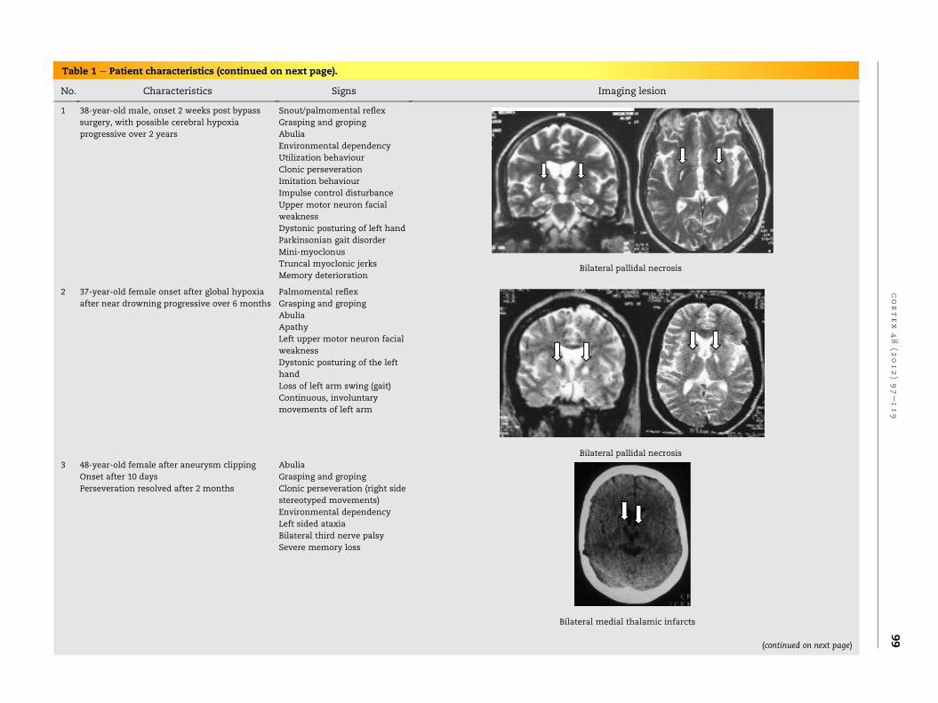
Table 1 e Patient characteristics (continued on next page).
No. Characteristics Signs Imaging lesion
1 38-year-old male, onset 2 weeks post bypass
surgery, with possible cerebral hypoxia
progressive over 2 years
Snout/palmomental reflex
Grasping and groping
Abulia
Environmental dependency
Utilization behaviour
Clonic perseveration
Imitation behaviour
Impulse control disturbance
Upper motor neuron facial
weakness
Dystonic posturing of left hand
Parkinsonian gait disorder
Mini-myoclonus
Truncal myoclonic jerks
Memory deteriorationBilateral pallidal necrosis
2 37-year-old female onset after global hypoxia
after near drowning progressive over 6 months
Palmomental reflex
Grasping and groping
Abulia
Apathy
Left upper motor neuron facial
weakness
Dystonic posturing of the left
hand
Loss of left arm swing (gait)
Continuous, involuntary
movements of left arm
Bilateral pallidal necrosis
3 48-year-old female after aneurysm clipping
Onset after 10 days
Perseveration resolved after 2 months
Abulia
Grasping and groping
Clonic perseveration (right side
stereotyped movements)
Environmental dependency
Left sided ataxia
Bilateral third nerve palsy
Severe memory loss
Bilateral medial thalamic infarcts
(continued on next page)
cortex
48
(2012)97e119
99

Table 1 e (continued on next page ).
No. Characteristics Signs Imaging lesion
4 41-year-old female acute onset with
improvement over 18 months
Abulia
Apathy
Grasping
Pout reflex
Clonic verbal perseveration
Intentional perseveration
Visual grasping (forced visual
following)
Utilization behaviour
Environmental dependency
Small haemorrhage left superior putamen (cavernoma)
5 50-year-old male hypertensive
intracerebral hemorrhage (ICH)
acute onset settled within 2 years
Abulia
Environmental dependency
Intentional perseveration
(verbal)
Impulse control disturbance
Short-term memory loss
Mild right hemiparesis
Fluent aphasia
Daytime somnolence
Left medial thalamus
cortex
48
(2012)97e119
100
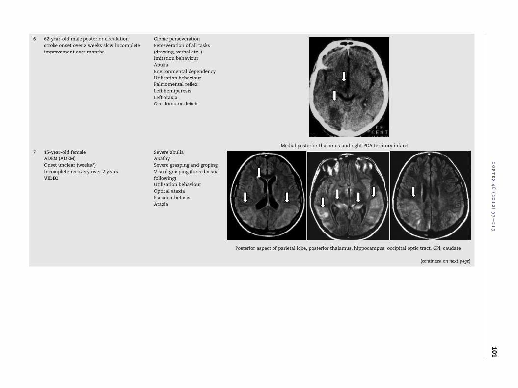
6 62-year-old male posterior circulation
stroke onset over 2 weeks slow incomplete
improvement over months
Clonic perseveration
Perseveration of all tasks
(drawing, verbal etc.,)
Imitation behaviour
Abulia
Environmental dependency
Utilization behaviour
Palmomental reflex
Left hemiparesis
Left ataxia
Occulomotor deficit
Medial posterior thalamus and right PCA territory infarct
7 15-year-old female
ADEM (ADEM)
Onset unclear (weeks?)
Incomplete recovery over 2 years
VIDEO
Severe abulia
Apathy
Severe grasping and groping
Visual grasping (forced visual
following)
Utilization behaviour
Optical ataxia
Pseudoathetosis
Ataxia
Posterior aspect of parietal lobe, posterior thalamus, hippocampus, occipital optic tract, GPi, caudate
(continued on next page)
cortex
48
(2012)97e119
101

Table 1 e (continued on next page ).
No. Characteristics Signs Imaging lesion
8 56-year-old male posterior circulation stroke
Onset over 2 weeks recovery over 1 year
Abulia
Apathy
Tonic perseveration
Severe grasping and groping
Environmental dependency
Short-term memory loss
Daytime somnolence
Dysarthria
Left hemiparesis
Ataxia
Gaze palsy
Medial & posterior thalamus, bilateral cerebellar infarct
9 71-year-old female unknown aetiology
VIDEO
Abulia
Grasping and groping
Visual grasping (forced visual
following)
Imitation behaviour
Clonic perseveration
Environmental dependency
Utilization behaviour
Ideomotor apraxia
Parkinsonism
Putamen, fasciculus occipito-frontalis
cortex
48
(2012)97e119
102

10 57-year-old female ischaemic stroke
VIDEO
Clonic perseveration
Abulia
Environmental dependency
Parkinsonian gait
Progressive memory loss
Right caudate head
11 71-year-old male gradual onset cerebral
lymphoma
Abulia
Grasping and groping
Utilization behaviour
Parkinsonism
Bilateral medial thalamus
(continued on next page)
cortex
48
(2012)97e119
103

Table 1 e (continued on next page ).
No. Characteristics Signs Imaging lesion
12 14-year-old female developed in childhood
primary cerebral tumour
VIDEO
Severe hemi-grasping
Left homonymous hemianopia
Left facial weakness
Right anterior thalamus, putamen, and globus pallidus
13 62-year-old male ischaemic stroke onset
over weeks
Abulia
Apathy
Clonic perseveration
Grasping
Environmental dependency
Aphasia
Left anterolateral thalamus
14 63-year-old female intracerebral
haemorrhage onset over few days
Abulia
Clonic perseveration
Ipsilateral head of caudate
cortex
48
(2012)97e119
104

15 36-year-old male cystic ependymoma Loss of impulse control
Abulia
Apathy
Grasp reflex (inconsistent)
Palmomental reflex
Parkinsonism
Vertical gaze palsy
Bilateral medial thalamus
16 56-year-old female ischaemic stroke
acute onset partial recovery over one week
Abulia
Apathy
Severe bilateral grasping
Ipsilateral (left) ataxia
Palatal tremor with
synchronous movements of left
face and diaphragm
Left medial thalamus
17 56-year-old male intracerebral
haemorrhage acute onset with
gradual worsening over 6 months
Abulia
Apathy
Grasping and groping
Tonic perseveration
Intentional perseveration
Left medial thalamus
cortex
48
(2012)97e119
105

Table 2 e Dysexecutive syndrome with site of lesion.
Symptom Proportiona Patient Lesion (No. of pt.)b
Abulia 16/17 Patient no.: 1, 2, 3, 4, 5, 6, 7, 8, 9, 10, 11, 13, 14, 15, 16, 17 Medial thalamus (7)
Caudate (3)
Pallidum (3)
Anterolat. thalamus (1)
Putamen (3)
Post. parietal lobes (1)
Cerebellum (1)
Apathy 8/17 Patient no.: 2, 4, 7, 8, 13, 15, 16, 17 Medial thalamus (4)
Pallidum (2)
Caudate (1)
Post. parietal lobes (1)
Anterolat. thalamus (1)
Utilization behaviour 6/17 Patient no.: 1, 4, 6, 7, 9, 11 Pallidum (2)
Thalamus (3)
Caudate (1)
Putamen (2)
Imitation behaviour 3/17 Patient no.: 1, 6, 9 Pallidum (1)
Medial thalamus (1)
Putamen (1)
Visual grasping 3/17 Patient no.: 4, 7, 9 Putamen (2)
Pallidum (1)
Caudate (1)
Posterior thalamus (1)
Grasping 13/17 Patient no.: 1, 2, 3, 4, 7, 8, 9, 11, 12, 13, 15, 16, 17 Medial thalamus (6)
Pallidum (5)
Anterolat. thalamus (2)
Caudate (1)
Putamen (1)
Clonic perseveration 8/17 Patient no.: 1, 3, 4, 6, 9, 10, 13, 14 Caudate head (2)
Pallidum (2)
Medial thalamus (2)
Putamen (1)
Anterolat. thalamus (1)
Please note some patients have more than one lesion.
a Number of patients displaying symptom/total of patients.
b Number of patients with lesion in this anatomical site.
c o r t e x 4 8 ( 2 0 1 2 ) 9 7e1 1 9106
3.3. Case 3
This case has been previously published (Fung et al., 1997). A
48-year-old female was admitted to the hospital following
a subarachnoid haemorrhage. After clipping of a ruptured
basilar aneurysm, she was somnolent. She displayed bilateral
third nerve palsy but showed no weakness of the extremities.
Ten days later, repetitive, stereotyped movements began on
the right side. She continuously rubbed the sole of her left foot
with the back of her right foot. She stated that this movement
felt pleasant. When not rubbing her left foot she instead
continuously wiggled her right foot. Similar repetitive waving
movements occurred in her right hand. Occasionally she
continuously rubbed or scratched her nose with her right
hand, eventually leading to skin ulceration of her nose and
cheek. A right sided grasp reflex and groping was very prom-
inent. Sensory limb ataxia also seemed to be present.
Although the movements disappeared within two months
and the ataxia improved, the woman was left with severe
short-term memory loss, abulia, apathy, grasping and
groping. A cranial computed tomography (CT) scan performed
three months after the surgery revealed bilateral posterior,
medial thalamic infarcts most likely representing a top of the
basilar artery occlusion involving the artery of Percheron.
3.4. Case 4
A 41-year-old female was admitted to hospital with auditory
hallucinations. She presented to hospital wearing two pairs of
undergarments even though it was very warm. When given
a pair of eyeglasses she put them on andwhen presentedwith
a second pair of glasses she also put them on over the other
glasses. When she put a third pair of glasses on over the other
two pairs, she could not comprehendwhy they did not stay on
her nose. When asked not to take the glasses she was unable
to suppress this behaviour. She did not speak spontaneously
but was oriented to person and place. She was able to name
objects correctly but would repeat the word when trying to
name the next object. For example, when asked to name
a pencil, she said ‘pencil’; however, when a key was presented
to her afterwards she repeated pencil. Sometimes she would
correct herself.
The same kind of behaviour was observed when asked to
perform a motor task. When asked to tap on the table twice,

Fig. 1 e Basal ganglia and thalamic lesions.
c o r t e x 4 8 ( 2 0 1 2 ) 9 7e1 1 9 107
she did so slowly. Immediately after this, she was asked to tap
her left shoulder,however, instead tappedthetable twiceagain.
Whenagainasked to tapher shoulder shefinallydidsobut then
continued to tap her shoulder when asked to touch her nose.
Similarly, when asked to copy a triangle, circle, and square, she
wrote theword triangle, and copied it twice correctly instead of
once, thenwent on towrite triangle at the circle and continued
to write triangle instead of drawing a circle.
Grasp reflexes in the hands and feet were present and
could not be suppressed. Also present was a prominent pout
reflex. When an object such as a pen was moved towards her
mouth, she opened her mouth in response. When a red light
was moved within her visual field, she followed it with her
gaze and was unable to divert her gaze elsewhere, even on
command; we term this response ‘forced visual following’.
Although unable to recognise the visual momentum of
a presented image, she was able to read a given text. No
spontaneousmovements or actions were observed apart from
fidgeting with her bandage; however, externally initiated
movements were repeated two to three times. This occurred
only with the right limbs. For example, when the left leg was
flexed three times, she continued flexing her right knee.
Similarly, when her right or left hand was placed sequentially
on the left shoulder, right shoulder and right leg, she continued
to touch both shoulders repeatedly with the right arm.
CT revealed an acute small left pallidal haemorrhage due
to a cavernoma. She recovered within 18 months.
3.5. Case 5
A 50-year-old right handed male was admitted to hospital
with an acute onset fluent aphasia and mild weakness of the
right limbs. CT scan showed an acute, small left dorsomedial
thalamic haemorrhage and an old right side lacunar pontine
infarct.
When he returned home, his family perceived him to be
a different person. His wife complained that he did not
participate in regular activities. Despite his previous enjoy-
ment of cooking, he had stopped preparing meals. When
encouraged to cook something, he would stop in themiddle of
the task, appearing to have forgotten to finish the job. He
displayed abulia, daytime somnolence, problems with short-
term memory, and failed to initiate any activity on his own.
When speaking, he showed limited vocabulary and tended to
repeat words from previous sentences. For example, when
asked what he was wearing, he replied ‘blue trousers’ (it was
blue pyjamas with a red bathrobe on top). When asked what
hewaswearing on top of that, hementioned ‘it is a bluee ume
actually it is red.’ As well, he had a tendency to repeat words. ‘I
was actually put into one of the relief units, where they kept me e
um e kept me in security. In premises which led to security e um e
security’. He was able to correctly name objects after some
consideration or after several attempts. When asked to name
a second object he first repeated the name of the prior object
before correcting himself.
He often became upset, lost his temper and seemed to
become angry easily. Within two years his behavioural
changes became less pronounced and he returned to a fairly
normal life although his speech never fully recovered.
3.6. Case 6
This case has been previously published (Fung et al., 1997).
A 62-year-old male was admitted with an acute onset

c o r t e x 4 8 ( 2 0 1 2 ) 9 7e1 1 9108
headache, drowsiness, unsteady gait, left hemiparesis, ataxia,
left homonymous hemianopia, and right oculomotor palsy.
CT and CT angiography revealed a top of the basilar
occlusion with bilateral infarcts in the posterior medial thal-
amus. Within one week his symptoms improved and he was
able to give short sensible answers but had the tendency to
repeat short phrases.
Over the following twoweeks, he developed unusualmotor
behaviour. Albeit spontaneous movements appeared rarely,
externally initiated even complexmovements were continued
unless terminated externally. This occurred only with the
right limbs. When passive movement was induced in the left
limbs, especially the left paretic leg, he mirrored the move-
ment with the non-paretic right leg.
As well, when asked to copy a square, triangle and circle he
did so but repeated each drawing several times (see Fig. 2).
When asked to copy the image only once and offered a 20
cents reward for doing so, he actually drew it only once.When
he was handed the coin he replied ‘thank you, thank you, thank
you, thank you’.
He showed marked abulia without spontaneous or goal
directed actions. He displayed utilization behaviour. The pal-
momental reflex was present.
3.7. Case 7 (see video online)
One week prior to admission this 15-year-old female suffered
from an upper respiratory tract infection. Following a stressful
Fig. 2 e Clonic perseveration when copying figures (case 6).
day, she was found lying in the shower. Although her eyes
were opened wide, she seemed to stare and did not respond.
The next morning she was confused and excitedly screaming
‘I’m not mad’. Over the following days she gradually declined
into a coma and required ventilation.
Supplementary video related to this article can be found
online at doi:10.1016/j.cortex.2011.03.014.
Initial CT, electroencephalogram (EEG) and cerebrospinal
fluid (CSF) studies were normal. Fluid attenuation inversion
recovery MRI (Magnetic Resonance Imaging) (MRI FLAIR)
revealed bilateral, hyper-intense lesions in the posterior part
of the parietal lobe and the hippocampal region. Angiography
of the cerebral arteries was normal, the EEG at that time
showed diffuse slowing, and a biopsy taken from one of the
lesions revealed non-specific inflammation. She was treated
with acyclovir and corticosteroids. Serology for neurotropic
viruses was negative and no metabolic disorder was found.
There was a very low titre Anti-nuclear antibody (ANA) (1:40).
Her symptoms slowly improved over the following weeks.
After twoweeks shewas able to breathe onher own. She could
stand and walk with an aid after six months. She would not
talk unless questioned and showed no emotional reaction
unless being directly involved in a social interaction. On
returning home, she did not participate in normal household
tasks unless specifically asked to do so. She answered ques-
tions sensibly using very brief, normally constructed senten-
ces. She was oriented to time, place, and date.
Examination six months after the initial presentation
revealed bilateral pseudoathetosis and ataxia,marked groping
and grasping. As well, she showed mouth grasping in
response to an object being placed close to her mouth. She
also displayedmarked utilization behaviour.When repeatedly
given eyeglasses she would continue to place each pair on top
of the other even though she did not require glasses. When
presented with food she would eat it. Whenever a visual
stimulus was presented within her visual field she would look
at it. When asked to look away, she inconsistently suppressed
the force to follow the visual stimulus for a short time but then
continued to look at it again.
She slowly recovered over several months and returned to
a fairly normal life. But she was never as active as before and
still displayed a minor ataxia which seemed to deteriorate
under visual guidance.
Follow up MRI (T2) taken eight years later revealed marked
cerebellar and occipital atrophy with bilateral pallidal
hyperintensities.
3.8. Case 8
A 56-year-old male was admitted to hospital with dysarthria,
left hemiparesis and ataxia, and gaze directed nystagmus.MRI
identified bilateral cerebellar (right> left) and thalamic
infarcts affecting the left mediodorsal and centromedian
nuclei, and parts of the right pulvinar.
After two weeks of a fluctuating course, the level of
consciousness, ataxia, left sided weakness, and dysarthria
slowly improved. However he remained very quiet and did not
initiate conversation or social interaction. He showed severe
grasping and groping. He maintained bizarre positions of the

c o r t e x 4 8 ( 2 0 1 2 ) 9 7e1 1 9 109
limbs for several minutes until externally terminated. At
times, when the examiner moved the right arm he would
move the left arm into a mirrored posture and maintain it in
that position.
Symptoms stabilized over the following year yet he
remained inactive and dependant on his environment and
suffered from memory deficits. He was lost to follow up.
3.9. Case 9 (see video online)
A 71-year-old female presented with sudden onset disorien-
tation, slowness of gait, poor balance, soft speech and tran-
sient right hemiparesis. She was very talkative and engaged in
social activity prior to the event. Since then she was very
passive, spentmost of the day sitting, doing nothing, napping,
and watching television without being particularly interested
in it and gained 20 kg of body weight. She needed help with
simple tasks such as dressing and feeding.
Supplementary video related to this article can be found
online at doi:10.1016/j.cortex.2011.03.014.
When examined 5 years after the onset of symptoms, she
gave appropriate and sensible answers to direct questions but
otherwise did not speak. She was fully oriented and could
perform simple arithmetic. Speech was soft but clear. The
affect was fatuously cheerful and untroubled. Facial expres-
sion was reduced, and her mouth was often open. There was
no sialorrhoea. Phonemic verbal fluency was severely and
category fluency was moderately impaired. Handwriting was
markedly micrographic but still legible. Constant repetitive
patting-stroking movements of the right hand were present.
She displayed a prominent, symmetrical rigidity and brady-
kinesia. Bilateral grasp and grope reflexes and forced visual
following present, which she could not inhibit. She immedi-
ately imitated absurd movements of the examiner during
a conversation. She could suppress shortly the imitation
behaviour if requested not to copy the examiner until she was
distracted. She was unable to mime simple tasks in the
absence of tools or objects (ideomotor apraxia), but could
perform complex tasks such as folding a piece of paper and
placing it in an envelope when given the objects. There was
marked hyperreflexia of the tendon jerks with extensor
plantar reflexes. The gait was characterized by short shuffling
steps with a broad base, the feet rotated out. Arm swing was
absent and the posture slightly stooped. Postural stability was
markedly impaired. Levodopa did not improve her symptoms.
MRI revealed increased T2 signal in the anterior 2/3 of the
putamen bilaterally, in addition to lesions around the anterior
horn of the lateral ventricle. There was a small peripheral
lesion in the right cerebellar hemisphere.
3.10. Case 10 (see video online)
This 57-year-old female, who was imprisoned for murder,
presented with an ankle fracture. During treatment for her
fracture, repetitive movements were observed. She com-
plained about a progressivememory loss, and her practitioner
recorded a peculiar gait. There is no history of psychiatric
disorder.
Supplementary video related to this article can be found
online at doi:10.1016/j.cortex.2011.03.014.
On examination, she showed continuous repetitive
movements of the right hand. The left hand displayed dys-
tonic posturing and minor repetitive movements. Further-
more she had the tendency to repeat phrases or short
sentences, which is highlighted in the following dialogue
between the author JM, who asked the patient JS what she
does with her continuously moving right hand:
JS: ‘I touch things, touch things, touch thing, . touch pretty
things.to see if it’s real, if it’s real, if it’s real.’
JM: ‘What about your speech?’
JS: ‘Now that’s funny, that’s funny, that’s funny, that’s funny.’
JM: ‘Did you have a stammer when you were younger’
JS: ‘No, no, no. I talked good, I talked good, I talked good.’
JM: ‘Why do you repeat the phrases?’
JS: ‘I can’t st- I don’t know, I don’t know, I don’t know, I don’t
know,’
When asked to copy objects she copied them over and over
again and had difficulty with copying a different object.
CT scan showed a lesion at the head of the right caudate
nucleus consistent with an old infarct.
3.11. Case 11
A 71-year-old male was referred with a cognitive decline. He
was obese with hypertension, diabetes and an extensive
smoking history. He was not oriented to time and place, but
could correctly state his name and address. When questioned
he gave slow, short answers and followed verbal commands
but did not initiate a conversation. He did not initiate any
movement, but when an object was placed in his palm he
would grab onto it and would not let go. When given a sunhat
he would put it on despite being in a room. When given
another hat he would place it on top of the previous one, and
so on. The same behaviour occurred when offered a pair of
eyeglasses. Furthermore, when given a meal he would
consume everything though remarking, when questioned,
that he was not hungry and repeat this behaviour
immediately.
CT scan showed a lesion involving the medial thalamus
bilaterally. Biopsy demonstrated primary central nervous
system (CNS) lymphoma.
3.12. Case 12 (see video online)
At age 12, this patient started to display unusual movements
of her left toes, accompanied by headache and vomiting.
Cranial MRI revealed a tumour in the right putamen, which
was subsequently resected at age 14. Post-operatively the
unusual behaviour of her left hand and foot worsened over
several weeks. The hand would occasionally ‘pick at’ her hair
or dress, or levitate and grasp onto objects. These movements
caused substantial social embarrassment. Furthermore she
described a flexion of her left toes, triggered by walking,
associated with a peculiar gait.
Supplementary video related to this article can be found
online at doi:10.1016/j.cortex.2011.03.014.

c o r t e x 4 8 ( 2 0 1 2 ) 9 7e1 1 9110
Neurological examination at the age of 26 revealed a left
homonymous hemianopia and mild left facial weakness.
Motor strength was normal as were deep tendon and plantar
reflexes. There were no signs of cerebellar or sensory
dysfunction. Gentle stroking over the sole of the foot provoked
forced sustained flexion of all toes. As well, gentle touching of
the palm provoked a forceful, sustained grasping of the
examiners fingers.When she tried to stand up froma chair the
left hand grabbed the hand rest and the patientwas not able to
release the hand.
We tested her ability to look away by designing a computer
program displaying a white dot in the middle of the screen
and then randomly displaying a red dot anywhere along
a horizontal line at the right or left half of the screen. The
patient was asked to look at the white dot in the middle.
Whenever a red dot occurred in the right or left visual field she
was asked to look to the opposite side of the red visual stim-
ulus. She did so without hesitation or any prosaccadic eye
movements.
MRI of the brain taken at the age of 26 showed a well
defined lesion in the right globus pallidus and putamen. Brain
FDG-PET demonstrated hypometabolism of the right superior
frontal and occipital lobe (see Fig. 3).
3.13. Case 13
A 62-year-old male with a prior history of type II diabetes,
hypertension, and coronary heart disease with atrial fibrilla-
tion was admitted to hospital with a non-fluent aphasia and
right hemiparesis. Previously he had ceased warfarin for joint
replacement surgery. The initial CT scan was unremarkable
but the MRI-brain performed 30 h later showed left antero-
lateral thalamic infarct involving parts of the capsule and an
occlusion of the distal internal carotid artery.
Within one week, the patient recovered from the right
hemiparesis but remained disoriented and aphasic. Following
discharge, he did not participate in his normal activities at
home becoming increasingly inactive and dependent on his
environment. Six months after the stroke his spouse reported
that he spent most of the day watching television, had gained
more than 10 kg in weight, and repeated useless tasks over
and over again. On examination, hewas not orientated to time
or place but could state his name and age. He answered simple
questions with short sentences sometimes searching for
words and had difficulty naming objects. He repeated words
or phrases three to four times and repeated simple motor
tasks (such as cleaning his glasses and adding sugar to his
coffee). He did not initiate any conservation or movement
unless externally initiated. Inconsistently, he held onto
objects that were placed into his palms. As well, he ate
anything offered to him.
Brain FDG-PET demonstrated left frontal hypometabolism
(see Fig. 3).
3.14. Case 14
A 63-year-old female presented with unusual movements of
the fingers. The movements were present most of the time
especially when watching television. When asked why
she rubbed her fingers against each other, she stated that the
fingers felt soft and the feeling was pleasant. The patient
complained about tiredness and did not like to go out and
meet with friends. She was a past smoker with 60 pack years.
The patient was a social worker in a retirement village but had
stopped working two years ago due to fatigue.
On examination, she demonstrated a continuous move-
ment of her left index finger and thumb, with continuous
rubbing of the fingertip to the tip of the thumb. Shewas able to
suppress this behaviour for a short time. When asked to wear
hand gloves the repetitive movement stopped, but she dis-
liked the gloves. The rest of the neurological examination was
normal.
MRI revealed a subacute intracerebral haemorrhage
involving the left caudate head.
3.15. Case 15
A 36-year-old male presented with profound behavioural
changes. He had started to urinate in public, lost interest in his
usual activities, and developed an unusual slow shuffling gait
and shake, mainly on the left.
Examination revealed an intermittent, low amplitude,
resting tremor, worse on the left, which increasedwith action.
There was no rigidity. He had a vacuous expression and was
hard to engage. He hardly participated in the consultation but
was able to respond with short and reasonably appropriate
answers to direct questions. He appeared unconcerned. Grasp
reflexes were inconsistently present bilaterally, and there was
a brisk palmomental reflex. He had vertical gaze palsy.
MRI revealed a dorsal midbrain tumour (cystic ependy-
moma), which extended to the medial thalamus bilaterally.
Subsequent diffusion-PET showed reduced frontal metabo-
lism (see Fig. 3). The patient died three months later.
3.16. Case 16
A 56-year-old female presented with an acute onset left side
ataxia, dizziness and unsteady gait. MRI-brain demonstrated
left medial posterior thalamic, and left superior cerebellar
artery infarcts.
Over one week the drowsiness and ataxia improved but
she did not engage in any social activity.
She did not initiate a conversation, however, she gave
sensible answers when prompted. She appeared unconcerned
about her condition. Severe bilateral grasping was observed.
She further suffered from palatal tremor extending to
synchronous movements of the left side of her face and dia-
phragm, associated with hypertrophic degeneration of the
superior olive. The abulia improved to some degree, but she
remains dependent on her environment two years later. PET
showed frontal hypometabolism (see Fig. 3).
3.17. Case 17
One morning a 56-year-old jeweller’s assistant remained in
bed. When his wife spoke to him he answered but did not
stand up. Neither did he initiate a conversation. Eventually his
wife persuaded him to take a shower; although he got under
the shower he did not turn the water on. When he walked he

Fig. 3 e Positron emission tomography.
c o r t e x 4 8 ( 2 0 1 2 ) 9 7e1 1 9 111

c o r t e x 4 8 ( 2 0 1 2 ) 9 7e1 1 9112
appeared to walk excessively slowly. From that time on he sat
and said nothing unless prompted.
When examined he sat on the bed making no eye contact.
He was oriented and knew who had won the recent election.
He did not, however, say anything unless spoken to. He
walked slowly and deliberately. Initially there were no grasp
reflexes detectable but six months after the initial presenta-
tion grasping and groping was present. When his arms were
placed in various positions, they remained there for prolonged
periods. When offered eyeglasses he put them on, and when
offered his wife’s eyeglasses he exchanged them for the
existing glasses, and would continue to do so without
comment. He showed tendency to repeat movements and/or
phrases once initiated.
CT scan of the brain showed a subacute small haemor-
rhage of anterior half of the right thalamus.
4. Discussion
Animal studies demonstrated a connection of the frontal
cortex to the basal ganglia. Alexander and colleagues
proposed five fronto-subcortical circuits (Fig. 4) (Alexander
and Crutcher, 1990; Alexander et al., 1986). Each circuit origi-
nates from a different frontal cortical area and projects to the
certain part of the striatum. Through the direct pathway via
the internal globus pallidus internus (GPi)/substantia nigra
and through the indirect pathway via the external globus
pallidus externus/substantia nigra back to GPi all loops
connect to different parts of the thalamus. From the thalamus
all circuits project back to one single area in the frontal cortex.
Antero- and retrograde labelling studies in animals
(Alexander et al., 1990) suggest that the fronto-subcortical
circuits are anatomically segregated and form parallel,
closed loops. However, other studies suggest additional open
Suppleme
SMA
Putame
Ventrolat.Caudolat.
Thalamu(VOL/VLM
APA
Frontal Eye Field
FEF
Caudate(body)
Caud.- dm.GPiVentrolat. SNr
Thalamus(lat.VAmc/MDpl)
DLC PPC
Lateral Orbitofrontal
LOF
Med.dm. GPiRostromed. SNr
Thalamus(med. VAmc/MDmc)
ACA STG/ITG
vm.Caudate(head)
Fig. 4 e Fronto-subcortical loops. ACA: anterior cingulate area;
cortex; dm: dorsomedialis; EC: entorhinal cortex; FEF: frontal ey
cortex; ITG: inferior temporal gyrus; LOF: lateral orbitofrontal co
SC: somatosensory cortex; STG: superior temporal gyrus; VOL:
VAmc: ventralis anterior pars magnocellularis; med.: medial; la
vent.: ventral; vm: ventromedial.
pathways from the same anatomical subregions in the stria-
tum, pallidum and substantia nigra, which project to func-
tionally non-corresponding motor and limbic subregions of
the basal ganglia (Joel andWeiner, 1997). Further, the thalamic
ventral anterior, ventral lateral, and mediodorsal nucleus
seem to facilitate exchange between different cortical circuits
(McFarland and Haber, 2002).
The anatomical existence of segregated loops has never
been demonstrated in humans for the lack of methods suit-
able to study projections from different areas of the frontal
lobes to the basal ganglia.
In the following we discuss the distinct dysexecutive
behaviour and correlate it to lesions in our case series. We
compare these results to other published cases with frontal
lobe and non-frontal lobe lesions and correlate the frontal
cortex and the affected frontal-subcortical circuit to the
behavioural abnormality (see also Cubillo et al., in press;
Langen et al., in press).
Table 2 andFig. 1 summarize the basal ganglia and thalamic
lesions grouped in order of the below discussed symptoms.
4.1. Abulia and apathy
The most common disturbance in our case series was the
reduction of self-generated voluntary and purposeful behav-
iour, and a lack of emotional response, which is referred to as
apathy or abulia. The term abulia and apathy are conceptually
ill defined. Both are used for similar or identical symptoms
(Starkstein and Leentjens, 2008). We would like to use abulia
for the cognitive and behavioural disturbance and the term
apathy for the lack of an emotional response.
Self-generated voluntary and purposeful behaviour was
reduced in all our cases except case 12. The most common
lesion (9/16) was found in the posterior medial part of the
thalamus (cases 3, 5, 6, 7, 8, 11, 15, 16 and 17) and in the left
nt Motor
n
GPi SNr
s)
MC/SC
Dorsolateral
DLC
Lat.dm. GPiRostrolat. SNr
Thalamus(lat. VApc/MDpc)
PPC APA
dl.Caudate(head)
Anterior Cingulate
ACA
Rostrolat. GPiVent. Pallidum
Rostrodors. SNr
Thalamus(post. med. MD)
ITG/STG HC/EC
VentralStriatum
APA: arcuate premotor area; DLC: dorsolateral prefrontal
e field; GPi: globus pallidus internus; HC: hippocampal
rtex; MC: motor cortex; PPC: posterior parietal cortex;
ventralis oralis lateralis; VLM: ventralis lateralis medialis;
t.: lateral; VApc: ventralis anterior pars parvocellularis;

c o r t e x 4 8 ( 2 0 1 2 ) 9 7e1 1 9 113
anterior thalamus (case 13). If the thalamus was affected
bilaterally patients showed no or very little spontaneous
behaviour. The most severely affected person (case 11) with
a midline lymphoma extending into both thalami did not
move or speak unless prompted.
Six of our 16 caseswith abulia did not suffer from lesions of
the thalamus. Two of our patients with abulia had lesion
affecting the caudate nucleus (cases 10 and 14). Cases 1, 2
suffered from pallidal necrosis after hypoxia. Two patients
(cases 4 and 9) with putaminal lesions also showed reduced
self-generated behaviour.
About half of our cases displayed a severely disturbed
affect and did not engage emotionally with their environment.
They displayed little concern or seemed not to suffer from
their condition. All patients with apathy also displayed abulia.
Abulia and apathy have been associated with many
neuropsychiatric conditions. The lack of spontaneous
behaviour is a common finding in different neurodegenerative
diseases like Alzheimer’s, Parkinson’s disease, or depression.
Functional imaging studies identified abnormalities of resting
blood flow and glucose metabolism in depression in the
orbital andmedial prefrontal cortex (Drevets, 1999) suggesting
a prefrontal lobe dysfunction.
Marshall et al. (2007) studied 41 probable Alzheimer’s
patients with FDG-PET. Those patients with a clinically
meaningful apathy (Subscale for the Assessment of Negative
Symptoms in Alzheimer Disease) showed a significantly
reduced tracer uptake in the anterior cingulate region, infe-
riorly to the medial orbitofrontal region and the bilateral
medial thalamus compared to those patients without apathy.
Apathy has been described in ischaemic brain lesions of
the caudate nucleus, the anterior and ventral globus pallidus,
and the dorsomedian and intralaminar thalamic nuclei (Jorge
et al., 2010).
Furthermore reduced self-generated behaviour has been
found in patients with lesions in the anterior cingulate cortex
(Brodmann’s area 24) and in the interconnected medial and
lateral orbitofrontal cortex (Brodmann’s areas 10, 11, 47)
(Amyes and Nielsen, 1953, 1955; Barris and Schuman, 1953;
Degos et al., 1993; Fesenmeier et al., 1990; Grunsfeld and
Login, 2006; Mega and Cohenour, 1997; Nemeth et al., 1988;
Nielsen, 1951; Nielsen and Jacobs, 1951). The anterior cingu-
late cortex is furthermore connected to the superior temporal
gyrus (Brodmann’s area 22). The lateral orbitofrontal cortex/
anterior cingulate cortex/superior temporal gyrus are con-
nected through the ventromedial part of the nucleus caudatus
head, the medial GPi to the medial and ventral anterior thal-
amus. Through themedial thalamus the loop reprojects to the
lateral orbitofrontal cortex. Furthermore the anterior cingu-
late cortex and superior temporal gyrus are connected to
hippocampal and entorhinal cortex and through the ventral
striatum, rostrolateral GPi, to the posterior medial thalamus
reprojecting to the anterior cingulate cortex.
The vast majority of our cases with abulia and apathy
suffered from posterior medial thalamic lesions, which is
consistent with the above described frontal-subcortical
circuits originating from the anterior cingulate cortex and
lateral orbitofrontal cortex.
One patient with a ventral anterior thalamic infarct, two
patients with caudate nucleus lesions and two others with
pallidal necrosis suffered fromabulia and apathywhich is also
part of this circuit.
Only two cases had lesions outside of the anterior cingulate
cortex and lateral orbitofrontal cortex fronto-subcortical
circuit. Both patients had lesions in the putamen, which has
been described by David and colleagues previously. David
et al. studied 22 patients with Alzheimer’s and Lewy Body
Dementia with (123I) Ioflupane-CIT Single Photon Emission
Computed Tomography (DaTSCAN) (David et al., 2008) which
is a marker of the presynaptic dopamine transporter. They
found a significant correlation between higher Apathy
Inventory Scores and reduced putaminal Ioflupane uptake
suggesting a loss of dopaminergic neurons projecting to the
putamen. Together with our two cases this study suggests an
involvement of the putamen in self-generated behaviour.
The occurrence of abulia in all but one of our cases suggests
that the fronto-subcortical loops originating from the orbito-
frontal cortex and the anterior cingulate cortex are not
segregated but interconnected to other frontal-subcortical
loops in humans.
4.2. Utilization and environmental dependency
Many of our patients displayed very little spontaneous
behaviour like case 11, who did not speak, move, or interact
with others unless prompted. However, when the right cue
was presented, he displayed elaborate automatic behaviour.
He utilized objects like a sunhat in the object correct way by
placing it on his head (utilization behaviour) (Lhermitte, 1983).
The behavioural patterns per se seemed to be preserved.
However, they did not occur spontaneously. An external cue
was needed to activate the behavioural pattern (environ-
mental dependency). Furthermore the behaviour appeared
automatically disregarding whether it was adequate or
purposeful. For example the patient placed one sunhat over
the next one on his head while sitting in a hospital room. This
utilization behaviour occurred in many of our patients with
abulia. They used eyeglasses which they did not need and
consumed food whenever it was placed in front of them,
despite the fact that they just had eaten a full meal. Even
when asked not to do so, they seemed unable to suppress the
urge to utilize the given object. Automatic and external driven
utilization behaviour was closely related to abulia in our case
series. Although we did not measure abulia quantitatively,
those patients displaying severe abulia showed more consis-
tent utilization behaviour, than those less affected.
We observed utilization behaviour 6 of our 17 patients
(cases 1, 4, 6, 7, 9 and 11). Cases 1 and 4 had pallidal lesions,
case 11 suffered from bilateral and case 6 from a right poste-
rior medial thalamic lesion, and case 7 had multiple lesions
among other involving both posterior thalami and GPi. Case 9
displayed lesions affecting the frontal white matter and the
putamen. All our cases with utilization behaviour displayed
abulia, suggesting that utilization behaviour is a concomitant
symptom of abulia.
Lhermitte first described utilization behaviour in a 62-year-
old female with a subarachnoid haemorrhage due to an
anterior communicating aneurysm after clipping of the
aneurysm (Lhermitte, 1983). She died one year after the

c o r t e x 4 8 ( 2 0 1 2 ) 9 7e1 1 9114
surgery and the autopsy revealed damage in the lower frontal
pole, the lateral orbitofrontal cortex.
Ishihara and colleagues presented a similar case of
a patient who displayed utilization behaviour after an infarct
of the left anterior cerebral artery affecting the superior
frontal cortex and subcortical white matter (Ishihara et al.,
2002). 99mTc-HMPAO Single Photon Emission Computed
Tomography (SPECT) demonstrated reduced uptake in the
ipsilateral left superior and medial frontal lobe. They
proposed utilization behaviour as a white matter disconnec-
tion syndrome disrupting the frontal thalamic pathways.
Utilization behaviour was also found after lesions of the
caudate nucleus (Degos et al., 1993; Rudd et al., 1998). Other
reports have described utilization behaviour in patients with
diffuse frontal lesions subsequent to herpes encephalitis
(Brazzelli et al., 1994), after paramedian thalamic infarcts
(Eslinger et al., 1991; Hashimoto et al., 1995) and following
lesions in the cingulate gyrus (De Renzi et al., 1996; Degos
et al., 1993; Fukui et al., 1993).
4.3. Imitation behaviour
Lhermitte considered imitation behaviour as the “first stage” of
utilization behaviour (Lhermitte et al., 1986). Imitation behav-
iour is an inborn atavistic behaviour, which has been linked to
the left parietal lobe (Muhlau et al., 2005), the anterior cingulate
cortex and the caudate nucleus (De Renzi et al., 1996). The
anterior cingulate cortex seems to suppress this atavistic
behaviour, possibly generated in the parietal lobe, so that the
behaviour is performed only when appropriate. Lhermitte
considered imitation behaviour to be a voluntary act, not
reflexive, as distinct from echopraxia (Dromard, 1905). Lher-
mitte argued that echopraxia was a more abrupt, automatic
behaviour that cannot be stopped. Although we did not test
eachpatient systematically, imitationbehaviour appeared tobe
rare in this population occurring only in cases 1, 6 and 9 who
had lesions in the pallidum, thalamus, and putamen respec-
tively. Patients with utilization behaviour did not necessarily
show imitation behaviour. However two of the three patients
who displayed imitation behaviour also displayed utilization
behaviour.This is inconcordancewith thefindingsofLhermitte
who reported imitation behaviour in only 40 of 75 patientswith
utilization behaviour. While two of his patients had deep
lesions (thalamic and caudate nucleus), all others had frontal
lobe pathology (Lhermitte et al., 1986) suggesting that imitation
behaviour is less common in lesions affecting the basal ganglia
or the thalamus compared to frontal lobe lesions.
4.4. Visual grasping
Like imitation behaviour, visual pursuit is an inborn behaviour
(Barten et al., 1971). The ability to look away (anti-saccade)
from a visual stimulus involves the frontal eye field (Fischer
et al., 1997; Fukushima et al., 2000; Munoz and Everling,
2004). Patients with unilateral frontal eye field lesions are
unable to suppress an automatic prosaccadic eye movement
to visual stimuli in the ipsilateral visual field, giving the
appearance of horizontal gaze palsy (Guitton et al., 1985).
The frontal eye field (Brodmann’s area 8) is interconnected to
the posterior parietal cortex and the dorsolateral prefrontal
cortex. These cortical areas project to the caudate nucleus, GPi
and ventral anterior nucleus of the thalamus before projecting
back to the frontal eye field.Munoz and Everling (2004) pointed
out that the posterior parietal cortex and frontal eye field are
also connected to the superior colliculus, which projects via
the cerebellum to the reticular formation.
‘Visual grasping’ occurred in only 3 of or 17 patients. Cases
4, 7 and 9 were unable to suppress the urge to look at a salient
visual stimulus without having a horizontal gaze preference.
Case 7 had extensive pathology affecting the posterior parietal
cortex, pulvinar, hippocampus, occipital optic tract, and
bilateral globus pallidus. Furthermore a cerebellar atrophy
was found years after the incident. Since the posterior parietal
cortex is interconnected with the frontal eye field it is not
surprising that case 7 displayed marked visual following.
Case4hada small lesion in the left putamen. Case9 showed
bilateral lesions in the putamen. The putamen is connected to
both supplementarymotor and arcuate premotor areas,which
each have connections to primary motor and somatosensory
cortices. The downstream structures involve the GPi and the
substantia nigra pars reticulata, as well as the ventral oro-
lateral and ventrolateromedial thalamic nucleus.
The putamen has not been associated with disturbed anti-
saccadic eye movements. Case 9 displayed a severe frontal
lobe syndrome with abulia, apathy, grasping, utilization, and
imitation behaviour as well as the inability to suppress a pro-
saccadic eyemovement towards a visual stimulus. If the small
lesion in the putamen is the cause of these symptoms of case
9, the putamen might have a much more prominent role in
frontal lobe function. All patients with visual grasping also
demonstrated manual grasping.
4.5. Grasping and groping
Manual grasping or groping was the second most common
executive dysfunction in our case series and occurred in 13 of
our 17 cases (cases 1, 2, 3, 4, 7, 8, 9, 11, 12, 13, 15, 16 and 17).
Grasping has been associated with dysfunction of the
contralateral supplementary motor area (Adie and Critchley,
1927; Wilson and Walshe, 1914) and anterior cingulate
cortex lesions (Hashimoto and Tanaka, 1998). The supple-
mentary motor area (Brodmann’s area 6) is interconnected
with the arcuate premotor area, the motor cortex and
somatosensory cortex. These cortical areas connect through
the putamen to the ventrolateral GPi and caudolateral sub-
stantia nigra pars reticulate to the nucleus ventralis oralis
lateralis and ventralis lateralis medialis of the thalamus and
reproject to the supplementary motor area.
In our case series grasping was most commonly found in
patients with medial thalamic and pallidal lesions. Case 12
displayed only unilateral grasping without any other dysex-
ecutive symptom. She was fully aware of this behaviour and
could not suppress it, which caused considerable social and
functional disability. She developed contralateral severe hemi-
grasping inthe lefthandandfootdue to rightputaminal tumour
in early childhood. The resection of the tumour left her with
damage to theputamen andpallidumand someminor changes
in the lateral anterior thalamus and the optic tract. Her brain
FDG-PET demonstrated hypometabolism of the right superior
frontal and occipital lobe. Although this patient suffered from

c o r t e x 4 8 ( 2 0 1 2 ) 9 7e1 1 9 115
a large defect involving her right putamenand globus pallidum,
she did not display any other dysexecutive behaviour and was
the only patient with “normal” spontaneous self-generated
behaviour. She sustained her lesion in early childhood, which
might have allowed for a greater remodelling of the frontal-
subcortical pathways limiting her deficits compared to those
who experienced their lesions later in life.
Case 13 who suffered from a small embolic left middle
cerebral artery (MCA) stroke involving his left ventral anterior
and ventral lateral thalamus displayed bilateral manual
grasping. The ventral lateral and ventral anterior nuclei of the
thalamus are not part of the above described frontal-
subcortical loop, opposing the segregation hypothesis of the
frontal-subcortical loops in humans.
Grasping was interpreted by Liepmann (1905) as a tonic
perseveration. According to Liepmann, perseveration occurs
as an error of self-generated behaviour. The activated ‘inner-
vation complex’ either persists or remains in a state of higher
excitability resulting in ongoing or recurrent identical behav-
iour. Liepmann concept of grasping was a self-generated
behaviour, which could not be altered.
Schuster and Pineas (Schuster, 1923; Schuster and Pineas,
1926) considered the grasp phenomenon unlike Liepmann as
areaction toasensorystimulusand identified twodistinct types.
The pursuing movements of an object that had touched the
hand, or crossed the field of vision was coined ‘groping’ (Nach-
greifen), in distinction from the involuntary closure of the hand
when an object was placed in the palm, which they called
‘grasping’ (Zwangsgreifen). They considered grasping to consist
of two components: a transient “snapping” movement
(Schnappbewegung) which the patient could release again and
a lasting grip (Umklammerung), which was provoked by an
object touching the hand sufficiently long. Schuster and Caspar
found that patients with forced grasping showed pathological
changes either in the basal ganglia or in the frontal lobe of the
opposite side. The frontal lesion involved the first frontal gyrus
or anterior genu of the corpus callosum. (Schuster, 1927;
Schuster and Caspar, 1930). Richter and Hines showed that iso-
lated destruction of the premotor area ononeside caused forced
grasping on the opposite side in Pithecus rhesus. However the
grasping phenomenon was transient in unilateral lesions and
persisted only after bilateral lesions (Richter and Hines, 1932).
Seyffarth and Denny-Brown (1948) distinguished five sub-
varieties of the instinctive grasp reaction: ‘Closing reaction’
of the thumb and fingers, the ‘final grip’ of the fingers/hand
onto the object, both provoked by a light stationary or moving
tactile stimulus to the palm. The ‘trap reaction’ is a sudden
tightening of the grip when the examiner tries to pull out the
fingers or the object placed in the hand. The ‘magnet reaction’
is used for a pursuing movement after a light touch of finger
tips of the patient. The ‘instinctive or visual groping’ for
a pursuing movement of the arm and hand provoked by a
visual stimulus. They reported that 6 of the 68 patients with
a grasp reaction suffered from a lesion in the basal ganglia,
they do not however present the exact location of the lesion.
DeRenzi and Barbieri investigated the grasp phenomenon in
491 unselected patients admitted to their unit (De Renzi and
Barbieri, 1992). Only patients with coma were excluded. They
systematically investigatedall patients for thepresenceof grasp
reflex, grasp reaction, foot grasping, and groping and found
a grasp phenomenon in 8% of all patients. They correlated the
symptoms to lesions in the brain based on CT imaging. In the
majority of their patients the grasp phenomenon occurred in
bothhands.Theyfoundgrasping tobeassociatedwith lesionsof
the supplementmotor area. They investigated then in a second
study44patientswith graspingor frontal lobe lesions to identify
the exact location within the frontal lobe which seems to be
associated with grasping. In 70% grasping was associated with
a lesion of the medial frontal lobe namely the premotor area.
Only few patients in both studies (two patients in the first study
and five in the second) had deep lesions involving the putamen
and the globus pallidus, and one patient with a lesion restricted
to the head of the caudate nucleus.
Hashimoto and Tanaka (1998) investigated systematically
the site of the brain lesion and the characteristics of patho-
logical grasping in 28 patients with hemispherical strokes
using Seyffarths and Denny-Brown’s taxonomy. They group-
ed the patients according to the site of lesion and found that
all seven patients who suffered from a stroke involving either
the supplement motor area or the anterior cingulate gyrus
displayed a grasping phenomenon which was minimal in
most of the patients. Only anterior cingulate gyrus lesions
produced visual groping and a magnet reaction and only
a mild grasping, where as patients with lesions restricted to
the supplement motor area exhibited a constant contralateral
grasp reflex. If both the supplement motor area and the
anterior cingulate gyrus area were affected patients displayed
moderate to severe grasping and groping.
We did not systematically investigate our patient with
regard to different grasp and groping phenomenon as sug-
gested by Seyffarth and Denny Brown. This might have led to
a under recognition of prehensile behaviour in our group. Our
findings highlight the role of the lentiform nucleus in case 12
who displayed severe hand and foot grasping as well as
groping to a lesser degree, which is consistent with De Renzi
and Barbieri’s finding. In our patient group the medial thal-
amuswas also consistently associated with bilateral grasping.
4.6. Perseveration
Liepmann distinguished three types of perseveration: Tonic
perseveration e the patient remains in a certain posture or is
caught in a self-generated behaviour; clonic perseveration e
the same task pattern recurs spontaneously several times
after first self-generated presentation, and intentional
perseveration, which is a recurrence of the same task pattern
after their first presentation whenever the patient tries to
perform a different one. Perseveration was the most frequent
motor disturbance observed in our series besides grasping.
4.7. Clonic perseveration
Clonic perseveration or repetitive movements were the most
common form (cases 1, 3, 4, 6, 9, 10, 13 and 14). Six of the eight
cases showed spontaneous, continuous repetitive move-
ments. Cases 1 and 6 displayed this behaviour only when
externally driven. Both these patients seemed to be
completely externally dependent in any task.
The two patients that displayed the most consistent clonic
perseveration had lesions of the caudate head ipsilateral to

c o r t e x 4 8 ( 2 0 1 2 ) 9 7e1 1 9116
the motor symptoms (cases 10 and 14). Especially case 10
showed marked motor and verbal perseveration. Cases 3 and
6 suffered from bilateral posterior thalamic infarcts. MRI
scans of cases 1 and 9 revealed lesions predominantly of the
globus pallidus and the putamen.
Luria and colleagues (Luria, 1965) were the first to associate
perseveration with frontal lobe lesions. Luria describes
a patient with pre-orbitofrontal cortex lesion due to olfactory
glioma who seemed to repeat tasks over and over again. The
examples of drawings accompanying his publication are very
similar to our case 6. This patient did not have difficulties in
switching from one task to the next.
Castaigne et al. have summarized patients with thalamic
lesions (Castaigne et al., 1981). Four of their 28 cases showed
spontaneous repetitive movements. One additional case
showed ‘involuntary movements’, however, no further
description of the movement is provided. All patients suffered
from thalamo-peduncular infarcts caused by embolism of the
basilarartery.Althoughall 10patientswith thalamo-peduncular
infarct presented with sudden onset of hemiparesis, gaze
disturbance or somnolence, the involuntary movements man-
ifested days or even weeks later. The common lesion among
these cases was of the dorsomedial nucleus of the thalamus.
Carrera and Bogousslavsky (Carrera and Bogousslavsky,
2006; Carrera et al., 2004) have correlated clinical symptoms
with thalamic lesions. They associated perseveration with
lesions of the anterior thalamus, which is in contrast to our
findings and the previous work of Castaigne and colleagues.
However, their concept of perseveration is limited to verbal
and paradigmatic perseveration and they did not report any
changes in motor behaviour.
4.8. Intentional perseveration
Intentional perseveration was only observed in cases 4 and 5.
Case 4 for example repeatedly wrote the word triangle not
only correctly under the triangle but also under the circle and
square. Case 4 showed a pallidal lesion and case 5 had a left
dorsomedial thalamic haemorrhage.
Luria (1965) presented a patient who had difficulties
switching between two tasks which Luria labelled as ‘inertia of
a previously recorded programme of action’. This patient had
a posterior frontal lobe lesion. This appears to be equivalent to
Liepmann’s intentional perseveration. Hudson (1968) has also
described intentional perseveration following a tumour of the
anterior temporal lobe causing (presumably) destruction of the
posterior and basolateral aspect of the frontal lobe. Annoni and
colleagues (Annoni et al., 1998) presented 42 stroke patients
withmotorperseveration,ofwhich27had lesionsoutsideof the
frontal lobe. They use a different taxonomy of perseveration.
Their ‘continuous perseveration’ has aspects of Liepmann’s
intentional perseveration. Unfortunately the localisation of
cerebrovascular lesion isnot sufficientlydetailedandno images
are available to allow correlation to the basal ganglia or the
thalamus. They used Luria’s graphic sequences to assess the
different types of perseveration. Some of these patients had
lenticular or temporoparietal lesions.
Woo and colleagues investigated executive deficits in 41
patients with probably Alzheimer’s dementia with FDG-PET
(Woo et al., 2010). They found a correlation of the Initiation/
Perseveration subscales of the Mattis Dementia Rating Scale
with resting hypometabolism in the bilateral mid-dorsolateral
frontal region (Brodmann’s areas 9 and 46). This subscales of
the Mattis Dementia Rating Scale focus on word and motor
fluency. They did not report which type of perseverative error
correlated to the reduced tracer uptake in the dorsolateral
frontal cortex. The dorsolateral frontal cortex is connected to
the head of the caudate nucleus, whichwas themost common
finding in our patient with clonic perseveration in our series.
Since there is no general accepted taxonomy of the term
perseveration, the literature on perseveration remains inho-
mogeneous and difficult to interpret.
4.9. Tonic perseveration
We did not observe tonic preservation in Liepmann’s defini-
tion. Case 8 would maintain his limbs in any position they
were placed in, even if they were grotesque postures. Liep-
mann’s concept of tonic perseveration required that the
‘innervation complex’ had to be activated and not externally
driven. Luria’s term of ‘inertia’ describes this phenomenon in
our case 8 better. Case 8 suffered a dorsomedial thalamic
infarct. Bianchi (1895) reported the same observations in
female cynocephalus monkeys following bifrontal resections
made anterior to the prefrontal sulcus.
Studies performed in monkeys have demonstrated that
resection of either orbitofrontal or dorsolateral frontal cortex
result in perseveration (Butter, 1969; Goldman, 1971; Goldman
et al., 1970; Milner et al., 1977; Sandson and Albert, 1984).
4.10. Dissocial behaviour
Damage to the ventromedial orbitofrontal cortex appears to
disrupt social behaviour with abnormal emotional processing.
The individuals show little emotional engagement (Bechara
et al., 2000) and possible aggressive behaviour and environ-
mental dependency (Tekin and Cummings, 2002; Sundram
et al., in press).
Dissocial or aggressive behaviour was uncommon in our
case series. Case 1 seemed to act aggressively on occasions
and suffered a bilateral pallidal necrosis; case 5 easily lost his
temper and suffered from a left dorsomedial thalamic hae-
morrhage. Case 10 had committedmurder, howeverwe do not
know whether the crime had occurred before or after her
ischaemic lesion in the right caudate head.
In those patients with an acute onset of the disease e.g.,
stroke, the behavioural changes developed days toweeks after
the incident. PET (see Fig. 3) was available in only a few of our
patients (cases 12, 13, 15 and 16). We found unilateral or
bilateral frontal hypometabolism in all of these cases sug-
gesting a down regulation of the frontal activity.
This case series highlights the role of the basal ganglia and
especially the thalamus for frontal lobe function. The limita-
tion of this case series lays in the purely clinical descriptions of
each case retrieved frommedical records.We did not quantify
any of the deficits andwe did not subject any of the patients to
a standardized clinical and psychometric test. Most of the
patients were seen many years ago by one of the authors (JM).
The imaging quality is poor and in most cases only selected
slices of the CT images were available. Further studies with

c o r t e x 4 8 ( 2 0 1 2 ) 9 7e1 1 9 117
standardized examination protocols investigating specific
aspects of dysexecutive behaviour are required to determine
the extent of thedeficit and correlate thiswith lesions location.
Diffusion-tensor MR tractography might help to under-
stand the frontal-subcortical connections in future studies
(Catani et al., in press; Thiebaut de Schotten et al., 2012) as
recently demonstrated elegantly by Little and colleagues
(2010) and Zappala et al. (in press) in traumatic brain injury.
5. Conclusion
We found dysexecutive behaviour in 17 patients with small
lesions in the basal ganglia and the thalamus. The most
common symptom in our group was a reduced self-generated
purposeful behaviour (abulia/apathy), which occurred in all
but one patient. Our case series suggest that abulia and apathy
are severe and common in patients with bilateral medial
posterior thalamic lesions disrupting the frontal-subcortical
pathway originating from the lateral orbitofortal cortex and
the anterior cingulate cortex.
The lesion pattern of cases with apathy was similar to that
of abulia indicating that abulia and apathy are concomitant
symptoms and follow the same frontal-subcortical pathways.
The loss of self-generated behaviour was contrasted by
elaborate external triggered automatic behaviour. Utilization
occurred together with abulia. Consequently we found utili-
zation behaviour in patients with lesions in the frontal-
subcortical circuits originating from the anterior cingulate
cortex/lateral orbitofrontal cortex.
The inability to voluntarily display a certain behaviour
pattern, which was preserved when externally triggered
suggests a disconnection syndrome.
Distinct and identical dysexecutive behaviour occurred in
patients who had lesions in different of the five frontal-
subcortical loops. This finding does not support a segrega-
tion of these circuits in humans.
Grasping and clonic perseveration were the most common
motor disorders in our group. Grasping was prominent in one
of our cases with a lesion of the lentiform nucleus supporting
previously published cases. Clonic perseveration seemed to be
related to the caudate head.
The delayed occurrence of behavioural changes after an
acute insult to the brain indicates neuronal remodelling in
down regulation of frontal activity.
Supplementary data
Supplementary data related to this article can be found online
at doi:10.1016/j.cortex.2011.03.014
r e f e r e n c e s
Adie W and Critchley M. Forced grasping and groping. Brain, 50(2):142e170, 1927.
Aimard G, Robert H, Boisson D, Confavreux C, and Devic M.Comportement d’utilisation au cours d’uneleucoencephalopathie multifocale progressive. RevueNeurologique (Paris), 139(4): 307e308, 1983.
Alexander GE and Crutcher MD. Functional architecture of basalganglia circuits: Neural substrates of parallel processing.Trends in Neurosciences, 13(7): 266e271, 1990.
Alexander GE, Crutcher MD, and DeLong MR. Basalgangliaethalamocortical circuits: Parallel substrates formotor, oculomotor, “prefrontal” and “limbic” functions.Progress in Brain Research, 85: 119e146, 1990.
Alexander, DeLong MR, and Strick PL. Parallel organization offunctionally segregated circuits linking basal ganglia andcortex. Annual Review of Neuroscience, 9: 357e381, 1986.
Allison RS. Perseveration as a sign of diffuse and focal braindamage. II. British Medical Journal, 2(5522): 1095e1101, 1966.
Amyes EW and Nielsen JM. Bilateral anterior cingulate gyruslesions; report of case. Bulletin of the Los Angeles NeurologicalSociety, 18(1): 48e51, 1953.
Amyes EW and Nielsen JM. Clinicopathologic study of vascularlesions of the anterior cingulate region. Bulletin of the LosAngeles Neurological Society, 20(3): 112e130, 1955.
Annoni G, Pegna A, Michel C, Estade M, and Landis T. Motorperseverations: A function of the side and the site of a cerebrallesion. European Neurology, 40(2): 84e90, 1998.
Barris RW and Schuman HR. Bilateral anterior cingulate gyruslesions; syndrome of the anterior cingulate gyri. Neurology,3(1): 44e52, 1953.
Barten, Birns B, and Ronch J. Individual differences in the visualpursuit behavior of neonates. Child Development, 42(1):313e319, 1971.
Bechara A, Damasio H, and Damasio AR. Emotion, decisionmakingand theorbitofrontal cortex.Cerebral Cortex, 10(3): 295e307, 2000.
Bhatia KP and Marsden CD. The behavioural and motorconsequences of focal lesions of the basal ganglia in man.Brain, 117(4): 859e876, 1994.
Bianchi L. The functions of the frontal lobes. Brain, 18(4) 1895.Brazzelli M, Colombo N, Della Sala S, and Spinnler H. Spared and
impaired cognitive abilities after bilateral frontal damage.Cortex, 30(1): 27e51, 1994.
Butter C. Perseveration in extinction and discrimination reversaltasks following selective frontal ablation in Macaca mulatta.Physiology and Behaviour, 4(2): 163e171, 1969.
Carrera E and Bogousslavsky J. The thalamus and behavior:Effects of anatomically distinct strokes. Neurology, 66(12):1817e1823, 2006.
Carrera E, Michel P, and Bogousslavsky J. Anteromedian, central,and posterolateral infarcts of the thalamus: Three varianttypes. Stroke, 35(12): 2826e2831, 2004.
Castaigne P, Lhermitte F, Buge A, Escourolle R, Hauw JJ, and Lyon-Caen O. Paramedian thalamic andmidbrain infarct: Clinical andneuropathological study.Annals ofNeurology, 10(2): 127e148, 1981.
Catani M, Dell’Acqua F, Vergani F, Malik F, Hodge H, Roy P, et al.Short frontal lobe connections of the human brain. Cortex,doi:10.1016/j.cortex.2011.12.001.
Cubillo A, Halari R, Smith A, Taylor E, and Rubia K. A review offronto-striatal and fronto-cortical brain abnormalities inchildren and adults with Attention Deficit HyperactivityDisorder (ADHD) and new evidence for dysfunction inadults with ADHD during motivation and attention. Cortex,doi:10.1016/j.cortex.2011.04.007
David R, Koulibaly M, Benoit M, Garcia R, Caci H, Darcourt J, et al.Striatal dopamine transporter levels correlate with apathy inneurodegenerative diseases. A SPECT study with partialvolume effect correction. Clinical Neurology and Neurosurgery,110(1): 19e24, 2008.
De Renzi E and Barbieri C. The incidence of the grasp reflexfollowing hemispheric lesion and its relation to frontaldamage. Brain, 115(1): 293e313, 1992.
De Renzi E, Cavalleri F, and Facchini S. Imitation and utilisationbehaviour. Journal of Neurology, Neurosurgery and Psychiatry,61(4): 396e400, 1996.

c o r t e x 4 8 ( 2 0 1 2 ) 9 7e1 1 9118
Degos JD, da Fonseca N, Gray F, and Cesaro P. Severe frontalsyndrome associated with infarcts of the left anteriorcingulate gyrus and the head of the right caudate nucleus.A clinico-pathological case. Brain, 116(6): 1541e1548, 1993.
Drevets WC. Prefrontal cortical-amygdalar metabolism in majordepression. Annals of New York Academy of Sciences, 877:614e637, 1999.
Dromard G. Etude psychologique et clinique sur l’echopraxie.Journal de Psychologie (Paris), 2: 385e403, 1905.
Eslinger PJ, Warner GC, Grattan LM, and Easton JD. “Frontallobe” utilization behavior associated withparamedian thalamic infarction. Neurology, 41(3): 450e452,1991.
Fesenmeier JT, Kuzniecky R, and Garcia JH. Akinetic mutismcaused by bilateral anterior cerebral tuberculous obliterativearteritis. Neurology, 40(6): 1005e1006, 1990.
Fischer B, Biscaldi M, and Gezeck S. On the development ofvoluntary and reflexive components in human saccadegeneration. Brain Research, 754(1e2): 285e297, 1997.
Fukui T, Hasegawa Y, Sugita K, and Tsukagoshi H. Utilizationbehavior and concomitant motor neglect by bilateral frontallobe damage. European Neurology, 33(4): 325e330, 1993.
Fukushima J, Hatta T, and Fukushima K. Development ofvoluntary control of saccadic eye movements. I. Age-relatedchanges in normal children. Brain and Development, 22(3):173e180, 2000.
Fung VS, Morris JG, Leicester J, Soo YS, and Davies L. Clonicperseveration following thalamofrontal disconnection: Adistinctive movement disorder. Movement Disorders, 12(3):378e385, 1997.
Goldman PS. Functional development of the prefrontal cortex inearly life and the problem of neuronal plasticity. ExperimentalNeurology, 32(3): 366e387, 1971.
Goldman PS, Rosvold HE, and Mishkin M. Evidence for behavioralimpairment following prefrontal lobectomy in the infantmonkey. Journal of Comparative and Physiological Psychology,70(3): 454e463, 1970.
Grunsfeld AA and Login IS. Abulia following penetrating braininjury during endoscopic sinus surgery with disruption of theanterior cingulate circuit: Case report. BMC Neurology, 6: 4, 2006.
Guitton D, Buchtel HA, and Douglas RM. Frontal lobe lesions inman cause difficulties in suppressing reflexive glances and ingenerating goal-directed saccades. Experimental Brain Research,58(3): 455e472, 1985.
Harlow J. Recovery from passage of an iron bar through the head.Proceedings of the Massachusetts Medical Society, 2: 725e728, 1868.
Hashimoto R and Tanaka Y. Contribution of the supplementarymotor area and anterior cingulate gyrus to pathologicalgrasping phenomena. European Neurology, 40(3): 151e158, 1998.
Hashimoto R, Yoshida M, and Tanaka Y. Utilization behavior afterright thalamic infarction. European Neurology, 35(1): 58e62, 1995.
Hudson AJ. Perseveration. Brain, 91(3): 571e582, 1968.Ishihara K, Nishino H, Maki T, Kawamura M, and Murayama S.
Utilization behavior as a white matter disconnectionsyndrome. Cortex, 38(3): 379e387, 2002.
Joel D and Weiner I. The connections of the primate subthalamicnucleus: Indirect pathways and the open-interconnectedscheme of basal gangliaethalamocortical circuitry. BrainResearch, Brain Research Reviews, 23(1e2): 62e78, 1997.
Jorge RE, Starkstein SE, and Robinson RG. Apathy followingstroke. Canadian Journal of Psychiatry, 55(6): 350e354, 2010.
Langen M, Leemans A, Johnston P, Ecker C, Daly E, Murphy CM,et al. Fronto-striatal circuitry and inhibitory control in autism:Findings from diffusion tensor imaging tractography. Cortex,doi:10.1016/j.cortex.2011.05.018.
Levy R and Dubois B. Apathy and the functional anatomy of theprefrontal cortexebasal ganglia circuits. Cerebral Cortex, 16(7):916e928, 2006.
Lhermitte F. ‘Utilization behaviour’ and its relation to lesions ofthe frontal lobes. Brain, 106(2): 237e255, 1983.
Lhermitte F, Pillon B, and Serdaru M. Human autonomy and thefrontal lobes. Part I: Imitation and utilization behavior: Aneuropsychological study of 75 patients. Annals of Neurology,19(4): 326e334, 1986.
LiepmannHL.Dieperseveration. In LiepmannHL (Ed), Uber Storungendes Handelns bei Gehirnkranken. Berlin: Karger, 1905: 115e127.
Lim JK and Yap KB. Bilateral caudate infarct e A case report.Annals Academy of Medicine Singapore, 28(4): 569e571, 1999.
Little DM, Kraus MF, Joseph J, Geary EK, Susmaras T, Zhou XJ,et al. Thalamic integrity underlies executive dysfunction intraumatic brain injury. Neurology, 74(7): 558e564, 2010.
Luria AR. Two kinds of motor perseveration in massive injury ofthe frontal lobes. Brain, 88(1): 1e10, 1965.
Marshall GA, Monserratt L, Harwood D, Mandelkern M,Cummings JL, and Sultzer DL. Positron emission tomographymetabolic correlates of apathy in Alzheimer disease. Archivesof Neurology, 64(7): 1015e1020, 2007.
McFarland NR and Haber SN. Thalamic relay nuclei of the basalganglia form both reciprocal and nonreciprocal corticalconnections, linking multiple frontal cortical areas. The Journalof Neuroscience, 22(18): 8117e8132, 2002.
Mega MS and Cohenour RC. Akinetic mutism: Disconnection offrontal-subcortical circuits. Neuropsychiatry, Neuropsychologyand Behavioural Neurology, 10(4): 254e259, 1997.
Milner AD, Ockleford EM, and Dewar W. Visuo-spatialperformance following posterior parietal and lateral frontallesions in stumptail macaques. Cortex, 13(4): 350e360, 1977.
Muhlau M, Hermsdorfer J, Goldenberg G, Wohlschlager AM,Castrop F, Stahl R, et al. Left inferior parietal dominance ingesture imitation: An fMRI study. Neuropsychologia, 43(7):1086e1098, 2005.
Munoz DP and Everling S. Look away: The anti-saccade task andthe voluntary control of eye movement. Nature ReviewsNeuroscience, 5(3): 218e228, 2004.
Nemeth G, Hegedus K, and Molnar L. Akinetic mutism associatedwith bicingular lesions: Clinicopathological and functionalanatomical correlates. European Archives of Psychiatry andNeurological Sciences, 237(4): 218e222, 1988.
Nielsen JM. Anterior cingulate gyrus and corpus callosum. Bulletinof the Los Angeles Neurological Society, 16(2): 235e243, 1951.
Nielsen JM and Jacobs LL. Bilateral lesions of the anteriorcingulate gyri; report of case. Bulletin of the Los AngelesNeurological Society, 16(2): 231e234, 1951.
Richter CP and Hines M. Experimental production of the graspreflex in the adult monkey by lesions of the frontal lobe.American Journal of Physiology, 101: 87e88, 1932.
Rudd R, Maruff P, MacCuspie-Moore C, Velakoulis D, Brewer W,and Pantelis C. Stimulus relevance in eliciting utilisationbehaviour: Case study in a patient with a caudate lesion.Cognitive Neuropsychiatry, 3(4): 287e298, 1998.
Sandson J and Albert ML. Varieties of perseveration.Neuropsychologia, 22(6): 715e732, 1984.
Schuster P. Zwangsgreifen und Nachgreifen, zweiposthemiplegische Bewegungsstorungen. Zeitschrift fur diegesamte Neurologie und Psychiatrie, 83(1): 586e609, 1923.
Schuster P. Autoptische Befunde bei Zwangsgreifen undNachgreifen. Zeitschrift fur die gesamte Neurologie undPsychiatrie, 108(1): 715e733, 1927.
Schuster P and Caspar J. Zwangsgreifen und Stirnhirn (sowieeinige Bemerkungen uber das occipitofrontale Bundel).Zeitschrift fur die gesamte Neurologie und Psychiatrie, 129(1):739e792, 1930.
Schuster P and Pineas H. Weitere Beobachtungen uberZwangsgreifen und Nachgreifen und deren Beziehungen zuahnlichen Bewegungsstorungen. Deutsche Zeitschrift furNervenheilkunde, 91: 16e56, 1926.

c o r t e x 4 8 ( 2 0 1 2 ) 9 7e1 1 9 119
Seyffarth H and Denny-Brown D. The grasp reflex and theinstinctive grasp reaction. Brain, 71(2): 109e183, 1948.
Sundram F, Deeley Q, Sarkar S, Daly E, Latham R, Craig M, et al.White matter microstructural abnormalities in the frontallobe of adults with antisocial personality. Cortex,doi:10.1016/j.cortex.2011.06.005
Starkstein SE and Leentjens AF. The nosological position ofapathy in clinical practice. Journal of Neurology, Neurosurgeryand Psychiatry, 79(10): 1088e1092, 2008.
Thiebaut de Schotten M, Dell’Acqua F, Valabregue R, andCatani M. Monkey to human comparative anatomy ofthe frontal lobe association tracts. Cortex, 48(1):81e95, 2012.
Tekin S and Cummings JL. Frontal-subcortical neuronal circuitsand clinical neuropsychiatry: An update. Journal ofPsychosomatic Research, 53(2): 647e654, 2002.
Wilson SAK and Walshe FMR. The phenomenon of “tonicinnervation” and its relation to motor apraxia. Brain, 37(2):199e246, 1914.
Woo BK, Harwood DG, Melrose RJ, Mandelkern MA, Campa OM,Walston A, et al. Executive deficits and regional brainmetabolism in Alzheimer’s disease. International Journal ofGeriatric Psychiatry, 25(11): 1150e1158, 2010.
Zappala G, Thiebaut deSchottenM, and Eslinger PJ. Traumatic braininjury and the frontal lobes: What can we gain with diffusiontensor imaging. Cortex, doi:10.1016/j.cortex.2011.06.020.