dysregulated phosphorylation of rab gtpases by lrrk2 … · research article open access...
TRANSCRIPT
RESEARCH ARTICLE Open Access
Dysregulated phosphorylation of RabGTPases by LRRK2 inducesneurodegenerationGa Ram Jeong1†, Eun-Hae Jang2,3,4†, Jae Ryul Bae1, Soyoung Jun2,3,4, Ho Chul Kang5, Chi-Hu Park6, Joo-Ho Shin7,Yukio Yamamoto8, Keiko Tanaka-Yamamoto4,8, Valina L. Dawson9,10,11, Ted M. Dawson9,11,12, Eun-Mi Hur2,3,4*
and Byoung Dae Lee1,13*
Abstract
Background: Mutations in leucine-rich repeat kinase 2 (LRRK2) are the most common cause of familial and sporadicParkinson’s disease (PD). Elevated kinase activity is associated with LRRK2 toxicity, but the substrates that mediateneurodegeneration remain poorly defined. Given the increasing evidence suggesting a role of LRRK2 in membraneand vesicle trafficking, here we systemically screened Rab GTPases, core regulators of vesicular dynamics, as potentialsubstrates of LRRK2 and investigated the functional consequence of such phosphorylation in cells and in vivo.
Methods: In vitro LRRK2 kinase assay with forty-five purified human Rab GTPases was performed to identify Rab familyproteins as substrates of LRRK2. We identified the phosphorylation site by tandem mass-spectrometry and confirmed itby assessing phosphorylation in the in vitro LRRK2 kinase assay and in cells. Effects of Rab phosphorylation onneurodegeneration were examined in primary cultures and in vivo by intracranial injection of adeno-associatedviral vectors (AAV) expressing wild-type or phosphomutants of Rab35.
Results: Our screening revealed that LRRK2 phosphorylated several Rab GTPases at a conserved threonine residue inthe switch II region, and by using the kinase-inactive LRRK2-D1994A and the pathogenic LRRK2-G2019S along with Rabproteins in which the LRRK2 site was mutated, we verified that a subset of Rab proteins, including Rab35, were authenticsubstrates of LRRK2 both in vitro and in cells. We also showed that phosphorylation of Rab regulated GDP/GTP-bindingproperty in cells. Moreover, in primary cortical neurons, mutation of the LRRK2 site in several Rabs caused neurotoxicity,which was most severely induced by phosphomutants of Rab35. Furthermore, intracranial injection of the AAV-Rab35-T72A or AAV-Rab35-T72D into the substantia nigra substantially induced degeneration of dopaminergic neurons in vivo.
Conclusions: Here we show that a subset of Rab GTPases are authentic substrates of LRRK2 both in vitro and in cells.We also provide evidence that dysregulation of Rab phosphorylation in the LRRK2 site induces neurotoxicity in primaryneurons and degeneration of dopaminergic neurons in vivo. Our study suggests that Rab GTPases might mediate LRRK2toxicity in the progression of PD.
Keywords: LRRK2, Rab GTPases, Phosphorylation, Neurodegeneration, Parkinson’s disease
* Correspondence: [email protected]; [email protected]†Equal contributors2Center for Neuroscience, Brain Science Institute, Korea Institute of Scienceand Technology (KIST), 5 Hwarang-ro 14-gil, Seongbuk-gu, Seoul 02792,South Korea1Department of Neuroscience, Graduate School, Kyung Hee University, Seoul,South KoreaFull list of author information is available at the end of the article
© The Author(s). 2018 Open Access This article is distributed under the terms of the Creative Commons Attribution 4.0International License (http://creativecommons.org/licenses/by/4.0/), which permits unrestricted use, distribution, andreproduction in any medium, provided you give appropriate credit to the original author(s) and the source, provide a link tothe Creative Commons license, and indicate if changes were made. The Creative Commons Public Domain Dedication waiver(http://creativecommons.org/publicdomain/zero/1.0/) applies to the data made available in this article, unless otherwise stated.
Jeong et al. Molecular Neurodegeneration (2018) 13:8 https://doi.org/10.1186/s13024-018-0240-1
BackgroundParkinson’s disease (PD) is the second most prevalentneurodegenerative disease, affecting about 1-3% of theelderly population [1]. Mutations in Leucine-rich repeatkinase 2 (LRRK2) comprise the leading cause of familialPD, and genome-wide association studies have also identi-fied LRRK2 as a risk locus for sporadic PD [2, 3]. LRRK2is a large multi-domain protein that encompasses a kinasedomain, a GTPase domain composed of a Ras of complex(ROC) and a C-terminus of ROC region, and severalprotein-protein interaction domains. Multiple lines of evi-dence indicate that LRRK2 toxicity is kinase-dependent[4–7], and the most common LRRK2 mutation,LRRK2-G2019S shows increased kinase activity towardgeneric kinase substrates [4, 5, 8–11]. Therefore, to under-stand the physiological functions of LRRK2 as well as themechanism by which mutations in LRRK2 contribute toPD pathogenesis, one of the greatest challenges in the fieldhas been to identify authentic LRRK2 substrates that areassociated with neurodegeneration. A previous study hassuggested that ribosomal protein s15 is an endogenoussubstrate of LRRK2 which links LRRK2 toxicity to alteredprotein synthesis and neurodegeneration [12]. However,LRRK2 is thought to be a multivalent kinase and theprecise mechanisms by which dysregulation of the kinaseactivity of LRRK2 causes neuronal toxicity are not fullyunderstood.The physiological functions of LRRK2 in neurons
remain to be determined, but LRRK2 has been broadlyimplicated in membrane dynamics and vesicle trafficking.Previous studies have shown that LRRK2 localizes tomembranous or vesicular structures, such as endosomes,lysosomes, multivesicular bodies, and autophagic vesicles[13–15], and increasing evidence suggests that LRRK2might have a role in endocytosis [16, 17], endolysosomalsorting [18], retromer-mediated trafficking [19], andautophagy [13, 14, 20–23]. Rab GTPases, which comprisemore than 60 members in the human genome, serve asmultifaceted organizers in almost all membrane andvesicle trafficking processes [24, 25]. Since the identifica-tion of the first LRRK2-related Rab GTPase Rab5b, phys-ical, genetic and functional interactions between LRRK2and many Rab family members have been reported [18,19, 26–28]. An unbiased search for interactors of LRRK2and a brain transcriptomics approach concurred thatLRRK2 interacts with Rab7L1 (also known as Rab29), anddysfunction of the LRRK2-Rab7L1 complex has beensuggested to cause neurotoxicity by disrupting the endoly-sosomal and autophagic pathways [13, 19, 23, 27]. Large-scale phosphoproteomic screens using fibroblasts fromknock-in mice that were genetically engineered to harboreither LRRK2-G2019S or inhibitor-resistantLRRK2-A2016T led to the identification of a singleLRRK2 substrate, Rab10 [29]. Further analysis with
selected Rab GTPases showed that a few more Rabs(Rab1b and Rab8a) were also directly phosphorylated byLRRK2 [29]. These findings are strongly suggestive of aphysiological role for Rab phosphorylation by LRRK2.Thus, it is worthwhile to undertake a systematic inquiryon the interplay of LRRK2 and Rab family GTPases, andperhaps an equally important task is to interrogate thefunctional consequence of Rab phosphorylation byLRRK2, particularly with respect to neurodegeneration.Here we performed an in vitro LRRK2 kinase assay
with forty-five human Rab family proteins to screen RabGTPases as potential substrates of LRRK2. Our screen-ing revealed that Rab1a, 1b, 3, 8a, 8b, and 35 are directlyphosphorylated by LRRK2, and we identified that Rabproteins are phosphorylated at a conserved threonineresidue in the switch II region. By using the kinase-inactive LRRK2-D1994A and the pathogenic mutantLRRK2-G2019S along with phosphomutants of Rabs, weconfirmed that a subset of Rab proteins, includingRab35, are authentic substrates of LRRK2 both in vitroand in cells. We also validated Rab phosphorylation atthe endogenous level by using kinase inhibitors ofLRRK2 and Lrrk2 knockout mice. Moreover, substitutionof the LRRK2 site in Rab1, 3, and 35 to either alanine oraspartate, but not the wild-types induced neurotoxicity,among which mutations in Rab35 caused the mostsevere phenotype. Furthermore, intracranial injection ofadeno-associated viral vectors (AAVs) expressingphosphomutants of Rab35 but not the wild-type into thesubstantia nigra induced degeneration of dopaminergicneurons in vivo. To the best of our knowledge, thisstudy is the first report on neurodegeneration of dopa-minergic neurons caused by a direct substrate of LRRK2in the mammalian brain.
MethodsGeneration of recombinant Rab proteinsEntry clone cDNA for each Rab GTPases was subclonedinto N-terminal GST tagged bacteria expression vector(pDEST15, Invitrogen, Carlsbad, CA, USA) using LRclonase (Invitrogen, Carlsbad, CA, USA) and the expres-sion vectors generated were transformed into BL21-AI™One Shot™ chemically competent E.coli (Invitrogen,Carlsbad, CA, USA). Protein expression was inducedusing 0.2% w/v L-arabinose (Sigma, St. Louis, MO, USA)for 1.5 h. Cells were pelleted (3000 g for 20 min) andthen lysed with sonication in the presence of chilled lysisbuffer (50 mM Tris-HCl (pH 7.5), 0.5% TX-100,150 mM NaCl, 0.5 mM EGTA, 2 mM DTT andComplete Protease Inhibitor Mixture). Cell lysates werecentrifuged at 15,000 g for 15 min, and the supernatantwas incubated with GSH-sepharose beads (GE health-care, Little Chalfont, UK) for overnight at 4°C. Afterincubation, the beads were washed with PBS containing
Jeong et al. Molecular Neurodegeneration (2018) 13:8 Page 2 of 17
150 mM NaCl and 0.1% TX-100 three times. For thekinase assay, beads were stored in PBS containing 30%glycerol at − 80°C.
In vitro LRRK2 kinase assaysRecombinant LRRK2 (Invitrogen, Carlsbad, CA, USA)and Rab proteins were incubated at 30°C for 30 min inkinase assay buffer (20 mM HEPES (pH 7.5), 5 mMEGTA, 20 mM β-glycerol phosphate, 20 mM MgCl2,50 μM ATP (Sigma, St. Louis, MO, USA), and0.5 μCi[γ-32P] ATP (PerkinElmer, Waltham, MA, USA)).The reaction was terminated by addition of Laemmilisample buffer, heated at 75°C for 10 min and then re-solved by 10% SDS-PAGE. After fixation with 10% aceticacid and 40% methanol, gels were first stained inCoomassie brilliant blue (CBB), and the amount of eachRab GTPase was quantified by measuring the intensityof Rab protein bands from CBB staining using ImageJ.Then, the gels were exposed to an X-ray film, and theamount of 32P incorporation into the substrate (Rabprotein of interest) and LRRK2 (autophosphorylation)was quantified by standard autoradiography usingImageQuant 6.0 software. The level of radiolabeled Rabproteins was normalized against both the amount of RabGTPases (from CBB staining) and the level of LRRK2autophosphorylation (from autoradiography) using thefollowing equation:
amount of 32P incorporated into substrateð Þ � 100; 000amount of Rab GTPaseð Þ � amount of 32P incorporated into LRRK2ð Þ
Identification of phosphorylation sites by tandem massspectrometryRecombinant LRRK2 and Rab8a proteins were incubatedat 30°C for 30 min in the kinase assay buffer (20 mMHEPES (pH 7.5), 5 mM EGTA, 20 mM β-glycerol phos-phate, 2 mM ATP, and 20 mM MgCl2). The reactionwas terminated by addition of Laemmili sample buffer,heated at 75°C for 10 min and then resolved by 10%SDS-PAGE. Gels were stained by CBB after fixation with10% acetic acid and 40% methanol. Excised gel bandswere destained with 50% ACN, shrunk with 100% ACN.Proteins in gels were then reduced for 30 min at 60 °Cby adding 1 mM DTT and then alkylated for 30 min indark place by the addition of 5.5 mM IAA solution. Thesamples were digested with sequence grade modifiedtrypsin for overnight at 30 °C in 0.1 M NH4HCO3.About 0.1 μg of protease was used for one gel band.Peptides were extracted from the gel slices with 66%ACN and 5% FA. The combined supernatants and ex-tracts were dried by speedvac, and phosphopeptideswere enriched by TiO2 (GL science, Tokyo, Japan)microcolumns packed in GELoader tips. A small plug ofC8 material was stamped out of a 3M Empore C8
extraction disk (3M, St. Paul, MN, USA) and placed atthe end of GELoader tip. TiO2 beads were suspended inacetonitrile and packed on top of the C8 disk using a1 mL disposable syringe. Peptide mixtures were dilutedfive times in loading buffer (1 M glycolic acid in 80%acetonitrile, 2-5% TFA) and loaded onto the TiO2 micro-columns. The column was washed with 10 μl of loadingbuffer followed by 40 μl of washing buffer (80%acetonitrile, 2% TFA). The samples were eluted using20-40 μl of ammonia water (10 μl of 25% ammonia solu-tion in 490 μl of water), pH 11. A small aliquot of eachof the eluates were acidified with 4-8 μl of formic acidand purified using a SepPak microcolumn (Waters,Wexford, Ireland). The resultant samples were dried byspeedvac. The resultant samples were dried by speedvacand stored at − 80 °C before analysis. The samples wereredissolved in 20 μL of 5% FA and analyzed by on-linenanoflow LC-MS/MS. All nano-LC-MS/MS experimentswere performed on an Ultimate 3000 system andnano-RSLC (Thermo Fisher Scientific, Bremen,Germany) connected to Mass Spectrometer with ananoelectrospray ion source. The tryptic digestedpeptides were separated in a 15 cm analytical column(Zorbax 300SB C18, 0.075 mm × 100 mm, Agilent Tech-nologies, Waldbronn, Germany) with a 90-min gradientfrom 5 to 60% acetonitrile in 0.1% formic acid. The ef-fluent from nanoLC was directly electrosprayed into themass spectrometer. The MS instruments (LTQ XL andLTQ-Orbitrap Elite, Thermo Fisher Scientific, Waltham,MA, USA) were operated in data-dependent mode toautomatically switch between full-scan MS and MS/MSacquisition. In LTQ, MS spectra were acquired over themass range of m/z 300–1600 Da in the ion trap at aresolution of 3000 and subsequently subjected to MS/MS with the five most intense peptide ions (chargestates ≥2), which were sequentially isolated and fragmen-ted in the linear ion trap by multistage activation (MSA;or pseudo-MS3). MS/MS spectra were identified byusing the MASCOT v2.3 (Matrix Science, London, UK)against latest uniprot human or mouse database. Tosearch the phosphorylation sites, we used the searchingparameters as the following: maximum missing cleavagesites, 4; fixed modification, carbamidomethyl (C);variable modification, oxidation (M), biotin (K), biotin(N-term), phospho(Y), and phospho(S, T). The toler-ances of MS spectra and MS/MS were used as 2 Da and1 Da respectively. After positive identification by Mascot2.3., we confirmed the results by manual inspection.
Rab plasmids and site-directed mutagenesisEntry clones for Rab GTPases were subcloned into N-ter-minal V5- or GFP-tagged expression vectors (pcDNA™ 3.1/nV5-DEST, pcDNA™-pDEST53) (Thermo Fisher Scientific,Waltham, MA, USA) using LR clonase (Invitrogen,
Jeong et al. Molecular Neurodegeneration (2018) 13:8 Page 3 of 17
Carlsbad, CA, USA). Point mutant plasmids of RabGTPases were generated using site-directed mutagenesiskit (Agilent Technologies, Santa Clara, CA, USA)according to the manufacturer’s protocol. Each entryclones were used as template and primer sequences wereas follows:Rab1a T75A (F: gaaagatttcgagcaatcacctcca-3′, R: tggag
gtgattgctcgaaatctttc-3′),Rab1a T75D (F: 5′-gaaagatttcgagacatcacctccagt-3′, R:
5′-actggaggtgatgtctcgaaatctttc-3′),Rab3c T94A (F: 5′-gaaagatacagggctatcaccacag-3′, R:
5′-ctgtggtgatagccctgtatctttc-3′),Rab3c T94D (F: 5′-gaaagatacagggatatcaccacagc-3′, R:
5′-gctgtggtgatatccctgtatctttc-3′),Rab8a T72A (F: 5′-gaacggtttcgggcgatcacaacgg-3′, R:
5′-ccgttgtgatcgcccgaaaccgttc-3′),Rab8a R72D (F: 5′-gaacggtttcgggacatcacaacggcc-3′, R:
5′-ggccgttgtgatgtcccgaaaccgttc-3′),Rab35 T72A (F: 5′-gagcgcttccgcgccatcacctcca-3′, R:
5′-tggaggtgatggcgcggaagcgctc-3′),Rab35 T72D (F: 5′-gagcgcttccgcgacatcacctccac-3′, R:
5′-gtggaggtgatgtcgcggaagcgctc-3′),Rab1a Q70L (F: 5′-cacagcaggcctggaaagatttc-3′, R: 5′-g
aaatctttccaggcctgctgtg-3′),Rab1a S25 N (F: 5′-gggttggaaagaattgccttcttc-3′, R: 5′-g
aagaaggcaattctttccaaccc-3′),Rab3c Q89L (F: 5′-cacagcaggcctggaaagataca-3′, R: 5′-t
gtatctttccaggcctgctgtg-3′),Rab3c T44 N (F: 5′-tgtggggaaaaattcttttctatt-3′, R: 5′-aa
tagaaaagaatttttccccaca-3′),Rab8a Q67L (F: 5′-cacagccggtctggaacggtttc-3′, R: 5′-g
aaaccgttccagaccggctgtg-3′),Rab8a T22 N (F: 5′-ggtggggaagaactgtgtcctgt-3′, R: 5′-a
caggacacagttcttccccacc-3′),Rab35 Q67L (F: 5′-cacagcggggctggagcgcttcc-3′, R: 5′-g
gaagcgctccagccccgctgtg-3′),Rab35 S22 N (F: 5′-tgtgggcaagaacagtttactgt-3′, R: 5′-a
cagtaaactgttcttgcccaca-3′).
Generation of phospho-Rab antibodiesPolyclonal antibodies against Rab1a phospho-T75 weregenerated by injection of KLH-conjugated phospho-peptide TAGQERFRpTITSSYYRG into rabbits. Antibodieswere purified from crude sera by using a sulfo-linkedimmobilized peptide affinity column (Thermo FisherScientific, Waltham, MA, USA) containing Rab1aphospho-peptide. The eluent was then applied to a sulfo--linked immobilized peptide affinity column containingnon-phosphorylated Rab1 peptide. We obtained purifiedphospho-antibody from the flow-through.
GTP binding assayMyc-tagged LRRK2s (wild-type, G2019S or D1994A)and/or V5-tagged Rabs (wild-type or their phospho-
mutants of Rab1a, 3c, 8a, 35) were transfected with Li-pofectamine 2000 (Invitrogen, Carlsbad, CA, USA) intoHEK-293 cells. Cells were lysed with GTP-binding buffer(50 mM Tris-HCl (pH 7.5), 0.5% Triton-X 100, 150 mMNaCl, 5 mM MgCl2, 5 mM EGTA, Protease InhibitorMixture (50 μM aprotinin, 50 μM leupeptin, and 50 μMpepstatin), and 0.2 mM PMSF) and centrifuged (15,000 gfor 15 min). Supernatants were incubated with immobi-lized γ-amino-hexyl-GTP agarose (Jena Bioscience, Jena,Germany) for 12 h at 4°C and then the beads were washedwith GTP-binding buffer containing 0.1% TX-100.GTP-bound Rabs were eluted by Laemmili sample bufferand subjected to SDS-PAGE for western blot analysis.
LRRK2 and Rab co-immunoprecipitationMyc-tagged LRRK2 and V5-tagged Rabs (Rab1a, 3c, 8a,or 35) were co-transfected with Lipofectamine 2000 intoHEK-293 cells. Cells were lysed in lysis buffer (50 mMTris-HCl (pH 7.5), 0.5% TX-100, 150 mM NaCl, 0.5 mMEGTA, protease inhibitor mixture (50 μM aprotinin, 50μM leupeptin, and 50 μM pepstatin, and 0.2 mM PMSF)and then centrifuged (15,000 g for 15 min). Supernatantswere incubated with anti-V5 or Myc antibodies coupledto protein-G-agarose for overnight at 4°C and thenwashed with PBS containing 150 mM NaCl and 0.1%TX-100 for three times. The beads bound to proteinswere eluted by Laemmili sample buffer and subjected toSDS-PAGE for western blot analysis.
Preparation of tissues for immunoblotWhole brains from Lrrk2 knock-out or wild-type mice(Fig. 3e), ventral midbrain regions from AAV-injectedmouse (Additional file 2: Figure S2), or various brainregions (olfactory bulb, cortex, hippocampus, striatum,ventral midbrain, cerebellum, and brain stem) fromC57/BL6 mice (Additional file 4: Figure S4) werehomogenized in lysis buffer (50 mM Tris-HCl (pH 7.5),0.5% TX-100, 150 mM NaCl, 0.5 mM EGTA, ProteaseInhibitor Mixture (50 μM aprotinin, 50 μM leupeptin,and 50 μM pepstatin), and 0.2 mM PMSF) with douncetissue grinder. The homogenates were incubated for30 min on ice and then centrifuged for 15 min at 4 °C.Protein levels of the supernatants were quantified withBCA kit (Thermo Fisher Scientific, Waltham, MA, USA)and western blot analysis was performed with anti--Rab35 (1:1000, 11,329-2-AP, Proteintech, Rosemont, IL,USA), anti-TH (1:2000, P40101, Pel Freez, Rogers, AK,USA), and anti-actin antibodies.
AnimalsAll experiments involving animals were performed in ac-cordance with the guidelines of the Institutional AnimalCare and Use Committee of the Korea Institute of Sci-ence and Technology and Kyung Hee University.
Jeong et al. Molecular Neurodegeneration (2018) 13:8 Page 4 of 17
Pregnant ICR mice and C57BL6 were purchased fromDBL (Eumseong, South Korea) and housed in the KoreaInstitute of Science and Technology Animal Facility.C57BL6 mouse (7-weeks old) were acclimatized for oneweek under conditions of controlled temperature (22 ±2°C), constant humidity, and a 12-h light/dark cycle, andfood and water were made available ad libitum. Allsurgical procedures were conducted according to theanimal welfare guidelines approved by the Kyung HeeUniversity Institutional Animal Care and Use Committee(KHUASP(SE)-16-074).
Primary cell culture and plasmid transfectionCortical neurons were prepared from embryonic day15.5 mice as described elsewhere. Cortices were dis-sected in HBSS (Gibco, Waltham, MA, USA), followedby digestion in papain (20 U ml− 1, Worthington, Lake-wood, NJ, USA) diluted in HBSS containing DNase(10 U ml− 1, Sigma, St. Louis, MO, USA) for 40 min at37 °C. Enzyme-digested cortices were washed threetimes with MEM (Cellgro, Manassas, VA, USA) contain-ing 10% heat inactivated fetal bovine serum (Hyclone,Little Chalfont, UK) and dissociated in culture medium.Dissociated neurons were then centrifuged to removethe supernatant, and cells were plated in 24-well disheswith 12 mm glass coverslips (5 × 10⁴ cells per well)coated with 100 μg ml− 1 poly-D-lysine (Sigma, Carlsbad,CA, USA). Cells were cultured in Neurobasal A (Gibco,Waltham, MA, USA) medium containing 1% Glutamax(Thermo Fisher Scientific, Waltham, MA, USA) and 2%B27 (Thermo Fisher Scientific, Waltham, MA, USA)supplements, and 1% penicillin/streptomycin. At oneday prior to transfection, media was replaced to Neuro-basal A (Gibco, Waltham, MA, USA) medium contain-ing 1% Glutamax and 2% B27 (Thermo Fisher Scientific,Waltham, MA, USA) supplements to remove antibiotics.At DIV 7, neurons were transfected with plasmid DNA(Myc-tagged LRRK2, GFP-conjugated Rab, or EGFP as acontrol) using Lipofectamine 2000 in Opti-MEMreduced serum medium. Neurons were fixed at theindicated time points and immunostained with eitheranti-myc (for cells transfected with LRRK2 constructs)or anti-GFP antibodies (for cells transfected with Rabconstructs) to identify transfected cells.
Immunostaining and fluorescence microscopyNeurons were fixed in a solution containing pre-warmed4% paraformaldehyde (PFA), 0.15% glutaraldehyde, and0.2% Triton X-100 dissolved in PBS (37 °C, 20 min). Fixedneurons were blocked in a blocking solution (2% BSA,0.2% Triton X-100 in PBS). Primary (GFP, PA1-9533,Thermo Fisher Scientific, Waltham, MA, USA, 1:500;Flag, TA100023, OriGene, Rockville, MD, USA) antibodieswere diluted in the blocking solution and secondary
antibodies (Alexa fluor 488, A11039, Invitrogen, Carlsbad,CA, USA) in PBS. All secondary antibodies (1:400-500)were incubated for 1 h at room temperature. Afterextensive rinsing with PBS, coverslips were mounted ontoglass slides for observation. All coverslips in any oneexperiment were fixed and processed together. Neuronswere viewed under an inverted microscope (AxioObserver Z1, Carl Zeiss MicroImging, Inc., Jena,Germany) equipped with epifluorescence optics. Imageswere captured with a CCD camera controlled by the ZENsoftware (Carl Zeiss MicroImaging, Inc., Jena, Germany).A 10× objective (0.45 NA) was used to record wholeneurons. Images were cropped to more clearly showneuronal cell morphology and improve conciseness of thepresentation.
LRRK2 toxicity assaysTransfected neurons were identified either by myc (forwile-type and mutants of LRRK2) or GFP immunostaining(for wild-types and mutants Rab proteins). Cells includedin the analysis were also positive for TuJ1 (Additional file1: Figure S1). For Fig. 5, neurons expressing LRRK2 orRab of interest (identified by either anti-myc or anti-GFPantibodies) that extended at least one neurite longer thantwice the cell body diameter were photographed, and thelength of the longest neurite in each neuron was manuallytraced using the measure/curve application of Zen bluesoftware (Carl Zeiss Microimaging, Inc.). Neurite lengthwas analyzed from three (for 1 and 7 day point) or five(for 2 day point) independent experiments, and in any oneexperiment, at least 20 transfected neurons per constructwere measured. For Fig. 6, neurons were also subjected toTUNEL staining (C10246, Invitrogen, Carlsbad, CA, USA)in accordance with the manufacturer’s protocol. Forquantification, images were taken from 15 to 20 (10X)randomly selected fields per each condition using a ZeissAxiocam fluorescent microscope with Axiovision 6.0 soft-ware. The percentage of neurons expressing the plasmidof interest exhibiting TUNEL-positive nuclei was quanti-fied. For Fig. 6a and b, six independent experiments wereperformed, and in any one experiment at least 40transfected neurons per construct were counted.
Generation of AAV constructs and virus particlesA plasmid for AAV vectors with the human synapsin 1gene promoter (hSyn) was used for the expression ofeGFP alone, or eGFP and hRab35 WT, hRab35 T72A, orhRab35 T72D via P2A peptide-mediated co-expressionsystem. AAV vectors were produced according to theprotocol provided by the Salk Institute viral vector corefacility (http://vectorcore.salk.edu/index.php) with slightmodifications. In brief, HEK-293 cells were co-transfected with a mixture of three plasmids, one AAVplasmid, the pHelper plasmid (Agilent Technologies,
Jeong et al. Molecular Neurodegeneration (2018) 13:8 Page 5 of 17
Santa Clara, CA, USA), and the serotype 1 plasmid(Vector Core of the University of Pennsylvania), usingcalcium phosphate. At 72 h after transfection, cells wereharvested and lysed by sonication. Then, AAV vectorswere collected from the cell lysate by gradient ultracen-trifugation using a Beckman NVT90 rotor at 183000 gfor 47 min, dialyzed in PBS with D-sorbitol, concen-trated by centrifugal filter devices (Millipore, Billerica,MA, USA), and stored at -80 °C. The AAV titer was esti-mated by quantitative PCR of DNase-I-treated AAV(2-4 × 10¹³ vector genome copies/ml).
Virus injection and stereological assessmentStereotaxic injections were performed on 8-week old mice.AAV-hSyn-P2A-eGFP, AAV-hSyn-hRab35 WT-P2A-eGFP,AAV-hSyn-hRab35 T72A-P2A-eGFP, or AAV-hSyn-hRab35 T72D-P2A-eGFP was injected unilaterally into thesubstantia nigra (A.P. -3.1. M.L. -1.4, D.V. -4.5) (0.3 μl persite) at a rate of 0.3 μl per 10 min with a 10 μl Hamiltonsyringe driven by a syringe pump. For each condition, fiveanimals were injected. Three weeks after virus administra-tion, the mice were anesthetized with pentobarbital (50 mgkg− 1, intraperitoneal injection) and perfused with PBSfollowed by 4% PFA (w/v in PBS). Brains were post-fixedwith 4% PFA overnight and cryoprotected in 30% sucrose(w/v in PBS) overnight. Forty μm coronal sections weremade throughout the brain including striatum or substan-tia nigra, and every fourth section was utilized for analysis.Sections were stained with rabbit polyclonal anti-tyrosinehydroxylase (TH) (1:2000, P40101, Pel Freez, Rogers, AK,USA) antibodies and visualized with biotinylated goat anti-rabbit IgG, followed by streptavidin-conjugated horseradishperoxidase (HRP) (Vectastain ABC kit, Vector Laborator-ies, Burlingame, CA, USA). Positive immunostaining wasvisualized with 3,3′-diaminobenzidine (DAB, Sigma, St.Louis, MO, USA) after reaction with hydrogen peroxide(DAB kit, Vector Laboratories). For Nissl staining, sectionswere stained with 0.5% cresyl violet acetate (Sigma, St.Louis, MO, USA). Sections were sequentially dehydratedwith in 25, 50, 75, 90, and 100% ethanol and cleared inxylene. Total numbers of TH-or Nissl-positive neurons inthe substantia nigra pars compacta were counted using theoptical fractionator probe of Stereo Investigator software(MicroBrightfield, Williston, VT, USA). Experimenterswere blinded to the treatment during stereologicalcounting. For immunohistochemistry, fixed brain sectionsincluding the substantia nigra were immunostained withanti-GFP (1:1000, A11120, Life Technologies, Carlsbad,CA, USA) and anti-TH (1:1000, ab112, Abcam,Cambridge, UK) antibodies overnight at 4 °C. Secondaryantibodies, Alexa 488 (1:1000, A11029, Life Technologies,and Carlsbad, CA, USA) and Alexa 568 (1:1000, A10042,Life Technologies, Carlsbad, CA, USA) were incubated for1 h at room temperature. After extensive rinsing with PBS,
sections were mounted onto glass slides for observation.Sections were viewed under an inverted microscope (LSM700 confocal laser scanning microscope (Carl ZeissMicroImging, Inc., Jena, Germany)).
Fractionation of membraneMembrane fraction was prepared, as described previously[30]. V5-tagged Rab35 wild-type, T75A, or T75D mutantswere transfected with Lipofectamine 2000 into HEK-293.Cells were washed once with PBS and scraped in lysisbuffer containing 0.25 M sucrose, 1 mM EDTA, 10 mMHepes-NaOH (pH 7.5), 1 mM MgCl2 and protease inhibi-tors. Cell lysates were passed through a 27 gauge needle10 times and centrifuged at 1000 g for 15 min at 4 °C.Protein levels were quantified using the BCA kit (ThermoFisher Scientific, Waltham, MA, USA). Equal amount ofprotein from each sample was loaded to centrifuge tubes(Beckman Coulter, Indianapolis, IN, USA) and centrifugedusing an SW 32 Ti swinging bucket rotor (BeckmanCoulter, Indianapolis, IN, USA) for 1 h at 100,000 g at 4 °C. The pellets were rinsed with lysis buffer andcentrifuged for 1 h at 100,000 g at 4 °C. Pellets wereresuspended in Laemmini sample buffer and subjected toSDS-PAGE. Western blot analysis was performed withanti-V5, anti-caveolin-1 (1:1000, 3267, Cell Signaling,Danvers, MA, USA), anti-EEA1 (1:1000, 3288, Cell Signal-ing, Danvers, MA, USA), anti-GOPC (1:1000, 8576, CellSignaling, Danvers, MA, USA), and anti-Hsp90 (1:1000,ab13492, Abcam, Cambridge, UK) antibodies.
StatisticsAll data presented in this study were either averages orrepresentative data from at least three independentexperiments, except the mass spectrometry data. Thenumber of independent experiments and sample size areindicated under each section of the Methods and in thefigure legends. Statistical analyses were conducted usingthe software Graph-Pad Prism (GraphPad Software 7,Inc.). Prior to determining statistical significance,Shapiro-Wilk test was performed to assess normality.For Figs. 4, 5, and 6, one-way ANOVA with Dunnett’spost hoc test was performed, and for Fig. 7, one-wayANOVA was followed by Tukey’s post hoc test. Datawere expressed as mean ± SD. *, **, *** and **** in thefigures denote p < 0.05, 0.01, 0.001, and 0.0001,respectively.
ResultsA targeted screen for LRRK2 substrates usingrecombinant Rab GTPasesTo investigate which of the Rab GTPases family are directsubstrates of LRRK2, we generated GST-fusion proteins forforty-five human Rab GTPases by Gateway cloning and per-formed in vitro kinase assays with wild-type LRRK2 (Fig. 1a).
Jeong et al. Molecular Neurodegeneration (2018) 13:8 Page 6 of 17
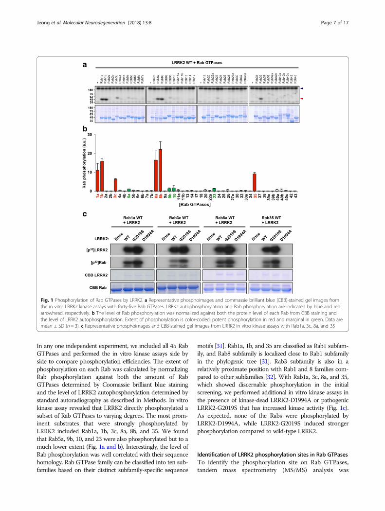
In any one independent experiment, we included all 45 RabGTPases and performed the in vitro kinase assays side byside to compare phosphorylation efficiencies. The extent ofphosphorylation on each Rab was calculated by normalizingRab phosphorylation against both the amount of RabGTPases determined by Coomassie brilliant blue stainingand the level of LRRK2 autophosphorylation determined bystandard autoradiography as described in Methods. In vitrokinase assay revealed that LRRK2 directly phosphorylated asubset of Rab GTPases to varying degrees. The most prom-inent substrates that were strongly phosphorylated byLRRK2 included Rab1a, 1b, 3c, 8a, 8b, and 35. We foundthat Rab5a, 9b, 10, and 23 were also phosphorylated but to amuch lower extent (Fig. 1a and b). Interestingly, the level ofRab phosphorylation was well correlated with their sequencehomology. Rab GTPase family can be classified into ten sub-families based on their distinct subfamily-specific sequence
motifs [31]. Rab1a, 1b, and 35 are classified as Rab1 subfam-ily, and Rab8 subfamily is localized close to Rab1 subfamilyin the phylogenic tree [31]. Rab3 subfamily is also in arelatively proximate position with Rab1 and 8 families com-pared to other subfamilies [32]. With Rab1a, 3c, 8a, and 35,which showed discernable phosphorylation in the initialscreening, we performed additional in vitro kinase assays inthe presence of kinase-dead LRRK2-D1994A or pathogenicLRRK2-G2019S that has increased kinase activity (Fig. 1c).As expected, none of the Rabs were phosphorylated byLRRK2-D1994A, while LRRK2-G2019S induced strongerphosphorylation compared to wild-type LRRK2.
Identification of LRRK2 phosphorylation sites in Rab GTPasesTo identify the phosphorylation site on Rab GTPases,tandem mass spectrometry (MS/MS) analysis was
a
b
c
Fig. 1 Phosphorylation of Rab GTPases by LRRK2. a Representative phosphoimages and commassie brilliant blue (CBB)-stained gel images fromthe in vitro LRRK2 kinase assays with forty-five Rab GTPases. LRRK2 autophosphorylation and Rab phosphorylation are indicated by blue and redarrowhead, respectively. b The level of Rab phosphorylation was normalized against both the protein level of each Rab from CBB staining andthe level of LRRK2 autophosphorylation. Extent of phosphorylation is color-coded: potent phosphorylation in red and marginal in green. Data aremean ± SD (n = 3). c Representative phosphoimages and CBB-stained gel images from LRRK2 in vitro kinase assays with Rab1a, 3c, 8a, and 35
Jeong et al. Molecular Neurodegeneration (2018) 13:8 Page 7 of 17
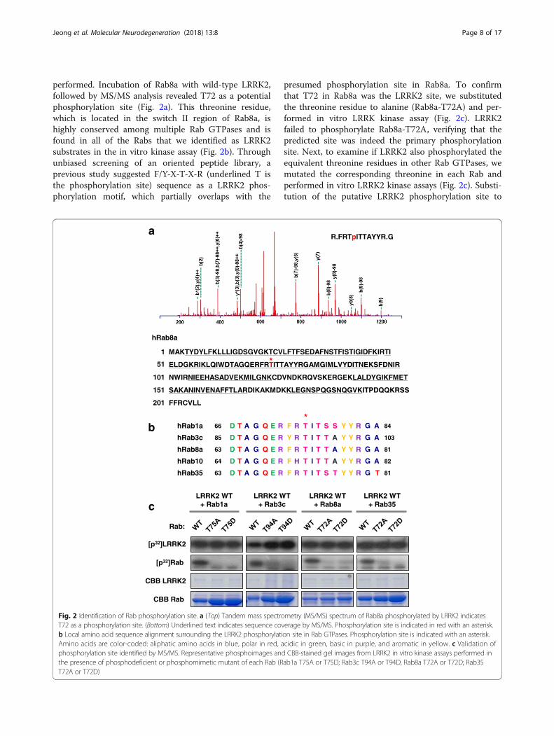
performed. Incubation of Rab8a with wild-type LRRK2,followed by MS/MS analysis revealed T72 as a potentialphosphorylation site (Fig. 2a). This threonine residue,which is located in the switch II region of Rab8a, ishighly conserved among multiple Rab GTPases and isfound in all of the Rabs that we identified as LRRK2substrates in the in vitro kinase assay (Fig. 2b). Throughunbiased screening of an oriented peptide library, aprevious study suggested F/Y-X-T-X-R (underlined T isthe phosphorylation site) sequence as a LRRK2 phos-phorylation motif, which partially overlaps with the
presumed phosphorylation site in Rab8a. To confirmthat T72 in Rab8a was the LRRK2 site, we substitutedthe threonine residue to alanine (Rab8a-T72A) and per-formed in vitro LRRK kinase assay (Fig. 2c). LRRK2failed to phosphorylate Rab8a-T72A, verifying that thepredicted site was indeed the primary phosphorylationsite. Next, to examine if LRRK2 also phosphorylated theequivalent threonine residues in other Rab GTPases, wemutated the corresponding threonine in each Rab andperformed in vitro LRRK2 kinase assays (Fig. 2c). Substi-tution of the putative LRRK2 phosphorylation site to
a
CBB LRRK2
[p32]LRRK2
CBB Rab
[p32]Rab
LRRK2 WT + Rab1a c
b
hRab8a
MAKTYDYLFKLLLIGDSGVGKTCVLFTFSEDAFNSTFISTIGIDFKIRTI
ELDGKRIKLQIWDTAGQERFRTITTAYYRGAMGIMLVYDITNEKSFDNIR
NWIRNIEEHASADVEKMILGNKCDVNDKRQVSKERGEKLALDYGIKFMET
SAKANINVENAFFTLARDIKAKMDKKLEGNSPQGSNQGVKITPDQQKRSS
FFRCVLL
1
51
101
151
201
*
Rab:
LRRK2 WT+ Rab3c
LRRK2 WT+ Rab8a
LRRK2 WT+ Rab35
hRab8a 63 81
hRab1a 8466
hRab35 63 81
hRab3c 85 103
hRab10
D
D
D
D
D
T
T
T
T
T
A
A
A
A
A
G
G
G
G
G
Q
Q
Q
Q
Q
R
R
R
R
R
F
F
F
Y
F
R
R
R
R
H
T
T*
T
T
T
I
I
I
I
I
T
T
T
T
T
T
S
S
T
T
A
S
T
A
A
Y
Y
Y
Y
Y
Y
Y
Y
Y
Y
R
R
R
R
R64 82
E
E
E
E
E
G
G
G
G
G
A
A
T
A
A
R.FRTpITTAYYR.G
Fig. 2 Identification of Rab phosphorylation site. a (Top) Tandem mass spectrometry (MS/MS) spectrum of Rab8a phosphorylated by LRRK2 indicatesT72 as a phosphorylation site. (Bottom) Underlined text indicates sequence coverage by MS/MS. Phosphorylation site is indicated in red with an asterisk.b Local amino acid sequence alignment surrounding the LRRK2 phosphorylation site in Rab GTPases. Phosphorylation site is indicated with an asterisk.Amino acids are color-coded: aliphatic amino acids in blue, polar in red, acidic in green, basic in purple, and aromatic in yellow. c Validation ofphosphorylation site identified by MS/MS. Representative phosphoimages and CBB-stained gel images from LRRK2 in vitro kinase assays performed inthe presence of phosphodeficient or phosphomimetic mutant of each Rab (Rab1a T75A or T75D; Rab3c T94A or T94D, Rab8a T72A or T72D; Rab35T72A or T72D)
Jeong et al. Molecular Neurodegeneration (2018) 13:8 Page 8 of 17
either alanine (TA) or aspartate residue (TD) in Rab1a,3c, 8a and 35 abolished phosphorylation in all Rabstested, verifying that the conserved threonine residueswere the major LRRK2 phosphorylation sites.
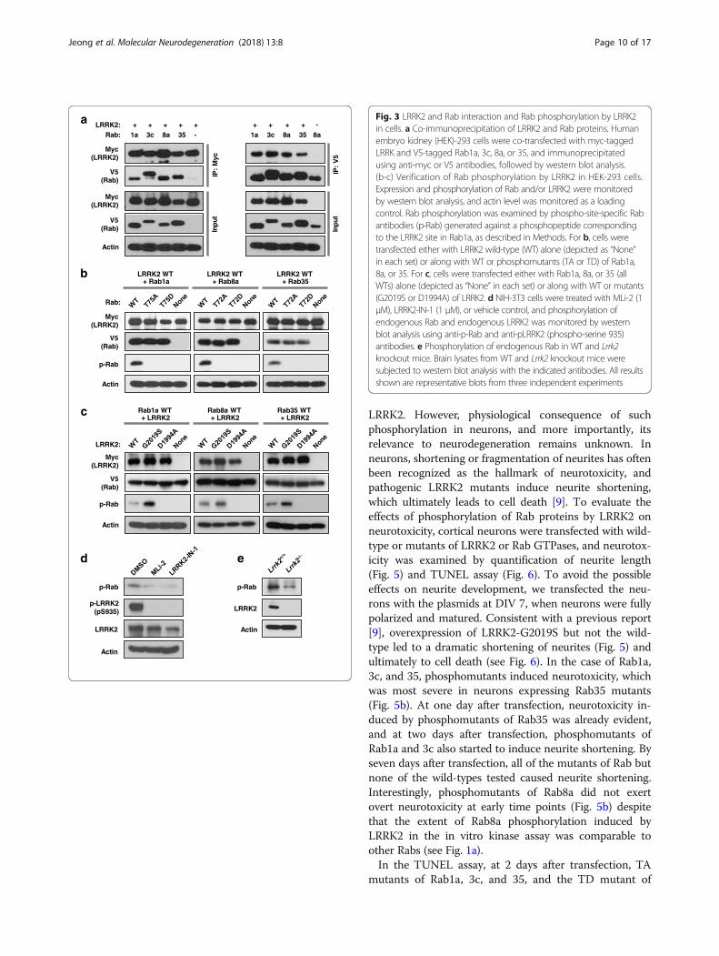
Phosphorylation of Rab by LRRK2 in cellsTo extend the findings from the in vitro kinase assays,we first investigated whether LRRK2 interacts with Rabsin cells. For co-immunoprecipitation analysis, we ectopi-cally expressed myc-tagged LRRK2 along with the V5-tagged Rab of interest in human embryonic kidney(HEK)-293 cells. Immunoprecipitation of cell lysateswith anti-myc antibodies, followed by immunoblottingwith anti-V5 antibodies revealed that LRRK2 was boundto Rab1a, 3c, 8a, and 35 (Fig. 3a). Reciprocal co-immu-noprecipitation experiments using antibodies against V5for immunoprecipitation, followed by immunoblottingwith anti-myc antibodies confirmed that all of the Rabstested physically interacted with LRRK2.To validate the in vitro phosphorylation of Rabs in
cells, we raised phospho-specific antibodies against aphosphopeptide TAGQERFRpTITSSYYRG, correspond-ing to the highly conserved sequence surrounding theLRRK2 site in Rab1a (see Fig. 2b). In HEK-293 cells,phosphorylation of Rab1a, 8a, and 35 was readilydetected when we co-expressed both LRRK2 and theRab of interest, but not when we overexpressed eitherLRRK2 or Rab (Fig. 3b and c). Despite the observationthat LRRK2 directly phosphorylated Rab3c in vitro, wefailed to detect phosphorylation of Rab3c from celllysates (data not shown), which might be because of aslightly different amino acid sequence close to theLRRK2 site (note the tyrosine residue in Rab3c insteadof phenylalanine in all other Rabs at position − 2 withrespect to the LRRK2 site). Interestingly, when we co-transfected Rab10 and LRRK2, the extent of phosphoryl-ation of Rab10 was comparable to those of Rab1a, 8a,and 35 (data not shown), despite the observation thatRab10 was not a prominent substrate of LRRK2 in thein vitro kinase assay (see Fig. 1a). When we co-trans-fected HEK-293 cells with phosphomutants (phosphomi-metic TD or phosphodeficient TA mutant) of the Rab ofinterest along with wild-type LRRK2, we could not de-tect any phosphorylation (Fig. 3b), confirming that theconserved threonine sites in Rab GTPases were the tar-gets of LRRK2 in cells. Furthermore, the extent of phos-phorylation of Rab1a, 8a, and 35 was enhanced byLRRK2-G2019S compared to wild-type LRRK2, whilenone of the Rabs were phosphorylated by kinase-deadLRRK2-D1994A (Fig. 3c). Together, these results con-firm the specificities of the phospho-Rab antibodies thatwe generated, and more importantly, provide unambigu-ous evidence that Rab1a, 8a, and 35 are phosphorylatedby LRRK2 both in vitro and in cells.
To examine phosphorylation of Rab proteins at the en-dogenous level, we first examined the effects of LRRK2inhibitors in NIH-3T3 fibroblast cells, which expressendogenous LRRK2. More specifically, we treated NIH-3T3 cells with two different inhibitors of LRRK2, eitherLRRK2-IN-1 (1 μM) or MLi-2 (1 μM), and examined thechanges in Rab phosphorylation using the aforemen-tioned phospho-specific Rab antibodies that we gener-ated (see Fig. 3b, c). Inhibition of endogenous LRRK2 bythe two inhibitors was confirmed by immunoblottingwith phospho-specific LRRK2 (phospho-serine 935) anti-bodies, and importantly, we confirmed that LRRK2-IN-1or MLi-2 markedly reduced phosphorylation of Rab (Fig.3d). Furthermore, we also confirmed that the level ofphospho-Rab was substantially diminished in brain ly-sates from Lrrk2 knock-out mice compared to wild-type(Fig. 3e). Collectively, these results provide strong sup-port for the notion that endogenous Rab proteins arephosphorylated by endogenous LRRK2.
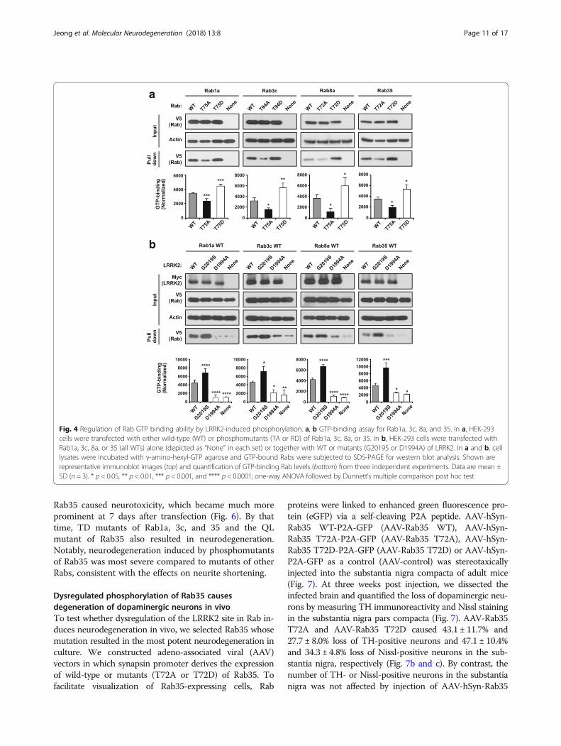
Phosphorylation of Rab by LRRK2 affects GTP-bindingTo elucidate the functional consequence of LRRK2-induced Rab phosphorylation, we investigated thechanges in GTP-binding ability of Rab GTPases afterLRRK2 phosphorylation. Rab GTPases cycle betweenactive (GTP-bound) and inactive (GDP-bound) states,which is controlled by three groups of regulatoryproteins: GTPase-activating proteins (GAPs), guaninenucleotide exchange factor (GEFs), and guaninenucleotide-dissociation inhibitors (GDIs). The conservedthreonine residue (LRRK2 site) resides in the switch IIregion, which undergoes a major conformational transi-tion between GDP- and GTP-bound states and coordi-nates the association with specific regulatory molecules[33]. We tested whether the mutation of the threonineresidue affected the GTP hydrolysis property of RabGTPases by using hydrolyzable GTP agarose, reflectiveof GTPase activity at the time of precipitation. For allRabs tested, GTP-binding was reduced by substitutingthe threonine residue to alanine but increased byreplacing it to aspartate (Fig. 4a). When LRRK2-G2019Swas co-expressed with the Rab of interest, GTP-bindingwas increased compared to wild-type LRRK2, whereasoverexpression of LRRK2-D1994A had little effect onGTP-binding (Fig. 4b). These results suggest that phos-phorylation of Rab proteins by LRRK2 kinase regulatesthe GDP/GTP exchange and that hyperactivation ofLRRK2 kinase increases GTP-bound Rabs in cells.
Mutations in the LRRK2 site in Rab GTPases causeneurodegeneration in cortical neuronsOur results described above and those reported by others[29] suggest that Rab GTPases are authentic substrates of
Jeong et al. Molecular Neurodegeneration (2018) 13:8 Page 9 of 17
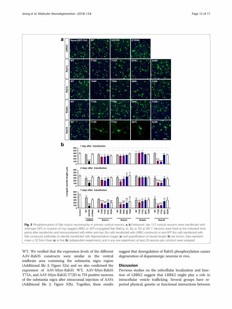
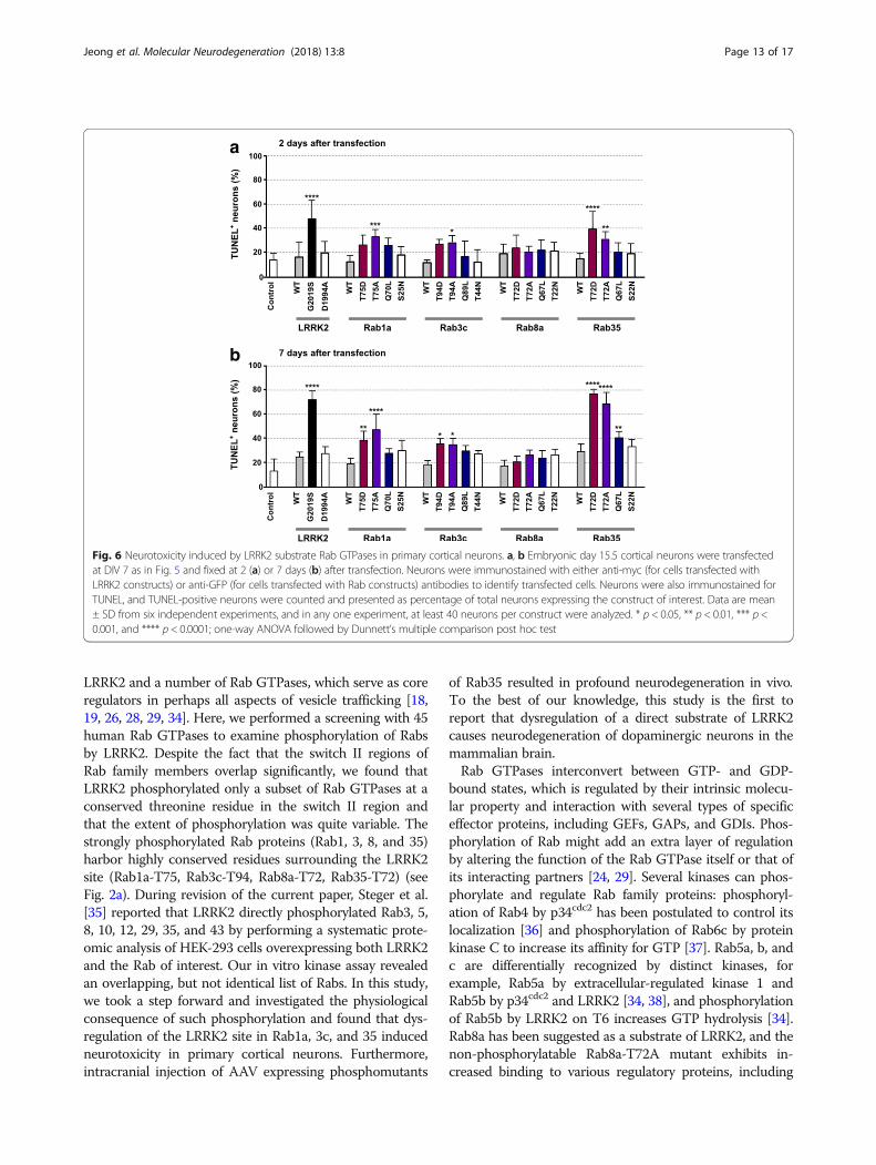
LRRK2. However, physiological consequence of suchphosphorylation in neurons, and more importantly, itsrelevance to neurodegeneration remains unknown. Inneurons, shortening or fragmentation of neurites has oftenbeen recognized as the hallmark of neurotoxicity, andpathogenic LRRK2 mutants induce neurite shortening,which ultimately leads to cell death [9]. To evaluate theeffects of phosphorylation of Rab proteins by LRRK2 onneurotoxicity, cortical neurons were transfected with wild-type or mutants of LRRK2 or Rab GTPases, and neurotox-icity was examined by quantification of neurite length(Fig. 5) and TUNEL assay (Fig. 6). To avoid the possibleeffects on neurite development, we transfected the neu-rons with the plasmids at DIV 7, when neurons were fullypolarized and matured. Consistent with a previous report[9], overexpression of LRRK2-G2019S but not the wild-type led to a dramatic shortening of neurites (Fig. 5) andultimately to cell death (see Fig. 6). In the case of Rab1a,3c, and 35, phosphomutants induced neurotoxicity, whichwas most severe in neurons expressing Rab35 mutants(Fig. 5b). At one day after transfection, neurotoxicity in-duced by phosphomutants of Rab35 was already evident,and at two days after transfection, phosphomutants ofRab1a and 3c also started to induce neurite shortening. Byseven days after transfection, all of the mutants of Rab butnone of the wild-types tested caused neurite shortening.Interestingly, phosphomutants of Rab8a did not exertovert neurotoxicity at early time points (Fig. 5b) despitethat the extent of Rab8a phosphorylation induced byLRRK2 in the in vitro kinase assay was comparable toother Rabs (see Fig. 1a).In the TUNEL assay, at 2 days after transfection, TA
mutants of Rab1a, 3c, and 35, and the TD mutant of
a
b
c
p-Rab
Myc(LRRK2)
Actin
V5(Rab)
Rab:
LRRK2 WT + Rab1a
LRRK2 WT + Rab8a
LRRK2 WT + Rab35
p-Rab
Myc(LRRK2)
Actin
V5(Rab)
Rab1a WT + LRRK2
Rab8a WT + LRRK2
Rab35 WT + LRRK2
LRRK2:
IP:
Myc
Inp
ut
LRRK2:Rab:
++ + + +-1a 3c 8a 35
Myc(LRRK2)
V5(Rab)
Myc(LRRK2)
V5(Rab)
IP:
V5
Inp
ut
+ + + +8a1a 3c 8a 35
Actin
-
d e
p-Rab
LRRK2
Actin
p-Rab
p-LRRK2(pS935)
LRRK2
Actin
Fig. 3 LRRK2 and Rab interaction and Rab phosphorylation by LRRK2in cells. a Co-immunoprecipitation of LRRK2 and Rab proteins. Humanembryo kidney (HEK)-293 cells were co-transfected with myc-taggedLRRK and V5-tagged Rab1a, 3c, 8a, or 35, and immunoprecipitatedusing anti-myc or V5 antibodies, followed by western blot analysis.(b-c) Verification of Rab phosphorylation by LRRK2 in HEK-293 cells.Expression and phosphorylation of Rab and/or LRRK2 were monitoredby western blot analysis, and actin level was monitored as a loadingcontrol. Rab phosphorylation was examined by phospho-site-specific Rabantibodies (p-Rab) generated against a phosphopeptide correspondingto the LRRK2 site in Rab1a, as described in Methods. For b, cells weretransfected either with LRRK2 wild-type (WT) alone (depicted as “None”in each set) or along with WT or phosphomutants (TA or TD) of Rab1a,8a, or 35. For c, cells were transfected either with Rab1a, 8a, or 35 (allWTs) alone (depicted as “None” in each set) or along with WT or mutants(G2019S or D1994A) of LRRK2. d NIH-3T3 cells were treated with MLi-2 (1μM), LRRK2-IN-1 (1 μM), or vehicle control, and phosphorylation ofendogenous Rab and endogenous LRRK2 was monitored by westernblot analysis using anti-p-Rab and anti-pLRRK2 (phospho-serine 935)antibodies. e Phosphorylation of endogenous Rab in WT and Lrrk2knockout mice. Brain lysates from WT and Lrrk2 knockout mice weresubjected to western blot analysis with the indicated antibodies. All resultsshown are representative blots from three independent experiments
Jeong et al. Molecular Neurodegeneration (2018) 13:8 Page 10 of 17
Rab35 caused neurotoxicity, which became much moreprominent at 7 days after transfection (Fig. 6). By thattime, TD mutants of Rab1a, 3c, and 35 and the QLmutant of Rab35 also resulted in neurodegeneration.Notably, neurodegeneration induced by phosphomutantsof Rab35 was most severe compared to mutants of otherRabs, consistent with the effects on neurite shortening.
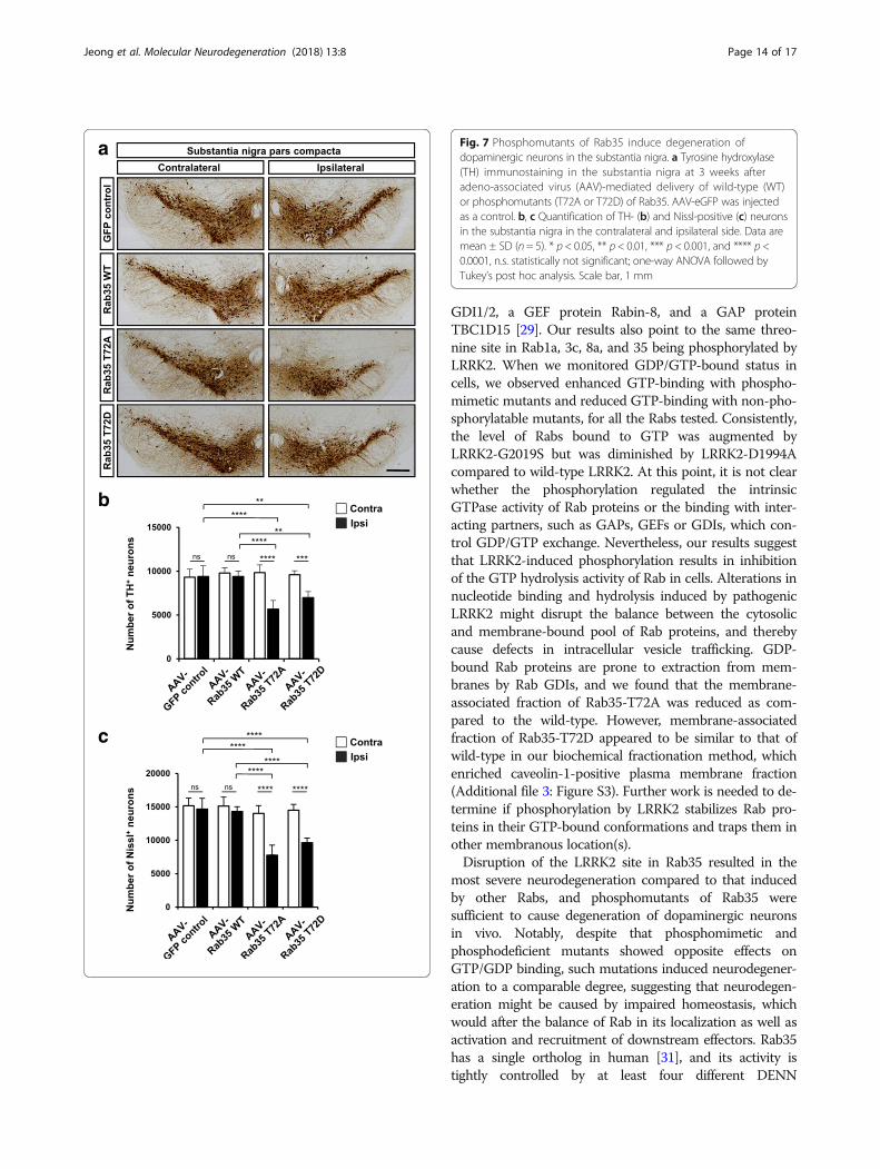
Dysregulated phosphorylation of Rab35 causesdegeneration of dopaminergic neurons in vivoTo test whether dysregulation of the LRRK2 site in Rab in-duces neurodegeneration in vivo, we selected Rab35 whosemutation resulted in the most potent neurodegeneration inculture. We constructed adeno-associated viral (AAV)vectors in which synapsin promoter derives the expressionof wild-type or mutants (T72A or T72D) of Rab35. Tofacilitate visualization of Rab35-expressing cells, Rab
proteins were linked to enhanced green fluorescence pro-tein (eGFP) via a self-cleaving P2A peptide. AAV-hSyn-Rab35 WT-P2A-GFP (AAV-Rab35 WT), AAV-hSyn-Rab35 T72A-P2A-GFP (AAV-Rab35 T72A), AAV-hSyn-Rab35 T72D-P2A-GFP (AAV-Rab35 T72D) or AAV-hSyn-P2A-GFP as a control (AAV-control) was stereotaxicallyinjected into the substantia nigra compacta of adult mice(Fig. 7). At three weeks post injection, we dissected theinfected brain and quantified the loss of dopaminergic neu-rons by measuring TH immunoreactivity and Nissl stainingin the substantia nigra pars compacta (Fig. 7). AAV-Rab35T72A and AAV-Rab35 T72D caused 43.1 ± 11.7% and27.7 ± 8.0% loss of TH-positive neurons and 47.1 ± 10.4%and 34.3 ± 4.8% loss of Nissl-positive neurons in the sub-stantia nigra, respectively (Fig. 7b and c). By contrast, thenumber of TH- or Nissl-positive neurons in the substantianigra was not affected by injection of AAV-hSyn-Rab35
a
b
Fig. 4 Regulation of Rab GTP binding ability by LRRK2-induced phosphorylation. a, b GTP-binding assay for Rab1a, 3c, 8a, and 35. In a, HEK-293cells were transfected with either wild-type (WT) or phosphomutants (TA or RD) of Rab1a, 3c, 8a, or 35. In b, HEK-293 cells were transfected withRab1a, 3c, 8a, or 35 (all WTs) alone (depicted as “None” in each set) or together with WT or mutants (G2019S or D1994A) of LRRK2. In a and b, celllysates were incubated with γ-amino-hexyl-GTP agarose and GTP-bound Rabs were subjected to SDS-PAGE for western blot analysis. Shown arerepresentative immunoblot images (top) and quantification of GTP-binding Rab levels (bottom) from three independent experiments. Data are mean ±SD (n = 3). * p < 0.05, ** p < 0.01, *** p < 0.001, and **** p < 0.0001; one-way ANOVA followed by Dunnett’s multiple comparison post hoc test
Jeong et al. Molecular Neurodegeneration (2018) 13:8 Page 11 of 17
WT. We verified that the expression levels of the differentAAV-Rab35 constructs were similar in the ventralmidbrain area containing the substantia nigra region(Additional file 2: Figure S2a) and we also confirmed theexpression of AAV-hSyn-Rab35 WT, AAV-hSyn-Rab35T72A, and AAV-hSyn-Rab35 T72D in TH-positive neuronsof the substantia nigra after intracranial injection of AAVs(Additional file 2: Figure S2b). Together, these results
suggest that dysregulation of Rab35 phosphorylation causesdegeneration of dopaminergic neurons in vivo.
DiscussionPrevious studies on the subcellular localization and func-tion of LRRK2 suggest that LRRK2 might play a role inintracellular vesicle trafficking. Several groups have re-ported physical, genetic or functional interactions between
a
b
Fig. 5 Phosphomutants of Rab induce neurotoxicity in primary cortical neurons. a, b Embryonic day 15.5 cortical neurons were transfected withwild-type (WT) or mutants of myc-tagged LRRK2 or GFP-conjugated Rab (Rab1a, 3c, 8a, or 35) at DIV 7. Neurons were fixed at the indicated timepoints after transfection and immunostained with either anti-myc (for cells transfected with LRRK2 constructs) or anti-GFP (for cells transfected withRab constructs) antibodies to identify transfected cells. Representative images (a) and quantification of neurite length (b) are shown. Data representmean ± SD from three (a) or five (b) independent experiments, and in any one experiment, at least 20 neurons per construct were analyzed
Jeong et al. Molecular Neurodegeneration (2018) 13:8 Page 12 of 17
LRRK2 and a number of Rab GTPases, which serve as coreregulators in perhaps all aspects of vesicle trafficking [18,19, 26, 28, 29, 34]. Here, we performed a screening with 45human Rab GTPases to examine phosphorylation of Rabsby LRRK2. Despite the fact that the switch II regions ofRab family members overlap significantly, we found thatLRRK2 phosphorylated only a subset of Rab GTPases at aconserved threonine residue in the switch II region andthat the extent of phosphorylation was quite variable. Thestrongly phosphorylated Rab proteins (Rab1, 3, 8, and 35)harbor highly conserved residues surrounding the LRRK2site (Rab1a-T75, Rab3c-T94, Rab8a-T72, Rab35-T72) (seeFig. 2a). During revision of the current paper, Steger et al.[35] reported that LRRK2 directly phosphorylated Rab3, 5,8, 10, 12, 29, 35, and 43 by performing a systematic prote-omic analysis of HEK-293 cells overexpressing both LRRK2and the Rab of interest. Our in vitro kinase assay revealedan overlapping, but not identical list of Rabs. In this study,we took a step forward and investigated the physiologicalconsequence of such phosphorylation and found that dys-regulation of the LRRK2 site in Rab1a, 3c, and 35 inducedneurotoxicity in primary cortical neurons. Furthermore,intracranial injection of AAV expressing phosphomutants
of Rab35 resulted in profound neurodegeneration in vivo.To the best of our knowledge, this study is the first toreport that dysregulation of a direct substrate of LRRK2causes neurodegeneration of dopaminergic neurons in themammalian brain.Rab GTPases interconvert between GTP- and GDP-
bound states, which is regulated by their intrinsic molecu-lar property and interaction with several types of specificeffector proteins, including GEFs, GAPs, and GDIs. Phos-phorylation of Rab might add an extra layer of regulationby altering the function of the Rab GTPase itself or that ofits interacting partners [24, 29]. Several kinases can phos-phorylate and regulate Rab family proteins: phosphoryl-ation of Rab4 by p34cdc2 has been postulated to control itslocalization [36] and phosphorylation of Rab6c by proteinkinase C to increase its affinity for GTP [37]. Rab5a, b, andc are differentially recognized by distinct kinases, forexample, Rab5a by extracellular-regulated kinase 1 andRab5b by p34cdc2 and LRRK2 [34, 38], and phosphorylationof Rab5b by LRRK2 on T6 increases GTP hydrolysis [34].Rab8a has been suggested as a substrate of LRRK2, and thenon-phosphorylatable Rab8a-T72A mutant exhibits in-creased binding to various regulatory proteins, including
a
b
Fig. 6 Neurotoxicity induced by LRRK2 substrate Rab GTPases in primary cortical neurons. a, b Embryonic day 15.5 cortical neurons were transfectedat DIV 7 as in Fig. 5 and fixed at 2 (a) or 7 days (b) after transfection. Neurons were immunostained with either anti-myc (for cells transfected withLRRK2 constructs) or anti-GFP (for cells transfected with Rab constructs) antibodies to identify transfected cells. Neurons were also immunostained forTUNEL, and TUNEL-positive neurons were counted and presented as percentage of total neurons expressing the construct of interest. Data are mean± SD from six independent experiments, and in any one experiment, at least 40 neurons per construct were analyzed. * p < 0.05, ** p < 0.01, *** p <0.001, and **** p < 0.0001; one-way ANOVA followed by Dunnett’s multiple comparison post hoc test
Jeong et al. Molecular Neurodegeneration (2018) 13:8 Page 13 of 17
GDI1/2, a GEF protein Rabin-8, and a GAP proteinTBC1D15 [29]. Our results also point to the same threo-nine site in Rab1a, 3c, 8a, and 35 being phosphorylated byLRRK2. When we monitored GDP/GTP-bound status incells, we observed enhanced GTP-binding with phospho-mimetic mutants and reduced GTP-binding with non-pho-sphorylatable mutants, for all the Rabs tested. Consistently,the level of Rabs bound to GTP was augmented byLRRK2-G2019S but was diminished by LRRK2-D1994Acompared to wild-type LRRK2. At this point, it is not clearwhether the phosphorylation regulated the intrinsicGTPase activity of Rab proteins or the binding with inter-acting partners, such as GAPs, GEFs or GDIs, which con-trol GDP/GTP exchange. Nevertheless, our results suggestthat LRRK2-induced phosphorylation results in inhibitionof the GTP hydrolysis activity of Rab in cells. Alterations innucleotide binding and hydrolysis induced by pathogenicLRRK2 might disrupt the balance between the cytosolicand membrane-bound pool of Rab proteins, and therebycause defects in intracellular vesicle trafficking. GDP-bound Rab proteins are prone to extraction from mem-branes by Rab GDIs, and we found that the membrane-associated fraction of Rab35-T72A was reduced as com-pared to the wild-type. However, membrane-associatedfraction of Rab35-T72D appeared to be similar to that ofwild-type in our biochemical fractionation method, whichenriched caveolin-1-positive plasma membrane fraction(Additional file 3: Figure S3). Further work is needed to de-termine if phosphorylation by LRRK2 stabilizes Rab pro-teins in their GTP-bound conformations and traps them inother membranous location(s).Disruption of the LRRK2 site in Rab35 resulted in the
most severe neurodegeneration compared to that inducedby other Rabs, and phosphomutants of Rab35 weresufficient to cause degeneration of dopaminergic neuronsin vivo. Notably, despite that phosphomimetic andphosphodeficient mutants showed opposite effects onGTP/GDP binding, such mutations induced neurodegener-ation to a comparable degree, suggesting that neurodegen-eration might be caused by impaired homeostasis, whichwould after the balance of Rab in its localization as well asactivation and recruitment of downstream effectors. Rab35has a single ortholog in human [31], and its activity istightly controlled by at least four different DENN
a
b
c
Fig. 7 Phosphomutants of Rab35 induce degeneration ofdopaminergic neurons in the substantia nigra. a Tyrosine hydroxylase(TH) immunostaining in the substantia nigra at 3 weeks afteradeno-associated virus (AAV)-mediated delivery of wild-type (WT)or phosphomutants (T72A or T72D) of Rab35. AAV-eGFP was injectedas a control. b, c Quantification of TH- (b) and Nissl-positive (c) neuronsin the substantia nigra in the contralateral and ipsilateral side. Data aremean ± SD (n = 5). * p < 0.05, ** p < 0.01, *** p < 0.001, and **** p <0.0001, n.s. statistically not significant; one-way ANOVA followed byTukey’s post hoc analysis. Scale bar, 1 mm
Jeong et al. Molecular Neurodegeneration (2018) 13:8 Page 14 of 17
(differentially expressed in normal and neoplastic cells)family of GEFs and five different TBC (Tre2/Bub2/Cdc16)family of GAPs [39]. Rab35 has been shown to regulate di-verse cellular processes, including endocytic recycling [40],exosome release [41], cytokinesis [42], and actin remodeling[30]. In the brain, Rab35 is expressed in multiple regions(Additional file 4: Figure S4), and in neurons, Rab35 seemsto control neurite outgrowth during development [43–46]and synaptic vesicle trafficking [47]. However, little isknown about its role in neurodegeneration. Interestingly,Rab35 was identified as a potential serum biomarker forPD through the analysis of proteomic profiles of PD pa-tients, and Rab35 was also elevated in the substantia nigraobtained from multiple PD animal models, includingMPTP-, rotenone-treated mice, and LRRK2-R1441C or-G2019S transgenic mice [48]. These results together withours imply that impaired function of Rab35, perhaps in partthrough dysregulated phosphorylation, might contribute toneurodegeneration in PD. Further studies are needed toexamine the extent, if any, to which the cellular processesknown to be regulated by Rab35 are altered in LRRK2-associated neurodegeneration and PD pathogenesis.Neurons are highly polarized cells and have extremely ar-
borized cellular architecture, which imposes a substantialburden on the trafficking system. Thus, it is not surprisingthat intracellular trafficking deficits are associated with anumber of neurodegenerative diseases, and PD is no ex-ception. Dysfunction of Rab GTPases and impaired mem-brane traffic might contribute to the onset and progressionof PD. Loss of Rab39b causes early-onset PD and Rab29resides in the PARK16 non-familial risk locus [49–52].Moreover, certain Rabs have been suggested to interactwith and modulate the function of key pathogenic proteins,such as LRRK2, α-synuclein and PTEN-induced kinase 1[17–19, 26, 29, 53–57]. Interestingly, overexpression ofRab1, 3a, and 8a, which we (this study) and others [29]suggest as LRRK2 substrates, could rescue α-synuclein-induced cytotoxicity in cell and animal model of PD [54,55], implying that Rab GTPases might be involved in mul-tiple pathways that play crucial roles in the pathogenesis ofPD. Therefore, further study of the Rab GTPases will notonly provide insight into how intracellular membrane dy-namics are orchestrated, but also facilitate the identifica-tion of molecules or pathways that can serve as therapeutictargets for the treatment of PD.
ConclusionsBy performing in vitro LRRK2 kinase assays with forty-five human Rab GTPases, here we identify a subset of RabGTPases, including previously identified as well as novelRabs, such as Rab35, as authentic substrates of LRRK2.We provide evidence that phosphorylation of RabGTPases by LRRK2 controls GDP/GTP exchange in cellsand that dysregulation of Rab phosphorylation in the
LRRK2 site causes neurodegeneration in primary neurons.Furthermore, we show that intracranial injection of AAVexpressing phosphomutants of Rab35 into the substantianigra induces profound degeneration of dopaminergicneurons in vivo. These results suggest that Rab GTPasesmight mediate LRRK2 toxicity in the etiology of PD.
Additional files
Additional file 1: Figure S1. Immunostaining of cortical neuronstransfected with Rab35 mutants. Representative images of embryonic day 15.5cortical neurons expressing mutants (T72A, T72D or Q67Q) of GFP-conjugatedRab35. Neurons were fixed at one day after transfection and immunostainedwith anti-GFP, anti-smi312 (axonal marker), and anti-GFAP (astrocyte marker)antibodies. Scale bar, 100 μm. (PPTX 9498 kb)
Additional file 2: Figure S2. Expression of AAV-Rab35 WT andphosphomutants after intracranial injection of AAV. At one week afterintracranial injection of AAV-eGFP (control), AAV-Rab35 WT, AAV-Rab35 T75Aor AAV-Rab35 T75D into the substantia nigra, mouse brains were preparedfor immunoblot (a) and immunohistochemistry (b). (a) Ventral midbrain wasdissected from the AAV-injected hemisphere and the tissue was homoge-nized. Immunoblots were performed with anti-Rab35, anti-TH, and anti-actinantibodies. Exogenous and endogenous Rab35 are indicated by red andblue arrowhead, respectively. (b) Cryosections of AAV-injected brainsincluding the substantia nigra region were immunostained with anti-GFP(green) and anti-TH (red) antibodies. Scale bar, 200 μm. (PPTX 3143 kb)
Additional file 3: Figure S3. Localization of Rab35 WT andphosphomutants. (a) HEK-293 cells were transfected with V5-taggedRab35 WT or phosphomutants (T72A or T72D). At 48 h after transfection,cells were harvested and membrane fractionation was performed asdescribed in Methods. Prepared membrane fractions were subjected toSDS-PAGE and immunoblotting with anti-caveolin-1 (plasma membranemarker), anti-EEA1 (early endosomal marker), anti-GOPC (golgi marker),and anti-Hsp90 (cytosol marker) antibodies. 2.5% of total lysates and 30%of membrane fractions were loaded for each immunoblot. (b) Quantificationof Rab35 protein levels normalized against input level of Rab35. Data aremean ± SD (n = 3). * p < 0.05; one-way ANOVA followed by Dunnett’smultiple comparison post hoc test. (PPTX 1972 kb)
Additional file 4: Figure S4. Expression of Rab35 in adult mouse brain.Analysis of Rab35 protein expression by immunoblot in various brainregions of adult mouse. (PPTX 1820 kb)
AbbreviationsAAV: Adeno-associated viral vectors; GAP: GTPase-activating protein;GDI: Guanine nucleotide dissociation inhibitor; GEP: Guanine nucleotideexchange factor; LRRK2: Leucine-rich repeat kinase 2; PD: Parkinson’s disease
FundingThis research was supported by the NIH grant (NIH/NINDS NS58377), KISTInstitutional Grant (2V05590, 2V05900), National Research Council of Science andTechnology (NST) Grant by the Korean government (MSIP) (CRC-15-04-KIST),grants from National Research Foundation (NRF) funded by the Korean Ministryof Science (NRF-2014R1A1A2055787), and the Brain Research Program throughthe National Research Foundation of Korea (NRF) funded by the Ministry ofScience, ICT & Future Planning (NRF-2016M3C7A1905386). T.M.D. is the Leonardand Madlyn Abramson Professor in Neurodegenerative Diseases.
Availability of data and materialsThe data generated and/or analyzed during this are included in the articleand materials are available from the corresponding authors on reasonablerequest.
Authors’ contributionsEMH and BDL designed the project and wrote the manuscript with allauthors’ input. GRJ performed most of the biochemical experiments andintracranial injections using AAVs generated and validated by YY and KT-Y.EHJ and SJ performed primary culture experiments and analyzed data. JRB
Jeong et al. Molecular Neurodegeneration (2018) 13:8 Page 15 of 17
performed the in vitro kinase screening using the plasmids provided by HCK.CHP and JHS performed mass spectrometry. VLD and TMD contributed tothe interpretation and discussion of the data and gave comments on thestudy. All authors reviewed and approved the manuscript.
Ethics approvalMice were housed, bred and treated according to the institutional guidelinesfor animal care and research protocols approved by Korea Institute ofScience and Technology and Kyung Hee University.
Consent for publicationNot applicable.
Competing interestsThe authors declare that they have no competing interests.
Publisher’s NoteSpringer Nature remains neutral with regard to jurisdictional claims inpublished maps and institutional affiliations.
Author details1Department of Neuroscience, Graduate School, Kyung Hee University, Seoul,South Korea. 2Center for Neuroscience, Brain Science Institute, Korea Instituteof Science and Technology (KIST), 5 Hwarang-ro 14-gil, Seongbuk-gu, Seoul02792, South Korea. 3Convergence Research Center for Diagnosis, Treatmentand Care System of Dementia, KIST, Seoul, South Korea. 4Division ofBio-Medical Science &Technology, KIST School, Korea University of Scienceand Technology, Seoul, South Korea. 5Department of Physiology, AjouUniversity School of Medicine, Suwon, South Korea. 6HuGex Co. Ltd.,Incheon, South Korea. 7Division of Pharmacology, Department of MolecularCell Biology, Samsung Biomedical Research Institute, Single Cell NetworkResearch Center, SungKyunKwan University School of Medicine, Suwon,South Korea. 8Center for Functional Connectomics, KIST, Seoul, South Korea.9Neurodegeneration and Stem Cell Program, Institute for Cell Engineeringand Department of Neurology, Johns Hopkins University School of Medicine,Baltimore, USA. 10Department of Physiology, Johns Hopkins University Schoolof Medicine, Baltimore, USA. 11Solomon H Snyder Department ofNeuroscience, Johns Hopkins University School of Medicine, Baltimore, USA.12Department of Pharmacology & Molecular Sciences, Johns HopkinsUniversity School of Medicine, Baltimore, USA. 13Department of Physiology,School of Medicine, Kyung Hee University, 26 Kyungheedae-ro,Dongdaemun-gu, Seoul 02447, South Korea.
Received: 25 August 2017 Accepted: 6 February 2018
References1. Lees AJ, Hardy J, Revesz T. Parkinson's disease. Lancet. 2009;373:2055–66.2. Tsika E, Moore DJ. Mechanisms of LRRK2-mediated neurodegeneration. Curr
Neurol Neurosci Rep. 2012;12:251–60.3. Healy DG, Falchi M, O'Sullivan SS, Bonifati V, Durr A, Bressman S, Brice A,
Aasly J, Zabetian CP, Goldwurm S, et al. Phenotype, genotype, andworldwide genetic penetrance of LRRK2-associated Parkinson's disease: acase-control study. Lancet Neurol. 2008;7:583–90.
4. Smith WW, Pei Z, Jiang H, Dawson VL, Dawson TM, Ross CA. Kinase activityof mutant LRRK2 mediates neuronal toxicity. Nat Neurosci. 2006;9:1231–3.
5. Greggio E, Jain S, Kingsbury A, Bandopadhyay R, Lewis P, Kaganovich A, vander Brug MP, Beilina A, Blackinton J, Thomas KJ, et al. Kinase activity isrequired for the toxic effects of mutant LRRK2/dardarin. Neurobiol Dis. 2006;23:329–41.
6. Lee BD, Shin JH, VanKampen J, Petrucelli L, West AB, Ko HS, Lee YI, Maguire-Zeiss KA, Bowers WJ, Federoff HJ, et al. Inhibitors of leucine-rich repeat kinase-2protect against models of Parkinson's disease. Nat Med. 2010;16:998–1000.
7. Deng X, Dzamko N, Prescott A, Davies P, Liu Q, Yang Q, Lee JD,Patricelli MP, Nomanbhoy TK, Alessi DR, Gray NS. Characterization of aselective inhibitor of the Parkinson's disease kinase LRRK2. Nat ChemBiol. 2011;7:203–5.
8. West AB, Moore DJ, Biskup S, Bugayenko A, Smith WW, Ross CA,Dawson VL, Dawson TM. Parkinson's disease-associated mutations inleucine-rich repeat kinase 2 augment kinase activity. Proc Natl Acad SciU S A. 2005;102:16842–7.
9. MacLeod D, Dowman J, Hammond R, Leete T, Inoue K, Abeliovich A. Thefamilial parkinsonism gene LRRK2 regulates neurite process morphology.Neuron. 2006;52:587–93.
10. Luzon-Toro B, Rubio de la Torre E, Delgado A, Perez-Tur J, Hilfiker S.Mechanistic insight into the dominant mode of the Parkinson's disease-associated G2019S LRRK2 mutation. Hum Mol Genet. 2007;16:2031–9.
11. Anand VS, Reichling LJ, Lipinski K, Stochaj W, Duan W, Kelleher K, PungaliyaP, Brown EL, Reinhart PH, Somberg R, et al. Investigation of leucine-richrepeat kinase 2 : enzymological properties and novel assays. FEBS J. 2009;276:466–78.
12. Martin I, Kim JW, Lee BD, Kang HC, Xu JC, Jia H, Stankowski J, Kim MS,Zhong J, Kumar M, et al. Ribosomal protein s15 phosphorylation mediatesLRRK2 neurodegeneration in Parkinson's disease. Cell. 2014;157:472–85.
13. Alegre-Abarrategui J, Christian H, Lufino MM, Mutihac R, Venda LL, AnsorgeO, Wade-Martins R. LRRK2 regulates autophagic activity and localizes tospecific membrane microdomains in a novel human genomic reportercellular model. Hum Mol Genet. 2009;18:4022–34.
14. Biskup S, Moore DJ, Celsi F, Higashi S, West AB, Andrabi SA, Kurkinen K, YuSW, Savitt JM, Waldvogel HJ, et al. Localization of LRRK2 to membranousand vesicular structures in mammalian brain. Ann Neurol. 2006;60:557–69.
15. Higashi S, Biskup S, West AB, Trinkaus D, Dawson VL, Faull RL, Waldvogel HJ,Arai H, Dawson TM, Moore DJ, Emson PC. Localization of Parkinson'sdisease-associated LRRK2 in normal and pathological human brain. BrainRes. 2007;1155:208–19.
16. Matta S, Van Kolen K, da Cunha R, van den Bogaart G, Mandemakers W,Miskiewicz K, De Bock PJ, Morais VA, Vilain S, Haddad D, et al. LRRK2controls an EndoA phosphorylation cycle in synaptic endocytosis. Neuron.2012;75:1008–21.
17. Shin N, Jeong H, Kwon J, Heo HY, Kwon JJ, Yun HJ, Kim CH, Han BS, Tong Y,Shen J, et al. LRRK2 regulates synaptic vesicle endocytosis. Exp Cell Res.2008;314:2055–65.
18. Dodson MW, Zhang T, Jiang C, Chen S, Guo M. Roles of the drosophilaLRRK2 homolog in Rab7-dependent lysosomal positioning. Hum Mol Genet.2012;21:1350–63.
19. MacLeod DA, Rhinn H, Kuwahara T, Zolin A, Di Paolo G, McCabe BD, MarderKS, Honig LS, Clark LN, Small SA, Abeliovich A. RAB7L1 interacts with LRRK2to modify intraneuronal protein sorting and Parkinson's disease risk. Neuron.2013;77:425–39.
20. Plowey ED, Cherra SJ 3rd, Liu YJ, Chu CT. Role of autophagy in G2019S-LRRK2-associated neurite shortening in differentiated SH-SY5Y cells. JNeurochem. 2008;105:1048–56.
21. Orenstein SJ, Kuo SH, Tasset I, Arias E, Koga H, Fernandez-Carasa I, Cortes E,Honig LS, Dauer W, Consiglio A, et al. Interplay of LRRK2 with chaperone-mediated autophagy. Nat Neurosci. 2013;16:394–406.
22. Schapansky J, Nardozzi JD, Felizia F, LaVoie MJ. Membrane recruitment ofendogenous LRRK2 precedes its potent regulation of autophagy. Hum MolGenet. 2014;23:4201–14.
23. Roosen DA, Cookson MR. LRRK2 at the interface of autophagosomes,endosomes and lysosomes. Mol Neurodegener. 2016;11:73.
24. Stenmark H. Rab GTPases as coordinators of vesicle traffic. Nat Rev Mol CellBiol. 2009;10:513–25.
25. Rivero-Rios P, Gomez-Suaga P, Fernandez B, Madero-Perez J, Schwab AJ,Ebert AD, Hilfiker S. Alterations in late endocytic trafficking related to thepathobiology of LRRK2-linked Parkinson's disease. Biochem Soc Trans. 2015;43:390–5.
26. Waschbusch D, Michels H, Strassheim S, Ossendorf E, Kessler D, GloecknerCJ, Barnekow A. LRRK2 transport is regulated by its novel interacting partnerRab32. PLoS One. 2014;9:e111632.
27. Beilina A, Rudenko IN, Kaganovich A, Civiero L, Chau H, Kalia SK, Kalia LV,Lobbestael E, Chia R, Ndukwe K, et al. Unbiased screen for interactors ofleucine-rich repeat kinase 2 supports a common pathway for sporadic andfamilial Parkinson disease. Proc Natl Acad Sci U S A. 2014;111:2626–31.
28. Gomez-Suaga P, Rivero-Rios P, Fdez E, Blanca Ramirez M, Ferrer I, Aiastui A,Lopez De Munain A, Hilfiker S. LRRK2 delays degradative receptor traffickingby impeding late endosomal budding through decreasing Rab7 activity.Hum Mol Genet. 2014;23:6779–96.
29. Steger M, Tonelli F, Ito G, Davies P, Trost M, Vetter M, Wachter S, LorentzenE, Duddy G, Wilson S, et al. Phosphoproteomics reveals that Parkinson'sdisease kinase LRRK2 regulates a subset of Rab GTPases. Elife. 2016;5:e12813.
30. Zhang J, Fonovic M, Suyama K, Bogyo M, Scott MP. Rab35 controls actinbundling by recruiting fascin as an effector protein. Science. 2009;325:1250–4.
Jeong et al. Molecular Neurodegeneration (2018) 13:8 Page 16 of 17
31. Pereira-Leal JB, Seabra MC. Evolution of the Rab family of small GTP-bindingproteins. J Mol Biol. 2001;313:889–901.
32. Stenmark H, Olkkonen VM. The Rab GTPase family. Genome Biol. 2001;2:REVIEWS3007.
33. Pfeffer SR. Structural clues to Rab GTPase functional diversity. J Biol Chem.2005;280:15485–8.
34. Yun HJ, Kim H, Ga I, Oh H, Ho DH, Kim J, Seo H, Son I, Seol W. An earlyendosome regulator, Rab5b, is an LRRK2 kinase substrate. J Biochem. 2015;157:485–95.
35. Steger M, Diez F, Dhekne HS, Lis P, Nirujogi RS, Karayel O, Tonelli F, MartinezTN, Lorentzen E, Pfeffer SR, et al. Systematic proteomic analysis of LRRK2-mediated Rab GTPase phosphorylation establishes a connection tociliogenesis. Elife. 2017;6:e31012.
36. van der Sluijs P, Hull M, Huber LA, Male P, Goud B, Mellman I. Reversiblephosphorylation–dephosphorylation determines the localization of rab4during the cell cycle. EMBO J. 1992;11:4379–89.
37. Fitzgerald ML, Reed GL. Rab6 is phosphorylated in thrombin-activatedplatelets by a protein kinase C-dependent mechanism: effects on GTP/GDPbinding and cellular distribution. Biochem J. 1999;342(Pt 2):353–60.
38. Chiariello M, Bruni CB, Bucci C. The small GTPases Rab5a, Rab5b and Rab5care differentially phosphorylated in vitro. FEBS Lett. 1999;453:20–4.
39. Chaineau M, Ioannou MS, McPherson PS. Rab35: GEFs, GAPs and effectors.Traffic. 2013;14:1109–17.
40. Sato M, Sato K, Liou W, Pant S, Harada A, Grant BD. Regulation of endocyticrecycling by C. Elegans Rab35 and its regulator RME-4, a coated-pit protein.EMBO J. 2008;27:1183–96.
41. Hsu C, Morohashi Y, Yoshimura S, Manrique-Hoyos N, Jung S, LauterbachMA, Bakhti M, Gronborg M, Mobius W, Rhee J, et al. Regulation of exosomesecretion by Rab35 and its GTPase-activating proteins TBC1D10A-C. J CellBiol. 2010;189:223–32.
42. Kouranti I, Sachse M, Arouche N, Goud B, Echard A. Rab35 regulates anendocytic recycling pathway essential for the terminal steps of cytokinesis.Curr Biol. 2006;16:1719–25.
43. Villarroel-Campos D, Henriquez DR, Bodaleo FJ, Oguchi ME, Bronfman FC,Fukuda M, Gonzalez-Billault C. Rab35 functions in axon elongation areregulated by P53-related protein kinase in a mechanism that involves Rab35protein degradation and the microtubule-associated protein 1B. J Neurosci.2016;36:7298–313.
44. Chevallier J, Koop C, Srivastava A, Petrie RJ, Lamarche-Vane N, Presley JF. Rab35regulates neurite outgrowth and cell shape. FEBS Lett. 2009;583:1096–101.
45. Kobayashi H, Fukuda M. Rab35 regulates Arf6 activity through centaurin-beta2 (ACAP2) during neurite outgrowth. J Cell Sci. 2012;125:2235–43.
46. Kobayashi H, Fukuda M. Rab35 establishes the EHD1-association site bycoordinating two distinct effectors during neurite outgrowth. J Cell Sci.2013;126:2424–35.
47. Uytterhoeven V, Kuenen S, Kasprowicz J, Miskiewicz K, Verstreken P. Loss ofskywalker reveals synaptic endosomes as sorting stations for synaptic vesicleproteins. Cell. 2011;145:117–32.
48. Chiu CC, Yeh TH, Lai SC, Weng YH, Huang YC, Cheng YC, Chen RS, HuangYZ, Hung J, Chen CC, et al. Increased Rab35 expression is a potentialbiomarker and implicated in the pathogenesis of Parkinson's disease.Oncotarget. 2016;7:54215–27.
49. Nalls MA, Pankratz N, Lill CM, Do CB, Hernandez DG, Saad M, DeStefano AL,Kara E, Bras J, Sharma M, et al. Large-scale meta-analysis of genome-wideassociation data identifies six new risk loci for Parkinson's disease. NatGenet. 2014;46:989–93.
50. Wilson GR, Sim JC, McLean C, Giannandrea M, Galea CA, Riseley JR,Stephenson SE, Fitzpatrick E, Haas SA, Pope K, et al. Mutations in RAB39Bcause X-linked intellectual disability and early-onset Parkinson disease withalpha-synuclein pathology. Am J Hum Genet. 2014;95:729–35.
51. Lesage S, Bras J, Cormier-Dequaire F, Condroyer C, Nicolas A, Darwent L,Guerreiro R, Majounie E, Federoff M, Heutink P, et al. Loss-of-functionmutations in RAB39B are associated with typical early-onset Parkinsondisease. Neurol Genet. 2015;1:e9.
52. Mata IF, Jang Y, Kim CH, Hanna DS, Dorschner MO, Samii A, Agarwal P,Roberts JW, Klepitskaya O, Shprecher DR, et al. The RAB39B p.G192Rmutation causes X-linked dominant Parkinson's disease. Mol Neurodegener.2015;10:50.
53. Chutna O, Goncalves S, Villar-Pique A, Guerreiro P, Marijanovic Z, Mendes T,Ramalho J, Emmanouilidou E, Ventura S, Klucken J, et al. The small GTPaseRab11 co-localizes with alpha-synuclein in intracellular inclusions and
modulates its aggregation, secretion and toxicity. Hum Mol Genet. 2014;23:6732–45.
54. Cooper AA, Gitler AD, Cashikar A, Haynes CM, Hill KJ, Bhullar B, Liu K, Xu K,Strathearn KE, Liu F, et al. Alpha-synuclein blocks ER-Golgi traffic and Rab1rescues neuron loss in Parkinson's models. Science. 2006;313:324–8.
55. Gitler AD, Bevis BJ, Shorter J, Strathearn KE, Hamamichi S, Su LJ, Caldwell KA,Caldwell GA, Rochet JC, McCaffery JM, et al. The Parkinson's disease proteinalpha-synuclein disrupts cellular Rab homeostasis. Proc Natl Acad Sci U S A.2008;105:145–50.
56. Goncalves SA, Macedo D, Raquel H, Simoes PD, Giorgini F, Ramalho JS,Barral DC, Ferreira Moita L. Outeiro TF: shRNA-based screen identifiesEndocytic recycling pathway components that act as genetic modifiers ofalpha-Synuclein aggregation, secretion and toxicity. PLoS Genet. 2016;12:e1005995.
57. Lai YC, Kondapalli C, Lehneck R, Procter JB, Dill BD, Woodroof HI, Gourlay R,Peggie M, Macartney TJ, Corti O, et al. Phosphoproteomic screeningidentifies Rab GTPases as novel downstream targets of PINK1. EMBO J. 2015;34:2840–61.
• We accept pre-submission inquiries
• Our selector tool helps you to find the most relevant journal
• We provide round the clock customer support
• Convenient online submission
• Thorough peer review
• Inclusion in PubMed and all major indexing services
• Maximum visibility for your research
Submit your manuscript atwww.biomedcentral.com/submit
Submit your next manuscript to BioMed Central and we will help you at every step:
Jeong et al. Molecular Neurodegeneration (2018) 13:8 Page 17 of 17