e-issn: comparative pharmacognosy atlas of pum kutaja
TRANSCRIPT
~ 160 ~
Journal of Pharmacognosy and Phytochemistry 2016; 5(3): 160-168
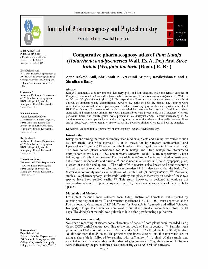
E-ISSN: 2278-4136 P-ISSN: 2349-8234 JPP 2016; 5(3): 160-168 Received: 11-03-2016 Accepted: 12-04-2016
Zope Rakesh Anil Research Scholar, Department of PG Studies in Dravyaguna SDM College of Ayurveda, Kuthpady, Udupi, Karnataka, India 574 118. Shrikanth P Associate Professor, Department of PG Studies in Dravyaguna SDM College of Ayurveda, Kuthpady, Udupi, Karnataka, India 574 118. KN Sunil Kumar Senior Research Officer, Department of Pharmacognosy, SDM Centre for Research in Ayurveda and Allied Sciences, Kuthpady, Udupi, Karnataka, India 574 118. Ravikrishna S Assistant Professor, Department of PG Studies in Dravyaguna SDM College of Ayurveda, Kuthpady, Udupi, Karnataka, India 574 118 T Shridhara Bairy Professor and Head Department of PG studies in Dravyaguna SDM College of Ayurveda, Kuthpady, Udupi, Karnataka, India 574 118 Correspondence Zope Rakesh Anil Research Scholar, Department of PG Studies in Dravyaguna SDM College of Ayurveda, Kuthpady, Udupi, Karnataka, India 574 118
Comparative pharmacognosy atlas of Pum Kutaja (Holarrhena antidysenterica Wall. Ex A. Dc.) And Stree
Kutaja (Wrightia tinctoria (Roxb.) R. Br.)
Zope Rakesh Anil, Shrikanth P, KN Sunil Kumar, Ravikrishna S and T Shridhara Bairy Abstract Kutaja is commonly used for amoebic dysentery, piles and skin diseases. Male and female varieties of Kutaja are mentioned in Ayurvedic classics which are sourced from Holarrhena antidysenterica Wall. ex A. DC. and Wrightia tinctoria (Roxb.) R. Br. respectively. Present study was undertaken to have a brief outlook of similarities and dissimilarities between the barks of both the plants. The samples were subjected to macro- and microscopic analysis, powder microscopy, physicochemical, phytochemical and HPTLC evaluation. Pharmacognostic analysis revealed both sources had crystals of calcium oxalate, stone cells and sclereids in common. However, phloem fibres were present only in W. tinctoria. Whereas, pericyclic fibres and starch grains were present in H. antidysenterica. Powder microscopy of H. antidysenterica showed parenchyma with starch grains and sclereids whereas, thin walled septate fibres and cut latex vessels were seen in W. tinctoria. HPTLC revealed similar Rf values in both the samples. Keywords: Adulteration, Comparative pharmacognosy, Kutaja, Phytochemistry.
Introduction Kutaja is one among the most commonly used medicinal plants and having two varieties such as Pum (male) and Stree (female) [1]. It is known for its Sangrahi (antidiarrheal) and Upashoshana (drying up) [2] properties, which makes it the drug of choice in Atisara (diarrhea). The two source plants considered for Pum Kutaja and Stree Kutaja are Holarrhena antidysenterica Wall. ex A. DC. and Wrightia tinctoria (Roxb.) R. Br. respectively [3], both belonging to family Apocynaceae. The bark of H. antidysenterica is considered as astringent, anthelmintic, amoebicidal and diuretic [4], and is used in amoebiasis [5], colic, dyspepsia, piles, diseases of the skin and spleen [4]. The bark of W. tinctoria is also known to be antidysenteric [6], and is used in treatment of piles and skin disorders [7]. It is also known that the bark of W. tinctoria is commonly used as an adulterant of Kurchi Bark (H. antidysenterica) [7]. Moreover, studies like pharmacognosy, antibacterial activity and physicochemistry on seeds of these two species have been studied earlier [8]. This study however, is designed to evaluate the comparative account of pharmacognostic and phytochemical components of bark of both species. Materials and Methods Fresh plant materials were collected from Udupi District of Karnataka, authenticated by referring the regional floras [9] and voucher specimens (14031401-02) were deposited at the Pharmacognosy department of S.D.M. Centre for Research in Ayurveda and Allied Sciences, Kuthpady, Udupi. Plant samples were washed and shade dried at room temperature for 10 days. The dried plant material was pulverized into a fine powder using a pulverizer. Macro-microscopic study Systematic recording of macroscopic characters of barks of both plants were recorded using Canon IXUS digital camera according to the text book of Pharmacognosy [10]. Samples were preserved in FAA (Formalin - 5ml + Acetic acid - 5ml + 70% Ethyl alcohol - 90ml) fixative solution for more than 48 hours. The preserved specimens were cut into thin transverse section using a sharp blade, followed by staining with saffranine [10]. A pinch of the sample was mounted on a microscopic slide with a drop of glycerin-water. Magnifications of the figures were indicated by the pre-calibrated scale-bars using Zeiss Axio Vision software.

~ 161 ~
Journal of Pharmacognosy and Phytochemistry
In order to supplement the descriptive part, photomicrographs in different magnifications of all necessary cells and tissues were taken in Zeiss Axio Lab trinocular microscope and Zeiss Stemi stereo microscope. Magnifications of the figures are indicated by the pre-calibrated scale-bars using Zeiss Axio Vision software. Physicochemical evaluation The percentage of physicochemical values like loss on drying, total ash, acid insoluble ash, alcohol soluble extractive, water soluble extractive were carried out as per the methods described in the Indian Pharmacopoeia [11]. Phytochemical study Powdered material was extracted in ethanol using cold maceration and was tested for various classes of active chemical constituents, using standard prescribed methods as described in Trease and Evans [10]. High Performance Thin Layer Chromatography (HPTLC) One gram of each powdered samples was extracted with 10 ml ethanol and kept for cold percolation for 24h and then filtered. Three and 6 µl of the above samples were applied on a pre-coated silica gel F254 on aluminum plates to a band width of 7 mm using Linomat 5 TLC applicator. The plate was developed in toluene: ethyl acetate (9.0:1.0). The developed plates were visualized under UV 254, 366 nm and then scanned under UV 254 and 366 nm. The plate was derivatised with vanillin sulphuric acid reagent to see constituents not detected under UV. Rf, colour of the spots and densitometric scan were recorded [12]. Results Holarrhena antidysenterica Wall. ex A. DC. dried stem bark appear in small recurved pieces that are 3 to 5 cm long and 0.5 to 2 cm thick, outer surface dark brownish, longitudinally wrinkled and bearing horizontal lenticels, inner surface brownish and rough [Fig. 1.1].
Fig 1: Macroscopy of H. antidysenterica and W. tinctoria
Fig. 1.1. H. antidysenterica
Outer surface of Wrightia tinctoria R. Br. dried stem bark, faintly longitudinally and transversely striated and intermittently displays small circular lenticels. Inner surface rough, having fibres and is buff in colour. [Fig. 1.2].
Fig 1.2: W. tinctoria Transverse section (TS) of stem bark of H. antidysenterica shows periderm, a wide stratified cortex and secondary phloem. Periderm consists of thin-walled and somewhat rectangular cork cells and 2 layers of phellogen. Phelloderm parenchymatous, containing prisms of calcium oxalate crystals and a few starch grains. Cortex shows groups of lignified pitted stone cells of different shapes upto 0.3 mm in size. In phloem region prisms, sclereids and stone cells can be seen. Medullary rays bi- or tri-seriate in different regions. Phloem parenchyma shows prisms of calcium oxalate crystals and starch in abundance. Phloem fibres absent. [Fig. 2].
Fig 2: Microscopy of bark of Holarrhena antidysenterica
Fig 2.1: TS of entire bark

~ 162 ~
Journal of Pharmacognosy and Phytochemistry
Fig 2.2 a: Stratified cork, cortex; b. phloem region with prisms, stone cells; c. sclereids group, d. starch grains and cambium TS of stem bark of W. tinctoria shows outermost multilayered cork, and parenchymatous cortex. Cortex interspersed with stone cells; phloem region shows fibers, medullary rays and laticiferous tubes and occasional isolated stone cells. Cortex made up of 12 to 15 layers traversed with usually isolated
prismatic crystals of calcium oxalate. Sieve tubes can be seen. Phloem wide, traversed with uni and sometimes multi-seriate medullary rays, isolated lignified fibres, and oval to circular wide latex canal. [Fig. 3].
Fig 3: Microscopy of bark of Wrightia tinctoria
Fig 3.1: TS of entire bark

~ 163 ~
Journal of Pharmacognosy and Phytochemistry
Fig 3.2 a: Cork and cortex; b. cortex with latex tubes and stone cells; c. phloem region with fibres and prisms; d. phloem rays, fibres and prisms Powder of H. antidysenterica is light brown, taste bitter; transversely and obliquely cut thin walled cork cells, groups of stone cells having different sizes and shapes. Cortical
parenchyma, thick walled with contents and starch grains, phloem tissue and few sclereids visible. [Fig. 4].
Fig 4: Powder microscopy of Holarrhena antidysenterica
Fig 4.1 a: Transversely cut cork; b. Cork cells; c. Obliquely cut cork cells; d. Cortical parenchyma; e. Parenchyma with content; f. Stone cell

~ 164 ~
Journal of Pharmacognosy and Phytochemistry
Fig 4.2 a: Parenchyma with content; b. Parenchyma with starch grains; c. Stone cell group; d. Phloem tissue; e. Thick walled cortical parenchyma; f. Sclereids
W. tinctoria powder shows presence of the fragments of quadrangular, pentagonal and hexagonal cork cells in surface view and sectional view, chlorenchyma of cortex and prisms of calcium oxalate; parenchymatous cells with content,
fragments of thin walled septate fibres; fibers with pitted walls; longitudinally cut latex vessels filled with granular contents. [Fig.5].
Fig 5: Powder microscopy of Wrightia tinctoria
Fig. 5.1 a: Obliquely cut cork cells; b. Transversely cut cork cells; c. Parenchyma with content; d. Chlorenchyma of cortex; e. Cork cells in surface view; f. Prisms of calcium oxalate

~ 165 ~
Journal of Pharmacognosy and Phytochemistry
Fig 5.2 a: Sclereid group; b. Fragment of fibre; c. Fragment of fibre; d. Pitted fibres; e. Latex vessel; f. Pitted fibres
Comparative pharmacognostic characters Many similar and dissimilar characters were observed between the barks as such and barks in powdered form of H. antidysenteica and W. tinctoria. The dissimilarities are as: the inner surface of H. antidysenterica bark is brown in colour and rough whereas W. tinctoria bark has pale brown and smoother inner surface. The stone cells are arranged throughout the section, and are arranged in concentric tangential bands in H. antidysenterica and are present only in cortical region in case of W. tinctoria. Prisms of calcium oxalate are comparatively less in W. tinctoria than in H. antidysenterica. Phloem fibres are present in W. tinctoria and absent in H. antidysenterica. Whereas pericyclic fibres are absent in W. tinctoria and present in H. antidysenterica. Physico-chemical and phytochemical results Loss on drying at 105º C was found to be almost same in both the samples. Total ash content was found to be more in W. tinctoria (12.86) than in H. antidysenterica (7.05). Similar results were observed in case of acid insoluble ash with values of 0.49 and 0.19 respectively. Water soluble extractive was more in H. antidysenterica (27.04) than in W. tinctoria (24.70). Alcohol soluble extractive did not show much variation, though. [Table-1] Table 1: Physico-chemical parameters of H. antidysenterica and W.
tinctoria
Parameter Results n=3 %w/w
H. antidysenterica W. tinctoria Loss On Drying 10.42 10.48
Total ash 7.05 12.86 Acid Insoluble Ash 0.19 0.49
Alcohol Soluble Extractive 10.25 10.46 Water Soluble Extractive 27.04 24.70
Alkaloids, carbohydrates, resins, steroids, saponins, tannins and terpenoids were present in both the species. The presence of coumarins, flavonoids and phenol was seen only in H. antidysenterica. [Table-2]
Table 2: Results of preliminary phytochemical tests of H.
antidysenterica and W. tinctoria
Test H. antidysenterica W. tinctoria Alkaloid + +
Carbohydrate + + Carboxylic acid - -
Coumarins + - Flavanoids + -
Phenol + - Quinone - - Resins + + Steroid + +
Saponins + + Tannin + +
Terpenoid + + Three bands were seen under H. antidysenterica with Rf values of 0.08, 0.48 and 0.83 under 366nm. Whereas in W. tinctoria only two bands were seen with 0.08 and 0.03 Rf. Seven bands were seen post derivatisation under H. antidysenterica with Rf values 0.07, 0.13, 0.16, 0.30 0.34, 0.51 and 0.80. While eight bands were observed under W. tinctoria with Rf values 0.15, 0.21, 0.35, 0.46, 0.52, 0.61, 0.67 and 0.90. [Table-3] On spectral comparison between both, the Rf values overlapping in both the samples under short and long UV radiations are 0.01, 0.73, 0.82 and 0.01 respectively. These graphs show the overlapping zones present in H. antidysenterica and W. tinctoria. [Fig. 6-9]
Table 3: Rf value of Alcohol extract of the Samples of Holarrhena antidysenterica and Wrightia tinctoria
At 254nm At 366nm Post Derivatisation HA WT HA WT HA WT
- - 0.08 (DF. Blue) 0.08 (LF. Blue) 0.07 (L. Purple) - - - 0.48 (LF. Blue) - 0.13 (L. Purple) - - - 0.83 (DF. Blue) 0.83 (DF. Blue) 0.16 (L. Purple) 0.15(L. Purple) - - - - - 0.21(L. Purple) - - - - 0.30 (D. Purple) - - - - - 0.34 (D. Purple) 0.35 (L. Purple) - - - - - 0.46 (D. Purple) - - - - 0.51 (D. Purple) 0.52 (D. Purple) - - - - - 0.61 (L. Purple) - - - - - 0.67 (L. Purple) - - - - 0.80 (D. Purple) - - - - - - 0.90 (D. Purple)
*L-Light, D-Dark, F-Fluorescent

~ 166 ~
Journal of Pharmacognosy and Phytochemistry
Fig 6: HPTLC photodocumentation of Alcohol extract of Holarrhena antidysenterica and Wrightia tinctoria
Track 1- Alcohol extract of Holarrhena antidysenterica 6µl Track 2 – Alcohol extract of Wrightia tinctoria 6µl Solvent system – Toluene: Ethyl acetate: (9:1
Fig 7: HPTLC Densitometric Scan at 254 nm
Fig 7a: Holarrhena antidysenterica
Fig 7b: Wrightia tinctoria
Fig 8: HPTLC Densitometric Scan at 366nm
Fig 8a: Holarrhena antidysenterica

~ 167 ~
Journal of Pharmacognosy and Phytochemistry
Fig 8b: Wrightia tinctoria
Fig 9: 3-D Chromatogram
Fig 9a: 3-D Chromatogram at 254 nm
Fig 9b: 3-D Chromatogram at 366nm Discussion Habit and habitat of both plants are similar as both of them belong to same family. Recurved shape and dark brownish colour of H. antidysenterica bark plays an important role in differentiating between both. The crystals of calcium oxalate are present in both the samples and have prismatic form. Stone cells/sclereids commonly occur in hard outer coats of seed and fruits and in the bark. In H. antidysenterica lignified pitted stone cells can be seen. Fibres are usually differentiated on the basis of the tissue in which they occur. Pericyclic fibers are seen only in H. antidysenterica bark which differentiates it from W. tinctoria bark. Fibres are absent in phloem of H. antidysenterica but presence of isolated lignified fibres in the phloem of W. tinctoria is a distinguishing character. Starch occurs in the form of granules (commonly known as starch grins). Their shape and size are characteristics of the species. They are found in the bark of H. antidysenterica and are absent in the other sample. More total ash value in W. tinctoria
indicates presence of more amounts of extraneous matter in it. Comparatively presence of more silicacious matter in W. tinctoria may be considered as a reason for more acid-insoluble ash value in it. The existence of close relationship between constituents of plants and their taxonomical status is established by extensive phytochemical screening. Such similarity can be seen in both these samples under study, as both of them belong to Apocynaceae family. Still H. antidysenterica showed presence of coumarins, flavonoids and phenol, which separates it from W. tinctoria. Overlapping zones in HPTLC indicates similarity in chemical components present in both samples. However they have to be evaluated pharmacologically and clinically. Conclusion W. tinctoria which is commonly used as an adulterant of H. antidysenterica bark, shows similar morphology and habitat. Pharmacognostic evidences show the presence of calcium

~ 168 ~
Journal of Pharmacognosy and Phytochemistry
oxalate crystals, stone cells and sclereids as key characters for differentiation. Phytochemically they are found to be similar with Rf value 0.01 and 0.73 in common when compared spectrally. This study is useful in the identification of both the species as well as in their differentiation from each other. Further scientific evaluation at molecular level, marker compounds and pharmacological confirmation is required however. References 1. Agnivesha. Vatsaka Kalpam. In: Shukla V, Tripathi RD
editors. Charaka Samhita. Volume II, Chaukhambha Sanskrit Pratishthan, Delhi, 2007, 827.
2. Agnivesha. Yajjah purusheeya. In: Shukla V, Tripathi RD editors. Charaka Samhita. Volume I, Chaukhambha Sanskrit Pratishthan, Delhi, 2006, 338.
3. Sharma PV. Dravyaguna-vijnana. Vol II, Chaukhambha Bharati Academy, Varanasi, 2009.
4. Khare CP. Indian Medicinal Plants An Illustrated Dictionary. Springer Science plus Business Media, New York, 2007, 312.
5. Gupta AK. Quality standards of Indian medicinal plants. Vol 4, Indian Council of Medical Research, New Delhi, 2006.
6. Bigoniya P, Rana A. Antidiarrheal and antispasmodic activity of Wrightia tinctoria Bark and its steroidal alkaloid fraction. Pharmacologyonline 2009; 3: 298-310
7. Khare CP. Indian Medicinal Plants An Illustrated Dictionary. Springer Science plus Business Media, New York, 2007, 720.
8. Jolly CI, Mecheri NR. Copmarative harmacognostical, physicochemical and antibacterial studies on seeds of Holarrhena antidysenterica Wall and Wrightia tinctoria R. Br. Indian J Pharm Sci. 1996; 58(2):51-54
9. Bhat GK. Flora of South Canara. Bhat GK, Udupi, 2014, 928.
10. Evans WC. Trease and Evans Pharmacognosy. Bailliere Tindall, London, 1989, 530.
11. Anonymous. Indian Pharmacopoeia. Edn 2, Government of India Publication, New Delhi, 2001.
12. Patel Rashmin, Patel Mrunali, Dubey Nitin, Dubey Nidhi, Patel Bharat. HPTLC Method Development and Validation - Strategy to Minimize Methodological Failures. J Food Drug Anal. 2012; 20(4):794-804.