e s t a b lish new zealand journal of in19 4 6 medical ... 65 no 1.pdf · the new zealand journal...
TRANSCRIPT

Established
in 1946
Volume 65 Number 1 April 2011ISSN 1171-0195
New Zealand Journal ofMedicalLaboratoryScience
Official Publication of the
New Zealand Institute of
Medical Laboratory Science
Incorporated
1

Conquer your space limitations • Reliability
• Easy to use
• Quality results
• Comprehensive system
STA Satellite®
Con
cept
ion
L2R
- ©
200
5 D
iagn
ostic
a St
ago
- A
ll ri
ghts
res
erve
d -
05/2
010
10-4053-AP STA Satellite Australie.indd 1 06/05/10 16:40

NZ J Med Lab Science 2011
1
Editor Rob Siebers, PGCertPH, FNZIC, FNZIMLS, FSB; University of Otago, Wellington, New Zealand
Deputy Editor Ann Thornton, CertMS, FNZIMLS; University of Otago, Wellington, New Zealand
Editorial BoardChris Kendrick, MSc, MNZIMLS; Massey University, Palmerston North, New ZealandMichael Legge, PhD, MSB, FIBMS, FNZIMLS; University of Otago, Dunedin, New ZealandAndrew Stewart, MSc, FIBMS, FNZIMLS; New Zealand Blood Service, Auckland, New ZealandKevin Taylor, BMLSc, PGDipMLSc; Canterbury Health Laboratories, Christchurch, New ZealandJohn Sterling, BSc(Hons), MLett, AFRCPA, MAIMS, FRMS; SA Pathology, Adelaide, AustraliaTony Woods, BA, BSc(Hons), PhD, MAIMS; University of South Australia, Adelaide, Australia
Statistical AdvisersNevil Pierse, MSc; University of Otago, Wellington, New ZealandGordon Purdie, BSc; University of Otago, Wellington, New Zealand
About the Journal The New Zealand Journal of Medical Laboratory Science is the official publication of the New Zealand Institute of Medical Laboratory Science (NZIMLS) who owns the copyright. No parts of this publication may be reproduced in any form without the written permission of the NZIMLS. The Journal publishes original articles, technical communications, review articles, case studies and letters to the Editor on all subjects pertaining to the practice of medical laboratory science. The journal is published three times per year in April, August and November. It is circulated to all NZIMLS members and universities and research institutes in New Zealand and overseas. Current circulation is about 2,200 copies per issue. Printed by Keyprint Printing Ltd, Auckland, New Zealand.
Brief instructions to authorsSubmit all material electronically to the Editor ([email protected] or [email protected]. Comprehensive instructions to authors can be found on the NZIMLS web site (www.nzimls.org.nz). With your submission include the author form in which each author’s contribution to the study is stated together with any possible conflicts of interest. This information will be published with the accepted manuscript. The authors are responsible for the scientific content and views. Opinions expressed in the Journal are not necessarily those of the Editors, Editorial Board or Council of the NZIMLS.
Indexing
The Journal is open access (www.nzimls.org.nz) and is abstracted by the Cumulative Index to Nursing and Allied Health Literature (CINAHL), EMBASE, EBSCO, the Australian Medical Index, Scopus, Google Scholarly and Thomson Gale. The Journal is a member of the World Association of Medical Editors (www.wame.org)
Subscription
Enquiries regarding subscription and address changes should be addressed to the NZIMLS at PO Box 505, Rangiora, New Zealand. Ph: +64 3 313 4761; Email: [email protected]
AdvertisingAdvertisement bookings and inquiries should be addressed to the Advertising Manager, Trish Reilly, 48 Towai Street, St Heliers, Auckland 1071, New Zealand. Ph: +64 9 575 5057; Fax: +64 9 575 0698; Email: [email protected]
N u m b e r 1Volume 65April 2011ISSN 1171-0195
N e w Z e a l a n d J o u r n a l o f
EditorialsThe role of the NZIMLS Council. “What we do” and “What can you do for us”Ken Beechey and Ross Hewett.......................................................2-3
Open access of the journalRob Siebers..........................................................................................4
Original articleOccurrence of blaampC in cefoxitin-resistant Escherichia coli and Klebsiella pneumoniae isolates from a North Indian tertiary care hospitalFarrukh Sobia, Mohammad Shahid, Anuradha Singh, Haris M. Khan, Indu Shukla and Abida Malik..........................................................5-9
Scientific letterThe influence of fetal bovine serum on protein expression in-vitro: a proteomics approachJody Anne Hazlett and Michael Legge...........................................10-11
Letter to the EditorThe clinical scientist in diagnostic pathologyAmanda Dixon-McIver......................................................................12
The author repliesMike Legge........................................................................................13
ReportThe Diversity of Pathology NZIMLS Annual Scientific MeetingRoss Hewett..................................................................................14-15
Book reviewMicroscopic Haematology (3rd Edition) by Gillian RozenbergReviewed by Cat Stevens....................................................................16
Regular featuresAdvertisers in this issue......................................................................3Australian Journal of Medical Science abstracts........................16-17Barry Edwards & Rod Kennedy Scholarship....................................22British Journal of Biomedical Science abstracts.........................17-21Correction to Life Members list........................................................24Index to Volume 64, 2010.................................................................25In this issue..........................................................................................4Instructions to authors........................................................................1Fellowship of the NZIMLS..................................................................26Journal questionnaire.......................................................................21Massey University student prize.......................................................26Med-Bio journal prize........................................................................22New products and services................................................................27NZIMLS journal prize.........................................................................22Olympus photo competition.............................................................24Pacific Way column.......................................................................28-29Special Interest Groups.................................................................30-35South Pacific Congress, Brisbane, 2011............................................36

NZ J Med Lab Science 2011
2
EditorialThe role of the NZIMLS Council. “What we do” and “What can you do for us.”Ken Beechey, Ross Hewett
__________________________________________“What does the Council do for us?” would be a common and reasonable question from the membership. Much like the hidden work of our profession to the public it is probably not until you are elected on to Council, attend meetings and deal with the regular correspondence that the full extent of Council’s involvement in serving our members and profession is realised.
Typically Council meets four times per annum – late November, in March before the South Island Seminar, during May before the North Island Seminar and in August preceding the Annual General Meeting (AGM). Following the AGM, the elected Council members are allocated portfolios (see below) that they are responsible for during the following 12 months, responding to enquiries fielded though the Executive Office and reporting back to Council at meetings.
The aim of this Editorial is to provide a brief insight into some of the functions of Council along with examples of our recent work. Opportunities on how the membership can become actively involved and help are also proffered under each section.
Regional representationYour five regional representatives network with contacts in each laboratory across their region reporting back to Council on local issues, concerns and items of interest. In recent years the Council representatives have endeavoured to improve communications back to their members through local newsletters post-Council meetings
Take time to find out who your local regional representative is and relay any issues to them regarding professional affairs. Council can only represent the members on the information and feedback received relating to our profession.
Financial governanceThe funding of the NZIMLS comes from the membership and their employers. The NZIMLS has employees and sub-contractors who manage the day to day business of the organisation. It is the responsibility of Council to ensure the proper use of these funds and to ensure, as a good employer, security of employees and manage the financial liabilities of all conferences and seminars. Without sufficient financial resources the NZIMLS would be unable to function and fulfil the needs of the membership.
Professional affairsThe NZIMLS has regular contact with other professional bodies (including MLSB, Universities, DHBNZ, MOH, PPTC, AIMS) throughout the year on issues such as qualifications and examination requirements, also course developments. Council is often requested to offer nominations and submissions to various Ministry of Health committees on issues, plus work relating to Clinical Scientists and the workforce.
Promoting the profession is another primary function of Council with annual involvement in career expos across the country.
Offer to participate in career expos in your area.
CPDThe CPD program run through the NZIMLS is one of three approved by the Medical Laboratory Science Board. Co-ordinated by Jillian Broadbent, designated Council members assist in the consideration of members’ enquiries on CPD point allocation and the programme in general.
Prepare questions for the on line classroom - obtain CPD points for yourself and help provide another avenue for your colleagues to also gain CPD points.
ExaminationsThe NZIMLS provides QMLT/QSST and Fellowship examinations. Although the good work in setting, marking and moderating examination papers are provided by our volunteer members, Council members work hard in the background with the Executive Office to assure the process is run in a timely manner and help audit the papers prior to being distributed to examinees.
Over the past few years Council has worked on standardising the syllabi and examination papers, providing log books for all disciplines and clarifying the guidelines for examiners and moderators. This, along with improved training opportunities for examiners and moderators, has led to a greater consistency in examination questions and standards.
Your continued input and feedback on syllabi content, logbooks and exams are fundamental to continued improvement to our professional examinations. Consider becoming an examiner or moderator.
Scientific meetings and seminarsThe Annual Scientific Meeting, Special Interest Groups and the North and South Island Seminars are all run under the auspices of the NZIMLS. A recent review and additions to the SIG Guidelines for running these events have been developed as an aid for the SIG convenors whilst also providing consistency and financial prudency.
These meetings cease to exist without papers and participation from our members. The success of the scientific content therefore lies within.
Help the continued success of our meetings by forward planning to offer presentations or posters to the SIG and conference convenors when asked. Also feedback to Council is welcomed on suggestions for good presenters (local and international) or topics for meetings.
CommunicationsThe NZIMLS continuously works on improving communications to its membership through the NZIMLS website, Journal and regular newsletters.
Write an article for the Journal.

NZ J Med Lab Science 2011
3
Feedback on the refreshed new look website and content.
Sponsorship/awardsThe NZIMLS offers the Barrie Edwards and Rod Kennedy Scholarships for which all applications are judged by designated members. The Hugh Bloore Poster prize is also awarded at the Annual Scientific Meeting. Apply for these awards and reap the financial benefits to help attend a scientific meeting.
MembershipAdvice and clarification on membership issues are also processed through Council.
Encourage your colleagues to become members.
CouncilIt is a rewarding task to serve on Council which covers a multitude of functions to promote the profession and to provide ongoing education and qualifications for its membership. It is always an important reminder that “you the members” are the NZIMLS and it is only by your continued support and input that will we continue to grow and raise the profile of our profession.
Communicate with Council through your regional representative, the Executive Office or Council member.Consider standing for Council and put something back into your profession.
NZIMLS Council Members
Top row from left to right: Tony Barnett (Region 4 rep), Kim Allan (Region 3 rep), Chris Pickett (Vice President), Jan Bird (Region 2 rep), Ross Hewett (Secretary/Treasurer)Bottom row: Rob Siebers (Journal Editor), Terry Taylor (Region 5 rep), Margie Matson (Region 1 rep), Ken Beechey (President), Jillian Broadbent (CPD Coordinator) Absent: Fran van Til (Executive Officer)
NZIMLS Council portfolios 2010-2011
CPD Jillian Broadbent / Chris Pickett / Ken BeecheyASM Fran van Til / Ross HewettProfessional Affairs Ken Beechey / Chris Pickett SIG Governance Ross Hewett / Fran van TilQMLT/QSST/QDT Kim Allan, Tony BarnettNewsletter Margie Matson / Sharon TozerPromotion/ Communication Terry Taylor / Executive OfficeMarketing Fran van Til / Ross Hewett
Careers Expos Wellington – Kim Allan Auckland – Margie Matson Christchurch–Ken Beechey + Some local volunteers Hamilton – Chris Pickett / Jan Bird Dunedin – Terry TaylorBoard of Studies AUT – Margie Matson Massey – Kim Allan Otago – Terry TaylorMembership Jan Bird / Sharon TozerClassroom Biochemistry – Margie Matson Microbiology – Tony Barnett Haematology – Ken Beechey Immunology – Terry Taylor Histology – Jillian BroadbentWebsite Chris Kendrick / Fran van TilDocumentation Fran van Til / Sharon TozerMLSB Liaison Ken Beechey, Fran van Til and Sharon TozerFellowship Rob Siebers, Ann Thornton and Jillian Broadbent.Finance Ross Hewett, Sharon TozerJournal Rob SiebersNZIMLS Governance Ross Hewett / Ken Beechey / Chris Pickett
Author information
Ken Beechey, FIBMS MNZIMLS, Section Head Haematology1 and President of the NZIMLS2 Ross Hewett, MNZIMLS, Laboratory Manager3 and Treasurer/Secretary of the NZIMLS2
1Canterbury Health Laboratories, Christchurch, New Zealand. E-mail: [email protected] Zealand Institute of Medical Laboratory Science, Rangiora, New Zealand3LabPLUS, Auckland, New Zealand
Carl Zeiss NZ Ltd....................................................outside back coverDiagnostica Stago.................................................inside front coveriSOFT.......................................................................23Medica Ltd.............................................................11Reed Global Resourcing........................................15Vital Diagnostics.....................................................inside back cover
In lieu of a full page advertisement from Abbott for this issue of the journal, Abbott has generously donated this amount to the Christchurch Earthquake appeal.
Advertisers in this issue_______________________

NZ J Med Lab Science 2011
4
In this issue_______________________In their Editorial, Ken Beechey and Ross Hewett, President and Treasure/Secretary of the NZIMLS respectively, discuss what the NZIMLS Council does for its members and suggest what members can do for the Institute.
Resistance to antibiotics is a worldwide problem. Sobia and colleagues from India analysed the occurrence of blaampC in cefoxitin-resistant isolates and evaluated the role of phenotypic methods for detection of AmpC-producers. They found that cefoxitin-resistant isolates showed multiple antimicrobial resistance and were resistant to more than three antibiotics and prevalence of blaampC was quite high. They conclude that a phenotypic test is insufficient to diagnose AmpC-producers and recommend that a genotypic test, like PCR, should be used.
Cell culture provides the opportunity to study and manipulate cells in-vitro which would otherwise be impossible to undertake in-vivo. During culture apparently normal cells may develop varying responses to agents, such as fetal bovine serum (FBS). Fox and Legge used a proteomics approach to identify whether proteins were affected by a simple modification of the FBS concentration in the culture media using 2 D gel electrophoresis. They found that the choice of FBS concentration will change the response of cell protein synthesis, which in turn has the potential to influence the outcome of cell culture results.
In this issue is a review by Cat Stevens of Gillian Rozenburg’s book, Microscopic Haematology. Cat found it to be is a beautiful book with superb images and concise yet comprehensive notes. Elsevier Australia, the publisher, has offered a 15% discount on the purchase price with free delivery to our readers. This offer ends 30 June, details in the Book Review section.
Rikki Penn was the winner of the Med-Bio Journal Prize for the best scientific article published in the Journal in 2010 while Vichet Khieng was the winner of the NZIMLS Journal Prize for the best case study published in the Journal in 2010. Both articles resulted from student projects and both came from the same laboratory, Southern Community Laboratories in Dunedin. Consider publishing in the Journal and you will be eligible for these prizes in 2011 (provided you are a financial member of the NZIMLS).
Last year the Olympus photo competition attracted a large number of excellent submissions with the winning photo submitted by Lynne Pomare from Aotea Pathology in Wellington. Olympus has kindly agreed to hold the competition again this year with entries closing Friday 16th September 2011. Details elsewhere in this issue.
EditorialOpen access of the journalRob Siebers
____________________The Institute’s journal has recently been accepted for indexing and listing by the Directory of Open Access Journals (DOAJ). The official aim of the DOAJ “...is to increase the visibility and ease of use of open access scientific and scholarly journals thereby promoting the increased usage and impact. The Directory aims to be comprehensive and cover all open access scientific and scholarly journals that use a quality control system to guarantee the content.”
Open access journals do not charge readers or institutions for access. To be included in the DOAJ it is mandatory for journals to allow users to “read, download, distribute, print, search, or link to the full text” of the journal’s articles. To be included, the journal must also exercise peer-review or editorial quality control. Editorial quality control on submitted articles must be through an editor, editorial board and/or a peer-review system.
The Institute’s journal has been categorised as a medical research journal with sub-headings of laboratory medicine, haematology, transfusion medicine, anatomic pathology, clinical biochemistry and clinical microbiology. To be included as a research journal it must report primary results of research or overview of research results (review articles) to a scholarly community. All articles are to be in full text and all scientific and scholarly subjects in its discipline are covered. A substantive part of the journal should consist of research papers and the target group should be primary researchers.
At present the DOAJ covers approximately 6,000 journals ensuring that about 20% of all published peer-reviewed articles from around the world are now freely accessible and available. The inclusion of the Institute’s journal in the DOAJ ensures that all its published papers are now freely available for the world’s scientific community. Of course, the Institute’s journal relies on submissions of quality articles from, but not exclusively, its members. Submissions have also come from non-NZIMLS members, both from within New Zealand and from other countries. Acceptance of articles by the journal is primarily dependant on their relevance to the broad area of medical laboratory science and scientific quality.
Now the Institute’s journal’s articles are open access and freely available, it is hoped that published articles from the journal will be more frequently cited in other biomedical journals and that scientists around the world will consider submitting their research articles to the New Zealand Journal of Medical Laboratory Science. Our journal has a proud tradition of publishing quality peer-reviewed scientific articles continuously for 65 years, informing and educating its readers and, in some cases, starting some of our members on their research career. In my own case, having had my first publication in the journal (1) gave me a taste for research culminating in my present academic position. At the journal we hope that you, as NZIMLS members, will support the journal by considering submitting your SIG, Users Group Meeting, Seminar and ASM presentations for wider dissemination to your colleagues in New Zealand and now, through listing in the DOAJ, more widely around the world.
ReferenceSiebers R, Clarkson K. Lipoprotein electrophoresis on cellulose acetate. N Z J Med Lab Technol 1972; 26: 112-3.
Author informationRob Siebers, PGCertPH FNZIC FNZIMLS FSB, Associate Professor1 and Editor of the New Zealand Journal of Medical Laboratory Science2
1School of Medicine and Health Sciences, University of Otago, Wellington, New Zealand. E-mail: [email protected] 2New Zealand Institute of Medical Laboratory Science, Rangiora, New Zealand

NZ J Med Lab Science 2011
5
Occurrence of blaampC in cefoxitin-resistant Escherichia coli and Klebsiella pneumoniae isolates from a North Indian tertiary care hospitalFarrukh Sobia, Mohammad Shahid, Anuradha Singh, Haris M. Khan, Indu Shukla and Abida Malik
__________________________________________AbstractAim: To analyse the occurrence of blaampC in cefoxitin-resistant isolates and to evaluate the role of phenotypic methods for detection of AmpC-producers.Methods: A total of 91 isolates (84 E. coli and 7 K. pneumoniae) that were resistant to cefoxitin and obtained during a period of six months were studied. Antibiotic susceptibility to third- and fourth-generation cephalosporins and other antibiotics were performed. blaampC was detected by PCR.Results: Cefoxitin-resistant isolates showed concomitant resistance to other antibiotics used. All 91 isolates showed multiple antimicrobial resistance and were found resistant to more than three antibiotics; maximum resistance (among cephalosporins) was noticed for ceftriaxone (89%) followed by cefpirome (80%). A total of 82.4 % isolates were found positive for ampC gene, however, 57.1 % were found AmpC-producers by modified three-dimensional extract test (MTDET). Out of 57.1 % isolates that were noticed as AmpC-producers by MTDET, 50.5 % were found positive on detection by PCR but 6 (6.6%) isolates were found negative for presence of blaampC gene. Among 26 (28.6%) AmpC-intermediate isolates, 20 (21.9%) were found to harbor blaampC genes while 6 (6.6%) were found negative for the same. However, of the 14.3 % isolates that were considered negative by TDET, 10.9 % gave positive results by PCR. Almost a similar frequency of occurrence of cefoxitin-resistant isolates harbouring blaampC was noticed from different hospital wards. Conclusion: Among cefoxitin-resistant isolates, prevalence of blaampC is quite high and a phenotypic test is insufficient to diagnose AmpC-producers and hence genotypic test, like PCR, should be used.
Key words: blaampC, cefoxitin resistance, TDET, PCR, E. coli, K. pneumonia
N Z J Med Lab Sci 2011; 65: 00-00
IntroductionThe worldwide use of antimicrobials has created enormous pressure for the selection of resistance among opportunistic bacterial pathogen. The resistance mechanism in bacteria to β-lactams is the production of β-lactamases that catalyze the hydrolysis of the β-lactam ring, preventing their interaction with the D,D-transpeptidases (1). During treatment with lactams, resistant mutants showing constitutive high levels of AmpC production are frequently selected leading to therapeutic failures (2). Bacteria over-expressing AmpC beta-lactamase are of major clinical concern as they confer resistance to beta-lactams, beta-lactam-beta-lactamase inhibitor combination and monobactams, but are found susceptible to fourth-generation cephalosporins (4GC) and carbapenems (3). Hence these two classes of drugs remain the only therapeutic options for such organisms (4,5). However, isolates harboring extended-spectrum beta-lactamases (ESBLs) along with AmpC offers resistance to 4GC as well (6).
The increasing awareness and improved recognition of ESBL-
producers have led to improved infection control measures to minimize spread of these emerging pathogens. With no published CLSI (formerly NCCLS) guidelines for proper identification and infection control measures, AmpC producing organisms infections may become a greater concern than ESBL producing organisms infections as they are increasing in prevalence (7,8). In addition MDR plasmids harboring both ESBL and AmpC genes are spreading among bacteria and are becoming a new emerging threat.
AmpC beta-lactamase poses a serious risk of transmission to hospitalized patients when colonized or infected ones are admitted, presenting a concern for hospital infection control surveillance of these resistance mechanisms. In the present study we analysed the occurrence of blaampC in a collection of cefoxitin-resistant isolates and evaluated the role of phenotypic methods for detection of AmpC-producers.
MethodsA total of 91 isolates (84 E. coli and 7 K. pneumoniae) were obtained from 91 hospitalised patients admitted to the JawaharLal Nehru Medical College & Hospital over a period of six months (January to June 2009) for the study. Demographic details of the patients were also noted. All these isolates were found to be resistant to cefoxitin and were isolated from urine, pus, semen, cervical swabs, drains and CSF.
Antimicrobial susceptibility testingAntimicrobial susceptibility testing was performed and interpreted as per CLSI guidelines (9). The following antibiotics (all supplied from Hi Media Laboratories, Mumbai, India) were tested: ceftriaxone (30μg), cefoperazone (75μg), cefixime (30μg), ceftazidime (30μg), cefepime (30μg), cefpirome (30μg), gentamicin (10μg), amikacin (30μg), ofloxacin (5μg), gatifloxacin (5μg) and aztreonam (30μg).
Phenotypic detection of AmpC-producersModified three-dimensional extract test (MTDET), as described by Shahid et al.(10) was performed on all cefoxitin-resistant isolates to identify AmpC producers among them. Briefly, 10-15 mg of bacterial wet weight was scraped from the culture plate and suspended in 0.5mL of peptone water in a sterile micro-centrifuge tube and incubated at 37˚С for one hour. Crude enzyme extract was prepared by repeated freezing-thawing. To ensure complete membrane lysis, the freezing-thawing was carried out five times. Lawn culture of E. coli ATCC 25922 was prepared on Mueller-Hinton Agar (MHA) plate and was incubated at 37˚С so that the plate died properly. Cefoxitin discs (30μg) were placed on the dried MHA plate. With a sterile scalpel a linear trench (3cm x 1mm) was prepared in agar at a distance of 5mm from edge of the disc in an outward radial direction. 50μl of enzyme preparation was carefully dispensed in the trench to avoid trench overfill. The inoculated media was incubated overnight at 37˚С. Enhanced growth of the surface organism at the point where the trench intersected the zone of inhibition towards the cefoxitin disc was interpreted as evidence for the presence of AmpC β-lactamase.
NZ J Med Lab Science 2011
6
Genotypic detection of blaampC genesblaampC genes were detected by PCR as described by Feria et al.(11) with some modifications. Briefly, the gene of interest was amplified in a total reaction volume of 50μl containing 10pmol each of primer (ampC-f, 5’CCC CGC TTA TAG AGC AAC AA 3’ and ampC-r, 5’TCA ATG GTC GAC TTC ACA CC 3’) that span universal region of ampC gene, 0.2mM of each dNTPs, 10mM Tris-HCl (pH 8.8), 50mM KCl, 2.0 mM MgCl2 and 1.25U Taq DNA Polymerase (Bangalore Genei, India). 2μL of template DNA was added to 48 μL of master mixture. The reaction mixture was placed in MJ-mini Bio-Rad thermal cycler (Bio-Rad, USA). The PCR amplification cycle was performed with cycling conditions consisting of an initial denaturation step at 95˚C for 15 min, followed by 35 cycles of 94˚C for 60 sec., 58˚C for 2 min., 72˚C for 3 min. and the process was completed with a final elongation step at 72˚C for 10 min. Amplified PCR products were analysed by gel electrophoresis with 2% agarose (Bangalore Genei, India) gel containing ethidium bromide. After electrophoresis DNA fragments were visualized by Bio-Rad Gel documentation system (Bio-Rad, USA).
RAPD typingGenotyping of cefoxitin-resistant isolates was done as described previously (12) in order to determine whether any specific clone was circulating in our hospital environment. The results were analyzed by using Bio-Rad Gel documentation system (Bio-Rad, USA).
Plasmid analysisPlasmid isolation was done in all isolates by the large scale alkaline lysis method as described previously (13). 10 μL of plasmid samples were electrophoresed in 0.8% agarose gel containing ethidium bromide. λ DNA double-digested with EcoRI and HindIII (Bangalore Genei, India) was used as a molecular weight marker.
Results and DiscussionAmpC producing strains which are intrinsically resistant to clavulanic acid are causing great concern as carbapenems are the only antibiotics effective against such strains (14). With the spread of these strains all over the world it is necessary to know their prevalence in a hospital so as to formulate a policy of empirical therapy in high risk units. It becomes equally important that information should be procured on an isolate from a patient so as to avoid misuse of extended spectrum cephalosporins. The routine susceptibility tests performed by clinical laboratories fail to detect these strains, which may lead to inappropriate and unsuccessful therapy of the patient and unnecessary usage of drugs (15).
On analyzing the clinical and demographic features (Table 1) in the present study it was observed that the mean ages of patients having infection of cefoxitin-resistant E. coli and K. pneumoniae were 30.4 and 37.7 years, respectively. The source of cefoxitin-resistant E. coli was pus (50.0 %), urine (38.1 %), drain (6.0 %), semen (3.6 %), and 1.2 % in both cervical swab and CSF, while 85.2 % K. pneumoniae were obtained from pus and 14.3% from urine. Maximum number of isolates were obtained from the surgery ward (33 E. coli and 4 K. pneumoniae) followed by the gynaecology and orthopaedics wards (19 E. coli and 2 K. pneumoniae & 17 E. coli and 1 K. pneumoniae respectively). Cefoxitin-resistance can be used to screen isolates for detecting possible AmpC production, but lack of permeation of porins has also been reported as one of the resistance mechanism of cefoxitin in AmpC non-producers (15).
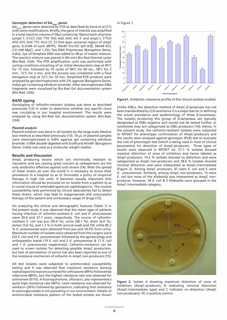
All test isolates were subjected to antimicrobial susceptibility testing and it was observed that maximum resistance (among cephalosporins) was encountered for ceftriaxone (89%) followed by cefpirome (80%), but the highest resistance rate was observed for aztreonam (91%). A fluoroquinolone, ofloxacin, also represented a quite high resistance rate (90%). Least resistance was observed for amikacin (26%) followed by gentamicin, indicating that resistance to aminoglycosides is not prevailing in our environment. Details of antimicrobial resistance pattern of the tested isolates are shown
in Figure 1.
56
72
63
89
67
80
74
90
50
26
91
0
10
20
30
40
50
60
70
80
90
100
cefop
erazo
ne
cefix
ime
cefta
zidim
e
ceftri
axon
e
cefep
ime
cefpi
rome
gatifl
oxac
in
oflox
acin
genta
micin
amika
cin
aztre
onam
Antimicrobial agents
Perc
enta
ge o
f res
ista
nt is
olat
es
Figure1. Antibiotic resistance profile of the clinical isolates studied.
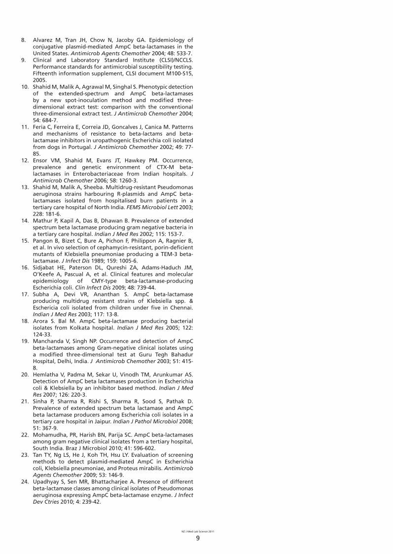
Unlike ESBLs, the detection method of AmpC β-lactamase has not been standardized by CLSI and hence it is a major barrier in defining the actual prevalence and epidemiology of these β-lactamases. The isolates producing this group of β-lactamase are typically designated as ESBL-negative and would not be tested further or sometimes they are categorized as ESBL-producers (16). Hence, in the present study, the cefoxitin-resistant isolates were subjected to MTDET for phenotypic confirmation of AmpC-producers and the results were analysed against genotypic (PCR) test to evaluate the role of phenotypic test (which is being used in most of clinical parameters) for detection of AmpC-producers. Three types of results were observed in MTDET viz. 57.1 % isolates showed maximal distortion of zone of inhibition and hence labeled as AmpC-producers, 14.3 % isolates showed no distortion and were categorized as AmpC non-producers and 28.6 % isolates showed minimal distortion and were interpreted as AmpC-intermediate (Figure 2). Among AmpC producers, 47 were E. coli and 5 were K. pneumoniae. Similarly, among AmpC non-producers, 13 were E. coli but none of the Klebsiella was interpreted as AmpC non-producer. However, 24 E. coli & 2 Klebsiella were grouped in the AmpC-intermediate category.
Figure 2. Isolate A showing maximum distortion of zone of inhibition (AmpC-producer), B indicating minimal distortion (AmpC-intermediate type) and C indicates no distortion (AmpC non-producers). PC is positive control.

NZ J Med Lab Science 2011
7
These isolates were subjected to genotypic detection of ampC gene to know the exact prevalence of AmpC-producers and to evaluate the exact role of phenotypic test like TDET in AmpC-detection. A total of 82.4 % isolates were found positive for ampC gene (Figure 3). Out of 57.1% isolates that were labeled as AmpC-producers by phenotypic detection method (TDET), 50.5 % were found positive on detection by PCR but 6 (6.6 %) isolates were found negative for presence of blaampC gene. Among 26 (28.6%) AmpC-intermediate isolates, 20 (21.9%) were found to harbor blaampC genes while 6 (6.6%) were found negative for the same. However, 14.3 % isolates were found negative by TDET but among these, 10.9 % gave a positive result on genotypic detection. Hence, a significant variation was observed in phenotypic and genotypic results and a significant number (9/91) of isolates harbors blaampC could not be detected by TDET. Probably some other mechanism is playing a part or some other enzyme resembling AmpC is produced by the isolates which inactivates the cefoxitin.
Figure 3. Agarose gel (2.0%) showing results of PCR for detection of blaampC genes. Molecular weight markers (High range DNA Ladder, Bangalore Genei, India) along with their sizes (in bp) are shown in Lane 1 & 15. Lane 2 shows positive control strain (Citrobacter D1) for blaampC gene. Lane 3 shows negative control with no DNA template. Lane 4-11 shows blaampC amplicons (634 bp) from clinical isolates while lane 12-14 shows negative clinical samples.
Out of 37 isolates obtained from the surgery ward, 31 (83.8%) showed the presence of blaampC genes. Similarly, 81.0% (17/21) and 72.2% (13/18) occurrence of blaampC genes were from the gynaecology and orthopaedics wards respectively (Table 1 for detailed results). These results indicate that blaampC harbouring isolates are in uniform circulation in our hospital environment.
Although reported with increasing frequency, the actual prevalence of AmpC beta-lactamase is still unknown as few studies have examined frequency of this class of beta-lactamase and they too have been described on the basis of phenotypic detection methods only. In India, 37.5 % and 47.8 % AmpC-producers have been reported from Chennai and Kolkata respectively (17,18). A total of 8 % isolates were reported as AmpC producers by Singhal et al.(2). Moreover, they have reported 36% of cefoxitin-resistant isolates as AmpC-producers which were confirmed by three Dimensional Extract test and also by AmpC disc test. They have categorized these phenotypically characterized isolates as strong (24.6%) and weak (11.5%) AmpC-producers by AmpC disc test, while 43 % AmpC-producers were reported by Manchanda & Singh (19). Hemlatha et al.(20) from Chennai observed 47.3 % AmpC producers in E. coli and Klebsiella isolates by an inhibitor-based method using boronic acid. Recently, Sinha et al.(21) have reported 24.0 % AmpC-producers in E. coli isolated from a tertiary care hospital in Jaipur and observed 27.5 % of AmpC non-producer isolates as cefoxitin-resistant. In a study conducted in Pondicherry, South India, 80.9% (51/63) isolates were described as AmpC-producers by AmpC disc method and 93.6% (59/63) by three-dimensional extract test method (22). Tan et al.(23) have reported AmpC activity in 49.8% isolates based
on phenotypic detection methods, while they observed blaampC in 47% isolates on PCR detection. Shahid et al.(6) reported 39.1% (18/46) Enterobacteriaceae isolates harbouring blaampC from Aligarh between 2003 and 2005. Recently, Upadhyay et al.(24) reported 59.4% isolates as AmpC-producers based on phenotypic test from Varanasi, though that study did not test for the blaampC gene.
Based on the reported phenotypic studies, it can be observed that there appears to be an increase in frequency of AmpC beta-lactamases as 59.4% have been reported in 2010 (24) as compared to 37.5% in 2003 (17). We have also observed an alarming rise in the prevalence of blaampC gene in the present study as compared to our previous reports.
RAPD typing of the tested isolates have demonstrated diversity in our bacterial population. On analyzing banding patterns, 64 clusters were observed, each giving its unique banding pattern. However, few bacterial isolates from the gynaecology, surgery and orthopaedics wards displayed a similar banding pattern. It can be concluded that probably the same clone is circulating in the gynaecology, surgery and orthopaedics wards as these wards are sharing the same block in our hospital building and therefore the chance of cross contamination is high.
All the 91 isolates were tested for the presence of plasmid and it was observed that there occurred a consistent presence of a single plasmid of ~ 23 kb (Figure 4). This finding was similar to that observed in our previous studies (6,13). Isolates showing the presence of blaampC genes also showed the presence of plasmid, except two isolates, where we obtained amplified product for the blaampC gene, but the plasmid was absent. These results indicate that blaampC gene is also present on chromosomes of a small proportion of the bacterial population.
Figure 4. Agarose gel (0.8 %) showing ~ 23 kb plasmids (Lanes 2-8) isolated from clinical samples. Lane 1 shows molecular weight marker (Lambda DNA double digested with Hind III and EcoRI, Bangalore Genei, India) along with their sizes (in kilo base pairs).
Early identification of these organisms is necessary as the appropriate treatment might reduce the spread of these resistant strains and consequently mortality in hospitalized patients can be reduced. This emphasizes the need for the detection of isolates that produce such enzymes and hence therapeutic failures and

NZ J Med Lab Science 2011
8
nosocomial outbreaks can be avoided. It can be concluded that among cefoxitin-resistant isolates, prevalence of blaampC is quite high in our region. We emphasize here that a genotypic test, like PCR, should be used for detection of AmpC-producers as a fair number of the isolates harboring blaampC could not be detected by TDET. Since few isolates found positive by TDET were not harboring blaampC, we presume some other mechanism exists for these isolates. Hence, we also suggest that, to understand the exact mechanism, a combination of phenotypic and genotypic method should be used.
AcknowledgementsThis study was partially funded by the Ministry of Science & Technology, Government of India as a Young Scientist Project (SR/FT/L-111/2006) awarded to M Shahid.
Author informationFarrukh Sobia, MSc, PhD Scholar Mohammad Shahid, MBBS MD PhD MNZIMLS, Associate ProfessorAnuradha Singh, MSc, PhD ScholarHaris M. Khan, MBBS MD, ProfessorIndu Shukla, MBBS MD, Professor Abida Malik, MBBS MD, Professor
Section of Antimicrobial Resistance Research & Molecular Biology, Department of Medical Microbiology, Jawaharlal Nehru Medical College & Hospital, Aligarh Muslim University, Aligarh, India.
Author contributions Farrukh Sobia and Anuradha Singh collected data, conducted experiments and substantively drafted the article. Haris Khan, Indu Shukla and Abida Malik advised on method evaluation and contributed to writing the article. Mohammad Shahid conceived the study, collected data, conducted experiments and substantively drafted the article. The authors declare no conflicts of interest.
Corresponding authorAssociate Professor Mohammad Shahid, Department of Medical Microbiology, Jawaharlal Nehru Medical College & Hospital, Aligarh Muslim University, Aligarh 202 002, India. E-mail: [email protected]
References1. Shahid M, Sobia F, Singh A, Malik A, Khan HM, Jonas D, et
al. Beta-lactams and beta-lactamase-inhibitors in current- or potential-clinical practice: a comprehensive update. Crit Rev Microbiol 2009; 35: 81-108.
2. Singhal S, Mathur T, Khan S, Upadhyay DJ, Chugh S, Gaind R, et al. Evaluation of methods for AmpC beta-lactamase in Gram negative clinical isolates from tertiary care hospitals. Ind J Med Microbiol 2005; 23: 120-4.
3. Philippon A, Arlet G, Jacoby GA. Plasmid-determined AmpC type β-lactamases. Antimicrob Agents Chemother 2002; 46: 1-11.
4. Girlich D, Naas T, Bellais S, Poirel L, Karim A, Nordmann P. Heterogeneity of AmpC cephalosporinases of Hafnia alvei clinical isolates expressing inducible or constitutive ceftazidime resistant phenotypes. Antimicrob Agents Chemother 2000; 44: 3220-3.
5. Thomson KS, Smith Moland E. Version 2000: the new beta-lactamases of Gram-negative bacteria at the dawn of new millennium. Microbes Infect 2000; 2: 1225-35.
6. Shahid M, Ensor VM, Hawkey PM. Emergence and dissemination of Enterobacteriaceae with plasmid-mediated CMY-6 and CTX-M-15 beta-lactamases in community in North-India. World J Microbiol Biotechnol 2009; 25: 1439-46.
7. Moland ES, Black JA, Ourada J, Reisbig MD, Hanson ND, Thomson KS. Occurrence of newer β-lactamases in Klebsiella pneumoniae isolates from 24 U.S. hospitals. Antimicrob Agents Chemother 2002; 46: 3837-42.
Table 1. D
emo
grap
hy, sp
ecimen
sou
rce and
ward
s from
wh
ere the cefo
xitin-resistan
t isolates (E. co
li and
Kleb
siella pn
eum
on
iae) were co
llected.
Isolate typ
eA
ge
Gen
der
% iso
late from
specim
en typ
e W
ard
Meanyrs
Ran
ge
Male
Female
Pus
Urin
eD
rainSem
enC
ervical sw
abC
SFSu
rgery
Gyn
ecolo
gy
Orth
op
aedics
Med
icine
Paediatrics
TB an
d
chest
diseases
E. coli
30.44m
to
74yrs38
4650
38.105.95
3.571.19
1.1933
1917
104
1
K. p
neu
mo
niae
37.722 to
60yrs
25
85.71-
--
14.29-
42
1-
--
Occu
rrence o
f b
laam
pC
31 (83.8%
)17 (81.0%
)13 (72.2%
)9 (90%
)4 (100%
)1 (100%
)
NZ J Med Lab Science 2011
9
8. Alvarez M, Tran JH, Chow N, Jacoby GA. Epidemiology of conjugative plasmid-mediated AmpC beta-lactamases in the United States. Antimicrob Agents Chemother 2004; 48: 533-7.
9. Clinical and Laboratory Standard Institute (CLSI)/NCCLS. Performance standards for antimicrobial susceptibility testing. Fifteenth information supplement, CLSI document M100-S15, 2005.
10. Shahid M, Malik A, Agrawal M, Singhal S. Phenotypic detection of the extended-spectrum and AmpC beta-lactamases by a new spot-inoculation method and modified three-dimensional extract test: comparison with the conventional three-dimensional extract test. J Antimicrob Chemother 2004; 54: 684-7.
11. Feria C, Ferreira E, Correia JD, Goncalves J, Canica M. Patterns and mechanisms of resistance to beta-lactams and beta-lactamase inhibitors in uropathogenic Escherichia coli isolated from dogs in Portugal. J Antimicrob Chemother 2002; 49: 77-85.
12. Ensor VM, Shahid M, Evans JT, Hawkey PM. Occurrence, prevalence and genetic environment of CTX-M beta-lactamases in Enterobacteriaceae from Indian hospitals. J Antimicrob Chemother 2006; 58: 1260-3.
13. Shahid M, Malik A, Sheeba. Multidrug-resistant Pseudomonas aeruginosa strains harbouring R-plasmids and AmpC beta-lactamases isolated from hospitalised burn patients in a tertiary care hospital of North India. FEMS Microbiol Lett 2003; 228: 181-6.
14. Mathur P, Kapil A, Das B, Dhawan B. Prevalence of extended spectrum beta lactamase producing gram negative bacteria in a tertiary care hospital. Indian J Med Res 2002; 115: 153-7.
15. Pangon B, Bizet C, Bure A, Pichon F, Philippon A, Ragnier B, et al. In vivo selection of cephamycin-resistant, porin-deficient mutants of Klebsiella pneumoniae producing a TEM-3 beta-lactamase. J Infect Dis 1989; 159: 1005-6.
16. Sidjabat HE, Paterson DL, Qureshi ZA, Adams-Haduch JM, O’Keefe A, Pascual A, et al. Clinical features and molecular epidemiology of CMY-type beta-lactamase-producing Escherichia coli. Clin Infect Dis 2009; 48: 739-44.
17. Subha A, Devi VR, Ananthan S. AmpC beta-lactamase producing multidrug resistant strains of Klebsiella spp. & Eschericia coli isolated from children under five in Chennai. Indian J Med Res 2003; 117: 13-8.
18. Arora S. Bal M. AmpC beta-lactamase producing bacterial isolates from Kolkata hospital. Indian J Med Res 2005; 122: 124-33.
19. Manchanda V, Singh NP. Occurrence and detection of AmpC beta-lactamases among Gram-negative clinical isolates using a modified three-dimensional test at Guru Tegh Bahadur Hospital, Delhi, India. J Antimicrob Chemother 2003; 51: 415-8.
20. Hemlatha V, Padma M, Sekar U, Vinodh TM, Arunkumar AS. Detection of AmpC beta lactamases production in Escherichia coli & Klebsiella by an inhibitor based method. Indian J Med Res 2007; 126: 220-3.
21. Sinha P, Sharma R, Rishi S, Sharma R, Sood S, Pathak D. Prevalence of extended spectrum beta lactamase and AmpC beta lactamase producers among Escherichia coli isolates in a tertiary care hospital in Jaipur. Indian J Pathol Microbiol 2008; 51: 367-9.
22. Mohamudha, PR, Harish BN, Parija SC. AmpC beta-lactamases among gram negative clinical isolates from a tertiary hospital, South India. Braz J Microbiol 2010; 41: 596-602.
23. Tan TY, Ng LS, He J, Koh TH, Hsu LY. Evaluation of screening methods to detect plasmid-mediated AmpC in Escherichia coli, Klebsiella pneumoniae, and Proteus mirabilis. Antimicrob Agents Chemother 2009; 53: 146-9.
24. Upadhyay S, Sen MR, Bhattacharjee A. Presence of different beta-lactamase classes among clinical isolates of Pseudomonas aeruginosa expressing AmpC beta-lactamase enzyme. J Infect Dev Ctries 2010; 4: 239-42.
NZ J Med Lab Science 2011
10
Scientific LetterThe influence of fetal bovine serum on protein expression in-vitro: a proteomics approachJody Anne Hazlett and Michael Legge
__________________________________________Cell culture provides the opportunity to study and manipulate cells in-vitro which would other wise be impossible to undertake in-vivo. In establishing and maintaining cell cultures the primary criteria relates to growth, morphology and response to experimental manipulation. However, it is well established that during culture apparently normal cells may develop varying responses to agents. Podansky et al identified altered glycosylation of insulin and insulin-like growth factor receptors in a Chinese hamster ovary cell-line, which caused binding specificity to change for insulin (1). Similar early research identified in-vitro induced modification of cells such as fetal bovine serum (FBS) concentration modifying cell glucose content and choice of buffer influencing glycosylation (2). Changes in glycosylation patterns have also been demonstrated between serum free media and media with FBS (3).
In the present research we wished to use a proteomics approach to identify whether proteins were affected by a simple modification of the FBS concentration in the culture media using 2 D-gel electrophoresis.
Dulbecco’s Modified Eagle Medium (DMEM) with high glucose (4500mg.L-1), L-glutamine and pryridoxine hydrochloride, without sodium pyruvate was obtained from Invitrogen, NZ. The antibiotics added to the culture media were, penicillin G, streptomycin sulphate and gentamicin sulphate (Sigma cell culture grade, Sigma, St Louis, USA). Fetal bovine serum was obtained from Invitrogen (NZ) and was all the same lot number for these experiments. The cells used were 3T3-LI pre-adipocytes and were cultured to confluence initially in DMEM containing 10% FBS in an atmosphere of 5% carbon dioxide in air at constant humidity and 37oC. Media was changed every second day.
At confluence the cells were washed in protein free media and sub-passaged into one of three DMEM media containing 5%, 10% and 15% FBS respectively and again cultured to confluence. At confluence each of the three cultures were harvested using 0.067% trypsin solution in sterile Dulbecco’s A PBS (Invitrogen, NZ), washed in protein-free media, then resuspended in protein-free media at constant volume and cell counts performed. The cells were then adjusted to constant cell number and pelleted by centrifugation, washed three times in sterile Dulbecco’s A PBS then the final pellet was suspended in Dulbecco’s A PBS (50 to 100mL) containing 5% phenyl methyl sulphonyl fluoride (PMSF). This was then sonicated on ice for 10 minutes, then centrifuged and the supernatant either analysed immediately or stored at -20oC until analysis.
Prior to electrophoresis the total protein concentration of each cell homogenate was determine spectrophotometrically in triplicate using the BCA assay (4). Two-dimensional protein electrophoresis was undertaken using constant protein loading as previously described (5,6) using an ampholyte pH range 3.5 to 10.0. Five gels were run in duplicate for each FBS concentration. Staining was with 0.025% Coomassie Brilliant Blue R (Sigma Chemical Company, St Louis, USA) and once de-stained were dried onto cellophane for analysis by densitometry.
Overall there was no change in expression for the majority of proteins identified on the gels. However, each of the FBS concentrations did modify the expression of a small number of low molecular proteins. A summary of the overall changes in protein expression for the three FBS concentrations is shown in Table 1. Most notable was the absence of proteins in the pI 4.5 to 4.7 and molecular weight 42kDa to 47kDa ranges for 5% FBS and a decrease in expression in the 15% FBS. In addition, cells in the 15% FBS demonstrated synthesis of two proteins not identified in the other two FBS concentrations, (pI 4.7, Mr80kDa and pI5.0, Mr92kDa).
Table 1. Protein expression in three concentrations of fetal bovine serum (FBS)
Fetal bovine Serum (%)
5 10 15Protein identification pI4.5, Mr 42kDa - + ⇓ pI4.6, Mr 45kDa - + ⇓ pI4.7, Mr 47kDa - + ⇓ pI5.6, Mr 57kDa ⇓ + ⇓ pI5.4, Mr 66kDa + + ⇓ pI4.7, Mr 80kDa - - ++ pI5.0, Mr92kDa - - ++
- = no expression; + = equal expression; ⇓ = decreased expression; ++ = new expression. NB: All expressions results are compared to 10% FBS.
Using a proteomics approach we have identified specific changes in protein expression in cell culture, which were FBS concentration specific. This information indicates that the simple choice of FBS concentration will change the response of cell protein synthesis, which in turn has the potential to influence the outcome of the cell culture results. We did not investigate different batches of FBS, which can be notoriously fickle in promoting cell growth and proliferation. However, as the only variable in this work was the FBS, the results obtained are the consequence of FBS concentration, which may in turn be reflected in differing batches of FBS.
We used 10% FBS as the reference concentration and in comparison identified three low molecular weight proteins were missing or had reduced expression from both 5% and 15% FBS media respectively. At present we can only surmise that the low concentration of FBS may have protein or ‘factors’ at a concentration too low to promote the synthesis of the missing proteins and the converse for the 15% FBS may have an inhibitory effect. Using the pI and molecular weight of the three missing proteins the most likely candidate is actin ([email protected]) a major cytoskeleton protein. In addition the likely candidate proteins identified in the 15%

NZ J Med Lab Science 2011
11
FBS but not in the other two FBS concentrations matches stress proteins. We have not, however, taken the identification of these further at this stage.
In summary, it has been demonstrated that the concentration of FBS has the potential to modify protein synthesis of at least five proteins in-vitro. We have not attempted to dissect the causative agent for this effect but we have identified (data not shown) that cell surface glycosylation patterns are also influenced by FBS concentrations, particularly the synthesis of 2-0-linked a-L-fucosyl units. This may indicate that there is an overall effect on the cell metabolic machinery by FBS, which may in turn provide some clues as to the reason various batch of FBS are problematic in cell culture.
References1. Podskalny JM, Rouiller DG, Grunberger G, Baxter RC,
McElduff A, Gorden P. Glycosylation defects alter insulin but not insulin-like growth factor 1 binding to Chinese hamster ovary (CHO) cells. J Biol Chem 1986; 261: 14076-81.
2. Megaw JM, Johson LD. Glycoprotein synthesized by cultured cells: effects of serum concentrations and buffers on sugar content. Proc Soc Exp Biol Med 1979; 161: 60-5.
3. Patel TP, Parekh RB, Moellering BJ, Prior CP. Different
culture methods lead to differences in glycosylation of a murine IgG monoclonal antibody. Biochem J 1992; 285 (Pt 3): 839-45.
4. Smith PK, Krohn RI, Hermanson GT, Mallia AK, Gartner FH, Provenzano MD et al. Measurement of protein using bicinchoninic acid. Anal Biochem 1985; 150: 76-85.
5. Hochstrasser DF, Harrington MG, Hochstrasser AC, Miller MJ, Merril CR. Methods for increasing the resolution of two-dimensional protein electrophoresis. Anal Biochem 1988; 173: 424-35.
6. Tian P, Legge M. Cryosolvent interaction with cellular actin using 3T3-LI cells as a model system. Cryobiology 2010; 61: 357-9.
Author information
Jody Anne Hazlett, BSc (Hons), Assistant Research Fellow1
Michael Legge, BSc PhD MSB FFSc(RCPA) FIBMS FNZIMLS, Associate Professor2
Departments of Physiology1 and Biochemistry2, University of Otago, Dunedin, New Zealand. E-mail: [email protected]
ACCEPT NO IMITATIONSThanks to years of experience in precision plastic molding, Simport offers you the widest choice of Histology Disposables on the market. By choosing Simport, you will be sure to find a cassette especially suited to fill your specific needs when processing regular tissue samples, single and multiple biopsies and also large specimens. Most models can be used with automated labeling machines. Simport also manu-factures many products to assist you with transportation, storage and staining of microscope slides. Here is a brief list of available products: Biopsy Foam Pads, Cassettes for Printers, Cytology Funnels, Disposable Base Molds, Dissecting Boards, Drain Racks, Embedding Rings, Microscope Slide Folder, Microscope Slide Mailer, Microscope Slide Staining Systems, Microscope Slide Storage Boxes, Microscope Slide Tray, Modular Storage Drawers, Prefilled, Specimen Containers, Tissue Capsules, Tissue Cassettes with Metal Lid, Tissue/Biopsy Cassettes with Plastic Lid.
• Biopsy Cassettes• Tissue Cassettes• Cassettes in Sleeves• Taped Cassettes• Slide Storage• Slide Staining• Slide Mailing• Dissection• Prefilled Sample Containers• Cytofunnels• Accessories
FREEPHONE 0800 106 100 FREEFAX 0800 688 883 EMAIL: [email protected] ORDER ON-LINE www.medica.co.nz
SimportPERFECT PARTNER IN HISTOLOGY
HISTOLOGY &CYTOLOGY

NZ J Med Lab Science 2011
12
Letter to the Editor__________________________________________The clinical scientist in diagnostic pathologyHaving recently returned from the United Kingdom, I had the opportunity to read my first NZIMLS journal for a number of years. I was very interested to see the leading article in the August issue of the NZIMLS journal titled “The clinical scientist in diagnostic pathology” (1). The article contains a number of inaccuracies which I feel should be identified and addressed before the Medical Laboratory Think Tank continues any further with their suggested changes to existing medical laboratory scientist training. Having practised as a Clinical Scientist, registered with the Health Professions Council in the UK for the past 10 years, I feel that I am able to clarify some of the inaccuracies that I have identified.
Unfortunately, this article references documents with regards to the training of clinical scientists in the UK that are outdated. The Department of Health in the UK, who are responsible for overseeing the national training programme, have since introduced a new career structure known as Modernising Scientific Careers (MSC). The first group of trainees entered the MSC programme this year following a successful pilot programme in 2009.
The information provided in the article appears to have been obtained from documents from the Royal College of Pathologists. This perhaps is the reason why the information supplied is outdated – the college is not directly involved with the training of clinical scientists, being more concerned with the training of Pathologists and administering post-registration examinations for registered scientists and is reliant on second-hand information. Training in the UK is generally provided (in the old training scheme that is described in the article) by the Professional Bodies of each discipline, eg. Association for Clinical Cytogenetics for Clinical Cytogenetics. I would like to clarify some areas:
1. The article states that the biomedical scientist is the equivalent of the New Zealand medical laboratory scientist. This is not entirely true and is dependent on the pathological discipline. Being a New Zealand registered Medical Laboratory Scientist did not exclude me from being registrable as a Clinical Scientist by the HPC. I was not a lone case and know of a number of other New Zealand trained scientists who also hold UK clinical scientist registration obtained through the international applicant route. New Zealand registered scientists currently work as both biomedical scientists and as clinical scientists.
2. Clinical Scientists in what I would now call the «old training scheme» did not usually exit with a PhD. In fact, the Clinical Scientists that I know with PhDs either held them before entering the training scheme or completed them years later post-training; either way Clinical Scientists with PhDs are definitely not the majority. A few lucky scientists are able to complete PhDs with the support of their institutions whilst still practising as Clinical Scientists, however they are a minority. Anyone wishing to pursue a PhD post training has to either resign from their job and apply for a PhD full-time position (as I did) or apply to undertake a PhD part-time which permits them to continue in active employment on a part-time basis.
3. The «old training scheme» that the Think Tank is looking to adopt as mentioned has now been superseded by a new training programme. I quote directly from the UK Department of Health “approximately 200 training posts in Life Sciences (under which the disciplines of Medical Laboratory Science fall), Physics & Engineering or Physiological Sciences to start in October 2011.
Successful candidates will join a three-year, fixed term, integrated training programme of workplace-based learning and a Master’s degree in their chosen specialism. Trainees will be employed by a single NHS Trust where they will be required to undertake a range of rotations, working in different departments (and possibly different trusts), before specialisation in the last two years of training. After this period of training, successful trainees will be in a position to apply for NHS posts as Healthcare Scientists and to the appropriate professional register.” 4. Clinical Scientists do not replace consultants. The title given is more accurately, Clinical Scientist with Consultant equivalence. This title is held, again, by a small number of suitable qualified Clinical Scientists who have usually undertaken the Fellowship examinations of the Royal College of Pathologists and usually involves the management of a large laboratory or a major departmental section. The Consultant Clinical Scientist, as they are sometimes referred to, although equivalent to a Consultant in the medical field by description, is not paid a Medical Consultant salary and generally acts in addition to their managerial role, as an advisor to the Consultants on the ward, which is in fact no different to the role that a head of laboratory or in some instances a head of section currently does in New Zealand. Importantly however, positions of this status are few and far between. I would strongly suggest that before any major changes are made to the way that medical laboratory scientists are trained in New Zealand, a review of current documentation is undertaken and would direct them to this web address: http://www.dh.gov.uk/en/Publicationsandstatistics/Publications/PublicationsPolicyAndGuidance/DH_113275 where the latest information with regards UK-training can be found. I do not particularly endorse either UK training scheme, but do think that it is important that decisions are made with accurate and update to date information.
Reference1. Legge M. The clinical scientist in diagnostic pathology. N
Z J Med Lab Sci 2010; 64 (2): 35-7.
Amanda Dixon-McIver, BMLSc MSc PhD, Senior ScientistIGENZ Ltd, PO Box 106 542, Auckland 1010, New Zealand
The author repliesDear EditorThank you for the opportunity to respond to Amanda’s letter. I will respond in the order of the points raised in the letter. A general comment first is that the Medical Laboratory Think Tank no longer exists and there is no identifiable structure at present to develop the concept of the Clinical Scientist any further than what the group arrived at during its life time. The Faculty of Science at the Royal College of Pathologists of Australasia (RPCA) has only recently been established and the Faculty Committee is yet to be elected. I would hope that there would be a positive development towards creating Clinical Scientists once the infrastructure has been established. It is not a correct assumption that the information relating to Clinical Scientists was obtained from the Royal College of Pathologists (UK). The acknowledgement to the College in the article was to acknowledge the considerable help they provided in allowing access to their databases to dissect membership and to identify discipline areas College Fellows were qualified in, which was used by the Think Tank. Not to obtain specific information relating to the training of Clinical Scientists in the UK, as this was readily available from the Association of Clinical Scientists as was registration information from the Health Practitioners Council

NZ J Med Lab Science 2011
13
(HPC).
Turning to the more specific points raised in the letter:1. The New Zealand BMLSc is regarded by the Institute of
Biomedical Science (IBMS) as equivalent to the Biomedical Science degree in the UK. The UK biomedical science degree is a protected title and can only be used by biomedical scientists, the UK equivalent of medical laboratory scientists here. Whereas the biomedical science degrees offered by New Zealand Universities do not qualify a person as a medical laboratory scientist. I did not say that holders of a BMLSc from New Zealand could not qualify as Clinical Scientists; I was drawing the distinction between the two occupational groups in the UK. There are a number of biomedical scientists in the IBMS and the Association of Clinical Scientists as well as holding Fellowships of the Royal College of Pathologists. I have discussed this issue of equivalence with the President and CEO of the IBMS when I have met with them and they were comfortable with the BMLSc and HPC registration as biomedical scientists. I would add that with a four year BMLSc degree, New Zealand graduates have also had success in obtaining the equivalent of UK Honours for further post-graduate study and salary progression.
2. I cannot understand the comment about the PhD issue, for which there seems to be some confusion. My comment relating to qualifying with a PhD related to the structure provided by the Royal College of Pathologists (UK) when non-medical scientists study for Fellowship. Over the period of time to qualify with Fellowship, completing a PhD may also undertaken. Obviously not all scientists will undertake a PhD and others will undertake Fellowship with a PhD already awarded. From the College database, 70% of those qualifying for Fellowship hold a PhD. The mechanism of how the PhD is achieved I have not discussed, although for Fellowship there is a requirement for records of continuous training. The increasing option of obtaining a professional doctorate whilst still working has been encouraged by the UK government and these graduates have been retained in the workforce.
3. I think we are looking at the same UK qualification route; however, there seems to be some confusion about the role of the “Think Tank”. Before its demise the members put forward a proposal to the RPCA for a possible training model for New Zealand should the Clinical Scientist role be developed. This was based on the concept of the UK system i.e. training to MSc level in a discipline with in-house discipline training (similar to registrar training). However, the group recognized that the scale of training and numbers would not be similar to the UK and that funding the model had also to be considered. In addition to these considerations there was the important issue of qualification transportability, especially in Australia, hence the direct involvement with the RPCA. AIMS were kept updated on these developments also as there were similar discussions going on in Australia with an interdisciplinary committee considering the same issue.
4. I did not say that Clinical Scientists replace medical consultants, my wording (in the context of the Fellowship of the Royal College of Pathologists) was: “The qualified clinical scientist is required to be registered with the HPC and can practise independently either at the consultant level or under the guidance of a consultant clinical scientist or medical practitioner in the specific discipline”. As I indicated in the article The Royal College of Pathologists recognises that attainment of consultant status will be medical consultant equivalents. The Clinical Pathology Accreditation (UK) agency, who are responsible for pathology accreditation, clearly states in its standards for laboratory accreditation that: “Each discipline shall be professionally directed by a consultant pathologist or a clinical scientist of equivalent status”. In
the reports considered by the ‘Think Tank’ there were clear divisions of responsibilities for both clinical scientists and pathologists which were recognized in working through the development of a possible training programme. In the “Modernising of Scientific Careers”, which Amanda mentions in her letter, the ‘caps’ on salary scales have been removed allowing the possibility of a non-medical scientist to reach and be paid at medical consultant level acknowledging special responsibilities and skills, which may be different to those of the medical consultant eg patient care. It is evident however not all scientists will achieve this which is no different to any career structure in any profession. I did mention in the article the necessity of ‘stopping off points’ in any career development and used the IBMS model of Extended and Expert Practice qualifications as an example to be considered. This would have to be structured in a different way for New Zealand, but offers an option for various levels of training and competencies.
In conclusion, we wait to see what the RPCA might decide on an option for New Zealand and Australia. The ‘Think Tank’ made excellent progress given the time and resources at its disposal and has made a solid, practical suggestion of extending the role of Medical Laboratory Scientists in the pathology workforce which would enhance the delivery of diagnostic pathology. The UK “Modernising Scientific Careers” has been a working concept for some time in various guises, commencing in 2008 with “Modernising Scientific Careers: The UK Way Forward”, and is still a very ‘fluid’ concept and it is early days in the implementation. This is highlighted with a comment from the IBMS CEO in December 2010 that the professions still have to sort out what the issues are relating to this and has been a specific area for discussion at recent IBMS conferences.
Mike Legge, FIBMS FNZIMLS PhD, Associate Professor & Director Medical Laboratory Science ProgrammeDepartment of Pathology, University of Otago, Dunedin

NZ J Med Lab Science 2011
14
The Diversity of Pathology NZIMLS Annual Scientific Meeting. Paihia, Bay of Islands, August 24th – 27th 2010Ross Hewett
__________________________________________After over 20 years, the NZIMLS ASM returned to Northland and despite a number of negative comments about the remoteness of the venue and the disruption around the country with quite a number of delegates not attending, the conference was extremely successful.
What made the conference was the wonderful location, the historic Bay of Islands, next to the Waitangi Treaty grounds and the venue, the Waitangi Hotel and conference centre.
The accommodation for the Bay of Islands was $139 per night per room regardless of it being single twin or double and including GST and breakfast. That was an amazing price and much cheaper than any of the major centres.
Living in the hotel in which the conference was being held made a huge difference and the pure scenic beauty added value to the wonderful scientific program that encompassed plenary and concurrent sessions.
With transport in the form of buses provided at no charge allowed for day delegates for the workshops or the scientific program allowing the location to not become an issue.
The participation and enthusiasm of the commercial delegates with an industry display and their sponsorship was at the same level as previous and feedback certainly confirmed the positivity of all who attended.
Tuesday 24th AugustTuesday was workshop day and they were very well attended. There were full day workshops on Pre-Analytical Processing (Heather Sharman, Kristen Kelly, Alice George, Jacqueline Munro and Jane Kendall) and Cell Markers (Michelle Petrasich and Kate Marson) ; with half day workshops on Fungi (Karen Rogers), Parasites (Linda Garcia) and Point of Care (Glen Devenie and Geoff Herd).
The workshops were followed by the Jim le Grice Ice Breaker, the usual and now traditional opening of the industry display sponsored by Abbott Diagnostics with Carl Zeiss being awarded best display.
Wednesday 25th AugustOn Wednesday, the opening plenary started with a Mihimihi, a traditional welcome from the local iwi to the delegates and with a blessing for a successful conference with Ross Hewett on behalf of the NZIMLS President declaring the conference open.
Dr Clare Ward and Kati Blattner spoke about the Rawene Health Centre and its role within primary health initiatives and clinics in the isolated areas of Northland and the major role that point of care testing played in the region.
The TH Puller Address was presented by Christine Pry. Christine started out her address by telling her story as a young lady pursuing the career of medical laboratory scientist, what a long
way we have come in the industry, the progress we have made and intermittently reminding everyone “you can achieve anything if you believe in yourself”.
The winner of the inaugural NZIMLS Rod Kennedy / Barrie Edwards travel scholarship was announced. It was Sandy Woods from Canterbury Health Laboratories; however Sandy was unable to attend the conference to receive the scholarship.
John Elliott was awarded life membership of the NZIMLS and spoke briefly of his work with the Pacific Paramedical Training Centre in Wellington and its role in the Pacific Islands.
The next plenary was four presentations on the roles of various laboratories in New Zealand. LabPLUS as a reference laboratory was presented by Ross Hewett, Gloria Crossley speaking about Taranaki Base Hospital as a regional laboratory, Wil Hermans presenting on a community based laboratory, Northland Pathology, and Viv Goldsmith the hospital network in Northland with Whangarei Base, Kaitaia, Bay of Islands and Dargaville Hospital laboratories. Each laboratory had its differences with a fascinating insight into their operations.
The concurrent sessions that afternoon were in biochemistry, haematology, laboratory management, pre-analytical processing and cytology.
The Hugh Bloore poster session was held on Wednesday evening and the winner of the Hugh Bloore Memorial Prize for the best poster was Nadia Al Anbuky from Microbiology Department, LabPLUS, Auckland for her poster entitled “Molecular epidemiology and susceptibility profiles of Clostridium difficile isolates in New Zealand, 2009”. The judges noted her poster to have a good background and aim with well-described methods and results. Her conclusions were supported by the data and the poster was well-laid out and easy to read. Overall her study was relevant to day-by-day medical microbiology.
Thursday 26th AugustThursday morning started with the Plenary on screening programs with an overview of the National Screening Programmes by Jane McEntee from the National Screening Unit. The aim of health screening is to reduce the morbidity and mortality of certain conditions as economically, in human and financial terms, as possible.
Dianne Webster was to present on the Ante-natal Down’s screening programme being shared between LabPLUS and CHL, but was unable to attend at the last moment and a suitable speaker was found to present Dianne’s talk.
Catherine Turner, a population health strategist gave an excellent talk on rheumatic fever and their success story in Whangaroa which involved screening school children with sore throats. The occurrence of rheumatic fever in Northland is eight times the national average, and she talked about the problems faced by

NZ J Med Lab Science 2011
15
medical staff.
The concurrent sessions followed for the rest of the day in biochemistry and inborn errors of metabolism, microbiology, histology, cytology, transfusion science and diagnostic genetics.
The NZIMLS Annual General Meeting was held at 4.30pm, which proceeded very quickly with no major issues being raised. A full report was published in the November 2010 journal.
On Thursday night there was the traditional conference dinner with over 200 attending. It was a remarkable and colourful night, the theme being Colonial New Zealand circa 1840. There were sealers and sailors, preachers and posers with many a maiden dispersed among the colonists.
Friday 27th AugustOn Friday morning Assoc Prof Don Love, LabPLUS presented on diagnostic genetics in the medical laboratory elaborating on the minute changes that are looked at in genes and their significance going from cytogenetics to custom arrays and parallel deep sequencing.
Dr Stephen Absalom gave an insight to pathology in the UK and the changes currently underway in light of the Carter report. What was a clinical lead pathology service is now under threat from financial drivers likely to significant damage the service.
Nadia Glavish, General Manager of Maori Health, ADHB presented
on cultural diversity and an understanding of Tikanga. This presentation was particularly relevant to our location and Naida gave us a very insightful presentation on the values and beliefs of Maori and how healthcare should to be practiced within their culture.
The concurrent sessions followed on education, diagnostic genetics and cytology. The buses left soon afterwards with everyone receiving a packed lunch for their journey back.
Our overseas speakers were wonderful and greatly appreciated the invitations to our conference. They were Nick Dudding -UK, Allan Wilson – UK, Markus Herrmann – Aus, Heather Sharman – Aus, Kevin Carpenter – Australia, Gordon McNair – UK, Lynne Garcia – USA, Imelda Bromilow – USA and Carol Turnbull – UK.
Many thanks to all the presenters, the organising committee of Margie Matson, Liz Pringle, Alice George, Christine Algie, Wil Hermans, Michele Petraish, Joe McDermott, Maree Gillies, Faith Taylor, Glen Devenie and Holly Perry. Grateful thanks to the NZIMLS Council for all their help and support and to Fran van Til and Sharon Tozer who made it all happen.
Many thanks to the members of the diagnostic industry, who made the trip, participated in the industry display and sponsored various aspects of the program and events.
And most importantly thank you to the delegates who were able to come and the colleagues from the Northland Laboratories who hosted us.
reedglobal.com.au
Sample Life in the UK
Reed Global Resourcing have an enviable reputation for providing quality locums to the UK's leading client base. We have ongoing contracts with a range of National Health Service (NHS) organisations and private clients to supply Medical Scientists across the UK. Our Australian based consultants have all previously lived and worked in the UK and will provide expert advice on the whole process from visas, professional registration and police checks to bank accounts, accommodation and limited company pay schemes - with Reed you are in very capable hands.
Why Reed Global Resourcing?• 50% HPC reimbursement*• 100% CRB (UK police check)*• Australian & UK based consultants• HUGE variety of locum positions across England, Scotland & Wales• Social events organised • Totally FREE service For a free information pack or to chat to one of our consultants, please contact:Phone: 1800 677 948 (Aus) or 0800 803 854 (NZ)Email: [email protected] us online: www.reedglobal.com.au
*terms & conditions apply

NZ J Med Lab Science 2011
16
Book Review____________________________________________Microscopic Haematology (3rd Edition) by Gillian RozenbergElsevier Australia. ISBN: 978 0 7295 4072 8.
Microscopic Haematology is a comprehensive guide to the microscopic appearance of peripheral blood and bone marrow in health and disease. This 3rd edition contains 450 beautiful, high quality, full colour images including Romanowsky, immunohistochemisty and cytochemical stains on peripheral blood and bone marrow.
This book is aimed at medical laboratory scientists, pathologists, clinical haematologists and students as a quick morphology reference tool. The online resources make it ideal for scientists’ CPD, teaching and student learning. It is an “Elsevier-evolve” title, enabling access to multiple on-line resources including 30 interactive case studies, supplementary information and the image collection from the book. While the Evolve-website itself is not particularly user-friendly, the content associated with this book is of a high standard. The cases are perhaps more suitable for students, recent graduates and trainees than for experienced morphologists. A downloadable “Student supplement” pdf details automated analysis, red cell indices, terminology, blood film examination, artifact and reporting.
The book begins with a comprehensive list of abbreviations (from ABL-1 to ZBTB16) and then is divided into four parts:
A. ErythrocytesB. Leucocytes and plateletsC. Paediatric haematology D. Blood parasites
Parts A, B and C begin with normal physiology and haematopoiesis, detailing each stage of normal maturation with corresponding photographs, then progress to benign and malignant pathology of each lineage. There are comprehensive sections on haemolytic anaemias, membrane and haemoglobin disorders with details and photos of some in vitro tests (HbC and HbS). Myeloid and lymphoid malignancies are described by the current WHO classification with morphology, cytogenetic, immunophenotypic and cytochemical features and beautiful colour images.
The book initially seems a little light on megakaryocyte maturation and platelet abnormalities, but these are discussed in detail in the paediatic haematology section. There are some amazing images of abnormal megakaryocytes.
The section on paediatric morphology is particularly interesting. It begins with a brief summary of normal cord blood before detailing neonatal and childhood benign and malignant disorders, again accompanied with beautiful images. A chapter on bone marrow failure initially seems out of place here, but describes conditions such as Fanconi anaemia, congenital dyserythropoiesis and Parvovirus B19 infection, which are typically diagnosed in children. There is also a valuable chapter on paediatric non-haematopoietic malignancies, detailing the haematology, cytogenetics and immunophenotypes associated with neuroblastoma, rhabdomyosarcoma and Ewing sarcoma.
Part D is divided into malarial and non-malarial parasitic infections. The diagnostic features of Plasmodium falciparum, vivax, malariae, ovale and knowlesi are described. It is exciting to see photos of the recently recognized fifth species of human-infecting malaria included in a diagnostic haematology book! The chapter on non-malarial parasites features details and images of microfilaria (Wuchereria bancrofti and loa loa), trypanosome, histoplasmosis
and leishmaniasis. This seems a little light, especially given increased migration and travel. Maybe the next edition will include other microfilaria species and babesiosis.
It is hard to find fault with this beautiful book. The layout is unusual, with photographs grouped together on the same pages rather than integrated with the appropriate text. This leads to a bit of flicking back and forth but this does make the layout clear, simple and easy to follow. There are no diagrams – just text and photos. Maybe a few diagrams would be useful but the text is concise and clear. After all, this is a book about haematology morphology rather than pathophysiology.
Microscopic Haematology (3rd edition) is a beautiful book with superb images and concise yet comprehensive notes. It is very accessible and would be extremely useful as a quick guide in a diagnostic laboratory and a valuable contribution to any haematology scientist’s reference collection.
Cat Ronayne, BMLSc DipGradProfessional Practice FellowDepartment of PathologyUniversity of Otago, Dunedin
* To receive 15% & free delivery off this title please visit our website: http://shop.elsevier.com.au/ISBN/9780729540728/Microscopic-Haematology/By-Gillian-Rozenberg
RRPA$135.00 Special NZJMLS 15% off A$114.75 (GST incl). Quote the following promotional code to receive the 15% discount & free delivery: NZJMLS. Offer ends 30 June 2011.
Australian Journal of Medical Science 2010 abstracts__________________________________Brown RD, Kabani K, Aklilu E, et al. Flow cytometry testing for PNH cells at a single referral laboratory during 2009. Austr J Med Sci 2010; 31: 4-8.
The definitive diagnosis of paroxsysmal nocturnal haemoglobinuria (PNH) requires testing by multiparameter flow cytometry to detect and quantitate the size of both the red blood cell (RBC) and white blood cell PNH clones. These clones have a reduced expression of specific proteins binding to the glycosyl-phosphatidyl-inositol (GPI) anchor. Testing and reporting of PNH has evolved in recent years with new markers replacing the more traditional markers (e.g. FLAER, CD24, CD66b replacing CD16, CD59 on granulocytes). The introduction of assays with a high sensitivity and specificity is essential to comply with both the regulatory requirements for the prescription of the new drug, Ecluzimab, and the new testing guidelines issued by the Australasian Flow Cytometry Group. We now review the results from 15 patients with PNH tested in our laboratory during 2009 and highlight important changes we have made to methodology.
Cohen P. The changing regulatory environment of in vitro diagnostics: implications for sponsors, manufacturers and

NZ J Med Lab Science 2011
17
laboratory users. Austr J Med Sci 2010; 31: 9-15.
Currently most in vitro diagnostic devices (IVDs, clinical pathology tests) are exempt from any form of pre-market assessment by the Australian Government Department of Health and Ageing and Ageing Therapeutic Goods Administration (TGA). The TGA believes that current regulation is not in line with international best practice nor is it ensuring that public or personal health are appropriately protected by the current regulation applied to the introduction of a rapidly developing array of new tests. The TGA has therefore proposed a revised regulatory framework that will now see all IVDs undergo pre-market regulatory assessment in accordance with their level of risk. The new frame work provides for four classes of IVDs ranging from Class 1 (lowest risk) to Class 4 (highest risk) and includes for the first time in-house manufactured laboratory tests. At the time of publication of this manuscript, it was anticipated that these changes would take effect from 1 July 2010 with a transition until 30 June 2014. The purpose, scope and details of how the revised framework will function and effect laboratory scientists and sponsors/manufacturers are the subject of this review.
Drummond A, Coyle L. An association between normal D-dimer levels at diagnosis and improved survival in a group of acute myeloid leukaemia patients. Austr J Med Sci 2010; 31: 44-52.
It has been accepted for many years that fibrin remodelling plays an important role in the ability of many solid tumours to increase in size and metastasise. Normal levels of both D-dimer and other components of the fibrinolytic system, measured at diagnosis, have been shown to relate to a favourable clinical outcome in many of these tumours. In some cases normal D-dimer levels have been shown to have equal or greater prognostic significance than more commonly used tumour markers such as carcinoembryonic antigen (CEA) and the tumour associated antigen CA 125. This study postulates that, in spite of apparent differences in tumour biology, normal D-dimer levels at diagnosis may have an equally favourable prognostic significance in patients with acute myeloid leukaemia (APML). Sixty-one patients who presented with de novo AML, exluding acute promyelocytic leukaemia (APML), from January 2000 until October 2008 were divided into two groups: those with normal (n=9) and those with elevated (n=52) D-dimer levels at diagnosis. Normal levels of D-dimer correlated with a lower peripheral blood white cell count and serum lactate dehydrogenase (LDH). Most importantly patients in the normal D-dimer group showed a significantly reduced rate of relapse and improved overall survival. If this effect is confirmed by a larger study it is proposed that D-dimer may be incorporated into a prognostic model for patients with AML.
Favaloro EJ. Harmonising quality to the lowest clinical diagnostic standard? The case against regulation of in vitro diagnostics (IVDs) for use in clinical diagnostic laboratories. Austr J Med Sci 2010; 31: 56-64.
A revised framework for the regulation of in vitro diagnostic devices (IVDs) will come into force on July 1, 2010 that aims to ‘ensure that public and personal health are adequately protected’, but which instead may lead to adverse outcomes in clinical diagnosis and management. The regulatory process aims to regulate all IVDs, including those used by clinical diagnostic laboratories, which are already subject to scrutiny as part of the current laboratory accreditation process. The intended aim of the IVDs regulatory process also appears to imply that the current procedure for regulation of clinical laboratories and their use of current IVDs is somehow inadequate and is placing the public at risk, although the evidence clearly indicates the opposing view, showing ongoing improvements over time in clinical diagnostic practice, driven by current accreditation requirements. In contrast, existing evidence related to regulation of IVDs in the UsA, for example, highlights several failings in this process that leads to ongoing use of
inappropriate test panels or methodologies in clinical diagnostics. This is largely due to direct and indirect costs associated with, and the effort needed to fulfil, regulatory requirements. The end result is that manufacturers of IVDs will conform to existing regulatory standards ahead of clinical best practice standards, and that future improvements to clinical best practice may not be translated into clinical practice because the regulatory burden may act as an impediment to manufacturers to undertake any future improvements in existing IVDs. It is therefore difficult to identify how the new framework will achieve its goal of ‘ensuring public and personal health’ with respect to the regulation of IVDs used in clinical diagnostic laboratories, since ‘public and personal health’ is best served by the timely introduction of appropriate diagnostics. Thus, the alternative outcome, that public and personal health will suffer following its implementation is the clear danger.
Streitburg GS, Angel L, Sikaris K, Bwititi P. Training organisations and their perceptions of graduate work skills. Austr J Med Sci 2010; 31: 122-130.
Given the increased automation and use of expert systems in pathology laboratories, especially within clinical biochemistry laboratories, syllabi for degree, diploma, and certificate courses need to keep pace with work requirements. The aim of this study was to determine the skills expectations held by tertiary institutions of their pathology graduates when they initially enter the workforce. Australian universities with AIMS accredited undergraduate degree courses, and TAFE colleges with courses leading to work in pathology laboratories, were surveyed by mailed questionnaires. The data obtained indicate that TAFE colleges expect their diploma and certificate graduates to be able to prepare reagents and samples and to perform analyses independently, but those institutions anticipate more supervision of their graduates e.g. for checking results. Universities and TAFE colleges have similarly high expectations of their degree and diploma graduates respectively with regard to having an understanding of quality control. There were differences in expectations relating to more complex activities such as evaluation of new assays, suggesting that university graduates are better equipped to carry out research.
British Journal of Biomedical Science 2010 abstracts__________________________________Mason CK. Goldsmith CE, Moore JE, et al. Optimisation of storage conditions for maintaining culturability of penicillin-susceptible and penicillin-resistant isolates of Streptococcus pneumoniae in transport medium. Br J Biomed Sci 2010; 67: 1-4.
Methods employed by the World Health Organization (WHO) are used during this study to determine the optimum storage conditions for maintaining the culturability of Streptococcus pneumoniae in skimmed milk, tryptone, glucose and glycerin (STGG) transport medium. A comparison of S. pneumoniae strains sensitive and resistant to penicillin showed no significant difference in their survival ability in STGG medium. Furthermore, it is confirmed that storage at -70°C remains most effective for maintaining viability by culture of S. pneumoniae. Storage at -20°C would only be acceptable in the short-term, while storage at +4°C is not recommended. Of note, this study has shown STGG medium at room temperature to be an efficient growth medium for pneumococci in the short-term. This work will help to establish robust sampling protocols when performing community studies to ensure culturability of comparison between community and laboratory pneumococci survival.
NZ J Med Lab Science 2011
18
Marques J, Barbosa J, Alves I, Moreira L. Staphylococcus aureus nasal and hand carriage among students from a Portuguese health school. Br J Biomed Sci 2010; 67: 5-8.
This study aims to compare the frequency of Staphylococcus aureus nasal carriage among students from a Portuguese higher health school. Antimicrobial susceptibility testing was also assayed in order to detect methicillin-resistant S. aureus (MRSA) strains among the isolates. Nasal swabs and fingerprints from 60 healthy nursing and pharmacy students were collected, followed by inoculation and incubation at 37°C for 24 h. All suspected S. aureus isolates were identified by routine laboratory procedures. The susceptibility to antimicrobial agents (tetracycline, gentamicin, chloramphenicol, amoxicillin/clavulanic acid, trimethoprim/sulphamethoxazole, oxacillin and vancomycin) of confirmed isolates was determined by a disc-diffusion method. Results showed 41.7% S. aureus colonisation among participants, and that the difference between nursing and pharmacy students was statistically significant. Antibiotic susceptibility testing demonstrated that S. aureus isolates showed variable sensitivity to antibiotics but, most importantly, were resistant to oxacillin and vancomycin. Although the frequency and prevalence of colonisation found is within the range previously described in healthy populations, increased resistance to antimicrobials and higher prevalence of MRSA among the student community was found.
Owusu L, Yeboah FA, Osei-Akota A, et al. Clinical and epidemiological characterisation of Burkitt’s lymphoma: an eight-year case study at Komfo Anokye Teaching Hospital, Ghana. Br J Biomed Sci 2010; 67: 9-14.
Endemic Burkitt’s lymphoma (BL) is a juvenile malignant neoplasm of B-lymphocyte origin, markedly affected by climate, vegetation and geographical location. This real country-based, cross-sectional, retrospective study reviews all out-patient clinical records of patients histologically and/or clinically diagnosed with BL from January, 2000 to December, 2007 at the Komfo Anokye Teaching Hospital, Ghana, a country within the malaria and lymphoma belts of the world. The aim of the study is to clinically and epidemiologically characterise all cases of BL over an eight-year period to ascertain the most common form of BL demographically prevalent. A mean age of 6.9±2.7 (range: 1-16) was observed. Males generally dominated in incidence (M:F=1.43:1, P< 0.001) and significantly with facial presentation (P<0.05). Females weakly dominated in abdominal tumour presentation (P>0.05). The age range 4-8 years was the high risk range (P< 0.001) for both sexes. Males were affected early in life (4-7 years) compared to their female counterparts (6-11 years). Of the 551 cases reviewed, 48.3%, 32.7%, 15.8% and 3.3% involved the face, abdomen, combined facial and abdominal and either facial or abdominal with central nervous system (CNS) involvement (usually paraplegia), respectively. An intriguing observation was evident between facial and combined facial and abdominal cases which exhibited reversed trends in incidence. Three regions within the forest zone showed significantly higher (P< 0.001) incidences compared to the seven cohorts from the coastal and savannah agro-ecological zones of Ghana. No region was explicitly associated with any particular clinical presentation. The study has shown that although BL can present with demographic patterns in prevalence within a given geographical location, no clinical characterisation is associated with such patterns.
Pengelly C, Wilson I, Hancock JT. Nitric oxide scavenging by food: implications for in vivo effects of diet. Br J Biomed Sci 2010; 67: 15-19.
Recent advice to the general public is to increase intake of fresh fruit and vegetables, a message based on the assumed benefits of the antioxidant content of plant substances. Although there have been numerous studies on the reactive oxide species scavenging of fresh food products, few studies have focused on whether
or not compounds in the diet can modulate the levels of nitric oxide (NO). Nitric oxide is a key signalling molecule that controls vasodilation and blood pressure, along with a range of other physiological events. Here, it is shown that commonly used food substances, such as cabbage, broccoli, kidney bean and oranges, all have the capacity to scavenge NO from solution, and therefore can potentially alter the level of NO in humans, with ramifications for the physiological systems that NO regulates. Using spinach, at least one element of the NO scavenging ability was shown to be heat-unstable, although heat-treating of other leaf materials had little effect, showing that NO scavenging will still occur after cooking. It is proposed that the NO scavenging of dietary components needs to be investigated more thoroughly before the full effects of increasing antioxidants through increased intake of fresh fruit and vegetables can truly be understood.
Alenzi FQ, El-Bolking ES, Salem ML. Protective effects of Nigella sativa oil and thymoquinone against toxicity induced by the anticancer drug cyclophosphamide. Br J Biomed Sci 2010; 67: 20-28.
Constituents of the Nigella sativa seed are reported to possess potent antioxidant effects. Treatment with anticancer drugs such as cyclophosphamide (CTX) is associated with significant toxicity due to over-production of reactive oxygen species, resulting in increased levels of oxidative stress. The aim of this study is to test whether or not N. sativa L oil (NSO) or its active ingredient, thymoquinone (TQ), can reduce CTX-induced toxicity. Male albino rats were treated with intraperitoneal administration of phosphate buffered saline (PBS) or 200 mg/Kg CTX followed by intragastric administration of NSO or TQ on alternate days for 12 days. Administration of NSO and TQ was initiated 6 h before or after CTX injection. Twenty-four hours after the last NSO and TQ treatment, blood and liver were harvested to analyse toxicity-related parameters. Treatment with CTX induced significant toxicity as shown by decrease in haemoglobin concentration and increases in blood sugar levels, activities of liver enzymes, bilirubin, urea, creatinine, lipids (triglyceride, cholesterol and low-density lipoprotein (LDL)-cholesterol) and lipid peroxidation in the liver. Treatment with NSO or TQ induced significant reduction in overall toxicity. The antitoxic effects of NSO and TQ were associated with induction of antioxidant mechanisms. These results suggest that administration of NSO or TQ can lower CTX-induced toxicity as shown by an up-regulation of antioxidant mechanisms, indicating a potential clinical application for these agents to minimise the toxic effects of treatment with anticancer drugs.
Fitzsimmons K, Bamber AI, Smalley HB. Infective endocarditis: changing aetiology of disease. Br J Biomed Sci 2010; 67: 35-41.
Infective endocarditis (IE) is an evolving disease resulting in high morbidity and mortality. Despite medical and diagnostic advances, the incidence of the disease has remained unchanged, reflecting the changing epidemiological and microbiological profile of IE. Classical risk factors such as rheumatic heart disease have now been overtaken by new risk factors including an ageing population, degenerative valve disease and intravenous drug use. The routine use of invasive procedures, implantable cardiac devices and prosthetic heart valves has served to increase the number of at-risk patients. The microbiology of IE mirrors the changing risk factors, with staphylococcal infections predominating over viridans streptococci. An overview of this rare disease is given describing current understanding, investigation and changing epidemiology and microbiology of IE.
Nwose EU, Richards RS, McDonald S, et al. Assessment of diabetic macrovascular complications: a prediabetes model. Br J Biomed Sci 2010; 67: 59-66.
Prediabetes is a condition that requires early intervention against diabetic macrovascular complications. This study aims to

NZ J Med Lab Science 2011
19
assess whether or not the likelihood of diabetes macrovascular complications occurring in prediabetes can be better estimated by a model combining a set of conventional and emerging biomarkers, with a view to improving cardiovascular disease (CVD) screening in individuals with elevated blood glucose levels associated with prediabetes. A total of 71 participants (female/male: 32/39) were divided into two groups - the prediabetic group (preDM: n=34) and the diabetic with cardiovascular complications group (DM+CVD: n=37). Blood glucose level (BGL), blood pressure (BP), total cholesterol (TC), high-density lipoprotein (HDL) and TC:HDL ratio, erythrocyte oxidative stress (as determined by reduced glutathione [GSH], malondialdehyde and methaemoglobin levels) and vascular events (D-dimer, homocysteine and whole blood viscosity) were measured. Statistical analysis was by binomial logistic regression modelling with forward likelihood ratio step procedures. A combination of BGL, BP, erythrocyte GSH and TC gave the best group identifications, with 28/34 (82.4%) and 29/37 (78.4%) members correctly identified in the preDM and DM+CVD groups, respectively. Six of the 34 (17.6%) prediabetes individuals were logistically identified as having diabetic macrovascular complications, but clinically did not qualify for CVD intervention under current screening models. The authors propose that a combination of BGL, BP, erythrocyte GSH and TC can provide a clinically acceptable standard for identifying CVD risk in individuals with prediabetes. This model provides a tool for early identification and targeted intervention in individuals with subclinical diabetes who are at risk of CVD.
Richards RS, Nwose EU. Blood viscosity at different stages of diabetes pathogenesis. Br J Biomed Sci 2010; 67: 67-70.
Hyperglycaemia-induced oxidative stress is implicated as a cause of increased whole blood viscosity (WBV), which is a clinically modifiable risk factor for cardiovascular disease (CVD). However, whether or not there is variation in WBV at different stages of diabetes mellitus (DM) has yet to be confirmed. The sensitivity of underlying oxidative stress has also yet to be investigated. A total of 154 participants representing different stages of DM pathogenesis were selected for the study. Healthy control, prediabetes, DM and DM+CVD groups were compared for variation in WBV levels. The prevalence of oxidative stress, indicated by abnormal levels of erythrocyte glutathione, malondialdehyde and methaemoglobin, associated with high WBV was evaluated. The results showed a statistically significant difference in WBV between groups (P<0.03). The level of viscosity was significantly lower in the control group relative to the prediabetes group (P<0.01) and DM+CVD group (P<0.04). There was no statistically significant difference between the DM+CVD and prediabetes groups. Greater than 76% prevalence of oxidative stress was shown to be associated with high WBV, reaching 95% prevalence in prediabetes. The study showed that WBV varies between individuals with different stages of diabetic macrovascular pathogenesis, including prediabetes. Redefining the criteria for use of WBV on the basis of sensitivity to underlying oxidative stress, rather than specificity to a disease condition, means that this easily performed test is an option to consider in an all-inclusive laboratory approach to early intervention against future diabetic macrovascular complications. This is particularly important for individuals with subclinical hyperglycaemia.
Worthington T, Dunlop D, Casey A, et al. Serum procalcitonin, interleukin-6, soluble intercellular adhesin molecule-1 and IgG to short-chain exocellular lipoteichoic acid as predictors of infection in total joint prosthesis revision. Br J Biomed Sci 2010; 67: 71-76.
The diagnosis of prosthetic joint infection and its differentiation from aseptic loosening remains problematic. The definitive laboratory diagnostic test is the recovery of identical infectious agents from multiple intraoperative tissue samples; however, interpretation of positive cultures is often complex as infection is frequently associated with low numbers of commensal microorganisms, in particular the coagulase-negative staphylococci
(CNS). In this investigation, the value of serum procalcitonin (PCT), interleukin-6 (IL-6) and soluble intercellular adhesion molecule-1 (sICAM-1) as predictors of infection in revision hip replacement surgery is assessed. Furthermore, the diagnostic value of serum IgG to short-chain exocellular lipoteichoic acid (sce-LTA) is assessed in patients with infection due to CNS. Presurgical levels of conventional serum markers of infection including C-reactive protein (CRP), erythrocyte sedimentation rate (ESR) and white blood cell count (WBC) is also established. Forty-six patients undergoing revision hip surgery were recruited with a presumptive clinical diagnosis of either septic (16 patients) or aseptic loosening (30 patients). The diagnosis was confirmed microbiologically and levels of serum markers were determined. Serum levels of IL-6 and sICAM-1 were significantly raised in patients with septic loosening (P=0.001 and P=0.0002, respectively). Serum IgG to sce-LTA was elevated in three out of four patients with infection due to CNS. In contrast, PCT was not found to be of value in differentiating septic and aseptic loosening. Furthermore, CRP, ESR and WBC were significantly higher (P=0.0001, P=0.0001 and P=0.003, respectively) in patients with septic loosening. Serum levels of IL-6, sICAM-1 and IgG to sce-LTA may provide additional information to facilitate the diagnosis of prosthetic joint infection.
Tazumi A, Nakanishi S, Meguro S, et al. Occurrence and characterisation of intervening sequences (IVSs) within 16S rRNA genes from two atypical Campylobacter species, C. sputorum and C. Curvus. Br J Biomed Sci 2010; 67: 77-81.
A polymerase chain reaction (PCR) method was carried out on 21 isolates of atypical Campylobacter sputorum (n=14) and C. curvus (n=7) using a primer pair to amplify the helix 11 region within 16S ribosomal RNA (rRNA) gene sequences. Following sequencing and alignment analysis, 14 C. sputorum (100%) and six C. curvus (86%) isolates were shown to carry intervening sequences (IVSs) in this region. Interestingly, the nucleotide sequences of all the IVSs were identical among the 14 C. sputorum isolates (n=5 C. sputorum biovar [bv] paraureolyticus; n=5 bv fecalis; n=4 bv sputorum). In addition, two different nucleotide lengths and sequences of IVSs were identified among the six C. curvus isolates. On the first prediction of the secondary structure model of the IVSs in 16S rRNA genes, stem and loop structures were identified. In the purified RNA fractions from the 20 Campylobacter isolates carrying IVSs, no 16S rRNA was evident. Instead, other smaller RNA fragments were identified. Thus, the primary 16S rRNA transcripts may have been fragmented in the 20 isolates. This is the first demonstration of atypical C. sputorum and C. curvus isolates carrying IVSs in the helix 11 region in 16S rRNA genes.
Lam TH, Cheng PS, Lai ST, et al. Evaluation of in-house and commercial genotyping assays for molecular typing of hepatitis C virus in Hong Kong. Br J Biomed Sci 2010; 67: 82-85.
This study aims to evaluate genotyping assays for hepatitis C virus (HCV). An in-house nucleic acid sequencing method is performed in parallel with the Roche Linear Array HCV genotyping test on 73 HCV-positive (66 clinical samples and seven proficiency testing quality control samples) and 12 HCV-negative samples (11 clinical samples and one proficiency testing sample). The performance of the in-house method was comparable with that of the Roche assay (concordance rate: 89.4%). Discordant results included four mixed infections missed by the in-house method, two false-negatives with the Roche assay, and three discrepant results. The in-house method exhibited a higher resolution (subtype vs. genotype level) at a lower running cost (25% of the commercial assay). The in-house method was also used to genotype 375 HCV clinical isolates to determine the genotypic distribution of HCV in Hong Kong between 2005 and 2008. A total of 441 (52.8%) clinical isolates proved to be genotype 1, which shows a poorer response to interferon therapy. Genotype 6 was the next most common (32.0%). Prevalence of genotypes 2 and 3 was 7.7% and 6.6%, respectively, and prevalence of genotypes 4 and 5 was 0.9% and 0%, respectively. Although the

NZ J Med Lab Science 2011
20
in-house nucleic acid sequencing method failed to detect a few cases of mixed HCV infection, its high resolution and low running cost make it suitable for surveillance and outbreak investigation.
Helme AJ, Ismail MN, Scarano FY, et al. Bactericidal efficacy of electrochemically activated solutions and of commercially available hypochlorite. Br J Biomed Sci 2010; 67: 105-108.
Electrochemical activation (ECA) has been developed as a quick and efficient method of hypochlorite production, and many claim increased efficacy when compared to conventional disinfectant solutions. Numerous potential applications, including hospital disinfection, waste-water treatment, routine drinking water disinfection and biological decontamination have been suggested. In this study, three solutions were produced by electrochemical activation of 0.5% NaCl and compared to commercially available NaOCl. The NaOCl concentration and pH of each solution was measured, and the minimum bactericidal concentration of each was determined using seven common microbial pathogens. All solutions were effective, the most significant of which was the ECA anolyte solution. This is notable due to its neutral pH and antimicrobial efficacy that is four times that of commercially available NaOCl. This process may lead to production of a highly effective yet non-caustic disinfectant that would have countless scientific, medical, military and public health applications.
El Melegy NZ, Aboulella HA, Abul-Fadi AM, Mohamed NA. Potential biomarkers for differentiation of benign prostatic hyperplasia and prostate cancer. Br J Biomed Sci 2010; 67: 109-112.
This study aims to evaluate the role of free/total prostate-specific antigen (PSA) ratio, serum total sialic acid level and cathepsin D activity in the differentiation of prostate cancer and benign prostatic hyperplasia (BPH). The study looked at 100 patients with BPH, 75 patients with organ-confined or locally advanced prostate cancer, and a control group of 50 healthy volunteers. Prostate cancer patients showed significantly higher total sialic acid level and cathepsin D activity and lower free/total PSA ratio than those in the BPH group. The results suggest that combined measurement of serum total sialic acid and/or cathepsin D activity with free/total PSA ratio could serve as a useful adjunct to conventional diagnostics for the differentiation of prostate cancer and BPH.
Sethi B, Taylor EL, Taylor J, Mitchell P. Stability of haematology parameters on the LH750: comparison of Sarstedt Monovettes and BD Vacutainers. Br J Biomed Sci 2010; 67: 113-119.
Haematological analysis of white blood cells, red blood cells and platelets is used to aid diagnosis and treatment. Although most laboratories aim to analyse haematology samples on the day of collection, this is not always possible, particularly when the laboratory is remote from the patient. The integrity of a haematological sample is known to depend on time and temperature: measurement technique has already been found to have an impact on stability. This study aims to evaluate whether or not the type of EDTA specimen tube affects the stability, and the effect on stability using two commonly used blood collection systems (Becton Dickinson Vacutainers and Sarstedt Monovettes). Blood was drawn from 20 volunteers and stored refrigerated. Haematological analysis was conducted on a Beckman Coulter LH750 haematology analyser at multiple time points up to 72 h. The results were examined using analysis of variance (ANOVA), to look for imprecision both within-run and between run. Stability assessment was performed using an in-house method based on the manufacturer’s stated precision limits. An analyte was classed as unstable when the cumulative SD/CV exceeded the precision limits of that assay. The method used to assess stability was found to provide robust stability information that matched data provided by the manufacturer and other researchers. Accurate full blood count results can be obtained on samples up to 48 h, provided that the samples are stored in a refrigerator. The tube type was found
to have minimal impact on the stability of haematological samples.
Negri M, Gonçalves V, Silva S, et al. Crystal violet staining to quantify Candida adhesion to epithelial cells. Br J Biomed Sci 2010; 67: 120-125.
In vitro studies of adhesion capability are essential to characterise the virulence of Candida species. However, the assessment of adhesion by traditional methods is time-consuming. The aim of the present study is the development of a simple methodology using crystal violet staining to quantify in vitro adhesion of different Candida species to epithelial cells. The experiments are performed using Candida albicans (ATCC 90028), C. glabrata (ATCC 2001), C. parapsilosis (ATCC 22019) and C. tropicalis (ATCC 750). A human urinary bladder epithelial cell line (TCC-SUP) is used. Yeast and epithelial cells were stained with crystal violet, epithelial cells were then destained using intermediate washing, and the dye in the yeast cells was extracted with acetic acid. The method was validated for the different Candida reference species by comparison with traditional microscope observation and enumeration. The method was then used to assess Candida adhesion to epithelial cells and also to silicone. For all Candida spp. high correlation values (r^sup 2^= 0.9724-0.9997) between the number of adherent yeasts (microscope enumeration) and absorbance values were obtained for an inoculum concentration >10^sup 6^ cells/mL. The proposed technique was easy to perform and reproducible, enabling the determination of adhesion ability of Candida species to an epithelial cell line.
Rao JR, Nelson D, Moore JE, et al. Non-coding small (micro) RNAs of Pseudomonas aeruginosa isolated from clinical isolates from adult patients with cystic fibrosis. Br J Biomed Sci 2010; 67: 126-132.
MicroRNAs are a class of small non-coding RNAs widely reported in eukaryotic multicellular organisms. In this study, a number of small non-coding micro (mi)RNA species in clinical isolates of prokaryote Pseudomonas aeruginosa were obtained from the sputum of adult patients with cystic fibrosis (CF) utilising a DynaExpress miRNA cloning kit, and five miRNAs of 16-47 nucleotides that were smaller than those encountered or described (80-100 nucleotides) previously in bacterial systems were described. This report presents data on these unknown cellular miRNAs cloned from P. aeruginosa isolates from CF patients. Adapting a computational miRNA prediction model that takes advantage of the highly conserved known miRNA hair pin stems regions, the results revealed that the fold structure of the microRNAs had a high homology to the recently reported human bacterial infection response (BiR)-related microRNA, mi-146, associated with the Toll-like receptor (TLR) family, which is the primary evolutionarily conserved sensors of pathogen-associated molecular patterns (PAMPs), and known to trigger host inflammatory and immune responses.
Nakanishi S, Tazumi A, Aihara N, et al. Structural analysis and expression of the full-length cytochrome P450 gene operon in Campylobacter lari. Br J Biomed Sci 2010; 67: 133-139.
Two sets of PCR primers are constructed to clone the cytochrome P450 structural gene, including putative promoter and terminator structures, and its adjacent genetic loci in Campylobacter lari isolates. The putative open reading frames (ORFs) of the P450 genes from 11 C. lari isolates (n=5 for urease-negative (UN) C. lari; n=6 urease-positive thermophilic Campylobacters [UPTC]) examined consisted of 1365 or 1371 bases (455 or 457 amino acid residues), differing from those of the other thermophilic Campylobacters (1359 [453] for C. jejuni and C. upsaliensis; 1368 [456] for C. coli). Each of the putative ORFs from the 11 isolates examined was also shown to carry start and stop codons and ribosome binding sites. Two putative promoter structures, consisting of sequences at the -35- and -10-like regions were also identified upstream of the ORFs. A single copy of the P450 gene in the genome was identified with
NZ J Med Lab Science 2011
21
UN C. lari JCM2530^sup T^ and UPTC CF89-12, based on Southern blot hybridisation analysis. In addition, when reverse transcription polymerase chain reaction (RT-PCR) analyses were carried out, the transcription of the P450 structural gene in C. lari organisms in vivo was confirmed. The transcription initiation site for the gene was also determined. High nucleotide sequence similarities (95.2-98.8%) of the full-length P450 structural gene were shown with each of the 12 C. lari isolates. The UN C. lari and UPTC organisms showed similar findings with the neighbour-joining method, based on the sequence information of the P450 structural gene.
Mboto CI, Fielder M, Davies-Russell A, Jewell AP. Hepatitis C virus prevalence and serotypes associated with HIV in The Gambia. Br J Biomed Sci 2010; 67: 140-144.
Hepatitis C virus (HCV) serotypes are important in the epidemiology and pathogenesis of HCV-related disease, but little is known of this connection in West Africa. Co-infection with human immunodeficiency virus (HIV) is associated with significant morbidity and mortality. This study aims to determine the prevalence of HCV and its serotypes associated with HIV in The Gambia. A total of 1500 individuals referred to the Royal Victoria Teaching Hospital for HIV serology between July and December, 2002 were screened for antibodies to HIV and subsequently for HCV, and seropositive samples were typed. This study shows HIV and HCV prevalence of 6.7% and 1.6 %, respectively, with a co-infection rate of 0.6%. Serotype 2 showed the highest prevalence (58.1 %), followed by serotype 1 (19.4%). Prevalence of HCV serotype 3 was 6.5 % and five samples were untypeable. Co-infection of HIV-1 with HCV serotype 1 showed a prevalence of 44.4%, and with HCV serotype 2 of 33.3%. The findings support the evidence to suggest the West African subregion as the origin of HCV serotype 2. It also demonstrates the need for routine HCV screening of HIV-infected persons and blood donations, and calls for further studies to elucidate the sources of the HCV virus.
Journal Questionnaire_____________________Below are 10 questions based on articles in the April 2011 Journal issue. Read the articles fully and carefully, most questions require more than one answer.
Answers are to be submitted through the NZIMLS web site. Make sure you supply your correct email address and membership number. It is recommended that you write your answers in a word document and then cut and paste your answers on the web site.
The site has been developed for use with Microsoft’s Internet Explorer web browser. If you are having problems submitting your questionnaire and you are using the Firefox web browser, try resubmitting from a computer or system using Microsoft’s Internet Explorer.
You are reminded that to claim valid CPD points for successfully completing the Journal questionnaire you must submit an individual entry. It must not be part of a consultative or group process. In addition, members who have successfully completed the Journal questionnaire can only claim 5 CPD points. You can not then claim additional CPD points for reading the articles from which the questions were derived.
The site will remain open until Friday 1st July 2011. You must get a minimum of 8 questions right to obtain 5 CPD points.
April 2011 journal questions1. The resistance mechanism in bacteria to β-lactams is?2. What test was used for the phenotypic detection of AmpC
producers.3. What are the only antibiotics effective against AmpC
producing strains.4. Failure in the laboratory to detect AmpC producing strains
may lead to what.5. What has also been reported as one of the resistance
mechanism of cefoxitin in AmpC non-producers.6. Why is early identification of cefoxitin-resistant organisms
necessary.7. Why should a genotypic test be used for detection of AmpC-
producers.8. What are the primary criteria in establishing and maintaining
cell cultures.9. Name two factors that can in-vitro induce modification of cells
and what are their effects.10. What is the main conclusion from the study by Hazlett and
Legge.
November 2011 journal questions and answers1. Until recently, what have been the standard tools used for the
diagnosis of malignant mesothelioma.Histochemical stains and electron microscopy.
2. Malignant mesothelioma is a solid tumour of what and how does it usually present.Solid tumour of the pleura. Usually presents with dyspnoea and chest pain.
3. Name the cytologic features of mesothelial cells that are shared by reactive and malignant mesothelial cells. Scalloped borders of cell clusters, intercellular windows, biphasic cytoplasm and low nuclear to cytoplasmic ratio.
4. Which genera that produce ESBL were resistant to all the antibacterial agents used in the study by Omoregie et al.Enterobacter, Proteus, Providencia and Alcaligenes.
5. Isolates that produces ESBL are common causes of what in patients in acute care hospitals.Enterobacter, Proteus, Providencia and Alcaligenes.
6. What is believed to be the major cause of mutation that has led to the emergence of ESBL.The wide spread use of third generation cephalosporins and aztreonams.
7. HLA-B27 is strongly associated with which diseases and the spondylarthropathies affect which organs.Ankylosing spondylitis, related spondylarthropathies and anterior uveitis. Spondylarthropathies affect the spine, joints, eyes and skin.
8. Which were the major HLA-B27 subtypes identified and what was the major HLA-B27 subtype in the Asian and European ethnic groups respectively.B*2702, B*2704 and B*2705.B*2704 in Asian and B*2705 in European.
9. What is macrocytosis generally attributed to.Alcoholism, B12 and folate deficiencies, or certain medications.
10. What is the current view on the mechanism on what macrocytosis in COPD is due to.Enhanced release of relatively immature, large red blood cells from the bone marrow.

NZ J Med Lab Science 2011
22
Med-Bio Journal Award______________________
New Zealand Institute of Medical Laboratory Science
The Barrie Edwards & Rod Kennedy Scholarships
The Barrie Edwards & Rod Kennedy scholarships are some of the most significant awards offered by the NZIMLS. The two scholarships provide winners with support to attend an international or national scientific meeting up to a maximum value of $7,500 for each. Applications for these prestigious scholarships are invited from Fellows, Members and Associate Members of the NZIMLS. Applicants must be a current financial member of the NZIMLS and have been a financial member for at least two concurrent years prior to application. To be eligible applicants must make an oral presentation or present a poster as 1st author at their nominated scientific meeting.
All applications will be considered by a panel consisting of the President and Vice-President of the NZIMLS and the Editor of the New Zealand Journal of Medical Laboratory Science (who are ineligible to apply for the scholarships). The applications will be judged on your professional and academic abilities together with your participation in the profession. The panel›s decision is final and no correspondence will be entered into.
Application is by letter. Please address all correspondence to:
NZIMLS Executive OfficerPO Box 505Rangiora 7440
There may be two scholarships awarded in each calendar year. Closing dates are - June 30th and December 20th in any given year. In your application letter please provide the following details:
• Full name, position, work address, email address and contact phone number
• The length of time you have been a financial member of the NZIMLS
• The conference you wish to attend - please provide dates• A budget comprising airfares, conference registration and
accommodation costs• The abstract of your intended oral or poster presentation
and whether it has been accepted for presentation (proof required)
• Your intentions to publish your results • State briefly your history of participation in the profession over
the last 5 years • State the reasons why you wish to attend your nominated
scientific meeting
Successful applicants will be required to provide a full written report on return which will be published in the Journal. If not intended to publish elsewhere, successful applicants will be required to submit their study results for consideration by the New Zealand Journal of Medical Laboratory Science.
Congratulations to Sandy Woods from the Biochemistry Department at Canterbury Health Laboratories who is the first recipient of the Barrie Edwards and Rod Kennedy Scholarship. Sandy will attend the South Pacific Congress in Brisbane next year where she intends to give an oral scientific presentation and prepare a conference report and paper for the Journal.
Med-Bio, a division of Global Science & Technology Ltd. offers an award for the best article published during the calendar year in the New Zealand Journal of Medical Laboratory Science worth $300. All financial members of the NZIMLS are eligible. The article can be an Original, Review or Technical Article. Excluded are Editorials, Reports, Fellowship Treatises or Case Studies (Case
Studies are judged under the NZIMLS Journal Prize)
No formal application is necessary but you must be a financial member of the NZIMLS to be eligible. The Editor and Deputy Editor will decide in December which article is deemed worthy of the award. Their decision will be final and no correspondence will be entered into.
Winner of the Med-Bio Journal Award for 2010 was Rikki Penn from Southern Community Laboratories at Dunedin Hospital for her article “Comparison of the Siemens vWF:RCo and the Instrumentation Laboratory HaemosIL von Willebrand factor activity assays for the diagnosis of von Willebrand disease”. N Z J Med Lab Sci 2010; 64: 40-43.
NZ J Med Lab Science 2011
23
• OneintegratedLaboratoryInformationsolutiontomanageandautomateallspecialistdisciplines
• 25yearsexperiencedeliveringlaboratorysystemsacrossEuropeandNewZealand
• Implementedandsupportedbyalocalteamwhounderstandyourbusiness
• NewZealandispartofagrowingcommunityofover300oftheworld’sleadinghospitalsanddiagnosticserviceprovidersusingiSOFTLaboratory
• LetiSOFTpoweryourlab
W www.isofthealth.com/Laboratories_NZ.aspxE [email protected] +61282516700
FindouthowiSOFTLaboratorycanincreaseefficiencyandproductivitywithcapabilitiessuchas:• Multifacilityintegrationandmanagement• Robustvolumeprocessing• Statisticalandmanagementreporting• Built-incustomisableworkflows• Advancedconfigurablebillingrulesengine
iSOFT in Labs?Absolutely.
InNewZealand?OfCourse!

NZ J Med Lab Science 2011
24
Council of the NZIMLS has approved an annual Journal prize ($300) for the best case study published in the Journal during the calendar year.
Case studies bring together laboratory results with the patient’s medical condition and are very educational. Many such studies are presented at the Annual Scientific Meeting, SIG meetings, and the North and South Island Seminars, yet are rarely submitted to the Journal for wider dissemination to the profession. Consider submitting your case study presentation to the Journal. If accepted, you are in consideration for
the NZIMLS Journal Prize and will also earn you CPD points. Please contact the Editor or any Editorial Board Member for advice and help. Contact details are on the NZIMLS web site (www.nzimls.org.nz) as are instructions to authors. Refer to the article “How to write a laboratory-based case study for the journal” published in the April 2010 issue of the journal, pages 22-23, for guidance.
No formal application is necessary but you must be a financial member of the NZIMLS during the calendar year to be eligible. All case studies accepted and published during the calendar year (April, August and November issues) will be considered. The Editor, Deputy Editor and the President of the NZIMLS will judge all eligible articles in December each calendar year. Their decision will be final and no correspondence will be entered into.
Winner of the NZIMLS Journal prize for 2010 was Vichiet Khieng for his case study “Vitamin D toxicity? A case study”. N Z J Med Lab Sci 2010; 64: 44-50.
NZIMLS Journal Prize_____________________
Correction to Life Members List_____________________
The Olympus Journal Imaging Competition_____________________
The NZIMLS Journal invites applications for the annual Olympus photo competition giving NZIMLS members the chance to win an Olympus digital camera and have their photo published in the journal.
The general theme is “Medical Laboratory Science”, so whether it is related to haematology / histology, laboratory personnel, instruments,
humour, or other, there’s plenty of scope for keen photographers to showcase their talents.
Olympus, a leading manufacturer of professional opto-digital products, has generously donated a digital camera as the prize for the best photo.
Entries should be submitted as an email attachment to Rob Siebers,
Editor of the NZIMLS Journal, at [email protected]. A title for the photo, together with the entrant’s name, place of work and email address, should accompany the attachment. Submissions can be in colour or black and white.
Entries close on 5pm on Friday 16th September 2011, with the winning photo appearing in the November 2011 issue of the Journal.
Judging will be carried out by the Editor, Deputy Editor and an Olympus representative. Their decision will be final and no correspondence will be entered into. Entrants must be current financial members of the NZIMLS to be eligible.
For further information about the competition, go to: www.nzimls.org.nz
In the November 2010 issue a list of current Life Members of the NZIMLS was published (N Z J Med Lab Sci 2010; 64: 108). Unfortunately one Life Member’s name, Marilyn Eales, was omitted for which the Editor apologises. The correct current NZIMLS Life Members list is:
Colvin Campbell Albert Nixon
Warren Dellow Jan Parker
Marilyn Eales Desmond Philip
John Elliot Dennis Reilly
Shirley Gainsford Trevor Rollinson
Harry Hutchings Gilbert Rose
Michael Lynch Robert Siebers
Ron Mackenzie Fran van Til
Paul McLeod Walter Wilson
Kevin McLoughlin

NZ J Med Lab Science 2011
25
Index to Volume 64, 2010_____________________EditorialsConflict of interest in peer-reviewed medical journals: the World Association of Medical Editors (WAME) position on a challenging problemLorraine E. Ferris, Robert H. Fletcher…………......…………………3-5
Editor’s comments on conflict of interest editorialRob Siebers…………………………………………………………………5
Viewpoint articlesCher bro’: a thought that may cross your mindJohn Waldon…………………………………………………………….....………6-10
The clinical scientist in diagnostic pathologyMike Legge..................................................................................35-37
Diagnosing a heart attack – should biomarkers be the be-all and end-all?Stewart Mann..............................................................................38-39
Original articlesDiazyme adenosine deaminase in the diagnosis of tuberculous pleural effusion: method evaluation and clinical experience in a New Zealand populationDavid Song, Andrea R. Lun, Weldon Chiu…………...................11-13
Comparison of the Siemens vWF:RCo activity assay and the HaemosIL vWF activity assay for the diagnosis of von Willebrand diseaseRikki Penn, Heather Signorini, Christopher John Kendrick…..............................................................................................40-43
Immunohistochemical detection of Glut-1 helps to distinguish between reactive mesothelium and malignant mesotheliomaJing Fung Kee..............................................................................70-73
Prevalence of extended spectrum β-lactamase among Gram-negative bacteria isolated from surgical wound and blood stream infections in Benin City, NigeriaRichard Omoregie, Isaac O. Igbarumah, Christopher A. Egbe, Helen O. Ogefere, Philomena I. Ogbogu.................................................................................................74-76
HLA-B27 polymorphism associated with disease in a New Zealand populationAndrew G. Stewart.....................................................................78-84
Prevalence of macrocytosis in patients with chronic obstructive pulmonary diseaseFrancis Fu-Sheng Wu, Chin-Fu Chang, Mei-Wen Wu, Nevil Pierse, Rob Siebers..................................................................................86-87
Case studiesHELLP syndrome – a case studyRobyn Barnett, Bronwyn Kendrick……….............................….14-17
Vitamin D toxicity? A case studyVichet Khieng, Catherine Stevens………..……..........................44-50
Technical articleEvaluation of the Nova Biomedical StatStrip glucose meterBarbara Mohn………………………….....................................….18-21
Scientific letterValidation of the “OTHERS” parameter on the Sysmex XE-2100 as a predictor for the presence of activated β-lymphocytes in the peripheral bloodEmma M. Cochrane, Kenneth Beechy........................................88-89
Education articleHow to write a laboratory-based case study for the JournalRob Siebers…………………………………………….….................22-23
TH Pullar Memorial AddressBelieve in yourself, and anything is possibleChristine Pry................................................................................67-68
Letters to the EditorMalaria and HIVOwen T. Mandisodza……………………….....…...............................23
The corresponding author repliesFrederick O. Akinbo…………………………......…...….....................23
Book reviewsDiagnostic Surgical Pathology of the Head and Neck (2nd Ed.) edited by Douglas R. GneppNorman A Firth…………………....………….....................................24
Lab Notes (2nd Ed.): Guide to Lab and Diagnostic Tests by Tracey B. HopkinsPhillip Wakem………………………………..................................24-25
A Guide to Laboratory Investigations (5th Ed.) by Michael McGheePhillip Wakem………………………..................................................25
Mosby’s Diagnostic & Laboratory Reference (9th Ed.) by Kathleen D. Pagana and Timothy J. PaganaPhillip Wakem………………….........................................................25
ReportsBiomedical Science Congress, Birmingham, UK, 2009Joe McDermott……………………...............................................52-55
President’s report 2010 Kevin Taylor......................................................................................90
Minutes of the AGM, Bay of Islands 2010.................................91-92
Abstracts of the ASM, Bay of Islands 2010..............................94-107
Life membership of the NZIMLS awarded to John Elliot.............108
Fellowship of the NZIMLS awarded to Andrew Stewart.............108
Author indexAkinbo, F.O…………………………………….....................................23Barnett, R………………………………….…......................................14Beechy, K...........................................................................................88Chang, C.F.........................................................................................86Chiu, W……………………………………...........................................11Cochrane, E.M..................................................................................88Egbe, C..............................................................................................74Ferris, L.L…………………………………………………….....................3Firth, N.A…………………………………………………......................24Fletcher, R.H………………………………………..................................3Igbarumah, I.O.................................................................................74Kee, J.F..............................................................................................70Kendrick, B……………………………………………….......................14Kendrick, C.J.....................................................................................40Khieng, V...........................................................................................44Legge, M...........................................................................................35Lun, AR…………………………………………....................................11Mandisodza, O.T………………………………...................................23Mann, S.............................................................................................38McDermott, J....................................................................................52Mohn, B…………………………………………………........................18Ogbogu, P.I.........................................................................................4Ogefere, H.O....................................................................................74Omoregie R......................................................................................74Penn, R..............................................................................................40Pierse, N............................................................................................86Pry, C.................................................................................................67Siebers, R…………………………………..................................5, 22, 86Signorini, H.......................................................................................40Song, D……………………….............................................................11Stevens, C..........................................................................................44Stewart, A.G.....................................................................................78Taylor, K............................................................................................90Wakem, P…………………………………..…….….......................24, 25Waldon, J………………………………………...…...............................6Wu, F.F.S.............................................................................................86Wu, M.W...........................................................................................86

NZ J Med Lab Science 2011
26
Fellowship of the New Zealand Institute of Medical Laboratory Science_____________________
Massey University – NZIMLS Student Award_____________________
The NZIMLS encourages members to consider Fellowship as an option for advancing their knowledge and career prospects. Fellowship provides an attractive option to academic postgraduate degrees at a fraction of the cost.
Fellowship of the NZIMLS may be gained by examination, by thesis or by peer-reviewed publications.
ExaminationConsists of two parts:a) Part 1: Two written papers each of three hours duration
b) Part 2: Upon successful completion of Part 1 a dissertation of 3000 - 5000 words
The dissertation may take the form of a review, development of a hypothesis or any other presentation that meets with the approval of the Fellowship Committee.
ThesisThe thesis must be based on the style of Master of Science by Thesis requirements of New Zealand Universities and not exceed 20,000 words.
PublicationsA minimum of seven peer-reviewed publications, of which the candidate must be first author of at least four, may be submitted for consideration. These need to have been published in international or discipline acknowledged scientific journals. A review of the submitted articles of 3000 – 5000 words must also be submitted. The candidate must state the contributions he or she made to the publications.
ExemptionCandidates who are holders of postgraduate or professional qualifications in Medical Laboratory Science may be exempt from the Part 1examinations but are still required to submit a dissertation for Fellowship.
Qualifications recognised by the NZIMLS for the purpose of exemption to sit the Part 1 examinations are:
• Fellowship of the Australian Institute of Medical Scientists (FAIMS), the Institute of Biomedical Science (FIBMS) and the Australasian Association of Clinical Biochemists (FAACB)
• An academic postgraduate qualification, normally at least a postgraduate diploma, in medical laboratory science or closely related subject. The course of study must meet the minimum requirement of one year’s full-time study
For full Fellowship regulations and application process visit the NZIMLS web site: www.nzimls.org.nz or contact the Fellowship Committee Chair, Associate Professor Rob Siebers at [email protected]
Samuel Bloomfield won the NZIMLS prize for 2010. He was the top third year BMLSc student at Massey University. Below is Samuel’s short biography.
I was born in Gisborne in 1990. I moved to Tawa with my family at age two, where I spent the remainder of my childhood. At the age of thirteen my family moved again to New Plymouth, where I spent my teenage years, before starting the study of medical laboratory science at Massey University, Palmerston North.
I grew up in a family of six with my parents and three younger brothers. Not being the sporty one in the family, I instead learnt to laugh at my un-sportiness, with the help of my open-minded mother. This drew me to a love of comedy, greatly influenced by my father’s love of dry humour and my mother’s love of comedians that only made fun of themselves.
My parents encouraged me as a child to play music, learning violin initially, but later moving to piano. Sadly, my love of music and motivation to practice music did not develop until my teenage years, where my father’s previous employment with radio introduced me to music ranging from the Woodstock to New Wave eras. However, for many years I was still able to put my limited musical skills to use, playing at local churches, even if my musical influences differed to those around me.
As a child I remember asking many questions and always playing make believe. This fuelled my imagination and quest for knowledge, evolving later into a love of novels, certain video games and debates with peers and friends. For some reason or another this did not lead me to excel through school, but I cannot deny these traits helped at University, along with hard work of course.
After my first year at University I received a scholarship and summer job from Taranaki Medlab, supposedly because I wore my Grandfather’s flash shoes at the interview. From then on I continued working at Taranaki Medlab every summer, learning how medical laboratories are run, which has inspired me to work hard for the remainder of my degree. This hard work later payed off when I received the Phil Pearce Memorial Prize in Clinical Biochemistry.
Currently I am starting my fourth year of medical laboratory science. I am doing my first placement in Biochemistry at Taranaki Medlab, due to the love of the theory behind it, and my second in Microbiology at Wellington hospital, because I have always been drawn to its practical side. Where my future leads me, I do not know; what the next step will be, I cannot tell, but as long as I am not standing still, I think I can cope.
Samuel Bloomfield

NZ J Med Lab Science 2011
27
New products and Services_ _ _ _ _ _ _ _ _ _ _ _ _ _ _ _ _ _ _ _ _MICROBIOLOGICAL SOLUTIONS - FROM SAMPLE PREPARATION TO BACTERIAL COUNTING
Medica Limited has recently partnered up with INTERSCIENCE, experts in microbiological solutions to help make microbiological analysis more reliable. INTERSCIENCE develops, manufactures and services a complete line of products dedicated to solid sample preparation and analysis as required by food laboratories, research institutes and pharmaceutical laboratories.
Gravimat® and BabyGravimat® - gravimetric dilutors
Gravimat® and BabyGravimat® dilutes a solid sample with the appropriate mass of solvent. All the dilution and distribution protocols used in laboratories are available. Dilution ratio is automatically saved to be re-used for the next dilution. Gravimat® is in compliance with the ISO 6887-1 norm.
BagMixer® - laboratory blender
BagMixer® isolates the microbial flora contained in and on the solid sample. The homogeneous mix obtained is representative of the global sample contamination. BagMixer® greatly reduces preparation time: 30 to 60 seconds are generally enough. Stainless steel, with removable and adjustable paddles, BagMixer® guarantees an efficient and easier laboratory work.
Spiral® - automatic plater
Spiral® plating method allows you to plate Petri dishes without any preliminary sample dilution. The sample is logarithmically deposited according to an Archimedes spiral pattern. The calibrated volume is known on any point of the Petri dish. Reading is made by reporting the
number of colonies counted to the deposited volume on a given sector. Spiral® allows the bacterial enumeration of a sample up to 106 UFC/ml on a single Petri dish and gives reproducible and repeatable results according to AFNOR V08-100 norm.
Scan® 100 - colony counter
Scan® 100 is an high-quality manual colony counter, remarkable for its exceptional ergonomics and its user-friendly features: data export via its USB port, anti-glare variable light for easier counting, ergonomically designed hand-rest.
Scan® 500 & Scan® 1200 - automatic colour colony counters
Scan® 500 and Scan® 1200 are high-end automatic colony counters developed for counting on Petri dishes, filtration m e m b r a n e s , Petrifilm™, Compact dry™ ... They can count chromogenic plates, automatically d i s t i n g u i s h confluent colonies, clustered colonies, air bubbles and impurities. Scan®
guarantee high-accuracy results and data saving by automatically recording the results and images; you can also export it to Excel™.
For pricing, ordering and specifications contact Medica Limited on freephone 0800 106 100 or email: [email protected]
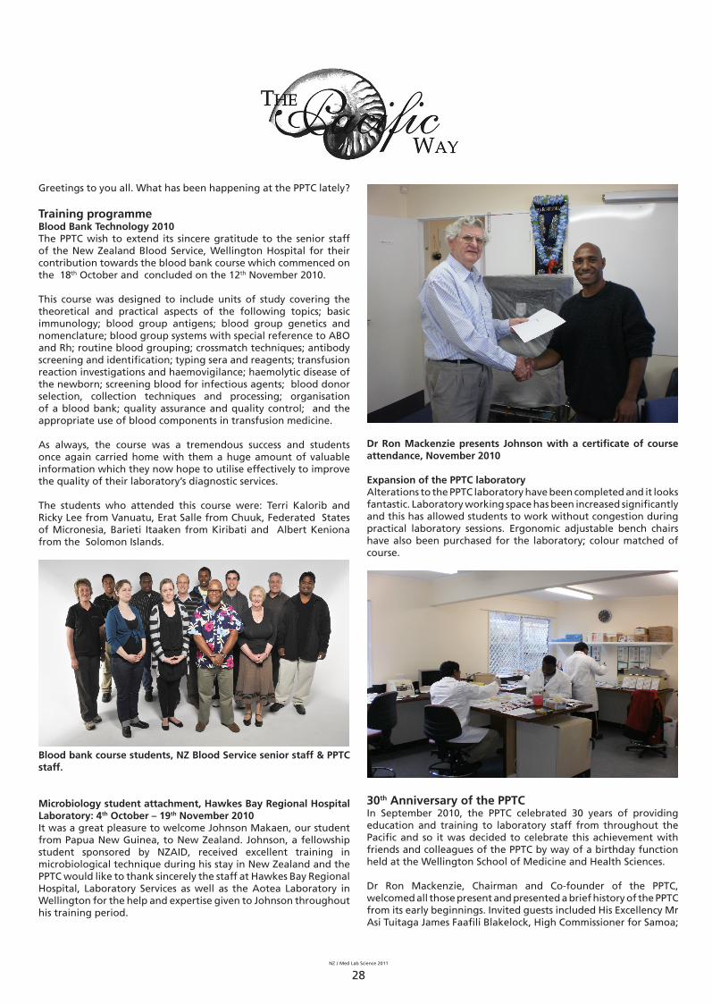
NZ J Med Lab Science 2011
28
Greetings to you all. What has been happening at the PPTC lately?
Training programmeBlood Bank Technology 2010The PPTC wish to extend its sincere gratitude to the senior staff of the New Zealand Blood Service, Wellington Hospital for their contribution towards the blood bank course which commenced on the 18th October and concluded on the 12th November 2010.
This course was designed to include units of study covering the theoretical and practical aspects of the following topics; basic immunology; blood group antigens; blood group genetics and nomenclature; blood group systems with special reference to ABO and Rh; routine blood grouping; crossmatch techniques; antibody screening and identification; typing sera and reagents; transfusion reaction investigations and haemovigilance; haemolytic disease of the newborn; screening blood for infectious agents; blood donor selection, collection techniques and processing; organisation of a blood bank; quality assurance and quality control; and the appropriate use of blood components in transfusion medicine.
As always, the course was a tremendous success and students once again carried home with them a huge amount of valuable information which they now hope to utilise effectively to improve the quality of their laboratory’s diagnostic services.
The students who attended this course were: Terri Kalorib and Ricky Lee from Vanuatu, Erat Salle from Chuuk, Federated States of Micronesia, Barieti Itaaken from Kiribati and Albert Keniona from the Solomon Islands.
Blood bank course students, NZ Blood Service senior staff & PPTC staff.
Microbiology student attachment, Hawkes Bay Regional Hospital Laboratory: 4th October – 19th November 2010It was a great pleasure to welcome Johnson Makaen, our student from Papua New Guinea, to New Zealand. Johnson, a fellowship student sponsored by NZAID, received excellent training in microbiological technique during his stay in New Zealand and the PPTC would like to thank sincerely the staff at Hawkes Bay Regional Hospital, Laboratory Services as well as the Aotea Laboratory in Wellington for the help and expertise given to Johnson throughout his training period.
Dr Ron Mackenzie presents Johnson with a certificate of course attendance, November 2010
Expansion of the PPTC laboratoryAlterations to the PPTC laboratory have been completed and it looks fantastic. Laboratory working space has been increased significantly and this has allowed students to work without congestion during practical laboratory sessions. Ergonomic adjustable bench chairs have also been purchased for the laboratory; colour matched of course.
30th Anniversary of the PPTCIn September 2010, the PPTC celebrated 30 years of providing education and training to laboratory staff from throughout the Pacific and so it was decided to celebrate this achievement with friends and colleagues of the PPTC by way of a birthday function held at the Wellington School of Medicine and Health Sciences.
Dr Ron Mackenzie, Chairman and Co-founder of the PPTC, welcomed all those present and presented a brief history of the PPTC from its early beginnings. Invited guests included His Excellency Mr Asi Tuitaga James Faafili Blakelock, High Commissioner for Samoa;

NZ J Med Lab Science 2011
29
His Excellency Mr Siaosi Taimani Ahio, Deputy High Commissioner for Tonga; Ms Mere Tora, First Secretary/ Acting head of Mission for Fiji; and Mr Jerry Talbot representing Red Cross. The PPTC was also delighted to have representatives from the New Zealand Aid Programme, Norman Kirk Memorial Trust, The University of Otago, the NZIMLS and Wellington Hospital attend the celebrations. An entertainment interlude was provided by the St Patricks College Barber Shop Quartet, which was very well received.
Co-Founders Drs Ron Mackenzie and Sandy Ford assisted by Marilyn Eales cut the anniversary cake
Training programme for 2011Courses at the PPTC1. Haematology and blood film examination: 21st
March – 15th April2. Biochemistry update: 4th July – 22nd July3. Blood transfusion science: 17th October - 11th
November
POLHN distance learning programmePPTC Diploma in Medical Laboratory Technology (2011)The restructured 2011 PPTC Diploma in Medical Laboratory Technology delivered through POLHN is scheduled to commence in the first week of March 2011. The Diploma has been restructured and now includes a “Laboratory Technology” module which has replaced the existing Immunology Module. The components of the latter have been incorporated into the Microbiology and Transfusion Science modules.
Laboratory Diagnosis of STIsA 12 module course has been prepared and teaching will commence through POLHN early in April. Following this theory component, which will detail the clinical and laboratory aspects of all the commonly encountered STIs including HIV, practical ‘wet’ workshops will be held in the laboratories of the Pacific Island countries from which students have registered.
Pacific travelFijiPhil and Christine attended the 2010 Regional ‘LABNET” Workshop held in Suva, Fiji from the 1st – 4th November 2010. Attendees included representatives from twenty Pacific Island countries along with representatives from SPC, PIHOA, NRL, CDC, WHO and Fiji National University School of Medicine, Nursing and Health Sciences. This meeting, at which the PPTC played a prominent role, was very successful and covered key issues including public health surveillance and the role of regional agencies and institutions in the strengthening of Pacific Island Laboratories, in response to the many and varied health challenges in the region.
Niue
Phil travelled to Niue on the 13th November 2010 and is grateful to Inosi Ravuvu for making his stay most welcoming. Apart from meeting health officials, Phil spent a good deal of the time in the laboratory carrying out assessments of the quality system, addressing such issues as EQA and QC and teaching haematology and blood film morphology as time permitted. KiribatiAlso in November, John visited the laboratory in Tarawa, Kiribati to carry out reviews of the laboratory and discuss on-going training requirements for staff. While there he also carried out an audit of the new TB Laboratory as part of the PPTC’s responsibility with the WHO’s StopTB DOTS programme.
In February Phil will represent the PPTC at the nurses and laboratory staff graduation ceremony where a number of laboratory staff will receive the DipMLT which they have completed through POLHN. Others will be awarded Certificates for the various modules that they have completed. Congratulations to these laboratory staff and those in other countries who have successfully completed these programmes.

NZ J Med Lab Science 2011
30
Special Interest Groups______________________________________Mortuary Technician and Forensic Pathology Special Interest Group
BackgroundThis was the second annual meeting of this group and represents a relatively small number of colleagues working in the mortuaries around New Zealand. The NZIMLS offers a QMLT examination in Mortuary Technique and Hygiene with anything from 3 – 6 candidates sitting the paper per year and the NZIMLS would like to support the formation of a SIG for this cohort.
Mortuary facilitiesIn New Zealand, mortuaries are funded by the Ministry of Justice as a contract between the Coronial Service of the Ministry and the DHB owning the facility. A base amount is paid to the DHB with each coronial autopsy being charged to the Ministry of Justice on a fee for service basis depending on the facility level and when it was performed.
There are three levels of mortuary facility covered in these contracts:
1. Level 1 – Body Storage - A location where bodies can be appropriately stored and viewed. There is no requirement for any post-mortem capability.
2. Level 2 – Standard Autopsy Facility – Level 1 plus a location where pathologists are capable of completing routine post-mortems. This level of facility must have implemented standard precautions to protect staff from unknown cases of infectious risk and comply with agreed standards. It must also hold IANZ accreditation against NZS: ISO 151 89.
3. Level 3 – Forensic Autopsy Facility – levels 1 and 2 plus a location that is capable of meeting the current requirements for protecting the “chain of evidence” in police investigations and Coroner inquests and inquiries. This is to include facilities to meet police requirements for homicide investigations which will have viewing facilities, access to specialist medical and police equipment and protection of police evidence. It must also accommodate full time forensic pathologist staff contracted to the National Forensic Pathology Service and make available sufficient technical staff to meet the needs of the forensic service.
The contract also stipulates the type of equipment for each type of facility and the level of competency for the Technical staff employed therein.
Level 1 has no need for a technician; level 2 is a minimum of one technician with QMLT (registration as a RMLT) and one technician considered suitable to work in the mortuary. Level 3 has as a minimum the same as level 2 plus a technical specialist in mortuary. The later isn’t too clearly defined as to what constitutes a “technical specialist” in mortuary and the type of competency or skills required at this level. The mortuary technicians are employed by their respective DHB’s or community laboratories and in most cases part of the laboratory / pathology service.
All mortuaries must meet the NZS/ISO 15189:2003 standard and use as a reference “Managing health and safety risks in New
Zealand mortuaries: guidelines to promote safe working conditions for the operation from a health and safety perspective”. All levels require a family / whanau viewing room, secure storage and refrigeration. Level 2 a separate “infectious” case room and level 3, the same as level 2 and capacity for 40 cases, separate viewing room, access to a conference room for police and facilities and systems and processes to support low copy number DNA testing if required.
National Forensic Pathology ServiceThis service was created in 2005 and is a contractual agreement between the Ministry of Justice and Auckland District Health Board. The purpose of the service, i.e. ADHB, is to employ regional forensic pathologists (RFPs) to provide the coroners and police with a national forensic pathology service.
The type of cases referred to a RFP, as well as routine coronial post-mortems, are all homicides and suspicious deaths; deaths requiring attendance by the police at autopsy; deaths in custody or police action related deaths; cases with ID issues; skeletal remains; accidents involving aviation and marine transport, trains and buses; multiple death incidents; workplace related deaths; diving and scuba related deaths; sudden infant death syndrome and deaths of young children; maternal deaths; infectious deaths such as TB, HIV, CJD etc; and all post-operative or post medical procedure deaths. There are RFPs situated in Auckland, Palmerston North, Wellington and Christchurch and they perform about 1800 pm’s per year in level 3 mortuaries for the cases listed above.
There are also a group of pathologists who perform routine coronial post-mortems for the MOJ and known as 2nd tier coronial pathologists. Their ability to perform coronial post-mortems is assessed by the RFP and recommended to the chief coroner and go onto the list of coronial pathologists.
The fast majority of post mortems in New Zealand (about 3,500 p.a.) are coronial post mortems with a now very small number of non-coronial / hospital post-mortems. The relationships between the stakeholders including the forensic pathologists, coronial pathologists, coroners, coronial service of the MOJ, mortuaries and the technicians, the NZ Police and the families, are determined by the Coroners Act and are quite complex. Managing these relationships as well as providing a service is quite a challenge.
Pathologist / technician relationshipAn effective working relationship is integral to the running of a successful mortuary service in fulfilling the needs of all stakeholders including the Coroners, police, funeral directors and importantly the deceased and the family of the deceased.
The technicians support the pathologists during an autopsy and afterwards in a number of different ways depending on the type of autopsy and the specific procedure the pathologist may follow. They have a huge responsibility in post autopsy reconstruction and preparation prior to release.
The technicians also need to be very familiar with the complexity of paperwork and documentation required by the coronial service and funeral directors. Labelling of samples and tracking these is also fundamental and key in chain of custody and forensic cases.
NZ J Med Lab Science 2011
31
Roles extension / advanced examination / trainingThe NZIMLS offers the QMLT examination for mortuary technicians and the syllabus and logbook development is currently under review. In the UK, there are two levels of examination for mortuary technicians and the NZIMLS has received permission to use both the syllabi and logbooks for something similar in NZ.
The proposed advanced QMLT examination and logbook would include a pre-requisite QMLT (or equivalent) and 3 years experience post QMLT exam and in depth knowledge and demonstrable skills in such areas as:• Anatomy and physiology• Medical photography• Comprehensive application of post-mortem examination
including disaster victim identification (DVI), chain of custody, DNA sampling, forensic toxicology, CT scanning, X-Ray, reconstruction.
• Deeper understanding of hygiene, hazards, infection, etc• Clear understanding of laws, codes and cultural competency
including return of tissue.• Clear understanding of a senior role in management of a
mortuary including IT, documentation, mortuary design, processes, ISO 15189 accreditation, teaching and the importance of all the stakeholders such as funeral directors, NZ Police, Coroners, families.
The newly formed SIG will engage with senior Mortuary Technicians and pathologists to propose such an advanced diploma with suitable syllabus and logbook. It is hoped therefore, this standard of competency will fulfil the “Technical Specialist in Mortuary” requirement of the MOJ contract for level 3 mortuaries for those newly entering this profession. Many of those with long experience in this field already have this level of competency and already work very closely with forensic and coronial pathologists.
There is already a confusion of titles such as mortuary technician, mortician, forensic technician, mortuary manager and forensic scientist. It would be appropriate to have clearly defined nationally accepted position titles, position descriptions and competency documents used along with similarities in training and practices.
It would be appropriate for the SIG to work with their respective employers and the forensic and coronial pathologists to establish such standards and the offering of a national training program with the NZIMLS QMLT and advanced QMLT examinations as recognisable qualifications of the program. The level 3 mortuaries could be used for block courses (1 -2 weeks) for the advanced training of those technicians working in level 2 mortuaries. The training and career development of Mortuary and Forensic technicians (and ML Scientists who may wish to specialise in this field) has been neglected for some time and needs a more structured program, especially in the newly developing areas of forensic science, DVI and necro-radiology (CT scanning).
Mortuary and Forensic Technician Special Interest Group Meeting, Wellington Hospital, 27 November 2010My attendance was twofold, firstly as Service Manager of the National Forensic Pathology Service (NFPS) and LabPLUS and secondly as Secretary / Treasurer of the NZIMLS.
The NFPS is a contracted national service established to manage Forensic Pathology in New Zealand between ADHB and the Ministry of Justice. ADHB employs Forensic Pathologists to carry out post-mortems for homicides, suspicious deaths and routine coronial cases. The relationship with a number of stakeholders including the coroners, coronial service of the MOJ, mortuaries and the technicians, the NZ Police and the families all determined by the Coroners Act is very complex and managing these relationships as well as providing a service quite a challenge.
I also needed to have some understanding of the environment in
which some of my colleagues, technicians and pathologists work and the duties they perform.
The NZIMLS provides an examination for Mortuary Technicians and is interested in supporting the formation of a Special Interest Group.
My presentation was to cover the establishment of the SIG and what it means, and to initiate the revision of the examination syllabus and development of a log book for the Mortuary Technician examination.
I arrived as Coroner Evans –“Grappling With New Issues”, Wellington Regional Coroner was presenting; however, his presentation covered the 2006 Coroners act and the role of the coroner under the new act. I was aware of both of these as a member of the NFPS advisory group and didn’t miss much.
The next speaker was Carmen Stewart – “Death by PowerPoint : The Blue Perspective”, police officer, Wellington Area Inquest Officer, qualified paramedic and embalmer. A unique individual with energy, humour, and a perspective on life that is refreshing and motivational. Carmen spoke of her love of the job, what it is to work with the pathologists and technicians in the Wellington Mortuary, to assist the coroners, and her contribution and sense of privilege to work with the deceased and their families. She also mentioned that as a sworn Police Officer, she can still hand out tickets for speeding, not wearing seat belts, running red lights and everything else police officers can do to up hold the law…. an enlightening and very worthwhile presentation.
After morning tea I followed with my presentation of the formation of the Mortuary and Forensic Technician SIG and NZIMLS update. It was well received and it is quite possible a number of non-mortuary techs and non-NZIMLS members / associates will attend this meeting regularly or even join the NZIMLS as associates.
Dr Martin Sage – “Clandestine Graves & Shaken Not Stirred – What have you got left?” is the Forensic Pathologist based in Christchurch and is under contract to the ADHB and NFPS. Martin’s presentation was on clandestine graves and what remains are left. He presented a number of case studies where the deceased by murder most foul had been buried, disposed of for sometime, discovered, dug up and exhumed for forensic examination. This included cases of bodies buried in the ground and their condition depending on soil type, length of stay, the conditions (wet, dry, temperature etc) and one case of a body being sealed in a barrel. What is left was very interesting biologically and forensically and hopefully sufficient evidence is found at post mortem to discover or determine the means of death and hopefully sufficient evidence for the police to lead to the conviction of the perpetrators. A very insightful and interesting presentation and I learnt a lot.
Bill Little – “The Total Mortuary Service and How We Interact” is the head mortuary technician operating out of the Southland Hospital Mortuary in Invercargill. Bill’s presentation was about the service and the day to day running of the mortuary. This included the practices, inter- relationships with the pathologists and the ability at the end of the week to discuss cases and de-brief. Clearly not all mortuaries and services throughout the country are the same and each has its own flavour. What appealed to Bill were his regular contact with the families of the deceased and the relationship with the funeral directors and other stakeholders within the community. A feeling of worth was especially derived from knowing this was something he could do for the deceased person and with care and attention. The number of PM’s aren’t huge say compared to Auckland or Christchurch and the service is very much community based.
Michelle Dunn – “Lifting the Lid on Grief” is a counsellor and Clinical Supervisor and spoke about grief, the process of grieving

NZ J Med Lab Science 2011
32
and the support mechanisms available for those grieving. Having lost her own child to pre-natal death, she became involved with SANDS (Stillborn & Neonatal Death Support) and trained as a support person for SANDS. This lead to further training and she finished a Bachelor of Applied Social Science and is now working in private practice after working in adult mental health and high school counselling. Michelle’s presentation was about lifting the lid on grief and an insight into the process and theory of grief. It would have particular helpful for those in the audience who may have contact with the family during their work life and day.
Although we do not encourage contact between the mortuary techs and the families because of the nature of their work and for them not to personalise it, there are some who are quite comfortable with it. It was particularly important for the audience to understand they must take care of themselves, as Michelle quoted a Zulu proverb, “You cannot wipe the tears from another person’s face, without getting your hands wet”.
At LabPLUS we have a mechanism in place for our Mortuary Techs to have 3 monthly visits to a clinical psychologist. This is to support them with their work and own well being. Points to consider• Seek support when you need it• Don’t assume others know how you are feeling• Acknowledge your feelings• Identify your needs• De-brief with work colleagues• Balance the hard stuff with humour• Check the work / life balance• Stay connected to your loved ones.
Over-all it was a refreshing and truly insightful presentation.
Marylou McGuinness, Maari Gray and Rachel Kinnaird – “Suturing the Sorrow; the Touch and Technique of Peri-natal Reconstruction” from the Wellington Hospital Mortuary jointly presented on the preparation, pre and post autopsy of the perinatal and stillbirth babies.
This was a very moving and emotional presentation which had the audience enthralled, silent and with more than a few tears. It involved much of the love and care these wee babies get in their very short lives and how they are returned to the families with new baby clothes or ones supplied by the families, and in baskets with a teddy bear and a certificate of life. All of these are supplied by SANDS and volunteers to make, buy, sew and knit the clothes. Just handling these wee babies may damage the skin of these fragile babies and they describe the technical processes and procedures used to ensure the damage is reduced. The suturing and repair following post-mortem is very delicate and requires a huge amount of care and attention.
This presentation was delivered with care and a huge sense of understanding and integrity and brought meaning and value to the work performed. While we often associate ourselves in pathology and laboratories with adding value to life, we as a profession do it also in death by giving meaning to a short life that did happen through care and pure professionalism.
Simon Manning: “A Little Life not a Little Loss” is a funeral director and embalmer from Wellington. His presentation covered the role of the funeral director in the journey of the deceased. The pathway from discovery, transportation, notification, the mortuary, the paperwork, the post mortem, collection, preparation, embalming, family, funeral and to The End. Once again, it was enlightening, sympathetic and intelligent.
I missed Dr John Rutherford’s presentation; John is the Regional Forensic pathologist (employed by ADHB) based in Wellington. John came to NZ from the UK after a falling out with the UK
coronial system and I’ve heard him present before. He was, however, talking about the UK system of technician training and although I was familiar with it, would have liked to have stayed. However I had a plane to catch back to Auckland.
In summary, the Mortuary Technician SIG meeting was extremely successful and worthwhile. It is totally acceptable for this unique and small group of colleagues within the profession to have an annual seminar. There will never be a huge number attending and will often run at a loss, however it is in the best interest of the NZIMLS and the profession as a whole to support this meeting.
Marylou McGuinness from Wellington Hospital Mortuary has agreed to be the Mort SIG convenor and the next SIG meeting will be in Christchurch towards the end of 2011.
Ross HewettNZIMLS Secretary / TreasurerLabPLUS Manager.
Transfusion Science Special Interest Group
TWENTY SECOND ANNUAL NICE WEEKENDDate: 20th – 22nd May 2011
Location: Bayview Wairakei ResortA Transfusion Science Educational Opportunity Organised by the NZIMLS TSSIG
The NICE Weekend (National Immunohaematology Continuing Education) is an educational meeting for all people working in Immunohaematology and/or blood services. As usual it will be held at the Bayview Wairakei Resort Hotel. Registration starts 5pm Friday evening. NICE Weekend finishes approx. 2.00pm Sunday.
As always, all Scientific Delegates are required to participate. They must present either a poster, or an oral presentation lasting 2 to 5 minutes, on any topic related to Immunohaematology or blood transfusion. It can be a case study, a discussion, a question, a problem for others’ to solve, etc. This will be followed by questions and discussion of the topic raised. This compulsory participation makes everyone nervous (yes, even the “old hands”) but it really is one of the reasons why the NICE Weekend is so successful.
There are awards, supplied by trades companies, for the best presentation and poster. We also like to distribute CDs with all the weekends’ power point presentations and posters to delegates who attend. If you would like to contribute an award, or sponsor CDs please contact Raewyn Cameron or Diane Whitehead.
Coming along and showing your support for NICE Weekend is very much appreciated and it’s a great opportunity to meet all sorts of people from all over NZ in the Transfusion Medicine Industry.
Registration FeesThe registration fee is still to be determined and you will be notified of this. Your registration fee entitles you to:• two nights (Friday 30th April and Saturday 1st May) accommodation
on a twin share basis (single room extra)• breakfast, morning and afternoon teas, and lunches on Saturday and
Sunday• dinner & disco on Saturday night. (Dress theme is “RED”’ (generally
everyone dresses up)• Friday night NICE games – a fun night with a few silly (not too
strenuous games). All for fun and a great way to get to know other attendees in a laugh-a-lot, non-professional environment! AND IT’S FREE!
AccommodationAccommodation is on a twin-share basis. You will be sharing a room with another attending delegate (same sex of course!). You may

NZ J Med Lab Science 2011
33
specify who you would like to share with if you wish to catch up with old friends. If you do not specify anyone, the organising team will endeavour to room you with another delegate from a similar sized site or within your own region. The idea is to meet other blood bankers and maybe make a new blood bank friend.
If you are not comfortable with sharing you may choose to pay the extra single room surcharge. (PLEASE NOTE: If this is your choice it will also be your cost. Employers do not usually pay this unless you have come to some arrangement.)
Accommodation on other nights can be arranged by Raewyn or Diane to get the discounted NICE weekend rate at Bayview Wairakei Resort.
User GroupsUser Groups are usually held on the Friday prior to NICE weekend. You may wish to organise your travel and leave days around these. They will all be held on site at Bayview Wairakei. These are organised through the individual companies and not by NICE team. Please
Please send registrations in by 14th April 2011.
Please complete registration form on line at www.nzimls.org.nz
Registration NotificationYou will be notified and sent any further information when your registration has been received. If you don’t hear from us we have not heard from you.
If you have any questions please contact the co conveners:
Raewyn Cameron (Rotorua) Diane Whitehead (Christchurch)07 349 7908 ph 03 3640 314027 418 0592 ph 021 211 5825
[email protected] [email protected]
contact them directly for further information. We, the co-conveners do usually know what’s going on though and may be able to help you. Transport costs will be your own responsibility.Please plan to arrive at the venue on Friday evening, as we have a full programme planned.
Trade representativesCompany representatives do attend our NICE weekend (they have to pay just like you!) as they have a vested interest in keeping up with the world of transfusion. They are not required to present but are around for the weekend – so make yourself known to them. They are a vital part of NICE weekend’s sponsorship (keeping your prices down) and are also a lot of fun!
NICE first timersIf this is your first NICE Weekend, we will introduce you to everyone, explain anything you don’t understand and make you feel at home. We will try to room you with someone who has been before to help you along.

NZ J Med Lab Science 2011
34
“Biochemistry in the City”NZIMLS presents
Biochemistry Special Interest Group MeetingSaturday June 18th, 2011
Waipuna Hotel and Conference Centre,58 Waipuna Road, Mt Wellington, Auckland
Guest speaker: Geoffrey Kellerman, AACB Roman Lecturer
Papers wanted: Special Chemistry, Toxicology, POCT, Automation, Chromatography, Endocrinology, Quality,
Immunology, Proteins, Lipids, Nucleic Acid, etc
Contact Sandy Woods: NZIMLS BSIG coordinator [email protected]
More details on www.nzimls.org.nz
What is this molecule? Come to the BSIG meeting and enter the draw with your answer to win a mystery prize.

NZ J Med Lab Science 2011
35
Registration and Coffee 9 am Scientific Programme begins 10 am sharp
Lunch 12 - 1 pm Programme ends approx 5pm
Dinner Oceania Room 7pm
On line registration will be available shortly on www.nzimls.org.nz
We are looking for presenters from all walks of life and
disciplines of Medical Laboratory Science.
Don’t be scared! Contact Nicky Beamish via e-mail
Prizes for best presentation and best first time presenter
North Island Seminar Saturday 28th May 2011 Te Papa Oceania Room Cable Street Wellington

NZ J Med Lab Science 2011
36
CALL FOR ABSTRACTS
AIMS NZIMLS South Pacific Congress 8-12 August 2011
Gold Coast Convention Centre, Queensland, Australia
Deadline for submission of abstracts 1 April 2011
Notification to authors, including accepting or rejecting abstracts
29 April 2011
Program released 27 May 2011
Early bird registration date closes 10 June 2011
The Australian Institute of Medical Scientists and the New Zealand Institute of Medical Laboratory Science is proud to host and invite you to the South Pacific Congress, 8-12 August 2011. The Congress will bring to the Gold Coast Convention Centre a top level forum of leading national and international speakers to address topical issues in medical science. SUBMISSION CATEGORIES The Congress theme ‘Lights! Camera! Action!’ has been chosen as a call to action for delegates to spend a focussed 4 ½ days in the vibrant Gold Coast at a Congress filled with topical and relevant presentations. Daily sub-themes will logically group presentations and have a little fun based on the Congress theme!
‘Waterworld’ – Water trauma/diseases ‘Basic Instinct’ – Back to basics ‘Aliens’ – The immune system ‘Back to the future’ – New technology ‘That’s all folks!’ – Closing day
SUBMITTING ABSTRACT This information is to be submitted electronically through the Congress website: www.alloccasionsgroup.com/AIMSNZIMLS11. All submissions are to include the following:
A 100 word biography 250 word abstract providing the essence of the presentation and how it links with the Congress theme(s) Full contact details (name, organisation, position, phone, fax, address and email).
Please refer to the Congress website for information regarding formatting requirements and submission guidelines.
SELECTION CRITERIA
Originality and interest Structure and clarity Scientific validity Potential significance
POSTER PRESENTATION You are welcome to send an abstract that will describe your poster submission. Please refer to the Congress website for information regarding formatting requirements and submission guidelines. Posters will be displayed for the duration of the Congress and the presenting author should be by their poster as often as possible, ideally during the breaks in the Congress. KEY TIME FRAMES For further information: Shanna Sheldrick, Manager - Conventions & Events Tracy Fisher, Coordinator - Conventions & Events All Occasions Management Telephone: +61 8 8125 2200 Fax: +61 8 8125 2233 Email: [email protected] Web: www.alloccasionsgroup.com/AIMSNZIMLS11

NZ J Med Lab Science 2011
37

NZ J Med Lab Science 2011
38
Laboratory Microscopy from Carl Zeiss
Carl Zeiss NZ Ltd
Ph: 0800 334 [email protected]
Axio Lab. A1Ergonomic. Easy to Use. Durable.
Primo Vert & Primo Vert Monitor Affordable. Easy-to-Use. Ergonomic.
•TUV-approvedergonomicconfiguration available(www.tuv.com)•Transmitted-LightoptionswithHalogen,LED daylightorLEDwarmlight•IntegratedLEDFluorescence,virtuallyno runningcost,UVandMercuryfree•DurabilitythroughtheuseofHighQuality Materials•SkinFriendlymaterialstoenhancecomfort•ExcellentZEISSopticalqualityforreliable results
•Excellentprice-performanceratio•Easy-to-Useconcept:autoon/offfunction, universalphasecontrast•Long-termmechanicalandopticalstability•Attractiveindustrialdesignusingskinfriendly materials•Modularilluminationwithhalogenlightor longerlastingLEDs.•UltimateConvenience:PrimoVertMonitor withIntegrateddigitalcamera,SDmemory cardslot,USBport
“High reliability and comfort for continuous microscopy work”
“The fast and convenient solution for assessing and checking living cells”
Axio Lab_Primo Vert Ad_267x180_NZIMLS Journal_Oct10.indd 1 12/10/2010 3:45:49 PM