e2f6: a unique regulator of post natal cardiac growth
TRANSCRIPT

E2F6: A Unique Regulator of
Post-natal Cardiac Growth, Death
and Function
Jennifer Lynn Major
Thesis submitted to the Faculty of Graduate and Postdoctoral Studies
in partial fulfillment of the requirements for the
PhD degree in Cellular and Molecular Medicine.
Department of Cellular and Molecular Medicine
Faculty of Medicine
University of Ottawa
© Jennifer Major, Ottawa, Canada, 2017

ii
FOR HUNTER.

iii
AUTHORIZATIONS
Title: The E2F Pathway in Cardiac Development
and Disease
Author: Jennifer Rueger
Publication: Springer eBook
Publisher: Springer
Date: Jan 1, 2011
Copyright © 2011, Springer Science+Business Media, LLC
Logged in as:
Jennifer Major
Order Completed
Thank you for your order.
This Agreement between Jennifer L Major ("You") and Springer ("Springer") consists of your license details and the terms and conditions provided by Springer and Copyright Clearance Center.
Your confirmation email will contain your order number for future reference.
Printable details.
License Number 4022540005708
License date Jan 05, 2017
Licensed Content Publisher Springer
Licensed Content Publication Springer eBook
Licensed Content Title The E2F Pathway in Cardiac Development and Disease
Licensed Content Author Jennifer Rueger
Licensed Content Date Jan 1, 2011
Type of Use Thesis/Dissertation
Portion Figures/tables/illustrations
Number of figures/tables/illustrations
2
Author of this Springer article Yes and you are a contributor of the new work
Order reference number
Original figure numbers Figures 1,2
Title of your thesis / dissertation E2F6: A Regulator of Cardiac Growth, Differentiation, and Death
Expected completion date Oct 2016
Estimated size(pages) 200

iv
Requestor Location Jennifer L Major Attn: Jennifer L Major
Billing Type Invoice
Billing address Jennifer L Major Canada Attn: Jennifer L Major
Total 0.00 CAD
ORDER MORE CLOSE WINDOW
Copyright © 2017 Copyright Clearance Center, Inc. All Rights Reserved. Privacy statement. Terms and Conditions. Comments? We would like to hear from you. E-mail us at [email protected]

v
Title: Interplay between the E2F pathway and β-adrenergic signaling in the pathological hypertrophic response
of myocardium
Author: Jennifer L. Major,Maysoon Salih,Balwant S. Tuana
Publication: Journal of Molecular and Cellular Cardiology
Publisher: Elsevier
Date: July 2015
Copyright © 2015 Elsevier Ltd. All rights reserved.
Logged in as:
Jennifer Major
Order Completed
Thank you for your order. This Agreement between Jennifer L Major ("You") and Elsevier ("Elsevier") consists of your license details and the terms and conditions provided by Elsevier and Copyright Clearance Center.
Your confirmation email will contain your order number for future reference.
Printable details.
License Number 4022531001921
License date Jan 05, 2017
Licensed Content Publisher
Elsevier
Licensed Content Publication
Journal of Molecular and Cellular Cardiology
Licensed Content Title Interplay between the E2F pathway and β-adrenergic signaling in the pathological hypertrophic response of myocardium
Licensed Content Author
Jennifer L. Major,Maysoon Salih,Balwant S. Tuana
Licensed Content Date July 2015
Licensed Content Volume
84
Licensed Content Issue n/a
Licensed Content Pages 12
Type of Use reuse in a thesis/dissertation
Portion full article
Format electronic
Are you the author of this Elsevier article?
Yes
Will you be translating? No

vi
Order reference number
Title of your thesis/dissertation
E2F6: A Regulator of Cardiac Growth, Differentiation, and Death
Expected completion date
Oct 2016
Estimated size (number of pages)
200
Elsevier VAT number GB 494 6272 12
Requestor Location Jennifer L Major Attn: Jennifer L Major
Total 0.00 CAD
ORDER MORE CLOSE WINDOW
Copyright © 2017 Copyright Clearance Center, Inc. All Rights Reserved. Privacy statement. Terms and Conditions. Comments? We would like to hear from you. E-mail us at [email protected]

vii
Journals
About Us | Careers | Contact Us | Help | 中文 | 日本語
View Basket | Sign In / Register
Search articles..
Search
Advanced Search
Journals A-Z
Arts & Humanities
Law
Medicine & Health
Science & Mathematics
Social Sciences
Academic Books & Online Resources
You are here:Home»Access & Purchase»Rights & Permissions»Author Self-Archiving Policy
o Author Self-Archiving Policy
Return to Rights & Permissions
Author Self-Archiving Policy
This policy sets out the ways in which Oxford University Press journal authors may self-archive
versions of their work on their own webpages, on institutional webpages, and in other repositories. Please be aware that policies and embargo periods may differ from journal to journal, so ensure that you have selected the correct journal policy.
Abstract and Citation information
Authors may reuse the Abstract and Citation information (e.g. Title, Author name, Publication dates) of their article anywhere at any time including social media such as Facebook, blogs and Twitter, providing that where possible a link is included back to the article on the OUP site. Preferably the link should be, or include, the Digital Object Identifier (DOI) which can be found in the Citation information about your article online.
Author’s Original Version
The Author’s Original Version (AOV) is defined here as the un-refereed author version of an article completed before submission of the article to the journal. This is sometimes referred to as the
“preprint” version. The author accepts full responsibility for the article, and the content and layout is set out by the author.

viii
Authors may reuse the AOV anywhere at any time, providing that once the article is accepted they provide a statement of acknowledgement, and that once the article has been published this acknowledgement is updated to provide details such as the volume and issue number, the DOI, and a link to the published article on the journal’s website:
This article has been accepted for publication in [Journal Title] Published by Oxford University Press.
Accepted Manuscript
The accepted manuscript (AM) is the final draft author manuscript, as accepted for publication by a journal, including modifications based on referees’ suggestions, before it has undergone copyediting, typesetting and proof correction. This is sometimes referred to as the post-print version.
Immediately upon publication authors may:
Immediately upload their AM to their own personal webpage (excluding commercial websites and repositories)
Immediately upload their AM to their institutional repository on the proviso that it is not made
publically available until after the specified embargo period
After the embargo period authors may:
Upload their AM to institutional repository or other non-commercial repositories and make it publicly
available. Accepted Manuscripts may not be uploaded to commercial websites or repositories, unless the website or repository has signed a licensing agreement with OUP allowing posting. For Profit social networking sites such as Researchgate and Academia.edu are considered “commercial” platforms.
Embargo periods
Embargo periods may vary between journals. For details of a journal’s specific embargo period, please see the information for each individual title on our Accepted Manuscript embargo page.
When uploading an accepted manuscript to a repository, authors should include the following acknowledgment as well as a link to the published version in the journal. This will connect the published version to the AM version in the repository and help ensure that the article is cited correctly.
This is a pre-copyedited, author-produced version of an article accepted for publication in [insert journal title] following peer review. The version of record [insert complete citation information here] is available online at: xxxxxxx [insert URL and DOI of the article on the OUP website].
Version of Record
The Version of Record (VOR) is defined here as the final typeset and edited version of the journal article that has been made available by OUP by formally and exclusively declaring the article “published”. This includes any ‘advanced access’ article even before the compilation of a volume issue.
The VOR as it appears in the journal following copyediting and proof correction may not be deposited by authors in institutional repositories or posted to third party websites and made publicly available unless the article is published on an Open Access model licence that allows for such posting. Authors may share their VOR with private groups within their institution or through private groups on non-commercial platforms that are signatories to the STM Voluntary principles for article sharing on Scholarly Collaboration Networks (SCN). The VOR may not be posted to Commercial Platforms, either publically or privately unless they have signed a direct agreement with OUP.

ix
This work is licensed under the Creative Commons Attribution 4.0 International License.
To view a copy of this license, visit http://creativecommons.org/licenses/by/4.0/ or send a
letter to Creative Commons, PO Box 1866, Mountain View, CA 94042, USA.

x
ABSTRACT
Rationale/Background: The adult mammalian heart has a very limited potential for
regeneration due to cardiomyocyte cell cycle withdrawal which occurs shortly after birth.
One potential avenue to repair the heart following stress/injury is to reprogram pre-
existing cardiomyocytes to re-enter the cell cycle. The E2F family is a group of
transcription factors which ubiquitously regulate the cell cycle, but it has previously been
difficult to fully appreciate their role in the post-natal myocardium due to either
redundancy or embryonic lethality of genetic models. Thus we generated a dominant
negative model of the E2F/Rb pathway via expression of the unique transcriptional
repressor E2F6 in postnatal myocardium. E2F6 transgenic (Tg) mice developed dose
dependent Dilated Cardiomyopathy (DCM) and sudden death without hypertrophy or
apoptosis. This was accompanied by the partial loss of E2F3 (critical for cardiac
development) and connexin-43 important for metabolic and electrical coupling.
Methods/Results: In this thesis E2F6-Tg mice were examined for markers of cardiac
differentiation/ function and exposed to stressors to evaluate the capacity for the E2F
pathway to regulate cardiomyocyte growth (isoproterenol) and death (doxorubicin and
cobalt chloride). E2F6-Tg mice were twice as sensitive to isoproterenol as their Wt
counterparts due to the observed activation of a β2-adrenergic survival pathway. Cardiac
hypertrophy in E2F6-Tg mice was accompanied by the rescue of E2F3 expression.
Treatment of neonatal cardiomyocytes isolated from Wt and E2F6-Tg pups with cobalt
chloride revealed a protective effect for E2F6 against apoptosis. Doxorubicin exposure

xi
led to the loss of E2F6 protein and abolished its protective effect. Examination of
neonatal hearts and cardiomyocytes isolated from them demonstrated a shift in the cell
cycle and metabolic profiles of E2F6-Tg myocardium. Tg cardiomyocytes show
decreased glycolysis and a dramatic increase in the regulator of ketolysis, β-
hydroxybutyrate dehydrogenase (BDH1), prior to DCM. The substrate of BDH1 (β-
hydroxybutyrate) was demonstrated to influence the levels of CX-43 in cardiomyocytes.
E2F6 also deregulated expression of T-cap which has been linked to human DCM.
Conclusions: I provide evidence that the E2F pathway can regulate growth, death, and
differentiation through a variety of mechanisms which link the cell cycle and metabolism
to growth and survival to critically govern post-natal cardiac function. Furthermore, I
reveal a new biomarker (BDH1) for early DCM which may be useful in diagnosis/
treatment of idiopathic cases of disease.

xii
TABLE OF CONTENTS
DEDICATION…………………………………………………………………… ii
AUTHORIZATIONS…………………………………………………………… iii
ABSTRACT………………………………………………………………………. x
TABLE OF CONTENTS………………………………………………………… xii
LIST OF TABLES………………………………………………………………... xvii
LIST OF FIGURES……………………………………………………………… xviii
LIST OF ABBREVIATIONS…………………………………………………… xxi
ACKNOWLEDGEMENTS……………………………………………………… xxiii
GENERAL INTRODUCTION………………………………………………… 1
The Cell Cycle…………………………………………………………………….. 3
The E2F Family………………………………………………………………… 5
E2F structure…………………………………………………………….. 5
E2F6: a non-classical E2F family member………………………………… 8
E2F Family Member Functions………………………………………………… 10
E2Fs in the heart…………………………………………………………… 12
E2Fs and Heart Failure………………………………………………………….. 13
Cardiac hypertrophy……………………………………………………..... 13
Cardiac metabolism………………………………………………………... 16
Apoptosis………………………………………………………………….. 18
E2F6 and Dilated Cardiomyopathy……………………………………………... 20
Dilated cardiomyopathy…………………………………………………… 21

xiii
Statement of the problem………………………………………………….. 22
Objectives………………………………………………………………… 23
CHAPTER 1……………………………………………………………………… 25
“Interplay between the E2F pathway and β-adrenergic signaling in the
pathological hypertrophic response of myocardium” Major JL, Salih M, and Tuana
B.S. (2015) Journal of Molecular and Cellular Cardiology, 84: 179-190
Abstract…………………………………………………………………………… 26
Introduction………………………………………………………………………. 28
Materials and Methods…………………………………………………………... 30
Mice and genotyping……………………………………………………… 30
Isoproterenol delivery……………………………………………………... 30
Echocardiography………………………………………………………..... 31
RNA isolation and reverse transcription-quantitative PCR……………….. 31
Western blot analysis……………………………………………………… 32
Immunohistochemistry……………………………………………............. 33
Statistics…………………………………………………………………… 33
Results…………………………………………………………………………….. 31
DCM without compensatory hypertrophy in E2F6 myocardium………… 33
E2F6 expression sensitizes the hypertrophic response to isoproterenol…... 36
E2F6 regulates the expression of β-adrenergic receptors…………………. 40
E2F6 up-regulates PKA and PDE4D expression in myocardium………… 42
E2F6 activates c-Src/ERK and Bcl2 in E2F6-Tg myocardium…………… 44
E2F6 down-regulates cardiac growth regulator AKT1……………………. 46

xiv
E2F6 differentially affects β2-AR, PKA-C, and Akt1 mRNA…………….. 48
E2F6 deregulates E2F3/Rb expression in myocardium…………………… 49
Discussion…………………………………………………………………………. 51
CHAPTER 2............................................................................................................. 58
“E2F6 Protein Levels Modulate the Apoptotic Response in Cardiomyocytes”
Major JL, Salih M, and Tuana BS. (2017) Cardiovascular Research, Submitted.
CVR-2017-59.
Abstract…………………………………………………………………………… 59
Introduction………………………………………………………………………. 61
Materials and Methods…………………………………………………………... 63
Results…………………………………………………………………………….. 63
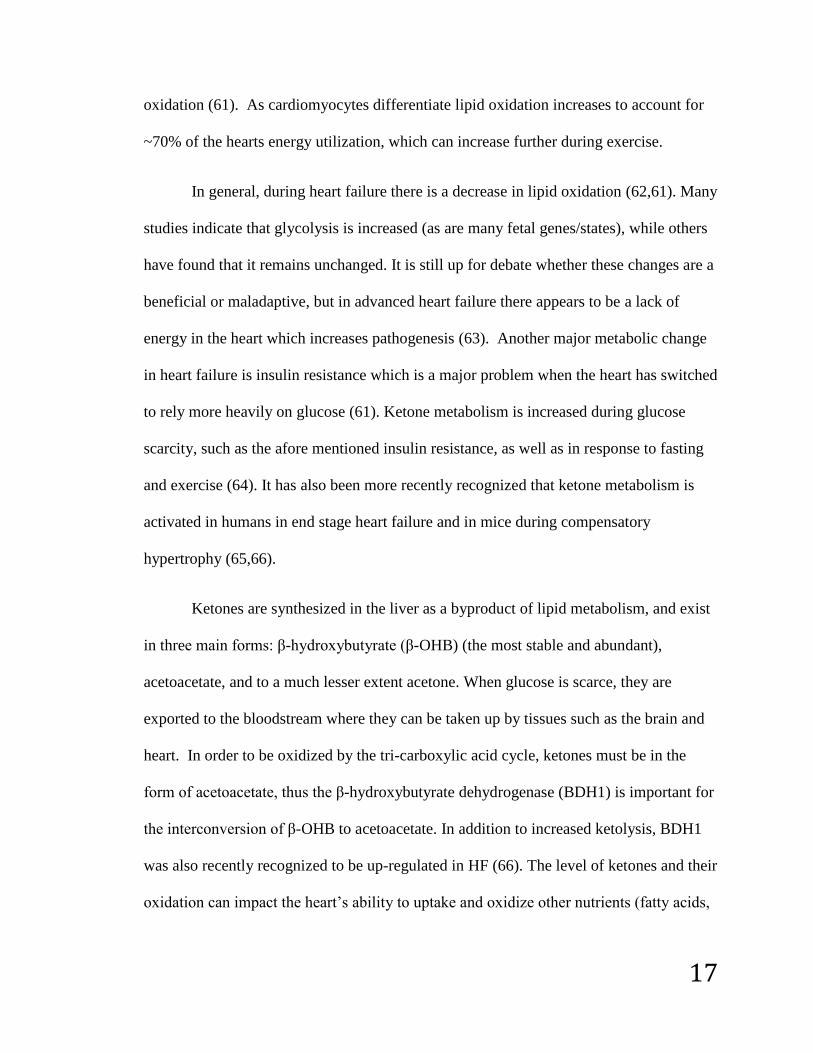
Genes involved in DNA replication and repair are upregulated in E2F6-Tg
myocardium……………………………………………………………….. 63
E2F6 protects neonatal cardiomyocytes from cobalt chloride induced
apoptosis…………………………………………………………………... 64
Doxorubicin induces apoptosis in Wt and E2F6-Tg neonatal
cardiomyocytes……………………………………………………………. 68
Doxorubicin promotes E2F6 loss in neonatal cardiomyocytes and HeLa… 70
Dox promotes E2F6 down-regulation via post-transcriptional mechanisms. 73
E2F6 impacts differentiation to induce dilated cardiomyopathy………….. 75
Discussion…………………………………………………………………………. 78
Mechanism of E2F deregulation during apoptosis………………………... 79
E2F6 protects cardiomyocytes from apoptosis via multiple mechanisms 79

xv
during cardiac development………………………………………………...
E2F6 induces DCM via deregulation of cell cycle not apoptosis…………. 80
CHAPTER 3……………………………………………………………………… 82
“E2F6 Impairs Glycolysis and Activates BDH1 Prior to Dilated
Cardiomyopathy” Major JL, Dewan A, Salih M, Leddy JJ, Tuana BS.
(2017)PLOS One. 12(1): p. e0170066.
Abstract…………………………………………………………………………… 83
Introduction………………………………………………………………………. 84
Materials and methods…………………………………………………………... 86
Mice and genotyping………………………………………………………. 86
RNA Analyses…………………………………………………………….. 87
Western Blot………………………………………...................................... 87
Neonatal Cardiomyocyte Isolation and Treatment………………………... 89
Seahorse Glycolysis Measurement……………………………………….. 89
Seahorse Fatty Acid Oxidation Measurement…………………………….. 90
Statistical Analyses………………………………………………………... 90
Results…………………………………………………………………………….. 90
E2F6 Induces the Early Expression of BDH1 in Neonatal Myocardium…. 90
E2F6 Impairs Glycolysis in Neonatal Cardiomyocytes…………………… 93
E2F6 Does Not Impact Fatty Acid Oxidation in Neonatal Cardiomyocytes 95
E2F6 Deregulates Cyclin Expression in Myocardium ……………………. 97
Ketones Regulate Connexin-43 in Neonatal Cardiomyocytes…………….. 98
Discussion…………………………………………………………………………. 101

xvi
GENERAL DISCUSSION………………………………………………………. 106
E2F6: a potential therapeutic tool against heart failure……………………… 108
Mechanisms for E2F6 induced dilated cardiomyopathy………………………. 112
Perspectives………………………………………………………………………. 116
RERFERENCES…………………………………………………………………. 117
APPENDICES……………………………………………………………………. 133
Appendix 1: Supplemental Information for Chapter 1………………………... 134
Appendix 2: Supplemental Information for Chapter 2………………………... 135
Appendix 3: Supplemental Information for Chapter 3……………………….. 139
Appendix 4: Supplemental Information for Discussion……………………….. 142

xvii
LIST OF TABLES
Table 1 Individual E2F/ Pocket protein family member knockouts....………………… 11
Table 2 Double and Triple E2F/Pocket Protein Knockout Studies ……………………..13

xviii
LIST OF FIGURES
Figure 1: E2F/DP family Structure……………………………………………….. 6
Figure 2: The E2F/RB pathway is regulated throughout the cell cycle………….. 8
Figure 3: E2F6-Tg mice develop early dilated cardiomyopathy without
significant compensatory hypertrophic growth……………………………………. 35
Figure 4: E2F6-Tg mice are twice as sensitive to isoproterenol………………….. 37
Figure 5: High dose isoproterenol induces hypertrophy in Wt and Tg
mice………………………………………………………………………………… 39
Figure 6: β-Adrenergic pathway affected by E2F6 expression…………………… 41
Figure 7: Phospholamban is only targeted by PKA in the presence of
isoproterenol……………………………………………………………………….. 44
Figure 8: Activation of c-Src/ERK/Bcl2 Survival Pathway in E2F6-Tg
myocardium………………………………………………………………………… 45
Figure 9: AKT1 activity is deregulated in E2F6-Tg myocardium……………….. 47
Figure 10: Effect of E2F6 on β2–AR, PKA-C, and AKT1 mRNA………………... 48
Figure 11: Deregulation of E2F3/Rb in Tg Mice under hypertrophic stimulation... 50
Figure 12: E2F6 induces the expression of genes involved in DNA replication
and repair in Tg myocardium………………………………………………………. 64
Figure 13: E2F6 protects neonatal cardiomyocytes from cobalt chloride induced
apoptosis…………………………………………………………………………… 66
Figure 14: Doxorubicin induces apoptosis in Wt and Tg neonatal cardiomyocytes 69
Figure 15: E2F6 protein is lost in response to apoptotic agents…………………... 71

xix
Figure 16: E2F6 and E2F1 protein levels are deregulated by apoptotic agents in
HeLa cells…………………………………………………………………………... 72
Figure 17: Mechanisms of E2F regulation in HeLa and cardiomyocytes…………. 74
Figure 18: E2F6-Tg hearts show alteration in cell cyclins and cardiac proteins….. 77
Figure 19: E2F6 Activates BDH1 Expression in Neonatal Myocardium…………. 91
Figure 20: E2F6 Impairs Glycolysis in Neonatal Cardiomyocytes……………… 94
Figure 21: E2F6 Does Not Impact Fatty Acid Oxidation in Neonatal
Cardiomyocytes…………………………………………………………………….. 96
Figure 22: E2F6 Deregulates Cyclin B1 Expression in Myocardium…………….. 97
Figure 23: β-OHB Regulates CX-43 Protein Expression in Neonatal
Cardiomyocytes……………………………………………………………………. 99
Figure 24: Summary Model……………………………………………………….. 107
Supplemental Figure 1: Fetal gene program is not activated in 2 Week old Tg
Hearts……………………………………………………………………………….. 134
Supplemental Figure 2: Phosphorylation status of troponin I (TnI) is not
changed in Tg myocardium………………………………………………………… 134
Supplemental Figure 3: Src not up-regulated in E2F6-Tg pup hearts……………. 138
Supplemental Figure 4: GLUT4 Transcription is Not Altered in E2F6-Tg
myocardium………………………………………………………………………… 139
Supplemental Figure 5: Seahorse Method of OCR is Specific to Fatty Acids…… 139
Supplemental Figure 6: CX-43 Expression is increased in Tg Cardiomyocytes
with Time in Culture………………………………………………………………. 140
Supplemental Figure 7: Promoter Analysis of Bdh1…………………………….. 141

xx
Supplemental Figure 8: E2F1 is decreased after dox (0.5μM 24hr) exposure in
neonatal cardiomyocytes…………………………………………………………… 142
Supplemental Figure 9: E2F6 is down-regulated post-transcriptionally in the
myocardium after birth……………………………………………………………... 143
Supplemental Figure 10: E2F6 inhibits myoblast fusion………………………… 144
Supplemental Figure 11: E2F6 is down-regulated post-transcriptionally during
C2C12 differentiation Day 0-Day 4………………………………………………... 145

xxi
LIST OF ABBREVIATIONS
2-DG 2-deoxy glucose
α-MHC Alpha myosin heavy chain
AKT Protein Kinase B
β-OHB Hydroxybutyrate
Anti-A Antimycin-A
Bcl2 B-cell lymphoma 2
Blm2 Bloom syndrome 2 homologue
BW Body weight
BDH1 Β-hydroxybutyrate dehydrogenase 1
β1AR Β1-Adrenergic receptor
β2-AR Β2-Adrenergic receptor
Blm2 Bloom syndrome 2 homologue
BSA Bovine serum albumin
cAMP Cyclic AMP
CDK Cyclin dependent kinase
CDKI Cyclin dependent kinase inhibitor
CHF Congestive heart failure
Chk1 Checkpoint Kinase 1
CREB cAMP response element-binding protein
CX-43 Connexin-43 (gap junction α1)
CVD Cardiovascular disease
DCM Dilated cardiomyopathy
Dnaja3 Chaperone protein hsp40
Dox doxorubicin
ECAR Extracellular acidification rate
Echo Echocardiography
EF Ejection fraction
ERK (1/2) Extracellular Receptor kinase (p44/p42)
FBS Fetal bovine serum
FCCP Carbonyl cyanide-4-phenylhydrazone
FS Fractional shortening
GAPDH Glyceraldehyde 3-phosphate dehydrogenase
GLUT4 Glucose transporter 4
HAT Histone acetyl-transferase
HCM Hypertrophic cardiomyopathy
HDAC Histone deacetylase
HF Heart failure
Iso isoproterenol
IVSs Interventricular septum thickness (systole)
IVSd Interventricular septum thickness (diastole)
NCM Neonatal cardiomyocyte
MAPK Mitogen activated protein kinase
MI Myocardial infarction

xxii
Myc-E2F6 E2F6 derived from transgene tagged with myc
OCR Oxygen consumption rate
OXCT1 succinyl-CoA: 3-oxoacid-CoA transferase
Pal palmitate
PDE4B Protein phosphodiesterase 4B
PDK4 Pyruvate dehydrogenase kinase 4
PKA Protein Kinase A
Pln phospholamban
LV Left ventricle
LVW Left ventricle weight
LVEDs Left ventricle end diameter (systole)
LVEDd Left ventricle end diameter (diastole)
Rad51 Recombinant protein A
Rb Retinoblastoma gene
RT-q-PCR Reverse transcription quantitative polymerase chain reaction
SEM Standard error of the mean
Ser serine
Src/ c-src Proto-oncogene tyrosine-protein kinase
T-cap Teliothenin (titin cap)
TnI Troponin I
TnT Troponin T
Tg Transgenic
Wt Wild type

xxiii
ACKNOWLEDGEMENTS
I would like to extend my sincere gratitude to the many people in my life who have helped me.
I would like to thank my supervisor: Dr. Balwant Tuana for giving me the opportunity to work on
such a unique project in his lab, for his guidance as well and freedom to design and test my own
ideas. I would also like to than Maysoon Salih- without her help and advice I do not think I could
have finished this thesis. I would also like to thank all current and past members of the Tuana lab
especially to Aaraf for all his help with the BDH1 project and to both him and Taha for great
discussions and being my IT support. Thank-you to Mayra and Jana, I learned so much from both
of you.
Thank-you to my best friends: Megan, Andrea, and Khierstyn, my sister Cyndi and my mother-
in-law Ruth for always having my back whatever the problem.
Thank-you to my parents, Dan and Jan Rueger, for always pushing me to do my best and reach
my potential. I’m so grateful to have such loving and supportive parents.
Thank-you to my wonderful husband Shane Major for being my partner and my support through
school and life.
Thank-you to my daughter Roxy for being the sunshine in my life.
Thank-you Hunter for always believing in me, you taught me so much-
This is for you kiddo!

1
GENERAL INTRODUCTION
Cardiovascular diseases are the leading cause of non-communicable disease
deaths, killing 17.5 million people world-wide in 2012 (1). In Canada, research and
medical intervention has extended life for patients with cardiovascular disease (CVD)
such that the survival rate has increased by 75% in the past six decades (2). Although
such an improvement is astounding, it also means that there is a high rate of Canadians
living with CVD and that produces an estimated economic burden of $20.9 billion
annually. Furthermore, the quality of life for those patients with CVD and heart failure is
lessened.
The heart is a highly specialized organ whose function is to pump blood which
will be delivered to the whole body providing both oxygen and nutrients. The mammalian
heart is a four chambered organ consisting of numerous cell types. The contractile cells
which make up the bulk of the weight are cardiomyocytes. The basic contractile unit of
the cardiomyocyte is called the sarcomere, which is organized into thick (actin) and thin
(myosin) filaments giving cardiac muscle its striated appearance. In order for
cardiomyocytes to communicate with one another and exert force they have a highly
developed cell membrane called the sarcolemma which has deep invaginations called t-
tubules which are rich in proteins and ion channels for excitation-contraction coupling.
Cardiomyocytes also have gap junctions which form channels across cells allowing for
communication between cardiomyocytes. The main gap junction protein expressed in the
ventricles (the cardiac chambers which pump blood to the body) is connexin43. Such a

2
specialized force comes at a cost: metabolism, which is why ~40% of cardiomyocyte
volume is occupied by mitochondria.
In order for appropriate differentiation and post-natal cardiac development to
proceed, cardiomyocytes withdraw from the cell cycle shortly after birth. There is
evidence indicating a limited amount of cardiomyocyte division in adult myocardium, but
it is generally believed that growth in the adult heart is restricted to hypertrophy (3,4).
This leaves the heart very sensitive to injury, since there is a limited potential to divide
and replenish damaged tissue. Thus following injury, such as myocardial infarction, acute
toxin/viral exposure, or long term exposure to stresses caused by chronic hypertension,
chemotherapy, etc. the heart undergoes cardiac remodeling in an attempt to rescue
function. This usually involves the induction of the pathological hypertrophic program.
While initially this may be beneficial, it ultimately causes disorganization of the
contractile apparatus resulting in inadequate pump function, chamber dilation, and cell
death leading to heart failure (HF).
A tremendous amount of research has gone into the use of stem cells to replenish
the heart mainly including the use of non-embryonic stem cells derived from bone
marrow and induced pluripotent stem cells (5). While studies have demonstrated the
therapeutic effects of these techniques they are limited by the ability of the cells to
remain in the heart as well as the many barriers in place to collecting and using stem cells
in humans. Another avenue to repair the heart is a cell cycle based approach to stimulate
pre-existing cardiomyocytes to re-enter the cell cycle to divide. Recent studies have
demonstrated that, for a brief window of time, neonatal mice have the potential to
regenerate lost/damaged cardiac tissue (6). Cell lineage tracing revealed that the newly

3
regenerated cardiac tissue consisted of cells that came from pre-existing cardiomyocytes
and not stem cells (7). Finally, the promotion of physiological over pathological
hypertrophy and the inhibition of apoptosis is an attractive way to save the failing heart.
In order to achieve this thorough understanding of the cardiac cell cycle, in particular the
switch from a proliferative to differentiated state that happens in the perinatal period is
essential.
The Cell Cycle
The cell cycle is a tightly regulated process which exists in five phases and has
numerous checkpoints to inhibit abnormal cell proliferation. It begins in Gap 1(G1)
during which cells grow and prepare for cell division. Cells which remain in G1 for
extended periods of time (such as cardiomyocytes) can exit the cell cycle and enter G0,
after which they may re-enter the cell cycle or differentiate. When the appropriate growth
cues are received cells pass through a restriction point in G1, after which they are
committed to the cell cycle. The next stage is the synthesis (S) phase in which DNA is
duplicated, followed by a second preparatory phase Gap 2 (G2). Next chromatin is
condensed and assembled at the mitotic plate (mitosis) and DNA and cellular components
are separated into two daughter cells in a process called cytokinesis. In the post-natal
heart cardiomyocytes can pass through the cell cycle without cytokinesis in a process
called endo-reduplication resulting in bi-nucleation.
Cell cycle regulation is managed in large by cyclin dependent protein kinases
(CDKs) which phosphorylate a wide range of proteins leading to the appropriate
expression of genes for coordinated induction and passage through the cycle. CDKs are

4
activated by the binding of cyclins whose type and level of expression fluctuates
throughout the cycle. During G1 cyclin D associates with CDK4 and CDK6, which is
important for passage through the G1/S restriction point. During S phase cyclins E or A
associate with CDK2 to create the S phase promoting factor, and cyclin B or A associates
with CDK1 to promote mitosis. While the embryonic heart contains large amounts of
cyclins and CDKs, they are down-regulated in quiescent adult cardiomyocytes, which
correlates with cell cycle withdrawal (8).
CDK inhibitors (CDKIs) negatively regulate CDKs by competing with cyclins for
binding, thereby blocking cell cycle progression. They exist in two groups: Cip/Kip (p21,
p27, and p57) which broadly inhibit all CDKs and the Ink4 family (p15, p16, p18, and
p19) which selectively inhibit CDK4/6. In contrast to cyclins, Cip members are highly
expressed in adult cardiomyocytes which have permanently withdrawn from the cell
cycle (8).
Together cyclins and CDKIs respond to cellular signals to activate or inactivate
the appropriate CDK complexes throughout the cell cycle, and this appears to be crucial
to cardiac development. Deletion of CDK2 and CDK4 is embryonic lethal due to cardiac
defects, and hyperplasia is observed in mice which over express cyclin D and CDK2 (8).
These defects have been attributed to the deregulation of the pocket protein Rb, and
consequently its effect on the E2F pathway. In fact, the pocket protein and E2F families
are amongst the most widely studied targets of CDK complexes. This is because the
E2F/Pocket protein families play a pivotal role in cell cycle control by regulating the
expression and repression of genes involved in cellular proliferation, differentiation, and
death.

5
The E2F Family
The E2F family is a group of transcription factors which regulate the expression
of genes which are involved in a wide variety of cellular processes including but not
limited to cellular proliferation, checkpoint control, differentiation, and apoptosis. The
family consists of nine members (including two forms of E2F3) which are expressed in
both tissue and temporal specific manners allowing for the complex regulation of
mammalian tissues. In the past, E2Fs were generally categorized as transcriptional
activators (E2F1-3a) or repressors (E2F3b-E2F8) based on their capacity to activate gene
expression and drive cell proliferation in conditions of over-expression. It is now more
widely recognized that these categories are over-simplistic and in reality the outcome of
gene expression will be dictated by numerous factors including tissue type, temporal
expression, and the availability of other transcription factors.
E2F structure
All E2F family members share a conserved winged helix motif for DNA binding
(Figure 1). E2F1-6 also share a differentiation protein (DP) dimerization domain
consisting of a leucine zipper and a marked box motif. These family members require
heterodimerization with DP proteins to form functional DNA binding complexes. Instead
of binding to DP proteins, E2F7 and E2F8 have two DNA binding complexes which
allow them to form homo and heterodimers with one another (9,10).

6
Figure 1: E2F/DP family Structure. Schematic of the E2F and DP family members. All
members share a DNA-binding domain and E2F1–6 dimerize with DPs through their
dimerization domain in order to form functional DNA-binding heterodimers. E2F7 and
E2F8 do not bind to DPs and instead have two DNA-binding domains. E2F1–5 share a C-
terminal domain that is responsible for transcriptional transactivation which also contains
a pocket-protein-binding domain. E2F1–3 share a cyclin-A-binding motif as well as a
nuclear localization signal in their amino-terminal while E2F4 and E2F5 share nuclear
export signals embedded in the DNA-binding domain. The C-terminus of E2F6 contains
a domain important for the recruitment of polycomb proteins for gene repression (11).
At their C-terminus E2F1-5 have a transactivation domain in common which
allows them to recruit the basal transcriptional machinery to activate gene expression
(Figure 1 and 2). Additionally, E2Fs may recruit histone acetyltransferases to relax

7
chromatin architecture, thereby facilitating gene expression (12). This is particularly
important at G1/S phase of the cell cycle to drive growth and proliferation (Figure 2).
Within the transactivation domain is a pocket protein binding motif, which when
occupied by the pocket proteins (Rb, p130, and p107) inhibits transcriptional activity.
The pocket proteins are able to recruit chromatin remodeling proteins such as histone
deacetylases (13) and methyltransferases (14) to silence gene expression. Repression of
E2F is crucial during G0 and early G1 phases of the cell cycle in order to block
proliferation (tumor suppressor) and also to achieve differentiation (Figure 2). In order to
drive proliferation pocket proteins must dissociate from E2Fs, which is achieved by the
cyclin dependent kinases, which phosphorylate pocket proteins when appropriate growth
cues are received.

8
Figure 2: The E2F/RB pathway is regulated throughout the cell cycle. During G0 E2F’s
transactivation domain is masked by pocket protein binding, resulting in E2F responsive
gene repression. Pocket proteins can recruit histone deacetylases (HDACs) and histone
methyltransferases (HMETs) to actively repress gene activity by chromatin remodeling.
As cells transition into G1/S phase, pocket proteins become phosphorylated by
cyclin/CDK complexes freeing E2F/DP complexes. Histone acetyl transferases (HATs)
are recruited to open chromatin architecture and the basal transcriptional machinery
activates the E2F-responsive genes necessary for cell cycle progression. (11)
E2F6: A non-classical E2F family member
The non-classical E2Fs include E2F 6-8 which do not contain transactivation or
pocket protein binding domains (Figure 1), and thus are not regulated by the conserved
E2F/Rb pathway. This adds another layer of complexity for E2F mediated gene
regulation, and makes them unique targets for modulation of the E2F pathway.

9
E2F6 was the first non-classical E2F family member to be recognized in the late
1990’s. Ectopic E2F6 was capable of repressing luciferase expression constructs
containing E2F-binding elements, leading to its recognition as a potent transcriptional
repressor (15,16,17,18). In support of its role as a repressor it has been demonstrated by
immunoprecipitation to interact with a group of chromatin remodeling enzymes called
the polycomb group (PcG) proteins. This group is responsible for the formation and
maintenance of heterochromatin during development (19). E2F6 interacts directly with
various members including Bmi1 and RYBP (20) and has also been isolated in PcG like
complexes (21). It was previously demonstrated that E2F6 only binds to promoters
during G1 (22,23) at which time it is normally up-regulated (18,24). Since then E2F6 has
been isolated in repression complexes during G0 as well (25,26). These complexes also
include other transcription factors such as Myc and Mga. Thus it seems highly plausible
that interactions with other proteins in different cell types and conditions could also
influence the behavior of E2F6.
The effect of E2F6 on the cell cycle is not entirely clear, but several studies have
demonstrated that it may delay progression in particular conditions. In growth arrested
NIH 3T3 cells, E2F6 expression delayed G1 exit while unsynchronized NIH 3T3 and
SAOS-2 cells were not affected by E2F6 (27). In another study over-expression of E2F6
in unsynchronized U-2 OS led to a delay in S-phase exit (15). In contrast, embryonic
fibroblasts from E2F6-/-
mice did not display any alteration in the cell cycle even under
conditions of growth arrest (28). This can likely be attributed to functional compensation
by other E2F family members such as E2F4. This was demonstrated in a chromatin

10
immunoprecipitation (ChIP) study in which E2F4 replaced E2F6 at gene promoters
during G1 and S phase in its absence (23).
The functional redundancy of E2F family members was highlighted in a ChIP
study in which E2F1, 4, and 6 were detected at similar gene promoters in HeLa and GM
cells (29). Surprisingly, 60% of the promoters E2F6 was detected at were genes which
were actively transcribed, providing evidence that E2F6 does not always behave as a
potent transcriptional repressor. Intriguingly, in Ntera2 cells E2F6 was the only E2F
family member which was detected at promoters of a unique set of genes (involved in
cell adhesion and glycoproteins), suggesting a novel role for gene regulation in these cells
(29).
E2F Family Member Functions
Given the size of the E2F/Rb families it is not surprising that there is a large level
of redundancy. This allows for complex gene regulation and given the importance of the
pathways regulated by E2F is likely intended to safeguard against disease due to
mutation. Despite the redundancy, individual knockouts have revealed some specific
roles for family members. A list of phenotypes associated with each family member is
described in Table 1.

11
Table 1. Individual E2F/ Pocket protein family member knockouts.
The individual knockout studies reveal that only one E2F family member, E2F3,
is required for viability. Double and triple knockout (Table 2) of E2Fs reveal that the
requirement for E2F3 is related to the timing of its expression during development, since
E2F1 and E2F2 can compensate for its loss when under the control of the E2F3a
promoter (43). In the case of E2F repressors, there is less room for error. Deletion of
Gene Phenotype Reference
E2F1 Higher tumor incidence in adults (30) (31)
E2F2 Increased susceptibility to infections, late-onset auto-immune
disease
(32)
E2F3 100% embryonic lethal in 129/sv pure strain. 75% embryonic
lethality in C57BL/6 × 129/sv. Cardiac proliferation defects.
Congestive heart failure in adults.
(33) (34)
(35)
E2F4 Defects in erythrocyte and gut epithelial differentiation. Early
death due to increased susceptibility to infection.
(36)
E2F5 Choroid plexus differentiation defect leading to non-lethal
hydrocephalus.
(37)
E2F6 Homeotic transformations of the axial skeleton, defects in
spermatocyte differentiation.
(28)
E2F7 No defects noted. (38)
E2F8 No defects noted. (38)
Rb Rb-/-
embryonic lethal, Rb-/+
develop pituitary and thyroid
cancer
(39,40)
p130 No defects noted. (41)
p107 No defects noted. (42)

12
more than one member in a subgroup (i.e. E2F4 and E2F5 or E2F7 and E2F8) is lethal
(Table 2). Since Rb is also required for embryonic viability Rb-/+
were studied. Deletion
of more than one pocket protein leads to embryonic lethality or cancer (Table 2 bottom).
Thus it seems that appropriate repression of the E2F pathway is critical to development.
Table 2. Double and Triple E2F/Pocket Protein Knockout Studies.
E2F function in the heart
Regulation of the E2F pathway is also critical to normal cardiac development.
Deletion of E2F3 leads to partial embryonic lethality due to defects in cardiac
proliferation (35). Additionally, mice which survive to adulthood eventually develop
dilated cardiomyopathy and die of congestive heart failure late in life. Deletion of pRb
Genes Phenotype Reference
E2F1;E2F2;
E2F3a
Partial embryonic lethal, perinatal lethal.
Reduction in white adipose tissue deposits.
(43)
E2F4;E2F5 Embryonic lethal due to differentiation
defects.
(44)
E2F7;E2F8 Embryonic lethal. Excess apoptosis (due to
increased E2F1 and p53) and blood vessel
dilation.
(38)
Rb-/+
; p130 Increased tumor incidence. (45)
Rb-/+
; p107 Embryonic lethal. (42)
Rb-/+
;p107 Embryonic lethal. Increased proliferation in
CNS and blood vessel endothelial cells, heart
defects.
(46)
p130;p107 Neonatal lethal. Deregulated limb
development.
(41)

13
and p107 is also embryonic lethal due to cardiac defects (46), and deletion of pRb
(cardiac restricted) and p130 causes abnormal cardiac hyperplasia (47). Furthermore, pRb
and p130 were demonstrated to be crucial in the maintenance of post-mitotic function in
adult cardiomyocytes (48). Thus appropriate repression of the E2F pathway by pocket
proteins is critical during both embryonic and postnatal cardiac development and
function.
E2Fs and Heart Failure:
When the heart is challenged by physical or chemical insult its initial response is
to remodel to increase cardiac function and output. This involves hypertrophy and shifts
in metabolism which although initially believed to be beneficial, eventually lead to
disruption of contraction and cell death (discussed below). The E2F/Rb pathway
regulates cell growth, death, and differentiation thus it is an attractive target for
therapeutic intervention.
Cardiac hypertrophy
Post-natal cardiac growth is restricted to hypertrophy which occurs under normal
conditions in response to increasing energy demands such as exercise and pregnancy in
what is termed physiological hypertrophy. Hypertrophy can also occur as a response to
physical and/or chemical challenges which results in a different set of processes resulting
in pathological hypertrophy, which will be discussed below (49,50). Hypertrophy can be
further sub-classified by the geometry in which the cardiomyocyte increases its size. In
concentric hypertrophy, sarcomeres are added in parallel which leads to a thickening of
the myocardial wall and less ventricular space. This occurs in response to pressure

14
overload such as that caused by hypertension. In eccentric hypertrophy sarcomeres are
added in series which results in left ventricle dilation (51,50). This occurs in response to
volume overload such as after myocardial infarction and is believed to be a greater risk to
the heart.
During hypertrophy the myocardium undergoes major transcriptional changes in
which it reverts to a more fetal like gene program to induce cell growth. Some of the
major players include the induction of natriuretic peptides ANP and BNP as well as a
switch from the α-myosin heavy chain to β-myosin heavy chain (51). It is also associated
with an increase in overall protein synthesis and the induction of proto-oncogenes.
Pathological cardiac hypertrophy involves a variety of receptors which upon stimulation
activate signal transduction pathways (51,50,52) which are briefly summarized below.
One of the main pathways activating cardiac hypertrophy is the increase of
intracellular Ca2+
which activates the calcineurin/ nuclear factor of activated T-cells
(NFAT) pathway. Calcineurin is a serine/threonine phosphatase which causes the de-
phosphorylation of NFAT and its subsequent translocation to the nucleus where it can
activate hypertrophic gene expression. Increased Ca2+
also activates the Ca2+
/calmodulin-
dependent kinase II (CaMKII) which can induce the transcription factor MEF-2 and the
expression of genes involved in hypertrophy.
Hypertrophy can also be stimulated via catecholamines which bind to adrenergic
receptors (α and β). Adrenergic receptors belong to the G-protein coupled receptor family
which activate the heterotrimeric G-proteins (Gα, and Gβγ). Upon stimulation, α-
adrenergic receptors bind to Gαq and activate phospholipase C which produces

15
diacylglycerol (DAG). DAG activates the mitogen activated protein kinases (MAPK) and
protein kinase C which results in hypertrophy and vasoconstriction. The β-adrenergic
receptors bind to Gαs which activates adenylate cyclase which produces cyclic-AMP
(cAMP). This activates the protein kinase A and MAPKs which phosphorylate various
proteins resulting in hypertrophy, vasodilation, and increased contraction.
Catecholamines also activate the calcineurin/NFAT pathways and CaMKII pathways
described above.
Another major activator of cardiac hypertrophy is the release of cytokines, such as
the interleukin 6 family (IL-6) and cardiotrophin (CT-1). Cytokines binds to glycoprotein
130 and activate multiple signal transduction pathways including the JAK/STAT, MAPK,
and PI(3)K/AKT which activate transcription factors which regulate the expression of
genes involved in cell growth.
In addition to the activation of transcription factors (such as MEF-2D, GATA4,
and NFAT), transcriptional reprogramming in hypertrophy is aided by changes in
chromatin architecture achieved by the competing activities of histone acetyltransferases
(HATs) and histone deacetylases (HDACs). Increased HAT activity is associated with the
increased transcription of genes involved in cell growth which can induce hypertrophy
and occurs after MI. In contrast, class II HDACs are exported from the nucleus during
hypertrophy thereby eliminating their inhibitory effect on transcription of pro-
hypertrophic genes. Class I HDACs also inhibit gene transcription, but their targets
include anti-hypertrophic genes, thus they have a pro-hypertrophic effect.

16
Given that the E2F/Rb family regulates the transcription of genes involved in cell
growth, and their interaction with HATs and HDACs, it is not surprising that they have
been implicated in cardiac hypertrophy. In neonatal cardiomyocytes isolated from rats,
expression of the classical E2Fs induced hypertrophy and the fetal gene program (53),
which was partially recapitulated in vivo via adenoviral delivery of E2Fs (54).
Furthermore, blocking the E2F pathway via pharmacological inhibition of E2F/DP
dimerization blocked neonatal cardiomyocyte growth and induction of the fetal gene
program (55). This appears to be clinically relevant as E2F1 and Rb were up-regulated in
human congestive heart failure patients, which was reversed after left ventricle assistance
device implantation (56) indicating a reversible relationship between E2F and human
cardiac hypertrophy.
Cardiac metabolism
It has become increasingly apparent that there is a cross-talk between the cell
cycle and metabolism (57,58). In hypertrophy and heart failure changes in metabolism
are also observed (59) which may in part reflect the alterations in gene expression
observed when the heart reverts from its differentiated state to a more “fetal like” growth
state as described above. Such changes can have a drastic effect on the heart, which beats
~100000 times per day, requiring an enormous amount of energy, while holding only a
small ATP reserve. In fact, the heart accounts for ~10% of the body’s total energy
expenditure although it only makes up ~0.5% of the actual body weight (60). In the
fetus, and immediately following birth, the majority of the heart’s energy supply is
glucose, with less than 20% of the heart’s ATP production coming from fatty acid
17
oxidation (61). As cardiomyocytes differentiate lipid oxidation increases to account for
~70% of the hearts energy utilization, which can increase further during exercise.
In general, during heart failure there is a decrease in lipid oxidation (62,61). Many
studies indicate that glycolysis is increased (as are many fetal genes/states), while others
have found that it remains unchanged. It is still up for debate whether these changes are a
beneficial or maladaptive, but in advanced heart failure there appears to be a lack of
energy in the heart which increases pathogenesis (63). Another major metabolic change
in heart failure is insulin resistance which is a major problem when the heart has switched
to rely more heavily on glucose (61). Ketone metabolism is increased during glucose
scarcity, such as the afore mentioned insulin resistance, as well as in response to fasting
and exercise (64). It has also been more recently recognized that ketone metabolism is
activated in humans in end stage heart failure and in mice during compensatory
hypertrophy (65,66).
Ketones are synthesized in the liver as a byproduct of lipid metabolism, and exist
in three main forms: β-hydroxybutyrate (β-OHB) (the most stable and abundant),
acetoacetate, and to a much lesser extent acetone. When glucose is scarce, they are
exported to the bloodstream where they can be taken up by tissues such as the brain and
heart. In order to be oxidized by the tri-carboxylic acid cycle, ketones must be in the
form of acetoacetate, thus the β-hydroxybutyrate dehydrogenase (BDH1) is important for
the interconversion of β-OHB to acetoacetate. In addition to increased ketolysis, BDH1
was also recently recognized to be up-regulated in HF (66). The level of ketones and their
oxidation can impact the heart’s ability to uptake and oxidize other nutrients (fatty acids,

18
glucose, and lactate) thus a better understanding of their part in cardiac metabolism is
important.
As mentioned above, there is a cross-talk between metabolism and the cell cycle,
thus it comes as no shock that the E2F/Rb pathway has been implicated in both. In
particular, the focus has been put on Rb which, in association with E2F1, inhibits
oxidative phosphorylation and mitochondrial activity (57). E2F1 can also drive glycolysis
via promotion of the pyruvate dehydrogenase kinase 4 and cyclin D. This is especially
important in cancer in which deregulation of the E2F pathway can promote proliferation
and alter metabolism to shift towards glycolysis promoting tumor growth (57). The role
of the E2F pathway in cardiac metabolism remains to be explored.
Apoptosis
Cardiac apoptosis is a major contributing factor to heart failure in particular
following myocardial infarction and dilated cardiomyopathy. Since cardiomyocytes have
little to no capacity to regenerate, lost cells are replaced with collagen leading to
myocardial stiffening and a further reduction in cardiac output.
Apoptosis is a program designed to kill the cell without the release of debris
which would cause inflammation (necrosis) (67). In this signaling cascade pro-caspases
are cleaved thereby initiating a protein proteolytic cascade, DNA fragmentation, cell
shrinking, and membrane blebbing. Apoptosis can be initiated via extrinsic pathways
involving death receptors (TNF) and caspase-8, or via intrinsic mechanisms which
involve changes in the mitochondria which activate the apoptoptosome and caspase-9.
Ultimately both pathways lead to the activation of the “executioner” caspase: capsase-3.

19
Intrinsic apoptosis can be initiated by negative factors including the withdrawal of
growth factors and hormones which normally suppress apoptosis, as well as by positive
factors such as hypoxia, radiation, toxins, and free radicals. These stimuli cause changes
in the permeability of the outer membrane of the mitochondria triggering the release of
pro-apoptotic proteins such as cytochrome c and apoptosis inducing factor (AIF) (67).
Cytochrome c binds to the apoptotic protease activating factor (APAF1) and pro-caspase
9 forming the apoptoptosome, while AIF is released at a later stage of apoptosis and
causes nuclear fragmentation.
The regulation of apoptotic events is largely regulated by the Bcl-2 family of
proteins which consist of both pro (ex. Bax, Bid, Bad) and anti-apoptotic (Bcl-2, Bcl-x)
members which regulate the release of cytochrome c. The Bcl-2 families of proteins in
turn are principally regulated by the tumor suppressor protein p53, which is deregulated
in most cancers.
The E2F pathway is widely recognized to regulate apoptosis (68). Rb is also a
tumor repressor which was initially discovered for its mutation in retinoblastoma, and is
deregulated in many cancers resulting in aberrant E2F activation and proliferation. In
addition to driving proliferation, E2F1 also has the capacity to initiate apoptosis via
multiple mechanisms including both p53 dependent and independent actions. E2F1
induces the expression of the cyclin-dependent kinase inhibitor p14ARF
, which inhibits
MDM2 and thereby stabilizes p53 (69,70,71). It also has the capacity to interact directly
with p53 through its cyclin-A binding domain (72), thus the availability of cyclin-A is
important for regulating E2F1’s decision between life and death. E2F1 can also directly

20
activate the expression of a variety of pro-apoptotic genes including caspases, bnip3, and
the p53 homologue: p73 (73,30).
E2F6 has been demonstrated to have anti-apoptotic properties in HEK-293 cells
which were treated with UV light (74) or exposed to the hypoxia mimetic agent cobalt
chloride (CoCl2) (75). This anti-apoptotic effect was believed to be achieved through
interference of the pro-apoptotic E2F1. E2F6 was also linked to prostate cancer and
resistance to apoptosis induced by chemotherapeutic agents (76,77,78). In these cases the
level of E2F6 is regulated by changes in expression of the microRNAs which targets it:
miR-31 and miR-185. When miR-31 is suppressed (by chromatin remodeling) cells are
resilient to death due to excess E2F6 (76) while treatment with an HDAC inhibitor
induced the activation of miR31 and the resultant loss of E2F6 and apoptosis (77).
Both E2F1 and E2F3 were demonstrated to activate apoptosis in neonatal
cardiomyocytes (53), as well as in the adult heart via viral administration (79). In
contrast, the role of E2F6 in cardiac apoptosis has yet to be evaluated.
E2F6 and Dilated Cardiomyopathy
Given the importance of the E2F pathway in the heart, and the complexity of the
E2F/Rb families we took a novel approach to assess the pathway via over-expression of
E2F6 to serve as a dominant negative regulator of the classical E2F pathway. Cardiac
specific expression was achieved using the cardiac α-myosin heavy chain promoter which
is pre-dominantly expressed in the postnatal period of cardiac development. During my
MSc I assessed these mice which developed dose dependent dilated cardiomyopathy
associated with the early loss of connexin-43- a regulator of cardiac communication and

21
contraction, and E2F3, the major regulator of cardiac development (80). Surprisingly,
E2F6 expression resulted in the robust induction of gene expression, including E2F
responsive pathways, which was presumably due to competition and deregulation of
other E2F family members which would normally recruit Rb to repress gene expression.
Perhaps even more surprising, was the fact that E2F6-Tg mice displayed no evidence of
pathological hypertrophy or apoptosis which are usually enhanced by E2F activity and
activated in DCM and HF.
Dilated cardiomyopathy:
Dilated cardiomyopathy (DCM) is the second most common cardiomyopathy in
Canada and worldwide. It affects both sexes and is the most prevalent cardiomyopathy in
children (81). Approximately 40% of DCM cases are hereditary, many of which are
caused by mutations in genes which code for proteins which regulate force generation
and transmission such as those expressed in the sarcomere (i.e. troponins I and T, titin,
titin-cap, tropomyosin, and myosin heavy chain). Mutations in genes which regulate
nuclear structure such as lamin A and emerin also cause DCM. A more exhaustive list
can be found in the review published by McNally and colleagues in the Journal of
Clinical Investigation (82). In addition to mutations, DCM is induced by a variety of
factors including myocardial infarction, alcohol and toxins (such as anthracyclines),
thyroid disease, chronic hypertension, and viral infections, but in about one third of cases
the cause is unknown.
Dilated cardiomyopathy is characterized by the dilation of the left ventricle and
thinning of the myocardial wall. This causes disorganization of the sarcomere (the

22
contractile unit of the heart) and leads to reduced systolic function which in humans is
described as a left ventricle ejection fraction of less than 50% and fractional shortening
less than 25% (83). DCM can also be accompanied by or cause arrhythmias and blood
clots. Since DCM has so many etiologies, histological results vary from patient to patient,
but usually cardiomyocyte loss is observed, nuclei are often enlarged and irregularly
shaped, and myofibril content is diminished (84). Dilated cardiomyopathy can be a
primary disease (most often in the case of genetic mutation) or can follow other
cardiovascular diseases and hypertrophy. One of the major problems in treating DCM is
that it can be asymptomatic for decades, and at the point in which symptoms arise, a
substantial amount of damage has already occurred.
Statement of the problem:
There are no treatments for DCM but current management strategies to improve
cardiac function in patients with the disease include β-adrenergic receptor blockers,
angiotensin converting enzyme inhibitors, and diuretics. Patients with arrhythmias can
also have pace-makers and cardiac defibrillators implanted. The lack of treatment options
and mechanistic information about idiopathic DCM warrants interrogation. During my
MSc studies I contributed to the development of a mouse model of DCM by targeting the
E2F pathway in the postnatal myocardium via the purported repressor E2F6 (80).
Contrary to what was expected, these mice presented with an enhanced E2F response and
DCM but without changes in cardiac growth or apoptosis. Thus, I hypothesize that
expression of E2F6 in post-natal myocardium would impact growth, death, and
differentiation leading to DCM. Furthermore, I hypothesize that through the exploration

23
of the E2F6-Tg model I will define how the E2F pathway regulates cardiomyocyte
growth, differentiation, and death with the hope of finding novel mechanisms,
biomarkers, and potential therapeutic targets for idiopathic DCM.
Objective/ Hypothesis 1:
E2F6 can repress pathological hypertrophy in myocardium.
In order to address if E2F6 could repress hypertrophy in myocardium I evaluated
cardiac growth during early post-natal cardiac development to determine if hypertrophy
preceded DCM. The capacity of E2F6 to block pathological hypertrophy was assessed
via administration of isoproterenol with osmotic mini pumps in Tg mice. These
experiments are described in the manuscript entitled “Interplay between the E2F pathway
and β-adrenergic signaling in the pathological hypertrophic response of myocardium”
which was published in the Journal of Molecular and Cellular Cardiology in July 2015
(Chapter 1).
Objective /Hypothesis 2:
E2F6 can repress apoptosis in cardiomyocytes
To determine if E2F6 can attenuate apoptosis in the heart, neonatal
cardiomyocytes were isolated from Wt and Tg mice and treated with the anthracycline
doxorubicin or the hypoxia agent CoCl2 to induce DNA damage and death. This study is
described in the manuscript entitled “E2F6 Protein Levels Modulate the Apoptotic
Response in Cardiomyocytes” which is under consideration for publication in
Cardiovascular Research (Chapter 2).

24
Objective/ Hypothesis 3:
E2F6 can inhibit the cardiac differentiation program via changes in the cell cycle.
To determine the impact of E2F6 on differentiation I assessed the expression of
proteins involved in the cell cycle and metabolism and their impact on the expression of
proteins which regulate cardiac contraction. This study is described in the manuscript
entitled “E2F6 Impairs Glycolysis and Activates BDH1 Prior to Dilated
Cardiomyopathy” which was published in PLoS One in January 2017 (Chapter 3) and is
also discussed in Chapter 2.

25
CHAPTER 1
“Interplay between the E2F pathway and β-adrenergic signaling in the pathological
hypertrophic response of myocardium”
Major JL, Salih M, and Tuana B.S. (2015) Journal of Molecular and Cellular
Cardiology, 84: 179-190.
Author contributions: JL Major performed experiments, analyzed data, and prepared the
manuscript under the supervision of BS Tuana. Technical support and advice was
provided by Maysoon Salih.

26
Abstract
The E2F/Pocket protein (Rb) pathway regulates cell growth, differentiation, and
death by modulating gene expression. We previously examined this pathway in the
myocardium via manipulation of the unique E2F repressor, E2F6, which is believed to
repress gene activity independently of Rb. Mice with targeted expression of E2F6 in
postnatal myocardium developed dilated cardiomyopathy (DCM) without hypertrophic
growth. We assessed the mechanisms of the apparent failure of compensatory
hypertrophic growth as well as their response to the β-adrenergic agonist isoproterenol.
As early as 2 weeks, E2F6 transgenic (Tg) mice present with dilated thinner left
ventricles and significantly reduced ejection fraction and fractional shortening which
persists at 6 weeks of age, but with no apparent increase in left ventricle weight: body
weight (LVW:BW). E2F6-Tg mice treated with isoproterenol (6.1 mg/kg/day) show
double the increase in LVW:BW than their Wt counterparts (32% vs 16%, p-value:
0.007). Western blot analysis revealed the activation of the adrenergic pathway in Tg
heart tissue under basal conditions with ~ 2-fold increase in the level of β2-adrenergic
receptors (p-value: 8.9E − 05), protein kinase A catalytic subunit (PKA-C) (p-value:
0.0176), activated c-Src tyrosine-protein kinase (p-value: 0.0002), extracellular receptor
kinase 2 (ERK2) (p-value: 0.0005), and induction of the anti-apoptotic protein Bcl2 (p-
value 0. 0.00001). In contrast, a ~ 60% decrease in the cardiac growth regulator: AKT1
(p-value 0.0001) and a ~ four fold increase in cyclic AMP dependent phosphodiesterase
4D (PDE4D), the negative regulator of PKA activity, were evident in the myocardium of
E2F6-Tg mice. The expression of E2F3 was down-regulated by E2F6, but was restored
by isoproterenol. Further, Rb expression was down-regulated in Tg mice in response to

27
isoproterenol implying a net activation of the E2F pathway. Thus the unique regulation of
E2F activity by E2F6 renders the myocardium hypersensitive to adrenergic stimulus
resulting in robust hypertrophic growth.
These data reveal a novel interplay between the E2F pathway, β2–
adrenergic/PKA/PDE4D, and ERK/c-Src axis in fine tuning the pathological hypertrophic
growth response. E2F6 deregulates E2F3 such that pro-hypertrophic growth and survival
are enhanced via β2-adrenergic signaling however this response is outweighed by the
induction of anti-hypertrophic signals so that left ventricle dilation proceeds without any
increase in muscle mass.
Abbreviations
β-AR, beta adrenergic receptor;
DCM, dilated cardiomyopathy;
ERK, extracellular receptor kinase;
PDE4D, 5′3′ cyclic AMP phosphodiesterase 4D;
PKA-C, protein kinase A catalytic subunit;
Pln, phospholamaban;
Rb, retinoblastoma gene product (pocket protein);
Ser, serine;
c-Src, protein tyrosine kinase
Keywords
Hypertrophy;
β-Adrenergic receptors;
E2Fs;
Protein kinase;

28
Phosphodiesterase
Introduction
Cardiac stress induces remodeling in an attempt to increase left ventricle mass to
improve pump function, which is associated with an increase in cardiomyocyte size,
protein synthesis, and changes in gene expression (85) (51). In pathological hypertrophy,
reactivation of the fetal gene program occurs to promote cardiac growth in the post-natal
heart. While this is believed to initially be an adaptive response to improve cardiac output
it causes aberrant protein build up and sarcomere disorganization resulting in inadequate
pump function (52). Cardiac hypertrophy can be induced by multiple factors including
chronic adrenergic stimulation, growth factors and hormones, as well as, by mechanical
stress induced by pressure or volume overload (86).
The E2F/Pocket protein pathway controls cellular growth, differentiation and
death in all cells including those of the myocardium (68). In vivo loss of function studies
have elegantly defined a critical role of the E2F/Pocket protein family members in
physiological cardiac growth and development (47), (35)and (46). The classical pathway
involves E2F1-5 which transactivate gene expression with their dimerization partners, DP
proteins (DP1-3), and are negatively regulated by the pocket proteins Rb, p130, and p107
(68).
Over-expression of the classical E2F family members has been demonstrated to
induce hypertrophy and apoptosis in neonatal cardiomyocytes, while inhibition of E2F
activity with pharmacological block of heterodimerization with DP is sufficient to inhibit
neonatal cardiomyocyte hypertrophy (55), (53) and (54). Further, activation of the classic

29
E2F/Rb family members has been noted in neonatal cardiomyocytes and mice stimulated
with hypertrophic agonists, as well as in human heart failure patients exhibiting
hypertrophy (55), (56) and (87). Thus, aberrant E2F expression can alter normal cardiac
growth patterns and furthermore, pathological stimuli can impact the E2F pathway.
E2F6-8 are unique family members which do not share the transactivation or pocket
protein binding domain and thus are able to regulate gene expression independently.
E2F6 is as a potent repressor of E2F responsive genes and can induce cell cycle arrest
through repression during G1/S phase of the cell cycle (15), (23), (24), (27) and (17). In
order to gauge the role of E2Fs in postnatal cardiac growth and function we previously
created a dominant negative transgenic (Tg) mouse model with the cardiac specific
expression of E2F6 (80). Expression of E2F6 resulted in dose dependent dilated
cardiomyopathy (DCM) and heart failure with sudden death (80). This was associated
with the loss of gap junction protein: connexin 43 which has been previously linked to
DCM and heart failure (80) (88) and (89). E2F3 was specifically down-regulated in
E2F6-Tg myocardium, and studies have shown that E2F3 is a critical regulator of growth
and development and its loss leads to DCM (35) and (80).
In addition, E2F6 induced genes involved in the G1/S transition, increased BrdU
incorporation, reactivated the fetal gene program, and activated the extracellular receptor
kinase pathway (ERK) in adult myocardium (80). Despite all these indications of cardiac
hypertrophy, there was no increase in muscle mass noted in the adult E2F6-Tg
myocardium. We hypothesized that deregulation of the E2F pathway rendered Tg mice
incapable of orchestrating an increase in muscle mass and instead went straight to cardiac
dilation leading to heart failure. In order to test this hypothesis we examined the

30
compensatory hypertrophic response with a focus on muscle build up in younger E2F6-
Tg mice and their response to the known hypertrophic agonist isoproterenol. While no
hypertrophy was apparent as early as two weeks, E2F6 expression rendered the
myocardium twice as sensitive to isoproterenol and resulted in marked activation of the
β-adrenergic signaling pathway on the one hand while increasing PDE4D activity and a
decrease in the growth regulator AKT1 on the other. Interestingly, isoproterenol was able
to restore the levels of E2F3 which correlated with muscle build up in Tg mice.
Materials and methods
Mice and genotyping
B6C3F1 mice expressing the E2F6 transgene under control of the αMHC
promoter and genotyping were previously described (80). The protocol was approved by
the University of Ottawa Institutional Care Committee. All surgery was performed under
isoflurane anesthesia, and all efforts were made to minimize animal suffering.
Isoproterenol delivery
Isoproterenol delivery was based on two published studies (90) and (91) with the
following modifications. Six week old Wt and Tg mice were anesthetized with 2%
isoflurane. Mini-osmotic pumps (2001D, Alzet) containing either 0.9% saline or
isoproterenol-HCl (Sigma Aldrich) dissolved in 0.9% saline were implanted
subcutaneously for eight days. Isoproterenol was delivered at the “low” rate of
6.1 mg/kg/d or the “high” rate of 12.2 mg/kg/d. Mice were subsequently analyzed by
echocardiography and/or sacrificed for histological or biochemical analysis.

31
Echocardiography
Echocardiography was performed on a Vevo 2100 high-resolution imaging
system (Visual Sonics, Toronto, ON, Canada) with a 40-MHz probe under 2% isoflurane
anesthesia. Cross-sectional M-mode recordings at the midventricular level were taken
and used to analyze left ventricle internal dimension, volume, wall thickness, mass,
fractional shortening, and ejection fraction with Vevo2100 1.4.1 software.
RNA isolation and reverse transcription-quantitative PCR
Total RNA extraction, first-strand cDNA synthesis, and qRT-PCR were
performed as previously described (80). Briefly, RNA was extracted from cardiac lysate
using the RNEasy Fibrous Tissue Mini Kit as per the manufacturer's protocol (Qiagen).
First-strand cDNA was synthesized from 2 μg RNA and oligoDT with SuperScriptII
reverse transcriptase (Invitrogen) as per the manufacturer's protocol. qPCR was
performed in the q-Rotor (Qiagen) using Fast Start SYBR Green (Roche). Gene
expression was normalized against 18S rRNA, and fold inductions were calculated using
the ΔΔCtmethod. Primer pairs used for qPCR are:
18S: 5′-CAGTTTCAGAGAGGTCTATTGCAC-3′ (sense)
and 5′-GCACTCACATGCCCATACTACATA-3′ (anti-sense)
AKT1: 5-ATAACGGACTTCGGGCTGTG-3′ (sense)
and 5-CTCGAACAGCTTCTCGTGGT-3′ (anti-sense)
ANP 5′-AGAGAGAGAAAGAAACCAGAGTGG-3′ (sense)
and 5′-GTCTAGCAGGTTCTTGAAATCCAT-3′ (anti-sense)

32
BNP 5′-GCTGGAGCTGATAAGAGAAAAGTC-3′ (sense)
and 5′-CAGGAGGTCTTCCTACAACAATT-3′ (anti-sense)
B2AR 5′-TGGTGCGAGTTCTGGACTTC-3′ (sense)
and 5′-GATCCACTGCAATCACGCAC-3′ (anti-sense)
PKA-C 5′-AAGAAGGGCAGCGAGCAG-3′ (sense)
and 5′-CGGTGCCAAGGGTCTTGAT-3′ (anti-sense)
Western blot analysis
Following treatment with isoproterenol or saline cardiac lysates were prepared
using an electric tissue homogenizer in RIPA buffer containing protease and phosphatase
inhibitors (Roche). Protein concentrations were estimated with the BCA kit (Thermo
Scientific). Protein (10–40 μg) were resolved on 10% SDS-polyacrylamide gels and
transferred to polyvinylidene fluoride membranes. The blots were probed with the
antibodies (listed below) which were diluted in TBS-T containing 5% non-fat dry milk.
The conversion of ECL substrate (Roche) was detected using film (Denville Scientific).
Band signals were assessed by densitometry using Image Lab Software 4.0.1 (Bio-Rad).
The following primary antibodies were used: AKT1 (2938, 1:1000), P-AKT1 (ser-473)
(9018, 1:1000), ERK1/2 (4695, 1:4000), P-ERK1/2 (Thr 202/Tyr204) (4370, 1:1500),
Non-P-Src(tyr 416) (2102, 1:1000), and P-Src (tyr 416) (6943, 1:1000) were purchased
from Cell Signaling, β1-AR (sc-568, 1: 2000), E2F3 (sc-878, 1:1000), and PKA-C (sc-
48412, 1:4000) were purchased from Santa Cruz Biotechnology, PLN (MA3-922,
1:20,000) was purchased from Affinity Bioreagents, P-PLN (ser16) (A010-12, 1:10,000)

33
was purchased from Badrilla, β2-AR (ab36956, 1:2000) and α-tubulin (ab176560,
1:30,000) were purchased from Abcam, PDE4D (12918-1-AP, 1:2000) was purchased
from Proteintech, GAPDH (RGM2, 1:50,000) was purchased from Advanced
Immunochemicals, Rb (554,136, 1:500) was purchased from Badrilla, and anti-myc clone
9E10 (in-house, 1:500) was used for the detection of myc-tagged E2F6. Secondary
antibodies: anti-mouse (115-035-003, 1:10,000) and anti-rabbit (111-035-045, 1:10,000)
were purchased from Jackson Immunochemicals.
Immunohistochemistry
Hearts were extracted from mice under anesthesia with 2% isoflurane, washed in
PBS, and fixed in 10% neutral buffered formalin for 24 h. Hearts were embedded in
paraffin blocks and 5μm longitudinal sections were cut and stained with hematoxylin and
eosin.
Statistics
All data are presented as means ± se. Two-group comparisons were done using
Student's t test using Graph Pad Prism (GraphPad, San Diego, CA, USA). A p-value of
< 0.05 was considered statistically significant.
Results
DCM without compensatory hypertrophy in E2F6 myocardium
We previously determined that postnatal cardiac expression of E2F6 results in
dilated cardiomyopathy (DCM) in the absence of any hypertrophy at the ages of three and
six months . We examined left ventricle (LV) structure of Wt and Tg mice at earlier ages
to determine if there was a hypertrophic response which preceded the presentation of
DCM. Echocardiographic examination of Wt and E2F6-Tg mice aged 2, 6, and 12 weeks

34
indicates that Tg hearts show statistically significant signs of dilated cardiomyopathy as
early as 2 weeks which worsen over time (Figure. 3). At 2 weeks Tg mice display a
significant increase in left ventricle internal dimension in both systole (LVIDs) (20%
increase p-value: 0.005) and diastole (LVIDd) (11% increase, p-value: 0.04) (Figure 3A
and 3B). They also show significant reduction in cardiac function as indicated by a lower
ejection fraction (EF) (18% decrease, p-value: 0.004) (Figure 3E) and fractional
shortening (FS) (21% decrease, p-value: 0.008) (Figure 3F) and a statistically
significantly thinner LV posterior wall thickness in diastole (LVPWd) (9% decrease, p-
value: 0.04) (Figure. 3D). No difference in body weight between Wt and Tg mice was
observed at any age (data not shown). We determined any changes in LV hypertrophy by
calculating the LV weight (determined by echo): body weight ratio (LVW: BW), and no
significant increase was noted at any age in Wt versus Tg hearts (Figure 3G).

35
Figure 3. E2F6-Tg mice develop early dilated cardiomyopathy without significant
compensatory hypertrophic growth. Echocardiographic examination of Wt (white bar) and
Tg (black bar) mice aged 2, 6, and 12 weeks. (A–B) Left ventricle internal diameter in
systole (LVIDs) and diastole (LVIDd). (C–D) Left ventricle posterior wall thickness in
systole (LVPWs) and diastole (LVPWd). (E) Ejection fraction. (F) Fractional shortening.

36
(G) Left ventricle weight: body weight. Results are presented as the mean ± sem. *:
P < 0.05 vs respective Wt, **: P < 0.01 vs respective Wt. (2 wk n = 7–9), (6 wk n = 8–10),
(12 wk n = 5–6).
Examination of the expression of the fetal genes ANP and β-MHC in 2 week old
Wt and Tg myocardium indicated that they remained unchanged (Appendix 1: Sup Fig
1). However, we have previously demonstrated that these genes are markedly reactivated
in six week old E2F6-Tg myocardium but in the absence of any hypertrophic growth
(80).
E2F6 expression sensitizes the hypertrophic response to isoproterenol
We tested the hypertrophic capacity of 6 week old E2F6-Tg mice by challenging
them with the β-adrenergic agonist isoproterenol. E2F6-Tg mice showed a two-fold
higher increase in LVW: BW in response to the low dosage of isoproterenol than their Wt
counterparts (Wt: 14%, p-value: 0.0144; Tg: 32% increase, p-value 0.0019) (Figure 4A
and 4B). Wt mice showed a significant increase in LVPW in response to this dose of
isoproterenol (11% increase, p-value: 0.02) (Figure 4C) and a slight improvement in
cardiac function as assessed by an increase in ejection fraction and fractional shortening
(Figure 4G and 4H). In contrast Tg mice did not display an increase in LVPW and
instead their hearts became more dilated (LVIDs: 17% increase, p-value: 0.02)
(Figure 4E) and pumped less efficiently as demonstrated by a lower ejection fraction
(37% decrease, p-value: 0.01) (Figure 4G) and fractional shortening (39% decrease, p-
value: 0.01) (Figure 4H).


38
Figure 4. E2F6-Tg mice are twice as sensitive to isoproterenol. Examination of Wt and
Tg mice treated with saline (white bar) or low dose isoproterenol (black bar). (A) Left
ventricle weight: body weight. (B) Representative transverse sections of Wt and Tg hearts
stained with hematoxylin and eosin following treatment with saline or low dose
isoproterenol. (C–H) Echocardiographic examination of Wt and Tg mice treated with saline
or isoproterenol. (C–D) Left ventricle posterior wall thickness in systole (LVPWs) and
diastole (LVPWd). (E–F) Left ventricle internal diameter in systole (LVIDd) and diastole
(LVIDs). (G) Ejection fraction. (H) Fractional shortening. Results are presented as the
mean ± sem. (n = 9–11) *: P < 0.05 vs respective saline, **: P < 0.01 vs respective saline.
When treated with a higher dose of isoproterenol (12.2 mg/kg/d) both Wt and Tg
mice had a 25% mortality rate (2/8). Surviving mice showed a ~ 35% increase in LVW:
BW (Figure 5A). Quantitative PCR analysis confirmed our previous results indicating
that Tg mice had significantly higher expression of the fetal genes ANP (~ 20 fold, p-
value: 0.001) and BNP (~ 5 fold, p-value: 0.026) in saline alone (Figure 5B and 5C). In
response to isoproterenol Wt mice show a significant increase in ANP (~ 8 fold increase,
p-value 0.05) and BNP (~ 3 fold increase, p-value: 0.03). Echocardiography revealed that
only Wt mice show a significant increase in LVPWs (20% increase, p-value: 0.008) in
response to “high” isoproterenol (Figure 5G). Both Wt and Tg mice show significant
increase in LVIDd (Figure 5G) and diminished cardiac function as indicated by a
decrease EF and FS in response to high isoproterenol (Figure 5H and 5I).

39
Figure 5. High dose isoproterenol induces hypertrophy in Wt and Tg mice. Examination
of Wt and Tg mice treated with saline (white bar) or high dose isoproterenol (black bar).
(A) Left ventricle weight: body weight. (B) Fold induction of ANP and (C) BNP transcripts
in Wt and Tg mice treated with saline or isoproterenol determined by qPCR in relation to
18SrRNA. (D–E) Left ventricle posterior wall thickness in systole (LVPWs) and diastole

40
(LVPWd). (F–G) Left ventricle internal diameter in systole (LVIDs) and diastole (LVIDs).
(H) Ejection fraction. (I) Fractional shortening. Results are presented as the mean ± sem.
(n = 6–7) *: P < 0.05vs respective saline, **: P < 0.01 vs respective saline.
E2F6 regulates the expression of β-adrenergic receptors
In order to determine why Tg mice were more sensitive to β-adrenergic
stimulation we examined the expression of the β-adrenergic receptors. Western blot
analysis with anti-β2-AR revealed polypeptides which migrated with molecular weights of
~ 52, 72, and 150 kDa. The immune-reactivity of the 52 kDa polypeptide was specifically
blocked by a peptide designed to squelch the anti-β2-AR peptide antibodies, thus we
analyzed the expression of this protein as the β2-AR (Figure 6A). Saline treated Tg mice
have ~ 2 fold more β2-AR than their Wt counterparts (p-value: 8.9 × 10E − 05) (Figure 6A
and 6B). Both Wt and Tg mice respond to isoproterenol with a significant increase in β2-
AR expression.


42
Figure 6. β-Adrenergic pathway affected by E2F6 expression. Heart lysate from Wt and
E2F6-Tg mice treated with saline or isoproterenol were analyzed in Western blots followed
by quantification using densitometry with α-tubulin as loading control. (A) Immunoblotting
with anti-β2-AR and anti-β1-AR and densitometric quantification: (B) β2-AR; (C) β1-AR.
(D) Immunoblotting with anti-PKA-C and anti-PDE4D and densitometric quantification:
(E) PKA-C; (F) PDE4D. White bars represent Wt samples and black bars represent Tg
samples. Results are presented as the mean ± sem. (n = 4) *: P < 0.05, **: P < 0.01, ***:
P < 0.0001.
Immunoblotting with anti-β1-AR detected several polypeptides. Two polypeptides
which migrated at ~ 60 kDa were enriched in isolated microsomes and are similar to what
was previously demonstrated in the rat heart (Figure 6A) (92). The expression of these
polypeptides was approximately the same in Wt and Tg in conditions of saline only, but in
response to iso they were significantly increased in Wt cardiac tissue (1.8 fold increase, p-
value: 0.005), but not Tg (Figure 6C). This left E2F6-Tg hearts with 65% less β1-AR (p-
value: 0.004) than their Wt counterparts in response to isoproterenol.
E2F6 up-regulates PKA and PDE4D expression in myocardium
Following stimulation, the β-adrenergic receptors activate G-coupled signaling
proteins which in turn activate multiple pathways to regulate cell growth vs. death.
Adenylate cyclase lies downstream of Gαs which synthesizes cyclic AMP-required to
activate protein kinase A. Western blot analysis of the catalytic subunit of protein kinase A
(PKA-C) revealed a single polypeptide which is ~ 40 kDa, similar to the predicted
molecular weight of PKA-C (42 kDa) (Figure 6D), which was up-regulated ~ 1.5 fold in

43
Tg mice (p-value: 0.0176) under basal conditions (Figure 6E). In response to isoproterenol
its levels were increased further in Tg hearts.
We also examined one of the negative regulators of the β-adrenergic pathway—
the cAMP-specific 3′5′-cyclic phosphodiesterase—PDE4D. Western blot analysis revealed
two polypeptides ~ 90 kDa (Figure 6D) which correspond to known cardiac splice
variants: PDE4D-5 (higher molecular weight band) and PDE4D-8/9 (lower molecular
weight band) (93). An approximately four-fold increase in all splice forms of PDE4D was
observed in Tg hearts treated with saline only (p-value: 0.0004) (Figure 6F). In response to
isoproterenol both Wt and Tg mice exhibited a significant increase in PDE4D (p-value:
0.006 and 0.0006 respectively) but the difference between Wt and Tg was reduced from 4
fold to 3 fold.
In order to examine the impact of PKA on substrate phosphorylation in E2F6-Tg
myocardium we assessed the phosphorylation (ser16) on phospholamban (PLN) a well-
known substrate of PKA involved in calcium signaling. Western blot examination of total
PLN revealed a polypeptide which migrated just below 10kDa in SDS-PAGE representing
the PLN monomer (Figure 7A). Examination with phospho-PLN (ser 16) revealed a single
polypeptide which migrated just below 10kDa (Figure 7A). In response to isoproterenol
both Wt and Tg hearts showed an increase in phospho: total PLN (WT: 1.5 fold, p-value:
0.05) (Tg: 2.2 fold: p-value 0.008) (Figure 7B).

44
Figure 7. Phospholamban is only targeted by PKA in the presence of isoproterenol. Heart
lysate from Wt and E2F6-Tg mice treated with saline or isoproterenol were analyzed in
Western blots followed by quantification using densitometry. (A) Immunoblotting with
anti-phospholamban (PLN) and phospho-phospholamban (ser 16) (P-PLN) and (B)
Quantification of phospho/total PLN. White bars represent saline treated samples and black
bars represent samples. Results are presented as the mean ± sem. (n = 4) *: P < 0.05, ***:
P < 0.001.
E2F6 activates c-Src/ERK and Bcl2 in E2F6-Tg myocardium
In addition to PKA, stimulation of the β-adrenergic pathway has also been
reported to activate the c-Src tyrosine kinase which can activate the extracellular receptor
kinase (ERK) a known regulator of cardiac growth and survival signaling (94). We
previously noted an increase in ERK activation in adult E2F6-Tg myocardium (80).
Western blot analysis with phospho-ERK (Thr 202/Tyr 204) and total ERK antibodies
revealed a 44 kDa species corresponding to ERK1, and a 42 kDa species corresponding to
ERK2 in SDS-PAGE (Figure 8A). Similar to what we previously found in older E2F6-Tg
mice, the ratio of phospho-ERK1/ERK1 was not significantly changed in Tg mice
(Figure 8B), but ERK2 showed a ~ 9 fold activation (p-value: 0.0005) (Figure 8C).

45
Figure 8. Activation of c-Src/ERK/Bcl2 Survival Pathway in E2F6-Tg myocardium.
Heart lysate from Wt and E2F6-Tg mice treated with saline or isoproterenol was analyzed
in Western blots followed by quantification using densitometry with α-tubulin or GAPDH
as loading controls. (A) Immunoblotting with anti-phosphorylated ERK1/2 and total
ERK1/2 and quantification of (B) phospho-ERK1/ERK1 and (C) phospho-ERK2/ERK2.
(D) Immunoblotting with anti-phosphorylated c-Src and non-phosphorylated c-Src and (E)
quantification of phospho/non-phosphorylated c-Src. (F) Immunoblotting with anti-Bcl2
and (G) quantification of Bcl2/Tubulin. White bars represent saline treated samples and

46
black bars represent isoproterenol treated samples. Results are presented as the
mean ± sem. (n = 4) ***: P < 0.001, ****: P < 0.0001.
Examination of Wt and Tg hearts with anti-phosphorylated (active) and non-
phosphorylated c-Src revealed a doublet ~ 60 kDa, the predicted size of c-Src (Figure 8D).
A 2 fold increase in the ratio of phosphorylated c-Src: non-phosphorylated c-Src was noted
in Tg mice in comparison to Wt (p-value 0.0002) (Figure 8E).
c-Src/ERK has been demonstrated to activate a survival signal through up regulation of the
anti-apoptotic/survival protein, Bcl2 (95). Western blot examination of Bcl2 revealed a
single polypeptide ~ 26 kDa which was barely detectable in Wt myocardium, but was
induced ~ 100 fold by E2F6 expression (p-value: 0.00001) (Figure 8F and 8G).
E2F6 down-regulates cardiac growth regulator AKT1
Western blot analysis of the major cardiac isoform of AKT involved in cardiac
growth—AKT1 (also known as protein kinase B) using a polyclonal antibody revealed 2
polypeptides, one of which was ~ 60 kDa the predicted molecular weight of AKT1
(Figure 9A), the other which was ~ 70 kDa and showed a similar expression pattern.
Densitometry of the 60 kDa polypeptide revealed a ~ 60% reduction in the amount of
AKT1 in Tg hearts in saline only conditions (p-value: 0.0006) (Figure 9B). Analysis of
AKT1 phosphorylation at serine 473, which is necessary for its activation, (with a specific
phospho antibody) revealed a 60 kDa phospho-peptide which was reduced in Tg
myocardium (Figure 9C) by ~ 33% (p-value: 0.04). The ratio of P-AKT1/AKT1 was
significantly increased by ~ 1.4 fold in Tg myocardium in saline only conditions (p-value:
0.04) (Figure 9D).

47
Figure 9. AKT1 activity is deregulated in E2F6-Tg myocardium. Heart lysate from Wt
and E2F6-Tg mice treated with saline or isoproterenol were analyzed in Western blots
followed by quantification using densitometry with GAPDH as loading control. (A)
Immunoblotting with anti-AKT1 and phospho-AKT1 (ser-473) and quantification of (B)
AKT1/GAPDH, (C) phospho-AKT1/GAPDH, and (D) phospho-AKT1/AKT1. White bars
represent Wt samples and black bars represent Tg samples. Results are presented as the
mean ± sem. (n = 4) *:P < 0.05, **:P < 0.01, ***: P < 0.001.
In response to isoproterenol total AKT1 levels were reduced by ~ 30% in Wt
myocardium (p-value: 0.05) while Tg were unchanged such that isoproterenol treatment
resulted in Wt and Tg having the same amount of total AKT. In contrast, P-AKT1 was
reduced by ~ 90% in Tg myocardium in response to isoproterenol (p-value: 0.003) (Figure
9A and 9C) as was the P-AKT1/AKT1 ratio (p-value 0001) (Figure 9D). In Wt hearts, P-

48
AKT1 was reduced by the same amount as total AKT1 (~ 30%, p-value: 0.009) which
resulted in no change in the activation status of AKT1.
E2F6 differentially affects β2-AR, PKA-C, and Akt1 mRNA
Since E2F6 is believed to serve as a transcriptional repressor, we assessed the
transcript levels of various genes whose protein products were deregulated in Tg
myocardium to determine if they were modulated at the transcriptional level. QPCR
analysis of PKA-C mRNA revealed a ~ 2.5 fold increase in its transcript levels in Tg
myocardium (Figure 10A) while the β2-AR and AKT1 mRNA levels were not significantly
changed (Figure 10B and 10C).
Figure 10. Effect of E2F6 on β2–AR, PKA-C, and AKT1 mRNA. QPCR was performed
on cDNA from Wt and Tg myocardium to determine the fold induction of (A) PKA-C (B)
β2-AR and (C) AKT1. Fold inductions were calculated in relation to 18sRNA. Results are
the mean ± sem. (n = 5) *: P < 0.05.

49
E2F6 deregulates E2F3/Rb expression in myocardium
In order to evaluate the effect of the E2F6 repressor on the E2F pathway in
response to hypertrophic stimulation we examined the levels of E2F family members. In
particular, we examined the expression of the E2F3 (including E2F3A and E2F3B) since it
is the only E2F family member critical to both neo-natal and postnatal cardiac growth, and
Rb because it is the major pocket protein expressed in postnatal development (35) and (48).
Western blots with anti-E2F3 revealed 3 major bands in cardiac lysates (Figure 11A),
which we previously described based on reference to control cell lysates (80). The doublet
which appears at ~ 45 kDa represents E2F3B and a higher molecular weight doublet
~ 55 kDa, present at much lower levels, represents E2F3A. A non-specific polypeptide
(42 kDa) was detected by this antibody which remained unchanged.

50
Figure 11. Deregulation of E2F3/Rb in Tg Mice under hypertrophic stimulation. Heart
lysate from Wt and E2F6-Tg mice treated with saline or isoproterenol were analyzed in
Western blots followed by quantification using densitometry with α-tubulin as loading
control. (A) Immunoblotting with anti-E2F3 and quantification of (B) E2F3A/α-tubulin and
(C) E2F3B/α-tubulin. (D) Immunoblotting with anti-Rb and quantification of (E) high
molecular weight Rb/α-tubulin and (F) low molecular weight Rb/α-tubulin. White bars
represent Wt samples and black bars represent Tg samples. Results are presented as the
mean ± sem. (n = 3–4) *: P < 0.05, **: P < 0.01, ***: P < 0.001, ****P < 0.0001.
Similar to what we previously found, E2F3B was drastically reduced (~ 80%, p-
value: 0.0001) in Tg hearts under saline conditions (Figure 11A and 11C). Levels of
E2F3B were decreased in Wt hearts in response to isoproterenol, and were significantly
increased by ~ 8 fold in Tg hearts (p-value 0.04). E2F3A is expressed at low levels in adult
myocardium in Wt and Tg hearts and was not different under basal conditions (Figure 11A

51
and 11B). Levels of E2F3A did not change in response to isoproterenol in Wt hearts, but
Tg hearts responded with a ~ 6 fold increase in E2F3A (p-value: 6.02E − 05) (Figure 11B).
Western blot examination with anti-Rb revealed two polypeptides of ~ 100kDa which
likely correspond to the phosphorylated (higher molecular weight) and non-phosphorylated
(lower molecular weight) forms of the Rb protein (Figure 11D). Analysis of both higher
and lower molecular weight polypeptides revealed no significant changes in Rb under
saline conditions. In Wt and Tg myocardium there was a shift from the higher molecular
weight polypeptide in saline to the lower molecular weight polypeptide in response to
isoproterenol (Figure 11D). In Wt hearts the lower molecular weight polypeptide was
significantly increased by 5 fold (p-value 0.02) but not in Tg hearts (Figure 11E). The
higher molecular weight polypeptide was significantly reduced in Tg hearts but not Wt
resulting in ~ 90% less high molecular weight Rb in isoproterenol treated Tg myocardium
(p-value: 0.03) (Figure 11F).
Discussion
Studies here reveal an intriguing cross-talk between the major regulators of
cellular growth and death: the E2F pathway, AKT1, and β-adrenergic signaling in the
control of pathological hypertrophy in vivo. The targeted expression of E2F6 in mouse
myocardium resulted in dilated cardiomyopathy as early as 2 weeks which increased in
severity over time in the absence of any change in muscle mass. Surprisingly, these mouse
hearts exhibit activation of the β-adrenergic signaling pathway including the specific
increase in β2 receptor protein levels, PKA activity, and c-Src/ERK activation. Despite
these indicators of growth, and a re-activation of the fetal gene program, E2F6-Tg mice fail

52
to elicit any notable cardiac hypertrophy. Activation of the negative regulator of the cAMP
pathway, PDE4D, and a reduction in cardiac growth regulator, AKT1, noted in E2F-Tg
mouse myocardium may attenuate the pro-hypertrophic growth signals thus potentially
limiting any increase in muscle mass. Further, the specific loss of E2F3 which is rescued by
adrenergic stimulation suggests that this E2F family member may play a critical role in the
hypertrophic response of the postnatal myocardium. The data also imply that normal E2F
activity is necessary to fine tune the signals that impinge upon mechanisms encompassing
β-adrenergic/PKA/PDE4D, c-Src/ERK, and AKT in control of cardiac hypertrophy. The
disruption of the E2F pathway results in an imbalance between pro and anti-hypertrophic
signals such that E2F6-Tg mice are unable to mount a compensatory hypertrophic growth
response and instead proceed directly to cardiac dilation.
E2F6-Tg hearts show a specific 2-fold up-regulation of the protein levels of
β2 adrenergic receptor under saline conditions while β1-AR is unchanged. A 1.5 fold
increase in the catalytic subunit of PKA protein was also evident and was reflected by a
similar increase in transcript levels indicating that E2F6 upregulated its expression via a
transcriptional mechanism. E2F6 Tg mice also presented with increased activation of c-Src
which is most likely due to phosphorylation by Gα in response to adrenergic
stimulation (96). c-Src can activate the mitogen activated protein kinase signaling pathway
via the Ras family of small GTPases and β-arrestin resulting in an increase in ERK activity
associated with cardiac growth (97) and (98). Thus E2F6 expression stimulated the pro-
hypertrophic β2-adrenergic signaling pathway resulting in PKA and c-Src/ERK activation,
but without the expected increase in cardiac muscle mass.

53
Examination of one of the downstream targets of PKA, phospholamban, revealed
that its phosphorylation status at serine 16 was not changed by E2F6 expression. Further,
no changes in the phosphorylation of another of PKA's substrates, troponin I, were noted
(Appendix 1: Sup Fig 2). This indicates that activation of PKA did not result in the
targeted phosphorylation of these protein substrates involved in excitation-contraction
coupling. Physiological response to PKA activation requires targeting via A kinase
anchoring proteins (AKAPs), thus it is possible that it did not compartmentalize with the
appropriate AKAP and substrate (99). Examination of the negative regulator of cAMP
signaling, PDE4D, revealed a four-fold increase in its protein levels in E2F6 Tg
myocardium. PDE4D was demonstrated to regulate cAMP levels in the sarcoplasmic
reticulum micro-domains which contain SERCA2a and phospholamban (100). This
suggests that the 4 fold increase in PDE4D in Tg myocardium could result in enhanced
cAMP degradation in the domain containing PLN, thus negating any phosphorylation due
to the increased PKA.
Another explanation for the discrepancy between pro-hypertrophic signals and the
apparent failure to elicit a significant muscle growth response could be the ~ 60% reduction
in AKT1, a critical regulator of both physiological and pathological hypertrophy (101).
Adrenergic stimulation activates the Phosphatidylinositol 3-kinase which phosphorylates
and activates AKT1. AKT1 phosphorylates a negative regulator of cardiac growth: the
glycogen synthase GSK3β resulting in its inactivation and allowing for cardiac growth.
Thus the down-regulation of AKT1 and its phosphorylation could represent the heart's
attempt to counteract the up regulation of the hypertrophic β-adrenergic system in response
to deregulated E2F activity.

54
AKT1 transcript levels were not altered in E2F6 Tg myocardium indicating that
its down-regulation involved post-transcriptional mechanisms. Paradoxically, the
phosphorylation of AKT1 at serine 473, the site which is necessary for its activity, also
targets AKT1 for degradation by the proteasome (102) and (103). Although AKT1 and P-
AKT1 were reduced in Tg myocardium, the ratio of P-AKT1/AKT1 was increased
suggesting that this could be the potential mechanism for its post-transcriptional loss in Tg
myocardium.
It is notable that adrenergic stimulation with isoproterenol did not rescue the
levels of AKT1 in Tg myocardium, and decreased AKT1 expression in Wt hearts. These
data indicate that the loss of AKT1 may represent a compensatory response to counter
adrenergic stimulation, but that it is not sufficient to block hypertrophy. Protein kinase A
was increased in both Wt and Tg myocardium following adrenergic stimulation, and PKA
targets GSK3β to control muscle growth (104). Thus the up-regulation of PKA in response
to isoproterenol may compensate the decrease in AKT1 allowing cardiac hypertrophy to
proceed in Wt and Tg mice (104).
Despite the absence of hypertrophy even in very young E2F6-Tg mice, they were
capable of mounting a hypertrophic growth response to β-adrenergic receptor stimulation
which was much more robust than that observed in their Wt counterparts. The most
obvious explanation for this effect is that Tg mice have double the amount of β2receptors to
respond to the hypertrophic stimulus. Furthermore, the signaling proteins downstream of
the β-receptors including PKA, c-Src, and ERK are already activated and primed for
signaling in E2F6-Tg myocardium. Thus E2F6 is capable of upregulating the critical
components of the pro hypertrophic signaling cascade which are effectively utilized by

55
isoproterenol enabling an immediate and potent growth response to the drug. PKA-C
appears to be activated at the transcriptional level while β2-AR, c-SRC, and ERK are
activated via post-transcriptional mechanisms.
Another key factor contributing to the hyper-sensitivity of Tg myocardium to
isoproterenol is that cardiac remodeling has already been activated as have anti-
hypertrophic genes ANP and BNP. Isoproterenol does not induce further activation of ANP
or BNP transcripts in E2F6-Tg mice, most likely because they are already highly up-
regulated. Thus, while isoproterenol induces ANP/BNP and a modest increase in LV
muscle mass and cardiac performance in Wt mice, Tg mice have already activated these
remodeling genes and proceed directly to a robust hypertrophic response. In fact, the
response of Tg mice to low dose isoproterenol is almost indistinguishable to the response
of Wt to the high dose which leads to marked LV hypertrophy and decreased contractility
that would be expected during heart failure in response to prolonged β-adrenergic
stimulation (105). In addition to regulating cardiac growth both E2F and β-adrenergic
signaling pathways also regulate cell death. In general, β1-AR and E2F activation are
associated with the induction of apoptosis in cardiomyocytes, while β2-AR and repression
of E2F are believed to enhance survival (95), (106), (73) and (107). In support of this,
repression of the E2F pathway via E2F6 led to the specific up-regulation of pro-survival β2-
AR and repressed the induction of pro-apoptotic β1-AR in response to isoproterenol. The
β2-adrenergic receptor was specifically demonstrated to activate the anti-apoptotic protein
Bcl2 via the ERK pathway (95) and both were up-regulated in E2F6-Tg myocardium. In
other studies it was demonstrated that the induction of ERK via β2-AR was dependent on c-
Src which is also activated in E2F6-Tg mice and can be induced via mechanical stretch

56
such as that imposed by dilation (98,108,109). Thus the activation of β2-AR, c-Src, ERK,
and Bcl2 may reflect the attempt of E2F6-Tg myocardium to mount a survival signal
following the stress of cardiac dilation which is not related to growth, hence the lack of
significant hypertrophy in these mice. In agreement, while ERK is often associated with
hypertrophy, ERK1−/− ERK2+/− mice can elicit cardiac growth in response to hypertrophic
stimuli, but show increased cell death indicating that ERK is required for survival, but not
growth in response to hypertrophic insult (110). Consistent with the activation of a survival
pathway, no apoptosis was detected in E2F6 mice at this stage (80), and further, E2F6 has
been demonstrated to inhibit apoptosis in cell culture (74,75). We observed an increase in
apoptosis in much older mice (6 months), at which point less than 40% of mice are alive,
suggesting that apoptosis was a consequence of end stage heart failure and not the
underlying cause of the DCM (80).
It is notable that E2F3 null mice which survive into adulthood also develop late
onset DCM, which was attributed to the loss of E2F3B (35). E2F6-Tg mice develop a much
more severe and earlier onset DCM which is associated with the specific loss of E2F3B as
well as the failure to increase the protein levels of the activator E2Fs (80). Thus the specific
deregulation of E2F3B by E2F6 induces cardiac dilation in the absence of a compensatory
hypertrophic response. Isoproterenol rescues E2F3B expression, induces E2F3A, and
attenuates the repressor Rb such that the E2F pathway is restored resulting in a robust
hypertrophic response. This study demonstrates that E2F6 can uniquely modulate E2F
activity and the hypertrophic response in postnatal myocardium through mechanisms
encompassing β-adrenergic signaling and stress related kinases and may serve as a novel
target in cardiac remodeling.

57
Funding sources
This work was funded by operating grants from the Heart and Stroke Foundation of
Ontario (54033) to BST.
Disclosures
None.
Acknowledgments
We thank Megan Fortier and Dr. Wael Maharsy for their technical help with
echocardiography and the Vevo software.

58
CHAPTER 2:
“E2F6 Regulates Stress Induced Apoptosis in Cardiomyocytes”
Major JL, Salih M, and Tuana BS. (2017) Cardiovascular Research. Submitted.
Author contributions: JL Major performed experiments, analyzed data, and prepared the
manuscript under the supervision of BS Tuana. Technical support and advice were given
by Maysoon Salih.

59
Abstract
Aims: The E2F/Rb pathway regulates cell growth, differentiation, and death. In
particular, E2F1 promotes apoptosis in all cells including the heart. E2F6, which
represses E2F activity, demonstrates anti-apoptotic properties, but its role in the heart
remains to be evaluated. We previously assessed the role of E2F6 in postnatal
myocardium in a transgenic (Tg) mouse which developed dilated cardiomyopathy (DCM)
in the absence of pathological growth or apoptosis. In this study we evaluate the anti-
apoptotic potential of E2F6 in the heart.
Methods and Results: Neonatal cardiomyocytes (NCM) from E2F6-Tg hearts showed
significantly less caspase-3 cleavage, a lower Bax/Bcl2 ratio, and improved cell viability
in response to various doses of cobalt chloride (CoCl2). In contrast, no difference in
apoptotic markers or cell viability was observed in response to Doxorubicin (Dox)
between Wt and Tg NCM. Dox caused a rapid and dramatic loss of E2F6 protein in Tg
NCM within 6hrs to non-detectable levels at 12 hrs. CoCl2 also promoted a loss of E2F6,
but it was still detectable at 24hrs. The level of e2f6 transcript was unchanged in Wt, but
was decreased in Tg NCM in response to Dox which was due to decreased levels of the
α-MHC gene (whose promoter was used to drive the E2F6 transgene). In HeLa, Dox
induced a dose dependent upregulation of the pro-apoptotic E2F1 protein with a marked
reduction in E2F6 levels while CoCl2 caused a reduction in E2F1 without affecting E2F6.
In HeLa, Dox induced Checkpoint kinase-1 activation and the loss of E2F6 via post-
transcriptional mechanisms. E2F6 up-regulated cyclin-E expression and decreased the
contractile protein teliothenin in E2F6-Tg myocardium.

60
Conclusions: These data imply that E2F6 may serve to protect cardiomyocytes from
apoptosis and improve survival although it can adversely impact differentiation leading to
DCM. Strategies to modulate its levels may be useful therapeutically to mitigate cell
death associated disorders.
Keywords: doxorubicin, cobalt chloride, cell survival, dilated cardiomyopathy, heart
failure
Non-standard Abbreviations and Acronyms:
αMHC- alpha myosin heavy chain
Chk- checkpoint kinase
DCM- Dilated Cardiomyopathy
Dox- Doxorubicin
NCM- Neonatal Cardiomyocytes
Rb-Retinoblastoma protein
T-cap- teliothenin
Tg- Transgenic
TnT- troponin T

61
Introduction
The heart has an estimated cardiomyocyte renewal rate of only ~1%, which is a
major obstacle in its repair following ischemia or exposure to toxins which induce
apoptosis. (4) Instead of cardiomyocyte proliferation after injury, the dead tissue is
replaced with collagen secreted by fibroblasts in a process called fibrosis. (111) This
causes the heart to stiffen and lose contractile force which leads to a cycle of blood not
being expelled from the heart, cardiomyocyte stretching, and cell death. Thus, finding
new ways to promote cardiac survival and limit apoptosis are key to treating the diseased
heart.
The E2F/pocket protein pathway is a major regulator of apoptosis in all cell types.
(68) The E2F family consists of eight transcription factors which regulate the expression
of genes which regulate a multitude of fate decisions in the cell. (112,113)
E2Fs1-5 are
evolutionarily conserved family members which are negatively regulated by binding of
the Retinoblastoma protein (Rb) family (113,112). The induction of apoptosis by E2F1
has been the most extensively studied. E2F1 can induce apoptosis via direct regulation of
pro-apoptotic genes including caspases, bnip3, and p73 (73,30). It can also activate
apoptosis via expression of the cyclin dependent kinase inhibitor p14ARF
, thereby
inhibiting MDM2 and stabilizing p53, or it can directly bind to p53 via its cyclin-A
binding domain to activate p53 dependent apoptosis. (69,70,72,71) Forced expression of
E2F1 has been demonstrated to induce apoptosis in neonatal cardiomyocytes as well as in
adult myocardium. (53,79,54)

62
E2F6 is a novel member of the E2F family which is capable of regulating E2F
dependent transcription independently of Rb. (15,16,20) It has been demonstrated to be
anti-apoptotic in HEK cells against UV damage and the hypoxia mimetic, cobalt chloride,
an effect which was partially attributed to its ability to out-compete the pro-apoptotic
E2F1. (75,74) The regulation of E2F6 via microRNAs, including miR-31 and miR-185,
was related to the efficacy of the chemotherapeutic induction of apoptosis in prostate
cancer and triple negative breast cancer respectively. (114,115,78) More recently, it was
discovered that the Epstein Barr Virus Nuclear antigen 3 (EBNA3), which is necessary
for EBV immortalization and the induction of EBV cancers, binds to and stabilizes E2F6
which inhibited the pro-apoptotic E2F1. (116) These studies suggest that E2F6 is an
important player in regulating apoptosis in cancer cells, but there is a lack of information
evaluating the potential for E2F6 to regulate apoptosis in the heart.
We previously explored the function of the E2F pathway in the post-mitotic heart
via expression of E2F6 in a transgenic (Tg) mouse. Expression of E2F6 led to E2F
dependent gene activation including genes involved in the cell cycle and DNA damage
repair due to competitive binding for E2F/Rb sites and the down-regulation of cardiac
transcriptional regulator: E2F3. (80,35). This resulted in E2F6 dose dependent dilated
cardiomyopathy (DCM), surprisingly without the normally expected pathological
hypertrophic or apoptotic responses (80).We detected the specific stimulation of a β2-
adrenergic survival signaling pathway in E2F6-Tg myocardium which induced the anti-
apoptotic protein Bcl2 via the ERK signaling cascade. In the present study we evaluate
the capacity for E2F6 to protect the heart from apoptosis by exposing neonatal
cardiomyocytes (NCM) from Wt and E2F6-Tg mice to chemical apoptotic insult.

63
Materials and Methods
Transgenic (Tg) mice with cardiac specific expression of E2F6 (B6C3F1) were
bred with WT and genotyped as previously described (80). Neonatal cardiomyocytes
(NCM) were collected from Wt and E2F6-Tg mice as previously described (117). HeLa
(Passage 3) were obtained from ATCC and grown in DMEM with 10% FBS. NCM and
HeLa were starved for 24 hours prior to the addition of cobalt chloride (CoCl2) (250μM-
1000μM) or Doxorubicin (Dox) (0.5μM-1.0μM) for 24hrs unless otherwise indicated.
Cell viability was determined using the Cell Titer Blue kit (Promega). Protein and RNA
analyses were performed using standard methods as previously described (117). A
detailed methods section and lists of primers and antibodies used can be found in the
online supplement (Appendix 2).
Results
Genes involved in DNA replication and repair are upregulated in E2F6-Tg
myocardium
Previously performed microarray analysis in E2F6-Tg hearts detected a
significant induction of genes involved in DNA repair (80). Reverse-transcription real
time PCR (RT-qPCR) analysis of Wt and E2F6-Tg myocardium confirmed the up-
regulation of: blm2- a helicase involved in DNA replication and repair (4-fold increase,
p<0.05), rad51-a recombinase involved in double strand break repair- (2 fold increase, p<
0.05), and chk1- a cell cycle kinase involved in single strand repair (3.6 fold increase, p<
0.05) (Figure 12). In contrast, Dnaja3 (otherwise known as hsp40)-a chaperone protein
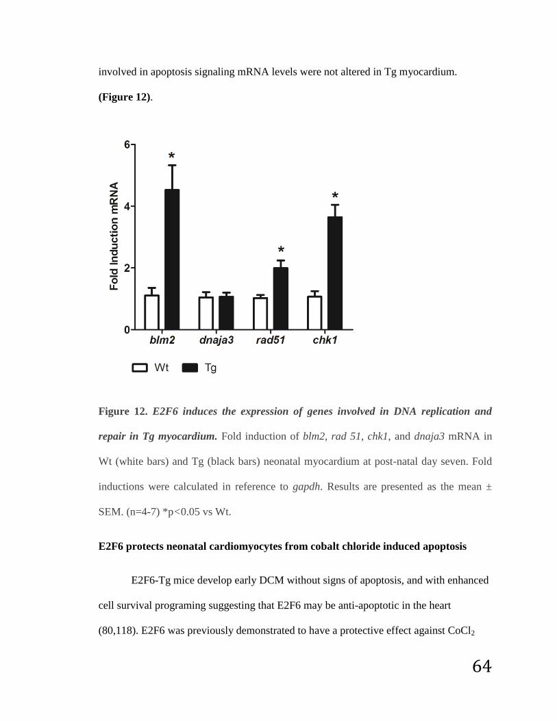
64
involved in apoptosis signaling mRNA levels were not altered in Tg myocardium.
(Figure 12).
Figure 12. E2F6 induces the expression of genes involved in DNA replication and
repair in Tg myocardium. Fold induction of blm2, rad 51, chk1, and dnaja3 mRNA in
Wt (white bars) and Tg (black bars) neonatal myocardium at post-natal day seven. Fold
inductions were calculated in reference to gapdh. Results are presented as the mean ±
SEM. (n=4-7) *p<0.05 vs Wt.
E2F6 protects neonatal cardiomyocytes from cobalt chloride induced apoptosis
E2F6-Tg mice develop early DCM without signs of apoptosis, and with enhanced
cell survival programing suggesting that E2F6 may be anti-apoptotic in the heart
(80,118). E2F6 was previously demonstrated to have a protective effect against CoCl2

65
induced apoptosis in HEK-293 cells. (75) In order to determine if E2F6 could serve to
protect the heart against apoptosis, neonatal cardiomyocytes (NCM) were isolated from
Wt and Tg hearts, exposed to different concentrations of CoCl2, and assayed for cell
survival and apoptosis. NCM were exposed to CoCl2 for a 24hr period which was
demonstrated to induce apoptosis in H9C2 cells (rat cardiomyocyte line). (119) Cell
viability analysis indicated that E2F6 had a significant protective effect on cardiomyocyte
survival post exposure to CoCl2 (250,500 and 1000μM) (Figure 13A). E2F6 consistently
and significantly increased NCM survival from 12% to as much as 50% at the highest
dose of CoCl2 (p<0.05) (Figure 13A).

66

67
Figure 13: E2F6 protects neonatal cardiomyocytes from cobalt chloride induced
apoptosis. (A) Percent viability of Wt and Tg NCM following 24hr treatment with the
indicated amounts of CoCl2. Results are presented as the mean ± SEM (n=8). *p<0.05 vs
Wt for each treatment. (B) Representative western blot of Wt and Tg NCM lysate
following 24hr treatment with 500μM CoCl2 examined with: anti-capsase3, anti-Bax, and
anti-Bcl2. Cleaved caspase 3 is denoted by an arrow. GAPDH was used as a loading
control. Myc-E2F6 represents E2F6 derived from the transgene. (C) Quantification of
cleaved caspase 3/full length caspase 3 and Bax/Bcl2 ratios based on densitometry of Wt
and Tg cardiomyocytes treated with CoCl2. Results are presented as the mean ± SEM
(n=3-4). *p<0.05 vs CoCl2 treated Wt.
To further assess the potential for E2F6 to protect against apoptosis in
cardiomyocytes Wt and Tg NCMs were treated with 500μM CoCl2 and examined for
molecular markers. While full length caspase 3 (~30kDa) was detected in all samples,
cleaved caspase 3 (~19kDa, denoted by arrow) was only detectable in CoCl2 treated
samples (Figure 13B). The ratio of cleaved caspase 3/full length caspase 3 was six times
greater in Wt in comparison to Tg (p<0.05) indicative of less caspase activation and
apoptosis in E2F6-Tg NCM (Figure 13C). Western analysis with pro-apoptotic anti-Bax
(~20kDa) and anti-apoptotic anti-Bcl2 (~26kDa) (Fig 2B) revealed a significantly lower
Bax/Bcl2 ratio in Tg cardiomyocytes (~60% less, p<0.05) (Figure 13C) also indicative
of less apoptosis.

68
Doxorubicin induces apoptosis in Wt and E2F6-Tg neonatal cardiomyocytes
Wt and Tg NCMs were next exposed to the chemotherapeutic agent doxorubicin
which is known to have damaging effects on the heart. (120) Cell viability analysis
demonstrated a similar impact on cell survival in Wt and Tg cardiomyocytes treated with
0.5μM or 1.0μM doxorubicin (~75% survival) (Figure 14A). Western blot analysis with
anti-caspase 3 detected a similar amount of caspase 3 cleavage in Dox treated Wt and Tg
NCMs (Figure 14B and 14C). Similarly, the level of p53 acetylation was equivalent in
Wt and Tg Dox treated cells suggesting that E2F6 did not protect cardiomyocytes from
Dox induced apoptosis. It is notable that myc-E2F6 protein was undetectable in Tg
cardiomyocytes following the doxorubicin treatment (Figure 14C).

69

70
Figure 14. Doxorubicin induces apoptosis in Wt and Tg neonatal cardiomyocytes. (A)
Percent viability of Wt and Tg NCM following 24hr treatment with the indicated
amounts of Dox. Results are presented as the mean ± SEM (n=8). *p<0.05 vs Wt for each
treatment. (B) Representative western blot of Wt and Tg NCM lysate following 24hr
treatment with 0.5µM Dox examined with: anti-capsase3, anti-p53, and anti-acetyl-p53.
Cleaved caspase 3 is denoted by an arrow. GAPDH was used as a loading control. Myc-
E2F6 represents E2F6 derived from the transgene. (C) Quantification of cleaved caspase
3/full length caspase 3 and acetyl p53/ p53 ratios based on densitometry of Wt and Tg
cardiomyocytes treated with Dox. Results are presented as the mean ± SEM (n=4).
*p<0.05 vs Dox treated Wt.
Doxorubicin promotes E2F6 loss in neonatal cardiomyocytes and HeLa
To estimate the effect of Dox on the disappearance of myc-E2F6 protein (derived
from the transgene), NCM were isolated from Tg mice and treated with 0.5μM
doxorubicin for 0 to 24hrs. Western blot analysis with anti-myc revealed that ~60% of
the expressed myc-E2F6 protein was lost by 6 hours (p-value <0.05) and it was virtually
undetectable in all samples 12hr post Dox exposure (Figure 15A and 15B). Myc-E2F6
was also decreased in response to CoCl2 (500μM), but there was increased variability
between samples and overall was less than that observed in response to Dox (Figure
15C, 15D, and 13B).

71
Figure 15. Transgenic E2F6 protein is lost in response to apoptotic agents. (A)
Representative western blot of Tg NCM lysate treated with 0.5μM doxorubicin for 0-24
hours examined with anti-myc and anti-caspase 3. Cleaved caspase 3 is denoted by an
arrow. GAPDH was used as a loading control. (B) Quantification of myc-E2F6 protein
levels in response to doxorubicin. Results are presented as the mean ± SEM (n=3).
*p<0.05 vs time 0. (C) Representative western blot of Tg NCM lysate treated with
500μM CoCl2 for 0-24 hours examined with anti-myc. GAPDH was used as a loading
control. (D) Quantification of myc-E2F6 protein levels in response to CoCl2. Results are
presented as the mean ± SEM (n=3). *p<0.05 vs time 0.

72
To determine if the effects on E2F6 were specific to cardiomyocytes or the
transgene itself, we treated HeLa cells with Dox or CoCl2 and examined endogenous
levels of E2Fs. Doxorubicin exposure (24hr) led to the dose-dependent down-regulation
of endogenous E2F6 protein in these cells (Figure 16A and 16B). In contrast, the pro-
apoptotic E2F1 was up-regulated in HeLa in response to doxorubicin treatment (Figure
16A and 16B). CoCl2 had no effect on endogenous E2F6 protein levels in HeLa while
E2F1 protein levels were decreased (Figure 16C and 16D).

73
Figure 16. E2F6 and E2F1 protein levels are deregulated by apoptotic agents in HeLa
cells. (A) Representative western blot of HeLa following 24hr treatment with the
indicated amounts of doxorubicin examined with: anti-E2F6, anti-E2F1, and anti-caspase
3. Cleaved caspase 3 is denoted by an arrow. GAPDH was used as a loading control. (B)
Quantification of E2F6 and E2F1 protein levels in HeLa in response to Dox. Results are
presented as the mean ± SEM (n=3). *p<0.05 vs untreated. (C) Representative western
blot of HeLa following 24hr treatment with the indicated amounts of CoCl2 examined
with anti-E2F6 and anti-E2F1. GAPDH was used as a loading control. (D) Quantification
of E2F6 and E2F1 protein levels in HeLa in response to CoCl2. Results are presented as
the mean ± SEM (n=3). *p<0.05 vs untreated.
Dox promotes E2F6 down-regulation via post-transcriptional mechanisms
RT-qPCR was performed on RNA isolated from HeLa cells to determine if the
Dox mediated deregulation of E2F6 and E2F1 were via transcriptional or post-
transcriptional mechanisms. No significant changes in either e2f6 or e2f1 transcripts were
observed in response to 0.5μM or 1μM doxorubicin (Figure 17A) suggesting that their
levels are regulated post-transcriptionally in HeLa. Transcript levels of e2f6 were not
examined in response to CoCl2 since the protein was stable.

74
Figure 17. Mechanisms of E2F regulation in HeLa and cardiomyocytes. (A) Fold
induction of e2f1 and e2f6 mRNA in HeLa following 24hr treatment with Dox. Fold
inductions were calculated in reference to gapdh. Results are presented as the mean ±
SEM (n=8). *p<0.05 vs untreated. (B) Western blot of HeLa lysate following 24hr
treatment with doxorubicin examined with anti-Chk1 and anti-p-Chk1. (C)
Quantification of p-Chk1/Chk1 in HeLa. Results are presented as the mean ± SEM (n=3).
*p<0.05 vs untreated. (D) Fold induction of e2f6 and α-mhc mRNA in Wt and Tg NCM

75
following 24hr treatment. Fold inductions were calculated in reference to 18S. Results are
presented as the mean ± SEM (n=6). *p<0.05 vs Wt untreated. ѰP<0.05 vs Tg untreated.
(E) Western blot of Wt and Tg NCM lysate following 24hr treatment of doxorubicin
examined with anti- Chk1 and anti- p-Chk1. ** Loading control is section of total protein
on blot using pre-stained protein loading buffer (Instant Bands, EZ Biolab).
It was demonstrated that E2F6 is phosphorylated by the Checkpoint kinase
(Chk1) following DNA replication stress which led to its dissociation from DNA. (121)
Since Chk1 is also activated by DNA damage, (122) and its mRNA was upregulated in
E2F6-Tg mice (Figure 12) we predicted that Chk1 might be involved in the post-
transcriptional regulation of E2Fs by Dox. Western blot with anti-Chk1 in HeLa detected
a polypeptide ~56kDa which was not altered by dox treatment (Figure 17B). Western
blot with anti-p-Chk1 (active) detected a doublet ~56kDa which showed a robust increase
in response to Dox (Figure 17B). The overall ratio of p-Chk1/Chk1 was significantly
increased by Dox in HeLa (Figure 17C).
We next examined NCM to determine the mechanism of E2F6 loss and if it was
similar to that observed in HeLa. RT-qPCR indicated that E2F6-Tg NCM have ~26 fold
higher levels of e2f6 transcript due to the transgene (p<0.05), but this level drops to only
six fold higher (p<0.05) following exposure to doxorubicin (Figure 17D). In contrast, the
level of endogenous e2f6 in Wt-NCM did not significantly change in response to dox
exposure (Figure 17D). We next examined α-Myosin heavy chain (αMHC) transcript
levels to determine if the E2F6 transgene was reduced due to a change in its promoter
(αMHC). RT-qPCR revealed that the levels of α-mhc transcript were reduced by 80% in
response to dox in both Wt and Tg NCM (Figure 17D).

76
Western blot examination of Chk1 in NCM revealed that it is expressed at similar
levels in Wt and Tg cells, but is not detectable following exposure to doxorubicin
(Figure 17E). The phosphorylated form of Chk1 was not detected in treated or untreated
NCM (Figure 17E).
E2F6 impacts differentiation to induce dilated cardiomyopathy
Our data in cardiomyocytes above and those reported by others in different cell
types indicate that E2F6 has anti-apoptotic properties, which may explain why E2F6-Tg
mice do not display the increased levels of apoptosis usually observed in DCM. Since
apoptosis is not the underlying cause of DCM in Tg hearts we examined other
mechanisms and noted that the G2/M cyclin, cyclin-B1, was specifically down-regulated
in E2F6-Tg myocardium implying a change in the cell cycle (117). Further analysis by
western blot shows that the S phase cyclin, cyclin-E1, is up-regulated by ~4 fold (p-value
<0.05) in E2F6-Tg pup hearts (Figure 18A and 18B).

77
Figure 18. E2F6-Tg hearts show alteration in cell cyclins and cardiac proteins. (A)
Representative western blot of Wt and Tg cardiac tissue examined with anti-cyclin-E1.
GAPDH was used as a loading control. Myc-E2F6 represents E2F6 derived from the
transgene. (B) Quantification of cyclin-E1 protein levels. Results are presented as the
mean ± SEM. (n=5). *p<0.05 vs Wt. (C) Representative western blot of Wt and Tg
cardiac tissue examined with anti-T-cap and anti-TnT. Tubulin was used as a loading
control. (D) Quantification of T-cap protein expression. Results are presented as the mean
± SEM. (n=3). *p<0.05 vs Wt.
As changes in cell cycle dynamics can lead to changes in differentiation we
examined cardiac specific gene expression in E2F6-Tg mice. Our initial microarray
analysis predicted the down-regulation of the sarcomere protein, teliothenin, otherwise
known as titin-cap (T-cap) (80).Western blot analysis with anti T-cap detects a

78
polypeptide ~19kDa which is down-regulated by ~70% in E2F6-Tg myocardium
(p<0.05) (Figure 18C and 18D). No change was observed in another sarcomere protein,
Troponin-T (TnT), levels in E2F6 hearts (Figure 18C).
Discussion
The E2F pathway is known to critically regulate proliferation, apoptosis, and
differentiation thus it has been studied extensively in cancer biology in terms of
inhibiting growth and increasing death in tumors. In fact, almost all cancers are
associated with aberrant E2F/Rb activity. (123,124) Less is known about how the E2F
pathway affects apoptosis in the myocardium, and even less is known about how the
atypical E2Fs, such as E2F6, contribute to apoptosis in the heart. In recent studies we
manipulated the E2F/Rb pathway in vivo in the postnatal heart by expressing the
repressor E2F6 (80,117,118).While the E2F pathway was deregulated and the mice
presented with DCM, there was surprisingly no significant increase in apoptosis which is
normally a hallmark of the disease. In the current study we find that E2F6 confers some
resilience towards cobalt chloride induced apoptosis in cardiomyocytes, but not to
doxorubicin. This specific protection appears to be related to the levels of E2F6 protein
(derived from the transgene) which are more profoundly downregulated following
doxorubicin exposure compared with CoCl2 thereby suggesting that E2F6 may serve to
protect. In HeLa cells Dox differentially impacts endogenous levels of the E2Fs by
upregulation of the pro-apoptotic E2F1 while downregulating E2F6. By contrast, CoCl2
treatment in HeLa caused no change in E2F6 and decreased the levels of apoptotic-E2F1
implying protection.

79
Mechanism of E2F deregulation during apoptosis
It does not appear that E2F6 levels are decreased by caspase 3 given that its
down-regulation in both HeLa and cardiomyocytes occurs prior to the activation of the
protease. It was demonstrated that E2F6 is phosphorylated by the Chk1 kinase (in
response to DNA replication stress) which resulted in its inability to bind to gene
promoters and an increase in E2F1 activity. (121) In our study we found that the
activation status of Chk1 (p-Chk1) is increased in HeLa in response to Dox at the same
time as E2F6 is down-regulated and E2F1 is up-regulated. Thus it is possible that Chk1
plays a part in E2F deregulation in response to Dox. In support, Chk1 and Chk2 were
demonstrated to phosphorylate and stabilize E2F1 promoting apoptosis. (125,126)
Furthermore, endogenous e2f6 transcript levels remain unaltered in both HeLa and Wt-
NCM suggesting it is normally regulated at the post-transcriptional level.
Although Chk1 has the potential to regulate E2F in HeLa, it does not appear to do
so in myocardium, as it is down-regulated in response to Dox in both Wt and Tg NCM.
Instead, myc-E2F6 protein levels are reduced via transcriptional mechanisms which are
likely related to the 80% reduction in α-mhc transcript since the transgene is under
control of its promoter.
E2F6 protects cardiomyocytes from apoptosis via multiple mechanisms during
cardiac development
In our previous studies we uncovered the activation of a survival pathway
involving β2-AR, c-src, ERK, and the 100-fold increase in Bcl2, supporting the notion
that E2F6 may confer resilience against cell death in the heart (118). In culture the overall

80
Bax/Bcl2 ratio was decreased in Tg NCM in response to dox, but without drug treatment
there was no difference in Bcl2 levels. The discrepancy in Bcl2 levels can be attributed to
that fact that the β2-AR survival pathway is likely only induced later on in E2F6-Tg
myocardium to compensate and protect against the stretch caused by DCM. In support of
this we did not observe the activation of the stretch sensitive c-src in neonatal Tg hearts
(Appendix 2: Sup Fig 3).
E2F6 induces DCM via deregulation of cell cycle not apoptosis
In cultured cardiomyocytes E2F6 likely protects via inhibition of E2F1 as was
previously demonstrated in HEK-293 cells. (75,74) In older neonates we observed the
activation of a DNA replication and repair gene profile. This activation could reflect a
cell survival pathway and/or a defect in the cell cycle, which is supported by the observed
changes in cyclin E and B1. A delay in cell cycle re-entry was observed in NIH-3T3 cells
over-expressing E2F6 following serum starvation implying that E2F6 does have the
capacity to alter cell cycle dynamics. (16)
Thus the DCM initiated by E2F6 appears to be related to changes in
differentiation program which impacts the function of critical proteins such as connexin-
43, involved in cardiomyocyte communication, and BDH1, involved in metabolic
signaling (80,117). Our data here further support this notion since the muscle specific T-
cap, involved in contraction, was significantly downregulated by E2F6. This is
particularly notable as mutations in T-cap as well as the proteins it interacts with (titin
and CSRP-encoded muscle LIM protein) are detected in human dilated and hypertrophic
cardiomyopathy. (127,128) Any defect in differentiation is likely linked to the down-

81
regulation of E2F3 in Tg myocardium (80,118), and the displacement of Rb (which
cannot interact with E2F6), which normally represses E2F gene expression during cardiac
and skeletal muscle development. (35,129,130,47)
Conclusions
In summary, the data here support a role for E2F6 levels in cardiac cell survival
and the potential for protection in drug induced cell death. Our data is consistent with
findings in other cell types where E2F6 has been implicated in attenuating apoptosis. The
data suggests multiple mechanisms by which E2Fs modulate cell growth, survival and
function in early and postnatal cardiac development.
Sources of Funding
This work was supported by an operating grant (540033) from the Heart and Stroke
Foundation of Canada to BST.
Conflict of Interest
None declared.

82
CHAPTER 3:
“E2F6 Impairs Glycolysis and Activates BDH1 Expression Prior to Dilated
Cardiomyopathy”
Major JL, Dewan A, Salih M, Leddy JJ, and Tuana BS. (2017) PLOS ONE.12 (1):
e0170066.
Author contributions: JL Major collected, analyzed data, and prepared the manuscript.
Aaraf Dewan aided in performing the Seahorse fatty acid and glucose oxidation
experiments and their data analysis. Technical support and advice were given by
Maysoon Salih. Dr. John Leddy aided in statistical analysis of Figure 23 and editing. All
experiments, data analysis, and manuscript preparation were done under the supervision
of BS Tuana.

83
Abstract
Rationale- The E2F pathway plays a critical role in cardiac growth and development,
yet its role in cardiac metabolism remains to be defined. Metabolic changes play
important roles in human heart failure and studies imply the ketogenic enzyme β-
hydroxybutyrate dehydrogenase I (BDH1) is a potential biomarker.
Objective- To define the role of the E2F pathway in cardiac metabolism and dilated
cardiomyopathy (DCM) with a focus on BDH1.
Methods and Results- We previously developed transgenic (Tg) mice expressing the
transcriptional repressor, E2F6, to interfere with the E2F/Rb pathway in post-natal
myocardium. These Tg mice present with an E2F6 dose dependent DCM and deregulated
connexin-43 (CX-43) levels in myocardium. Using the Seahorse platform, a 22%
decrease in glycolysis was noted in neonatal cardiomyocytes isolated from E2F6-Tg
hearts. This was associated with a 39% reduction in the glucose transporter GLUT4 and
50% less activation of the regulator of glucose metabolism AKT2. The specific reduction
of cyclin B1 (70%) in Tg myocardium implicates its importance in supporting glycolysis
in the postnatal heart. No changes in cyclin D expression (known to regulate
mitochondrial activity) were noted and lipid metabolism remained unchanged in neonatal
cardiomyocytes from Tg hearts. However, E2F6 induced a 40-fold increase of the Bdh1
transcript and 890% increase in its protein levels in hearts from Tg pups implying a
potential impact on ketolysis. By contrast, BDH1 expression is not activated until
adulthood in normal myocardium. Neonatal cardiomyocytes from Wt hearts incubated
with the ketone β-hydroxybutyrate (β-OHB) showed a 100% increase in CX-43 protein

84
levels, implying a role for ketone signaling in gap junction biology. Neonatal
cardiomyocyte cultures from Tg hearts exhibited enhanced levels of BDH1 and CX-43
and were not responsive to β-OHB.
Conclusions- The data reveal a novel role for the E2F pathway in regulating glycolysis in
the developing myocardium through a mechanism involving cyclin B1. We reveal BDH1
expression as an early biomarker of heart failure and its potential impact, through ketone
signaling, on CX-43 levels in E2F6-induced DCM.
Introduction
The failing heart shows transcriptional and metabolic remodeling which may have
detrimental effects on cardiac function (59,131). Given the extensive energy requirements
of the heart, and its limited ATP reserve, understanding the mechanisms which regulate
cardiac metabolism is critical to the understanding of heart function and failure (132,62).
In the normal adult heart, fatty acid oxidation accounts for up to 90% of the ATP
production while glycolysis supplies the remainder. (61). The heart shows a remarkable
capacity to adapt to substrate utilization under stress. In the failing heart, a reduction of
fatty acid oxidation and changes in glycolysis are observed (61,63). It was recently
demonstrated that there is an increase in ketone metabolism and the ketogenic enzyme, β-
hydroxybutyrate dehydrogenase 1 (BDH1), in human and mouse heart failure
(133,134,66,65).
Ketones are synthesized in the liver and exist in three main forms: β-
hydroxybutyrate (β-OHB) the most stable and abundant form, acetoacetate, and acetone
(135). Ketone bodies arise from the incomplete oxidation of long chain fatty acids. They

85
are utilized as an alternative energy source when glucose is scarce, such as during fasting,
exercise, and in many disease states such as diabetes (135,136). Glucose is a vital nutrient
source in the developing heart and during heart failure, thus ketones could be an
important alternative energy source during metabolic stress (59). It is therefore critical to
understand how the heart uses ketones to adapt to substrate utilization during cardiac
development and disease.
The E2F family is a group of nine transcription factors which regulate cell
proliferation, hypertrophy, and death (113,68). Recently, the transcriptional activator,
E2F1, has been demonstrated to promote glycolysis via regulation of the pyruvate
dehydrogenase kinase (PDK4) and inhibition of histone deacetylases (HDACS) to
regulate metabolism (137,138,139,140). In association with its modulator,
Retinoblastoma protein (Rb), E2F1 can also repress oxidative phosphorylation (141,142).
E2F6 is a unique E2F family member which is believed to repress E2F responsive genes
independently of Rb (27,20). We previously examined the contribution of the E2F
pathway in post-natal mouse myocardium by cardiac specific expression of E2F6 which
led to the early-onset of dilated cardiomyopathy (DCM) without hypertrophy or apoptosis
(80), Contrary to expectation, E2F6-Tg mice showed activation of E2F responsive genes
(80). This was achieved via down-regulation of E2F3 (critical for cardiac development)
and competitive binding at E2F sites which are normally repressed via E2F/Rb in the
post-natal heart (143,75,27). In essence, E2F6 serves as a dominant negative of E2F/Rb
in the heart.
E2F6-Tg mice display an early reduction in connexin-43 (CX-43) and reduced
conductivity which is a hallmark of DCM and heart failure (35). Here we further examine

86
the changes in the post-natal myocardium of E2F6-Tg mice with the view to define early
bio-markers and novel pathways which may be useful for understanding and treating
idiopathic DCM. We note the early induction of BDH1 in neonatal Tg myocardium and a
relationship between ketones and CX-43 expression in cardiomyocytes. We further note
that deregulation of the E2F pathway impairs glycolysis which may have triggered the
induction of BDH1 and the resulting changes in CX-43 leading to DCM.
Methods
Mice and Genotyping
Previously described Tg mice with cardiac specific expression of E2F6 (under
control of the α-Myosin heavy chain promoter) (B6C3F1) were bred with WT (B6C3F1)
mice (80). All animal work was conducted according to the University of Ottawa’s
institutional animal care committee guidelines. The protocols were approved by the
University of Ottawa's institutional animal care committee: cmm-1725, cmm-1723. All
possible steps were taken to ameliorate animal suffering. Mouse pups were euthanized
by decapitation. Adult mice were euthanized via carbon dioxide inhalation.
Approximately 110 mice were used in this study. This number was required in order to
obtain sufficient sample size for appropriate biochemical and statistical analyses.
Genotyping was performed via DNA extraction from mouse ear (adult) or tail
(pup) clip and PCR using the Phire Tissue Direct (Thermo Scientific) kit as per the
manufacturer’s instructions. Primers spanning the 6th
intron of E2F6 were used
(ATCACAGTACATATTAGGAGCAC- sense, and GGTGCGGCTACCAGTCTACA–

87
anti-sense) which result in the amplification of a long fragment (988bp) in Wt mice and a
long and short fragment (342bp) in Tg mice which were separated on a 1.3% agarose gel.
RNA Analyses
RNA was extracted from cardiac lysate at post-natal day 7 using the RNEasy
Fibrous Tissue Mini Kit as per the manufacturer’s protocol (Qiagen). First-strand cDNA
was synthesized from 2μg RNA and oligoDT with SuperScriptII reverse transcriptase
(Invitrogen) as per the manufacturer's protocol. qPCR was performed in the q-Rotor
(Qiagen) using Fast Start SYBR Green (Roche) according to the manufacturer’s
instructions. Gene expression was normalized against Gapdh, and fold inductions were
calculated using the ΔΔCt method. Primer pairs used for qPCR are: Bdh1:
AAGCACTGGAAGCAGACACAT (sense), ACACTTAGGGCTTTTCCTGGG (anti-
sense), Oxct1: CTGGAGTTTGAGGACGGCAT (sense),
TCCGCATCAGCTTCGTCTTT (anti-sense), GLUT4:
GCAGATCGGCTCTGACGATG (sense), GCCACGTTGCATTGTAGCTC (anti-sense),
and Gapdh: GCAGTGGCAAAGTGGAGATT (sense) and
TCTCCATGGTGGTGAAGACA (anti-sense).
Western Blot
Neonatal cardiomyocytes were rinsed twice with PBS and frozen at -80oC. Plates
were thawed on ice in RIPA (50mM Tris (pH 7.4), 1mM EDTA, 150μM NaCl, 0.25%
deoxycholic acid, 1% NP-40) containing protease and phosphatase inhibitors (Roche).
Plates were scraped and lysates were syringed with a 21G needle. Cardiac lysates were

88
prepared with an electric tissue homogenizer in RIPA with protease and phosphatase
inhibitors.
Cardiac and cell lysates were centrifuged at 12800g for 10min at 4oC. Protein
concentrations were determined using the BCA assay (Thermo Scientific). Lysates (13-
50ug) were run on gradient (5-15%) SDS-PAGE gels in 3X loading dye (Cell Signaling).
Gels were transferred to PVDF membrane (Millipore) in transfer buffer (25mM Tris,
190mM Glycine, 20% methanol) overnight at 4oC. Membranes were blocked in TBST
(1M Tris, 290mM NaCl, 0.1% Tween, pH7.2) with 5% milk. The blots were probed with
primary and secondary antibodies (listed below) which were diluted in TBST with 5%
milk. The conversion of ECL substrate (Roche) was detected on film. Band signal
intensities were quantified by densitometry using Image Lab Software4.0.1 (Bio-Rad).
The following primary antibodies were used: GLUT4 (2213, 1:1000), AKT2
(6063, 1:1000), p-AKT2 (ser 479) (8599, 1:1000), cyclin D1 (2978, 1:1000), cyclin D3
(2936, 1:2000), and cyclin B1 (4138, 1:1000) were purchased from Cell Signaling, BDH1
(MA5-15594, 1:1000) and GAPDH (MA5-15738, 1:5000) were purchased from Thermo
Scientific, CX-43 (ab11370, 1:100000) was purchased from Abcam, OXCT1 (12175-1-
AP , 1:10000) was purchased from Proteintech, and myc (which detects myc-tagged
E2F6) (11667149001, 1:2000) was purchased from Roche. Secondary antibodies anti-
mouse (115-035-003, 1:15000) and anti-rabbit (111-035-045, 1:15000) were purchased
from Jackson Immunochemicals.

89
Neonatal Cardiomyocyte Isolation and Treatment
Hearts were collected from Wt and Tg mice at post-natal day 1 following
decapitation. Hearts were rinsed in HBSS and incubated in 0.5% trypsin dissolved in
HBSS overnight at 4oC while genotyping was performed. Hearts were grouped in twos or
threes (by genotype) for digestion with 0.5% Collagenase type II (Gibco) dissolved in
HBSS (4x10min). Cells were collected after each digestion via centrifugation at 3000g
for 3 minutes and resuspended in DMEM supplemented with 10% FBS, 1%
penicillin/streptomycin, and 1% non-essential amino acids. Total suspensions were plated
on uncoated 10cm dishes to remove fibroblasts (45min x2). Cardiomyocytes were seeded
onto 0.1% gelatin coated plates (300000/well on XFe24 well plate for Seahorse metabolic
analysis, or 1x10⁶/well on 6 well plates for ketone treatment) and allowed to attach for 48
hours in a 37oC incubator with 5% CO2.
Paired neonatal cardiomyocytes were treated with or without 3.8mM β-
hydroxybutyrate (Sigma) dissolved in water for 24 hours, or used for glycolysis and fatty
acid oxidation measurements (described below).
Seahorse Glycolysis Measurement
Cardiomyocyte media was replaced with bicarbonate and glucose free DMEM
containing 2mM glutamine. The extracellular acidification rate (ECAR) was measured
using an XFe24 Extracellular Flux Analyzer (Seahorse Bioscience). Triplicate pre-
injection readings were taken to establish a baseline, followed by triplicate reading after
sequential treatment with 20mM glucose, 2mg/mL oligomycin, and 100mM 2-deoxy-
glucose. ECAR was corrected to baseline pre-injection measurements.

90
Seahorse Fatty Acid Oxidation Measurement
Cardiomyocytes were starved for 24hr in substrate limited medium (DMEM,
0.5mM glucose, 1.0mM Glutamine, 0.5mM L-carnitine, 1% FBS) and replaced with fatty
acid oxidation assay medium (111mM NaCl, 4.7mM KCl, 1.25mM CaCl2, 2.0mM
MgSO4, NaH2PO4, 2.5mM glucose, 0.5mM L-carnitine, and 5mM HEPES).
Cardiomyocytes were treated with or without 40µM etomoxir for 15min. The oxygen
consumption rate (OCR) was measured using the XFe24 Extracellular Flux Analyzer
(Seahorse Bioscience). Triplicate pre-injection readings were recorded to establish
baseline and cardiomyocytes were sequentially treated with either 17µM BSA or 100 µM
palmitate conjugated to BSA and: 2mg/mL oligomycin, 1μM Carbonyl cyanide-4-
trifluoromethoxy phenylhydrazone, and 1µM Antimycin-A. OCR was corrected to
baseline pre-injection measurements.
Statistical Analyses
All data were analyzed with a student t-test with the exception of the protein
expression data in (Figure 23D) which was analyzed with a two way mixed ANOVA
with repeated measures. The level of significance was set at P<0.05 in all cases.
Results
E2F6 Induces the Early Expression of BDH1 in Neonatal Myocardium
E2F6 expression in postnatal myocardium led to dose dependent DCM associated
with decreased levels of CX-43 (80). Our previous microarray results predicted the up-
regulation of Bdh1 in E2F6-Tg hearts thus we performed RT-q-PCR on mRNA collected

91
from Wt and Tg pup hearts for validation. A 40 fold increase of Bdh1 transcript was
detected in Tg myocardium (P<0.05) (Figure 19A). BDH1 protein was barely detectable
in Wt pup hearts but was markedly upregulated by 890% (P<0.05) in E2F6 Tg hearts at
post-natal day 1 (P1) (Figure 19B and 19C). Since BDH1 is up-regulated in the failing
adult human heart (65) we examined its expression at 6 weeks of age, at which time Tg
mice show full onset of DCM (118). Western blot analysis did not reveal any difference
in BDH1level between Tg and Wt hearts at this time (Figure 19B and 19C).

92
Figure 19. E2F6 Activates BDH1 Expression in Neonatal Myocardium. (A) Bdh1 (β-
hydroxybutyrate dehydrogenase 1) and Oxct1 (succinyl-CoA: 3-oxoacid-CoA
transferase) transcript levels in Wt and Tg myocardium 7 days after birth. Expression is
normalized to Gapdh. Results represent mean±SEM values (n=5-7). (B) Representative
immunoblots of protein from Wt and Tg mouse myocardium at post-natal day 1 (pups)
and 6wks of age (adult) examined with the anti-BDH1 and anti-OXCT1. (C)
Densitometric quantification of BDH1 and OXCT1 expression from immunoblots.
Expression is normalized against GAPDH. Results represent mean±SEM values (n=4).
(D) Immunoblot of protein from Wt and Tg myocardium with BDH1 at the indicated
time points after birth.*P<0.05.
We next examined the developmental expression of BDH1 protein in post-natal
myocardium between P1 and 6wks. In normal developing myocardium BDH1 protein is
undetectable at P1 and appears at 6 weeks of age (Figure 19D). On the other hand, in
E2F6-Tg hearts, BDH1 expression appears to be maximally expressed as early as P1 with
this level maintained at P7, 3 weeks, and 6 weeks. It should be noted that symptoms of
DCM arise as early as 2 weeks in E2F6-Tg mice (118), thus BDH1 is markedly elevated
prior to the early development of DCM and maintained at this level into adulthood
(Figure 19D).
The induction of BDH1 expression by E2F6 was specific since the succinyl CoA:
3-oxoacid CoA transferase (OXCT1) (another rate limiting enzyme in ketone
metabolism) was not changed at the mRNA (Figure 19A) or protein level in heart tissue
from pup or adult E2F6-Tg mice (Figure 19B and 19C).

93
E2F6 Impairs Glycolysis in Neonatal Cardiomyocytes
Since ketone metabolism is increased when glucose is scarce, we measured
glycolysis in in Tg mice to gauge whether changes in glycolytic rates may have
stimulated BDH1 expression. We used the Seahorse method to monitor the extracellular
acidification rate (ECAR) as a measure of glycolysis which produces lactic acid. In
nutrient starved neonatal cardiomyocytes (pre-injection) ECAR measurements were
similar in Wt and Tg, but Tg ECAR readings were lower following the addition of
glucose and oligomycin (inhibitor of ATP synthase that drives glycolysis) (Figure 20A).
The competitive inhibitor of glucose, 2-Deoxy-glucose (2-DG), caused the ECAR
readings to return to baseline in both Wt and Tg cardiomyocytes demonstrating the
specificity of the experiment to glucose (Figure 20A). Calculation of basal glycolysis
revealed a 22% decrease in E2F6 expressing cardiomyocytes (P<0.05) (Figure 20B). No
significant changes in glycolytic capacity or reserve were noted.

94
Figure 20. E2F6 Impairs Glycolysis in Neonatal Cardiomyocytes. (A) Glycolytic rates
for Wt and Tg neonatal cardiomyocytes pre-injection, and following the sequential
addition of: glucose, oligo (oligomycin), and 2-DG (2-deoxy glucose). ECAR
(extracellular acidification rate) is normalized against pre-injection rates. Results
represent mean±SEM values (n=8). (B) Glycolytic measurements (basal glycolysis,
glycolytic capacity, glycolytic reserve) in Wt and Tg neonatal cardiomyocytes. (C)
Representative immunoblot analyses of protein extracts from Wt and Tg myocardium at

95
postnatal day 1 with anti: GLUT 4 (glucose transporter 4), T-AKT2 (total protein kinase
B), and p-AKT2 (phospho (ser 474)- protein kinase B). (D) Densitometric quantification
of immunoblots. Expression is normalized against GAPDH (n=4). *P<0.05.
Glucose entry into cardiomyocytes is a rate limiting step in glycolysis, thus we
examined changes in the cardiac glucose transporter: GLUT4.Western blot analysis of
protein isolated from Wt and Tg myocardium at P1 with anti-GLUT4 detected a GLUT4
monomer of ~50kDa and high molecular weight polypeptides which are GLUT4
oligomers (Figure 20C). Quantification of all polypeptides revealed a 39% decrease in
total GLUT4 protein (P<0.05) (Fig 2D). RT-q-PCR revealed no change in GLUT4
transcript levels in Tg myocardium (Appendix 3: Sup Fig 4) indicating its loss is post-
transcriptional.
We also examined the expression of another major regulator of glucose
metabolism: AKT2 (also known as protein kinase B). Western blot analysis with anti-
total AKT2 (T-AKT2) revealed a 100% increase in E2F6-Tg hearts (P<0.05), while
analysis of phosphorylated (active) AKT2 revealed no change (Figure 20C and 20D).
The ratio of phospho: total AKT2 was decreased by ~50% (P<0.05) in Tg myocardium
indicating less AKT2 activation as a fraction of the total AKT2 pool (Figure 20D).
E2F6 Does Not Impact Fatty Acid Oxidation in Neonatal Cardiomyocytes
In the failing human heart the up-regulation of BDH1 is believed to be an
adaptation to a decrease in lipid metabolism (134,66). Thus, we also assessed lipid
metabolism in neonatal cardiomyocytes from Wt and Tg mice. We used the seahorse
method to measure the oxygen consumption rate (OCR) which revealed no change in

96
fatty acid oxidation in E2F6-expressing cardiomyocytes at pre-injection, or following the
sequential addition of palmitate (fatty acid), oligomycin (inhibitor of ATP synthase),
Carbonyl cyanide-4-phenylhydrazone (FCCP/uncoupler) or antimycin-A (electron
transport chain inhibitor) (Figure 21). Cardiomyocytes treated with etomoxir (inhibitor
of fatty acid entry) showed no significant increase in OCR beyond baseline, thereby
demonstrating the specificity of the experiments to fatty acids (Appendix 3: Sup Fig 5).
Figure 21. E2F6 Does Not Impact Fatty Acid Oxidation in Neonatal Cardiomyocytes.
Oxygen consumption rate (OCR) in Wt and Tg neonatal cardiomyocytes following 24hr
glucose starvation. Results were normalized against baseline (pre-injection).
Cardiomyocytes were sequentially incubated with BSA or palmitate, oligo (oligomycin),
FCCP (Carbonyl cyanide-4-phenylhydrazone), and Anti-A (Antimycin-A). Results
represent mean±SEM values (n=7-8).

97
E2F6 Deregulates Cyclin Expression in Myocardium
The E2F/Rb pathway is believed to drive glycolysis via the transcriptional
regulation of cyclins (144). Thus, we examined the expression of various cyclins in Wt
and Tg myocardium at post-natal day 1. Cyclin D1 and D3 expression levels were not
altered in cardiac tissue from E2F6-Tg mice (Figure 22A) while cyclin B1 was reduced
by 70% in E2F6-Tg myocardium (P<0.05) (Figure 22A and 22B).

98
Figure 22. E2F6 Deregulates Cyclin B1 Expression in Myocardium. (A) Representative
immunoblot of protein isolated from Wt and Tg myocardium at post-natal day 1 with
anti- cyclin D1, anti-cyclin D3, and anti-cyclin B1. (B) Densitometric quantification of
cyclin immunoblots. Expression is normalized against GAPDH. Results represent
mean±SEM values (n=4). *P<0.05.
Ketones Regulate Connexin-43 in Neonatal Cardiomyocytes
Recent evidence indicates that ketones can enhance CX-43 protein in endothelial
cells via the Extracellular Receptor Kinase (ERK) pathway (145). We previously noted
the deregulation of ERK activity and a loss of CX-43 protein in adult Tg mice (80), and
in the present study we detected a 50% down-regulation of CX-43 protein at post-natal
day 1 (P<0.05) (Figure 23A and 23B).

99
Figure 23. β-OHB Regulates CX-43 Protein Expression in Neonatal Cardiomyocytes.
(A) Representative immunoblot of protein isolated from Wt and Tg myocardium at post-
natal day 1 with anti-CX-43. (B) Densitometric quantification of CX-43 immunoblot.
Expression is normalized against GAPDH. Results represent mean±SEM values (n=4).
(C) Representative immunoblot analyses of protein from Wt and Tg neonatal
cardiomyocytes following incubation with or without β-OHB (β-hydroxybutyrate). (D)
Densitometric quantification of CX-43 in treated and non-treated neonatal

100
cardiomyocytes. Expression is normalized against GAPDH. Results represent the
mean±SEM values (n=3-5). (E) Representative immunoblot analysis of BDH1 in Wt and
Tg neonatal cardiomyocytes. *P<0.05.
We hypothesized that the excess BDH1present in Tg hearts could limit the
amount of β-OHB in the cardiomyocyte via its conversion to acetoacetate and subsequent
availability for the ketone catabolic pathway. This could in turn inhibit the signaling
capacity of β-OHB (the most stable and abundant ketone) and thus may contribute to the
observed down-regulation of CX-43 in Tg hearts. To test this we examined the amount
of CX-43 in neonatal cardiomyocytes incubated with or without the β-OHB. Wt
cardiomyocytes incubated with β-OHB showed a 100% increase of CX-43 (P<0.05)
(Figure 23C and 23D), similar to what was recently reported in endothelial cells (145).
In contrast, β-OHB had no significant impact on CX-43 expression in Tg cardiomyocytes
supporting our hypothesis that BDH1 reduces ketone signaling.
Surprisingly, the reduction of CX-43 noted in vivo (Figure 23A) was reversed in
culture of Tg cardiomyocytes (Figure 23B). In order to rule out the possibility that
BDH1 was reduced in culture, thereby reversing its effect on CX-43 expression, we
assessed its level in cultured cardiomyocytes. Western blot with anti-BDH1 detected its
presence in neonatal cardiomyocytes cultured from Tg mice, but not Wt, indicating that
its loss is not responsible for the rescue of CX-43 in culture (Figure 23E). This suggests
the presence of factors in the culture media which specifically promote expression of CX-
43 in neonatal cardiomyocyte from Tg hearts. In support of this hypothesis, Tg
cardiomyocytes showed an increase in CX-43 expression over time in culture (Appendix
3: Sup Fig 6).

101
Discussion
The E2F pathway regulates early cardiac development through mechanisms that
impact cell proliferation, hypertrophy, and death (47,53,146,55). We previously
demonstrated that the E2F pathway can also impact post-natal cardiac function via
manipulation of the pathway in vivo with the repressor E2F6 (80). E2F6 resulted in dose
dependent DCM associated with inappropriate activation of E2F responsive genes, but
without changes in cardiac growth or death (80). Further, our data also revealed that the
E2F pathway can impact β-adrenergic signaling in the post-natal heart (118). Here we
demonstrate that deregulation of the E2F pathway impairs glycolysis in the developing
postnatal myocardium, and markedly enhances that expression of the ketogenic enzyme,
BDH1, which was linked to the down-regulation of CX-43. Thus we present notable
evidence that appropriate modulation of the E2F pathway is critical for regulating cardiac
metabolism and, consequently, function of post-natal myocardium.
E2F6-Tg mice present with an early reduction of CX-43 and a defect in cardiac
function potentially accounting for the noted DCM and sudden death (80). Recent studies
demonstrated that β–OHB can enhance CX-43 expression in endothelial cells (145)
which we have now also noted in Wt neonatal cardiomyocytes. This suggests that ketones
may uniquely modulate the gap junction levels in different cell types. It should be noted
that CX-43 expression was rescued in culture of Tg cardiomyocytes while BDH1 was
still highly expressed. Thus other factors appear to have an effect on CX-43 expression in
culture conditions. In this regard, multiple MAPK pathways have been implicated in the
post-transcriptional regulation of CX-43, and these were deregulated in E2F6-Tg mice

102
(147), (145) (80). It is possible that deregulation of MAPKs may have sensitized E2F6-
Tg cardiomyocytes to growth factors in the culture media.
Despite differences in vitro and in vivo both neonatal cardiomyocytes and hearts
from Tg mice show a marked induction of BDH1 and a diminished capacity to regulate
CX-43 expression. In the heart, Tg-mice do not enhance CX-43 levels to the appropriate
level required for cardiac function, and in culture, Tg cardiomyocytes are unresponsive to
CX-43 ketone signaling pathways. In both cases this likely involves the marked
induction of BDH1 protein which could diminish the available β-OHB (via its conversion
to a less stable ketone) thereby driving it towards the ketone catabolic pathway and
reducing its ability to act as a signaling molecule to enhance the amount of CX-43. The
resulting down-regulation of CX-43 in Tg myocardium would alter cardiomyocyte
communication and function from a very young age, potentially leading to the early
DCM noted. In this regard, CX-43 is reported to be down-regulated in the diabetic rat
heart and in many types of heart failure (148,149,150,151). Further, metabolic
remodeling is known to occur in heart failure with enhanced BDH1 and ketone signaling
(65,66). Hence our data here may critically serve to link enhanced BDH1 and the loss of
CX-43 in the adult failing heart as well. It has previously been demonstrated that an
increase in BDH1 transcript levels in both mouse and human cardiac tissue is associated
with enhanced BDH1 activity and ketone metabolism (66,65). While we have not
measured BDH1 activity here, the robust increase in both transcript and protein levels
induced by E2F6 imply an enhanced BDH1enzyme activity as early as postnatal day 1.
The relationship between E2F, BDH1, and CX-43 highlights the emerging idea
that ketones not only serve as energy substrates, but are also important signaling

103
molecules which can regulate gene expression during nutrient scarcity/adaptation.
Ketones can initiate undefined signaling cascades via binding to G-protein coupled
receptors, or by direct interaction with and inhibition of HDACs (64,152). Rb recruits
HDACs to repress E2F responsive genes (14,13), which we have shown to be inhibited
by E2F6 expression in the heart (80). Furthermore, we found that E2F6 also regulates G-
coupled protein receptor signaling pathways (118), suggesting a novel relationship
between E2F/Rb, HDACs, and ketones which critically impacts myocardial growth,
function, and metabolism.
It is plausible that BDH1 was induced by E2F6 in Tg myocardium to compensate
for the impairment in glycolysis (which is the major source of energy in the neonatal
heart). The decrease in GLUT4 protein could have limited glucose entry to E2F6-Tg
cardiomyocytes, and when coupled with the observed decrease in AKT2 activation
(which regulates glucose homeostasis) could account for the impaired glycolysis (153).
Alterations in glucose metabolism have been previously linked to the E2F/Rb pathway as
demonstrated by double knock-out of E2F1/E2F2 which impaired insulin production and
caused diabetes (154). Additionally, studies have demonstrated that E2F/Rb promote
glycolysis over oxidative phosphorylation in muscle and fat tissue via the deregulation of
cyclin, in particular cyclin D1 (144,155). Thus it is a logical extension that E2F6, which
inhibits E2F/Rb activity, would also inhibit glycolysis. E2F6-Tg mice express less
cyclinB1 which could represent a delay in the cardiac cell cycle (i.e. not reaching mitosis)
similar to what was observed in E2F6 expressing 3T3 cells which accumulated in S phase
(27). Such a shift in the cell cycle could cause a shift in the metabolic profile as well. The
specific change in cyclin B1 may involve the reduction of E2F3 in E2F6-Tg myocardium

104
since it is a critical regulator of the cell cycle during perinatal cardiac development
(35,80). This suggests that E2F3 may also be a key regulator of cyclin B1 and glycolysis
in the heart. Further, since fatty acid oxidation is not increased in response to the
depression of glycolysis, ketolysis may be a necessary adaptation as an alternative energy
source.
It is interesting to note that BDH1 levels were not increased in the adult (6wk old)
Tg-myocardium which are experiencing heart failure. This may reflect the fact that fatty
acids are the major energy substrate at this time and fatty acid metabolism appeared to be
unaffected in E2F6 Tg mice. This implies that the induction of BDH1 may be an early
marker for metabolic stress/changes in the myocardium that precede heart failure. There
is a window of overlap between enhanced BDH1 expression and the early symptoms of
DCM around 3 weeks supporting the correlative relationship (118). This may be
particularly useful information for ~40% of cases of idiopathic dilated cardiomyopathy
and early-onset of disease.
The robust increase of Bdh1 transcript by E2F6 at birth and the lack of change in
other ketogenic enzymes, such as OXCT1, imply a specific role for E2Fs through a
transcriptional mechanism. A basic promoter analysis did not detect an E2F consensus
binding site in the Bdh1 promoter (Appendix 3: Sup Fig 7). This does not rule out direct
transcriptional regulation as E2F6 has been demonstrated to bind to DNA at non-
consensus sites and activate transcription (25,29). A nuclear-factor (NF)-kappaB site was
predicted by the promoter analysis 1938bp upstream of the BDH1 transcription start site
(core score 1.00). This is of particular interest because E2Fs can bind to and de-regulate
NF-kappaB in the context of inflammation and metabolism (156,157). Thus it is possible

105
that the deregulation of E2Fs in E2F6-Tg myocardium may have enabled NF-kappaB to
flip a metabolic switch activating BDH1 in the neonatal heart. In support of this, E2F1-3
levels normally drop in the adult myocardium (8) when we have demonstrated that BDH1
is normally expressed.
The data here demonstrate a novel role for the E2F pathway in regulating
metabolism in post-natal myocardium since its perturbation by E2F6 leads to aberrant
glycolysis and the induction of the ketogenic enzyme: BDH1. These changes appear to
impact ketogenic signaling to deregulate CX-43 levels and thereby disrupt cardiac
function. The potent early induction of BDH1 prior to a discernable cardiac pathology
implies that BDH1 is an early biomarker of metabolic stress and DCM. The information
here could provide new insight into the early diagnosis and potential treatment options in
idiopathic cases of DCM.
Acknowledgements
We thank Dr. Mary Ellen Harper for kind use of the Flux Analyzer/ Sea Horse platform.
Disclosures
None.

106
GENERAL DISCUSSION
The works in this thesis elucidate the in vivo capacity for the E2F pathway to
regulate cell death, hypertrophy, and differentiation in the post-natal heart by
manipulating its activity with E2F6 in a cardiac specific Tg mouse model. E2F6-Tg mice
developed early onset DCM without hypertrophy or apoptosis suggesting that E2F6 had
protective properties against these pathological processes. My data clearly shows that
E2F6 deregulates E2F3 which is a master regulator of the cardiac cell cycle and the only
E2F family member required for viability. I propose that via alteration of E2F3 (and
subsequently its association with Rb), E2F6 altered the dynamics of cell cycle withdrawal
in Tg myocardium leading to metabolic changes which impact differentiation and
function leading to DCM (summarized in Figure 24). Use of the E2F6-Tg model may
aid in the understanding of idiopathic cases of DCM which may prove to be of
significance in the field.

107
Figure 24: A summary model for E2F6 induced dilated cardiomyopathy. E2F6
expression inhibits E2F3 with impacts on cell cycle/metabolism and thereby cardiac
differentiation via inhibition of E2F3/Rb. These changes cause the down-regulation of
CX-43 and T-Cap leading to cardiac remodeling which proceeds directly to dilation
without hypertrophy or apoptosis via inhibition of E2F3 and E2F1. Dilation activates the
stretch sensitive c-src and a β2-AR survival pathway, which protects Tg myocardium
from apoptosis. Activation of the β2 pathway sensitizes the heart to isoproterenol leading
to the rescue of E2F3 and the induction of pathological hypertrophy. The increased
activity of ERK may also impact the post-transcriptional loss of CX-43 via miR-206 (80)
which enhances disease progression.

108
E2F6: a potential therapeutic tool in heart failure
The lack of hypertrophy and apoptosis in E2F6-Tg mice is particularly curious
given that these are hallmarks of heart failure and DCM. Cell growth and apoptosis are
well known to be regulated by the E2F pathway thus I hypothesized that E2F6 had
repressed them. When challenged with the hypertrophic agent, isoproterenol, E2F6-Tg
mice were not resilient, but instead were twice as sensitive as their Wt counterparts. Their
increased sensitivity can likely be attributed to activation of a β2 adrenergic receptor
pathway. Intriguingly, E2F3 was downregulated in Tg myocardium and was rescued by
isoproterenol, suggesting that E2F activity is central and necessary for cardiac
pathological hypertrophy to occur (Figure 24). In support of this notion, Bicknell and
colleagues found that E2F/DP activity was necessary for pathological hypertrophy in
NCM (55). Thus it seems that the E2F pathway, in particular E2F3, is critical to regulate
cardiac pathological hypertrophy in vitro and in vivo.
The β2 adrenergic pathway described in Chapter 1 was previously demonstrated to
activate cell survival in cardiomyocytes (95). Thus I hypothesized that its activation
could explain the lack of apoptosis in Tg mice despite their advanced disease. In support
of its anti-apoptotic properties, E2F6 was capable of attenuating the apoptotic response to
cobalt chloride in cardiomyocytes. In contrast to the adult heart, the protection conferred
to NCM by E2F6 did not include an increase in Bcl-2. In cancer cell lines E2F6 inhibited
apoptosis via inhibition of E2F1 (Figure 24) (75,78). E2F6 was able to out-compete
E2F1 for DNA binding in in vitro assays (15,17), and we previously detected the
presence of myc-E2F6 on gene promoters in adult Tg myocardium (80). This suggests
that E2F6 has the capacity to out-compete pro-apoptotic E2F family members in the

109
heart. Unlike in cancer cell lines (such as HeLa used in Chapter 2), my preliminary data
suggests that E2F1 was not up-regulated in cardiomyocytes in response to doxorubicin
(Appendix 4: Sup Fig 8). This suggests that another E2F family member may be
responsible for inducing apoptosis. In addition to E2F1, E2F3 was also capable of
inducing apoptosis in neonatal cardiomyocytes (53). Furthermore, I demonstrated that
E2F3 expression was enhanced by isoproterenol along with the β1-AR which can induce
cardiomyocyte cell death (95). Since E2F6 repressed E2F3 expression in Tg
myocardium it is possible that E2F6 may exert protection against apoptosis in
cardiomyocytes via inhibition of E2F3. Thus future experiments should focus on the
balance between the repressor E2F6 and activators: E2F1 and E2F3 in cardiomyocytes.
Although E2F6 holds promise for battling apoptosis in the heart, my studies were
limited by the reduction of myc-E2F6 in response to drug exposure. In the case of
doxorubicin the loss of E2F6 was similar (albeit more rapid) to what I observed in HeLa
cells treated with dox, but via completely different mechanisms. In HeLa and Wt-NCM
e2f6 transcript levels did not change in response to dox, implying that endogenous E2F6
protein was deregulated at the post-transcriptional level by dox. Post-transcriptional
regulation of E2F6 also appears to be important in cardiac development in which its
transcript is maintained while protein is decreased (Appendix 4: Sup Fig 9). Unlike
other E2F family members, E2F6 is not targeted to the proteasome via p19ARF
, and the
mechanisms which regulate its turn-over are not known. Bertoli and colleagues found
that E2F6 was phosphorylated by Chk1 in HEK cells which were subjected to DNA
replication stress (121). Chks have also been demonstrated to phosphorylate and stabilize
pro-apoptotic E2F1. Thus it is possible that E2F6 was down-regulated and E2F1 was up-

110
regulated at the post-transcriptional level by Chk1 phosphorylation. Phosphorylation of
E2F6 could signal for its degradation, or its dissociation from DNA could destabilize it,
thereby leaving E2F1 (which is stabilized by Chk) free to induce apoptosis. The use of
commercially available Chk1/2 inhibitors could be used to test this hypothesis.
In cardiomyocytes, Chk1 is unlikely to regulate E2Fs given the fact that it is
undetectable following dox exposure. Since E2F1 does not appear to be up-regulated by
dox (Appendix 4: Sup Fig 8), and E2F6 is normally expressed at very low levels in the
post-natal heart, this is not particularly surprising. In fact, it is reasonable to propose that
the lack of E2F6 in adult myocardium may actually sensitize it to doxorubicin exposure
resulting in cardiomyopathy. Additionally, the interruption of α-mhc by dox, which
caused the loss of the E2F6 transgene (via its promoter), may also sensitize the heart to
dox (120).
In contrast to dox, cobalt chloride did not deregulate E2F6 protein levels and
decreased the pro-apoptotic E2F1 in HeLa. Although myc-E2F6 was retained in Tg
cardiomyocytes for a longer period of time (than in response to dox) and did convey
some resilience to apoptosis it was still down-regulated. Since E2F6 protein is stable in
HeLa in response to CoCl2, it is compelling to predict that myc-E2F6 is down-regulated
at the level of its αMHC promoter (which is reduced by dox and in heart failure), but this
remains to be confirmed by RT-qPCR. If it is merely a problem of promoters, the
potential to use E2F6 as a therapeutic target to reduce apoptosis in the heart would be
increased. Future experiments should also explore the response of Tg cardiomyocytes (or
perhaps rat cardiomyocytes transfected with E2F6 under the control of a different

111
promoter) with other apoptotic agents and hypoxia/re-oxygenation to determine if E2F6
could be a useful tool for cardio-protection.
Similarly, the ability of E2F6 to protect against other hypertrophic agents should
also be evaluated. In particular, the ability for E2F6 to regulate the alpha adrenergic
response could be tested (via administration of angiotensin or endothelin) which could
prove a valuable tool against hypertension. Another useful model of hypertrophy in mice
is trans-aortic constriction (TAC). This could prove problematic in Tg mice since TAC is
usually performed on adult mice due to the difficulty of the surgery on such a small
animal, and the Tg mouse heart is already compromised by DCM. In support of this, the
pre-existing DCM likely contributed to the increased sensitivity of Tg mice to
isoproterenol. It is probable that since Tg myocardium was already remodeled it could
not remodel itself any further upon stimulation. In support of this, while Wt mice showed
an activation of the fetal gene program after iso administration, the program was already
activated in Tg mice and was not further increased by drug exposure.
An alternative to using adult Tg mice would be to treat neonatal cardiomyocytes
from Wt and Tg mice. It would also be beneficial to extract normal adult cardiomyocytes
(using the Langendorf system) and infect them with E2F6 (via adenovirus). This way the
cells would not be initially compromised and would be more similar to the adult diseased
heart. To take it one step further, E2F6 could also be delivered to adult mice prior to the
administration of hypertrophic and/or apoptotic reagents and tested for increased survival
and cardiac performance in vivo.

112
Mechanisms for E2F6 induced dilated cardiomyopathy
While it is astounding that E2F6-Tg mice develop DCM without preceding
hypertrophy and the limited induction of apoptosis, the question still remains: why do
they develop DCM? I believe the answer to this question is hidden in the cell cycle
during early post-natal cardiac development. When one thinks of the E2F pathway, the
cell cycle immediately comes to mind as E2F regulates virtually all facets of it including:
cell cycle gene repression during G0, the induction of genes involved in growth during G1
and DNA replication during S-phase, and checkpoint control. As mentioned, the exact
role of E2F6 in the cell cycle remains undefined although evidence has demonstrated that
E2F6 can cause an accumulation of cells in S-phase and delay cell cycle re-entry (27).
My initial microarray/qPCR analysis of Tg hearts (P7) during my MSc revealed
alterations in cell cycle related genes, in particular those involved in S phase implying a
potential accumulation (80). We did observe an increase in BrdU incorporation in adult
Tg- myocardium (a common observation in HF) but there was no increase in neonatal
pup hearts (80). The BrdU experiment was performed at P7 to mirror the micro-arrays,
but it is possible that a difference could be observed at an earlier time, especially when
considering that cardiomyocytes withdraw from the cell cycle a few days after birth (day
5). Thus, a potential defect or delay in cell cycle withdrawal could have occurred which
was no longer detectable at P7. I did observe an increase in the S-phase cyclin (cyclin E1)
and a decrease in the G2/M phase cyclin (cyclin B1) in younger Tg hearts, which would
imply a prolonged S phase. In order to fully assess this possibility, future studies should
include the evaluation of more cell cycle markers and fluorescent activated cell sorting in
Wt vs. Tg NCM.

113
As alluded to in the manuscripts, a defect in the cell cycle of Tg mice would
likely be related to the down-regulation of E2F3 (Figure 24). In embryonic stem cells,
Oct3/4 expression inhibited proliferation with increased expression of E2F6 and reduced
E2F3, and was reversible by the addition of E2F3 (158). In post-natal myocardium
growth does not include cardiomyocyte proliferation, but a similar phenomenon was
observed in the context of hypertrophy. In essence, E2F6 inhibited pathological growth in
the diseased heart with a reduction of E2F3 and this was reversed by iso and the rescue of
E2F3 expression.
In addition to growth and proliferation, E2F3 was demonstrated to regulate early
cardiac development (35,129). Furthermore, in a partial mixed strain of E2F3-/-
in which
a quarter of mice survive into adulthood, 85% of those go on to develop late-onset DCM
(35). This implies that E2F3 is also important to adult cardiac structure and function. It
was suggested by the authors that this phenotype could be caused by the disorganization
of sarcomere proteins. This is quite feasible as mutations in sarcomere proteins make up
about one third of all cases of DCM. In our study, I demonstrated a 70% decrease in the
amount of the sarcomere protein: Titin-cap. This protein is important for recruiting
proteins like muscle lim protein to the z-line, and point mutations which inhibit T-cap
function cause DCM (127).
It is interesting to note that the time of contractile perturbation is also important,
as exemplified by the over-expression of tropomodulin which caused early onset DCM
when it accumulated shortly after birth, but was asymptomatic when expressed later on in
development (159). This suggests that the cardiomyocyte is particularly sensitive during
this critical time-point of peri-natal development and cell cycle withdrawal. Thus, if E2F6

114
caused an alteration in cardiac function early on it could have major consequence like
early-onset DCM. I noted a significant decrease in connexin43 in E2F6-Tg mice which
occurred as early as P1. The loss of CX-43 was demonstrated in a desmin model of DCM
(160), and generally speaking its down-regulation is a hallmark of HF (89). Thus the
down-regulation of critical players in cardiac function and contraction (such as CX-43
and T-cap) at an early age could imply a defect or delay in cell cycle withdrawal which
leads to aberrant differentiation and DCM (Figure 24).
E2F3 was also demonstrated to be an important player in skeletal muscle
differentiation and development (161). I attempted to explore the role of E2F6 in muscle
development by expressing E2F6 in C2C12 cells (murine myoblast cell line) and
inducing differentiation via serum starvation (replacing feeding media with 2% horse
serum induces myotube formation). This resulted in a 30% reduction in the fusion index
(measure of differentiation) of myc-E2F6 transfected C2C12 in comparison to 6myc-
empty vector transfected cells (Appendix 4: Sup Fig 10). Unfortunately, the mechanisms
for this defect could not be fully explored because myc-E2F6 expression was lost on the
first day of serum starvation (when selection media was removed). These results imply
that E2F6 has a negative effect on differentiation, which is supported by its reduced
expression during C2C12 differentiation (Appendix 4: Sup Fig 11) and cardiac
development (80).
To maintain a differentiated state E2F3 would impart its activity via the
recruitment of the pocket proteins. In fact, deletion of Rb and p130 in myocardium led to
hyperplasia in vivo (47), and in cardiomyocytes inhibited post-mitotic differentiation
(143). It is also interesting to note that Rb’s repressor function was inhibited in an

115
emerin model of DCM due to its inappropriate nuclear docking and subsequent E2F gene
activation (162,163). Since E2F6 can out-compete for DNA binding (we detected it at
E2F responsive gene promoters in Tg hearts), and it does not interact with pocket
proteins it is logical that E2F6 would inhibit pocket protein mediated repression and
differentiation, especially when considering that Rb’s localizer, E2F3, is down-regulated.
This is supported by the transcriptional activation of hundreds of genes in E2F6-Tg
hearts, in particular E2F responsive genes (80). Chromatin Immunoprecipitation (ChIP)
of Rb would be required to validate its displacement in Tg myocardium, but this has
proven difficult since the antibodies which best detect Rb in the heart target its binding
domains which are inaccessible during ChIP.
Rb and E2F1 have also been implicated in regulating cellular metabolism. In
general, they tend to drive glycolysis over oxidative phosphorylation. Given that E2F6
seems to impair E2F/Rb function in Tg myocardium it was perhaps not surprising that we
observed metabolic changes in E2F6-Tg neonates. Metabolic shifts, or “metabolic
remodeling” has been recognized as an important change during HF and connected to the
cell cycle. E2F6-Tg mice present with impaired glycolysis without a compensatory
increase in fatty acid oxidation. Since glucose is still an important energy source for the
young cardiomyocyte (and the failing heart), this could have a detrimental effect on Tg
hearts shortly after birth as well as in their diseased state. We detected the expression of
the ketogenic enzyme: BDH1 at post-natal day 1, six weeks prior to when it is detectable
in the normal heart. The potent early induction of this enzyme implies that it could
increase ketone metabolism and affect ketone signaling pathways. Of particular interest,
is the potential for BDH1expression in the down-regulation of CX-43 (Figure 24).

116
Unfortunately, we were not capable of fully assessing our hypothesis (that excess BDH1
reduced β-OHB and thereby CX-43- Figure 24) since CX-43 was rescued in culture
conditions. Despite this complication, Tg-cardiomyocytes did not increase CX-43 in
response to β-OHB like their Wt counterparts, thereby implying that BDH1 could have
inhibited β-OHB signaling in Tg NCM. Future studies should focus on the mechanism
for the transcriptional up-regulation of BDH1. Co-transfection of cells with a BDH1
luciferase construct and E2F family members could be used to determine if it is a direct
transcriptional target of E2F.
Perspective
In summary, E2F6 appears to impact multiple facets which determine cardiac
development and function, ultimately causing DCM in Tg mice. Given these results it
would be beneficial to screen human samples of DCM for mutations in E2F coding
regions and upstream sequences to determine if mutations in E2F occur in DCM. Further,
our results imply that BDH1 may be an early biomarker of the disease, and that further
investigation of the interplay between the cell cycle and metabolism could be useful in
the detection and treatment of DCM. This is especially important since DCM has no cure
and its cause remains unknown in one third of patients. Finally, the studies reported in
this thesis have unveiled the potential for E2F6 in promoting cell survival via multiple
mechanisms throughout post-natal cardiac development implying a therapeutic potential
for E2F6 in a wide range of cardiac diseases.

117
REFERENCES
1. Organization WH. World Health Organization. [Online].; 2016. Available from:
http://www.who.int/gho/ncd/mortality_morbidity/cvd/en/.
2. Foundation HaS. Statistics. [Online].; 2012 [cited 2016 09 29. Available from:
http://www.heartandstroke.com/site/c.ikIQLcMWJtE/b.3483991/k.34A8/Statistics.
htm.
3. Senyo SE, Lee RT, Kuhn B. Cardiac regeneration based on mechanisms of
cardiomyocyte proliferation and differentiation. Stem Cell Research. 2014; 13: p.
532-541.
4. Bergmann O, Bhardwaj RD, Bernard S, Zdunek S,BHF, Walsh S, Zupicich J, et al.
Evidence for cardiomyocyte renewal in humans. Science. 2009; 324: p. 98–102.
5. Singh A, Singh A, Sen D. Mesenchymal stem cells in cardiac regeneration: a
detailed progress report of the last 6 years (2010–2015). Stem Cell Research and
Therapy. 2016; 7: p. 82.
6. Porello ER, Mahmoud AI, Simpson E, Hill JA, Richardson JA, Olson EN, et al.
Transient Regenerative Potential of the Neonatal Mouse Heart. Science. 2011;
331(6020): p. 1078-1080.
7. Senyo SE, Steinhauser ML, Pizzimenti CL, Cai L, Wang M, Wu TD, et al.
Mammalian heart renewal by pre-existing cardiomyocytes. Nature. 2013;
493(7432): p. 433-436.
8. Ahuja P, Sdek P, MacLellan WR. Cardiac myocyte cell cycle control in
development, disease, and regeneration. Physiological Reviews. 2007; 87: p. 521-
544.
9. DiStefano L, Rugaard Jensen M, Helin K. E2F7, a novel E2F featuring DP-
independent repression of a subset of E2F-regulated genes. EMBO Journal. 2003;
22: p. 6289-6298.
10. Maiti B, Li J, de Bruin A, Gordon F, Timmers C, Opavsky R, et al. Cloning and
characterization of mouse E2F8, a novel mammalian E2F family member capable
of blocking cellular proliferation. The Journal of Biological Chemistry. 2005;
280(18): p. 18211-18220.

118
11. Rueger J, Tuana BS. The E2F Pathway in Cardiac Developement and Disease. In
Ostadal B, editor. Genes and Cardiovascular Function.: Springer Science ; 2011. p.
29-41.
12. Taubert S, Forrini C, Frank SR, Parisi T, Fuchs M, Chan HM, et al. E2F-dependent
histone acetylation and recruitment of the Tip60 acetyltransferase complex to
chromatin in late G1. Molecular and Cellular Biology. 2004; 24: p. 4564-4556.
13. Ferreira R, Magnaghi-Jaulin L, Robin P, Harel-Bellan A, Trouche D. The three
members of the pocket proteins family share the ability to repress E2F activity
through recruitment of a histone deacetylase. Proceedings of the National Academy
of Sciences of the United States of America. 1998; 95: p. 10493-10498.
14. Brehm A, Miska EA, McCance DJ, Reid JL, Bannister AJ, Kouzarides T.
Retinoblastoma protein recruits histone deacetylase to repress transcription. Nature.
1998; 391: p. 597-601.
15. Cartwright P, Muller H, Wagener C, Holm K, Helin K. E2F-6: a novel member of
the E2F family is an inhibitor of E2F-dependent transcription. Oncogene. 1998; 17:
p. 611-623.
16. Gaubatz S, Wood JG, Livingston DM. Unusual proliferation arrest and
transcriptional control properties of a newly discovered E2F family member, E2F-
6. Proceedings of the National Academy of Sciences of the United States of
America. 1998; 95: p. 9190-9195.
17. Trimarchi JM, Fairchild B, Verona R, Moberg K, Andon N, Lees JA. E2F-6, a
member of the E2F family that can behave as a transcriptional repressor.
Proceedings of the National Academy of Sciences of the United States of America.
1998; 95: p. 2850-2855.
18. Dahme T, Wood J, Livingston JM, Gaubatz S. Two different E2F6 proteins
generated by alternative splicing and internal translation initiation. European
Journal of Biochemistry. 2002; 269: p. 5030-5036.
19. Sparmann A, van Lohuizen M. Polycomb silencers control cell fate, development
and cancer. Nature Reviews Cancer. 2006; 6: p. 846-856.

119
20. Trimarchi JM, Fairchild B, Wen J, Lees JA. The E2F6 transcription factor is a
component of the mammalian Bmi1-containing polycomb complex. Proceedings of
the National Academy of Sciences of the United States of America. 2001; 98: p.
1519-1524.
21. Atwool C, Oddi S, Cartwright P, Prosperini E, Agger K, Steensgaard P, et al. A
noevl repressive E2F6 complaex containing the polycomb group protein, EPC1,
that interacts with EZH2 in a proliferation-specific manner. Journal of Biological
Cehmistry. ; 280.
22. Takahashi Y, Rayman J, Dynlacht B. Analysis of promoters binding by the E2F
and pRB families in vivo: distinct E2F proteins mediate activation and repression.
Genes and Developement. 2000; 14: p. 804-816.
23. Giangrande PH, Zhu W, Schlisio S, Sun X, Mori S, Gaubatz S. A role for E2F6 in
distinguishing G1/S- and G2/M-specific transcription. Genes and Developement.
2004; 18: p. 2941-2951.
24. Lyons TE, Salih M, Tuana BS. Activating E2Fs mediate transcriptional regulation
of human E2F6 repressor. American Journal of Physiology. Cell Physiology. 2006;
290: p. C189-C199.
25. Ogawa H, Ishiguro K, Gaubatz S, Livingston DM, Nakatani Y. A complex with
chromatin modifiers that occupies E2F-and myc-responsive genes in G0 cells.
Science. 2002; 296: p. 1132-1136.
26. Deshpande AM, Akunowicz JD, Reveles XT, Patel BB, Saria EA, Gorlick AG, et
al. PHC3, a component of the hPRC-H complex, associates with E2F6 during G0
and is lost in osteosarcoma tumors. Oncogene. 2007; 26: p. 1714-1722.
27. Gaubatz S, Wood JG, Livingston DM. Unusual proliferation arrest and
transcriptional control properties of a newly discovered E2F family member, E2F-
6. Proceedings of the National Academy of Sciiences of the United States of
America. 1998; 95: p. 9190-9195.
28. Storre J, H. E, Fuchs M, Ullmann D, Livingston D, S. G. Homeotic transformations
of the axial skeleton that accompany a targeted deletion of E2f6. EMBO Reports.
2002; 3: p. 695-700.

120
29. Xu X, Bedia M, Jin VX, Rabinovich A, Oberley MJ, Green R, et al. A
comprehensive ChIP-chip analysis of E2F1, E2F4, and E2F6 in normal and tumor
cells reveals interchangeable roles of E2F family members. Genome Research.
2007; 17: p. 1550-1561.
30. Field SJ, Tsai F, Kuo F, Zubiaga AM, Kaelin WGJ, Livingston DM, et al. E2F-1
Functions in Mice to Promote Apoptosis and Suppress Prolioferation. Cell. 1996;
85(4): p. 549-561.
31. Yamasaki L, Jacks T, Bronson R, Goillot E, Harlow E, Dyson NJ. Tumor Induction
and Tissue Atrophy in Mice Lacking E2F-1. Cell. 1996; 85: p. 537-548.
32. Murga M, Fernandez-Capetillo O, Field SJ, Moreno B, Borlado LR, Fuijwara Y, et
al. Mutation of E2F2 in mice causes enhanced T lymphocyte proliferation, leading
to the development of autoimmunity. Immunity. 2001; 15: p. 959-970.
33. Humbert PO, Verona R, Trimarchi JM, Rogers C, Dandapani S, Lees JA. E2f3 is
critical for normal cellular proliferation. Genes and Developement. 2000; 14(6): p.
690-703.
34. Cloud JE, Rogers C, Reza TL, Ziebold U, Stone JR, Picard MH, et al. Mutant
Mouse Models Reveal the Relative Roles of E2F1 and E2F3 In Vivo. Molecular
and Cellular Biology. 2002; 22(8): p. 2663-2672.
35. King JC, Moskowitz IPG, Burgon PG, Ahmad F, Stone JR, Seidman JG, et al.
E2F3 plays an essential role in cardiac development and function. Cell Cycle. 2008;
7: p. 3775-3780.
36. Humbert PO,RC, Ganiastas S, Landsberg RL, Trimarchi JM, Dandapani S,
Brugnara C, et al. E2F4 is essential for normal erythrocyte maturation and neonate
viability. Molecular Cell. 2004; 6: p. 281-291.
37. Lindeman J, Dagnino L, Gaubatz S, Xu Y, Bronson RT, Warren HB, et al. A
specific, non-proliferative role for E2F-5 in choroid plexus function revealed by
gene targeting. Genes and Developement. 1998; 12: p. 1092-1098.
38. Li J, Ran C, Li E, Gordon F, Comstock G, H. S, et al. Synergistic Function of E2F7
and E2F8 is Essential for Cell Survival and Embryonic Develeopment.
Developemental Cell. 2008; 14(1): p. 62-75.

121
39. Jacks T, Fazeli A, Schmitt EM, Bronson RT, Goodell MA, Weinberg RA. Effects
of an rb mutation in the mouse. Nature. 1992; 359: p. 295-300.
40. Lee EYP, Chang C, N. H, Wang YJ, Lai C, Herrup K, et al. Mice deficient for Rb
are nonviable and show defects in neurogenesis and haematopoiesis. Nature. 1992;
359(6393): p. 288-294.
41. Cobrinik D, Lee MH, Hannon G, Mulligan G, Bronson RT, Dyson N, et al. Shared
role of the pRB-related p130 and p107 proteins in limb development. Genes and
Developement. 1996; 10: p. 1633-1644.
42. Lee MH, Williams BO, Mulligan G, Mukai S, Bronson RT, Dyson N, et al.
Targeted disruption of p107: functional overlap between p107 and Rb. Genes and
Developement. 1996; 10: p. 1621-1632.
43. Tsai SY, Opavsky R, Sharma N, Wu L, Naidu S, Nolan E, et al. Mouse
developement with a single E2F activator. Nature. 2008; 458(137): p. 1142.
44. Rempel R, Saenez-Robels M, Storms R. Loss of E2F4 Activity Leads to Abnormal
Developement of Multiple Cell Lineages. Moecular Cell. 2000; 6: p. 270-281.
45. Dannenberg JH, Schuijff L, Dekker M, vdVM, te Riele H. Tissue-specific tumor
suppressor activity of retinoblastoma gene homologs p107 and p130. Genes and
Developement. 2004; 18: p. 2952-2962.
46. Berman SD, West JC, Danielan PS, Caron AM, Stone JR, Lees JA. Mutations of
p107 exacerbates the consequences of Rb loss in embryonic tissues and causes
cardian and blood vessel defects. Proceedings of the National Academy of Sciences
of the UnitedStates of America. 2009; 106: p. 14932-14936.
47. MacLellan WR, Garcia A, Oh H, Frenkel P, Jordan M, Roos KP, et al. Overlapping
roles of pocket proteins in the myocardium are unmasked by germ line deletion of
p130 plus heart-specific deletion of Rb. Molecular and Cellular Biology. 2005; 25:
p. 2486-2497.
48. Sdek P, Zhao P, Wang Y, Huang C, Ko CY, Butler PC, et al. Rb and p130 control
cell cycle gene silencing to maintain the postmitotic phenotype in cardiac
myocytes. Journal of Cell Biology. 2011; 194: p. 407-423.
49. Shimizua I, Minaminoa T. Physiological and pathological cardiac hypertrophy.
Journal of Molecular and Cellular Cardiology. 2016; 97: p. 245-262.

122
50. Bernardo BC, Weeks KL, Pretorius L, McMullen JR. Molecular distinction
between physiological and pathological cardiac hypertrophy: Experimental findings
and therapeutic strategies. Pharmacology & Therapeutics. 2010; 128(1): p. 191-
227.
51. Barry SP, Davidson SM, Townsend PA. Molecular regulation of cardiac
hypertrophy. International Journal of Biochemistry and Cell Biology. 2008; 40: p.
2023-2039.
52. Rohini A, Agrawal N, Koyani CN, Singh R. Molecular targets and regulators of
cardiac hypertrophy. Pharmacology Research. 2010; 61: p. 269-280.
53. Ebelt H, Hufnagel N, Neuhaus P, Neuhaus H, Gajawada P, Simm A, et al.
Divergent siblings: E2F2 and E2F4 but not E2F1 and E2F3 induce DNA synthesis
in cardiomyocytes without activation of apoptosis. Circulation Research. 2005; 96:
p. 509-517.
54. Ebelt H, Zhang Y, Kampke A, Xu J, Schlitt A, Buerke M. E2F2 expression induces
proliferation of terminally differentiated cardiomyocytes in vivo. Cardiovascular
Research. 2008; 80: p. 219-226.
55. Vara D, Bicknell KA, Coxon CH, Brooks G. Inhibition of E2F Abrogates the
Development of Cardiac Myocyte Hypertrophy. The Journal of Biological
Chemistry. 2003; 278: p. 21388-21394.
56. Wohlschlaeger J, Schmitz KJ, Takeda A, Takeda N, Vahlhaus C, Stypmann J, et al.
Reversible regulation of the retinoblastoma protein/E2F-1 pathway during "reverse
cardiac remodelling" after ventricular unloading. J Heart Lung Transplant. 2010;
29: p. 117-124.
57. Aguilar V, Lluis Fajas L. Cycling through metabolism. EMBO Molecular
Medicine. 2010; 2(9): p. 338-348.
58. Kaplon J, van Dam L, Peeper D. Two-way communication between the metabolic
and cell cycle machineries: the molecular basis. Cell Cycle. 2015; 14(13): p. 2022-
2032.
59. Tuunanen H, Knuuti J. Metabolic remodelling in human heart failure.
Cardiovascular Research. 2011; 90: p. 251-257.

123
60. McClave SA, Sinder HL. Dissecting the energy needs of the body. Current Opinion
in Clinical Nutrition and Metabolic Care. 2001; 4(2): p. 143-147.
61. Stanley WC, Recchia FA, Lopaschuk GD. Myocardial Substrate Metabolism in the
Normal and Failing Heart. Physiological Reviews. 2005; 85: p. 1093-1129.
62. Doenst T, Nguyen TD, Abel ED. Cardiac Metabolism in Heart Failure Implications
Beyond ATP Production. Circulation Research. 2013; 113: p. 709-724.
63. van Bilsen M, van Nieuwenhoven FA, van der Vusse GJ. Metabolic remodelling of
the failing heart: beneficial or detrimental? Cardiovascular Research. 2009; 81: p.
420-428.
64. Newman JC, Verdin E. Ketone bodies as signaling metabolites. Trends in
Endocrinology and Metabolism. 2014; 25: p. 42-52.
65. Bedi KCJ, Snyder WD, Brandimarto J, Moez A, Mesaros C, Worth AJ, et al.
Evidence for Intramyocardial Disruption of Lipid Metabolism and Increased
Myocardial Ketone Utilization in Advanced Human Heart Failure. Circulation.
2016; 133: p. 706-716.
66. Aubert G, Martin OJ, Horton JL, Lai L, Vega RB, Leone TC, et al. The Failing
Heart Relies on Ketone Bodies as a Fuel. Circulation. 2016; 133: p. 698-705.
67. Elmore S. Apoptosis: A Review of Programmed Cell Death. Toxicologic
Pathology. 2007; 35: p. 495-516.
68. Polager S, Ginsberg D. E2F - at the crossroads of life and death. Trends in Cell
Biology. 2008; 18: p. 528-535.
69. Nahle Z, Polakoff J, Davuluri RV, McCurrach ME, Jacobson MD, Narita M, et al.
Direct coupling of the cell cycle and cell death machinery by E2F. Nature Cell
Biology. 2002; 4: p. 859-864.
70. Haupt Y, Maya R, Kazaz A, Oren M. Mdm2 promotes the rapid degradation of
p53. Nature. 1997; 387: p. 296-299.
71. Hiebart SW, Packham G, Strom D, Haffner R, Oren M, Zambetti G, et al.
E2F1:Dp1 induces p53 and overrides survival factors to trigger apoptosis.
Molecular and Cellular Biology. 1995; 5: p. 6864-6874.

124
72. Hseih J, Yap D, O'Connor DJ, Fogal V, Fallis L, Chan F, et al. Novel Functions of
the Cyclin A Binding Site of E2F in Regulating p-53 Induced Apoptosis in
Response to DNA Damage. Molecular Cell Biology. 2002; 22: p. 78-93.
73. Yurkova N, Shaw J, Blackie K, Weidman D, Jayas R, Flynn B, et al. The Cell
Cycle Factor E2F-1 Activates Bnip3 and the Intrinsic Death Pathway in Ventricular
Myocytes. Circulation Research. 2008; 102: p. 472-479.
74. Yang W, Wang Z, Zhu Y, Yang H. E2F6 negatively regulates ultraviolet-induced
apoptosis via modulation of BRCA1. Cell Death and Differentiation. 2006; 14: p.
807-817.
75. Yang WW, Shu B, Zhu Y, Yang HT. E2F6 Inhibits Cobalt Chloride-Mimetic
Hypoxia-induced Apoptosis through E2F1. Molecular Biology of the Cell. 2008;
19: p. 3691-3700.
76. Bhatnagar N, Li X, Padi SK., Zhang Q, Tang MS, Guo B. Downregulation of miR-
205 and miR-31 confers resistance to chemotherapy-induced apoptosis in prostate
cancer cells. Cell Death Discovery. 2010; 1: p. e105.
77. Zhang Q, Sun M, Zhou S, Guo B. Class I HDAC inhibitor mocetinostat induces
apoptosis by activation of miR-31 expression and suppression of E2F6. Cell Death
Discovery. 2016; 2: p. 16036.
78. Tang H, Liu P, Yang L, Xie X, Ye F, Wu M, et al. miR-185 Suppresses Tumor
Proliferation by Directly Targeting E2F6 and DNMT1 and Indirectly Upregulating
BRCA1 in Triple-Negative Breast Cancer. Molecular Cancer Therapeutics. 2014;
13(12): p. 3185-3197.
79. Agah R, Kirschebaum LA, Abdellatif M, Truong LD, Chakraborty S, Michael LS,
et al. Adenoviral Delivery of E2F-1 Directs Cell Cycle Reentry and p53-
independent Apoptosis in Post Mitotic Adult Myocardium. Journal of Clinical
Investigation. 1997; 100: p. 2722-2728.
80. Westendorp B, Major JL, Nader M, Salih M, Leenen FHH, Tuana BS. The E2F6
repressor activates gene expression in myocardium resulting in dilated
cardiomyopathy. FASEB Journal. 2012; 26(6): p. 2569-2579.
81. Mozaffarian D, Benjamin EJ, Go AS, Arnett DK, Blaha MJ, Cushman M, et al.
Heart Disease and Stroke Statistics—2016 Update. Circulation. 2016;: p. e1-e323.

125
82. McNally EM, Golbus JR, Puckelwartz MJ. Genetic mutations and mechanisms in
dilated cardiomyopathy. Journal of Clinical Investigation. 2013; 123(1): p. 19-26.
83. Hershberger RE, Morales A. Dilated Cardiomyopathy Overview [internet]. Seattle:
University of Washington; 1993-2017 [cited 2017 1 24. Available from:
https://www.ncbi.nlm.nih.gov/books/NBK1309/.
84. Luk A, Ahn E, Soor GS, Butany J. Dilated cardiomyopathy: a review. Journal of
Clinical Pathology. 2009; 62(3): p. 219-225.
85. McKinsey TA, Olson EN. Toward transcriptional therapies for the failing heart:
chemical screens to modulate genes. Journal of Clinical Investigation. 2005; 115: p.
538-546.
86. Demos-Davies KM, Ferguson BS, Cavasin MA, Mahaffey JH, Williams SM,
Spiltoir JI. HDAC6 contributes to pathological responses of heart and skeletal
muscle to chronic angiotensin-II signaling. American Journal of Physiology. Heart
Circulation Physiology. 2014; 307: p. H252-H258.
87. Mori J, Basu R, McLean BA, Das SK, Zhang L, Patel VB. Agonist-induced
hypertrophy and diastolic dysfunction are associated with selective reduction in
glucose oxidation: a metabolic contribution to heart failure with normal ejection
fraction. Circulation Heart Failure. 2012; 5: p. 493-503.
88. Gustein DE, Morley GE, Tamaddon H, Vaidya D, Schneider MD, Chen J.
Conduction slowing and sudden arrhythmic death in mice with cardiac-restricted
inactivation of Connexin43. Circulation Research. 2001; 88: p. 333-339.
89. Severs NJ, Dupont E, Coppen SR, Halliday D, Inett E, Baylis D,ea. Remodelling of
gap junctions and connexin expression in heart disease. Biochimica et Biophysica
Acta Biomembranes. 2004; 1662: p. 138-148.
90. Song K, Backs J, McAnally J, Qi X, Gerard RD, Richardson JA, et al. The
transcriptional coactivator CAMTA2 stimulates cardiac growth by opposing class
II histone deacetylases. Cell. 2006; 125: p. 453-466.
91. Fielitz J, van Rooij E, Spencer JA, Shelton JM, Latif S, van der Nagel R, et al. Loss
of muscle-specific RING-finger 3 predisposes the heart to cardiac rupture after
myocardial infarction. Proceedings of the National Academy of Sciences of the
United States of America. 2007; 104: p. 4377-4382.

126
92. Merlet N, Piriou N, Rozec B, Grabherr A, Lauzier B, Trochu JN, et al. Increased
beta2-adrenoreceptors in doxorubicin-induced cardiomyopathy in rat. PLos One.
2013; 8: p. e64711.
93. Richter W, Catherine SL, Conti M. Splice variants of the cyclic nucleotide
phosphodiesterase PDE4D are differentially expressed and regulated in rat tissue.
Biochemical Journal. 2005;: p. 803-811.
94. Luttrell LM, Ferguson SSG, Daaka Y, Miller WE, Maudsley S, Della Rocca GJ, et
al. B-arrestin-dependent formation of B2 adrenergic receptor-Src protein kinase
complexes. Science. 1999; 283: p. 655-661.
95. Shin S, Kim T, Ho-Sung L, Ho-Sun K, Jun Hyuk KJ, Lee JY, et al. The switching
role of b-adrenergic receptor signaling in cell survival or death decision of
cardiomyocytes. Nature Communications. 2014; 5: p. 5777.
96. Corre I, Baumann HS. Regulation of Gi2 proteions of v-fms-induced proliferation
and transformation via Src-kinase and STAT3. Oncogene. 1999; 18: p. 6335-6342.
97. Bueno OF, Molkentin JD. Involvement of extracellular signal-regulated kinases 1/2
in cardiac hypertrophy and cell death. Circulation Research. 2002; 91: p. 776-781.
98. Wang S, Gong H, Jiang G, Ye Y, Wu J, You J, et al. Src is required for mechanical
stretch-induced cardiomyocyte hypertrophy through angiotenisin II type 1 receptor-
dependent B-arrestin2 pathways. PLos One. 2014;: p. e92926.
99. Scott JD, Santana LF. A-kinase anchoring proteins: getting to the heart of the
matter. Circulation. 2010; 121: p. 1264-1271.
100. Beca S, Heli PB, Simpson JA, Zhao D, Farman GP,JPP, al e. Phosphodiesterase 4D
regulates baseline sarcoplasmic reticulum Ca2+ release and cardiac contractility,
independently of L-type Ca2+ current. Circulation Research. 2011; 109: p. 1024-
1030.
101. Chang Z, Zhang Q, Feng Q, Xu J, Teng Q, al e. Deletion of Akt1 causes heart
defects and abnormal cardiomyocyte proliferation. Developemental Biology. 2010;
347: p. 384-391.
102. Wu Y, Ouyang W, Lazorchak AS, Liu D, Shen H, Su B. mTOR complex 2 tragets
Akt for proteasomal degradation via phosphorylation at the hydrophobic motif.
Journal of Biological Chemistry. 2011; 286: p. 14190-14198.

127
103. Liao Y, Hung MC. Physiological regulation of Akt activity and stability. American
Journal of Translational Research. 2012; 2: p. 19-42.
104. Fang X, Yu SX, Lu Y, Bast RC, Woodgett JR, Mills GB. Phosphorylation and
inactivation of glycogen synthase kinase 3 by protein kinase A. Proceedings of the
National Academy of Sciences of the United States of America. 2000; 97: p.
11960-11965.
105. Tomita H, Nazmy M, Kajimoto K, Yehia G, Molina CA, Sadoshima J. Inducible
cAMP early repressor (ICER) is a negative-feedback regulator of cardiac
hypertrophy and an important mediator of cardiac myocyte apoptosis in response to
B-adrenergic receptor stimulation. Circulation Research. 2003; 93: p. 12-22.
106. Shaw J, Yurkova N, Zhang T, Gang H, Aguilar F, Weidman D, et al. Antagonism
of E2F-1 regulated Bnip3 transcription by NFKB is essential for basal cell survival.
Proceedings of the National Academy of Scienec of the United Staes of America.
2008; 105: p. 20734-20739.
107. D. D, Konecny F, Zou J, Sun X, von Harsdorf R. Anti-apoptotic of the E2F
transcription factor 4 (E2F4)/p130, a member of retinoblastoma gene family in
cardiac myocytes. Journal of Molecular and Cellular Cardiology. 2012; 53: p. 820-
828.
108. Shenoy SK, Drake MT, Nelson CD, Houtz DA, Xiao K, S. M, et al. B-Arrestin-
dependent, G protein-independent ERK1/2 activation by the B2 adrenergic
receptor. Journal of Biological Chemistry. 2006; 281: p. 1261-1273.
109. Ruwhof C, van der Laarse A. Mechanical stress-induced cardiac hypertrophy:
mechanisms and signal transduction pathways. Cardiovascular Research. 2000; 47:
p. 23-27.
110. Purcell NH, Wilkins BJ, York A, Saba-El-Leil MK, Meloche S, Robbins J, et al.
Genetic inhibition of cardaic ERK1/2 promotes stress-induced apoptosis and heart
failure but has no effect on hypertrophy in vivo. Proceedings of the National
Academy of Sciences of the United States of America. 2007; 104: p. 14074-14079.
111. Travers JG, Kamal FA, Robbins J, Yutzey KE, Blaxall BC. Cardiac Fibrosis: The
Fibroblast Awakens. Circulation Research. 2016; 118(6): p. 1021-1040.
112. Trimarchi JM, Lees JA. Sibling rivalry in the E2F family. Nature Reviews
Molecular Cell Biology. 2002; 3: p. 11-20.

128
113. Dyson DK, Dimova NJ. The E2F transcriptional network: old acquaintances with
new faces. Oncogene. 2005; 24: p. 2810-2826.
114. Bhatnagar N, Li X, Padi SK, Zhang Q, Tang MS, Guo B. Downregulation of miR-
205 and miR-31 confers resistance to chemotherapy-induced apoptosis in prostate
cancer cells. Cell Death and Disease. 2010; 1: p. e105.
115. Zhang Q, Padi SK, Tindall DJ, Guo B. Polycomb protein EZH2 suppresses
apoptosis by silencing the proapoptotic miR-31. Cell Death and Disease. 2014; 5:
p. e1486.
116. Pei Y, Banerjee S, Sun Z, Chandra Jha H, Saha A, Robertson ES. EBV Nuclear
Antigen 3C Mediates Regulation of E2F6 to Inhibit E2F1 Transcription and
Promote Cell Proliferation. PLOS Pathogens. 2016; 12(8): p. e1005844.
117. Major JL, Dewan A, Salih M, Leddy JJ, Tuana BS. E2F6 Impairs Glycolysis and
Activates BDH1 Expression Prior to Dilated Cardiomyopathy. PLOS One. 2017;
12(1): p. e0170066.
118. Major JL, Salih M, Tuana BS. Interplay between the E2F pathway and β-adrenergic
signaling in the pathological hypertrophic respnse of myocardium. Journal of
Molecular and Cellular Cardiology. 2015; 84: p. 179-190.
119. Shu B, Yang WW, Yang HT. Expression pattern of E2F6 in physical and chemical
hypoxia-induced apoptosis. Acta Physiologican Sinica. 2008; 60(1): p. 1-10.
120. Chatterjee K, Zhang J, Honbo N, Karliner JS. Doxorubicin Cardiomyopathy.
Cardiology. 2010; 115(2): p. 155-162.
121. Bertoli C, Klier S, McGowan C, Wittenberg C, de Bruin RA. Chk1 inhibits E2F6
repressor function in response to replication stress to maintain cell-cycle
transcription. Current Biology. 2013; 23: p. 1629-1637.
122. Cimprich KA, Cortez D. ATR: An Essential Regulator of Genome Integrity. Nature
Reviews Molecular and Cellular Biology. 2008; 9(8): p. 616-627.
123. Chen H, Tsai S, Leone G. Emerging roles of E2Fs in cancer: An exit from cell
cycle control. Nature Reviews Cancer. 2009; 9(11): p. 785-797.
124. Knudsen ES, Wang JYJ. Targeting the RB-pathway in cancer therapy. Clinical
Cancer Research. 2010; 16(4): p. 1094-1099.

129
125. Stevens C, Smith L, La Thangue NB. Chk2 activates E2F-1 in response to DNA
damage. Nature Cell Biology. 2003; 5(5): p. 401-409.
126. Urist M, Tanaka T, Poyurovsky MV, Prives C. p73 induction after DNA damage is
regulated by checkpoint kinases Chk1 and Chk2. Genes and Developement. 2004;
18(24): p. 3041-3054.
127. Hayashi T, Arimura T, Itoh-Satoh M, Ueda K, Hohda S, Inagaki N, et al. Tcap gene
mutations in hypertrophic cardiomyopathy and dilated cardiomyopathy. Journal of
the American Academy of Cardiology. 2004; 44(11): p. 2192-2201.
128. Bos JM, Poley RN, Ny M, Tester DJ, Xu X, Vatta M, et al. Genotype-phenotype
relationships involving hypertrophic cardiomyopathy-associated mutations in titin,
muscle LIM protein, and telethonin. Molecular Genetics and Metabolism. 2006;
88(1): p. 78-85.
129. Asp P, Acosta-Alvear D, Tsikitis M, van Oevelen C, Dynlacht BD. E2f3b plays an
essential role in myogenic differentiation through isoform-specific gene regulation.
Genes and Developement. 2009; 23: p. 37-53.
130. Sdek P, Zhao P, Wang Y, Huang C, Ko CY, Butler PC, et al. Rb and p130 control
cell cycle gene silencing to maintain the postmitotic phenotype in cardiac
myocytes. Journal of Cell Biology. 2011; 194(3): p. 407-423.
131. Baskin KK, Winders BR, Olson EN. Muscle as a "mediator" of systemic
metabolism. Cell Metabolism. 2015; 21: p. 237-248.
132. Kolwicz Jr SC, Purohit S, Tian R. Cardiac Metabolism and its Interactions With
Contraction, Growth, and Survival of Cardiomyocytes. Circulation Research. 2013;
113: p. 603-613.
133. Janardhan A, Chen J, Crawford PA. Altered Systemic Ketone Body Metabolism in
Advanced Heart Failure. Texas Heart Institute Journal. 2011; 38: p. 533-538.
134. Kolwicz SC, Airhart S, Tian R. Ketones Step to the Plate: A Game Changer for
Metabolic Remodeling in Heart Failure? Circulation Research. 2016; 133: p. 689.
135. Dedkova EN, Blatter LA. Role of β-hydroxybutyrate, its polymer poly-β-
hydroxybutyrate and inorganic polyphosphate in mammalian health and disease.
Frontiers in Physiology. 2014; 5: p. 260.

130
136. Kanikarla-Marie P, Jain SK. Hyperketonemia and ketosis increase the risk of
complications in type 1 diabetes. Free Radical Biology and Medicine. 2016; 95: p.
268-277.
137. Palomer X, Álvarez-Guardia D, Davidson MM, Chan TO, Feldman AM, Vázquez-
Carrera M. The Interplay between NF-kappaB and E2F1 Coordinately Regulates
Inflammation and Metabolism in Human Cardiac Cells. PLOS One. 2011; 6: p.
e19724.
138. Hsieh MCF, Das D, Sambandam N, Zhang MQ, Nahlé Z. Regulation of the PDK4
Isozyme by the Rb-E2F1 Complex. The Journal of Biological Chemistry. 2008;
283: p. 27410-27417.
139. Ramjiawan A, Bagchi RA, Blant A, Albak L, Cavasin MA, Horn T, et al. Roles of
histone deacetylation and AMP kinase in regulation of cardiomyocyte PGC-1α
gene expression in hypoxia. American Journal of Physiology. Cell Physiology.
2013; 304: p. C1064-72.
140. Ferguson BS, McKinsey TA. Non-sirtuin histone deacetylases in the control of
cardiac aging. Journal of Molecular and Cellular Cardiology. 2015; 83: p. 14-20.
141. Fajas L, Annicotte JS. E2F1 at the crossroad of proliferation and oxidative
metabolism. Cell Cycle. 2011; 10: p. 4193-4194.
142. Wu M, Seto E, Zhang J. E2F1 enhances glycolysis through suppressing Sirt6
transcription in cancer cells. Oncotarget. 2015; 6: p. 11252-11263.
143. Sdek P, Zhao P, Wang Y, Maclellan R. Rb and p130 control cell cycle gene
silencing to maintain the postmitotic phenotype in cardiac myocytes. Journal of cell
Biology. 2011; 194: p. 407-423.
144. Sakamaki T, Casimiro MC, Ju X, Quong AA, Katiyar S, Liu M, et al. Cyclin D1
determines mitochondrial function in vivo. Molecular and Cellular Biology. 2006;
26: p. 5449-5569.
145. Ho CF, Chan KW, Yeh HI, Kuo J, Liu HJ, Wang CY. Ketone bodies upregulate
endothelial connexin43 (Cx43) gap junctions. The Veterinary Journal. 2013; 198:
p. 696-701.

131
146. Kirshenbaum LA, Abdellatif M, Chakraborty S, Schneider MD. Human E2F-1
reactivates cell cycle progression in ventricular myocytes and represses cardiac
gene transcription. Developemental Biology. 1996; 179: p. 402-411.
147. Kalma Y, Granot I, Galiani D, Barash A, Dekel N. Luteinizing Hormone-Induced
Connexin 43. Endocrinology. 2004; 145: p. 1617-1624.
148. Kostin S, Dammer S, Hein S, Klovekorn WP, Baure EP, Schaper J. Connexin 43
expression and distribution in compensated and decompensated cardiac
hypertrophy in patients with aortic stenosisConnexin 43 expression and distribution
in compensated and decompensated cardiac hypertrophy in patients with aortic
stenosis. Cardiovascular Research. 2004; 62(1): p. 426-436.
149. Ai X, Pogwizd SM. Connexin 43 Downregulation and Dephosphorylation in
Nonischemic Heart Failure Is Associated With Enhanced Colocalized Protein
Phosphatase Type 2A. Circulation Research. 2005; 96: p. 54-63.
150. Boulaksil M, Winckels SK, Engelen MA, Stein M, van Veen TA, Jansen JA, et al.
Heterogeneous Connexin43 distribution in heart failure is associated with dispersed
conduction and enhanced susceptibility to ventricular arrhythmias. European
Journal of Heart Failure. 2010; 12(9): p. 913-921.
151. Lin H, Ogawa K, Imanaga I, Tribulova N. Remodeling of connexin 43 in the
diabetic rat heart. Molecular and Cellular Biochemistry. 2006; 290(1): p. 69-78.
152. Shimazu T, Hirschey MD, Newman J, He W, Shirakawa K, Le Moan N, et al.
Suppression of oxidative stress by β-hydroxybutyrate, an endogenous histone
deacetylase inhibitor. Science. 2013; 339: p. 211-214.
153. Gonzalez E, McGraw TE. The Akt kinases: isoform specificity in metabolism and
cancer. Cell Cycle. 2009; 8: p. 2502-2508.
154. Iglesias A, Murga M, Laresgoiti U, Skoudy A, Bernales I, Fullaondo A, et al.
Diabetes and exocrine pancreatic insufficiency in E2F1/E2F2 double-mutant mice.
The Journal of Clinical Investigation. 2004; 113: p. 1398-1407.
155. Dali-Youcef N, Mataki C, Coste A, Messaddeq N, Giroud S, Blanc S, et al.
Adipose tissue-specific inactivation of the retinoblastoma protein protects against
diabesity because of increased energy expenditure. Proceedings of the National
Academy of Sciences of the United States of Amreica. 2008; 104: p. 10703–10708.

132
156. Chen M, Capps C, Willerson JT, Zoldhelyi P. E2F-1 regulates nuclear factor-
kappaB activity and cell adhesion: potential antiinflammatory activity of the
transcription factor E2F-1. Circulation. 2002; 106: p. 2707-2713.
157. Palomer X, Álvarez-Guardia D, Davidson MM, Chan TO, Feldman AM, Vazquez-
Carrera M. The Interplay between NF-kappaB and E2F1 Coordinately Regulates
Inflammation and Metabolism in Human Cardiac Cells. PLOS One. 2011; 6: p.
e19724.
158. Kanai D, Ueda A, Akagi T, Yokota T, Koide H. Oct3/4 directly regulates
expression of E2F3a in mouse embryonic. Biochemical and Biophysical Research
Communications. 2015; 459: p. 374-378.
159. Sussman MA, Welch S, Cambon N, Klevitzky R, Hewett T, Price R, et al.
Myofibril degeneration caused by tropomodulin overexpression leads to dilated
cardiomyopathy in juvenile mice. Journal of Clinical Investigation. 1998; 101(1): p.
51-61.
160. Gard JJ, Yamada K, Green KG, Eloff BC, Rosenbaum DS, Wang X, et al.
Remodeling of gap junctions and slow conduction in a mouse model of desmin-
related cardiomyopathy. Cardiovascular Research. 2005; 67(3): p. 539-547.
161. Asp P, Acosta-Alvear D, Tsikitis M,vOC, Dynlacht BD. E2f3b plays an essential
role in myogenic differentiation through isoform-specific gene regulation. Genes
and Developement. 2009; 23: p. 37-53.
162. Markiewicz E, Dechat T, Foisner R, Quinlan RA, Hutchiinson CJ. Lamin A/C
Binding Protein LAP2 is Required for Nuclear Anchorage of Retinoblastoma
Protein. Molecular Biology of the Cell. 2002; 13: p. 4401-4413.
163. Melcon G, Koslov S, Cutler DA, Sullivan T, Hernandez L, Zhao P, et al. Loss of
emerin at the nuclear envelope disrupts the Rb1/E2F and MyoD pathways during
muscle regeneration. Human Molecular Genetics. 2006; 15: p. 637-651.
164. Clarke AR, Maandag ER, van Roon M, van der Lugt NMT, van der Valk M,
Hooper ML, et al. Requirement for a functional rb-1 gene in murine development.
Nature. 1992; 359(6393): p. 328-330.
165. Courel M, Friesenhahn L, Lees JA. E2f6 and Bmi1 cooperate in axial skeletal
development. Development Dynamics. 2008; 237: p. 1232-1242.

133
APPENDICES
Appendix 1: Supplemental Information for Chapter 1
Supplemental Figure 1. Fetal gene program is not activated in 2 Week old Tg Hearts.
Fold induction of ANP and β–MHC transcripts in 2 week old Wt (white) and Tg (black)
myocardium calculated by qPCR in relation to 18SrRNA. Results are presented as the
mean ± SEM. (n=5-6).
Supplemental Figure 2. Phosphorylation status of troponin I (TnI) is not changed in
Tg myocardium. (A) Western blot analysis of phosphorylated (p-Tni) and total (Tni) in
Wt and Tg cardiac lysates. (B) Densitometric quantification of p-Tni/Tni. Results are
presented as the mean ± SEM. (n=4).

134
Appendix 2: Supplemental Information for Chapter 2
Online Materials and Methods Supplement
Mice & Genotyping
Transgenic (Tg) mice with cardiac specific expression of E2F6 (B6C3F1) under
control of the α-MHC promoter were bred with WT B6C3F1 (80). All animal work was
performed in accordance with the Guide for the Care and Use of Laboratory Animals
published by the US National Institutes of Health (NIH Publication No. 85-23, revised
2011). The protocols were approved by the University of Ottawa's institutional animal
care committee: cmm-1725, cmm-1723. All possible steps were taken to ameliorate
animal suffering. Mouse pups were euthanized by decapitation.
Genotyping was performed via DNA extraction from mouse ear (adult) or tail
(pup) clip and PCR using the Phire Tissue Direct (Thermo Scientific) kit as per the
manufacturer’s instructions. Primers spanning the 6th
intron of E2F6 were used
(ATCACAGTACATATTAGGAGCAC- sense, and GGTGCGGCTACCAGTCTACA–
anti-sense) which result in the amplification of a long fragment (988bp) in Wt mice and a
long and short fragment (342bp) in E2F6-Tg mice.
Neonatal Cardiomyocyte Isolation
Neonatal cardiomyocytes isolation was performed as previously described (117).
Briefly, hearts were collected from Wt and Tg mice at P1, rinsed in HBSS, and incubated
in 0.5% trypsin dissolved in HBSS overnight at 4oC. Hearts were digested in 0.5%
Collagenase type II (Gibco) dissolved in HBSS and cells were collected after each
digestion via centrifugation at 3000g for 3 minutes and resuspended in feeding media
(DMEM, 16% media-199,10% horse serum, 5% FBS, 1% penicillin/streptomycin, and
1% non-essential amino acids). Total suspensions were plated on uncoated 10cm dishes
to remove fibroblasts. Cardiomyocytes were seeded (1x10⁶/well on 6 well plates for

135
protein analysis or 1x10⁵/well on 96 well plate for viability assay) onto 0.1% gelatin
coated plates and allowed to attach for 48 hours in a 37oC incubator with 5% CO2.
HeLa Cell Culture
HeLa (passage 3) were obtained from ATCC and suspended in feeding media
(DMEM supplemented with 10% FBS and 1% penicillin/streptomycin). Cells were kept
in a 37oC incubator with 5% CO2.
Induction of Apoptosis and Cell Viability Assay
Cardiomyocyte and/or HeLa feeding media was replaced with starving media
(DMEM, 16% media-199, 1% FBS, 1% penicillin/streptomycin) for 24hr prior to the
addition of doxorubicin (0.25μM-1μM) or cobalt-chloride (250μM -1000μM) for 24
hours except where indicated. For the cell viability analysis cell titer blue reagent
(resazurin-10ul) was added to each well and cells were incubated at 37oC with 5% CO2
for 3 hours as per the manufacturer’s protocol (Promega). Absorbance at 570nm and
590nm were recorded by the Synergy H1 plate-reader (Biotek). Percent viability was
calculated as the difference between 570 and 590nM readings in treated cells compared
to their own untreated controls. Viability experiments were performed in triplicate (n=8).
Protein Extraction and Western Blot
Protein extraction and western blot was performed as previously described (117).
Briefly, lysates were collected in RIPA (50mM Tris (pH 7.4), 1mM EDTA, 150μM
NaCl, 0.25% deoxycholic acid, 1% NP-40) containing protease and phosphatase
inhibitors (Roche) and 5mM sodium butyrate to inhibit histone deacetylases. Lysates
were centrifuged at 12800g for 10min at 4oC and protein concentrations were determined
using the BCA assay (Thermo Scientific). Lysates (20-40ug) were run on gradient (5-
15%) SDS-PAGE gels in 3X loading dye (Cell Signaling). Gels were transferred to
PVDF membrane (Millipore) in transfer buffer (25mM Tris, 190mM Glycine, 20%
methanol) overnight at 4oC. Membranes were blocked and antibodies were diluted in
TBST (1M Tris, 290mM NaCl, 0.1% Tween, pH7.2) containing 5% milk. Following

136
ECL (Roche), band signals were assessed by densitometry using Image Lab Software
4.0.1 (Bio-Rad).
The following primary antibodies were used: p53(2524, 1:1000, mouse), acetyl
p53 (lys379) (2570, 1:1000, rabbit), caspase 3 (9662,1:1000, rabbit), Chk1(2G1D5)
(2360, mouse, 1:1000), p-Chk1(Ser345) (2348,1:1000, rabbit), and cyclin E1 (HE12)
(4129, 1:1000, mouse) were purchased from Cell Signaling, Bcl2 (sc-3782, 1:500,
mouse) and E2F1 (sc-251, 1:1000, mouse) were purchased from Santa Cruz
Biotechnology, α-Tubulin (ab176560, 1:30000, rabbit) and Teliothenin (T-cap)
(ab133646, mouse, 1:1000) were purchased from Abcam, GAPDH (MA5-15738, 1:5000,
mouse) was purchased from Thermo-Scientific, anti-E2F6 (MABE57, 1:500, mouse) was
purchased from Millipore, and myc (11667149001, 1:2000, mouse) was purchased from
Roche. Secondary antibodies: anti-mouse (115-035-003, 1:20000) and anti-rabbit (111-
035-045, 1:20000) were purchased from Jackson Immunochemicals.
RNA isolation and Reverse Transcription real time PCR
Total RNA extraction, first-strand cDNA synthesis, and qRT-PCR were
performed as previously described (80). Briefly, RNA was extracted from HeLa and
NCM using the RNEasy kit (Qiagen) and from cardiac lysate using the RNEasy Fibrous
Tissue Mini Kit as per the manufacturer’s protocol (Qiagen). First-strand cDNA was
synthesized from 1μg RNA and oligoDT with SuperScriptII reverse transcriptase
(Invitrogen) as per the manufacturer's protocol. qPCR was performed in the q-Rotor
(Qiagen) using Fast Start SYBR Green (Roche). Gene expression was normalized against
18S rRNA or gapdh as indicated, and fold inductions were calculated using the ΔΔCt
method. Primer pairs used for qPCR are:
18S: 5’-CAGTTTCAGAGAGGTCTATTGCAC-3’ (sense) and 5’-
GCACTCACATGCCCATACTACATA-3’ (anti-sense),
Chk1: 5’-CGCCACATCAGGTGGTATGT-3’ (sense) and 5’-
GGACACGTAGGCTGGGAAAA (anti-sense),
Rad51: 5’- TGTACATTGACACCGAGGGC-3’ (sense) and 5’-
CCGCCCTGAGTAGTCTGTTC-3’ (anti-sense),

137
Blm2: 5’-TAAGCCTGAGTGAGGATCATGGC-3’ (sense) and 5’-
TTTGTGGAGTGGAGACTCAGTG-3’ (anti-sense),
E2F6: 5’-AAGGGGCGGAGATGATGACC-3’ (sense) and 5’-
GCCCCAAAGTTGTTCAGGTCAGAT -3’ (anti-sense),
human E2F6: 5’-GTATGCAGCCTTGCTGTTGA-3’ (sense) and 5’-
AGTCCCTCAAGGAGCTCACA -3’ (anti-sense),
human E2F1: 5’-GGGCTCTAACTGCACTTTCG-3’ (sense) and 5’-
AGGGAGTTGGGGTATCAACC -3’ (anti-sense)
Statistics
Data were analyzed with a student t-test (Figures: 12, 13C/D, 14 C/D, 18), or
ANOVA (one-way Figures: 15B/D. 16B/D, 17C) (two way Figures: 13A, 14A, 17A,
17D) with Bonferroni post-hoc test. The level of significance was set at P<0.05 in all
cases.
Supplemental Figure 3. Src not up-regulated in E2F6-Tg pup hearts. Representative
immunoblot of Wt and Tg myocardium at postnatal day 1 probed with anti-src and p-src.
(B) Quantification of p-src: non-p-src based on densitometry.

138
Appendix 3: Supplemental Information for Chapter 3
Supplemental Figure 4. GLUT4 Transcription is Not Altered in E2F6-Tg
myocardium. GLUT4 transcript levels from Wt and Tg myocardium 7 days after birth.
Expression is normalized to Gapdh. Results represent mean±SEM values (n=5-7).
Supplemental Figure 5. Seahorse Method of OCR is Specific to Fatty Acids.
Normalized oxygen consumption rate (OCR) of Wt and Tg neonatal cardiomyocytes
following 24hr glucose starvation and treatment with etomoxir. Cardiomyocytes were
treated with either BSA or palmitate, followed by the addition of oligomycin (Oligo),
Carbonyl cyanide-4-phenylhydrazone (FCCP), and Antimycin-A (Anti-A). Results
represent mean±SEM values (n=7-8).

139
Supplemental Figure 6. CX-43 Expression is Increased in Tg Cardiomyocytes with
Time in Culture. (A) Representative immunoblot of CX-43 in neonatal cardiomyocytes
from Tg mice at 16, 36, and 72hr post-plating. (B) Quantification of CX-43 immunoblots.
Results are presented as the mean ± SEM (n=3). *P<0.05.
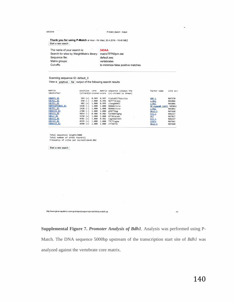
140
Supplemental Figure 7. Promoter Analysis of Bdh1. Analysis was performed using P-
Match. The DNA sequence 5000bp upstream of the transcription start site of Bdh1 was
analyzed against the vertebrate core matrix.

141
Appendix 4: Supplemental Figures for Discussion
Supplemental Figure 8: E2F1 is decreased after dox (0.5μM 24hr) exposure in
neonatal cardiomyocytes. Western blot examined with anti E2F1. E2F1 is denoted by the
* (correct band determined by comparison to E2F1 expression vector).

142
Supplemental Figure 9: E2F6 is down-regulated post-transcriptionally in the
myocardium after birth. (A) Fold induction of E2F family members in the heart at
various stages during murine development. Fold Inductions were calculated in reference
to GAPDH (n=6). (B) Representative western blot of cardiac lysates from mice at various
stages during development probed with anti-E2F6. (C) Quantification of E2F6 protein in
the mouse heart in comparison to GAPDH.
0
0.5
1
1.5
2
2.5
3
E2F1 E2F3A E2F3B E2F4 E2F6
Fold
ind
uct
ion
E-16
E-20
P-1
P-7
6Wk

143
Supplemental Figure 10: E2F6 inhibits myoblast fusion. Transient transfection of
C2C12 with (A) 6-myc or (B) E2F6. Cells were stained with mf20 (red) and DAPI (blue).
Fusion index (C) n=8 fields of view. ** P-value<0.01

144
Supplemental Figure 11: E2F6 is down-regulated post-transcriptionally during C2C12
differentiation Day 0-Day 4. (A) Fold induction of E2F6 transcript relative to GAPDH
(n=3). (B) Western blot analysis of E2F6 protein levels during differentiation. Over-
expression of E2F6 in C2C12 is shown on the right as a positive control for anti-E2F6.