early carcinoma of the esophagus associated with …mj-med-u-tokai.com/pdf/330103.pdf · early...
TRANSCRIPT

― 13―
Tokai J Exp Clin Med., Vol. 33, No. 1, pp. 13-16, 2008
IntroductIon
Achalasia is an idiopathic primary esophageal mo-tor disorder characterized by insufficient relaxation of the lower esophageal sphincter to muscle and the absence of esophageal peristalsis. Decreased and/or di-minished myenteric ganglia have been reported in the lower esophagus (1, 2). Achalasia is known as a risk factor of esophageal carcinoma (3, 4, 5). Fagge first reported the relationship between achalasia and esoph-ageal carcinoma in 1872 by their autopsy findings (6). Since this initial observation, the increased risk of developing esophageal carcinoma has been postulated in the patients with long-standing achalasia (7). The incidence of carcinoma in achalasia patients has been reported to range from 1.7 to 20% (3, 8-17). Chronic inflammatory irritation by retained food may induce carcinogenesis of the esophageal squamous epithelium. In this report, a case of endoscopically resected early esophageal carcinoma developing after the operation for achalasia is described.
cAse rePort
A 63-year-old woman was suffering from dysphagia to solids and liquids from 1992. She had been treated conservatively, such as calcium-channel blocker and pneumatic dilatation for symptomatic achalasia in the other hospital. But, her symptom became worse pro-gressively, and she noted a 5kg weight loss over in four
months. The patient was admitted to Tokai University Hospital on November 5, 2002 for evaluation of dysphagia. Physical examination at admission did not reveal abnormalities. Laboratory data were within nor-mal limits, including tumor makers such as SCC and CEA, except for slight anemia.
Esophagography of double contrast examination demonstrated marked dilatation of the esophagus proximal to gastroesophageal junction, measuring 7.5cm in diameter (Figure1). The Upper GI series also showed sigmoid-type elongation and aperistalsis of the distal half of the esophagus. Conventional endoscopic examination revealed the dilatated and atonic esopha-gus with a large amount of diet staying inside. The entire esophageal mucosa demonstrated significant hyperplastic changes of stratified squamous epithe-lium, but iodine-unstained lesion was not detected by chromoendoscopic examination (Figure 2A, B). The preoperative diagnosis was achalasia, sigmoid type and Grade III (18). The surgical operation by Tokai University method (19), Heller’s long esophagomyec-tomy, Hill’s posterior cardiopexy, fundoplication (4/5 of circumference) and selective proximal vagotomy us-ing a laparotomy with upper median abdominal skin incision, were performed on November 19, 2002. The postoperative course was uneventful and the patient was discharged two weeks later. She has been well without complaint of dysphagia after the operation.
Endoscopic examination with iodine staining was
early carcinoma of the esophagus Associated with Achalasia treated by endoscopic mucosal resection: report of A case
Osamu CHINO1, Hideo SHIMADA1, Yoshifumi KISE1, Takayuki NISHI1, Tadashi HARA1, Soichiro YAMAMOTO1, Makiko TANAKA2, Hiroshi KAJIWARA3, Hiroshi KIJIMA4,
Hiroyasu MAKUUCHI1
Departments of Surgery1, Critical Care and Emergency Medicine2, Pathology3, Tokai University School of Medicine Department of Pathology4, Hirosaki University School of Medicine
(Received October 16, 2007; Accepted December 10, 2007)
A case of endoscopically resected early esophageal carcinoma associated with achalasia is reported. A 63-year-old woman was made diagnosis of esophageal achalasia, sigmoid type and grade III. the patient was operated by tokai university method, Heller’s long esophagomyectomy, Hill’s posterior cardiopexy, fundoplication and selective proximal vagotomy using a laparotomy. two years and six months after the operation, an early carcinoma of type 0-IIb, 1cm in size, was detected in the upper thoracic esophagus, and treated by endoscopic mucosal resection using eemr-tube method. Pathological examination revealed pro-liferation of squamous cell carcinoma in situ (tis: m1). the entire esophageal mucosa around the carcinoma demonstrated hyperplastic changes of stratified squamous epithelium and foci of dysplastic changes. In the patient of achalasia, food stasis in esophagus is thought to induce chronic hyperplastic esophagitis, convert-ing eventually to malignant transformation. Achalasia is known as a risk factor of esophageal carcinoma. early operation or good drainage of the esophageal lumen might reduce the risk. long-term follow-up for patients of achalasia by endoscopic screening is recommended.
key words: Achalasia of esophagus, esophageal carcinoma, carcinogenesis, endoscopic mucosal resection
Osamu Chino, Department of Surgery, Tokai University School of Medicine, 143 Shimokasuya, Isehara, Kanagawa, 259-1193 Japan Tel: +81-463-93-1121 Fax: +81-463-95-6491 E-mail: [email protected]

O. CHINO et al. /Esophageal carcinoma in achalasia.
― 14―
performed every six months and esophagitis was well controlled without marked hyperplastic change after the operation. Two years and six months after the op-eration, an early carcinoma of type 0-IIb, 1cm in size, was detected at 23cm from incisor in the upper thorac-ic esophagus by endoscopy (Figure 3A,B). Computed tomography and echogram showed no evidence of metastasis. Endoscopic mucosal resection (EMR) using
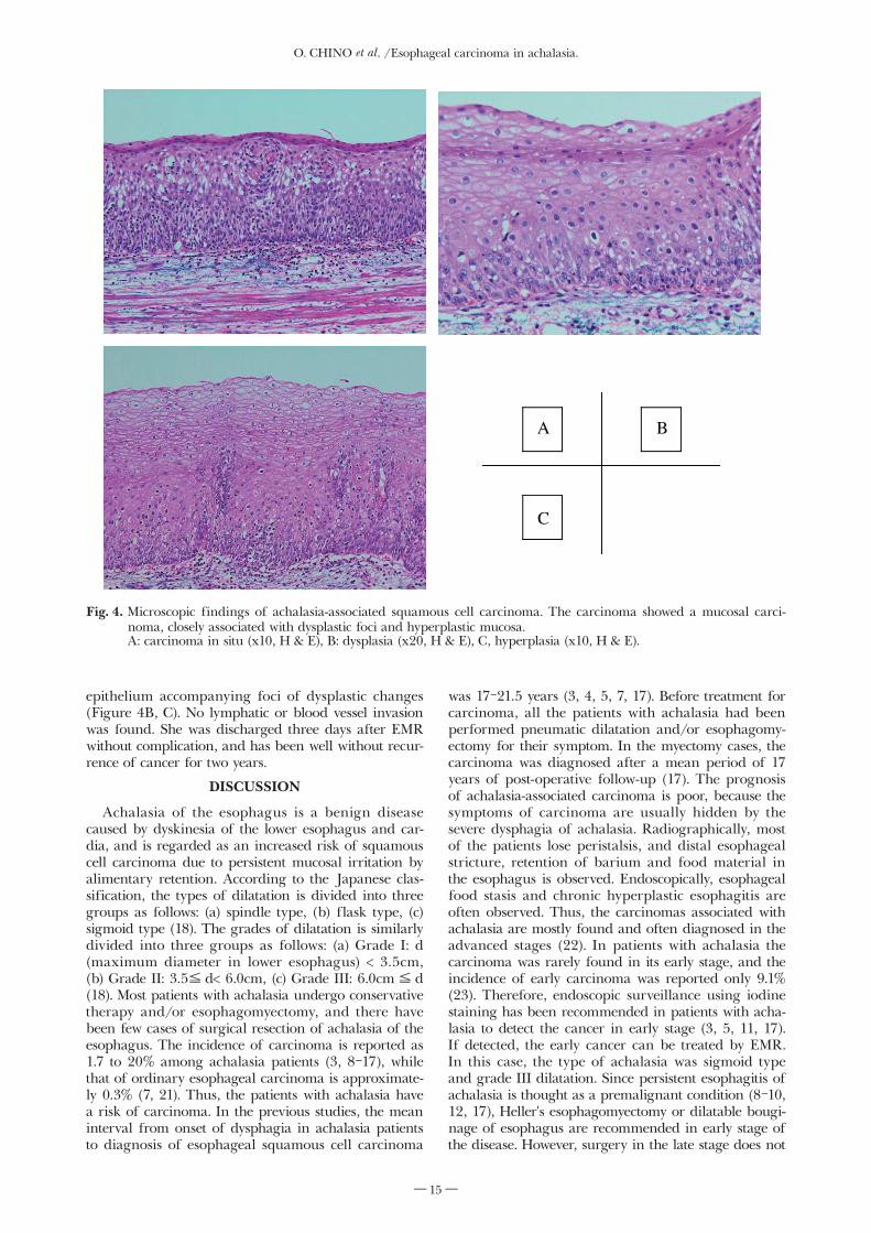
EEMR-tube method was performed on October 13, 2005 (Figure 3C, D). Pathological examination of the resected specimen revealed proliferation of squamous cell carcinoma, closely associated with dysplastic foci. The invasion of carcinoma cells was limited to the squamous epithelium (Tis: m1) (Figure 4A) (20). The entire esophageal mucosa around the carcinoma dem-onstrated hyperplastic changes of stratified squamous
Fig. 2. Endoscopic view of achalasia in preoperative status. A: Conventional endoscopic examination revealed the presence of a dila-
tated and atonic esophagus with marked hyperplastic changes of stratified squamous epithelium, B: iodine-unstained lesion was not detected in the upper thoracic esophagus by chromoendoscopic examination.
Fig. 1. Esophagography of double contrast examination dem-onstrated marked dilatation of the esophagus proximal to gastroesophageal junction, measuring 7.5cm in diameter. The patient was made diag-nosis as esophageal achalasia, sigmoid type and Grade III.
Fig. 3. An early carc inoma of type 0 - I Ib, 1cm in size, developed at 23cm from incisor in the upper thoracic esophagus after the opera-tion. Endoscopic mucosal resection using EEMR-tube method was performed. A: conventional endoscopic view, B: chromoendoscopic view with iodine staining, C: endoscopic mucosal resec-tion, D: macroscopic view of the resected specimen by EMR with iodine staining.
A B
A B
C D
O. CHINO et al. /Esophageal carcinoma in achalasia.
― 15―
epithelium accompanying foci of dysplastic changes (Figure 4B, C). No lymphatic or blood vessel invasion was found. She was discharged three days after EMR without complication, and has been well without recur-rence of cancer for two years.
dIscussIon
Achalasia of the esophagus is a benign disease caused by dyskinesia of the lower esophagus and car-dia, and is regarded as an increased risk of squamous cell carcinoma due to persistent mucosal irritation by alimentary retention. According to the Japanese clas-sification, the types of dilatation is divided into three groups as follows: (a) spindle type, (b) flask type, (c) sigmoid type (18). The grades of dilatation is similarly divided into three groups as follows: (a) Grade I: d (maximum diameter in lower esophagus) < 3.5cm, (b) Grade II: 3.5≦ d< 6.0cm, (c) Grade III: 6.0cm≦ d (18). Most patients with achalasia undergo conservative therapy and/or esophagomyectomy, and there have been few cases of surgical resection of achalasia of the esophagus. The incidence of carcinoma is reported as 1.7 to 20% among achalasia patients (3, 8-17), while that of ordinary esophageal carcinoma is approximate-ly 0.3% (7, 21). Thus, the patients with achalasia have a risk of carcinoma. In the previous studies, the mean interval from onset of dysphagia in achalasia patients to diagnosis of esophageal squamous cell carcinoma
was 17-21.5 years (3, 4, 5, 7, 17). Before treatment for carcinoma, all the patients with achalasia had been performed pneumatic dilatation and/or esophagomy-ectomy for their symptom. In the myectomy cases, the carcinoma was diagnosed after a mean period of 17 years of post-operative follow-up (17). The prognosis of achalasia-associated carcinoma is poor, because the symptoms of carcinoma are usually hidden by the severe dysphagia of achalasia. Radiographically, most of the patients lose peristalsis, and distal esophageal stricture, retention of barium and food material in the esophagus is observed. Endoscopically, esophageal food stasis and chronic hyperplastic esophagitis are often observed. Thus, the carcinomas associated with achalasia are mostly found and often diagnosed in the advanced stages (22). In patients with achalasia the carcinoma was rarely found in its early stage, and the incidence of early carcinoma was reported only 9.1% (23). Therefore, endoscopic surveillance using iodine staining has been recommended in patients with acha-lasia to detect the cancer in early stage (3, 5, 11, 17). If detected, the early cancer can be treated by EMR. In this case, the type of achalasia was sigmoid type and grade III dilatation. Since persistent esophagitis of achalasia is thought as a premalignant condition (8-10, 12, 17), Heller's esophagomyectomy or dilatable bougi-nage of esophagus are recommended in early stage of the disease. However, surgery in the late stage does not
Fig. 4. Microscopic findings of achalasia-associated squamous cell carcinoma. The carcinoma showed a mucosal carci-noma, closely associated with dysplastic foci and hyperplastic mucosa.
A: carcinoma in situ (x10, H & E), B: dysplasia (x20, H & E), C, hyperplasia (x10, H & E).
C
BA

O. CHINO et al. /Esophageal carcinoma in achalasia.
― 16―
seem to be effective for achalasia-mediated carcinogen-esis.
In the previous studies, histological examination of the resected esophageal specimens demonstrated marked hyperplastic changes of stratified squamous epithelium and multiple foci of dysplastic changes. The squamous cell carcinomas were of well to moderately differentiated type with low-grade atypia, closely as-sociated with dysplastic foci (17, 24, 25). We speculate that food stasis induces chronic hyperplastic esopha-gitis, leading eventually to malignant transformation of esophageal epithelial cells, in accordance with dysplasia-carcinoma sequence (8-10, 12, 17).
The previous immunohistochemical investigations regarding p53 accumulation of esophageal squamous cell carcinoma show frequent over-expression of p53 in both achalasia-associated carcinoma and dysplasia (17, 26, 27). Immunohistochemical studies of p21 and p16 expression suggest that the cell cycle might be increased in achalasia-associated carcinoma because of persistent inflammation (17, 27-30). Under these con-ditions of accelerated cell cycle, the achalasia-associated carcinoma may develop according to the dysplasia-carcinoma sequence.
In conclusion, we reported a case of early esopha-geal carcinoma associated with postoperative achalasia treated by EMR. Achalasia is known as a risk factor of esophageal carcinoma. Long-term follow-up for patients of achalasia by endoscopic screening is recom-mended.
Acknowledgements
The authors express the sincere thanks to Akihiko Serizawa M.T. (Department of Pathology, Tokai University School of Medicine) for the technical assis-tance and cooperation.
reFerences
1) Goldblum JR, Rice TW, and Richter JE: Histopathologic fea-tures in esophagomyotomy specimens from patients with achala-sia. Gastroenterology 111: 648-654, 1996.
2) Goldblum JR, Whyte RI, Orringer MB and Appleman HD: Achalasia. A morphologic study of 42 resected specimens. Am J Surg Pathol 18: 327-337, 1994.
3) Meijssen MAC, Tilanus HW, Blankenstein M, Hop WCJ, and Ong GL: Achalashia complicated by oesophageal squamous cell carcinoma: a prospective study in 195 patients. Gut 33: 155-158, 1992.
4) Sandler RS, Nyren O, Ekbom A, Eisen GM, Yuen J and Josefsson S: The risk of esophageal cancer in patients with acha-lasia. JAMA 274: 1359-1362, 1995.
5) Streitz Jr JM, Ellis Jr FH, Gibb SP, and Heatley GM: Achalasia and squamous cell carcinoma of the esophagus: Analysis of 241 patients. Ann Thorac Surg 59: 1604-1609, 1995.
6) Fagge CH: A case of simple stenosis of the esophagus, followed by epithelioma. Guy’s Hosp Rep 17: 413, 1872.
7) Seliger G, Lee T, and Schwartz S: Carcinoma of the proximal esophagus, a complication of long-standing achalasia. Am J Gastoenterol 57: 20-25, 1972.
8) Pierce WS, MacVaugh H III, and Johnson J: Carcinoma of the esophagus arising in patients with achalaisa of cardia. J Thorac Cardiovasc Surg 59: 335-339, 1970.
9) Carter R, and Brewer LA: Achalasia and esophageal carcinoma: studies in early diagnosis for improved surgical management. Am J Surg 130: 114-120, 1975.
10) Just-Viera JO, and Haight C: Achalasia and carcinoma of the esophagus. Surg Gynecol Obstet 128: 1081-1095, 1969.
11) Lortat-Jacob JL , Richard CA, Fekete F, and Testart J:
Cardiopasm and esophageal carcinoma: report of 24 cases. Surgery 66: 969-975, 1969.
12) Wychulis AR, Woolam GL, Anderson HA, and Ellis FH: Achalasia and carcinoma of the esophagus. JAMA 215: 1638-1641, 1971.
13) Benedict EB, and Joske RA: The role of benign esophageal obstruction in thedevelopment of carcinoma of the esophagus. J Thorac Surg 36: 749-755, 1959.
14) Camera-Lopes LH: Carcinoma of the esophagus as a complica-tion of mega-esophagus: an analysis of seven cases. Am J Dig Dis 6: 742-756, 1961.
15) Harkins JR, and McLaughlin JS: The association of carcinoma of the esophagus with achalasia. J Thorac Crdiovasc Surg 69: 355-360, 1975.
16) Norton GA, Postlethwait RW, and Thompson WM: Esophageal carcinoma: A summary of populations at risk. Suoth Med J 73: 23-27, 1980.
17) Chino O, Kijima H, Shimada H, Nishi T, Tanaka H, Oshiba G, Kise Y, Kajiwara H, Tsuchida T, Tnakaka M, Tajima T, Makuuchi H: Clinicopathological studies of esophageal carci-noma in achalasia: Analyses of carcinogenesis using histological and immunohistochemical procedures. Anticancer Research 20: 3717-3722, 2000.
18) Japanese Society for Esophageal Disease (ed). Descriptive Rules for Achalasia of the esophagus, 3rd ed. Tokyo: Kanehara Inc, 1983.
19) Makuuchi H, Machimura T, Soh Y, Shimada H, Mizutani K, Kanno K, Sugihara T, Sasaki T, Tajima T, Mitomi T. Pathophysiology of esophago -gastric junction and op -eration in patients with achalasia. The Japanese Journal of Gastroenterological Surgery 23: 2477-2481, 1990 (Abstract in English).
20) Japanese Society for Esophageal Disease (ed). Guide lines for the Clinical and Pathologic Studies on Carcinoma of the Esophagus, 10th ed. Tokyo: Kanehara Inc, 2007.
21) Ackerman L, and Del Regato J. Cancer, St.Louis, C.V. Mosby & Co., 1962.
22) Peracchia A, Segalin A, Bardini R, Roul A, Baessato M. Esophageal carcinoma and achalasia: Prevalence, incidence and results of treatment. Hepato-Gastroenterol 38: 514-516, 1991.
23) Mine H, Nakamura T, Kohno H, Yaita A, Rai Y, Tohgi K, Nakamori H, Kubota H, Masuo K. Evaluation of esophageal carcinoma concomitant with achalasia –A review collected from 139 institutes in Japan-. General Thoracic and Cardiovascular Surgery 32: 2041-2047, 1984 (Abstract in English).
24) Porschen R, Molsberger G, Kuhn A, Sarbia M, and Borchard F: Achalasia-associated squamous cell carcinoma of the esophagus: Flow-cytometric and histological evaluation. Gastroenterolgy 108: 545-549, 1995.
25) Loviscek LF, Cenoz MC, Badaloni AE, and Agarinakazato O: Early cancer in achalasia. Disease of the Esophagus 11: 239-247, 1998.
26) Moskaluk CA, Heitmiller R, Zahurak M, Schwab D, Siransky D, and Hamilton SR: p53 and p21WAF1/CIP1/SDI1 gene products in Barrett esophagus and adenocarcinoma of the esophagus and esophagogastric junction. Human Pathology 27: 1211-1220, 1996.
27) Chino O, Kijima H, Shimada H, Nishi T, Tanaka H, Kise Y, Kenmochi T, Himeno S, Machimura T, Tanaka M, Inokuchi S, Tajima T, Osamura Y. Makuuchi H: Accumulation of p53 in esophageal squamous cell carcinoma. International Journal of Molecular Medicine 8: 359-363, 2001.
28) Ohashi K, Nemoto T, Eishi Y, Matsuno A, Nakamura K, and Hirokawa K. Expression of the cyclin dependent kinase inhibitor p21WAF1/CIP1 in oesophageal squamous cell carcinomas. Virchows Arch 430: 389-395, 1997.
29) El-Deiry WS, Tokino T, Velculescu VE, Levy DB, Parsons R, Trent JM, Lin D, Mercer WE, Kinzler KW, and Vogelstein B: WAF1, a potential mediator of p53 tumor suppression. Cell 75: 817-825, 1993.
30) Igaki H, Sasaki H, Tachimori Y, KatoH, Watanabe H, Kimura T, Harada Y, Sugimura T, and Terada M: Mutation frequency of the p16/CDKN2 gene in primary cancers in upper digestive tract. Cancer Research 55: 3421-3423, 1995.