early childhood in focus 7 - open research onlineoro.open.ac.uk/33493/1/developing_brains.pdf ·...
TRANSCRIPT

EARLY CHILDHOOD IN FOCUS 7
Developing Brains
EARLY CHILDHOOD IN FOCUSSeries edited by Martin Woodhead and John Oates
Early Childhood in Focus is a series of publications produced by the Child and Youth Studies Group at The Open University, United Kingdom, with the support of the Bernard van Leer Foundation.
The series provides accessible and clear reviews of the best and most recent available research, information and analysis on key policy issues, offering clear messages on core policy topics and questions, spanning all aspects of early childhood care and education, as well as the full age range, from infancy through to the early years of school.
Each publication is developed in consultation with world leaders in research, policy, advocacy and children’s rights. Many of these experts have written summaries of key messages from their areas of work especially for the series, and the accuracy of the content has been assured by independent academic assessors, themselves experts in the field of early childhood.
The themes of the series have been chosen to reflect topics of research and knowledge development that address the most significant areas of children’s rights, and where a deeper understanding of the issues involved is crucial to the success of policy development programmes and their implementation.
These publications are intended to be of value to advocates for the rights of children and families, to policy makers at all levels, and to anyone working to improve the living conditions, quality of experience and life chances of young children throughout the world.
Developing Brains
Editors
John OatesAnnette Karmiloff-Smith
Mark H. Johnson
EARLY CHILDHOOD IN FOCUS 7
SERIES EDITORSMartin WoodheadJohn OatesChild and Youth Studies GroupThe Open UniversityMilton Keynes, United Kingdom
SERIES ADVISERRobert Myers, independent consultant, Mexico
To obtain further copies of this and other publications in the Early Childhood in Focus series, visit: www.bernardvanleer.org
Also in the series:Attachment RelationshipsEarly Childhood and Primary EducationDeveloping Positive IdentitiesEffective Early Childhood ProgrammesSupporting ParentingCulture and Learning
Copyright © 2012 The Open University First published 2012 by The Open UniversityChild and Youth Studies GroupThe Open UniversityWalton Hall, Milton KeynesMK7 6AAUnited Kingdom
with the support of:Bernard van Leer FoundationPO Box 823342508 EH The HagueNetherlands
All rights reserved. No part of this publication may be reproduced, stored in a retrieval system, transmitted or utilised in any form or by any means, electronic, mechanical, photocopying, recording or otherwise, without written permission from the publisher or a licence from the Copyright Licensing Agency Ltd. Details of such licences may be obtained from the Copyright Licensing Agency Ltd, Saffron House, 6–10 Kirby Street, London EC1N 8TS.
A catalogue record for this title is available from the British Library.
Designed by Agnes BorszekiText edited by Margaret MellorPrinted in the United Kingdom by Cambrian Printers, AberystwythISBN 978-1-78007-321-7

The foundations of brain architecture are established early in life through a continuous series of dynamic interactions in which environmental conditions and personal experiences have a significant impact on how genetic predispositions are expressed. Because specific experiences affect specific brain circuits during specific developmental stages – referred to as sensitive periods – it is vitally important to take advantage of these early opportunities in the developmental building process. That is to say, the quality of a child’s early environment and the availability of appropriate experiences at the right stages of development are crucial in determining the strength or weakness of the brain’s architecture, which, in turn, determines how well he or she will be able to think and to regulate emotions.
(National Scientific Council on the Developing Child, 2007, p. 1)
Without the threats of biological and psychosocial risks, and with a caregiving environment that supports cognitive and social–emotional development, children experience healthy brain development that enables them to reach toward their developmental potential. With this strong foundation, they build lifespan developmental trajectories that enable them to benefit from family, community, and educational opportunities …. Interventions to reduce risks and support early child development will yield lifetime gains that contribute to the achievement and sustainability of improved development in the next generation.
(Walker et al., 2011, p. 1335)
The emerging science of brain development shows that to develop properly, a child’s growing brain needs nurturing long before formal schooling starts at age 6 or 7. Investments in prenatal health and early childhood development programs that include education and health are essential to realize this potential.
(World Bank, 2011, p. 4)
EDITORSJohn Oates, Senior Lecturer in Developmental Psychology, Child and Youth Studies Group, The Open University, United Kingdom
Annette Karmiloff-Smith, Professorial Research Fellow, Developmental Neurocognition Lab, Centre for Brain and Cognitive Development, Birkbeck, University of London, United Kingdom
Mark H. Johnson, Director, Centre for Brain and Cognitive Development, Birkbeck, University of London, United Kingdom
CONTRIBUTORSMichelle de Haan, Reader, Centre for Developmental Cognitive Neuroscience, University College London, United Kingdom
Ellie Dommett, Lecturer in Psychology, Brain and Behavioural Sciences, The Open University, United Kingdom
Teodora Gliga, Research Fellow, Centre for Brain and Cognitive Development, Birkbeck, University of London, United Kingdom
Elizabeth Isaacs, Senior Research Fellow, Institute of Child Health, University College London, United Kingdom
Eamon McCrory, Senior Lecturer, Developmental Risk and Resilience Unit, University College London, United Kingdom
Charles A. Nelson, Professor of Paediatrics, Children’s Hospital Boston/Harvard Medical School and Harvard Center on the Developing Child, United States of America
Gaia Scerif, Lecturer, Attention, Brain and Cognitive Development Group, Department of Experimental Psychology, University of Oxford, United Kingdom
Núria Sebastián-Gallés, Director, SAP Research Group, Universitat Pompeu Fabra, Barcelona, Spain
Joan Stiles, Professor, Department of Cognitive Science, University of California, San Diego, United States of America
Faraneh Vargha-Khadem, Professor of Developmental Cognitive Neuroscience, University College London Institute of Child Health, and Great Ormond Street Hospital for Children, United Kingdom
ACADEMIC ASSESSORSarah-Jayne Blakemore, Professor of Cognitive Neuroscience, Institute of Cognitive Neuroscience, University College London, United Kingdom

ContentsPreface ....................................................................................................ix
I. Children’s brains ............................................................................ 1
The structure of the human brain .......................................................2 Localisation of function .......................................................................4
Building blocks of the brain ................................................................6 Development of the cerebral cortex ....................................................8 Neurotransmitters .............................................................................10 Systems and pathways ......................................................................12 Unique brain; unique child ...............................................................14
Brain imaging methods .....................................................................16
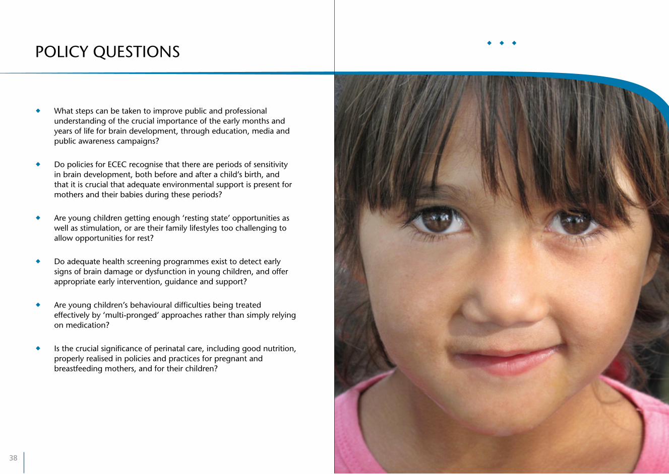
POLICY QUESTIONS .........................................................................18
II. What develops? .......................................................................... 21
Before birth ......................................................................................22 Neural growth and pruning ..............................................................24 Myelination and cognitive development ...........................................26 Sensitive periods ...............................................................................28 Localisation and lateralisation ............................................................30 Networks and resting states ..............................................................32 The social brain .................................................................................34 Objects of attention ..........................................................................36
POLICY QUESTIONS .........................................................................38
III. Environmental influences ....................................................... 41
Development of the visual system .....................................................42 Language acquisition in the monolingual and bilingual child ............44 The importance of sleep for learning ................................................46 Diet and nutrition in brain development ...........................................48 The effects of early psychosocial deprivation .....................................50 Maltreatment, genetics and brain development ................................52
POLICY QUESTIONS .........................................................................54
References ...............................................................................................56
Illustrations ........................................................................................... 60

ix
PrefaceThe language of neuroscience is increasingly being used to support assertions about many aspects of human life. A recent series of studies (Weisberg et al., 2008) found that explanations of human behaviour that included irrelevant neuroscience words, such as ‘frontal lobe brain circuitry’, were rated by non-experts as significantly more credible than explanations without the extra words. This should alert us to the need to ‘look behind the headlines’ of popular reporting of neuroscience findings and not to accept at face value arguments that rely on ‘latest findings by brain scientists’ to make their points.
The effects of early experiences on children’s development have been especially subject to ‘latest findings’ reporting. There is good scientific justification for many of the insights that are being offered; scientists around the world have been using the new tools and methods of developmental neuroscience to great effect, and there are important findings that have been replicated and have great relevance for policies influencing children’s lives. But there is also a risk in reporting this research – of overstating what is known and the policy implications. This volume in the Early Childhood in Focus series aims to present an overview of the most significant areas of research, starting in the first section with sufficient basic explanation of the brain and how it functions for the research to be understood by people with little or no previous knowledge of the field.
The second section gives an overview of the developmental processes involved as the child’s brain grows and matures in constant interaction with the environment, from conception through to adulthood. For brain development continues through life as experience builds memories and learning, shaping structures and functions of the brain. But the 9 months before birth and the early years of a child’s life include especially important and sensitive periods, because the growth and development of the brain’s architecture and processes are vastly greater during this time than later.
Enough is now known to be able to say clearly that, for children to reach their full potential, supporting the healthy development of their brains is paramount and the powerful effects of early environments, both physical and social, can no longer be ignored. The third section of this volume focuses on these influences.
It is important to recognise that research into the development of children’s brains is still a field of study in its early stages, with many unanswered questions. Thus, for those concerned with making use of knowledge from this field as the rationale for interventions and support for children and families, it would be rash to accept without question strong assertions about essential and very specific ingredients for healthy brain development, especially assertions that seek to justify particular approaches to childcare and education.
We hope that this volume will help you to better understand and critically evaluate reports of new findings from the expanding and increasingly important field of developmental neuroscience.
John Oates, Annette Karmiloff-Smith, Mark H. Johnson, Editors

As a child’s brain develops, the various parts become progressively more specialised as specific neural circuits develop for specific functions.
Although functions become localised to an extent, the brain is a complex organ in which many parts work in unison.
Early brain development is dependent on appropriate experiences; the young brain is a very responsive and ‘plastic’ part of the body, with a vast number of neurons and connections between them.
Pathways between parts of the brain become established along the more active connections, forming systems supporting different sensory, cognitive, emotional and behavioural functions.
The uniqueness of each child results from the complex actions between genes that control brain growth and formative experiences from the child’s environment, involving both sensitivity and resilience.
Children’s brainsI.
2
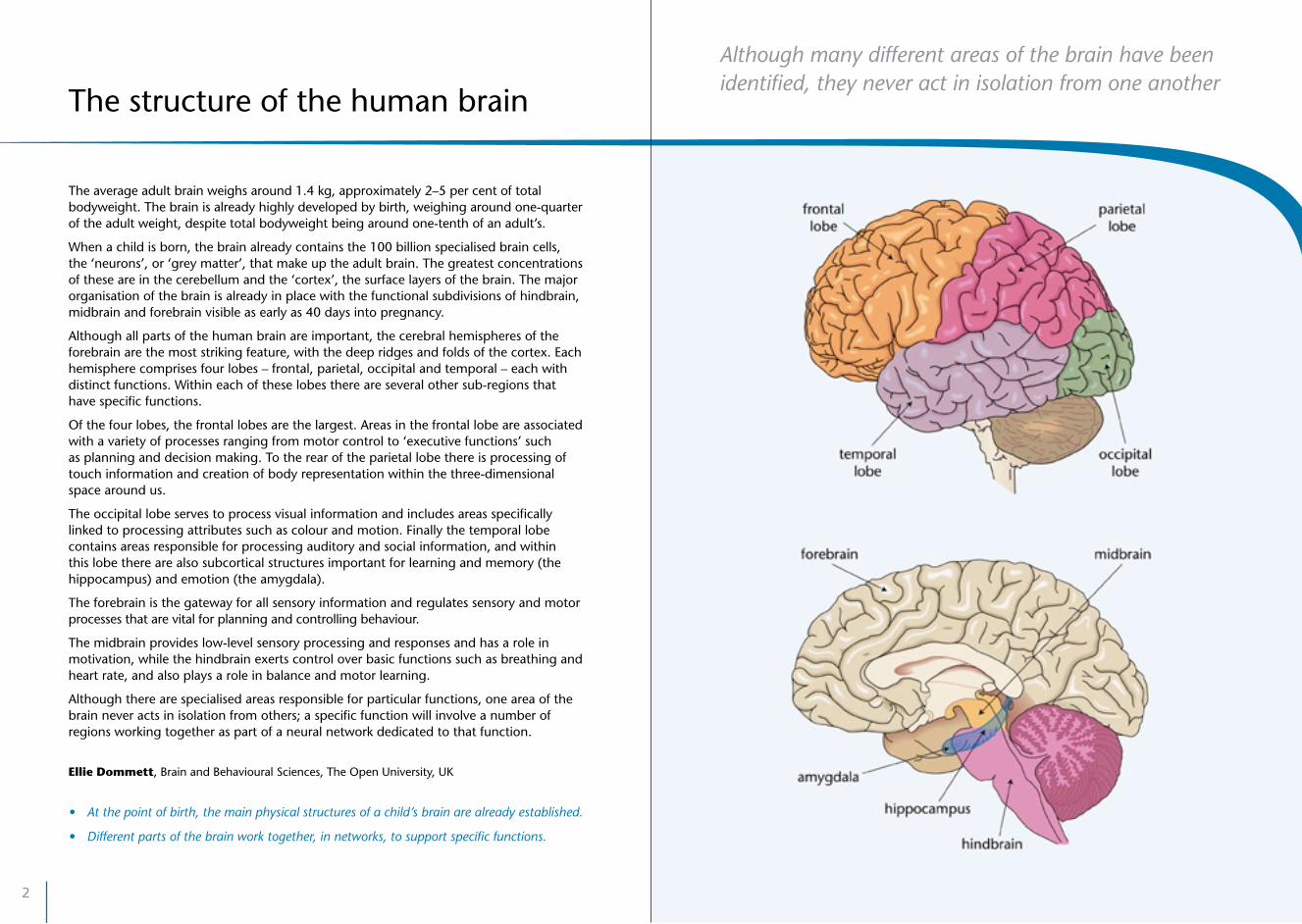
Although many different areas of the brain have been identified, they never act in isolation from one another
The average adult brain weighs around 1.4 kg, approximately 2–5 per cent of total bodyweight. The brain is already highly developed by birth, weighing around one-quarter of the adult weight, despite total bodyweight being around one-tenth of an adult’s.
When a child is born, the brain already contains the 100 billion specialised brain cells, the ‘neurons’, or ‘grey matter’, that make up the adult brain. The greatest concentrations of these are in the cerebellum and the ‘cortex’, the surface layers of the brain. The major organisation of the brain is already in place with the functional subdivisions of hindbrain, midbrain and forebrain visible as early as 40 days into pregnancy.
Although all parts of the human brain are important, the cerebral hemispheres of the forebrain are the most striking feature, with the deep ridges and folds of the cortex. Each hemisphere comprises four lobes – frontal, parietal, occipital and temporal – each with distinct functions. Within each of these lobes there are several other sub-regions that have specific functions.
Of the four lobes, the frontal lobes are the largest. Areas in the frontal lobe are associated with a variety of processes ranging from motor control to ‘executive functions’ such as planning and decision making. To the rear of the parietal lobe there is processing of touch information and creation of body representation within the three-dimensional space around us.
The occipital lobe serves to process visual information and includes areas specifically linked to processing attributes such as colour and motion. Finally the temporal lobe contains areas responsible for processing auditory and social information, and within this lobe there are also subcortical structures important for learning and memory (the hippocampus) and emotion (the amygdala).
The forebrain is the gateway for all sensory information and regulates sensory and motor processes that are vital for planning and controlling behaviour.
The midbrain provides low-level sensory processing and responses and has a role in motivation, while the hindbrain exerts control over basic functions such as breathing and heart rate, and also plays a role in balance and motor learning.
Although there are specialised areas responsible for particular functions, one area of the brain never acts in isolation from others; a specific function will involve a number of regions working together as part of a neural network dedicated to that function.
Ellie Dommett, Brain and Behavioural Sciences, The Open University, UK
• Atthepointofbirth,themainphysicalstructuresofachild’sbrainarealreadyestablished.
• Differentpartsofthebrainworktogether,innetworks,tosupportspecificfunctions.
The structure of the human brain
4
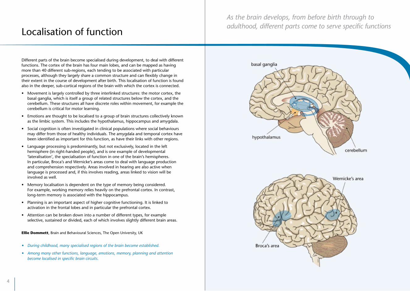
As the brain develops, from before birth through to adulthood, different parts come to serve specific functions
Different parts of the brain become specialised during development, to deal with different functions. The cortex of the brain has four main lobes, and can be mapped as having more than 40 different sub-regions, each tending to be associated with particular processes, although they largely share a common structure and can flexibly change in their extent in the course of development after birth. This localisation of function is found also in the deeper, sub-cortical regions of the brain with which the cortex is connected.
• Movementislargelycontrolledbythreeinterlinkedstructures:themotorcortex,thebasal ganglia, which is itself a group of related structures below the cortex, and the cerebellum. These structures all have discrete roles within movement, for example the cerebellum is critical for motor learning.
• Emotionsarethoughttobelocalisedtoagroupofbrainstructurescollectivelyknownas the limbic system. This includes the hypothalamus, hippocampus and amygdala.
• Socialcognitionisofteninvestigatedinclinicalpopulationswheresocialbehavioursmay differ from those of healthy individuals. The amygdala and temporal cortex have been identified as important for this function, as have their links with other regions.
• Languageprocessingispredominantly,butnotexclusively,locatedinthelefthemisphere (in right-handed people), and is one example of developmental ‘lateralisation’, the specialisation of function in one of the brain’s hemispheres. In particular, Broca’s and Wernicke’s areas come to deal with language production and comprehension respectively. Areas involved in hearing are also active when language is processed and, if this involves reading, areas linked to vision will be involved as well.
• Memorylocalisationisdependentonthetypeofmemorybeingconsidered. For example, working memory relies heavily on the prefrontal cortex. In contrast, long-term memory is associated with the hippocampus.
• Planningisanimportantaspectofhighercognitivefunctioning.Itislinkedtoactivation in the frontal lobes and in particular the prefrontal cortex.
• Attentioncanbebrokendownintoanumberofdifferenttypes,forexampleselective, sustained or divided, each of which involves slightly different brain areas.
Ellie Dommett, Brain and Behavioural Sciences, The Open University, UK
• Duringchildhood,manyspecialisedregionsofthebrainbecomeestablished.
• Amongmanyotherfunctions,language,emotions,memory,planningandattentionbecome localised in specific brain circuits.
Localisation of function

6
Many different cell types work together in the brain to form and support communicating networks
The brain is made up of around 100 billion specialised cells called neurons.
Each neuron consists of four key parts that allow it to perform its function.
• Dendrites: branch-like protrusions from the neuron, which serve to receive incoming signals from other neurons.
• Cell body: the hub of the neuron, which serves to integrate all incoming information by summing the signals together.
• Axon: a long fibre, along which electrical impulses, ‘action potentials’, are transmitted.
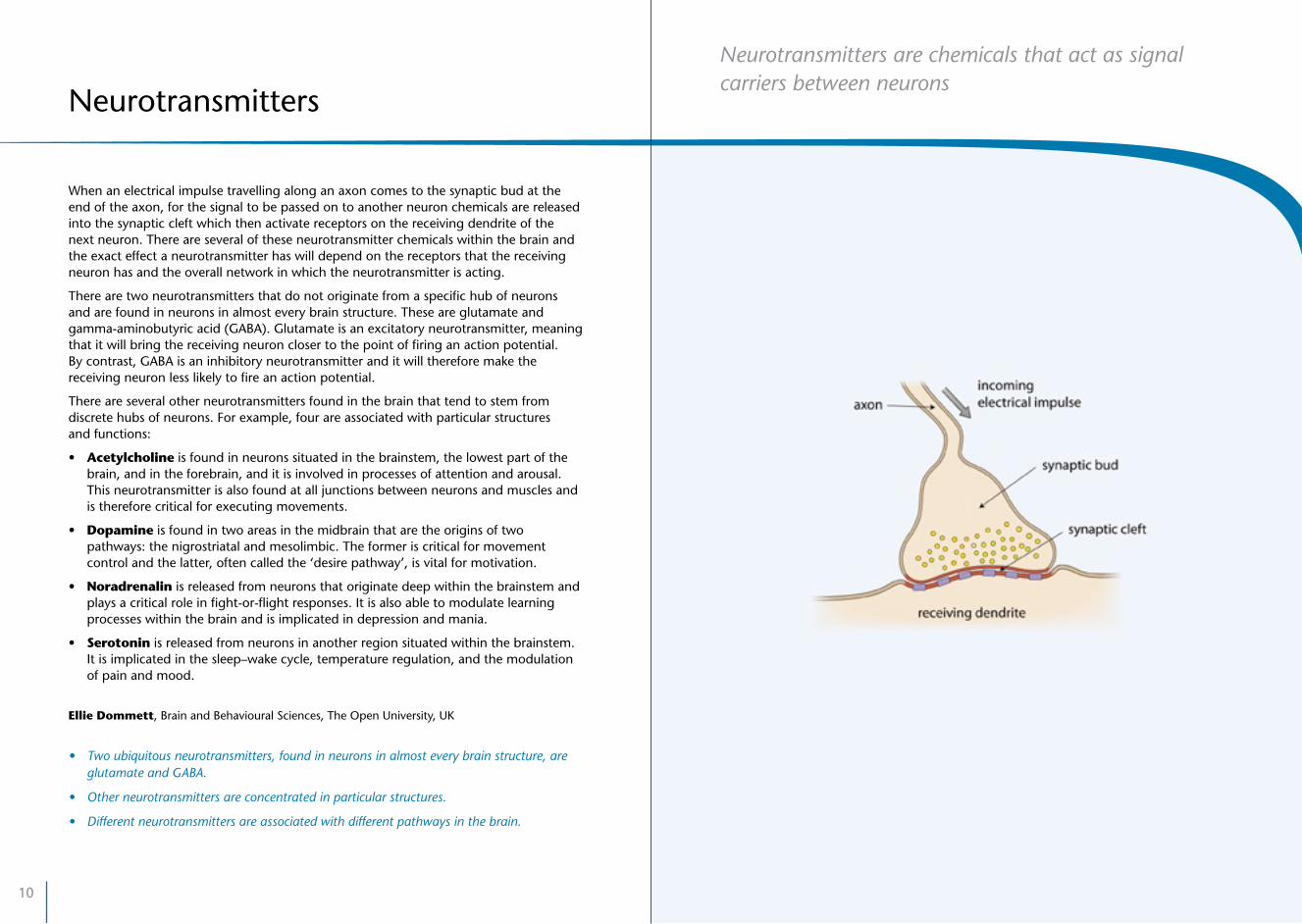
• Axon terminals: these are points at the end of an axon where the signal passes to another axon. In most of these the signal is converted from an electrical signal into a chemical one for transmission to the next neuron.
At the axon terminals the neuron will contact the dendrites of another neuron. However, this contact is not direct because in most cases there is a small gap between the two neurons, called a synaptic cleft. This gap prevents the electrical signal continuing directly from the first to the second neuron. Therefore, the action potential triggers the release of a specific chemical messenger, a ‘neurotransmitter’, from the first neuron, which is able to diffuse across the gap to reach the second neuron where it can cause a small change in the electrical properties of the neuron, allowing the signal to continue. For this to happen, the second neuron has to be able to receive the released neurotransmitter and this is done by specific receptors on the neuron dendrite.
There are many different types of neuron, some extending great distances with axons reaching from the spinal cord to the toe while others only stretch a tiny fraction of that distance within a single brain region.
It should also be noted that neurons are not the only cells within the brain. They are in fact greatly outnumbered by various different types of glial cells which serve to support the function of neurons and perform various roles in different ways, for example by ensuring sufficient oxygen and nutrients are available for neurons. The dendrites, axons and glial cells make up the so-called ‘white matter’ of the brain.
Ellie Dommett, Brain and Behavioural Sciences, The Open University, UK
• Therearemanydifferenttypesofneuron.
• Someneuronsextendgreatdistanceswithinthebody,whileothersaremicroscopicallyshort.
Building blocks of the brain
8
During development, the cortex builds up in a series of layers as neurons migrate to their destinations, and as a set of regions for particular functions
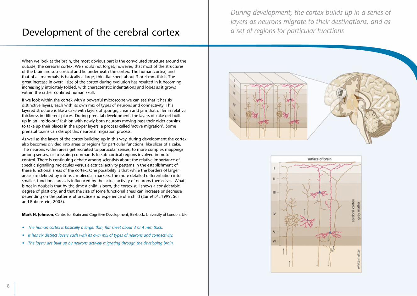
When we look at the brain, the most obvious part is the convoluted structure around the outside, the cerebral cortex. We should not forget, however, that most of the structures of the brain are sub-cortical and lie underneath the cortex. The human cortex, and that of all mammals, is basically a large, thin, flat sheet about 3 or 4 mm thick. The great increase in overall size of the cortex during evolution has resulted in it becoming increasingly intricately folded, with characteristic indentations and lobes as it grows within the rather confined human skull.
If we look within the cortex with a powerful microscope we can see that it has six distinctive layers, each with its own mix of types of neurons and connectivity. This layered structure is like a cake with layers of sponge, cream and jam that differ in relative thickness in different places. During prenatal development, the layers of cake get built up in an ‘inside-out’ fashion with newly born neurons moving past their older cousins to take up their places in the upper layers, a process called ‘active migration’. Some prenatal toxins can disrupt this neuronal migration process.
As well as the layers of the cortex building up in this way, during development the cortex also becomes divided into areas or regions for particular functions, like slices of a cake. The neurons within areas get recruited to particular senses, to more complex mappings among senses, or to issuing commands to sub-cortical regions involved in motor control. There is continuing debate among scientists about the relative importance of specific signalling molecules versus electrical activity patterns in the establishment of these functional areas of the cortex. One possibility is that while the borders of larger areas are defined by intrinsic molecular markers, the more detailed differentiation into smaller, functional areas is influenced by the actual activity of neurons themselves. What is not in doubt is that by the time a child is born, the cortex still shows a considerable degree of plasticity, and that the size of some functional areas can increase or decrease depending on the patterns of practice and experience of a child (Sur et al., 1999; Sur and Rubenstein, 2005).
Mark H. Johnson, Centre for Brain and Cognitive Development, Birkbeck, University of London, UK
• Thehumancortexisbasicallyalarge,thin,flatsheetabout3or4mmthick.
• Ithassixdistinctlayerseachwithitsownmixoftypesofneuronsandconnectivity.
• Thelayersarebuiltupbyneuronsactivelymigratingthroughthedevelopingbrain.
Development of the cerebral cortex
10
Neurotransmitters are chemicals that act as signal carriers between neurons
When an electrical impulse travelling along an axon comes to the synaptic bud at the end of the axon, for the signal to be passed on to another neuron chemicals are released into the synaptic cleft which then activate receptors on the receiving dendrite of the next neuron. There are several of these neurotransmitter chemicals within the brain and the exact effect a neurotransmitter has will depend on the receptors that the receiving neuron has and the overall network in which the neurotransmitter is acting.
There are two neurotransmitters that do not originate from a specific hub of neurons and are found in neurons in almost every brain structure. These are glutamate and gamma-aminobutyric acid (GABA). Glutamate is an excitatory neurotransmitter, meaning that it will bring the receiving neuron closer to the point of firing an action potential. By contrast, GABA is an inhibitory neurotransmitter and it will therefore make the receiving neuron less likely to fire an action potential.
There are several other neurotransmitters found in the brain that tend to stem from discrete hubs of neurons. For example, four are associated with particular structures and functions:
• Acetylcholine is found in neurons situated in the brainstem, the lowest part of the brain, and in the forebrain, and it is involved in processes of attention and arousal. This neurotransmitter is also found at all junctions between neurons and muscles and is therefore critical for executing movements.
• Dopamine is found in two areas in the midbrain that are the origins of two pathways: the nigrostriatal and mesolimbic. The former is critical for movement control and the latter, often called the ‘desire pathway’, is vital for motivation.
• Noradrenalin is released from neurons that originate deep within the brainstem and plays a critical role in fight-or-flight responses. It is also able to modulate learning processes within the brain and is implicated in depression and mania.
• Serotonin is released from neurons in another region situated within the brainstem. It is implicated in the sleep–wake cycle, temperature regulation, and the modulation of pain and mood.
Ellie Dommett, Brain and Behavioural Sciences, The Open University, UK
• Twoubiquitousneurotransmitters,foundinneuronsinalmosteverybrainstructure,areglutamate and GABA.
• Otherneurotransmittersareconcentratedinparticularstructures.
• Differentneurotransmittersareassociatedwithdifferentpathwaysinthebrain.
Neurotransmitters

12
The various systems within the brain that are responsible for particular functions are made up of connections between different parts which form ‘pathways’ between them. These connections can be very long, with axons of neurons stretching between distant brain regions, and different pathways are associated with different neurotransmitters. For example, two very important pathways together form the mesolimbic system, and the two main neurotransmitters involved in them are dopamine and serotonin. This system connects parts of the brainstem (the hindbrain and midbrain) with different areas of the cortex, associated with different functions, and is concerned primarily with controlling how the individual relates to and behaves in their environment. The mesolimbic system is sometimes described as a ‘primitive’ part of the brain because it arose early in evolution.
The dopaminergic (‘desire’) pathway links the parts of the brainstem that are active when motivating stimuli are experienced to the parts of the prefrontal cortex that control attention and executive functions. It supports individuals in behaving in ways that maximise rewards. This is clearly important for survival, but it can also be a source of problems such as the development of addictive behaviour.
The serotonergic pathway (with serotonin as the main neurotransmitter) can be seen as the ‘well-being’ pathway. It connects parts of the brainstem with the cortex, including the prefrontal areas, and also with other areas involved in memory, mood and activity levels. Disorders of this pathway are associated with anxiety, depression and obsessive–compulsive behaviour.
Both of these pathways work together to support motivated, organised behaviour and associated emotional states. The maturation and strengthening of these pathways in early childhood contribute to a child’s growing abilities to engage in more complex, planned behaviour.
Medication for mood, attention and activity disorders in children acts by changing the ways that the neurotransmitters operate in these pathways.
John Oates, Child and Youth Studies Group, The Open University, Milton Keynes, UK
• Maturationandstrengtheningofpathwaysinearlychildhoodcontributetoachild’sgrowing capacities for complex, planned behaviour.
• Thevarioussystemsinthebrainaremadeupofconnectionsbetweendifferentpartswhich form ‘pathways’.
• Themesolimbicsystemplaysanimportantroleincontrollinghowanindividualbehavesin their environment.
Systems and pathways
The mesolimbic system supports motivated behaviour and associated emotional states

14
Differences between children become apparent very soon after they are born: some are irritable while others are calmer. Some are more alert than others, and some are more sociable. Psychologists use the term ‘temperament’ to refer to characteristics such as these which are predominantly biologically based, having a large genetic component along with influences from the period before birth.
Evidence is accumulating from animal and human studies that stress experienced by pregnant mothers, along with deficiencies in their diets, can have short-term as well as long-term effects on brain development (Mulder et al., 2002), with consequences for the behavioural characteristics of children, and their development.
There also multiple genetic factors influencing brain structure (Thompson et al., 2001; Wright et al., 2002), and genes that have so far been identified as playing such roles show significant variations (polymorphisms) with consequences for children’s temperaments. A particular issue that is receiving attention from researchers is the effects of polymorphisms of genes associated with neurotransmission, such as in the dopamine and serotonin systems. For example, variations in the length of repeat sequences of the gene DRD4 which codes for a type of dopamine receptor in the mesolimbic system have been shown to be associated with differences in children’s attachment with their caregivers, and also to interact in complex ways with differences in mothers’ caregiving (Gervai, 2009).
This is a developing field of research, and it is becoming clear that there are many different gene–gene interactions involved in the causation of temperamental differences between children. Factors in children’s environments also interact in complex ways with these differences. For example, it seems that some genetic profiles may be protective for a child in one environment, but in another environment may conversely make a child more vulnerable (Belsky and Pluess, 2009).
These complex interacting factors and processes mean that each child is indeed unique, a further support for the notion that ‘one size does not fit all’ when it comes to helping children to overcome adversity and achieve their fullest potential.
John Oates, Child and Youth Studies Group, The Open University, Milton Keynes, UK
• Differencesintemperamentbetweennewbornsareaffectedbygeneticvariations,alongwithinfluencesfromtheperiodbeforebirth.
• Manydifferentgeneinteractionsarerelatedtotemperamentaldifferencesbetweenchildren.
• Complexinteractingfactorsandprocessesmeanthateachchild’sbrainistrulyunique,and this is true even of identical twins.
Unique brain; unique child
There are many different gene and gene–environment interactions involved in temperamental differences between children

16
One reason for the current interest in relating the growth and structures of the brain to children’s development comes from advances in methods which allow research ideas to be generated and tested more readily than previously. One set of tools involves brain imaging – creating ‘functional’ maps of brain activity based on changes in brain metabolism, blood flow, or electrical activity. The three main techniques that are used to study development are event-related potentials (ERP), magnetic resonance imaging (MRI), and near infra-red spectroscopy (NIRS).
ERP uses sensors on the scalp to measure tiny changes in the electrical activity of the brain generated as groups of neurons fire together. This method is good for detecting the rapidly changing activity that is characteristic of the brain. Usually, the data from many trials are averaged, to screen out natural rhythms of the brain that are unrelated to the presentation of a stimulus. With a large number of sensors, mathematical analyses can infer the likely locations of electrical activity within the brain.
Structural MRI (sMRI) allows brain anatomy to be imaged, while functional MRI (fMRI) also allows the non-invasive measurement of cerebral blood oxygenation, an indicator of surrounding neural activity, with a fine spatial resolution in millimetres but with a coarse temporal resolution of several seconds. While fMRI does not have the time resolution of ERP, it has better spatial resolution. fMRI has recently been extended to infants as well as children, although because the person has to remain still, babies can only be studied during sleep.
NIRS is a relatively new imaging method that takes advantage of the much thinner skull in babies (Lloyd-Fox et al., 2009). Infants wear a cap with light emitters and detectors. Tiny changes in light absorption due to changes in blood oxygenation in the brain give maps of functional activity similar to those from fMRI. Because the method is much less sensitive to movement, data can be recorded while young children are awake and actively engaged in a task.
While these techniques are used in medical diagnosis as well as in neuroscience research, there are significant differences between these two applications. In diagnosis, the focus is on the individual, whereas in research, data from many individuals are commonly combined and averaged to give clear, generalised pictures of brain activity. This is so that the ‘noise’ inherent in brain imaging can be reduced. It is important to recognise that, where this is done, it can also obscure differences that mark the individuality of the development of every person’s brain.
(adapted from Mareschal et al., 2004)
Brain imaging methods
A range of different methods is now available to researchers wishing to study the development and activity of children’s brains
• Event-relatedpotential(ERP)techniquesmeasuresmallelectricalchangesonthescalpsurfaces to pick up rapid temporal changes in brain activity and infer which areas are active.
• Functionalmagneticresonanceimaging(fMRI)senseschangesinbloodoxygenlevelswithin the brain and is good at localising activity but poor at sensing rapid changes.
• Nearinfra-redspectroscopyisanewermethodthatuseslightabsorptionandislessaffected by movements of the head, hence suitable for use with younger children.

18
® ® ®
® Are persons who are responsible for policy decisions and their implementation sufficiently knowledgeable about young children’s brain development?
® What procedures exist to scan for new neuroscience research findings of significance for early childhood education and care?
® How effectively are relevant insights from research disseminated among public services, NGOs and others responsible for promoting young children’s development?
® Are there adequate continuing professional development programmes in operation to ensure that those responsible for making and delivering policies are able to assess the scientific validity of claims regarding neuroscience implications for early childhood education and care?
® If healthy brain development is part of every child’s rights in early childhood, what are the implications for national policies?
POLICY QUESTIONS

21
A child’s brain starts to develop just a few days after conception.
A vast amount of brain growth happens before a child is born, at first through abundant creation of neurons and then through extensive axonal connections forming between them.
Just before birth and in the following year, brain systems and pathwaysbecomeestablishedasoften-activeneuronssurvive and less frequently activated neurons die.
As myelin sheaths form around nerve fibres towards the end of gestation and in early childhood, the efficiency of signal transmission improves.
As systems and pathways develop, different functions become relatively localised in specific brain areas and some become more lateralised in one or other of the two hemispheres of the brain.
Because different elements of the brain have their growth peaks at different times, there are ‘sensitive periods’ when different environmentalinfluencesareimportant.
‘Resting periods’, when the brain is less involved with external tasks but is nonetheless very active, are important in development.
What develops?II.

22
The brain of the foetus begins to form even before a mother may become aware that she is pregnant
Four weeks after conception, before a mother may even realise that she is pregnant, the foetal brain is already beginning to form. It is important now and further into pregnancy that the mother’s diet contains enough folic acid, a lack of which can restrict brain development and lead to spina bifida (an incomplete closure of the spine, exposing the spinal cord). For the next 4 months, brain cells form at an astonishing rate, around 250,000 every minute. After this, new cell formation slows, while vast numbers of axon interconnections among the neurons are made.
By the third month of gestation, the nervous system is sufficiently developed for basic physical reflexes to be present, as well as reactions such as kicking and arm flexing. In the fourth month the eyes and ears have already connected to the developing brain and the foetus reacts to sounds and bright lights. During these early months, many neurons are migrating from where they were formed to their final destinations and, as they migrate, they maintain most of the connections already made. Much of this migration is towards the outer layers of the young brain, forming the neuron-dense cortex.
By 5 months, the movements of the foetus become more controlled and varied as the parts of the brain that control motor behaviour mature. By the sixth month, new neuron growth has greatly reduced, while many more connections among neurons are being established through multiple dendrites (branches) forming on the axons, and learning can be seen as the foetus shows habituation (reduced responding) to repeated stimuli such as the same sounds. Electrical activity of the foetal brain can be detected by 7 months.
The mother’s diet continues to be important, since an adequate supply of nutrients is needed to build the nervous system and there are risks of damage from toxins (McEwen, 1987). The mother’s psychological well-being also affects brain development; stress during pregnancy has effects on the foetus that are evident after birth and in some cases may be long-lasting (Mulder et al., 2002). During the final stages of pregnancy, the number of neurons begins to decline as cell death eliminates those that are not actively involved in the developing brain pathways and systems.
John Oates, Child and Youth Studies Group, The Open University, Milton Keynes, UK
• About250,000newbraincellsareformedeveryminuteinthefoetusduringthefirst4months of gestation.
• Anadequatesupplyofnutrientsisneededtobuildthecomponentsofthenervoussystem.
• Amother’spsychologicalwell-beingduringpregnancyaffectsbraindevelopment.
Before birth
24
12
3
4a
4b
4c
5
6
1
2
3
4a
4b
4c
5
6
1
2
3
4a
4b
4c
5
6
1
2
3
4a
4b
4c
5
6
The basic architecture of the human brain develops before a child is born; most of the neurons a child will ever have are produced by the middle of gestation and by birth they have organised to form the cortex and other important brain structures. The major white matter pathways that make up the brain’s information processing networks are also present. Yet, brain development is far from complete in the newborn for, after birth, the child’s experience will play an increasingly important role in shaping and refining the major brain pathways and cortical networks.
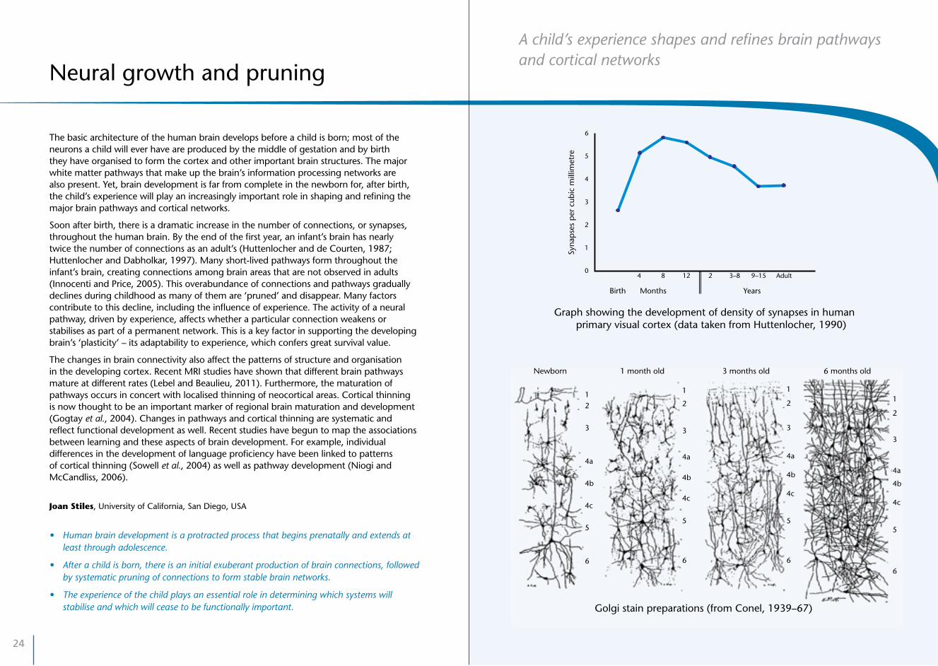
Soon after birth, there is a dramatic increase in the number of connections, or synapses, throughout the human brain. By the end of the first year, an infant’s brain has nearly twice the number of connections as an adult’s (Huttenlocher and de Courten, 1987; Huttenlocher and Dabholkar, 1997). Many short-lived pathways form throughout the infant’s brain, creating connections among brain areas that are not observed in adults (Innocenti and Price, 2005). This overabundance of connections and pathways gradually declines during childhood as many of them are ‘pruned’ and disappear. Many factors contribute to this decline, including the influence of experience. The activity of a neural pathway, driven by experience, affects whether a particular connection weakens or stabilises as part of a permanent network. This is a key factor in supporting the developing brain’s ‘plasticity’ – its adaptability to experience, which confers great survival value.
The changes in brain connectivity also affect the patterns of structure and organisation in the developing cortex. Recent MRI studies have shown that different brain pathways mature at different rates (Lebel and Beaulieu, 2011). Furthermore, the maturation of pathways occurs in concert with localised thinning of neocortical areas. Cortical thinning is now thought to be an important marker of regional brain maturation and development (Gogtay et al., 2004). Changes in pathways and cortical thinning are systematic and reflect functional development as well. Recent studies have begun to map the associations between learning and these aspects of brain development. For example, individual differences in the development of language proficiency have been linked to patterns of cortical thinning (Sowell et al., 2004) as well as pathway development (Niogi and McCandliss, 2006).
Joan Stiles, University of California, San Diego, USA
• Humanbraindevelopmentisaprotractedprocessthatbeginsprenatallyandextendsatleast through adolescence.
• Afterachildisborn,thereisaninitialexuberantproductionofbrainconnections,followedby systematic pruning of connections to form stable brain networks.
• Theexperienceofthechildplaysanessentialroleindeterminingwhichsystemswillstabilise and which will cease to be functionally important.
Neural growth and pruning
Newborn 1 month old 3 months old 6 months old
Golgi stain preparations (from Conel, 1939–67)
Syna
pse
s p
er c
ubic
mill
imet
re
Graph showing the development of density of synapses in human primary visual cortex (data taken from Huttenlocher, 1990)
4 8 12 2 3–8 9–15 Adult
6
5
4
3
2
1
0
Birth Months Years
4 8 12 2 3–8 9–15 Adult
6
5
4
3
2
1
0
Birth Months Years
Syna
pse
s p
er c
ubic
mill
imet
re
Graph showing the development of density of synapses in human primary visual cortex (data taken from Huttenlocher, 1990)
A child’s experience shapes and refines brain pathways and cortical networks
26
Good diet, for pregnant mothers and young children, is important for normal myelination and cognitive development
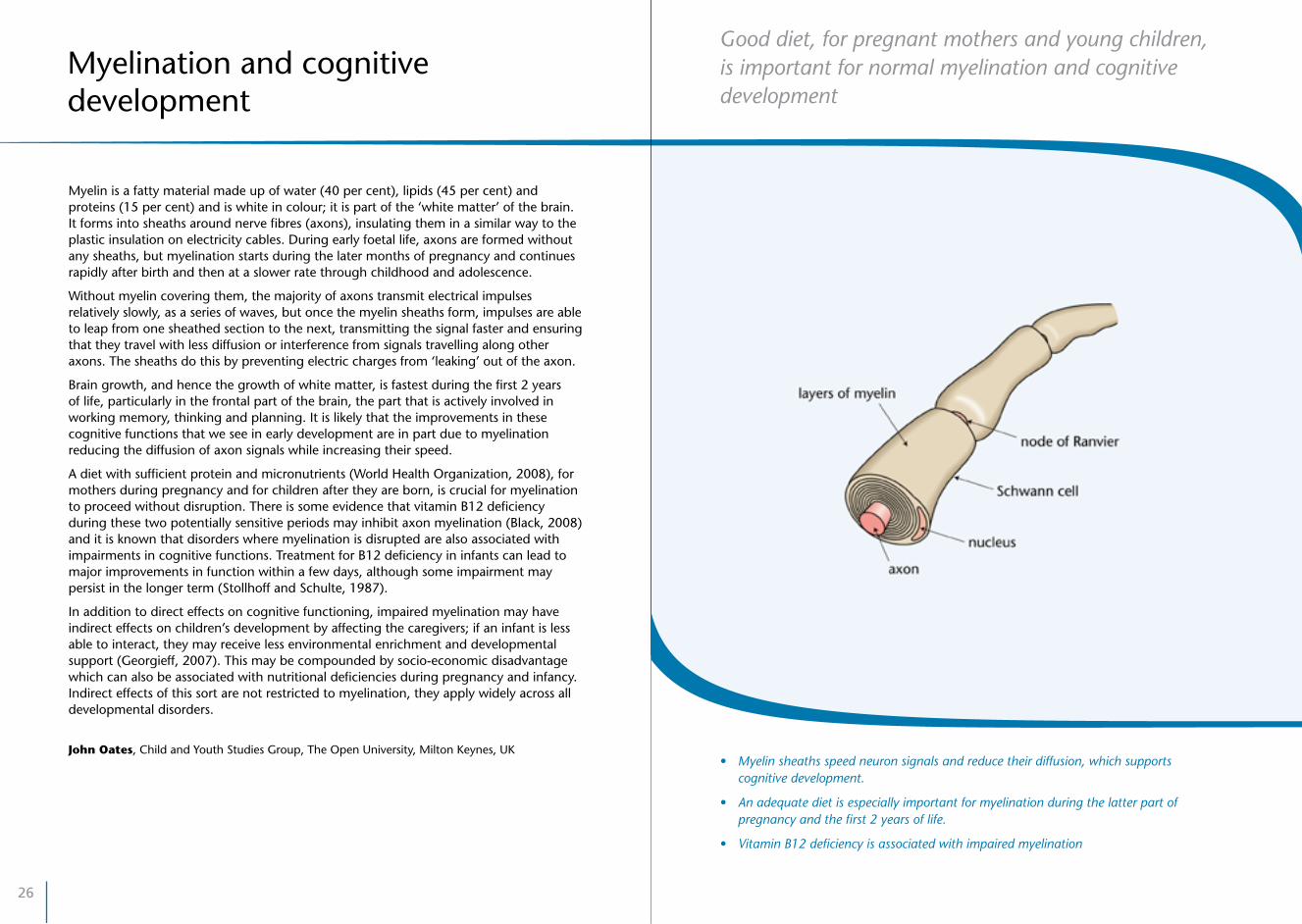
Myelin is a fatty material made up of water (40 per cent), lipids (45 per cent) and proteins (15 per cent) and is white in colour; it is part of the ‘white matter’ of the brain. It forms into sheaths around nerve fibres (axons), insulating them in a similar way to the plastic insulation on electricity cables. During early foetal life, axons are formed without any sheaths, but myelination starts during the later months of pregnancy and continues rapidly after birth and then at a slower rate through childhood and adolescence.
Without myelin covering them, the majority of axons transmit electrical impulses relatively slowly, as a series of waves, but once the myelin sheaths form, impulses are able to leap from one sheathed section to the next, transmitting the signal faster and ensuring that they travel with less diffusion or interference from signals travelling along other axons. The sheaths do this by preventing electric charges from ‘leaking’ out of the axon.
Brain growth, and hence the growth of white matter, is fastest during the first 2 years of life, particularly in the frontal part of the brain, the part that is actively involved in working memory, thinking and planning. It is likely that the improvements in these cognitive functions that we see in early development are in part due to myelination reducing the diffusion of axon signals while increasing their speed.
A diet with sufficient protein and micronutrients (World Health Organization, 2008), for mothers during pregnancy and for children after they are born, is crucial for myelination to proceed without disruption. There is some evidence that vitamin B12 deficiency during these two potentially sensitive periods may inhibit axon myelination (Black, 2008) and it is known that disorders where myelination is disrupted are also associated with impairments in cognitive functions. Treatment for B12 deficiency in infants can lead to major improvements in function within a few days, although some impairment may persist in the longer term (Stollhoff and Schulte, 1987).
In addition to direct effects on cognitive functioning, impaired myelination may have indirect effects on children’s development by affecting the caregivers; if an infant is less able to interact, they may receive less environmental enrichment and developmental support (Georgieff, 2007). This may be compounded by socio-economic disadvantage which can also be associated with nutritional deficiencies during pregnancy and infancy. Indirect effects of this sort are not restricted to myelination, they apply widely across all developmental disorders.
John Oates, Child and Youth Studies Group, The Open University, Milton Keynes, UK
Myelination and cognitive development
• Myelinsheathsspeedneuronsignalsandreducetheirdiffusion,whichsupports cognitive development.
• Anadequatedietisespeciallyimportantformyelinationduringthelatterpartofpregnancy and the first 2 years of life.
• VitaminB12deficiencyisassociatedwithimpairedmyelination

28
100
80
60
40
20
00–8
weeks8–38weeks
0–12months
1–12years
Age (log scale)
Gen
e ex
pre
ssio
n (%
of m
axim
um)
12–20years
20+years
EMBRYONIC FOETALBIRTH
INFANCY CHILDHOOD ADOLESCENCE ADULTHOOD
Cell proliferation
Progenitors and immature neurons
Synapse development
Dendrite development
Myelination
Differences in the strength of gene expression for different elements of the developing brain are associated with periods of enhanced sensitivity
Different components of the brain have peak periods of construction at different times during development from conception through early childhood (Kang et al., 2011). These periods of expansive formation and growth are controlled by different genes that are turned on and off by time- and location-related processes. These changes in gene expression are at their greatest during foetal development and early infancy.
In the first 2 months after conception, the most strongly expressed genes are those that control the proliferation of new neurons and associated cells in the foetal brain. The dominance of this process falls off rapidly up to the time of birth, when it is only at around one-tenth of its initial strength. Gene expression for new neurons is almost completely suppressed by the age of 6 years. During the later months of foetal growth, gene expression increases for the growth of synapses connecting the neurons and the axon dendrites which allow for multiple connections from each neuron, reaching a maximum by 6 months after a child’s birth. The genes that control myelination of the axons only reach half of their expression strength by the time of birth and continue to increase their influence for a further 12 months.
These dramatic changes in the growth peaks of different components of the brain, and the maturation of structures and processes that depend on them, mean that there are sensitive periods when environmental conditions are more likely to have specific effects. For example:
The infant–caregiver relationship depends on the quality and availability of caregiving early in life, the same period that is sensitive to the effect of iron deficiency on myelination and density of dopamine receptors.
(Walker et al., 2011, p. 1327)
While research into such sensitive periods is progressing apace, with many unknowns still to be addressed, the Harvard University National Scientific Council on the Developing Child states that:
Because low-level circuits mature early and high-level circuits mature later, different kinds of experiences are critical at different ages for optimal brain development, a concept called age-appropriate experience. Soon after birth, basic sensory, social, and emotional experiences are essential for optimizing the architecture of low-level circuits. At later ages, more sophisticated kinds of experiences are critical for shaping higher-level circuits.
(National Scientific Council on the Developing Child, 2007, p. 4)
John Oates, Child and Youth Studies Group, The Open University, Milton Keynes, UK
Sensitive periods
• Differencesinthestrengthofexpressionofgenesthatcontrolthebuildingofthechild’sbrain lead to peak periods of growth of different brain components during gestation and infancy.
• Thesedifferencesareassociatedwithaseriesofdifferentsensitiveperiodswhenenvironmentalinfluencesareespeciallyimportant.
• Researchiscurrentlyinvestigatingthespecificenvironmentalinfluencesthatarecriticalinthese sensitive periods.
.

30
Genetic predisposition and environmental stimulation work together to support brain localisation and specialisation during childhood
The activity of the brain cortex in newborn infants is thought to be less localised than in older children and adults, and it tends to be equally distributed across the two hemispheres of the brain. With experience and maturation, different parts of the cortex gradually become more specialised, forming circuits for specific functions, in some cases in one or the other hemisphere. Some functions, particularly those to do with the physiology of the body and the senses, become established very early in life. Others, such as the control of movements for crawling and then walking, emerge somewhat later, while yet others, such as mental time travel and forward planning, develop even later.
It is important to note that even though specific neural networks are associated with specific functions, the brain nevertheless works as a set of closely interconnected parts and even simple functions will be associated with some level of activity in parts other than the focal area(s) (Gottlieb et al., 1997).
Given an environment that provides sufficient and appropriate stimulation, such as, for example, a rich language environment, the genetic predisposition for specific parts of the brain to become the focal areas for specific functions will unfold. In the case of language, this means that Broca’s area, on the left-hand side of the brain, becomes important for understanding and producing speech (Neville et al., 1991). Several aspects of cognitive function become lateralised in this way during childhood, finding their focal areas of activity on one side or the other of the brain. In general, though, both parts of the brain work together; it is inaccurate to think of ‘right-brain’ and ‘left-brain’ functions as being separate.
During this long period of increasing specialisation and lateralisation, the cortex remains a very adaptable organ and, in cases of acquired damage, it is able to rescue an ‘at risk’ function by accommodating it in the opposite hemisphere to the one where the function is normally established. Initially, there is a lot of redundancy in the young brain, which gives it this flexibility, and the specialisation that follows reduces the capacity for the developing brain to overcome the effects of damage. However, if damage occurs to the same area on both sides of the brain, the capacity for recovery is severely reduced or even completely lost (Stiles, 1998; Stiles et al., 2009).
Because different functions localise and lateralise at different times in development, and the brain’s ‘plasticity’ reduces as children get older, early diagnosis and treatment of any damage or lack of expected function are very important, to maximise the chances of good outcomes.
John Oates, Child and Youth Studies Group, The Open University, Milton Keynes, UK
Faraneh Vargha-Khadem, Developmental Cognitive Neuroscience Unit, University College London Institute of Child Health, and Great Ormond Street Hospital for Children, UK
Localisation and lateralisation
• Childhoodisatimeofincreasingspecialisationandlateralisationofbrainfunctions.
• Inearlychildhood,theeffectsofacquireddamagetoanareainonehemisphereofthebrain can be reduced by the equivalent area in the other hemisphere taking over the developing function.
• Fortreatmentoffocalbraindamagetobeeffective,earlyandcarefuldiagnosisofthelossof function and assessment of the child’s developmental level are crucial.
Left Right
Adult fMRI scan showing greater activity (coloured areas) in the left hemisphere of people’s brains when listening to short spoken sentences

32
Resting or ‘default’ states may be particularly important in early childhood development, and may be linked to the functional development of the brain structures
When you are not engaged in a specific task and your mind seems to be idle, there is still a lot of activity in your brain. This ‘resting state’ activity is now being intensively studied using EEG and fMRI, showing specific electrical activity such as ‘alpha’ band activity and coordinated networks of active regions. Resting state activity has been identified even in young babies, sometimes resembling that seen in adults (Fair et al., 2009). Resting states may be particularly important in early childhood development, possibly linked to the development of brain structures (Gordon et al., 2011) and to recovery after damage (Merabet and Pascual-Leone, 2010). Previous experience may be reflected in this resting state activity and so play a part in sculpting brain connections based on early environments.
Mental processes involve networks of different brain regions, each with their own particular specialisations. Studies of the efficiency of different kinds of networks show that so-called ‘small world’ networks are most efficient, and that adult brains have this kind of connectivity pattern. In contrast to the grid pattern of streets found in many cities, ‘small world’ networks are more like the clusters of small streets in a village that is linked to other such villages by fast highways. Recent research, often examining brain resting states, is showing developmental changes in both segregation of brain regions during development (reducing the number of connections with nearby regions) and integration (regions joining up with more long-range connections between them) (Fair et al., 2009).
Adult networks have a more hierarchical structure that works well to support top-down relations between one part of the network and another (Supekar et al., 2009). While hierarchical networks have a number of computational advantages, they are also less plastic and more vulnerable to damage or noise in the particular parts at the top of the hierarchy. The network arrangements of children’s brains may be more flexible and plastic in response to new sensory inputs or environmental contexts.
Mark H. Johnson, Centre for Brain and Cognitive Development, Birkbeck, University of London, UK
• Attimeswhenachildisphysicallyatrest,andnotobviouslymentallyengagedwithatask, their brain is still highly active.
•Changesinchildren’sthinkingseemtobelinkedtochangesinbrainnetworks.
•‘Restingstate’brainactivitymaybeimportantforthetypicaldevelopmentofchildren’sbrains.
Networks and resting states

34
Different areas in the child’s brain become specialised for recognising different aspects of the social world
Human infants are born with a strong drive to interact with other people on whom they depend for care (such as shelter, food, affection) and learning (for example language, cultural norms, skills). Because human interaction is guided by goals and beliefs and not just by physical laws, attending to and interacting with other people require different skills and brain systems – sometimes referred to as the ‘social brain’. The recent development of infant-friendly ways to image the workings of the brain has allowed researchers to see the ‘social brain’ in action from early in life.
Human faces and voices are intrinsically rewarding for the human newborn, as shown by the fact that infants orient to and enjoy them over other kinds of visual or auditory stimulation (Johnson et al., 1991). If these initial preferences are responded to appropriately – if infants are surrounded by a rich, stimulating social environment – they allow them to learn rapidly about people’s appearance and behaviour. As this happens, different areas in the child’s brain become progressively specialised for recognising different aspects of the social world: human movement (Lloyd-Fox et al., 2009), human voice (Dehaene-Lambertz et al., 2002) or human faces (Gliga and Dehaene-Lambertz, 2006). The specialisation of a specific brain area that is linked to perceiving faces allows children to improve gradually their ability to discriminate people’s faces.
In parallel to learning about people, infants’ brains are prepared to communicate with, and learn from, adults. Because attention is initially quite limited in babies, when something needs to be learned caregivers make use of attention-getting signals, which are very similar across cultures. This involves establishing eye contact, using a ‘sing-song’ voice (referred to as motherese or parentese) and/or repeatedly calling the infant’s name. Brain imaging studies have shown that an area of the infant’s brain – the prefrontal cortex – responds to these kinds of signals as early as 5 months of age (Grossman et al., 2010). Eye contact is effective in attracting infants’ attention from birth (Farroni et al., 2007) and the eyes remain for a long time the face element that infants prefer to look at and that generate the strongest brain responses (Gliga and Dehaene-Lambertz, 2006). This is no surprise, as the eyes are a rich source of information about someone’s intentions or emotions.
Not all children develop an interest in interacting and learning from others, notably children diagnosed as having various degrees of autism spectrum disorders. Research in understanding the causes of this disorder is ongoing; one of the hypotheses being tested is that there may be an impairment in the early development of the ‘social brain’.
Teodora Gliga, Centre for Brain and Cognitive Development, Birkbeck, University of London, UK
The social brain
• Interactingwiththesocialworldrequiresthedevelopmentofspecialisedbrainmechanisms – the ‘social brain’.
• Infantsarebornwithbiasestoorienttohumanvoicesandfacesandespeciallytohumaneyes, allowing infants to learn about people and from people.
• Impairmentsinthesefunctionsmaybelinkedtoautisticspectrumdisorders.

36
Attention modulates children’s learning in our complex world
Social and physical environments are very complex, and yet adults are able to select efficiently what is relevant for encoding into memory, learning and action planning. Attention is the multifaceted set of skills that enable adults to be so effective in selecting what is pertinent while ignoring distractions, but it also plays a role when maintaining goals in mind and inhibiting inappropriate behaviours. It is therefore no surprise that attention influences learning from the very beginning of childhood. Seminal work has demonstrated that newborns’ attention is attracted automatically by salient objects (such as faces), and that these early orienting behaviours are gradually replaced by more controlled attention, crucial to learning about those stimuli. Prefrontal and parietal regions of the brain interact with other networks to facilitate their increasing specialisation and fine-tuning to environmental stimuli (Johnson, 2011). Later in development, good attentional skills also give preschool children a head start in numeracy and literacy, predicting how well they do at school entry and subsequently modulating working memory, a key player in school outcomes (Astle and Scerif, 2011).
Difficulties with attention also explain why some children struggle to learn, and can be an important focus for intervention. For example, recent studies have shown how children with attention deficit hyperactivity disorder (ADHD), at risk of poor outcomes both in and outside the classroom, have difficulties modulating neural networks that are actively engaged in controlling action; they also have problems suppressing active networks when thinking (Fair et al., 2010). Motivational incentives, such as rewarding sustained attention, can be effective in improving the balance across such excitatory and inhibitory networks and may interact synergistically with psycho-stimulants to bring children with ADHD to perform at the level of children who do not have attention difficulties (Liddle et al., 2011).
Attention modulates what young children learn from their environment, with some processes (like attentional control over action) continuing to improve well into adolescence and young adulthood.
Gaia Scerif, Attention, Brain and Cognitive Development Group, Department of Experimental Psychology, University of Oxford, UK
• Attentionandattentiondifficultiesaffectlearningacrossdomains.
• Attentionalprocessesarediversebut,asweunderstandthembetter,seemmodifiablethrough targeted cognitive intervention.
• Motivationalincentivesandpsycho-stimulantscanworkinsynergytohelpchildrenwithattention disorders.
Objects of attention
38
® ® ®
® What steps can be taken to improve public and professional understanding of the crucial importance of the early months and years of life for brain development, through education, media and public awareness campaigns?
® Do policies for ECEC recognise that there are periods of sensitivity in brain development, both before and after a child’s birth, and that it is crucial that adequate environmental support is present for mothers and their babies during these periods?
® Are young children getting enough ‘resting state’ opportunities as well as stimulation, or are their family lifestyles too challenging to allow opportunities for rest?
® Do adequate health screening programmes exist to detect early signs of brain damage or dysfunction in young children, and offer appropriate early intervention, guidance and support?
® Are young children’s behavioural difficulties being treated effectively by ‘multi-pronged’ approaches rather than simply relying on medication?
® Is the crucial significance of perinatal care, including good nutrition, properly realised in policies and practices for pregnant and breastfeeding mothers, and for their children?
POLICY QUESTIONS
41
A child’s visual system is critically dependent on environmental stimulation for full development.
Exposure to a rich language environment is crucial if a child is to develop good language skills; learning more than one language can be a benefit.
For healthy brain development, a child needs a good diet and sufficient rest and sleep.
Neglect, abuse and other forms of maltreatment have serious negative consequences for children’s brain development and cause subsequent psychological problems.
Children differ in their vulnerability and resilience to potentially harmfulinfluencesontheirdevelopment.
Environmental influencesIII.

42
Babies must learn to see, much as they learn to walk or talk
Babies use their eyes to explore the world from the time they are born, even before they can use their hands and legs to grasp or crawl. Even so, vision is one of the least developed senses at birth. Babies must learn to see, much as they learn to walk or talk. In order for this to happen, babies need visual stimulation.
Some of the stimulation that the visual system needs happens when babies are still inside the womb. Cells in the visual pathway generate their own spontaneous activation as preparation for the stimulation that will come from the outside world after birth. If babies are born preterm, this process can be disrupted and affect visual development.
After birth, visual stimulation from the outside world begins. Its effects can be studied in children who experience reversible blindness caused by dense cataracts that are surgically treated. The final clarity of vision (acuity) achieved in the treated eye is never quite normal, with a larger impairment the earlier the period of visual deprivation occurred (Le Grand et al., 2001). Visual stimulation is also necessary for other aspects of vision, including contrast sensitivity, motion perception and face processing. However, the timing and duration of the period when normal visual input is necessary varies widely for these different aspects of vision, ranging from a few months after birth to more than the first 10 years of life. Even when a mature level of function is attained, there is a further period of stabilisation when normal visual experience is necessary to retain the level of skill achieved. It is thought that this long period of development allows the child’s visual system to adapt to the specifics of the seen environment in which they grow up (Fox et al., 2010).
Visual input is also important for development in other areas. Children born with non-reversible blindness show delays in motor, language and cognitive development, reduced integration of intact senses and impaired social skills (Warren, 1984). These are probably a combination of a direct effect of the loss of visual stimulation and its secondary consequences. For example, vision plays a role in early social abilities such as eye contact and joint attention, which in turn influence the development of skills such as language.
It is clear that impairments in vision early in life have lasting consequences for visual skills, with knock-on consequences for other domains.
Michelle de Haan, Centre for Developmental Cognitive Neuroscience, University College London, UK
• Thedevelopmentofvisualfunctionstartsbeforebirthasthevisualpathwayspontaneouslyself-activates.
• Visualdevelopmentafterbirthcontinuesatleastuntil10yearsofageanddependsonadequate environmental stimulation.
• Atypicalvisualdevelopmentcanhavenegativeeffectsinotherareas,suchassocialdevelopment.
Development of the visual system

44
Infants growing up in bilingual environments adapt their language learning strategies to the richer input that they receive
Acquiring and using our first language is a complex but surprisingly fast achievement which, despite apparent effortlessness, results from an intricate array of perceptual and cognitive processes. Sensitivity to speech sounds begins prenatally, and newborns already show some remarkable capacities related to language. They are able to notice that some languages sound different from others, such as English and Japanese. By 5 months, infants are sensitive to the difference between more similar languages, such as Dutch and English, or even between dialects such as American and British English (Kuhl, 2004).
Research has shown that newborns are able to discriminate all the sounds of the languages of the world, even if they have never heard them before. But by the end of the first year of life, they only retain the ability to distinguish between the sounds that they have heard spoken by the people around them (Werker and Tees, 2005). By the time they begin to speak, they already have quite complex knowledge of the language(s) that they have been exposed to, including many words (Kuhl, 2009).
A large percentage of the infants around the world are exposed from birth to more than one language in their environment. Early bilingualism has long-term effects on the underlying brain structures devoted to language and perhaps to other cognitive abilities. Many parents are worried by the possibility that bilingual exposure may ‘confuse’ their children. Research with young infants has clearly shown that there are no difficulties in dealing simultaneously with more than one language.
Research carried out with newborns and 4-month-old infants exposed to either dissimilar languages, such as French and English or Philippino (Tagalo) and English, or to similar ones, such as Spanish and Catalan (two Romance languages), demonstrates that bilingual infants show equivalent capacities of language discrimination to monolingual infants (Sebastián-Gallés, 2010). In fact, they may even be slightly better. Bilingual children also learn the sounds of their two languages, and can identify the first words, at the same time as monolingual infants. Interestingly, although they perform equivalently, they use different strategies. Infants growing up in bilingual environments adapt their learning strategies to cope better with the nature of their double input. Taken together, research with bilingual-to-be infants shows that not only do they keep their pace with their monolingual peers, but that the refined brain mechanisms that they bring to bear on the acquisition of two languages have the potential to enhance other aspects of their cognitive development.
Núria Sebastián-Gallés, Universitat Pompeu Fabra, Barcelona, Spain
• Speechperceptionstartsprenatallyandcontinuesactivelyduringthefirstmonthsoflife.
• Infantsknowalotaboutlanguagebeforetheyproducetheirfirstwords.
• Bilingualinfantsnotonlykeeppacewiththeirmonolingualpeersbutshowenhanceddevelopment in some other aspects of their cognitive development.
Language acquisition in the monolingual and bilingual child

46
Sleep has a crucial role in learning and memory
It used to be thought that sleep was when the brain took a rest, ticking over for vital functions like breathing. In reality, some parts of our brain are more active during sleep than when we are awake (Kahn et al., 1996). Between infancy and adulthood, we spend more than one-third of our lives asleep, during which the body replenishes its energy and the brain re-processes experiences stored during waking hours.
Research on bird brains provides important clues to the role of sleep. Chicks learn their species song by copying the mother bird’s song. In a research study (Rauske et al., 2003) one group of zebra finch chicks practised their tweeting, followed by a silent rest during which they stayed awake. The second group also had a break, but it was used to induce sleep. The chicks that remained awake had reduced brain activity when resting, whereas the sleeping chicks’ brains were highly active, as if they were still processing their mothers’ song. Moreover, they learned faster and more accurately. Similar research was undertaken with two groups of cats (Frank et al., 2001). Both initially received the same training; then one group slept for 6 hours, while the other was kept awake and received 6 extra hours’ training. Yet the group that had half the training, but slept, learned far better than the group that had double the training. Both studies attest to the crucial role of sleep in learning and memory.
Research with humans has yielded similar findings, with recent work (Fischer et al., 2007; Backhaus et al., 2008) highlighting long-term learning effects of sleep even in infants. Infants who napped within 4 hours of exposure to an artificial language remembered the general grammatical pattern of the language 24 hours later, whereas those who had not napped showed no evidence of remembering the language (Hupbach et al., 2009). The findings support the view that infants’ frequent napping plays an essential role in consolidating knowledge in long-term memory.
We can therefore conceive of sleep, not just as a rest period, but also as a cognitive process in which activity in certain brain regions plays a critical role in learning and memory throughout the lifetime (Hill et al., 2007). This suggests that sleep intervention programmes might be used to enhance learning.
Annette Karmiloff-Smith, Centre for Brain and Cognitive Development, Birkbeck, University of London, UK
• Partsoftheinfant’sbrainaremoreactiveduringsleepthanwhenawake.
• Sleepisessentialforconsolidatinglearningintolong-termmemory.
• Adequatesleepisimportantthroughoutthelifespan.
The importance of sleep for learning

48
Diet is a most important environmental variable, particularly during the first 2 years of rapid brain growth
Diet and nutrition in brain development
During pregnancy, all the nutrients needed for the foetal brain’s development and functioning come from the mother’s food intake, via her bloodstream and through the placenta into the foetal bloodstream. After birth nutrients come from the mother’s milk, if she is breastfeeding, or formula, as well as from supplementary foods in the infant’s diet. Studies of cognitive outcomes (Benton, 2008) suggest strongly that early nutrition modifies the physical structures of the brain on which these functions depend, but hard evidence for this is thin on the ground, partly because data from animal studies are hard to extrapolate to humans. There were no suitable methods for examining brain structure in living humans until advances in neuroimaging made it possible to see subtle structural changes related to early diet. The best evidence that nutrition actually causes such changes comes from neuroimaging studies in randomised controlled trials but so far there are few of these.
Animal research and cognitive studies have shown that certain micronutrients (metals and vitamins) have specific, critical roles in brain development (Delange, 2000; Lozoff and Georgieff, 2006). Iron level, for example, may affect neurotransmitter synthesis while fatty acids affect their release. Differences in macronutrient (proteins and calories) intake can affect the volume of the caudate nucleus, a brain structure associated with Verbal IQ (Isaacs et al., 2008). A key issue is the role of breast milk in cognitive development – a higher percentage of breast milk in the diet of infant boys has been shown to be associated with greater white matter volume in the brain and higher Verbal IQ (Isaacs et al., 2010). It has been suggested that long-chain polyunsaturated fatty acids in breast milk promote cognitive development, mainly because of their effects on neuronal membranes and neural transmission.
Many unanswered questions remain regarding the dietary needs for optimal brain/cognitive development. Key factors are likely to be nutrient dose, duration of exposure and a child’s sex. The same nutrient may exert different effects on different structures at different developmental stages. Some structures, such as the hippocampus and white matter, seem particularly vulnerable to dietary deficiencies. Despite this complexity, diet is among the most modifiable of environmental factors, holding out the promise of policies that support adequate nutrition for children in the early years having a significant positive impact, as our knowledge base increases.
Elizabeth Isaacs, Institute of Child Health, University College London, UK
• Evidencesuggeststhatadequateintakesofmicronutrientsarecrucialfordevelopingbrains.
• Thereisneedforfurtherresearchintospecificdietaryeffectsonbraindevelopment.
• Becausedietisarelativelyeasilymodifiedenvironmentalfactor,itshouldbeanimportantfocus for child health policies.

50
Early experience, particularly parent–child relationships, plays a critical role in brain development
Our understanding of brain development has increased exponentially over the past few decades. And, although much remains to be discovered, we can state with great certainty that experience, and its timing, play critical roles in brain development.
One example comes from children experiencing psychosocial deprivation, such as in institutions with poor standards of social support and care. Over the past decade, several investigators have focused their attention on such children, and the findings can be summarised as follows. First, children with a history of institutionalisation show reduced brain metabolism in the prefrontal cortex and the temporal lobe, and disturbances in white matter in various brain regions (Chugani et al., 2001; Eluvanthingal et al., 2006). Second, several groups have reported significant reductions in grey and white matter volumes and enlarged relative amygdala volumes in previously institutionalised children (Mehta et al., 2009; Tottenham et al., 2011). Third, the Bucharest Early Intervention Project (Nelson et al., 2009) has reported less cortical brain activity among institutionalised children compared to never-institutionalised children. When the Project’s sample turned 42 months of age, previously institutionalised children who had been placed in foster care before the age of 2 years showed EEG activity that more closely resembled the never-institutionalised children than the institutionalised children; at age 8 years, children placed in foster care before age 2 years showed EEG activity closely similar to that of never-institutionalised children, whereas children placed after 2 years looked virtually identical to currently institutionalised children. Finally, evidence from event-related potentials (ERPs) revealed subtle perturbations in face and emotion processing and, as with our EEG data, dramatic reduction in the size of various ERP components among institutionalised children prior to placement in foster care. At follow-up, however, the children placed in foster care were beginning to resemble those in the never-institutionalised group (see Marshall et al., 2004, 2008; Parker et al., 2005a, 2005b; Moulson et al., 2009a, 2009b; Nelson et al., 2009; Vandewert et al., 2010).
Overall, these findings point to the power of early experience, particularly parent–child relationships, as playing a critical role in brain development.
Charles A. Nelson, Children’s Hospital Boston/Harvard Medical School and Harvard Center on the Developing Child, USA
• Experienceanditstimingplayacriticalroleinbraindevelopment.
• Psychosocialdeprivation,suchasmayoccurininstitutionalcare,canhaveseriousandpermanent negative effects on brain development.
• Forchildrenseparatedfromtheirparents,beingplacedinfostercarebeforetheyare2yearsold can avoid the developmental delays that can otherwise arise from institutional care.
The effects of early psychosocial deprivation

52
• Adversecare-givingexperiencescanaffectbrainstructuresandfunctionandimpactonpsychological development.
• Geneticvariationsmaymakesomemaltreatedchildrenmorevulnerabletomaladaptiveoutcomes while being protective for others.
Maltreatment during childhood affects brain structure and function, with consequences for children’s psychological and emotional development
Children who experience maltreatment (sexual, physical or emotional abuse or neglect) are more likely to develop psychological problems (Curry and Widom, 2010). Recent research has begun to show how adverse caregiving experiences can affect brain structures and function, and how these may in turn impact on psychological and emotional development (McCrory et al., 2011).
There is now reliable evidence that childhood adversity is associated with atypical development of the hypothalamic-pituitary-adrenal (HPA) axis which controls the release of stress hormones, with patterns of both reduced and exaggerated responsiveness being reported. These patterns have also been associated with psychiatric problems in adulthood, including depression and post-traumatic stress disorder (Heim et al., 2008).
Reductions in the volume of the hippocampus (a key structure in memory processing) have been reported in structural MRI studies of adults who experienced abuse as children (Woon and Hedges, 2008; Treadway et al., 2009). In children, experiencing maltreatment has been associated with reduced volume of the corpus callosum, a white matter structure that connects the two brain hemispheres, and reduced grey matter volume in the orbitofrontal cortex, an area implicated in aspects of social processing (McCrory et al., 2011). There is some evidence of increased amygdala volume, but only in children who have experienced early institutionalisation, a rather extreme form of early adversity (Sonuga-Barke, 2009). fMRI studies have found increased functional response in the amgydala, an area known to be critically important in threat detection and processing. Children exposed to family violence – either physical abuse or witnessing domestic violence – show increased activation of the amygdala to angry but not sad faces. This suggests a hypervigilant response to social threat, and fits with psychological data indicating that physically abused children may learn to automatically allocate greater attention to social threat cues (McCrory et al., 2011).
However, the effects of environmental adversity may be moderated by genotype. Evidence is now accumulating that some common genetic polymorphisms may make some maltreated children more vulnerable to maladaptive outcomes. It is possible that structural and functional differences at the neural level make these children more sensitive to adverse emotional environments. Other genetic polymorphisms may be protective in adverse situations, explaining why even in the same family children show individual differences in their coping strategies. A new field of research is now also beginning to open up, investigating possible epigenetic effects of caregiving and maltreatment. Specifically, researchers are investigating whether early maltreatment may serve to alter genetic expression later in a child’s life (McCrory et al., 2010).
Eamon McCrory, Developmental Risk and Resilience Unit, University College London, UK
Maltreatment, genetics and brain development

54
® ® ®
® Are teachers, care workers and other practitioners aware of the factors that put young children’s brain development at risk?
® What public health systems are in operation to screen for and identify potential environmental risks for negative effects on brain development during pregnancy and early childhood? Are they adequate?
® Are there public education programmes that are operating effectively to educate parents about the minimal standards of care, nutrition and living environments needed to avoid neurodevelopmental harms?
® Are practitioners sufficiently well educated in strategies to help parents to avoid or ameliorate ‘toxic’ stress?
® What policies, programmes and practices are in place to support families in maintaining stable and growth-promoting relationships?
® Are children being given adequate opportunities to sleep soundly and for sufficiently long periods, and are parents aware of the importance of sleep for brain development?
® Are there robust and sensitive child protection procedures in place to detect early, and rapidly deal with, cases of child neglect and abuse?
® How can children’s brain development be promoted as a national and community priority, in ways that take account of diverse living conditions, cultural expectations for mothers and children, and resources to support them?
POLICY QUESTIONS

56 57
Gordon, E.M., Lee, P.S., Maisog, J.M., Foss-Feig, J., Billington, M.E., VanMeter, J. and Vaidya, C.J. (2011) ‘Strength of default mode resting state connectivity relates to white matter integrity in children’, Developmental Science, vol. 14, pp. 738–51.
Gottlieb, G., Wahlsten, D., and Lickliter, R. (1997) ‘The significance of biology for human development: a developmental psychobiological systems view’ in Lerner, R. (ed.), HandbookofChildPsychology:Vol.1.Theory(5th edn), New York, NY, Wiley.
Heim, C.D., Newport, J., Mletzko, T., Miller, A.H. and Nemeroff, C.B. (2008) ‘The link between childhood trauma and depression: insights from HPA axis studies in humans’, Psychoneuroendocrinology, vol. 33, pp. 693–710.
Hill, C.M., Hogan, A.M. and Karmiloff-Smith, A. (2007) ‘To sleep, perchance to enrich learning?’, Archives of Disease in Childhood, vol. 92, pp. 637–43.
Hupbach, A., Gómez, R., Bootzin, R. and Nadel, L. (2009) ‘Nap-dependent learning in infants’, Developmental Science, vol. 12, pp. 1007–12.
Huttenlocher, P.R. (1990) ‘Morphometric study of human cerebral cortex development’, Neuropsychologia, vol. 28, pp. 517–27.
Huttenlocher, P.R. and Dabholkar, A.S. (1997) ‘Regional differences in synaptogenesis in human cerebral cortex’, Journal of Comparative Neurology, vol. 387, pp. 167–78.
Huttenlocher, P.R. and de Courten, C. (1987) ‘The development of synapses in striate cortex of man’, HumanNeurobiology, vol. 6, pp. 1–9.
Innocenti, G.M. and Price, D.J. (2005) ‘Exuberance in the development of cortical networks’, Nature Reviews Neuroscience, vol. 6, pp. 955–65.
Isaacs, E.B,. Fischl, B.R., Quinn, B.T., Chong, W.K., Gadian. D.G. and Lucas, A. (2010) ‘Impact of breast milk on IQ, brain size and white matter development’, Pediatric Research, vol. 67, pp. 357–62.
Isaacs, E.B., Gadian, D.G., Sabatini, S., Chong, W.K., Quinn, B.T., Fichl, B. and Lucas A. (2008) ‘The effect of early human diet on caudate volume and IQ’, Pediatric Research, vol. 63, pp. 308–14.
Johnson, M.H. (2011) ‘Interactive specialization: a domain-general framework for human functional brain development?’, Developmental Cognitive Neuroscience, vol. 1, pp. 7–21.
Johnson, M.H., Dziurawiec, S., Ellis, H.D. and Morton J. (1991) ‘Newborns’ preferential tracking of face-like stimuli and its subsequent decline’, Cognition, vol. 40, pp. 1–19.
Kahn, A., Dan, B., Groswasser, J., Franco, P. and Sottiaux, M. (1996) ‘Normal sleep architecture in infants and children’, Journal of Clinical Neurophysiology, vol. 13, pp. 184–97.
Kang, H.J., Kawasawa, Y.I., Cheng, F., Zhu, Y., Xu, X., Li, M. et al. (2011) ‘Spatio-temporal transcriptome of the human brain’, Nature, vol. 478, pp. 483–89.
Kuhl, P.K. (2004) ‘Early language acquisition: cracking the speech code’, Nature Reviews Neuroscience, vol. 5, pp. 831–43.
Kuhl, P.K. (2009) ‘Early language acquisition: phonetic and word learning, neural substrates, and a theoretical model’, in Moore, B., Tyler, L. and Marslen-Wilson, W. (eds), The Perception of Speech: From sound to meaning, Oxford, Oxford University Press.
Lebel, C. and Beaulieu, C. (2011) ‘Longitudinal development of human brain wiring continues from childhood into adulthood’, Journal of Neuroscience, vol. 31, pp. 10937–47.
Le Grand, R., Mondloch, C.J., Maurer, D. and Brent, H.P. (2001) ‘Early visual experience and face processing’, Nature, vol. 410, p. 890.
Liddle, E.B., Hollis, C.P., Batty, M.J., Groom, M.J., Totman, J.J., Liotti, M. et al. (2011) ‘Task-related Default Mode Network modulation and inhibitory control in ADHD: effects of motivation and methylphenidate’, Journal of Child Psychology and Psychiatry, vol. 52, pp. 761–71.
Lloyd-Fox, S., Blasi, A., Volein, A., Everdell, N., Elwell, C.E. and Johnson, M.H. (2009) ‘Social perception in infancy: a near infrared spectroscopy study’, Child Development, vol. 80, pp. 986–99.
Lozoff, B. and Georgieff, M.K. (2006) ‘Iron deficiency and brain development’, Seminars in Pediatric Neurology, vol. 13, pp. 158–65.
McCrory, E., De Brito, S.A. and Viding, E. (2010) ‘Research review: the neurobiology and genetics of maltreatment and adversity’, Journal of Child Psychology and Psychiatry, vol. 51, pp. 1079–95.
ReferencesAbbott, A. (2011) ‘Brain child’. Nature, vol. 478, p. 443. Astle, D.E. and Scerif, G. (2011) ‘Interactions between attention and VSTM: lessons from
developmental and individual differences’, Neuropsychologia, vol. 49, pp. 1435–45.Backhaus, J., Hoeckesfeld, R., Born, J., Hohagen, F. and Junghanns, K. (2008) ‘Immediate as well as
delayed post learning sleep but not wakefulness enhances declarative memory consolidation in children’, Neurobiology of Learning and Memory, vol. 89, pp. 76–80.
Belsky, J. and Pluess, M. (2009) ‘Beyond diathesis stress: differential susceptibility to environmental influences’, Psychological Bulletin, vol. 135, pp. 885–908.
Benton, D. (2008) ‘The influence of children’s diet on their cognition and behaviour’, European Journal of Nutrition, vol. 47 (Supp. 3), pp. 25–37.
Black, M.M. (2008) ‘Effects of vitamin B12 and folate deficiency on brain development in children’, Food and Nutrition Bulletin, vol. 29, pp. 126–31.
Chugani, H.T., Behen, M.E., Muzik, O., Juhasz, C., Nagy, F. and Chugani, D.C. (2001) ‘Local brain functional activity following early deprivation: a study of post institutionalized Romanian orphans’, Neuroimage, vol. 14, pp. 1290–301.
Conel, J.L. (1939–1967) ThePostnatalDevelopmentoftheHumanCerebralCortex, Cambridge, MA, Harvard University Press.
Curry, J. and Widom, C.S. (2010) ‘Long-term consequences of child abuse and neglect on adult economic well-being’, Child Maltreatment, vol. 15, pp. 111–20.
Dehaene-Lambertz, G., Dehaene, S. and Hertz-Pannier, L. (2002) ‘Functional neuroimaging of speech perception in infants’, Science, vol. 298, pp. 2013–15.
Delange, F. (2000) ‘The role of iodine in brain development’, Proceedings of the Nutrition Society, vol. 59, pp. 75–79.
Eluvanthingal, T.J., Chugani, H.T., Behen, M.E., Juhász, C., Muzik, O., Maqbool, M. and Makki, M. (2006) ‘Abnormal brain connectivity in children after early severe socioemotional deprivation: a diffusion tensor imaging study’, Pediatrics, vol. 117, pp. 2093–100.
Fair, D.A., Cohen, A.L., Power, J.D., Dosenbach, N.U., Church, J.A., Miezin, F.M., Schlaggar, B.L. and Petersen, S.E. (2009) ‘Functional brain networks develop from a “local to distributed” organization’, PLoS Computational Biology, vol. 5, e1000381.
Fair, D.A., Posner, J., Nagel, B.J., Bathula, D., Dias, T.G., Mills, K.L., Blythe, M.S., Giwa, A., Schmitt, C.F. and Nigg, J.T. (2010) ‘Atypical default network connectivity in youth with attention-deficit/hyperactivity disorder’, Biological Psychiatry, vol. 68, pp. 1084–91.
Farroni, T., Massaccesi, S., Menon, E. and Johnson, M.H. (2007) ‘Direct gaze modulates face recognition in young infants’, Cognition, vol. 102, pp. 396–404.
Fischer, S., Wilhelm, I. and Born, J. (2007) ‘Developmental differences in sleep’s role for implicit off-line learning: comparing children with adults’, Journal of Cognitive Neuroscience, vol. 19, pp. 214–27.
Fox, S., Levitt, P. and Nelson, C.A. (2010) ‘How the timing and quality of early experiences influence the development of brain architecture’, Child Development, vol. 81, pp. 28–40.
Frank, M.G., Issa, N.P. and Stryker, M.P. (2001) ‘Sleep enhances plasticity in the developing visual cortex’, Neuron, vol. 30, pp. 275–87.
Georgieff, M.K. (2007) ‘Nutrition and the developing brain: nutrient priorities and measurement’, American Journal of Clinical Nutrition, vol. 85, pp. 614–20.
Gervai, J. (2009) ‘Environmental and genetic influences on early attachment’, Child and Adolescent PsychiatryandMentalHealth, vol. 3, pp. 1–25.
Gliga, T. and Dehaene-Lambertz, G. (2006) ‘Structural encoding of body and face in human infants and adults’, Journal of Cognitive Neuroscience, vol. 17, pp. 1328–40.
Gogtay, N., Giedd, J.N., Lusk, L., Hayashi, K.M., Greenstein, D., Vaituzis, A.C. et al. (2004) ‘Dynamic mapping of human cortical development during childhood through early adulthood’, Proceedings of the National Academy of Science, vol. 101, pp. 8174–9.

58 59
Sowell, E.R., Thompson, P.M., Leonard, C.M., Welcome, S.E., Kan, E. and Toga, A.W. (2004) ‘Longitudinal mapping of cortical thickness and brain growth in normal children’, Journal of Neuroscience, vol. 24, pp. 8223–31.
Stiles, J. (1998) ‘The effects of early focal brain injury on lateralization of cognitive function’, Current Directions in Psychological Science, vol. 7, pp. 21–6.
Stiles, J., Nass, R.D., Levine, S.C., Moses, P. and Reilly, J.S. (2009) ‘Perinatal stroke: effects and outcomes’ in Yeates, K.O., Ris, M.D., Taylor, H.G. and Pennington B. (eds), Pediatric Neuropsychology Research, Theory, and Practice (2nd edn), New York, NY, The Guildford Press.
Stollhoff, K. and Schulte, F.J. (1987) ‘Vitamin B12 and brain development’, European Journal of Pediatrics, vol. 146, pp. 201–5.
Supekar, K., Musen, M. and Menon, V. (2009) ‘Development of large-scale functional brain networks in children’, PLoS Biology, vol. 7, e1000157.
Sur, M., Angelucci, A. and Sharma, J. (1999) ‘Rewiring cortex: the role of patterned activity in development and plasticity of neocortical circuits’, Journal of Neurobiology, vol. 41, pp. 33–43.
Sur, M. and Rubenstein J.L. (2005) ‘Patterning and plasticity of the cerebral cortex’, Science, vol. 310, pp. 805–10.
Thompson, P., Cannon, T., Narr, K., van Erp, T., Poutanen, V., Huttunen, M., Lonnqvist, J. et al. (2001) ‘Genetic influences on brain structure’, Nature Neuroscience, vol. 4, pp. 83–95.
Tottenham, N., Hare, T.A., Milner, A., Gihooly,T., Zevin, J. and Casey, B.J. (2011) ‘Elevated amygdala response to faces following early deprivation’, Developmental Science, vol. 14, pp. 190–204.
Treadway, M.T., Grant, M.M., Ding, Z., Hollon, S.D., Gore, J.C. and Shelton, R.C. (2009) ‘Early adverse events, HPA activity and rostral anterior cingulate volume in MDD’, PLoS ONE, vol. 4, e4887.
Walker, S.P., Wachs, T.D., Grantham-McGregor, S., Black, M.M., Nelson, C.A., Huffman, S.L. et al. (2011) ‘Inequality in early childhood: risk and protective factors for early child development’, The Lancet, vol. 378, pp. 1325–38.
Warren, D.H. (1984) Blindness and Early Childhood Development (2nd edn), New York, NY, AFB Press.Weisberg, D.S., Keil, F.C., Goodstein, J., Rawson, E. and Gray, J.R. (2008) ‘The seductive allure of
neuroscience explanations’, Journal of Cognitive Neuroscience, vol. 20, pp. 470–7.Werker, J.F. and Tees, R.C. (2005) ‘Speech perception as a window for understanding plasticity and
commitment in language systems of the brain’, Developmental Psychobiology, vol. 46, pp. 233–61.Woon, F.L. and Hedges, D.W. (2008) ‘Hippocampal and amygdala volumes in children and
adults with childhood maltreatment-related posttraumatic stress disorder: a meta-analysis’, Hippocampus, vol. 18, pp. 729–36.
World Bank (2011) Learning for All: Investing in people’s knowledge and skills to promote development, World Bank Group Education Strategy 2020. Washington DC, The International Bank for Reconstruction and Development/The World Bank; also available online at: http://siteresources.worldbank.org/EDUCATION/Resources/ESSU/Education_Strategy_4_12_2011.pdf (Accessed January 2012).
World Health Organization (2008) WHOGlobalStrategyonDiet,PhysicalActivityandHealth:Aframework to monitor and evaluate implementation’, Geneva, WHO.
Wright, I.C., Sham, P., Murray, R.M., Weinberger, D.R. and Bullmore, E.T. (2002) ‘Genetic contributions to regional variability in human brain structure: methods and preliminary results’, NeuroImage, vol. 17, pp. 256–71.
McCrory, E., Stephane, A., De Brito, S.A. and Viding, E. (2011) ‘The impact of childhood maltreatment: a review of neurobiological and genetic factors’, Frontiers in Psychiatry, vol. 2, pp. 1–14.
McEwen, B.S. (1987) ‘Steroid hormones and brain development: some guidelines for understanding actions of pseudohormones and other toxic agents’, EnvironmentalHealthPerspectives, vol. 74, pp. 177–84.
Mareschal, D., Johnson, M. and Grayson, A. (2004) ‘Brain and cognitive development’ in Oates, J. and Grayson, A. (eds), Cognitive and Language Development in Children, Oxford, Blackwell Publishing.
Marshall, P.J., Fox, N.A. and the Bucharest Early Intervention Project Core Group (2004) ‘A comparison of the electroencephalogram between institutionalized and community children in Romania’, Journal of Cognitive Neuroscience, vol. 16, pp. 1327–38.
Marshall, P., Reeb, B.C., Fox, N.A. and the Bucharest Early Intervention Project Core Group (2008) ‘Effects of early intervention on EEG power and coherence in previously institutionalized children in Romania’, Development and Psychopathology, vol. 20, pp. 845–59.
Mehta, M.A., Golembo, N.I., Nosarti, C., Colvert, E., Mota, A., Williams et al. (2009) ‘Amygdala, hippocampal and corpus callosum size following severe early institutional deprivation: the English and Romanian Adoptees Study Pilot’, Journal of Child Psychology and Psychiatry, vol. 50, pp. 943–51.
Merabet, L.B. and Pascual-Leone, A. (2010) ‘Neural reorganization following sensory loss: the opportunity of change’, Nature Reviews: Neuroscience, vol. 11, pp. 44–52.
Moulson, M.C., Fox, N.A., Zeanah, C.H. and Nelson, C.A. (2009a) ‘Early adverse experiences and the neurobiology of facial emotion processing’, Developmental Psychology, vol. 45, pp. 17–30.
Moulson, M.C., Westerlund, A. and Nelson, C.A. (2009b) ‘The effects of early experience on face recognition: an event-related potential study of institutionalized children in Romania’, Child Development, vol. 80, pp. 1039–56.
Mulder, E.J., Robles de Medina, P.G., Huizink, A.C., Van den Bergh, B.R., Buitelaar, J.K. and Visser, G.H. (2002) ‘Prenatal maternal stress: effects on pregnancy and the (unborn) child’, Early HumanDevelopment, vol. 70, pp. 3–14.
National Scientific Council on the Developing Child (2007) The Timing and Quality of Early Experiences Combine to Shape Brain Architecture, Working Paper 5, available online at: http://www.developingchild.net (Accessed January 2012).
Nelson, C.A., Furtado, E.A., Fox, N.A. and Zeanah, C.H. (2009) ‘The deprived human brain’, American Scientist, vol. 97, pp. 222–9.
Neville, H.J., Nicol, J.L., Barss, A. and Forster, K.I. (1991) ‘Syntactically based sentence processing classes: evidence from event-related brain potentials’, Journal of Cognitive Neuroscience, vol. 3, pp. 151–65.
Niogi, S.N. and McCandliss B.D. (2006) ‘Left lateralized white matter microstructure accounts for individual differences in reading ability and disability’, Neuropsychologia, vol. 44, pp. 2178–88.
Parker, S.W, Nelson, C.A. and the Bucharest Early Intervention Project Core Group (2005a) ‘The impact of early institutional rearing on the ability to discriminate facial expressions of emotion: an event-related potential study’, Child Development, vol. 76, pp. 54–72.
Parker, S.W, Nelson, C.A. and the Bucharest Early Intervention Project core group (2005b) ‘An event-related potential study of the impact of institutional rearing on face recognition’, Development and Psychopathology, vol. 17, pp. 621–39.
Rauske, P.L., Shea, S.D. and Margoliash, D. (2003) ‘State and neuronal class-dependent reconfiguration in the avian song system’, Journal of Neurophysiology, vol. 3, pp. 1688–701.
Sebastián-Gallés, N. (2010) ‘Bilingual language acquisition: where does the difference lie?’, HumanDevelopment, vol. 53, pp. 245–55.
Sonuga-Barke, E.J. (2009) ‘Amygdala, hippocampal and corpus callosum size following severe early institutional deprivation: the English and Romanian Adoptees Study Pilot’, Journal of Child Psychology and Psychiatry, vol. 50, pp. 943–51.

60 61
IllustrationsFront cover – Italy. Beatrice. © John OatesOpposite p. 1 – UK. Erin. © John Oatesp. 3 – Structure of the human brain. © Open Universityp. 5 – Localisation of function. © Open Universityp. 7 – Building blocks of the brain. © Open Universityp. 9 – Adapted from ThePostnatalDevelopmentoftheHumanCerebralCortex (Conel, 1935–67).
© President and Fellows of Harvard Collegep. 11 – Neurotransmitters. © Open Universityp. 13 – Dopamine and serotonin pathways. © Open Universityp. 15 – Hungary. Children in a Sure Start centre. © John Oatesp. 17 – Uppsala, Sweden. Eight-month-old Nellie has 128 electrodes attached to her head in order
for scientists to monitor her brain activity at the University of Uppsala (April 2007). ‘The Baby Lab’, as the research centre on children’s sensory motor and cognitive development is called, has several projects examining babies’ brain functions, including a project to better understand autism. © Ke Ericson/Aurora/SpecialistStock
p. 19 – Hungary. Armando. © John Oatesp. 20 – Dhaka, Bangladesh. A child looks through a hole on the main gate of a building, March
2010. © Chandan Robert Rebeiro/Still Picturesp. 23 – Development of the brain of the foetus. © Open Universityp. 25 – Neural growth and pruning. © Open Universityp. 27 – Myelination. © Open Universityp. 29 – Sensitive periods. From Abbott, A. (2011) ‘Brain child’, Nature, vol. 478, p. 443.p. 31 – Adult fMRI scan showing greater activity in the left hemisphere of people’s brains when
listening to short spoken sentences. © 2012 Kate Watkins, Department of Experimental Psychology, University of Oxford
p. 33 – Hamburg, Germany. Foreign boy sitting bored on the wayside. © argus/Mike Schroeder/Still Pictures
p. 35 – Mother and daughter. © Zoonar/Kristina Afanasyeva/SpecialistStockp. 37 – Kabul, Afghanistan. Girls’ school in Kabul. © Ton Koene/Lineair/Still Picturesp. 39 – Hungary. Helena. © John Oatesp. 40 – Baghdad, Iraq, Al Rahma Centre for Abandoned Street Children. A 12-year-old girl looks
through a torn blanket that has replaced the door that used to be there before looting. Along with many other girls, she ran away from the orphanage after it was attacked by looters just after the war broke out, but has now returned. © Shehzad Noorani/Still Pictures
p. 43 – UK. Katie. © John Oatesp. 45 – Portrait of family. © Design Pics/Still Picturesp. 47 – Alaska, USA, Alaska Regional Hospital SC. Newborn sleeping. © Chris Arend/Alaska Stock/
SpecialistStockp. 49 – UK. Somali boy. © John Oatesp. 51 – Iran. In an orphanage run by the Kerman Welfare Organization, Mariam (about 14 months
old) constantly rocks from left to right and right to left – a sign of deprivation. She is suffering from lack of physical contact and attention. © Shehzad Noorani/Still Pictures
p. 53 – Hungary. Child left alone. © John Oatesp. 55 – Hungary. Orsi. © John OatesBack cover – Orlando, Florida, USA. Sam Kornylak (5 years) plays with sea creature toys on an
ocean-blue blanket in a hotel room. © Andrew Kornylak/Aurora/SpecialistStock

9 781780 073217
ISBN 978-1-78007-321-7
Sarah-Jayne Blakemore, Professor of Cognitive Neuroscience at University College London Institute of Cognitive Neuroscience and Royal Society University Research Fellow, UK
This important publication on the early development of the human brain, from fertilisation through the first few years of life, is written by leading experts in the field. It will provide the interested policy maker, researcher or practitioner with the key findings from neuroscience and their potential implications for policy.
With the support of:
www.bernardvanleer.org