early collapse is not an obligate step in protein folding · early collapse is not an obligate step...
TRANSCRIPT

Early Collapse is not an Obligate Step inProtein Folding
Jaby Jacob1,2, Bryan Krantz1, Robin S. Dothager1, P. Thiyagarajan2
and Tobin R. Sosnick1,3*
1Department of Biochemistryand Molecular Biology, TheUniversity of Chicago, ChicagoIL 60637, USA
2Intense Pulsed Neutron SourceArgonne National LaboratoryArgonne, IL 60439, USA
3Institute for BiophysicalDynamics, The University ofChicago, Chicago, IL 60637USA
The dimensions and secondary structure content of two proteins whichfold in a two-state manner are measured within milliseconds of denatur-ant dilution using synchrotron-based, stopped-flow small-angle X-rayscattering and far-UV circular dichroism spectroscopy. Even upon a jumpto strongly native conditions, neither ubiquitin nor common-type acyl-phosphatase contract prior to the major folding event. Circular dichroismand fluorescence indicate that negligible amounts of secondary andtertiary structures form in the burst phase. Thus, for these two denaturedstates, collapse and secondary structure formation are not energeticallydownhill processes even under aqueous, low-denaturant conditions. Inaddition, water appears to be as good a solvent as that with high concen-trations of denaturant, when considering the over-all dimensions of thedenatured state. However, the removal of denaturant does subtly alterthe distribution of backbone dihedral f,c angles, most likely resulting ina shift from the polyproline II region to the helical region of theRamachandran map. We consider the thermodynamic origins of thesebehaviors along with implications for folding mechanisms and computersimulations thereof.
q 2004 Elsevier Ltd. All rights reserved.
Keywords: protein folding; small-angle X-ray scattering; circular dichroism;kinetics; polyproline II*Corresponding author
Introduction
The nature of the earliest events in protein fold-ing remains a topic of intense interest. One view isthat upon transfer of a denatured protein from agood solvent condition (i.e., high temperature,high denaturant concentration) to a poor solventcondition (i.e., low temperature, low denaturant
concentration), the polypeptide chain collapsesfrom a random coil conformation to a stablecompact state.1 – 21 In addition, protein foldingsimulations often tend to exhibit early collapse andsecondary structure formation.22–28 Furthermore,intra-chain contact time-scales can benanoseconds,21,29–31 although overall chain relaxationcould be slower.32 Hence, one of the earliest events inprotein folding arguably is an early collapse phase.33
However, over 30 small proteins fold via a two-state mechanism without the accumulation ofearly intermediates.34,35 By definition for two-stateproteins, only the native and unfolded states arepopulated during the folding process. In this idealtwo-state situation, all native properties arerecovered in the same kinetic step includingcollapse, secondary and tertiary structure for-mation. Two-state mechanisms are often supportedby the “chevron” analysis of the denaturant depen-dence of folding rates.36 If the equilibrium valuesfor free energy, DG, and surface burial, mo-value,are equivalent to those calculated from kineticmeasurements alone, then all the energy change
0022-2836/$ - see front matter q 2004 Elsevier Ltd. All rights reserved.
Present address: B. Krantz, Department ofMicrobiology and Molecular Genetics, Harvard MedicalSchool, 200 Longwood Ave., Boston, MA 02115, USA.
E-mail address of the corresponding author:[email protected]
Abbreviations used: csp, cold-shock protein; ctAcP,common-type acylphosphatase; HEWL, hen egg-whitelysozyme; HX, hydrogen exchange; CD, circulardichroism; Dphysiological, physiological denatured stateunder low-denaturant conditions; FRET, fluorescenceresonance energy transfer; GdmCl, guanidiniumchloride; Io, scattering intensity at zero angle; PPII,polyproline II; P(r), pair distance distribution function;Rg, radius of gyration; SAXS, small-angle X-rayscattering; Ub, ubiquitin.
doi:10.1016/j.jmb.2004.02.065 J. Mol. Biol. (2004) 338, 369–382

and surface burial in the entire two-state reactioncan be accounted for in the observed kineticphase. When these criteria are satisfied, stablespecies with significant free energy or surface areaburial may not accumulate prior to the rate-limiting step. Therefore, a separate early collapsephase which buries surface area is inconsistentwith the highly cooperative folding behaviorobserved for many small proteins.
Yet, sub-millisecond “burst-phase” circulardichroism (CD), fluorescence and fluorescenceresonance energy transfer (FRET) signals havebeen observed for two-state proteins.9,20,21,35,37 – 43
We have proposed that these signal changes oftenrepresent the response of the unfolded state to thenew, poorer solvent condition rather than theformation of a distinct intermediate.35,44 – 46 It isunclear how much chain contraction is associatedwith this relaxation process.
Small angle X-ray scattering (SAXS) is an idealtool to measure the dimensions of macromoleculesin solution.47 The technique is particularly usefulfor the characterization of unfolded states whichare not readily studied by high-resolutiontechniques.48 – 51 Furthermore, recent advances intime-resolved SAXS methods at synchrotronsources have made it possible to measure thedimensions of the refolding protein within milli-seconds of the initiation of refolding.4,5,8,11,12,14,16,43,52,53
Direct evidence for early collapse to a compactconformation has been obtained from SAXSmeasurements, albeit on multi-state proteins5,10 – 15
that contain disulfide bonds16 or prostheticgroups.8,52 A compact intermediate was observedfor ubiquitin (Ub) refolding in the presence of 45%ethylene glycol at 220 8C.43 For the two-statemodel system, Protein L, however, SAXS measure-ments indicated that no collapse occurs in theburst phase; although this result was at a single,elevated denaturant concentration, 1.5 M guani-dinium chloride (GdmCl).54
To more extensively investigate the possibility ofearly collapse and secondary structure formationas well as the nature of the unfolded state undernative conditions, we have performed time-resolved SAXS and CD222 nm measurements on twoproteins that are known to fold in a two-statemanner, mammalian ubiquitin containing a F45Wsubstitution (Ub, 76 aa)35,55 and common-type acyl-phosphatase (ctAcP, 98 aa).56,57 Upon dilution toless than 1 M denaturant, we find that bothproteins fail to exhibit any appreciable chaincontraction on the sub-millisecond time-scale.Whereas refolding ctAcP has no burst CD signal,Ub’s burst signal is slightly greater than its nativevalue. However, the thermally denatured stateshares this property. Hence, the burst signals forboth proteins represent a solvent dependentresponse of the unfolded state, rather than the for-mation of specific intermediate. Burst CD signalsas well as unfolded sloping baselines in general,likely are due to shifts from polyproline II (PPII)to helical backbone conformations in the denatured
ensemble. In total, these results demonstrate thatupon transfer to aqueous conditions, only minimallocal changes in the polypeptide structure occur,and an early collapse phase is not an obligatorystep of protein folding.
Results
Time-resolved SAXS
We examine the compaction of Ub and ctAcP atthe earliest stages of folding using SAXS at 20 8C(Figures 1 and 2). The purpose of these measure-ments is to identify whether the polypeptideundergoes an early collapse phase. Rate constantsare not determined with this method, becausethey can be readily obtained using either CD orfluorescence measurements. Hence, we measurethe dimensions of the protein using SAXS only ata single early time point at each denaturant con-centration during continuous-flow, where therefolding solution is aged for ,2.5 milliseconds.This strategy reduces the number of sample-intensive measurements (,15 mg protein/point)and permits us to examine the burst phase overan extensive denaturant range.
Equilibrium melting measurements reveal acooperative unfolding transition at highdenaturant concentration. This transition iscommensurate with that obtained from far-UV CD(Figure 3). Native Ub58 and ctAcp59 have radii ofgyration, Rg ¼ 13:9 A and 14.6 A, which agree withtheir known values, 12.9 A and 13.9 A, respect-ively. When the proteins unfold, their Rg
values increase to ,26 A and ,31 A, respectively.The behavior of the Rg values as a function ofdenaturant concentration is consistent with atwo-state folding model with the free energybeing linearly dependent upon denaturantconcentration according to DGð½denaturant�Þ ¼DGH2O
Equil þ mo½denaturant� (Table 1).Time-resolved measurements indicate that right
after dilution of denaturant, neither protein under-goes a measurable reduction in the Rg (Figure 1).This result holds down to the lowest concen-trations we have investigated (0.65 M GdmCl forUb and 0.88 M urea for ctAcP). Generally, thepopulation of folded molecules is negligible inthese continuous-flow measurements, because thefolding time is much longer than the dead-time.For ctAcP, kf , 1 s21, the native population neverexceeds 0.5%.
The fastest folding rate of Ub is 120 s21 at 0.65 MGdm. At this condition, ,25% of the molecules areestimated to have refolded within the dead-time.This fractional native population should result ina 2.5 A reduction in the observed Rg, which isslightly greater than the ,1 A value that weobserve (i.e., we observe a 10%, rather than a 25%native fraction). SAXS measurements inherentlyare sensitive to this amount of native molecules,as seen in the equilibrium measurements
370 Lack of Early Collapse in Protein Folding

(Figure 3A). The sample has mixed with denatur-ant according to the scattering intensity(Figure 2A). Potentially, part of the 1.5 Adiscrepancy, which is slightly larger than the repro-
ducibility of the time-resolved measurements, isdue to an over-estimation of the dead-time, whichdepends upon the location of the X-ray beam onthe capillary.
Figure 1. Equilibrium and time-resolved SAXS on Ub and ctAcP. Representative Guinier plots for equilibrium(upper panels) and burst-phase data (lower panels) obtained for Ub (left side) and CtAcP (right side). Rg values wereobtained from a Guinier analysis performed using data range Qmax & 1.3/Rg. Measurements were carried out at 20 8Cin 50 mM Hepes, pH 7.0 (Ub) or 50 mM sodium acetate buffer, pH 5.5 (ctAcP). Unfolded samples were in 6 MGdmCl (Ub) or 8 M urea (ctAcp).
Figure 2. X-ray scattering intensity and oligomeric state. Intensity values extrapolated to zero angle, I0, for (A) Uband (B) ctAcP. I0 increases with oligomeric state and the electron contrast between protein and solvent. The equi-librium values for the folded proteins are for monomers as the observed Rg agrees with that calculated using thehigh-resolution structures. The similarity between the equilibrium and kinetic I0 values, therefore, implies that duringthe kinetic study the proteins remained as monomers. The small discrepancy in the values at the lower denaturant con-centrations may be due to the mild difference in electron contrast between a folded protein and the more solvated,unfolded chain present in the burst phase. The decrease in I0 at higher denaturant concentration for both proteins isdue to the reduction in the electron contrast between the protein and the increasingly electron dense solvent.
Lack of Early Collapse in Protein Folding 371

Global shape changes can be analyzedqualitatively by means of a Kratky plot (Figure 4).In this plot of Q2IðQÞ versus Q2, the scatteringprofile of a globular particle has a characteristicpeak, whereas the profile for extended particleseither increases at high Q, if it is an ideal randomcoil, or plateaus, if there is some degree of localstructure in an otherwise extended chain. For bothproteins, the Kratky plots obtained in the equi-librium measurements exhibit a cooperativeunfolding transition with increasing denaturantgoing from a compact globular conformation to anextended coil. This transition is the same asobserved with the Rg values. The plots for thefolded proteins are peaked while the denaturedproteins tend to increase at high Q, indicative ofan open structure.
The burst-phase Kratky plots at all finaldenaturant concentrations do not exhibit the peakassociated with a compact conformation. However,a small down turn or plateau occurs atQ . 0.13 A21, which is not observed in the equi-librium data of the unfolded state at highdenaturant concentration. This downturn corre-sponds to changes in the local structure. Theselocal changes, however, do not result in a netchange in the Rg:
Shape changes are also seen in the pair-distribution function PðrÞ (Figure 4C and D). Thisfunction is the probability distribution of vectorlengths within the particle. The folded proteinshave a compact conformation, while the burst-phase and equilibrium denatured states areexpanded to a similar degree for both proteins.
Scattering intensity and oligomeric state
The oligomeric state of the protein is determinedfrom the scattering intensity at zero angle, I0: For amonodisperse system, the I0 value is related to theprotein’s molecular weight (MW), concentration(C, in mg/ml), and the electron density (r)difference, or contrast, between the protein andsolvent:47
I0 / ðrprotein 2 rsolventÞ2MW £ C ð1Þ
The I0 value can be obtained from the y-intercept ofthe Guinier plot.
The I0 values for both the equilibrium andkinetic measurements increase at lower denaturantconcentration (Figure 2). This increase is due to thelarger contrast between the protein and solvent,which is a result of decreasing electron density ofthe solvent with decreasing denaturant concen-tration. The higher electron density of GdmCl,used in the Ub studies, compared to density ofurea, used in the ctAcP studies, leads to a largerchange in I0 as a function of denaturant concen-tration for Ub.
The equilibrium Rg values for native Ub andctAcP agree with their known structures,indicating that they are monomeric in solution.The similarity between the equilibrium and kineticI0 values therefore indicates that the refoldingproteins are monomeric as well. At the lowestdenaturant concentrations, the equilibrium andkinetic I0 values do slightly diverge. However, thiscomparison is between a folded protein and an
Figure 3. Denaturant dependence of equilibrium and burst phase Rg and CD values. Rg values are obtained from therepresentative Guinier analysis shown in Figure 1 while CD222 nm values were obtained from data such as shown inFigure 5.
Table 1. Thermodynamic parameters from SAXS and CD222 nm measurements
SAXS CD222 nm
mo (kcal mol21 M21) Cm (M) mo (kcal mol21 M21) Cm (M)
Ub 1.74 ^ 0.09 3.99 ^ 0.02 1.63 ^ 0.32 3.84 ^ 0.17ctAcP 1.69 ^ 0.36 4.16 ^ 0.09 1.19 ^ 0.25 3.80 ^ 0.16
372 Lack of Early Collapse in Protein Folding

extended chain, which has different electrondensity contrast as there is a larger hydration shellassociated with the extended chain.5,54,60,61 Thiseffect accounts for the difference, although aminor amount of aggregation cannot be ruled out.
CD and fluorescence monitored folding
Prior fluorescence-monitored studies havedemonstrated that Ub and ctAcP satisfy the two-state, chevron criteria.35,55 – 57 The equilibriumvalues for the free energy and surface burial areidentical with the values obtained according toDG ¼ DG‡
f 2 DG‡u and mo ¼ mu þ mf: This result
establishes that no significant surface is buriedand no significant free energy is expended prior tothe major folding event.
Here, we follow the folding of Ub and ctAcPusing CD222 nm and fluorescence, which aremeasured simultaneously. Far-UV CD monitorsthe secondary structure content while fluorescencesenses a change in the environment of the trypto-phan and tyrosine residues in each protein.
For both proteins, the equilibrium CDdenaturant titrations exhibit the same cooperativeunfolding transition observed by SAXS (Figure 3and Table 1). For Ub, the entire time-course ofrefolding is monitored (Figure 5) and the equi-librium CD values are obtained after folding hasgone to completion. Due to the slow folding ratesfor ctAcP, pre-equilibrated samples are used toobtain the equilibrium CD values.
In the kinetic studies, ctAcP exhibits a mildburst-phase CD222 nm signal (Figure 3B), while thefluorescence data lack a burst phase altogether(data not shown). The burst CD amplitudeincreases as the urea concentration is reduced,forming a near-linear extension of the unfoldedprotein’s base-line.
For Ub, as GdmCl concentrations are reducedbelow 2.5 M, the burst CD222 nm signal is largerthan what would be expected from a linear extra-polation (Figure 3A). At 0.5 M GdmCl, the burstCD222 nm signal is slightly larger than that of thenative state. Nevertheless, the folding rate retainsits denaturant dependence and the folding arm of
Figure 4. Globularity of Ub and ctAcP. Kratky and PðrÞ plots for (A,C) Ub and (B,D) ctACP after normalizing thedata using I0 values. Kratky plots serve to distinguish between compact (folded) conformations for which a peak isobserved in the data, and extended (unfolded) conformations for which there is either an increase in intensity at highQ for a random coil conformation or a plateau if there is some local structure. For clarity, only a few representativeplots are shown. The PðrÞ plots similarly indicate that the folded states are compact, while both the burst phase anddenatured states are similarly extended particles.
Lack of Early Collapse in Protein Folding 373

the chevron remains linear (Figure 5B). Hence, nosurface burial occurs in the burst phase. Inaddition, the slope of the folding arm, the mf-value,is 1.30(^0.02) kcal mol21 M21, which agrees withour previous value of 1.35(^0.02) kcal mol21 M21.55
In contrast to our demonstration of two-statefolding behavior for Ub, Roder and co-workersobserved chevron roll-over and a fluorescent burstphase at 25 8C, but not at 8 8C.62,63 Accordingly,they proposed that an early folding intermediatedoes populate at the higher temperature. We wereunable to observe either a roll-over or a burstphase with our stopped-flow apparatus,35,55 whichhas a one-millisecond, rather than a three-milli-second dead-time. In addition, we used bothcontinuous-flow and double-jump protocols,which minimized the contribution of slow foldingphases due to proline mis-isomerization and aggre-gation. Their data taken on the slower apparatus,however, contained significant amounts of theslower phases, which were included in the fittingprocess (see Figure 3D of Krantz et al.35). Thisissue was most pronounced when the fast phasewas nearly complete in the instrumental dead-time. As a result, the true folding rate was underes-timated at the lowest denaturant concentrations at25 8C, which inadvertently created a roll-over anda burst phase.
A time-resolved SAXS study conducted in 45%ethylene glycol at 220 8C observed the rapid for-mation of a compact, highly helical intermediate.43
As this species did not populate under conditionsused here, it presumably formed due to thepresence of the concentrated cosolvent and thelow temperature.
In summary, for the Ub burst phase, the poly-peptide: (i) does not collapse, (ii) does not have adistinct fluorescence signal (data not shown),(iii) does not show any increase in surface burial,and (iv) does not form hydrogen bonds.64 Theabsence of these changes requires some alternative
explanation for the origin of the burst-phaseCD222 nm signal. In addition, it is unclear why theCD222 nm burst phase differs from previous work.64
Therefore, additional thermal denaturation experi-ments were conducted to confirm ourobservations.
Thermal denaturation of Ub
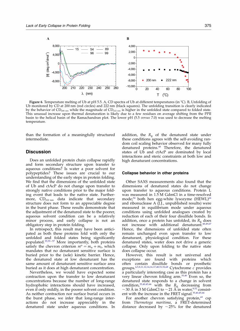
In order to compare the CD222 nm signal in thechemically denatured state to the thermallydenatured state, we measured the far-UV CDspectra at higher temperatures (Figure 6). Changesin the CD spectra indicate that, at 95 8C, the proteinhas thermally denatured. In particular, the CD200 nm
signal goes from positive to negative, indicating anincrease in the amount of coil or PPII structure,defined as the extended conformation withdihedral angles F ¼ 2758 and C ¼ 1508.
The thermally denatured state has a stronger(more negative) CD222 nm signal than the nativestate. This unusual increase upon thermal unfold-ing may be due in part to native Ub’s low CD222 nm
value which results65 from its high PPII content(32%, 24% and 34% for b, PPII, and a dihedralangles, respectively). The increase in CD222 nm
upon thermal denaturation probably is due tosome residues with native dihedral angles in thePPII basin adopting helical f, c angles in thedenatured state (see below).
In summary, both the burst phase at 0.5 MGdmCl and the thermally denatured protein havenearly the same CD222 nm signal. In addition, theabsolute magnitude of the burst phase change issmall (DCD , 3000 deg cm2 dmol21). These results,in combination with the lack of a change in the Rg,fluorescence signal, surface burial and hydrogenbond content between the burst phase andU6 M Gdm argue that the burst-phase CD signalrepresents the adjustment of the denatured stateto the new solvent condition, Dphysiological, rather
Figure 5. Denaturant dependence of Ub folding monitoring CD. A, Refolding traces at the GdmCl concentrationsindicated. B, Rate constants are obtained from both the CD222 nm and fluorescence traces, measured simultaneously.The continuous line is a fit to the higher accuracy fluorescence rates assuming a linear dependence of the free energyof folding. The fitted slope, or mf value, is within the statistical precision of our previous measurements, and representsthe amount of surface buried going from the denatured state to transition. Rates could not be obtained from CD222 nm
measurements at the two lowest denaturant concentrations as almost the entire CD signal is lost in the burst phase.
374 Lack of Early Collapse in Protein Folding
than the formation of a meaningfully structuredintermediate.
Discussion
Does an unfolded protein chain collapse rapidlyand form secondary structure upon transfer toaqueous conditions? Is water a poor solvent forpolypeptides? These issues are crucial to ourunderstanding of the early steps in protein folding.We find that the dimensions of the unfolded stateof Ub and ctAcP do not change upon transfer tostrongly native conditions prior to the major fold-ing event that leads to the native state. Further-more, CD222 nm data indicate that secondarystructure does not form to an appreciable degreein the burst phase. These results demonstrate thatthe adjustment of the denatured state to the poorer,aqueous solvent condition can be a relativelyminor process, and early collapse is not anobligatory step in protein folding.
In retrospect, this result may have been antici-pated as both these proteins fold with only theunfolded and folded states being significantlypopulated.35,55 – 57 More importantly, both proteinssatisfy the chevron criterion mo ¼ mu þ mf, whichmandates that no denaturant sensitive surface isburied prior to the (sole) kinetic barrier. Hence,the denatured state at low denaturant has thesame amount of denaturant sensitive surface areaburied as it does at high denaturant concentration.
Nevertheless, we would have expected somecontraction upon the transfer to low denaturantconcentrations because the number of long-rangehydrophobic interactions should have increased,even if only mildly, in the poorer solvent condition.As neither contraction nor surface burial occurs inthe burst phase, we infer that long-range inter-actions do not increase appreciably in thedenatured state under aqueous conditions. In
addition, the Rg of the denatured state underthese conditions agrees with the self-avoiding ran-dom coil scaling behavior observed for many fullydenatured proteins.48 Therefore, the denaturedstates of Ub and ctAcP are dominated by localinteractions and steric constraints at both low andhigh denaturant concentrations.
Collapse behavior in other proteins
Other SAXS measurements also found that thedimensions of denatured states do not changeupon transfer to aqueous conditions. Protein Lwas measured in 1.5 M GdmCl in a time-resolvedmode;54 both hen egg-white lysozyme (HEWL)66
and ribonuclease A (J.J., unpublished results) weremeasured in equilibrium mode under aqueousconditions using unfolded analogues created byreduction of each of their four disulfide bonds. Inaddition, once a protein has unfolded, its Rg doesnot increase with additional denaturant.48,49,67
Hence, the dimensions of unfolded state oftenremain unchanged even upon transfer to lowdenaturant, physiological condition. For thesedenatured states, water does not drive a genericcollapse. Only upon folding to the native statedoes collapse occur.
However, this result is not universal andexceptions are found with proteins whichoften contain disulfide bonds or prostheticgroups.4,5,8,11,12,14,16,17,48,51,52,68 Cytochrome c providesa particularly interesting case as this protein has avery linear chevron folding arm.35,40 Even so, thedenatured state responds to a change in solventcondition,35,40,45,46 with the Rg decreasing from,30 A in 3 M GdmCl to ,21 A in water,8,52 consist-ent with the increase in the FRET signal.35,40,45,46
For another chevron satisfying protein,69 cspfrom Thermotoga maritima, a FRET-determineddistance decreased by ,25% for the denatured
Figure 6. Temperature melting of Ub at pH 5.5. A, CD spectra of Ub at different temperatures (in 8C). B, Unfolding ofUb monitored by CD at 200 nm (red circles) and 222 nm (black squares). The unfolding transition is clearly indicatedby the behavior of CD200 nm while the magnitude of CD222 nm is higher in the unfolded state compared to folded state.This unusual increase upon thermal denaturation is likely due to a few residues on average shifting from the PPIIbasin to the helical basin of the Ramachandran plot. The lower pH (5.5 versus 7.0) was used to decrease the meltingtemperature.
Lack of Early Collapse in Protein Folding 375

protein upon transfer to 0.5 M GdmCl.9,19 Althoughthe large size of the two chromophores (four or sixaromatic rings) opens up the possibility of theirassociation, the denatured state lacking them alsocontracted, as assayed by tryptophan quenching.21
In addition, in the refolding of the csp from Bacilluscaldolyticus, up to 70% of a FRET signal is quenchedin the burst phase.20 Nevertheless, Magg & Schmidconclude that in spite of this quenching, the burst-phase species cannot be a partially folded inter-mediate. Potentially, some of the change in theFRET signal is due to a different local environmentresulting from a shift in F,C conformers. Never-theless, SAXS measurements, which directlymonitor chain dimensions, would be extremelyhelpful in quantifying the amount of contractionthat can occur in these otherwise two-state systemswhich give FRET burst signals.
Thermodynamic considerations
For over 30 small proteins, stable species do notaccumulate on the unfolded side of the sole kineticbarrier.34,35,70 As intra-chain contact times are sub-microseconds, polymer-style collapse could be onthis time-scale. As this time-scale is three to eightorders of magnitude faster than the overall foldingtimes, the unfolded chain has ample time tocollapse before it traverses the folding barrier.However, the chain does not do so for these highlycooperative proteins. Therefore, the absence ofstable collapsed species prior to acquisition of thenative state must be due to thermodynamic ratherthan kinetic constraints.
Although hydrophobic interactions areenhanced upon the shift to aqueous conditions,collapse still is inhibited by the loss of confor-mational entropy, both backbone and side-chain,and the desolvation of main-chain polar groups.As there is no increase in hydrophobic burial inthe denatured state ðmo ¼ mu þ mfÞ, we concludethat hydrophobic association must be difficult toachieve without a costly desolvation of the polarbackbone.
Previous results further support this reasoning.Kinetic 2H/H amide isotope effect experimentsdemonstrate that surface burial occurs com-mensurately with native hydrogen bond formationalong the folding pathway.35,70 Stable hydrogenbonded secondary structure cannot be divorcedfrom surface burial, thereby precluding earlycollapse without a significant amount of hydrogenbonding. Consistently, burst-phase hydrogen bondformation has not been detected by hydrogenexchange experiments on two-state proteins.41,64,71
From a theoretical vantage point, stable back-bone desolvation is very demanding as it can onlyoccur in a context that satisfies hydrogen bondingrequirements. The burial of each unsatisfied hydro-gen bond partner costs several kcal mol21.72 Giventhat the net stability of a protein often is less than10 kcal mol21, even a small number of failuresresults in an unstable collapsed species. Therefore,
a generic, hydrophobic collapse is unlikely. Due tothe nearly universal trend for two-state folding insmall proteins, these considerations are likely tobe general, although exceptions can occur forfunctional reasons.17
Folding mechanisms
Main-chain burial requirements inhibit non-native chain condensation strongly, and in conjunc-tion with entropic considerations, the early steps inthe conformational search for the transition stateare uphill in free energy. Stable species are difficultto form, and present only on the native side of thebarrier. By the time the chain has achieved a stableconformation, it has such a significant proportionof surface burial and hydrogen bond formationthat it will have already traversed the kineticbarrier.
Based upon these and other considerations, weproposed that formation of a stable collapsedspecies is the intrinsic rate-limiting step in proteinfolding.45,73,74 The limiting step itself is the for-mation of a transition state structure with asufficient number of long-range contacts whichadequately define the native topology.75 At thispoint, the nucleation event, a considerable amountof configuration entropy is lost and folding canproceed rapidly downhill.
Implications to computer simulations
Even though folding studies with computersimulations probe single molecule trajectories,their average behavior should be consistent withour ensemble measurements (Figure 7). For a two-state system, the protein will jump back and forthbetween the unfolded and folded states.9,19,76
Transiently populated intermediates manifestthemselves as brief stops between the two levels.77
Hence, for simulations starting from a realisticunfolded state, the chain should, on an average,retain the properties of the unfolded state prior toa sharp transition leading to the native state. Arealistic unfolded state would be a self-avoidingchain with the appropriate residue-dependentRamachandran basin distributions, rather than, forexample, a fully extended state that rapidlycontracts.
Before the major folding transition, the chain canbriefly sample collapsed conformations. However,the time-averaged population of such confor-mations should be much less than that of theunfolded state in order to agree with the experi-mental results. A greater population fractionwould represent the formation of a stable collapsedintermediate before the rate-limiting step. We donot observe these collapsed species in burst-phaseSAXS measurements. Even a low accumulationlevel of a collapsed species with significant surfaceburial would be detectable in an ensemble experi-ment. In addition to having a smaller Rg, theintermediate would have buried surface that
376 Lack of Early Collapse in Protein Folding

would reduce the denaturant dependence offolding rate and produce a “roll-over” in thechevron folding arm. Observed folding arms oftenare linear to better than 10% of the mf-value. There-fore, the fraction of time that the chain in a foldingsimulation is allowed to sample a non-native,collapsed conformation is severely limited.
Furthermore, all native-like properties should beacquired in the major transition, or shortly there-after. Otherwise, the average of multiple simu-lations would be inconsistent with the observationthat all the native properties are acquired in thesame kinetic step.
Molecular origin of burst phase signals
Ub and ctAcP’s denatured states exhibit no con-traction upon the shift to aqueous conditions, butthe amount of short-range structure increases. ForctAcP, the change in CD is fully accounted for bya sloping base-line of the unfolded state. All thestopped-flow CD data on kinetically two-stateproteins we are aware of share this property41,42,78
although the base line sometimes has mildcurvature.44,46,79
For Ub, the burst-phase CD has pronouncedincrease starting at 2.5 M GdmCl. Both the burstphase at 0.5 M GdmCl and the thermallydenatured protein have larger CD signal than thenative state. The difference may be due to theunusually low native CD222 nm value resultingfrom its high (24%) PPII content.65 For both Uband cytochrome c,46 the thermally denatured stateis a better approximation of Dphysiological than is theunfolded state at high denaturant concentration.
In summary, for all two-state systems con-sidered, burst-phase CD signals simply representthe response of the unfolded state to the change insolvent condition. The origin of these signals isunclear as they often are not associated with stablehydrogen bonded structure.35,46,64,80 The spectro-scopic signals could be due to transient, itineranthelix formation running throughout the chain. Wepropose that the residual CD signals could alsoresult from a shift in the average backbonedihedral angle distribution from PPII to helicalconformers. This increase in the number of peptidegroups with f, c angles in the helical basin of the
Ramachandran plot does not necessarily involveany organized helical structure. Shifts to theb-basin can be considered and would produce thesame qualitative result, but this basin is not signifi-cantly populated in the denatured state.81 – 85
PPII has a positive CD222 nm signal, and is thedominant conformation in the denatured state.86 – 94
PPII has a more exposed backbone and increasedsolvent–protein interactions compared to thehelical conformation.95– 99 Additionally, denatur-ants enhance PPII conformations (N. Kallenbach,personal communication).
Small shifts in basin populations even for shortamino acid stretches are sufficient to quantitativelyaccount for the denaturant dependence of theburst-phase CD. Even one to three consecutivehelical residues have 210,000 mdeg cm2 dmol21
to 220,000 mdeg cm2 dmol21, respectively88,100
(R. Woody, personal communication). A subtle20% shift from the helical basin to the PPIIbasin, using values of 0 mdeg cm2 dmol21 and215,000 mdeg cm2 dmol21 for [u]PPII and [u]helix,respectively, would decrease the CD222 nm signalby 3000 mdeg cm2 dmol21. (This decrease iswhat we observe in Ub’s burst phase.)
In spite of the increase in helical basin popu-lation, the Rg does not change. However, thissame result is found even when extensive amountsof isolated helix are formed in methanol101,102 andtrifluoroethanol solutions.66
Conclusions
Upon a jump to native conditions, the denaturedchain remains extended for both Ub and ctAcP.Hence, water is a sufficiently good solvent thatgeneric intra-chain contacts do not outweighprotein–solvent interactions to produce an earlycollapse phase. The local backbone changesthat do occur are likely the result of a minorredistribution from PPII to helical conformers.These results place severe constraints onfolding mechanisms and computer simulations ofcooperatively folding proteins. The origins ofthese observations are due, in part, to thermo-dynamic issues including backbone desolvation,backbone and side-chain entropy, and hydrogen
Figure 7. Two-state foldingbehavior and the lack of an earlycollapse phase places constraintson computer simulations. Theblack illustrative trace is consistentwith ensemble behavior, as theobservable probe has either thevalue of the unfolded state or nativestate for the majority of the trajec-tory. Only unstable, short-lived
collapsed species exist prior to major folding transition at tfold: Likewise, post-transition state intermediates areshort-lived compared to average folding time, obtained from multiple trajectories. The blue and red trajectories areinconsistent with ensemble two-state folding behavior and the present SAXS data, as collapsed intermediates populate,and the kinetic data will not satisfy the chevron criteria, mo ¼ mu þ mf:
Lack of Early Collapse in Protein Folding 377
bonding requirements. However, it is unclearwhether strong protein–solvent interactions andlack of collapse are exclusive to protein sequenceswhich fold in a two-state manner. Potentially,these properties are applicable to generic poly-peptides, suggesting that chains of a-amino acidresidues were chosen in part for having theseproperties, which would tend to reduce misfoldingand aggregation.
Materials and Methods
Expression and purification
Proteins were expressed and purified as described35,55
in multiple batches to produce gram-scale quantities forSAXS experiments.
Small-angle X-ray scattering
SAXS experiments were carried out at the SAXSinstrument on the BioCAT ID-18 beam-line of ArgonneNational Laboratory’s Advanced Photon Source. Datawere collected using a single chip (8.6 cm £ 4.9 cm)CCD area detector and exposure times were approxi-mately 0.4 seconds for each measurement. Typically,four measurements were recorded at each concentrationand these were averaged to obtain a single data point.Sample to detector distance was ,2 m and energy ofX-ray radiation was set to 12 keV.
Experiments used a Biologic SFM 400 stopped-flowapparatus55 with a 1.0 mm diameter cylindrical quartzcapillary of 0.01–0.02 mm wall thickness. In order tominimize the dead-time in kinetic measurements, SFM400 was mounted at the beam-line so that the X-raybeam passed through the lowest part of the capillarytube minimizing the distance between the mixer and thepoint where the X-rays pass through the capillary tube.Pre-equilibrated samples at the appropriate denaturantconcentrations were used to obtain equilibrium data.A constant flow (flow speed 0.5 ml s21) was maintainedto avoid radiation damage. Final protein concentrationwas ,1.5 mg ml21. In acquiring the kinetic data, theunfolded protein (concentration ,12 mg ml21) wasdiluted eightfold to appropriate denaturant concen-tration and data were acquired at a constant flow speedof 4 ml s21, corresponding to refolding dead-time of,2.5 milliseconds. The buffer background scatteringwas obtained from an otherwise identical configuration.
Measurements were carried out at 20 8C in 50 mMHepes, pH 7.0 (Ub) or 50 mM sodium acetate buffer, pH5.5 (ctAcP). Unfolded samples were in 6 M GdmHCl(Ub) or 8 M urea (ctAcP).
CD and fluorescence measurements
Far-UV circular dichroism spectroscopy was per-formed with a Jasco Model 715 CD spectropolarimeter.Equilibrium CD spectra were recorded every 1 nm at2-nm resolution in a 1 cm pathlength cell. The kineticmeasurements used the Biologic SFM 4 stopped-flowapparatus interfaced with the Jasco spectropolarimeterand a 0.8 mm path length cuvette. The resolution wasset to 2 nm and between ten and 40 traces were averagedat each condition. Simultaneous acquisition of thefluorescence signal was achieved using a second photo-
multiplier tube mounted at 908 to the incident beam andusing a 300–400 nm band pass filter.
Time-dependent change in CD was placed on anabsolute scale using the identical protein solution in thestandard spectrophotometer sample holder. Fl valueswere normalized by measurements on the folded andthe unfolded protein. The signal from the buffer wassubtracted at every denaturant concentration, and burst-phase amplitudes shown in Figure 3 were extrapolatedback to zero time. The data in the burst phase areacquired by averaging 0.15–0.2 seconds of data in thecontinuous flow phase. Even though the dead volume(volume between mixer and the point of observation) ishigher compared to that in SAXS measurements, ahigher flow-speed of 6 ml s21 was used in these measure-ments leading to approximately the same dead-time of2.5 milliseconds.
Identical buffer conditions were maintained in the CDand fluorescence measurements as for the SAXSmeasurements, except for lower protein concentrations.Final protein concentration was 92 mM and 69 mM forUb and ctAcP, respectively. Approximately 40 shotswere averaged at each condition. To reduce buffer andsample absorbance, temperature melting of Ub was car-ried out in 5 mM sodium acetate at a protein concen-tration of 19 mM and pH 5.5. The lower pH was used tolower the melting temperature.
Data analysis
SAXS reports on the average structure of particles insolution. X-rays are scattered by the sample and the scat-tering profile is measured at very low angles u.103 Dataare presented as the scattering intensity per solid angle,IðQÞ, where the scattering vector Q, is defined asQ ¼ 4p sin u=l, l is the X-ray wavelength, and u is thehalf scattering angle. At low Q # 1.3/Rg, the dimensionsof a particle can be determined from the width of theinner part of the scattering profile, which can be approxi-mated as a Gaussian, IðQÞ ¼ I0 e2Q2R2
g=3: The Rg is theroot-mean-square of the distances of all regions to thecenter-of-mass of the particle. Typically, the Rg isobtained from the slope of the low-angle region of aGuinier Plot of ln IðQÞ versus Q2:
To measure the Rg value accurately, scattering datamust be taken across angles where the Gaussianapproximation holds. Our reliable data starts atQ ¼ 0.02 A21, or QRg ¼ 0:6 for the largest size(Rg ¼ 30– 32 A for unfolded ctAcP). This low Qmin
value, along with the linearity in the Guinier plot andthe expected behavior of I0, demonstrate that the Rg canbe reliably obtained from these data.
More precise structural parameters were derived froma PðrÞ analysis using the entire scattering profile. The PðrÞfunction has a maximum at the most probable distancein the object (e.g., slightly larger than the radius for asphere) and goes to zero at the maximum dimension,dmax, of the object (e.g. the diameter). P(r) functionswere calculated according to:
PðrÞ ¼1
2p2
ðQmax
Qmin
IðQÞQr sinðQrÞdQ ð2Þ
using the indirect Fourier inversion algorithms devel-oped by Svergun†.
† GNOM small-angle scattering data processing bymeans of the regularization technique. http://www.srs.dl.ac.uk/ncd/computing/manual.gnom.html
378 Lack of Early Collapse in Protein Folding

The equilibrium Rg values were fitted to a linear freeenergy relationship:
Rg ¼
ffiffiffiffiffiffiffiffiffiffiffiffiffiffiffiffiffiffiffiffiffiffiffiffiffiffiffiffiffiffiffiffiffiffiffiffiffiffiffiffiffiffiffiffiffiffiffiffiffiffiffiffiffiffiffiffiR2
d þR2
n 2 R2d
1 þ e2ðmCu2m½den�Þ=RT
sð3Þ
where Rn and Rd are the Rg of native and denatured pro-tein, respectively, m is the equilibrium m-value and Cu isthe midpoint of the collapse transition.49 The equilibriumCD data were fitted to standard two-state transitionswith sloping linear baselines:
CD ¼ CDu þ Su½den�
þCDf þ Sf½den�2 CDu 2 Su½den�
1 þ e2ðmCm2m½den�Þ=RTð4Þ
where CDf and CDu represent the CD signals of thefolded and unfolded states in water, respectively, Sf andSu represent the denaturant dependencies of thesevalues, and Cm, the midpoint of the transition.
The kinetic data were analyzed using the “chevronanalysis” of the denaturant dependence of folding rateconstants104 where the standard free energy of folding,DG8, along with the standard activation free energy forfolding DG‡
f , and unfolding, DG‡u, are linearly dependent
on denaturant concentration:
DG8ð½den�Þ ¼ DGH2OEquil þ mo½den� ¼ 2RT lnðKuÞ ð5aÞ
DG‡f ð½den�Þ ¼ 2RT lnðkH2O
f Þ þ mf½den� þ Constant ð5bÞ
DG‡uð½den�Þ ¼ 2RT lnðkH2O
u Þ2 mu½den� þ Constant ð5cÞ
where R is the universal gas constant, T is the tempera-ture, and the dependence on denaturant concentration,the m-values, report the degree of surface area burialduring the folding process. When equilibrium andkinetic folding reactions are effectively two-state and arelimited by the same activation barrier, the equilibriumvalues for the standard free energy and surface burialcan be calculated from the kinetic measurements accord-ing to DG8 ¼ DG‡
f 2 DG‡u and mo ¼ mu þ mf: Parameters
were fit using non-linear least-squares algorithmsimplemented in the Microcal Origin software package.
Acknowledgements
We thank K. Plaxco, S. W. Englander, N.Kallenbach, R. Woody, A. Garcia, G. Rose, A.Kentsis, R. Pappu, D. Raleigh, and members ofour group for discussions, unpublished resultsand preprints, and Dr Elena Kondrashkina forexpert assistance at the beam-line. This work issupported by grants from the NIH (T.R.S.), TheUS Department of Energy, BES-Materials Science,under contract W-31-109-ENG-38 (P.T.), The Univ.of Chicago-Argonne National LaboratoryCollaborative Seed Grant Program (T.R.S. & P.T.),and Packard Foundation Interdisciplinary ScienceProgram (T.R.S., P.T., D. Lynn, S. Meredith andR. S. Berry). Use of the Advanced Photon Sourcewas supported by the US Department of Energy,Basic Energy Sciences, Office of Science, under con-tract No. W-31-109-ENG-38. BioCAT is a National
Institutes of Health-supported Research CenterRR-08630.
References
1. Hagen, S. J. & Eaton, W. A. (2000). Two-state expan-sion and collapse of a polypeptide. J. Mol. Biol. 297,781–789.
2. Agashe, V. R., Shastry, M. C. & Udgaonkar, J. B.(1995). Initial hydrophobic collapse in the foldingof barstar. Nature, 377, 754–757.
3. Ballew, R. M., Sabelko, J. & Gruebele, M. (1996).Direct observation of fast protein folding: the initialcollapse of apomyoglobin. Proc. Natl Acad. Sci.USA, 93, 5759–5764.
4. Pollack, L., Tate, M., Finnefrock, A., Kalidas, C.,Trotter, S., Darnton, N. et al. (2001). Time resolvedcollapse of a folding protein observed with smallangle X-ray scattering. Phys. Rev. Letters, 86,4962–4965.
5. Chen, L., Wildegger, G., Kiefhaber, T., Hodgson,K. O. & Doniach, S. (1998). Kinetics of lysozymerefolding: structural characterization of a non-specifically collapsed state using time-resolvedX-ray scattering. J. Mol. Biol. 276, 225–237.
6. Parker, M. J., Dempsey, C. E., Lorch, M. & Clarke,A. R. (1997). Acquisition of native beta-strand top-ology during the rapid collapse phase of proteinfolding. Biochemistry, 36, 13396–13405.
7. Nath, U. & Udgaonkar, J. B. (1997). Folding oftryptophan mutants of barstar: evidence for aninitial hydrophobic collapse on the folding path-way. Biochemistry, 36, 8602–8610.
8. Akiyama, S., Takahashi, S., Kimura, T., Ishimori, K.,Morishima, I., Nishikawa, Y. & Fujisawa, T. (2002).Conformational landscape of cytochrome c foldingstudied by microsecond-resolved small-angle X-rayscattering. Proc. Natl Acad. Sci. USA, 99, 1329–1334.
9. Lipman, E. A., Schuler, B., Bakajin, O. & Eaton, W. A.(2003). Single-molecule measurement of proteinfolding kinetics. Science, 301, 1233–1235.
10. Segel, D. J., Eliezer, D., Uversky, V., Fink, A. L.,Hodgson, K. O. & Doniach, S. (1999). Transientdimer in the refolding kinetics of cytochrome ccharacterized by small-angle X-ray scattering.Biochemistry, 38, 15352–15359.
11. Segel, D. J., Bachmann, A., Hofrichter, J., Hodgson,K. O., Doniach, S. & Kiefhaber, T. (1999). Character-ization of transient intermediates in lysozymefolding with time-resolved small-angle X-rayscattering. J. Mol. Biol. 288, 489–499.
12. Eliezer, D., Chiba, K., Tsuruta, H., Doniach, S.,Hodgson, K. O. & Kihara, H. (1993). Evidence ofan associative intermediate on the myoglobinrefolding pathway. Biophys. J. 65, 912–917.
13. Doniach, S., Bascle, J., Garel, T. & Orland, H. (1995).Partially folded states of proteins: characterizationby X-ray scattering. J. Mol. Biol. 254, 960–967.
14. Chen, L., Hodgson, K. O. & Doniach, S. (1996). Alysozyme folding intermediate revealed by solutionX-ray scattering. J. Mol. Biol. 261, 658–671.
15. Arai, S. & Hirai, M. (1999). Reversibility andhierarchy of thermal transition of hen egg-whitelysozyme studied by small-angle X-ray scattering.Biophys. J. 76, 2192–2197.
16. Arai, M., Ito, K., Inobe, T., Nakao, M., Maki, K.,Kamagata, K. et al. (2002). Fast compaction ofalpha-lactalbumin during folding studied by
Lack of Early Collapse in Protein Folding 379

stopped-flow X-ray scattering. J. Mol. Biol. 321,121–132.
17. Choy, W. Y., Mulder, F. A., Crowhurst, K. A.,Muhandiram, D. R., Millett, I. S., Doniach, S. et al.(2002). Distribution of molecular size within anunfolded state ensemble using small-angle X-rayscattering and pulse field gradient NMRtechniques. J. Mol. Biol. 316, 101–112.
18. Dill, K. A. & Shortle, D. (1991). Denatured states ofproteins. Annu. Rev. Biochem. 60, 795–825.
19. Schuler, B., Lipman, E. A. & Eaton, W. A. (2002).Probing the free-energy surface for protein foldingwith single-molecule fluorescence spectroscopy.Nature, 419, 743–747.
20. Magg, C. & Schmid, F. X. (2004). Rapid collapseprecedes the fast two-state folding of the coldshock protein. J. Mol. Biol. 335, 1309–1323.
21. Buscaglia, M., Schuler, B., Lapidus, L. J., Eaton,W. A. & Hofrichter, J. (2003). Kinetics of intra-molecular contact formation in a denatured protein.J. Mol. Biol. 332, 9–12.
22. Zagrovic, B., Snow, C. D., Khaliq, S., Shirts, M. R. &Pande, V. S. (2002). Native-like mean structure inthe unfolded ensemble of small proteins. J. Mol.Biol. 323, 153–164.
23. Shimada, J. & Shakhnovich, E. I. (2002). Theensemble folding kinetics of protein G from an all-atom Monte Carlo simulation. Proc. Natl Acad. Sci.USA, 99, 11175–11180.
24. Duan, Y. & Kollman, P. A. (1998). Pathways to aprotein folding intermediate observed in a 1-micro-second simulation in aqueous solution. Science, 282,740–744.
25. Shen, M. Y. & Freed, K. F. (2002). All-atom fastprotein folding simulations: the villin headpiece.Proteins: Struct. Funct. Genet. 49, 439–445.
26. Alonso, D. O. V. & Daggett, V. (1998). Moleculardynamics simulations of hydrophobic collapse ofubiquitin. Protein Sci. 7, 860–874.
27. Linhananta, A., Zhou, H. Y. & Zhou, Y. Q. (2002).The dual role of a loop with low loop contactdistance in folding and domain swapping. ProteinSci. 11, 1695–1701.
28. Mayor, U., Guydosh, N. R., Johnson, C. M.,Grossmann, J. G., Sato, S., Jas, G. S. et al. (2003).The complete folding pathway of a protein fromnanoseconds to microseconds. Nature, 421, 863–867.
29. Lapidus, L. J., Eaton, W. A. & Hofrichter, J. (2000).Measuring the rate of intramolecular contact for-mation in polypeptides. Proc. Natl Acad. Sci. USA,97, 7220–7225.
30. Krieger, F., Fierz, B., Bieri, O., Drewello, M. &Kiefhaber, T. (2003). Dynamics of unfoldedpolypeptide chains as model for the earliest stepsin protein folding. J. Mol. Biol. 332, 265–274.
31. Chang, I. J., Lee, J. C., Winkler, J. R. & Gray, H. B.(2003). Bioinorganic Chemistry Special Feature: theprotein-folding speed limit: intrachain diffusiontimes set by electron-transfer rates in denaturedRu(NH3)5(His-33)-Zn-cytochrome c. Proc. NatlAcad. Sci. USA, 100, 3838–3840.
32. Qiu, L., Zachariah, C. & Hagen, S. J. (2003). Fastchain contraction during protein folding: “fold-ability” and collapse dynamics. Phys. Rev. Letters,90, 168103.
33. Sadqi, M., Lapidus, L. J. & Munoz, V. (2003). Howfast is protein hydrophobic collapse? Proc. NatlAcad. Sci. USA, 100, 12117–12122.
34. Jackson, S. E. (1998). How do small single-domainproteins fold? Fold. Des. 3, R81–R91.
35. Krantz, B. A., Mayne, L., Rumbley, J., Englander,S. W. & Sosnick, T. R. (2002). Fast and slowintermediate accumulation and the initial barriermechanism in protein folding. J. Mol. Biol. 324,359–371.
36. Jackson, S. E. & Fersht, A. R. (1991). Folding ofchymotrypsin inhibitor 2.1. Evidence for a two-state transition. Biochemistry, 30, 10428–10435.
37. Elove, G. A., Chaffotte, A. F., Roder, H. & Goldberg,M. E. (1992). Early steps in cytochrome c foldingprobed by time-resolved circular dichroism andfluorescence spectroscopy. Biochemistry, 31,6876–6883.
38. Roder, H. & Colon, W. (1997). Kinetic role of earlyintermediates in protein folding. Curr. Opin. Struct.Biol. 7, 15–28.
39. Shastry, M. C. & Roder, H. (1998). Evidence forbarrier-limited protein folding kinetics on themicrosecond time scale. Nature Struct. Biol. 5,385–392.
40. Chan, C. K., Hu, Y., Takahashi, S., Rousseau, D. L.,Eaton, W. A. & Hofrichter, J. (1997). Submillisecondprotein folding kinetics studied by ultrarapidmixing. Proc. Natl Acad. Sci. USA, 94, 1779–1784.
41. Scalley, M. L., Yi, Q., Gu, H., McCormack, A., Yates,J. R., 3rd & Baker, D. (1997). Kinetics of folding ofthe IgG binding domain of peptostreptococcalprotein L. Biochemistry, 36, 3373–3382.
42. Sato, S., Luisi, D. L. & Raleigh, D. P. (2000). pHjump studies of the folding of the multidomainribosomal protein L9: the structural organization ofthe N-terminal domain does not affect theanomalously slow folding of the C-terminaldomain. Biochemistry, 39, 4955–4962.
43. Qin, Z., Ervin, J., Larios, E., Gruebele, M. & Kihara,H. (2002). Formation of a compact structuredensemble without fluorescence signature earlyduring ubiquitin folding. J. Phys. Chem. B, 106,13040–13046.
44. Qi, P. X., Sosnick, T. R. & Englander, S. W. (1998).The burst phase in ribonuclease A folding andsolvent dependence of the unfolded state. NatureStruct. Biol. 5, 882–884.
45. Sosnick, T. R., Mayne, L. & Englander, S. W. (1996).Molecular collapse: the rate-limiting step in two-state cytochrome c folding. Proteins: Struct. Funct.Genet. 24, 413–426.
46. Sosnick, T. R., Shtilerman, M. D., Mayne, L. &Englander, S. W. (1997). Ultrafast signals in proteinfolding and the polypeptide contracted state. Proc.Natl Acad. Sci. USA, 94, 8545–8550.
47. Svergun, D. I. & Koch, M. H. J. (2003). Small-anglescattering studies of biological macromolecules insolution. Rep. Prog. Phys. 66, 1735–1782.
48. Millet, I. S., Doniach, S. & Plaxco, K. W. (2002).Toward a taxonomy of the denatured state: smallangle scattering studies of unfolded proteins.Advan. Protein Chem. 62, 241–262.
49. Millet, I. S., Townsley, L. E., Chiti, F., Doniach, S. &Plaxco, K. W. (2002). Equilibrium collapse and thekinetic “foldability” of proteins. Biochemistry, 41,321–325.
50. Sosnick, T. R. & Trewhella, J. (1992). Denaturedstates of ribonuclease A have compact dimensionsand residual secondary structure. Biochemistry, 31,8329–8335.
51. Flanagan, J. M., Kataoka, M., Shortle, D. &
380 Lack of Early Collapse in Protein Folding
Engelman, D. M. (1992). Truncated staphylococcalnuclease is compact but disordered. Proc. NatlAcad. Sci. USA, 89, 748–752.
52. Pollack, L., Tate, M. W., Darnton, N. C., Knight, J. B.,Gruner, S. M., Eaton, W. A. & Austin, R. H. (1999).Compactness of the denatured state of a fast-foldingprotein measured by submillisecond small-angleX-ray scattering. Proc. Natl Acad. Sci. USA, 96,10115–10117.
53. Semisotnov, G. V., Kihara, H., Kotova, N. V.,Kimura, K., Amemiya, Y., Wakabayashi, K. et al.(1996). Protein globularization during folding. Astudy by synchrotron small-angle X-ray scattering.J. Mol. Biol. 262, 559–574.
54. Plaxco, K. W., Millett, I. S., Segel, D. J., Doniach, S. &Baker, D. (1999). Chain collapse can occur con-comitantly with the rate-limiting step in proteinfolding. Nature Struct. Biol. 6, 554–556.
55. Krantz, B. A. & Sosnick, T. R. (2000). Distinguishingbetween two-state and three-state models forubiquitin folding. Biochemistry, 39, 11696–11701.
56. Taddei, N., Chiti, F., Paoli, P., Fiaschi, T.,Bucciantini, M., Stefani, M. et al. (1999). Thermo-dynamics and kinetics of folding of common-typeacylphosphatase: comparison to the highly homo-logous muscle isoenzyme. Biochemistry, 38,2135–2142.
57. Krantz, B. A., Srivastava, A. K., Nauli, S., Baker, D.,Sauer, R. T. & Sosnick, T. R. (2002). Understandingprotein hydrogen bond formation with kineticH/D amide isotope effects. Nature Struct. Biol. 9,458–463.
58. Vijay-Kumar, S., Bugg, C. E., Wilkinson, K. D.,Vierstra, R. D., Hatfield, P. M. & Cook, W. J. (1987).Comparison of the three-dimensional structures ofhuman, yeast, and oat ubiquitin. J. Biol. Chem. 262,6396–6399.
59. Thunnissen, M. M., Taddei, N., Liguri, G., Ramponi,G. & Nordlund, P. (1997). Crystal structure ofcommon type acylphosphatase from bovine testis.Structure, 5, 69–79.
60. Svergun, D. I., Richard, S., Koch, M. H., Sayers, Z.,Kuprin, S. & Zaccai, G. (1998). Protein hydration insolution: experimental observation by X-ray andneutron scattering. Proc. Natl Acad. Sci. USA, 95,2267–2272.
61. Merzel, F. & Smith, J. C. (2002). Is the first hydrationshell of lysozyme of higher density than bulkwater? Proc. Natl Acad. Sci. USA, 99, 5378–5383.
62. Khorasanizadeh, S., Peters, I. D. & Roder, H. (1996).Evidence for a 3-state model of protein folding fromkinetic analysis of ubiquitin variants with alteredcore residues. Nature Struct. Biol. 3, 193–205.
63. Khorasanizadeh, S., Peters, I. D., Butt, T. R. & Roder,H. (1993). Folding and stability of a tryptophan-containing mutant of ubiquitin. Biochemistry, 32,7054–7063.
64. Gladwin, S. T. & Evans, P. A. (1996). Structure ofvery early protein folding intermediates: newinsights through a variant of hydrogen exchangelabelling. Fold. Des. 1, 407–417.
65. Sreerama, N. & Woody, R. W. (2003). Structuralcomposition of betaI- and betaII-proteins. ProteinSci. 12, 384–388.
66. Hoshino, M., Hagihara, Y., Hamada, D., Kataoka,M. & Goto, Y. (1997). Trifluoroethanol-induced con-formational transition of hen egg-white lysozymestudied by small-angle X-ray scattering. FEBSLetters, 416, 72–76.
67. Noppert, A., Gast, K., Zirwer, D. & Damaschun, G.(1998). Initial hydrophobic collapse is not necessaryfor folding RNase A. Fold. Des. 3, 213–221.
68. Flanagan, J. M., Kataoka, M., Fujisawa, T. &Engelman, D. M. (1993). Mutations can cause largechanges in the conformation of a denatured protein.Biochemistry, 32, 10359–10370.
69. Perl, D., Welker, C., Schindler, T., Schroder, K.,Marahiel, M. A., Jaenicke, R. & Schmid, F. X.(1998). Conservation of rapid two-state folding inmesophilic, thermophilic and hyperthermophiliccold shock proteins. Nature Struct. Biol. 5, 229–235.
70. Krantz, B. A., Moran, L. B., Kentsis, A. & Sosnick,T. R. (2000). D/H amide kinetic isotope effectsreveal when hydrogen bonds form during proteinfolding. Nature Struct. Biol. 7, 62–71.
71. Sosnick, T. R., Mayne, L., Hiller, R. & Englander,S. W. (1994). The barriers in protein folding. NatureStruct. Biol. 1, 149–156.
72. Honig, B. & Yang, A. S. (1995). Free energy balancein protein folding. Advan. Protein Chem. 46, 27–58.
73. Sosnick, T. R., Mayne, L., Hiller, R. & Englander,S. W. (1995). Peptide and Protein Folding Workshop,Philadelphia, PA.
74. Sosnick, T. R. & Englander, S. W. (1996). What limitsprotein folding. In Dynamics and the Problem ofRecognition in Biological Macromolecules (Jardetzky,O. & Lefevre, J-F., eds), vol. 288, pp. 65–71, PlenumPress, New York.
75. Plaxco, K. W., Simons, K. T. & Baker, D. (1998).Contact order, transition state placement and therefolding rates of single domain proteins. J. Mol.Biol. 277, 985–994.
76. Deniz, A. A., Laurence, T. A., Beligere, G. S., Dahan,M., Martin, A. B., Chemla, D. S. et al. (2000). Single-molecule protein folding: diffusion fluorescenceresonance energy transfer studies of the denatura-tion of chymotrypsin inhibitor 2. Proc. Natl Acad.Sci. USA, 97, 5179–5184.
77. Tan, E., Wilson, T. J., Nahas, M. K., Clegg, R. M.,Lilley, D. M. & Ha, T. (2003). A four-way junctionaccelerates hairpin ribozyme folding via a discreteintermediate. Proc. Natl Acad. Sci. USA, 100,9308–9313.
78. Zitzewitz, J. A., Bilsel, O., Luo, J., Jones, B. E. &Matthews, C. R. (1995). Probing the foldingmechanism of a leucine zipper peptide by stopped-flow circular dichroism spectroscopy. Biochemistry,34, 12812–12819.
79. Takei, J., Chu, R. A. & Bai, Y. (2000). Absence ofstable intermediates on the folding pathway ofbarnase. Proc. Natl Acad. Sci. USA, 97, 10796–10801.
80. Robertson, A. D. & Baldwin, R. L. (1991). Hydrogenexchange in thermally denatured ribonuclease A.Biochemistry, 30, 9907–9914.
81. Smith, L. J., Bolin, K. A., Schwalbe, H., MacArthur,M. W., Thornton, J. M. & Dobson, C. M. (1996).Analysis of main chain torsion angles in proteins:prediction of NMR coupling constants for nativeand random coil conformations. J. Mol. Biol. 255,494–506.
82. Serrano, L. (1995). Comparison between the phi dis-tribution of the amino acids in the protein databaseand NMR data indicates that amino acids havevarious phi propensities in the random coil confor-mation. J. Mol. Biol. 254, 322–333.
83. Penkett, C. J., Redfield, C., Dodd, I., Hubbard, J.,McBay, D. L., Mossakowska, D. E. et al. (1997).NMR analysis of main-chain conformational
Lack of Early Collapse in Protein Folding 381

preferences in an unfolded fibronectin-bindingprotein. J. Mol. Biol. 274, 152–159.
84. Swindells, M. B., MacArthur, M. W. & Thornton,J. M. (1995). Intrinsic phi, psi propensities of aminoacids, derived from the coil regions of knownstructures. Nature Struct. Biol. 2, 596–603.
85. Avbelj, F. & Baldwin, R. L. (2003). Role of backbonesolvation and electrostatics in generating preferredpeptide backbone conformations: distributions ofphi. Proc. Natl Acad. Sci. USA, 100, 5742–5747.
86. Shi, Z., Olson, C. A., Rose, G. D., Baldwin, R. L. &Kallenbach, N. R. (2002). Polyproline II structure ina sequence of seven alanine residues. Proc. NatlAcad. Sci. USA, 99, 9190–9195.
87. Shi, Z., Woody, R. W. & Kallenbach, N. R. (2002). Ispolyproline II a major backbone conformation inunfolded proteins? Advan. Protein Chem. 62,163–240.
88. Eker, F., Griebenow, K. & Schweitzer-Stenner, R.(2003). Stable conformations of tripeptides inaqueous solution studied by UV circular dichroismspectroscopy. J. Am. Chem. Soc. 125, 8178–8185.
89. Pappu, R. V. & Rose, G. D. (2002). A simple modelfor polyproline II structure in unfolded states ofalanine-based peptides. Protein Sci. 10, 2437–2455.
90. Creamer, T. P. & Campbell, M. N. (2002). Determi-nants of the polyproline II helix from modelingstudies. Advan. Protein Chem. 62, 263–282.
91. Dukor, R. K. & Keiderling, T. A. (1991). Reassess-ment of the random coil conformation: vibrationalCD study of proline oligopeptides and related poly-peptides. Biopolymers, 31, 1747–1761.
92. Krimm, S. & Tiffany, M. L. (1974). The circulardichroism spectrum and structure of unorderedpolypeptides and proteins. Isr. J. Chem. 12, 189–200.
93. Wilson, G., Hecht, L. & Barron, L. D. (1996).Residual structure in unfolded proteins revealedby Raman optical activity. Biochemistry, 35,12518–12525.
94. Keiderling, T. A. & Xu, Q. (2002). Unfolded
peptides and proteins studied with infrared absorp-tion and vibrational circular dichroism spectra.Advan. Protein Chem. 62, 111–161.
95. Adzhubei, A. A. & Sternberg, M. J. (1994). Conser-vation of polyproline II helices in homologousproteins: implications for structure prediction bymodel building. Protein Sci. 3, 2395–2410.
96. Creamer, T. P. (1998). Left-handed polyproline IIhelix formation is (very) locally driven. Proteins:Struct. Funct. Genet. 33, 218–226.
97. Mezei, M., Fleming, P. J., Srinivasan, R. & Rose,G. D. (2004). Polyproline II helix is the preferredconformation for unfolded polyalanine in water.Proteins: Struct. Funct. Genet. In the press.
98. Kentsis, A., Mezei, M., Gindin, T. & Osman, R.(2004). Unfolded state of polyalanine is asegmented polyproline II helix. Proteins: Struct.Funct. Genet. In the press.
99. Garcia, A. E. (2004). Characterization of non-alphahelical conformations in Ala peptides. Polymers, 45,669–676.
100. Chin, D. H., Woody, R. W., Rohl, C. A. & Baldwin,R. L. (2002). Circular dichroism spectra of short,fixed-nucleus alanine helices. Proc. Natl Acad. Sci.USA, 99, 15416–15421.
101. Kamatari, Y. O., Konno, T., Kataoka, M. & Akasaka,K. (1998). The methanol-induced transition and theexpanded helical conformation in hen lysozyme.Protein Sci. 7, 681–688.
102. Kamatari, Y. O., Konno, T., Kataoka, M. & Akasaka,K. (1996). The methanol-induced globular andexpanded denatured states of cytochrome c: astudy by CD fluorescence, NMR and small-angleX-ray scattering. J. Mol. Biol. 259, 512–523.
103. Glatter, O. & Kratky, O. (1982). Small Angle X-rayScattering, Academic Press, London.
104. Matthews, C. R. (1987). Effects of point mutationson the folding of globular proteins. MethodsEnzymol. 154, 498–511.
Edited by C. R. Matthews
(Received 11 December 2003; received in revised form 23 February 2004; accepted 24 February 2004)
382 Lack of Early Collapse in Protein Folding