early colonial burial practices for perinates at the
TRANSCRIPT

71
Early colonial burial practices for perinates at the Parramatta convict hospital, NSW.
DENISE DONLON, MARY CASEY, WOLFGANG HAAK AND CHRISTINA ADLER
The skeletal remains of six perinatal infants were found during archaeological excavation of the Parramatta convict hospital. Perinatal refers to the period around birth, from 24 weeks gestation to 7 postnatal days. Two individuals were found in a double grave dated to the second convict hospital c.1800–c.1810, one individual in a storage cellar dated to c.1820 (second hospital) and three individuals in a pit dated to c.1830–1844 (third hospital). The completeness and preservation of the excavated skeletal remains varies according to the form of burial. Probable ancestry, sex and age of the individuals were examined using morphological and molecular evidence. European ancestry for two individuals has been indicated by mtDNA making these of historical significance in this period of early Australian-British settlement. The death and burial of these infants gives insight into late eighteenth and early nineteenth-century attitudes to death and Australian colonial society.
INTRODUCTIONThis paper describes six perinatal skeletons excavated in part of the Parramatta convict hospital. The mortuary practices provide some insight into late eighteenth and early nineteenth-century Australian settler society. The burials are described in their archaeological context, as this has considerable bearing on preservation, dating and interpretation. The individuals are briefly described and morphological and genetic analyses are summarised.
Few bones of perinates and infants have been found in Australian cemeteries. Burials of perinates are often overlooked or not studied, possibly because of the perception
that they are very poorly preserved compared with adult bones and teeth (Guy et al. 1997). The bones of children are known to be particularly prone to decay during burial since they are more porous and have a higher collagen content than adult skeletons (Buckberry 2000). The small size of the bones makes them more likely to be displaced and they may simply be missed due to inexperience of the excavators (Baker et al. 2005).
Other reasons for the scarcity of perinatal skeletons may be because of the type of mortuary practices observed. Mortuary practices for foetuses and neonates may differ to those for adults in many societies. They may be miscarried or stillborn in a hospital and therefore be disposed of by hospital
Figure 1: Location plan of the Parramatta convict hospital, Parramatta Justice Precinct site shaded grey. Casey & Lowe
Aust_Hist_Archeology_Vol26.indd 71 18/5/09 4:06:29 PM

authorities. Infanticide may result in using a clandestine method of burial.
Very few historic cemeteries have been excavated in Australia and even fewer of those have included remains of perinatal skeletons. New South Wales has seen the excavation of the Old Sydney Burial Ground, Sydney Town Hall (Birmingham and Liston 1976; Lowe and Mackay 1992; Casey & Lowe 2008), the Destitute Children’s Asylum at Randwick in Sydney (Austral/ Godden Mackay 1997) and Cadia cemetery at Orange (Edward Higginbotham 2002). In Adelaide, South Australia, St Mary’s cemetery was excavated and the skeletal remains investigated (Pate and Adams 2000; Coussens et al. 2002). In 2000 and 2001 Lang Park in Brisbane, Queensland was excavated to reveal a cemetery which dated from 1843–1875 (Rains and Pragnell 2002; Haslam et al. 2003). Of the 397 burials excavated, 161 of these were determined to be children on the basis of coffin length. Of these, 135 had ‘no skeleton remaining’ at excavation and 26 had ‘poor’ skeletal preservation (Glenys McGowan pers. comm. 2009). While not strictly speaking a cemetery, a mass grave for the Batavia victims at Beacon Island, Western Australia was excavated in 2001 (Paterson and Franklin 2004). The only sites where juvenile skeletons as young as those described here have been found are the Cadia Cemetery near Orange and the Destitute Children’s Asylum at Randwick, both in New South Wales.
Both Cadia Cemetery and the Destitute Children’s Asylum at Randwick contained perinatal individuals. Both of these cemeteries date from the 1860s until the 1890s. At the Destitute Children’s Asylum at Randwick 65 juvenile skeletons were excavated, and evidence of only a single perinatal cranial vault fragment was found (Austral/Godden Mackay 1997). The bone
had actually rotted away leaving hair and a dark stain where the cranial vault would have been and there was also a dark stain in the region of the knee joints. The burial was not on the plan of the cemetery or in the records and was probably a clandestine burial. At Cadia, of 111 skeletons one presented as between 7–8 months in utero, three as consistent with 8 months in utero to birth and 13 as neonatal (Estelle Lazer pers comm.)
BACKGROUND AND INVESTIGATIONThe first convict hospital in Parramatta was established
c.1790 initially as two long sheds built in the form of a tent (Tench 1793 (1979):196). By1792 the second convict hospital was built to replace the dilapidated earlier hospital. The second hospital was more substantial, being built of brick, and was in use from 1792 until c.1818 (Collins 1798 (1975), vol 1:207; Casey & Lowe 2005, 2006).
The former Parramatta convict hospital, near the corner of George and Marsden Streets, is an archaeological site on the State Heritage Register and is of State heritage significance (Fig. 1). Between March 2005 and January 2008 Casey & Lowe undertook archaeological investigations to expose, record and retain in situ the footprint of most of the surviving remains of Parramatta’s second and third convict hospitals. In some areas testing and salvage of the archaeological remains was permitted. The archaeological program found the extensive remains of the third convict hospital (1818–c.1844) and the partial remains of the second convict hospital (1792–1818). The focus of the salvage work was in the southwest corner of the hospital (Figs 2, 3, 4) which was once bounded by a series of fences and walls. This area would have been the ‘backyard’
72
Figure 2: Plan of the hospital area and the main structures from the second and third convict hospitals. The black structures are the third hospital (1818-c.1844) and the grey structures belong to the second hospital (1792-1818). Casey & Lowe
Aust_Hist_Archeology_Vol26.indd 72 18/5/09 4:06:31 PM

and appears to have been used for rubbish disposal, and cut through by drains and various phases of sewers. The 1870s toilets were also located nearby. This area also appeared to be one of the most intact parts of the site in terms of built-up deposits from the eighteenth to twentieth centuries and there was limited disturbance by twentieth-century buildings. As an
isolated corner of the hospital, always at the very back of the two convict hospitals and behind their outbuildings, it became a place to dispose of ‘rubbish’ and also to bury two small perinates in a shallow grave. The human remains which are the subject of this paper were found in three separate archaeological contexts in this small area of the site (Figs 3, 4).
73
Figure 3: Detail of the second convict hospital period remains, the storage cellar and double infant burial, and the third hospital rubbish pit. All were located in the southwest corner. Casey & Lowe
Figure 4: General photo of the back corner of the second and third convict hospitals. The double burial is covered with navy blue protective material, it is immediately east of the storage cellar, and the rubbish pit which contained quantities of animal bone and remains of three perinates is far right, next to the boundary wall. Looking south, scale 1 m. Casey & Lowe
Aust_Hist_Archeology_Vol26.indd 73 18/5/09 4:06:34 PM

74
In August 2006 a small grave was exposed in the Stage 2c salvage area. Excavation revealed the skeletal remains of two sets of juveniles (Casey & Lowe 2006:15–16; Miskella 2005–2006). Another perinatal skeleton was found in the backfill of a storage cellar and three more in a rubbish pit (Figs 3, 4). As we were dealing with significant late eighteenth and early nineteenth-century deposits all this material was 100 per cent sieved through two nested sieves, with 5 mm and 2 mm mesh, to allow recovery of all artefacts. By this stage we had already identified remains of a human hand and some human teeth in a rubbish pit. Once the hand bones were found all the bone previously recovered during sieving from the large rubbish pit full of burnt bone, mostly animal, was re-examined to determine if more of this bone might be human. In addition to this the specialist cataloguing the faunal bone found further evidence of human bone in a third context, the backfill (c.1820) of a storage cellar. All these archaeological deposits were within a small area of approximately 8 m by 5 m, indicating a strong and continuing preference for the disposal of human remains in this isolated area.
The double burial
Just east of the storage cellar in the last triangular segment of the salvage area to be excavated was a small shallow burial (6410). This contained the skeletal remains of two perinates, PJP A and PJP B (Fig. 5). This near-rectangular cut measured 600 x 330 mm and was only 70 mm deep. It cut through the upcast material (6470) from the c.1800 storage cellar which indicates it was later than the construction of the storage cellar. This cellar was not part of the original hospital construction phase as it cuts through an earlier drain, but how much later is not clear. It was buried below a layer of rubble brick and roofing tiles (6408) which was itself sealed by a charcoal-rich deposit (6339). The stratigraphy dated the burial to between the late 1790s and early 1800s. This shallow grave contained two fills. The upper fill (6411) was a mix of brown sandy loam and lighter subsoil with brick, tile and glass fragments probably pressed in from 6408, the brick and roofing tile deposit. The lower fill (6454) was a
fine mid brown sandy loam which contained the remains of the two perinates laid out in anatomical position (Fig. 5). The burial was orientated east-west with the heads at the western end. Only a small segment of the brick rubble fill (6408) which covered the burial was exposed making it difficult to see what this rubble/demolition material was associated with. The rest of the rubble and the associated structures were within the grounds of the convict hospital and are retained in situ. It is also possible that other burials may survive within this in situ area (Miskella 2005–2006:115).
The stratigraphic relationships of the burial and associated contexts are important to understanding the date or phasing of the burial and its association with the operation of the second convict hospital. It is essential to draw on the evidence provided by associated artefacts to explore the likely date of this phasing. The brick and roofing tiles in the deposit (6408) covering the burial were the same as those used in the construction of the convict hospital and probably relate to either a rebuilding phase, also found further to the north, or to the demolition of the hospital or the nearby storage cellar c.1818. The roofing tiles (SL1) probably were manufactured no later than 1810 (Stocks 2008a:50–51, 2008b this volume). The six fragments of ceramics found within this context were also typical of an early date; all being locally-made early pottery. Two of the vessels were slipped rather than glazed, also typical of early local pottery when there was limited access to glazes (Ward 2007:18, 175, 180; Casey 1999). Therefore it is likely that the dating for this burial is between the late 1790s and early 1800s, sometime prior to demolition of the second hospital c.1818.
The discovery of a baby burial was not completely unexpected at an early hospital site where burial practices for stillborn or newborn babies may have been haphazard, especially perhaps if the convict mother had also died or had no real desire to have the baby in the first place or there were no people to provide support to the mother and the new-born infant. The burial with an east-west orientation is fairly typical of Christian burials, except that they were buried in unconsecrated ground. Their burial within the grounds of the hospital indicates they
Figure 5: The two perinates partly excavated within the double burial. Due to the fragility of the bones detailed excavation was undertaken at the Shellshear Museum. The skulls are located on the left side of the photo. PJP A is to the north of PJP B. Looking north, scale 50 cm with 10 cm gradations. Casey & Lowe
Aust_Hist_Archeology_Vol26.indd 74 18/5/09 4:06:37 PM
75
were not baptised prior to their death but does suggest that there was some care and attention paid to how they were buried if not where they were buried.
Who were the most likely mothers of these two infants? Probably convict women, possibly ones who sought refuge at the convict hospital as there was limited accommodation for convict women in Parramatta prior to 1818 unless they provided it for themselves, through whatever means, unless they were assigned servants or hut keepers. The first Female Factory, built c.1804 above the gaol immediately across the river from the hospital, did not offer accommodation although some women were reputed to have lived within the factory rooms. Governor Hunter had sought to have the transportation of women stopped as he considered them troublesome. Governor King wanted to provide them with accommodation at the factory or on assignment. In addition some worked at the hospital and the incorrigible ones may have been sent to the coal works at Newcastle. In 1806, of the 196 women maintained by government 72 were regarded as incorrigible and ‘employed at the linen and woollen manufacturies’ immediately cross the river (Liston 2008:31–32).
Dr Luttrell, a surgeon at the Parramatta convict hospital, was the subject of an inquiry in 1813. Part of his duties was to attend the women at the Female Factory. It was reported that he failed to attend them promptly and that they ‘suffered from his lack of attention’ (Liston 2008:32). One woman had become ill after delivering her baby at the factory and others had been denied medicine when ill and were accused of being drunk rather than sick. It appears that as late as 1820 pregnant convict women were housed on the upper floor of the original factory with repeat offenders (Liston 2008:34).
The presence of these two perinatal infants at the hospital suggests that the two mothers were likely to have been in poor health and had been removed from their likely accommodation at the Female Factory. It is quite possible that the mothers, along with their infants, died at the hospital but unlike them were buried at St Johns cemetery, Parramatta. It is also possible that they lived long enough to see their children
buried with some elements of respect within the grounds of the hospital. The double burial represents a much stronger measure of concern and caring for the dead infants than the following perinates who were found in a rubbish pit and the backfill of a storage cellar when it was demolished c.1820.
Excavation of the double burialThe grave was excavated over a period of two days. The burial contained two fills, an upper fill and a lower fill. The lower fill contained the bones of two perinates (PJP A and PJP B) in approximate anatomical positions. There was no sign of a coffin or any artefacts. The bodies of the perinates were lying east-west with the heads to the west and possibly with their faces to the north (Fig. 5). The bodies were probably lying on their backs but somewhat in foetal positions with the arms and legs curled towards the north. In both cases the heads were slightly anterior of the pelves. The upper limbs were lying alongside the bodies.
Only small brushes, wooden sculpturing tools and plastic spoons were used in the excavation. Bones and soil were removed and they were placed in a purpose-made ‘anatomical’ form made of rubber in order to keep track of the often difficult to identify bone fragments. Final ‘excavation’ of some bones was done back in the laboratory. The bones were slightly damp when excavated and then were allowed to dry slowly in the lab. Sieves with 2 mm meshes were used to sieve any deposit. The remains were cleaned by brushing and some of the more fragile bones were consolidated with Paraloid B–72. The vertebral bodies were left untreated, as it was planned they would be used in chemical analyses.
The backfill of the storage cellar
Cutting the eastern end of an early box drain (6324) was a rectangular brick structure, a storage cellar (6330), constructed during the occupation of the second convict hospital. The eastern half of the structure was cut by the southern boundary stone wall (5669) c.1844 and was below the later 1870s privies (6363) and brown loam deposit (6333) (Fig. 4). A number of
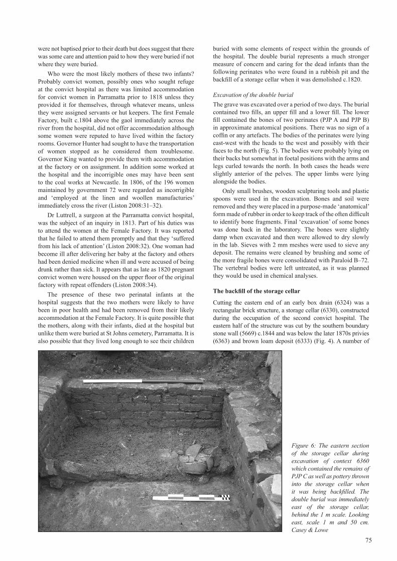
Figure 6: The eastern section of the storage cellar during excavation of context 6360 which contained the remains of PJP C as well as pottery thrown into the storage cellar when it was being backfilled. The double burial was immediately east of the storage cellar, behind the 1 m scale. Looking east, scale 1 m and 50 cm. Casey & Lowe
Aust_Hist_Archeology_Vol26.indd 75 18/5/09 4:06:39 PM
76
fills were found within the interior of the storage cellar. Context 6360 contained the perinatal remains of PJP C. This fill deposit was located within the eastern half of the cellar which was excavated separately from the western section (Fig. 6). The base of the interior of the storage cellar measured 3.9 by 1.3 m.
The fill 6360 was a mid-brown, friable, sandy loam containing brick, stone and roof tile fragments, shell mortar and charcoal specks. It also contained a number of artefacts including lead-glazed pottery and Chinese ceramics (Fig. 6). The fill (6360) was excavated in 200 mm spits and 100 per cent sieved through nested sieves, with 5 mm and 2 mm mesh, to allow recovery of all artefacts from the deposits. The fill (6331) in the western half of the storage cellar is considered to be the same fill and was excavated in the same manner (Miskella 2005–2006:112–114). Therefore artefacts from either context provide a date range for the backfilling of the cellar and the date for the disposal of PJP C. It is noted that a c.1870s L-shaped footing cut through the backfill of the cellar and possibly introduced some artefact contamination to these deposits.
Where we know the manufacturing dates for the artefacts found in eastern fill (6330) they date between 1780 and 1802 and two fragments of smoking pipes date from c.1820. Both of these fragments of pipe were found in spit 2, indicating they were probably intact deposits. Where we have manufacturing dates for artefacts in the western fill (6331) they generally range from 1780 to 1810. The late date of the pipes in the eastern fill indicates that the cellar was probably not backfilled until after the third hospital was operating in 1818 but shortly thereafter. Fill 6336, a deposit beneath 6360, also contains a rose head nail with a rectangular section dating from c.1820. The rest of the known manufacturing dates for 6336 are 1780 to 1810 and fit well with the later backfilling. Some near complete lead-glazed vessels were found in the eastern part of the lower backfill and are clearly of an early date (Miskella 2005–2006:112–115).
The rubbish pit
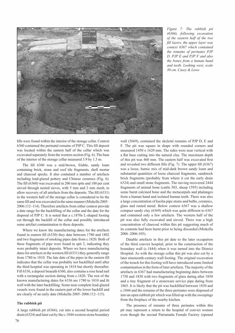
A large rubbish pit (6366), cut into a second hospital period drain (6324) and later cut by the c.1844 western stone boundary
wall (5669), contained the skeletal remains of PJP D, E and F. The pit was square in shape with rounded corners and measured 1450 x 1620 mm. The sides were near vertical with a flat base cutting into the natural clay. The maximum depth of this pit was 460 mm. The eastern half was excavated first and revealed two different fills (Fig. 7). The upper fill (6367) was a loose, humic mix of mid-dark brown sandy loam and substantial quantities of loose charcoal fragments, sandstock brick fragments (probably from where it cut the early drain 6324) and small stone fragments. The sieving recovered 2444 fragments of animal bone (cattle 503, sheep 1595) including some burnt calcined bone and the metacarpals and phalanges from a human hand and isolated human teeth. There was also a large concentration of kaolin pipe stems and bulbs, ceramics, glass and rusted metal. Below context 6367 was a shallow compact sandy clay (6369) which was quite different to 6367 and contained only a few artefacts. The western half of the pit was also fully excavated and sieved. There was a high concentration of charcoal within this pit suggesting much of its contents had been burnt prior to being discarded (Miskella 2006–2006:105).
Datable artefacts in this pit date to the later occupation of the third convict hospital, prior to the construction of the boundary wall (c.1844) when it was turned into the District Hospital. As with the storage cellar this pit was also cut by a later nineteenth-century wall footing. The original excavation of the trench for this footing will have introduced some limited contamination in the form of later artefacts. The majority of the artefacts in 6367 had manufacturing beginning dates between 1750 and 1830 with two fragments of glass dating after 1850 and a tiny fragment of a stoneware service pipe dating from 1865. It is likely that the pit was backfilled between 1830 and c.1844 and the remains of the three perinates were disposed of into an open rubbish pit which was filled up with the sweepings from the fireplace of the nearby kitchen.
The presence of remains of three perinates within this pit may represent a return to the hospital of convict women even though the second Parramatta Female Factory (opened
Figure 7: The rubbish pit (6366), following excavation of the eastern half of the two fill layers, the upper layer was context 6367 which contained the remains of perinates PJP D, PJP E and PJP F and also the bones from a human hand and teeth. Looking west, scale 50 cm. Casey & Lowe
Aust_Hist_Archeology_Vol26.indd 76 18/5/09 4:06:42 PM
77
1822) later had a hospital wing attached to the original factory buildings (Buchanan plan 1833). It is likely that the mothers of these three perinates were in poor health or had difficulties with the births and were bought to the hospital during this period. The distinctly different method of disposal of these three perinates is striking when compared to the earlier double burial. These little babies were thrown out into the rubbish and their remains may have been partially exposed until the rubbish pit was closed or the next lot of burnt rubbish was thrown onto the top of dead infants. This was probably a similar pattern of burial for PJP C in the backfill of the storage cellar.
Storage Cellar and Rubbish Pit Skeletal Remains
A perinatal skeleton (PJP C) was located in the storage cellar while three perinatal skeletons (PJP D, PJP E, PJP F) were found in the rubbish pit. Postcranial bones were also found in the pit. These could be matched with skeletons PJP D, PJP E and PJP F. Also found in the pit were the some bones of a single human hand as well as eight isolated teeth, many with dental caries. It is likely the hand was amputated but there were no marks on the bone to support this possibility but it is difficult to see how else the bones would have gotten into the pit.
Because the bones recovered from the storage cellar and the pit were in such good condition, it was not considered necessary to consolidate them with Paraloid B–72.
METHODSSkeletal analysis
Complete skeletal inventories and dental charts were produced. The preservation of the bones was described by eye and under a low power (x7) dissecting microscope. The Gross Preservation Index (GPI) which involved four categories of classification, ranging from poor to excellent were used (Haynes et al. 2002). The whole skeleton and any features of interest were photographed
Any skeletal and dental pathology was recorded. Standard cranial, post-cranial and dental measurements and cranial, post-cranial and dental epigenetic traits were recorded. Measurements of the bones were those developed by Fazekas and Kosa (1978) and modified by Buikstra and Ubelaker (1994). Definitions of the measurements of the teeth can be found in Barrett et al. (1964). Two sets of measurements were taken for all skeletons in order to minimise recorder error. Post-excavation analysis focused on the biological profile of the individuals. Age was determined mainly from both the size and morphological stage of development of the bones. Data for age estimates were drawn from Fazekas and Kosa (1978) and Scheuer and Black (2000).
Sex determination from perinatal bones is problematic, as sexually dimorphic traits do not become apparent until puberty. Methods using juvenile remains of known sex from Spitalfields have had limited success (accuracy of 70%–90%) with morphological features of the mandible and ilium of the pelvis (Schutkowski 1993). In 2002 Coussens and colleagues (2002) used mandibular criteria to examine differences in the robusticity index of the femur and humerus of children between 0 and 4 years from St Mary’s Anglican Church in South Australia. However the incomplete bones did not allow this method to be used. In Chile, Sutter (2003) had some success (an accuracy of 81.5%) using the greater sciatic notch
depth and dental arch criterion in children of known sex from 0 to 5 years. Weaver (1980) found a nonmetric feature called auricular surface elevation to accurately assign 75 per cent of female and 92 per cent of male skeletons. It worked best for foetal rather than perinatal or infant skeletons. While the above methods have had mixed success in determining sex in perinatal remains, sex determination was nevertheless attempted when the relevant bones were present.
There was also an attempt to obtain nuclear DNA from skeletons A and B. Only one individual (A) had any teeth and the dentition was incomplete. De Vito and Saunders (1990) obtained 76 per cent to 90 per cent correct classification of sex in deciduous teeth from children in nineteenth-century historic Canadian cemeteries. Unfortunately the appropriate teeth were not available to use this method.
Types of pathology that were investigated were trauma (particularly birth trauma), dental disease, specific infections and congenital conditions.
Causes of death in perinates and infants include obstructed labour, exposure to infection, haemorrhage, birthing practices, infanticide and abortion (spontaneous or induced). Congenital syphilis and tuberculosis can cause spontaneous abortion (Lewis 2007). Any evidence for these conditions was explored.
Genetic analysis
Of great interest was the possible ancestry of the remains. In terms of cultural context, the possible ancestry of each individual is: British (non-convict), Australian-born settler, convict (British born, including Irish) or Australian Aboriginal. Admittedly there is some limited possibility of other European or African ancestry but this is a low probability. It is generally not possible to determine ancestry from the morphology of the bones of such young individuals. Extraction of mitochondrial DNA (mtDNA) was attempted on the petrous temporal bones of the two perinates from the grave. No attempt was made to extract mtDNA from the bones found in the storage cellar and pit as it was considered they would be too contaminated.
The aim of the analysis was to determine maternal ancestry via mitochondrial DNA (mtDNA) haplogroup testing, to clarify if the two individuals represented by the skeletal remains were related and to facilitate possible future comparison with analysed samples of DNA isolated from any individual claiming to be a maternal relative of the deceased. Likely period of burial was determined by Mary Casey using detailed analysis of the stratigraphy and related artefacts.
All aDNA work was conducted in a dedicated ancient DNA laboratory. The pre-PCR laboratory is located in a building free of all molecular biology work. While a second laboratory accommodates post-PCR analyses. The amount of human DNA within the laboratories is minimised by following strict protocols; all workers may enter only after a shower, wearing freshly laundered clothes, a body suit, shoe covers, boots, facemask, face shield and triple gloving. All surfaces in the lab are routinely triple wiped with bleach, decon and isopropanol. The laboratory is irradiated with ultraviolet (UV) (280-400nm) light overnight (≈13hours). All consumables are externally bleached and UV irradiated.
Sample PreparationThe petrous temporal bones and vertebral bodies for both perinates were used for DNA extraction. Decontamination
Aust_Hist_Archeology_Vol26.indd 77 18/5/09 4:06:42 PM

78
of samples included UV irradiating the medial and lateral surfaces of the petrous temporal bone and superior and inferior surfaces of the vertebral bodies, for 20 minutes each, wiping all surfaces with bleach soaked kimwipes and completely removing the outer surface with a Dremel Drill sanding disk. The petrous temporal bones were sectioned to remove bone between the internal auditory meatus and the sigmoid sulcus using a diamond drill (35 000 RPM). The bone removed was then powdered to a fine grain using a microdismembrenator at 3300 RPM / 30 sec. The remaining petrous temporal bone was stored at 4oC. The complete vertebral bodies were powdered.
DNA ExtractionThe bone powder was digested and lysed by rotating the samples overnight at 37oC with 3.33mL of extraction buffer (0.5M EDTA, pH 8.5, 0.5% N-lauryl Sarcosine, 20mg/ml Protinase K). An extraction blank was included for every 3–4 extractions. DNA was extracted using a phenol/chloroform/isoamyl alcohol pH 8.0 (25:24:1) method as described previously (Haak et al. 2005). The supernatant was transferred to a 50 KDa 15 ml amplicon tube (Amplicon, Millipore) and washed twice with UV irradiated, hepafiltered water.
Polymerase Chain Reaction (PCR) and real time (q) PCRThe complete mtDNA hypervariable sequence region I (16024-16365) and II (73-340) and 22 haplogroup determining, single nucleotide polymorphisms (SNP’s) from the coding region were amplified using single and multiplex PCR. Standard PCRs using Amplitaq Gold (Applied Biosystems) were conducted in 25µl volumes using 1x Buffer Gold, 2.5mM MgCl2, 0.25mM of each dNTP (Fermentas), 400µM of each primer, 1mg/ml RSA (Sigma-Aldrich), 2 U of Amplitaq Gold Polymerase and 2µl of DNA extract. Thermocycling conditions consisted of an initial enzyme activation at 95°C for 6min, followed by 50 cycles of denaturation at 94°C for 30s, annealing at 58°C for 30s and elongation at 72°C for 30s and a single final extension time at 60°C for 10min. Each PCR reaction included the two extraction blanks per sample, as
well as a minimum of two PCR negatives. PCR products were checked by electrophoresis on 3.5% agarose TBE gels.
qPCR was used to determine the amount of DNA in the samples prior to amplification and assess the authenticity based on the assumption there is an inverse relationship between DNA quantity and fragment length for degraded, ancient DNA (Cooper and Poinar 2000; Pruvost 2004). Two different length fragments were amplified from the HVSI; 141 bp from L16117, H16218 and 179 bp from L16209, H16348. All qPCR’s were carried out in 10µL, containing Express SYBR® Green ER Supermix Universal (Invitrogen), Rabbit Serum Albumin (10mg/ml), forward and reverse primers (10µM), distilled water and 1µL of DNA extract or water for the PCR blank. The reaction conditions were as follows: 95°C for 5 min and 50 cycles of 95°C for 10s, 58°C for 20s and 72°C for 15s. The starting quantity of DNA in the ancient samples was determined by comparison to a standard curve of a known amount of DNA. Ancient qPCR’s were run in triplicate with extraction and PCR blanks, and PCR standards (positive control) run in duplicate. Amplifications were performed on Rotor-Gene 6000 and analysis on Rotor-Gene 6000 Series Software 1.7. Both samples displayed appropriate molecular behaviour for degraded DNA, exhibiting an inverse relationship between fragment length and DNA quantity. There was a greater amount of DNA of the shorter 141bp fragment length for perinate 1 (PJP A) (73800 DNA copies/µL) and for perinate 2 (PJP B) (600 DNA copies/µL) compared to the 179 bp fragment, which contained for perinate 1 (60 DNA copies/µL) and for perinate 2 (15 DNA copies/µL).
SequencingAll PCR and qPCR products were purified for sequencing using 5µl of PCR product with 0.7 U of SAP and 0.6 U of ExoI (both Fermentas) and incubating at 37°C for 40 min, followed by heat inactivation at 80°C for 10 min. All successful PCR products were directly sequenced in both directions using the Big Dye Terminator 3.1 Kit (Applied Biosystems) as per manufacturer’s instructions. Sequencing products were
Figure 8: Skeletal remains of the two perinates (PJP A and PJP B) found in the double burial, scale10 cm. Russell Workman
Aust_Hist_Archeology_Vol26.indd 78 18/5/09 4:06:46 PM

79
purified using Cleanseq magnetic beads (Agencourt, Becker Coulter) according to the manufacturer’s protocol. Sequencing products were separated on the 3130xl Genetic Analyzer (Applied Biosystems) and the resulting sequences were edited and aligned relative to the Cambridge Reference Sequence using the software Sequencher.
RESULTSInventories and condition
The two perinates in the grave, PJP A and PJP B had many of the long bones, ribs, pectoral and pelvic girdles, hands and feet missing and, as would be expected, the skulls were disarticulated. No teeth were in situ in their jaws. The teeth consisted of crowns only, were extremely delicate and many were chipped post-mortem (Fig. 8).
The bones from the grave appeared non-greasy and porous with many bones showing loss of the cortical bone and exposure of the spongy bone. There was no sign of any plant invasion of the bones or of any animal damage to the bones. All of the long bones recovered were broken. These breaks appeared to have occurred post-mortem as they were sharp and at right angles to the long axes of the bones as would be expected in bones which have lost their fat and moisture and therefore their elasticity.
The best preserved bones were the vertebral bodies, the petrous portions of the temporal bones and the body of the sphenoid and the pars basilaris. Using the index suggested by Haynes and colleagues (2002) these bones have a gross preservation index (GPI) of 3, which is good and the remainder of the bones a GPI of 1 which is poor.
The pH of the grave fill was 7.4, thus slightly alkaline and showed elevated calcium which is unusual for the soils of the Parramatta region and which may indicate human activity. The calcium may have come from concrete structures, lime or shell. Elevated phosphorus levels may have been the result of decay of bones (Sydney Environmental and Soil Laboratory 2008a).
The skeletal remains from the storage cellar consisted of a single perinatal skeleton (PJP C) (Fig. 9). The bones appeared non-greasy and porous with some bones showing loss of the cortical bone and exposure of the spongy bone. However the bones were in most cases better preserved than those in the grave. There was no sign of any plant invasion of the bones nor any animal damage to the bones. Almost all of the lower limbs were missing with the exception of some bones of the right foot. The bones of the skull were disarticulated. All long bones of the upper limbs were broken, probably post-mortem as the breaks are at right angles to the long axes of the bones. Using the index suggested by Haynes and colleagues (2002) the above listed bones have a gross preservation index (GPI) of 3, which is good.
The soil from the storage cellar had a pH of 8.4 making it moderately alkaline. The calcium levels were high which may suggest associated lime from mortar. Phosphorus levels were also high, suggesting human impact. Such conditions are good for bone preservation (Sydney Environmental and Soil Laboratory 2008b).
Skeleton PJP D consisted of an incomplete disarticulated skull, a few ribs, one vertebral body, and right ilium, left and right humeri, and partial radii and right ulna, both femora, tibiae and fibulae. All long bones were broken, probably post-mortem as the breaks are at right angles to the long axes of
the bones (Fig. 10). The bones appeared non-greasy. There was no sign of any plant invasion of the bones or any animal damage to the bones. These bones have a gross preservation index (GPI) of 3, which is good (Haynes et al. 2002).
Skeleton PJP E consisted of a right petrous temporal bone, two cranial fragments and numerous ribs and rib fragments while skeleton PJP F was represented by a right petrous temporal bone only (Fig. 10). Both sets of bones appeared non-greasy. There was no sign of any plant invasion of the bones or any animal damage to the bones. These bones have a gross preservation index (GPI) of 3, which is good (Haynes et al. 2002).
The soil from the pit had a pH of 8.1 making it moderately alkaline. The calcium levels were high which may suggest associated lime from mortar. Phosphorus levels were also high, suggesting human impact. There was a lower nutrient status than in the storage pit. Such conditions are very good for bone preservation (Sydney Environmental and Soil Laboratory 2008c).
Age
Age for all individuals was based on measurements of the bones of the crania and long bones in all cases. In skeleton PJP A the development of the teeth was also used. The age estimate for skeleton PJP A from the double grave was based on the size of the pars basilis, pars petrosa and sphenoid. A combination of the age estimates gave a range of 33 to 40 weeks (Table 1).
In skeleton PJP B, also from the double grave, the age was based on the size of the pars basilis, pars petrosa, sphenoid and zygomatic. A range of 25 to 39 weeks in utero was obtained.
The age of skeleton PJP C from the storage cellar was based on the size of the pars latralis, pars petrosa, zygomatic,
Figure 9: Skeletal remains of PJP C found in the storage cellar, scale 10 cm. Russell Workman
Aust_Hist_Archeology_Vol26.indd 79 18/5/09 4:06:46 PM

80
scapula, humerus, radius, ulna and pelvis. Age ranged from 33 to 37 weeks in utero.
The age of skeleton PJP D (from the pit) was based on the size of pars petrosa and the sphenoid, humerus, pelvis, femur, tibia and fibula. Age ranged between 31 to 40 weeks in utero. The age of skeletons PJP E and PJP F (from the pit) were based on the pars petrosa only and ranged between 20 to 21 weeks in utero. However the morphology of this bone suggested 24 weeks (Weaver 1998). Therefore a range of 20–24 weeks in utero is assumed.
Sex
Various methods of sex determination were made but conflicting results were obtained. Nevertheless the results are given here in case in the distant future it may be possible to obtain nuclear DNA and therefore conclusively determine sex. If this were the case then it would allow these methods to be independently tested. In the ilium of individual PJP A, the arch criterion suggested a female while the curvature of the iliac crest suggested male (Schutkowski 1993). Therefore no clear determination of sex could be made.
Individual PJP C did not show any eversion of the gonial region of the mandible suggesting this individual was a female (Schutkowski 1993). The innominate displayed a broad greater sciatic notch, female arch criterion and female curvature of the iliac crest but at the same time displayed a male-like depth for the greater sciatic notch (Schutkowski 1993). Individual PJP C shows no elevation of the auricular region suggesting it was male. Therefore no clear determination of sex could be made.
Individual PJP D had a round anterior dental arcade suggesting it was female (Schutkowski 1993). It also displayed a female greater sciatic notch angle, female arch criterion and female iliac crest curvature but male greater sciatic notch depth. No clear determination of sex could be made.
Ancestry and Date
Archaeological evidence suggests all skeletal remains are almost certainly British given the context, that is, all were found
within the boundaries of the second convict (1792–c.1818) and the third convict hospitals (1818–c.1844) (Casey & Lowe 2005, 2006). The date range for the burial would suggest a mother of British origin especially as the mother would have been a convict to have a baby in the hospital and there were no locally-born ‘convicts’ although it is just possible either of the mothers could have been a ‘currency lass’ if the burial was as late as 1808 and they had committed a local crime but the possibilities of this are quite slim. Therefore a locally-born non-Aboriginal woman as the mother or mothers is excluded on two counts.
Mitochondrial haplogroup typing was carried out by Dr Wolfgang Haak and Christina Adler at the Australian Centre for Ancient DNA at the University of Adelaide. DNA was extracted from the petrous temporal bones and vertebrae bodies for both perinates and mtDNA fragments of HVS I and II could be reliably amplified from both perinates buried together in the grave. In addition, 22 basic SNPs characteristic of the main branches of the mtDNA phylogeny were typed in a multiplex assay to confirm haplogroup status of both.
According to the genetic analysis of PJP A, it matches the revised Cambridge Reference Sequence and falls into a haplotype (European Hg H2) which is typical European. This is consistent with the archaeological context. The most likely conclusion is that A is the child of a British-born convict. Skeleton PJP B falls basal into haplogroup Hg N* (16223T), which is one of the deep branching lineages which are rare everywhere in Eurasia (including Australians). To distinguish PJP B from Australian-specific subhaplogroups of N, parts of the HVS II were amplified and sequenced. According to the sequencing results of the HVS II it is highly likely that individual B is also of European origin. The genetic analysis was replicated. PJP A was haplogroup H2 that is the Cambridge reference sequence. PJP B was basal haplogroup N and contained the mutation C16233A in the hypervariable region 1 (HVS1). Therefore identical results were found for anatomically isolated elements (vertebral bodies) for both PJP A and PJP B.
No attempt was made to extract mtDNA from the perinate PJP C bones as it was considered they would be
Figure 10: Skeletal remains of PJP D, PJP E and PJP F found in the rubbish pit, scale10 cm. Russell Workman
Aust_Hist_Archeology_Vol26.indd 80 18/5/09 4:06:50 PM

too contaminated. The context suggests they are dated to the demolition phase of the second convict hospital c.1820 and are probably of British ancestry.
It is highly unlikely that PJP D, PJP E and PJP F are of Aboriginal ancestry. It was decided not to investigate for the presence of DNA in the bones of these individuals as they were found in a pit and would probably have been subject to greater chance of contamination. These are dated to the 1830s/1840s, to the later occupation of the third convict hospital (1818–c.1844) and thus are most likely of European ancestry.
Personal identification
Given that mtDNA has been extracted from the two individuals in the grave then it is hypothetically possible that these could be compared with mtDNA of possible relatives and therefore result in a possible identification. This depends on a lot of assumptions, for example, we must know the names of female convicts at this time in Parramatta and they or their sisters must have gone on to have other children. However, the likelihood of this happening is very slim.
The fact that the two perinates were buried together in the same grave did make us wonder if they may have been related in some way. Given the different ages of A and B it is unlikely they were twins (even if one had died in utero). The other possibility is that they may have been the children of sisters, that is, cousins. Genetic results indicate the two skeletons are not maternally related. The control region sequences (np 15997–16410) of the two Parramatta perinates are different.
Pathology and cause of death
There was no sign of trauma or disease in any of the skeletal remains found, therefore no indication of the cause of death. As these perinatal remains were not found in the context of a cemetery, they do not throw any light on mortality rate in this period. Below is a summary of findings for all skeletons (Table 1).
DISCUSSIONThe age of the remains in the grave are estimated to date between c.1800–1810 making them possibly the oldest European skeletal remains yet found in Australia (on the Australian mainland). Genetic analysis of the two individuals in the grave strongly suggests they are of European origin. Combined with the archaeology it is suggested they were probably the offspring of British-born convicts. The age of the remains from the storage cellar and pit, while not as old as those from the grave, are still reasonably old, especially for
juvenile remains. A comparison of these findings with those of historic Australian cemeteries shows it is fairly rare to find the skeletal remains of such young individuals (Table 2). The successful extraction of DNA from two of the remains and the good condition given their age gives hope for future research into early European juvenile remains.
The ages of the individuals ranged between 20–24 weeks to 33–40 weeks in utero. It is possible that they were stillborn or that all survived birth but died shortly after. The causes of death of these perinates are unknown but the very presence of them in a hospital suggests the mothers may have been suffering from complications. Without actually knowing for certain the true biological ages of the perinates it is not possible to say anything about their growth and development for their ages. Assessment of sex gave mixed results with some individuals showing signs of both sexes. It is suggested that these results on sex determination are not particularly reliable.
The historical nature of these remains offers a rare opportunity to provide insight into the disposal of perinatal human remains in late eighteenth and early nineteenth-century Australia. The two individuals in the grave were buried according to traditional Christian method in terms of their orientation but they were not placed in individual graves and they were not baptised and were therefore buried in unconsecrated ground. That they were buried at all is perhaps surprising and suggests that they may have lived longer than the individuals recovered from the storage cellar and pit or because of the wishes of their mothers. Perinates PJP A and PJP B were buried with a great deal more care than the four perinates buried in the later storage cellar and rubbish pit; indicating that there may have been more concern for the infants during the earlier period of the occupation of the second convict hospital than eight or more years later for PJP C or 20 to 30 years later with PJP D, PJP E and PJP F. This is perhaps surprising in light of the difficult nature of early convict life and the poor provisions at the hospital during this period. But it may also represent a closer knowledge of individuals within the smaller population of the early colony, or possibly the relative rarity of births to convict women at this time. It is also likely that the disposal methods for the later perinates represents a greater medicialisation of the management of births within hospitals and the speedy removal of the perinates from their mothers and their disposal into conveniently open rubbish pits.
The bone preservation showed some variation depending on whether they were recovered from the pit or from the grave. Overall the bones recovered from the pit were better preserved than those from the grave. However, given the age of these remains and the fact that they are of such young
81
Table 1: Summary of findings for all skeletons from the Parramatta convict hospital site.
SKELETON PJP A PJP B PJP C PJP D PJP E PJP F
LOCATION GRAVE GRAVE STORAGE CELLAR PIT PIT PIT
Age 1800–1818 1800–1818 c.1820 1830s/ 1840s 1830s/ 1840s 1830s/ 1840s
Condition poor poor good good good good
Ancestry European mt(DNA)
European mt(DNA)
Presumed European
Presumed European
Presumed European
Presumed European
Age (wks in utero) 33–40 25–39 33–37 31–40 20–24 20–24
Sex Unknown Unknown Unknown Unknown Unknown Unknown
pH of soil 7.4 7.4 8.4 8.1 8.1 8.1
Aust_Hist_Archeology_Vol26.indd 81 18/5/09 4:06:51 PM
82
individuals, it is surprising that they have survived at all and in such good condition. It is especially interesting that mtDNA could be extracted from the petrous temporal bones. These are the parts of the temporal bones which house the organs of hearing and balance. They are particularly robust and well protected in and under the skull.
The possibility of stable isotope analysis was investigated however it appears that little could be gained at this stage of such research in Australia. Samples will be stored in the event of future research.
These perinatal remains are considered to be of outstanding scientific and historical significance given their antiquity for offspring of European women (probably British-born convicts) in the very early days of British settlement in Australia and are therefore part of a rare historic experiment, the transportation of convicts to New South Wales.
ACKNOWLEDGEMENTSDepartment of Commerce, Attorney-General’s Office and Brookfield Multiplex funded the archaeological program and the reporting on the human remains and the excavation generally. The helpful referees and the many others who assisted in various stages of the project: Abi Cryerhall who identified the burial and preliminary excavation; Franz Reidel and Jill Miskella for illustrations and archaeological context from the trench reports; Tania Stellini who initially identified the hand and other human bones; Caroline Wilby who identified further human and non-human bone during cataloguing of faunal bone; Sarah Croker, Marcus Robinson, Christina Adler who provided assistance with sieving and identification of non-human bones; Russell Workman who did the photography of the skeletal remains; Estelle Lazer for information on the Cadia skeletons and Tony Lowe.
BIBLIOGRAPHYAUSTRAL ARCHAEOLOGY/GODDEN MACKAY 1997
POW Project 1995, Randwick Destitute Children’s Asylum Cemetery, Archaeological Investigation, unpublished report for Sydney Eastern Area Health Service and Heritage Council of NSW.
BAKER, B.J, DUPRAS, T. L. and TOCHERI M. W. 2005 Osteology of Infants and Children, A&M University Press, Texas.
BARRETT M.J., BROWN T., ARATO G. and OZOLS I.V. 1964. ‘Dental observations on Australian Aborigines: buccolingual crown diameters of deciduous and permanent teeth’, Australian Dental Journal 9:280–285.
BIRMINGHAM, J. AND LISTON, C. 1976 Old Sydney Burial Ground 1974, emergency excavation in the City of Sydney, Studies in Historical Archaeology No. 5, Australian Society for Historical Archaeology, Sydney
BUCHANAN, W. 1833 Plan of the Female Factory, November 1833 National Archives UK (formerly Public Records Office), PRO MPH 91(9).
BUCKBERRY, J. 2000 ‘Missing, presumed buried? Bone diagenesis and the under-representation of Anglo-Saxon children’, Assemblage 5:1–14.
BUIKSTRA, J.E AND UBELAKER, D.H (eds) 1994 Standards for data collection from human skeletal remains, Arkansas Archaeological Survey Research Series No.44.
CASEY, M. 1999 ‘Local pottery and dairying at the DMR Site, Brickfields, Sydney, New South Wales’, Australasian Historical Archaeology 17:3–37.
CASEY & LOWE 2005 Excavation permit application, Parramatta Hospital site, Marsden Street, Parramatta, unpublished report for the Department of Commerce, http://www.caseyandlowe.com.au/pdf/parra/pjphistory.pdf
Table 2: Summary of historic European juvenile skeletal remains found in Australia.
SITE DATE AGE No.
Batavia mass grave, Abrolhos Is, WA.(Paterson and Franklin 2004)
1629 < 1 year5–6 years12–16 years
111
Parramatta Justice Precinct, NSW 1800–18181800–1818c.18201830s–1840s1830s–1840s
33–40 weeks in utero25–39 weeks in utero33–37 weeks in utero31–40 weeks in utero20–24 weeks in utero
11112
Lang Park, Brisbane, Queensland(Rains and Pragnell 2001, McGowan pers. comm. 2009)
1843 - 1875 Juveniles, neonates possible(determined on the basis of coffin length, 135 had no skeletal remains left, 26 were in poor condition.)
162
Cadia, Orange NSW(Lazer pers comm 2008.)
1860s–1880s 7–8 months in utero8 months in utero
13
St Mary’s Anglican Church Adelaide, SA.(Coussens et al. 2002)
1847–1925 birth–24 months3–4 yearsneonates
23113
Randwick Destitute Children’s Asylum, NSW (Austral Archaeology/Godden Mackay 1997)
1860s–1880s neonate1–3 years ±2 months4–9 years ± 6 months10–12 years ± 12 months
114414
Aust_Hist_Archeology_Vol26.indd 82 18/5/09 4:06:51 PM

83
CASEY & LOWE 2006. Preliminary Results, archaeological investigation Stage 2c, Parramatta Hospital Site, Marsden & George Streets, Parramatta, unpublished report prepared for Multiplex, September 2006, http://www.caseyandlowe.com.au/reptpjp.htm.
CASEY & LOWE 2009 Archaeological investigation, Parramatta Justice Precinct, the former Parramatta Convict Hospital Site, cnr Marsden & George Streets, Parramatta, unpublished report prepared for NSW Dept of Commerce, Brookfield Multiplex on behalf of the NSW Attorney-General’s Office, in preparation.
COOPER, A. and H. N. POINAR. 2000 ‘Ancient DNA: do it right or not at all’, Science 289:1139.
COUSSENS, A, ANSON, T., NORIS, R AND HENNEBERG, M. 2002. ‘Sexual dimorphism in the robusticity of long bones of infants and young children’, Anthropological Review 65:3–16.
EDWARD HIGGINBOTHAM & ASSOCIATES 2002 Report on the archaeological excavation of the Cadia Cemetery, Cadia Road, Cadia, NSW, 1997–1998, unpublished report for Cadia Holdings Pty Ltd.
FAZEKAS, I.G. AND KOSA, F. 1978 Forensic fetal osteology, Akademiai Kiado, Budapest.
GUY, H., MASSET, C. AND BAUD, C.A. 1997 ‘Infant taphonomy’, International Journal of Osteoarchaeology 7:221–229.
HAAK, W., P. FORSTER, B. BRAMANTI, S. MATSUMURA, G. BRANDT, M. TANZER, R. VILLEMS, C. RENFREW, D. GRONENBORN, K. W. ALT, and J. BURGER. 2005 ‘Ancient DNA from the first European farmers in 7500-year-old Neolithic sites’, Science 310:1016-1018.
HAYNES, S., SEARLE, J.B., BRETMAN, A. and DOBNEY, K.M. 2002 ‘Bone preservation and ancient DNA: the application of screen methods for predicting DNA survival’, Journal of Archaeological Sciences 29: 585–592.
HASLAM, M, PRAGNELL, J, KIRKWOOD, L, MCKEOUGH, MURPHY, A and LOY. T 2003 ‘A Lang Park mystery: analysis of remains from a 19th century burial Brisbane, Queensland’, Australian Archaeology 56:1–7.
JOHNSTON, F.E. AND ZIMMER, L.O. 1989 ‘Assessment of growth and age in the immature skeleton, in Isçan, M.Y. and Kennedy, K.A.R. (eds) Reconstruction of life from the skeleton, Alan R. Liss, New York, pp. 11–22.
KROGMAN, M. M. AND ISCAN, M. Y.1986 The human skeleton in forensic medicine, Charles C. Thomas, Springfield Illinois.
LEWIS, M.E. 2007 The Bioarchaeology of Children: perspectives from biological and forensic Anthropology, Cambridge University Press, Cambridge
LISTON, C. 2008 ‘Convict women in the female factories of New South Wales’, in Hendriksen, G. and C. Liston (eds) Women Transported, life in Australian’s convict female factories, Parramatta Heritage Centre and University of Western Sydney, Parramatta, pp. 29–52.
LOWE, A. and MACKAY, R. 1992 ‘Old Sydney Burial Ground, Australasian Historical Archaeology 10:15–23.
MISKELLA, J. 2005–2006 Draft trench report, Stage 1 & 2c, unpublished report for Casey & Lowe on behalf of
Brookfield Multiplex, Dept of Commerce and Attorney-General’s Office.
MOLLESON, T. and COX, M. 1993 The Spitalfields Project: The middling sort. Volume 2: The anthropology, CBA Research Report 86; Council for British Archaeology.
PATE, D. and ADAMS, W.H. 2000 ‘News from the Pacific Rim. Excavation of St Mary’s Anglican Cemetery, Adelaide, South Australia’, Palaeopathology Newsletter 111:6–7.
PATERSON, A. and FRANKLIN, D. 2004 ‘The 1629 mass grave for Batavia victims, Beacon Island, Houtman Abrolhos Islands, Western Australia’, Australasian Historical Archaeology 22: 71–78.
PRUVOST, M. and GEIGL, E. 2004. ‘Real-time quantitative PCR to assess authenticity of ancient DNA amplification’, Journal of Archaeological Science 31:1191-1197.
RAINS, K. and PRAGNELL, J. 2002 ‘Background to the University of Queensland Archaeological Services Unit’s Lang Park Salvage Excavations: History, significance assessment and methods’, Queensland Archaeological Research 13:21–30
SCHEUER, L. and BLACK, S. 2000 Developmental Juvenile Osteology, Academic Press, NY.
SCHUTOWSKI, H. 1993 ‘Sex determination of infant and juvenile skeletons: 1. Morphological features’, American Journal of Physical Anthropology 90:199–205.
STOCKS, R. 2008a Building materials report, former Parramatta convict hospitals site, Parramatta Justice Precinct, Marsden and George Streets, Parramatta, unpublished report to Casey & Lowe, draft, July 2008.
STOCKS, R. 2008b ‘New evidence for local manufacture of artefacts at Parramatta, 1790–1830’, Australasian Historical Archaeology 26: 29–44.
STOCKS, R. 2009 Miscellaneous, metals and organics report, former Parramatta convict hospitals site, Parramatta Justice Precinct, Marsden and George Streets, Parramatta, unpublished report to Casey & Lowe, draft, January 2009.
SYDNEY ENVIRONMENTAL AND SOIL LABORATORY 2008a Multiple Analysis Profile. Batch no. 4313, sample no.1 Grave Fill Parramatta.
SYDNEY ENVIRONMENTAL AND SOIL LABORATORY 2008b Multiple Analysis Profile. Batch no. 5990, sample no.1 Parramatta.
SYDNEY ENVIRONMENTAL AND SOIL LABORATORY 2008c Multiple Analysis Profile. Batch no. 5990, sample no 2 Parramatta.
WARD, R. 2007 Ceramics report, former Parramatta convict hospitals site, Parramatta Justice Precinct, Marsden and George Streets, Parramatta, unpublished report to Casey & Lowe, draft, August 2007.
WEAVER, D.S. 1980. ‘Sex differences in the ilia of a known sex and age sample of fetal and infant skeletons’, American Journal of Physical Anthropology 52:191–195.
Aust_Hist_Archeology_Vol26.indd 83 18/5/09 4:06:52 PM