early detection of subungual melanoma in situ proposal of abcd … · 2017-12-26 · jh lee, et al...
TRANSCRIPT

JH Lee, et al
36 Ann Dermatol
Received March 7, 2017, Revised May 4, 2017, Accepted for publication June 21, 2017
Corresponding author: Dong-Youn Lee, Department of Dermatology,Samsung Medical Center, Sungkyunkwan University School of Medicine, 81 Irwon-ro, Gangnam-gu, Seoul 06351, Korea. Tel: 82-2-3410-3543, Fax: 82-2-3410-3869, E-mail: [email protected]
This is an Open Access article distributed under the terms of the Creative Commons Attribution Non-Commercial License (http://creativecommons.org/licenses/by-nc/4.0) which permits unrestricted non-commercial use, distribution, and reproduction in any medium, provided the original work is properly cited.
Copyright © The Korean Dermatological Association and The Korean Society for Investigative Dermatology
pISSN 1013-9087ㆍeISSN 2005-3894Ann Dermatol Vol. 30, No. 1, 2018 https://doi.org/10.5021/ad.2018.30.1.36
ORIGINAL ARTICLE
Early Detection of Subungual Melanoma In Situ: Proposal of ABCD Strategy in Clinical Practice Based on Case Series
Jae Ho Lee, Ji-Hye Park, Jong Hee Lee, Dong-Youn Lee
Department of Dermatology, Samsung Medical Center, Sungkyunkwan University School of Medicine, Seoul, Korea
Background: Prevalence of subungual melanoma (SUM) in Asian population is relatively high and early clinical de-tection improves both quality of life and survival. Objective: We sought to establish screening strategy for SUM in situ. Methods: We retrospectively reviewed medical records of 8 patients pathologically diagnosed as SUM in situ between January 2015 and September 2016. Results: All patients in our study were adults and had solitary lesion. In all cases, lon-gitudinal melanonychia as well as background pigmentation in entire nail plate was observed. Seven (87.5%) cases had periungual pigmentation. Clinical features of SUM in situ can be summarized according to new criteria categorized under initial letters of alphabet, namely nail ABCD of SUM in situ; “A” stands for adult age (age >18 years); “B” for brown bands in brown background; “C” for color in peri-ungual skin; “D” for one digit. Our strategy was to suspect SUM in situ if solitary longitudinal melanonychia in adult was satisfying either “B” or “C” in ABCD. All cases were sus-pected as SUM in situ under nail ABCD rule, and histological examination confirmed diagnosis. Sensitivity and specificity of nail ABCD was 100% and 96.6%, respectively, regarding our previously published 18 SUM in situ and unpublished 28 nail matrix nevi cases as well as 8 SUM in situ presented here.
Conclusion: ABCD rule is simple and sensitive clinical strat-egy for early detection of SUM in situ. (Ann Dermatol 30(1) 36∼40, 2018)
-Keywords-Clinical, Criteria, Nail unit, Retrospective studies, Subungual melanoma in situ
INTRODUCTION
Subungual melanoma (SUM) is a malignant neoplasm of melanocytes that arises from the nail matrix. In the early phase of SUM, it usually presents as longitudinal brown or black band on nail plate (melanonychia), and can be mis-diagnosed as benign nail pigmentation disorders such as nail matrix nevi or subungual lentigo1-3. Although SUM is rarely diagnosed in Caucasian, the prevalence in Asian population is relatively high. In a retrospective study in Japanese population, SUM accounted for approximately 10% of cutaneous melanoma3. According to our data in a single tertiary institute, SUM constitutes about 18% of cu-taneous melanoma in Korea4. Prognosis of SUM was gen-erally poor in the past, because the diagnosis was fre-quently delayed. One of the most common causes of de-lay in SUM diagnosis is lack of early recognition5. Although an “ABCDEF rule” for early clinical detection of SUM had been proposed6 and it seems to be useful, the 6 features of this criterion, particularly the “family history of melanoma or dysplastic nevus syndrome,” are not always observed in cases of SUM in situ.The aim of our study is to suggest simple clinical strategy for SUM in situ for early clinical detection by analyzing clinical features of 8 cases of SUM in situ.

ABCD Rule for SUM In Situ
Vol. 30, No. 1, 2018 37
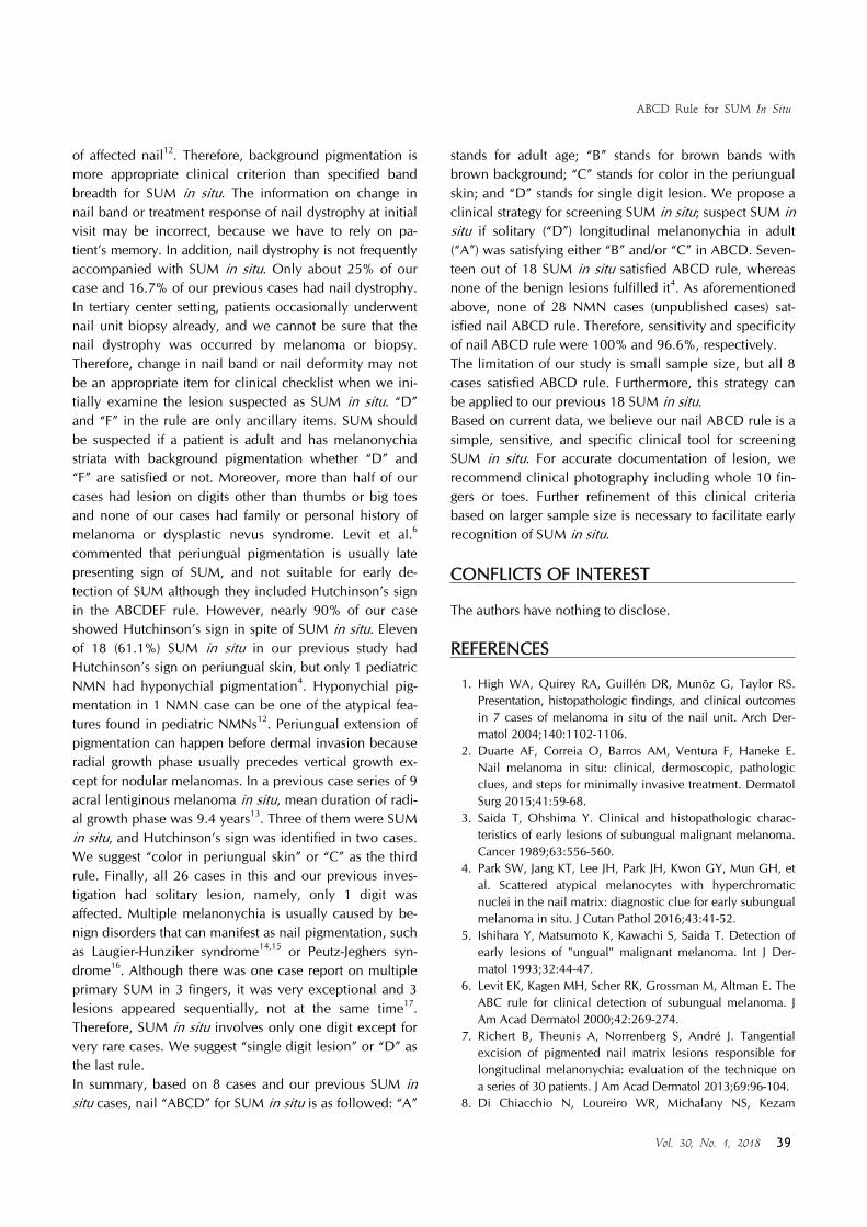
Fig. 1. Clinical photographs of 8 subungual melanoma in situ cases. Note all cases have entirely pigmented nail plate compared to unaffected ones (arrow in case 5). Hutchinson’s sign was observed except for case 1. Cases 2 and 6 had nail deformity ahead of nail matrix biopsy.
MATERIALS AND METHODS
After receiving approval from the institutional review board of Samsung Medical Center (IRB no. 2016-08-187- 001), 8 cases of SUM in situ diagnosed at the Samsung Medical Center from January 2015 to September 2016 were identified and retrospectively reviewed. Histopathology slides, clinical records, and clinical photographs of the cases were included. Data gathered from the electronic medical record included gender, age, age of onset, loca-tion of the lesion, clinical features including pattern of melanonychia, presence of periungual pigmentation (Hut-chinson’s sign) and nail deformity, biopsy and surgical method, and pathological information.All patients underwent nail matrix biopsy. After local anes-thesia with 2% lidocaine, nail plate was extracted and about 5 mm longitudinal incisions were made at both ends of the proximal nail fold to expose nail matrix. Nail matrix specimens were obtained at nail matrix with the darkest pigmentation or nail matrix corresponding to the darkest band on nail plate by 3 different methods: 3 mm punch
biopsy; longitudinal incision from nail matrix to hypo-nychium; or transverse shave biopsy (similar to the tan-gential excision of nail matrix described in literatures7,8, but matrix was shaved after nail plate extraction). Wide excision of nail unit was performed in all cases. Pathologi-cal diagnosis of SUM in situ was based on our previous in-vestigation of histological findings of SUM in situ: diag-nosis of SUM in situ was made if increased number of scattered atypical melanocytes with large hyperchromatic nuclei were observed in nail matrix without evidence of dermal invasion in the excision specimen.
RESULTS
Of the 8 cases of SUM in situ, 3 (37.5%) cases were male and 5 (62.5%) were female. The age at diagnosis ranged from 30 to 70 years (mean age, 56.3 years). The duration of pigmentation ranged from 6 months to 16 years (mean duration, 6.3 years). The thumb (25%) and big toe (12.5%) were most commonly affected. All cases were presented as longitudinal melanonychia in a single digit (Fig. 1).

JH Lee, et al
38 Ann Dermatol
Table 1. Clinical features, method of biopsy, and surgical management in 8 cases
Case SexAge (yr)
Onset (yr)
LocationTotal
melanonychiaColor
Nail dystrophy
Hutchinson’s sign
Biopsy Surgery
1 F 43 6 Right 2nd finger + Tan to brown +, PBx − Punch WE/FF2 F 66 16 Left 4th finger + Black + + Incision WE/FF3 M 69 3 Left 2nd finger + Tan to brown − + Incision WE/SG4 F 62 3 Right 1st finger + Tan to brown +, PBx + Punch WE/SG5 M 49 10 Left 2nd toe + Tan to brown − + Shave WE/SG6 F 61 5 Right 1st toe + Tan to brown + + Punch WE/SG7 M 30 7 Right 4th finger + Tan to brown +, PBx + Punch WE/SG8 F 70 0.5 Left 1st finger + Tan to brown − + Punch WE/SG
F: female, M: male, PBx: nail dystrophy due to previous biopsy, WE: wide excision, FF: free flap from distant site, SG: skin graft(full thickness).
Colors of melanonychia striata were black, dark brown, or tan. Besides, entire nail plate was faintly or darkly pig-mented (i.e., total melanonychia) in all cases, exhibiting homogeneously black (1 case) or variegated shades of tan or brown (7 cases). Periungual pigmentations on hypo-nychium or proximal nail fold were found in 7 (87.5%). In short, all patients were older than 18 and had solitary mel-anonychia with brown background pigmentation or peri-ungual pigmentation. Nail plates in 2 patients (Fig. 1; cas-es 2 and 6) had deformity ahead of nail matrix biopsy, pre-sumably due to the effect of melanoma. Nail dystrophy in 3 cases (Fig. 1; cases 1, 4, and 7) were caused by previous nail matrix biopsy before visiting us. Three-millimeter punch biopsy was performed in 5 patients. Longitudinal incision and nail matrix shave biopsy were used in re-maining 2 (cases 2 and 3) and 1 (case 5) patients, respec-tively. All patients received wide excision with distant free flap or full-thickness skin graft; no evidence of dermal in-vasion was found in all cases. Clinical findings, method of biopsy, and surgical procedures that patients received are summarized in Table 1.
DISCUSSION
Amputation of affected phalanx was performed for the sur-gical management of SUM in situ in the past, but several recent literatures advocated the phalangeal bone conserv-ing surgery in early stage9-11. Therefore, early detection and diagnosis of SUM is important because it can improve quality of life as well as 5-year survival rate. Our study provides a clinical screening tool for SUM in situ. In 1989, Saida and Ohshima3 described 5 clinical features of SUM in situ: noticed after middle age; pigmented band broader than 6 mm; brownish band with shades of tan to black; Hutchinson’s sign; and slight degree of nail deformity. Levit et al.6 reviewed this and other literatures to propose
the “ABCDEF” mnemonic for SUM: “A” stands for age, ranged from 20 to 90 years in Asian or African-American race; “B” stands for pigmented black-brown band with breadth wider than 3 mm; “C” stands for rapid change in nail band or lack of change in nail dystrophy despite ap-propriate treatment; “D” stands for digit, namely, most commonly in thumb of dominant hand; “E” stands for ex-tension of pigmentation on periungual skin; “F” stands for family or personal history of dysplastic nevus syndrome or melanoma. Although this mnemonic seems to be useful, items need to be added or corrected for SUM in situ. First, all patients were adults (i.e., older than 18 years). Previous ABCDEF rule specified the range of age, but we simplified “A” as “adult age”. All of 18 cases of SUM in situ in our previous study were adults as well4. In addition, all of our cases had faint to dark background pigmentation in entire nail plate. Cases 1, 3, 4, 5, and 8 might be confused with partial melanonychia, but background pigmentation ad-jacent to the dark band was identified by comparing with unaffected opposite finger/toe or adjacent fingers/toes (Fig. 1, arrow in case 5). Thus, we suggest that “dark brown bands in brown background” is a characteristic of SUM in situ, adding background pigmentation to “B” of previous ABCDEF rule. We recommend clinical photograph should include all 10 fingers or toes for correct description of lesion. Background pigmentation can differentiate benign nail pigmentation disorder from SUM in situ as well. In our previous study, 18 SUM in situ were included. Fourteen out of 18 (77.8%) SUM in situ had entirely pig-mented nail plate4. On the other hand, in our clinicopa-thological analysis of 28 nail matrix nevus (NMN) cases (not published), all except for 2 total melanonychia cases exhibited sharp band margin and normal colored nail plate adjacent to the lesion. Two total melanonychia cases were children. Moreover, in another study on NMN, there were adult cases with band breadth wider than two thirds
ABCD Rule for SUM In Situ
Vol. 30, No. 1, 2018 39
of affected nail12. Therefore, background pigmentation is more appropriate clinical criterion than specified band breadth for SUM in situ. The information on change in nail band or treatment response of nail dystrophy at initial visit may be incorrect, because we have to rely on pa-tient’s memory. In addition, nail dystrophy is not frequently accompanied with SUM in situ. Only about 25% of our case and 16.7% of our previous cases had nail dystrophy. In tertiary center setting, patients occasionally underwent nail unit biopsy already, and we cannot be sure that the nail dystrophy was occurred by melanoma or biopsy. Therefore, change in nail band or nail deformity may not be an appropriate item for clinical checklist when we ini-tially examine the lesion suspected as SUM in situ. “D” and “F” in the rule are only ancillary items. SUM should be suspected if a patient is adult and has melanonychia striata with background pigmentation whether “D” and “F” are satisfied or not. Moreover, more than half of our cases had lesion on digits other than thumbs or big toes and none of our cases had family or personal history of melanoma or dysplastic nevus syndrome. Levit et al.6 commented that periungual pigmentation is usually late presenting sign of SUM, and not suitable for early de-tection of SUM although they included Hutchinson’s sign in the ABCDEF rule. However, nearly 90% of our case showed Hutchinson’s sign in spite of SUM in situ. Eleven of 18 (61.1%) SUM in situ in our previous study had Hutchinson’s sign on periungual skin, but only 1 pediatric NMN had hyponychial pigmentation4. Hyponychial pig-mentation in 1 NMN case can be one of the atypical fea-tures found in pediatric NMNs12. Periungual extension of pigmentation can happen before dermal invasion because radial growth phase usually precedes vertical growth ex-cept for nodular melanomas. In a previous case series of 9 acral lentiginous melanoma in situ, mean duration of radi-al growth phase was 9.4 years13. Three of them were SUM in situ, and Hutchinson’s sign was identified in two cases. We suggest “color in periungual skin” or “C” as the third rule. Finally, all 26 cases in this and our previous inves-tigation had solitary lesion, namely, only 1 digit was affected. Multiple melanonychia is usually caused by be-nign disorders that can manifest as nail pigmentation, such as Laugier-Hunziker syndrome14,15 or Peutz-Jeghers syn-drome16. Although there was one case report on multiple primary SUM in 3 fingers, it was very exceptional and 3 lesions appeared sequentially, not at the same time17. Therefore, SUM in situ involves only one digit except for very rare cases. We suggest “single digit lesion” or “D” as the last rule. In summary, based on 8 cases and our previous SUM in situ cases, nail “ABCD” for SUM in situ is as followed: “A”
stands for adult age; “B” stands for brown bands with brown background; “C” stands for color in the periungual skin; and “D” stands for single digit lesion. We propose a clinical strategy for screening SUM in situ; suspect SUM in situ if solitary (“D”) longitudinal melanonychia in adult (“A”) was satisfying either “B” and/or “C” in ABCD. Seven-teen out of 18 SUM in situ satisfied ABCD rule, whereas none of the benign lesions fulfilled it4. As aforementioned above, none of 28 NMN cases (unpublished cases) sat-isfied nail ABCD rule. Therefore, sensitivity and specificity of nail ABCD rule were 100% and 96.6%, respectively.The limitation of our study is small sample size, but all 8 cases satisfied ABCD rule. Furthermore, this strategy can be applied to our previous 18 SUM in situ. Based on current data, we believe our nail ABCD rule is a simple, sensitive, and specific clinical tool for screening SUM in situ. For accurate documentation of lesion, we recommend clinical photography including whole 10 fin-gers or toes. Further refinement of this clinical criteria based on larger sample size is necessary to facilitate early recognition of SUM in situ.
CONFLICTS OF INTEREST
The authors have nothing to disclose.
REFERENCES
1. High WA, Quirey RA, Guillén DR, Munõz G, Taylor RS. Presentation, histopathologic findings, and clinical outcomes in 7 cases of melanoma in situ of the nail unit. Arch Der-matol 2004;140:1102-1106.
2. Duarte AF, Correia O, Barros AM, Ventura F, Haneke E. Nail melanoma in situ: clinical, dermoscopic, pathologic clues, and steps for minimally invasive treatment. Dermatol Surg 2015;41:59-68.
3. Saida T, Ohshima Y. Clinical and histopathologic charac-teristics of early lesions of subungual malignant melanoma. Cancer 1989;63:556-560.
4. Park SW, Jang KT, Lee JH, Park JH, Kwon GY, Mun GH, et al. Scattered atypical melanocytes with hyperchromatic nuclei in the nail matrix: diagnostic clue for early subungual melanoma in situ. J Cutan Pathol 2016;43:41-52.
5. Ishihara Y, Matsumoto K, Kawachi S, Saida T. Detection of early lesions of "ungual" malignant melanoma. Int J Der-matol 1993;32:44-47.
6. Levit EK, Kagen MH, Scher RK, Grossman M, Altman E. The ABC rule for clinical detection of subungual melanoma. J Am Acad Dermatol 2000;42:269-274.
7. Richert B, Theunis A, Norrenberg S, André J. Tangential excision of pigmented nail matrix lesions responsible for longitudinal melanonychia: evaluation of the technique on a series of 30 patients. J Am Acad Dermatol 2013;69:96-104.
8. Di Chiacchio N, Loureiro WR, Michalany NS, Kezam

JH Lee, et al
40 Ann Dermatol
Gabriel FV. Tangential biopsy thickness versus lesion depth in longitudinal melanonychia: a pilot study. Dermatol Res Pract 2012;2012:353864.
9. Sureda N, Phan A, Poulalhon N, Balme B, Dalle S, Thomas L. Conservative surgical management of subungual (matrix derived) melanoma: report of seven cases and literature review. Br J Dermatol 2011;165:852-858.
10. Nakamura Y, Ohara K, Kishi A, Teramoto Y, Sato S, Fujisawa Y, et al. Effects of non-amputative wide local excision on the local control and prognosis of in situ and invasive subungual melanoma. J Dermatol 2015;42:861- 866.
11. Shin HT, Jang KT, Mun GH, Lee DY, Lee JB. Histo-pathological analysis of the progression pattern of subungual melanoma: late tendency of dermal invasion in the nail matrix area. Mod Pathol 2014;27:1461-1467.
12. Ohn J, Choe YS, Mun JH. Dermoscopic features of nail
matrix nevus (NMN) in adults and children: a comparative analysis. J Am Acad Dermatol 2016;75:535-540.
13. Kim JY, Choi M, Jo SJ, Min HS, Cho KH. Acral lentiginous melanoma: indolent subtype with long radial growth phase. Am J Dermatopathol 2014;36:142-147.
14. Kemmett D, Ellis J, Spencer MJ, Hunter JA. The Laugier- Hunziker syndrome--a clinical review of six cases. Clin Exp Dermatol 1990;15:111-114.
15. Lalosevic J, Zivanovic D, Skiljevic D, Medenica L. Laugier- Hunziker syndrome--case report. An Bras Dermatol 2015; 90(3 Suppl 1):223-225.
16. Valero A, Sherf K. Pigmented nails in peutz-jeghers syndrome. Am J Gastroenterol 1965;43:56-58.
17. Liu Y, Wang L. The rare occurrence of three subungual melanomas in one patient. J Cutan Pathol 2012;39:286- 288.