early-life effects of vitamin d: a focus on pregnancy and
TRANSCRIPT

Vitamin D
Reprinted with permission from:Ann Nutr Metab 2020;76(suppl 2):16–28
Early-Life Effects of Vitamin D: A Focus on Pregnancy and Lactation
Carol L. Wagner Bruce W. Hollis
Division of Neonatology, Department of Pediatrics, Medical University of South Carolina, Charleston , SC , USA
Carol L. Wagner Medical University of South Carolina 10 McClennan Banks Drive, MSC 915 Charleston, SC 29425 (USA) wagnercl @ musc.edu
© 2020 Nestlé Nutrition Institute, Switzerland/S. Karger AG, Basel
Key Messages
• The active form of vitamin D – 1,25-dihydroxyvitamin D (1,25[OH] 2 D) – increases during pregnancy, remains elevated throughout, and, unlike at other times during the lifecycle, is directly affected by circulating 25-hydroxyvitamin D (25[OH]D) concentration with the optimal point of conversion of 25(OH)D to 1,25(OH) 2 D at 100 nmol/L (40 ng/mL).
• Lactation has increased demands on the mother regarding nutrient intake delivered through her breast milk to her recipient infant: when a mother is vitamin D deficient, her milk is deficient, which can be remedied by direct infant supplementation; however, this treats only the infant.
• A safe alternative during lactation to infant supplementation is direct maternal vitamin D supplementation at higher doses than usual (6,400 IU/day), improving the vitamin D status of the mother, the content of the milk, and, consequently, the vitamin D status of the infant, effectively treating both the mother and the infant.
DOI: 10.1159/000508422
Keywords 1,25-dihydroxyvitamin D · 25-hydroxyvitamin D ·
Cholecalciferol · Calcidiol · Clinical nutrition · Human
nutrition · Infancy and childhood · Lactation · Pregnancy
Abstract Vitamin D is an endocrine regulator of calcium and bone me-
tabolism. Yet, its effects include other systems, such as innate
and adaptive immunity. Unique to pregnancy, circulating
1,25-dihydroxyvitamin D (1,25[OH] 2 D) increases early on to
concentrations that are 2–3 times prepregnant values. At no
other time during the lifecycle is the conversion of 25-hy-
droxyvitamin D (25[OH]D) to 1,25(OH) 2 D directly related and
optimized at ≥ 100 nmol/L. Vitamin D deficiency appears to
affect pregnancy outcomes, yet randomized controlled trials
of vitamin D supplementation achieve mixed results depend-
ing on when supplementation is initiated during pregnancy,
the dose and dosing interval, and the degree of deficiency at
the onset of pregnancy. Analysis of trials on an intention-to-
treat basis as opposed to the use of 25(OH)D as the interme-
diary biomarker of vitamin D metabolism yields differing re-
sults, with treatment effects often noted only in the most
deficient women. Immediately after delivery, maternal circu-
lating 1,25(OH) 2 D concentrations return to prepregnancy
baseline, at a time when a breastfeeding woman has in-
creased demands of calcium, beyond what was needed dur-
ing the last trimester of pregnancy, making one question why
1,25(OH) 2 D increases so significantly during pregnancy. Is it
to serve as an immune modulator? The vitamin D content of
mother’s milk is directly related to maternal vitamin D status,
and if a woman was deficient during pregnancy, her milk will
be deficient unless she is taking higher doses of vitamin D.
Because of this relative “deficiency,” there is a recommenda-
tion that all breastfed infants receive 400 IU vitamin D 3 /day
starting a few days after birth. The alternative – maternal sup-
plementation with 6,400 IU vitamin D 3 /day, effective in safe-
ly raising maternal circulating vitamin D, that of her breast
milk, and effective in achieving sufficiency in her recipient

Vitamin D during Pregnancy and Lactation 17Reprinted with permission from:Ann Nutr Metab 2020;76(suppl 2):16–28DOI: 10.1159/000508422
breastfeeding infant – remains a viable option. Additional re-
search is needed to understand vitamin D’s influence on
pregnancy health and the effect of maternal supplementa-
tion on breast milk’s immune signaling.
© 2020 Nestlé Nutrition Institute, Switzerland/
S. Karger AG, Basel
Conception Onward
From the moment of conception, there are tremendous
changes that must occur for growth and shaping of a single-
cell organism to billions of cells as the construct of diverse
systems, which function in concert to form a living human
being. It is in the context of this timing, this concert of matter
and energy transfer across cells, that we can appreciate what
is happening surrounding conception. Conception does not
occur in a hostile or nonnurturing environment, yet the very
invasion of extravillous cytotrophoblasts into the uterine wall
is an invasive and inflammatory process [1–3] . Pregnancy is a
state of change and flux that must balance between negen-
tropy—organization of tissue—and cellular death and apop-
tosis – necessary for refinement of tissue and organ structure.
The very event of conception is dependent upon a functional
neuroendocrine system in both the mother and father, a
functioning uterus with a rich lining to allow for invasion of
the extravillous cytotrophoblasts into the uterine wall, and a
dynamic synchrony of cell division and cell death. The initia-
tion of human life at the moment of conception involves a
myriad of ancient signaling hormones, which include vitamin
D [4, 5] .
Vitamin D as Preprohormone
Long known as an endocrine facilitator in its role as a prepro-
hormone affecting calcium and bone metabolism and ho-
meostasis, vitamin D is something more as well. Our under-
standing of vitamin D has expanded in the decades since its
discovery in the early 20th century. There are provocative ex-
perimental models in animals that extend to observational
and some clinical trials in humans, which suggest that vitamin
D plays a role in both innate and adaptive immunity, affecting
our ability to survive infectious insults as well as long-latency
diseases, such as autoimmune diseases and cancers, all of
which depend on a balanced and functional immune system
[6] .
There are 2 forms of vitamin D: ergocalciferol (or vitamin
D 2 , which is synthesized by plants and fungi) and cholecalcif-
erol (or vitamin D 3 , which is synthesized in the skin of humans
and animals). Humans can metabolize both forms of vitamin
D. Pre-vitamin D 3 is synthesized in the epidermal layer of the
skin by keratinocytes mainly in the stratum basale and stra-
tum spinosum when 7-dehyrocholesterol is exposed to ul-
traviolet B light in the wavelength of 290–320 nm [7] . Through
this photolytic energy transfer, pre-vitamin D is formed, and
with further thermally induced isomerization in the skin, the
parent compound vitamin D 3 is produced. Vitamin D 3 is car-
ried into the bloodstream bound to vitamin D-binding pro-
tein (VDBP) or, less frequently, to albumin. Once vitamin D
(either form D 2 or D 3 ) enters the circulation, either through
epidermal transfer or intestinal absorption, it associates with
VDBP, a 58-kD globular protein that binds vitamin D and its
metabolites with various affinities based on the number and
position of polar functional groups and/or methyl groups [8] .
The initial step in the metabolic activation of vitamin D is the
enzyme-catalyzed insertion of an OH group at carbon 25;
this oxidation process is primarily a hepatic microsomal func-
tion mostly by CYP2R1, a 25-hydroxylase [9] , producing
25-hydroxyvitamin D (25[OH]D), the most abundant circulat-
ing form of vitamin D [10] .
As shown in Figure 1 (from Hollis and Wagner [11] , with
permission), following formation in the liver, 25(OH)D appears
in the circulation – bound primarily to VDBP. The half-life of
the parent compound is 12–24 h, while that of its first me-
tabolite 25(OH)D is 2–3 weeks. The conversion of 25(OH)D to
the active hormone 1,25-dihydroxyvitamin D (1,25[OH] 2 D)
through the CYP27B1 enzyme mainly occurs in the proximal
tubules of the kidney, and then it is carried throughout the
body also bound to VDBP.
Unlike 25(OH)D, 1,25(OH) 2 D has a much shorter half-life
of 4–8 h. VDBP preferentially binds 25(OH)D with higher af-
finity than 1,25(OH) 2 D or the parent compound [12] . The high
affinity of VDBP for the vitamin D and its metabolites, coupled
with the excessive binding capacity, keeps “free” or unbound
concentrations of vitamin D and its metabolites at quite low
relative concentrations [13, 14] . This is important because only
the “free” concentrations of the vitamin and its metabolites
have transmembrane diffusion capabilities, thus exerting their
Vitamin D plays a role in both
innate and adaptive immunity,
affecting our ability to survive
infectious insults as well as
long-latency diseases

Wagner/Hollis Reprinted with permission from:Ann Nutr Metab 2020;76(suppl 2):16–28
18
DOI: 10.1159/000508422
biologic function. What influences vitamin D status through-
out the lifecycle is parathyroid hormone (PTH). When circulat-
ing 1,25(OH) 2 D concentrations decrease, PTH increases, af-
fecting intestinal absorption of vitamin D and conversion of
vitamin D from its precursor in the skin. The measurement of
intact PTH (iPTH) has long been considered an indicator of
vitamin D deficiency and is used as a marker [15] .
All vitamin D moieties are capable of binding to the vitamin
D receptor (VDR). As shown in Figure 1 , the conversion of vi-
tamin D to 25(OH)D and of 25(OH)D to 1,25(OH) 2 D in the
nuclear membrane of the cell is not limited to the liver and
kidneys, respectively; keratinocytes and many cells through-
out the body, including monocytes, macrophages, and pros-
tate and breast cells, can convert vitamin D 3 to 25(OH)D and
then to the active form 1,25(OH) 2 D [16, 17] . 1,25(OH) 2 D’s en-
docrine effects include the following classic triad of action: (1)
increase in intestinal calcium (as Ca 2 + ions) absorption
through the actions of calbindin; (2) increase in urinary cal-
cium reabsorption; and (3) regulation of PTH in a negative
feedback loop that allows calcium to be absorbed from the
gastrointestinal tract, reabsorbed from urine, and metabo-
lized from bone in order to maintain calcium homeostasis
within the body. Because calcium is essential to all tissues and
organs, particularly the heart, skeletal muscle, and brain, the
body will claim calcium, if necessary, from the skeleton. In
individuals with vitamin D deficiency, only trace amounts of
vitamin D will be found in the body because whatever comes
into the circulation is quickly converted to 25(OH)D and then
to 1,25(OH) 2 D to maintain calcium homeostasis [18] . For this
reason, 1,25(OH) 2 D is not the indicator of vitamin D status and
why 25(OH)D with its longer half-life should be used.
Another important factor influencing the conversion rate
of 25(OH)D to 1,25(OH) 2 D is the tissue transport mechanism
for these secosteroids referred to as the megalin-cubilin sys-
tem. The megalin-cubilin endocytic system [19] serves as an
essential delivery system of 25(OH)D to the 25(OH)D-1-α-
hydroxylase in the kidney, necessary in the conversion of
25(OH)D to 1,25(OH) 2 D [19] . This system also exists in the
parathyroid glands and, therefore, plays an important role in
the endocrine function of vitamin D to maintain calcium ho-
meostasis. Interestingly, the megalin-cubilin system also
functions in the placenta and likely orchestrates maternal-
fetal calcium homeostasis [20] . For those tissues that lack this
endocytic transport system, free circulating concentrations of
vitamin D moieties reach target cells through passive diffu-
sion. For additional information, there are excellent reviews
available that detail vitamin D metabolism in the nonpregnant
individual [17, 21–23] .
Also of importance is that 1,25(OH) 2 D itself is responsible
for reducing 1,25(OH) 2 D concentrations in cells primarily by
stimulating its catabolism through the induction of 24-hy-
droxylase, 24CYP24A1. This enzyme hydroxylates both 25(OH)
D and 1,25(OH) 2 D in the 24 position to form 24,25(OH) 2 D and
1,24,25(OH) 3 D [24] . As is discussed next, during pregnancy,
there is increased 1,25(OH) 2 D concentration presumed to be
due to decreased catabolism.
Vitamin D and tissue homeostasisDiet + UV
MilkMilk
megalin-mediated megalin-mediated
Kidney
1- hydroxylase
1- hydroxylase
Liver
Placenta
VDR
25 hydroxylase
25(OH)D
Regulation ofcell growth
Breast, colon, skin, brain,ovary, prostate, etc.
Active hormone1,25(OH)2D(autocrine)
Vitamin Dt1/2 24 h
Prohormone25(OH)D
t1/2 3 weeks
Active hormone1,25(OH)2D
t1/2 2 h
Bone health (endocrine)
Increased calcium absorption
Fig. 1. Diagram of the metabolic processes providing vitamin D and its metabolites to various tissues of the body. Tissue distribu-tion of vitamin D and 25(OH)D based on sim-ple diffusion (red arrows) or endocytosis (green arrows). Endocytosis requires the tis-sue-specific megalin-cubilin system, where-as simple diffusion is primarily controlled by the dissociation constant of the vitamin D compound for VDBP. Bolder red lines indi-cate greater diffusion rates due to a higher dissociation constant. t 1/2 , half-life. (Hollis and Wagner [11] , 2013, with permission.)

Vitamin D during Pregnancy and Lactation 19Reprinted with permission from:Ann Nutr Metab 2020;76(suppl 2):16–28DOI: 10.1159/000508422
Differences in Vitamin D Metabolism during Pregnancy
From early on in pregnancy, circulating 1,25(OH) 2 D concen-
trations increase without the predicted surge in PTH that
causes a rise in calcitriol in nonpregnant individuals. While
calcitriol is synthesized by the placenta, during pregnancy it
is mainly synthesized by the kidneys [25] . There appears to be
a slower rate of catabolism of 1,25(OH) 2 D to 24,25(OH) 2 D
[26] . What purpose does this early and sustained rise in
1,25(OH) 2 D serve? There has been much speculation about
this. It has been theorized for decades that this increase dur-
ing pregnancy was due to increased fetal calcium require-
ments, most notable during the last trimester [27] . Elevated
circulating 1,25(OH) 2 D was also thought to continue during
lactation [28] , but later, with more sensitive assay methodol-
ogy surrounding the measurement of 1,25(OH) 2 D, this was
shown not to be the case [29, 30] . The return to prepreg-
nancy circulating concentrations of 1,25(OH) 2 D during lacta-
tion is poorly understood and suggests that the role of
1,25(OH) 2 D during pregnancy may be for reasons that extend
beyond calcium metabolism and which surround vitamin D’s
role in immune function [25] . The above occurs in the pres-
ence of a continued high calcium requirement of the breast-
feeding infant of at least 200–350 mg/day for growth that is
comparable to fetal requirements during the last trimester of
pregnancy.
Specific to pregnancy, there are changes in states of in-
flammation: early in pregnancy, there is inflammation – to
allow the conceptus to invade the uterine milieu and for a
network of channels between maternal and embryo to de-
velop that give rise to the placenta, following a time of relative
quiescence of those inflammatory processes that facilitate
fetal growth beginning in the middle of the first trimester to-
ward the end of pregnancy, with a return to a relatively in-
flammatory state with the onset of labor and the expulsion of
the placenta [3] . Pregnancy represents tremendous change
in numerous systems with most notable increases in estro-
gen, progesterone, human placental growth factor, the inter-
leukins, as well as 1,25(OH) 2 D. Each has its purpose, but with
any system, various growth factors and cytokines do not op-
erate in isolation, but there is much interaction.
There is evidence that maternal vitamin D deficiency –
however this is defined – affects maternal and fetal out-
comes. Although scientific inquiry on the topic with pub-
lished observational and clinical vitamin D supplementation
trials did not consistently appear in the literature until the late
1970s/early 1980s [31] , there is historical information as early
as the 1940s with halibut liver oil – rich in both vitamins A and
D and other vitamins – given as a supplement to pregnant
women that showed benefit [32] . Specifically, a study con-
ducted by the People’s League of Health in 1938–1939 in-
volving over 5,000 pregnant women who were randomized
to receive a cocktail of vitamins and halibut liver oil (a source
of both vitamins A and D) or placebo was rediscovered by
Olsen and Secher [32] and the results published in 1990. This
nutritional supplement was superior compared to control in
achieving reductions in preterm birth and preeclampsia.
Since that time, studies that have focused on one nutrient
instead of a combined nutritional supplement, with the ex-
ception of higher-dose vitamin D studies in the most defi-
cient women, and more recently in systematic reviews and
meta-analyses, have failed to demonstrate this effect. Much
research has occurred with far more studies published each
year on the topic. With those trials, there have been mixed
results, with some studies showing a positive effect and oth-
ers showing a minimal or no effect. There are, however, in-
disputable findings surrounding gene expression on the basis
of maternal vitamin D status.
Focusing on vitamin D, the metabolism of this important
preprohormone during pregnancy is vastly different when
compared to the nonpregnant state. As noted earlier,
1,25(OH) 2 D increases 2- to 3-fold within days of conception,
while 25(OH)D remains relatively stable within a certain range
[33–35] . It is 25(OH)D which crosses the placenta to the fetus
and, thus, is the main pool of vitamin D in the fetus, not
1,25(OH) 2 D; the fetus must synthesize 1,25(OH) 2 D from that
pool. While the main source of the increased 1,25(OH) 2 D dur-
ing pregnancy comes from the kidney, its other source is the
placenta, with VDR and regulatory metabolic enzymes syn-
thesized in the placenta and decidua. This is considered a
potential critical point in the immunomodulation at the ma-
ternal-fetal interface and raises the question if maternal hy-
povitaminosis D during pregnancy leads to pregnancy-relat-
ed disorders [36, 37] .
There is historical information
as early as the 1940s with
halibut liver oil – rich in both
vitamins A and D and other
vitamins – given as a
supplement to pregnant
women that showed benefit

Wagner/Hollis Reprinted with permission from:Ann Nutr Metab 2020;76(suppl 2):16–28
20
DOI: 10.1159/000508422
Genetic Studies and Vitamin D Status
There is an increasing number of genetic studies to evaluate
vitamin D’s effect on gene expression. One of the first was a
study by Al-Garawi et al. [38] who, in their post hoc analysis
of a randomized clinical trial of maternal vitamin D supple-
mentation in women who themselves or of whom a first-
degree relative had allergy or asthma, sought to explore vita-
min D’s effect on genomic changes during pregnancy, which
is one of the first reports of its kind. Women were randomized
at 10–18 weeks of gestation to 400 and 4,400 IU vitamin D 3 /
day [39] with the primary outcome wheezing in the offspring
at 3 and 6 years. An analysis of a subset of blood samples for
RNA gene expression changes between the first and third tri-
mesters was conducted. Using significance of analysis of mi-
croarrays (SAM) and clustered weighted gene co-expression
network analysis (WGCNA) to identify major biological tran-
scriptional profiles between those time points, 5,839 signifi-
cantly differentially expressed genes were studied. Tran-
scripts from these genes clustered into 14 co-expression
modules, of which 2 (associated with immune defense path-
ways, extracellular matrix reorganization, and Notch signal-
ing and transcription factor networks) showed significant
correlation with maternal 25(OH)D concentrations. The find-
ings show that maternal gene expression changes during
pregnancy are affected by maternal vitamin D status, which,
in turn, is a direct reflection of maternal vitamin D supple-
mentation.
Additional evidence of vitamin D’s effect on gene expres-
sion comes from Baca et al. [40] and another from Barchitta
et al. [41] in their focus on vitamin D-related genes. Baca et al.
[40] conducted a meta-analysis of 2 large cohorts – the Epi-
demiology of Vitamin D Study (EVITA) and the Collaborative
Perinatal Project (CPP) – where the combined analysis of
more than 4,000 randomly selected samples showed that the
maternal genotypes of 7 SNPs in VDR, 3 SNPs in GC (VDBP),
and 1 SNP in the flanking regions of Cyp27B1 were associated
with maternal vitamin D status as expressed as the log25(OH)
D concentration. Adjusting for multiple comparisons, 1 SNP in
VDR and 2 SNPs in GC remained significant. The investigators
theorized that SNPs in VDR may influence circulating 25(OH)
D by changing the rate at which 25(OH)D is hydroxylated ei-
ther directly or indirectly through a negative feedback loop.
The 2 SNPs in GC are likely related to the response of an in-
dividual to vitamin D supplementation, with certain GC poly-
morphisms associated with an attenuated or refractory re-
sponse to supplementation compared to other genotypes,
such as 1S or 2 [42] .
Barchitta et al. [41] conducted a study to examine the as-
sociation of VDR polymorphisms and preterm birth and neo-
natal anthropometric measures. Utilizing the Italian “Mamma
and Bambino” cohort ( n = 187), they studied the most com-
mon polymorphisms – BsmI, ApaI, FokI and TaqI. The inves-
tigators found that for the FokI polymorphism, gestational du-
ration (age) and birth weight (that are clearly linked) were sta-
tistically significantly lower with increasing number of the A
allele. In addition, when compared to mothers with the GG or
GA genotype, those mothers who carried the AA genotype
had a higher risk of preterm birth (OR 12.049, 95% CI 2.606–
55.709, p = 0.001). Further, the BsmI polymorphism appeared
to be protective against preterm birth, both allelic (A vs. G: OR
0.74, 95% CI 0.59–0.93) and recessive (AA vs. GG + AG: OR
0.62, 95% CI 0.43–0.89, p = 0.0001). Mothers with the AA
genotype exhibited a 12-fold increased risk of preterm birth
that was independent of sociodemographic characteristics,
lifestyle, vitamin D intake/use of supplements, type of delivery,
and parity. The results of this study were combined with ear-
lier reported studies, which strengthened the robustness of
these findings.
These genetic studies collectively suggest that genotyp-
ing of common allelic variants and polymorphisms may play
an important role in vitamin D metabolism during pregnancy.
The findings further suggest that certain functional genetic
variants may contribute to vulnerability or risk of vitamin D
deficiency. The findings suggest that there may be subgroups
of women based on their genotype profile for relevant vita-
min D-related genes who would benefit from certain dosing
regimens while others would not. The changes in gene ex-
pression from the first trimester compared to the third may
also suggest that the prescription of one vitamin D supple-
ment dose throughout pregnancy does not meet the physi-
ological needs of the pregnant woman and might be based
more on convenience than what is needed for optimal vita-
min D status.
Maternal gene expression
changes during pregnancy are
affected by maternal vitamin
D status, which, in turn, is a
direct reflection of maternal
vitamin D supplementation
Vitamin D during Pregnancy and Lactation 21Reprinted with permission from:Ann Nutr Metab 2020;76(suppl 2):16–28DOI: 10.1159/000508422
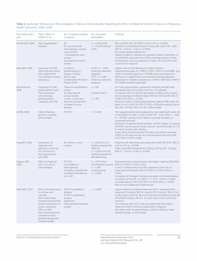
Table 1. Systematic Reviews and Meta-Analyses of Various Clinical Studies Reporting the Effect of Maternal Vitamin D Status on Pregnancy Health Outcomes, 2018–2019
First author [ref.], year
Topic: Effect of Vitamin D on …
No. of studies included in analysis
No. of pooled participants
Findings
Amraei [65], 2018 Risk of gestational diabetes
26: 8 cross-sectional6 prospective, nested case-control7 retrospective case-control5 prospective cohort studies
n = 5,464 GDMn = 15,039 without GDM
Risk of GDM, OR 1.18, 95% CI 1.01–1.35, p < 0.0001;25(OH)D concentration lower in those with GDM, OR –0.26, 95% CI –0.39 to –0.14, p < 0.0001No world regional differencesStated limitations: differences between studies in definition of low 25(OH)D and criteria for GDM; potential impact of other confounders, such as pregnancy wt gain, SES, and skin color could not be explored
Baca [40], 2018 Expression of VDR SNPs and association with log25(OH)D concentration during pregnancy
2 cohorts:Epidemiology of Vitamin D Study (EVITA) and Collaborative Perinatal Project (CPP)
EVITA: n = 1,958 randomly selected/ analyzedCPP: n = 4,285 randomly selected/ analyzed
Higher rates of vitD deficiency in black mothersMaternal genotypes of 7 SNPs in VDR, 3 SNPs in GC/VDBP, and 1 SNP in flanking regions of CYP27B1 were associated with difference in log25(OH)D concentration during pregnancyAdjusting for multiple comparisons, 1 SNP in VDR and 2 SNPs in GC/VDBP remained significant
Barchitta [41], 2018
Evaluation of VDR polymorphisms and their association with neonatal anthropometric measures and PTB
“Mamma and Bambino” cohort11 observational clinical studies3 case-control plus Mamma and Bambino included in meta-analysis
n = 187
Unclear total N
n = 763
For FokI polymorphism, gestational duration and BW were decreased with an increase in the No. of A allele(s)Compared with GG and GA genotypes, mothers who carried the AA genotype exhibited higher PTB risk (OR 12.049, 95% CI 2.606 to 55.709, p = 0.001)Protective effect of BsmI polymorphism against PTB under the allelic (A vs G: OR 0.74, 95% CI 0.59–0.93) and recessive (AA vs GG+AG: OR 0.62, 95% CI 0.43–0.89) models
Bi [66], 2018 Infant/offspring growth, morbidity and mortality
24 RCTs n = 5,405 VitD supplemented during pregnancy associated with lower risk of SGA (RR 0.72, 95% CI 0.52 to 0.99; RD –5.6%, 95% CI –0.86 to –10.34%), without risk of fetal or neonatal mortality or congenital anomalyNeonates of supplemented mothers: at birth, higher circulating 25(OH)D, serum calcium levels, and wt, carried through to 3, 6, 9, and 12 months after delivery Lower rates of fetal/neonatal mortality of mothers receiving 2,000 IU vitD/day and risk not reduced above that dose (RR 0.35, 95% CI 0.59 to 0.80)
Fang [67], 2019 Association of maternal vitD deficiency (<20 ng/mL/<50 nmol/L) during pregnancy with LBW
16 cohort or case-control
n = 8,403 from 8 studies analyzed for LBW riskn = 11,867 from 10 studies analyzed for BW differences
Maternal vitD deficiency associated with LBW (OR 2.39, 95% CI 1.25 to 4.57, p = 0.008)Total mean BW decreased by 0.08 kg or 80 g (OR –0.08 kg, 95% CI –0.10 to –0.06, p < 0.001)
Fogacci [68], 2019
Effect of maternal vitD-S on risk of preeclampsia
27 RCTsLow degree of heterogeneity3 studies excluded that included multivitamins with vitD
n = 4,777 totalVitD treatment group n = 2,487Control groupn = 2,290
Decreased risk of preeclampsia with higher maternal 25(OH)D (OR 0.37, 95% CI 0.26 to 0.52)If vitD-S initiated before 20 wks’ gestation, lower risk of maternal preeclampsia (OR 0.35, 95% CI 0.26 to 0.52, p < 0.001)Increased vitD dosage inversely associated with preeclampsia risk (slope of log OR –1.1, 95% CI –1.73 to –0.46, p < 0.001 corresponding to OR 0.33, 95% CI 0.18 to 0.63, p < 0.001)Risk not associated with maternal age
Gallo [69], 2019 Risk of vitD deficiency in mother and neonateRisk of abnormal maternal homeostatic model assessment of insulin resistanceEffect on BWRisk of preeclampsia, cesarean section, gestational age and neonatal length
20 RCTs qualitative analysis17 RCTs quantitative analysisSignificant heterogeneity between studies
n = 2,844 Good evidence to support maternal vitD-S, increased both maternal (13 studies, MD 14.1 ng/mL [35.2 nmol/L]; 95% CI 9.6 to 18.6 ng/mL [24.0 to 46.4 nmol/L]) and neonatal (cord blood) 25(OH)D (9 studies, MD 9.7, 5.2, 14.2 ng/mL [24.2, 12.9, 35.5 nmol/L])Fair evidence that vitD-S was associated with decreased maternal HOMA-IR and increased BW in offspringNull effect seen for preeclampsia, mode of delivery, infant gestational age, or birth length

Wagner/Hollis Reprinted with permission from:Ann Nutr Metab 2020;76(suppl 2):16–28
22
DOI: 10.1159/000508422
Table 1 (continued)
First author [ref.], year
Topic: Effect of Vitamin D on …
No. of studies included in analysis
No. of pooled participants
Findings
Li [70], 2019 VitD-S during pregnancy and the risk of wheezing in offspring
4 prospective cohorts3 RCTs
n = 6,068 mother/child pairs
Inverse relationship between maternal vitD intake during pregnancy and occurrence of wheezing in offspring (pooled OR 0.68, 95% CI 0.55 to 0.83, p < 0.01)Inverse relationship between maternal vitD intake during pregnancy and eczema but not significant (pooled OR 0.95, 95% CI 0.75 to 1.21, p = 0.66)Reported U-shaped dose curve between maternal vitD intake and risk of wheezing in offspring, with lowest risk in 800-IU group but were not able to control for timing of dose, maternal asthma, parental smoking, and other potential confounders
Maugeri [71], 2019
Effects of vitD-S on birth size
13 RCTs17 comparison groups
Maternal vitD-S associated with BW (12 RCTs; MD 103.17 g, 95% CI 62.29 to 144.04), length (6 RCTs; MD 0.22 cm, 95% CI 0.11 to 0.33), and head circumference (6 RCTs; MD 0.19 cm, 95% CI 0.13 to 0.24)Also associated with reduced risk of LBW (3 RCTs; RR 0.40, 95% CI 0.22 to 0.74) and SGA (5 RCTS; RR 0.69, 95% CI 0.51 to 0.92)
Ojo [72], 2019 Effect of vitD-S on glycemic control in women with GDM
5 RCTs n = 173 Compared to controls, vitD-S associated with decrease in fasting blood glucose (mean 0.46 mmol/L, 95% CI −0.68, −0.25, p < 0.001), glycated hemoglobin (mean 0.37%, 95% CI −0.65, −0.08, p < 0.01), and serum insulin concentration (mean 4.10 μIU/mL, 95% −5.50, −2.71, p < 0.001)
Pacheco-González [73], 2018
Prenatal vitD status and later offspring respiratory and allergy outcomes
34 observational26 separate study populations25 longitudinal and 1 case-control16 countries represented
n not listed Risk of RTIs: comparing highest with lowest 25(OH)D category, pooled OR 0.64 (95% CI 0.47, 0.87)Positive borderline association with lung function at school age (FEV1 z-score coefficient 0.07, 95% CI –0.01, 0.15) No associations found for wheeze, asthma, atopic eczema, allergic rhinitis, and allergic sensitization
Santamaria [74], 2018
Prenatal vitD status and offspring growth, adiposity, and metabolic health
30 observational n = 35,032 mother/offspring pairs
Low prenatal vitD associated with lower BW (g) (MD −100.69, 95% CI −162.25, −39.13), increased risk of SGA (OR 1.55, 95% CI 1.16, 2.07), and an elevated wt (g) in infants at the age of 9 months (MD 119.75, 95% CI 32.97, 206.52)No associations between prenatal vitD status and other growth parameters at birth, age 1 year, 4–6 yrs, or 9 yrs, or with diabetes type 1
Shen [75], 2018 Effect of maternal or neonatal (cord blood) vitD status on later risk of wheezing 5 yrs of age and >5 yrs
3 RCTs33 cohort studies
n = 1,619 No statistically significant association between maternal or cord blood 25(OH)D or intake early in life and asthma either at 5 or >5 yrs
Shi [76], 2019 Maternal vitD intake during pregnancy and later risk of asthma and wheeze in offspring
10 observational, with 14 independent reports
2,073 incident cases of asthma1,875 cases of wheezeTotal 23,030 mother/child pairs
Compared to offspring of nonsupplemented mothers, offspring of vitD-S mothers with reduced risk of asthma or wheeze in infants Combined OR infant wheeze 0.65 (95% CI 0.54 to 0.79) and asthma 0.78 (95% CI 0.69 to 0.89)
Tous [77],2019 Association of low prenatal 25(OH)D (using 3 different threshold levels), PTB, and anthropometric and neurodevelop-mental outcomes in offspring
54 observational n = 67,484 Mothers with 25(OHD threshold value of <30 nmol/L, at greater risk of offspring with:– lower BW (MD –87.82 g, 95% CI –119.73 to –55.919)– lower head circumference (MD –0.19 cm, 95% CI –0.32 to –0.06)– increased risk of SGA and PTB (OR 1.59, 95% CI 1.24 to 2.03)With threshold of <50 nmol/L, offspring with: – increased risk for SGA (OR 1.43, 95% CI 1.08 to 1.91)– increased risk for PTB (OR 1.38, 95% CI 1.08 to 1.52When maternal 25(OH)D 75 nmol/L, not associated with BW, SGA status, or PTBOffspring of vitD insufficient/deficient mothers had lower scores on mental index (OR –1.12, 95% CI –1.82 to 0.42) and language (OR –0.35, 95% CI –1.00 to 0.31, but not statistically significant)

Vitamin D during Pregnancy and Lactation 23Reprinted with permission from:Ann Nutr Metab 2020;76(suppl 2):16–28DOI: 10.1159/000508422
Vitamin D Clinical Trials during Pregnancy
The issue with nutrient studies is that they often are designed
more like a drug study, where the starting concentration of
the “drug” is zero, compared with a nutrient study, such as vi-
tamin D, where there is some vitamin D concentration in ev-
eryone, and, thus, baseline 25(OH)D concentration is variable
and not zero. Heaney [43] described the qualities that should
define a nutrient study:
1 basal nutrient status must be measured, used as an inclu-
sion criterion for entry into the study, and recorded in the
report of the trial;
2 the intervention must be large enough to change nutrient
status and must be quantified by suitable analysis;
3 the change in nutrient status produced in those enrolled in
the report of the trial must be measured and reported;
4 the hypothesis to be tested must be that a change in nutri-
ent status produces the sought-after effect; and
5 the status of other nutrients must be optimized to guaran-
tee that the nutrient being studied is the only nutrition-
related limiting factor in the response.
One might add another stipulation to the list – that the
nutrient being investigated has to follow an appropriate dos-
ing schedule to match what happens naturally. In the case of
vitamin D, there is a plethora of data that show substantial
physiological differences between daily, weekly, and monthly
vitamin D dosing regimens [11] .
In reviewing past clinical trials of vitamin D, most lack all 5
of the Heaney criteria and often the 6th dosing criterion. While
systematic reviews and meta-analyses provide larger com-
bined sample sizes, the analyses of several limited studies only
compound the problem. More recently, the rigor of clinical
trials with increased sample sizes has improved the consis-
tency of pooled/aggregate data, and some compelling evi-
dence from these clinical trials suggests that vitamin D suffi-
ciency during pregnancy enhances maternal and fetal health.
The “translation” of the laboratory data to the clinic and bed-
side supports this emerging concept that vitamin D plays a
role not only in calcium homeostasis and bone function but
also in immune function. Beyond the scope of this review for
an exhaustive summary, below are some of the highlights of
those systematic reviews and meta-analyses to date, with an
emphasis on the salient findings, and their strengths and
weaknesses.
As mentioned in the attributes of a well-designed nutrient
study as outlined by Heaney [43] , part of the issue is that these
supplementation trials have varied by the onset of supple-
mentation during pregnancy, the dosing and timing of that
dose, the degree of vitamin D deficiency at the onset of the
trial, and different methodology used in measuring 25(OH)D.
There have been numerous systematic reviews and meta-
analyses on the topic, also with mixed findings. Restricting
systematic reviews and meta-analyses to the 2 recent years
2018–2019, there are over 30 publications on the topic of vi-
tamin D and pregnancy outcomes. The analyses cover such
topics as gestational age, infant birth weight, gestational dia-
betes and insulin resistance, small-for-gestational age, pre-
eclampsia, and maternal and neonatal vitamin D status at de-
Table 1 (continued)
First author [ref.], year
Topic: Effect of Vitamin D on …
No. of studies included in analysis
No. of pooled participants
Findings
Yuan [78], 2019 Association of maternal 25(OH)D and risk of preeclampsia
1 nested case-control20 additional clinical studies
n = 122 women with preeclampsian = 480 controlsn = 39,031 participants: 3,305 with preeclampsia, various ethnicities
65.6% with preeclampsia had 25(OH)D <50.0 nmol/L vs. 55.3% of controls25(OH)D was significantly lower in women with preeclampsia than in controls (median [IQR] 43.3 [35.5, 55.2] vs. 47.5 [37.6, 60.4] nmol/L, p = 0.014) Women with 25(OH)D <50.0 nmol/L with 65% increase in preeclampsia risk (95% CI 1.02 to 2.69) compared with women with 25(OH)D 50.0 to 74.9 nmol/LMeta-analysis showed that low 25(OH)D concentrations were associated with significantly increased risk of preeclampsia by 62% (pooled OR 1.62, 95% CI 1.36 to 1.94), and risk effect of low 25(OH)D concentrations existed in most subgroups
Zhang [79], 2018
Effect of maternal vitD status on risk of GDM
87 observational and 25 RCTs
n = 55,859 in observational studiesn = 2,445 in RCTs
Low 25(OH)D associated with increased GDM (OR 1.850, 95% CI 1.471 to 2.328)25(OH)D associated with fasting glucose and HOMA-IR index
BW, birth weight; GDM, gestational diabetes mellitus; HOMA-IR, homeostatic model assessment of insulin resistance; LBW, low BW; MD, mean difference; OR, odds ratio; PTB, preterm birth; RTCs, randomized clinical trials; SNPs, single nucleotide polymorphisms; SGA, small for gestational age; SES, socioeconomic status; vitD, vitamin D; vitD-S; vitamin D supplementation; VDR, vitamin D receptor; wt, weight; wks, weeks; yrs, years.
Wagner/Hollis Reprinted with permission from:Ann Nutr Metab 2020;76(suppl 2):16–28
24
DOI: 10.1159/000508422
livery. Compared to analyses performed in earlier years when
there were few published randomized controlled trials that
were often plagued with small sample sizes, the more recent
reviews consistently have shown benefit of maternal vitamin
D supplementation during pregnancy. The highlights of some
of the larger systemic reviews and meta-analyses published in
the past 2 years (2018–2019) are summarized in Table 1 . With
each review, there is evidence that there are still limitations to
the clinical studies and there is a need for continued research,
especially with genetic and epigenetic considerations in place
and design of nutrient studies that take into account the Hea-
ney criteria [43] .
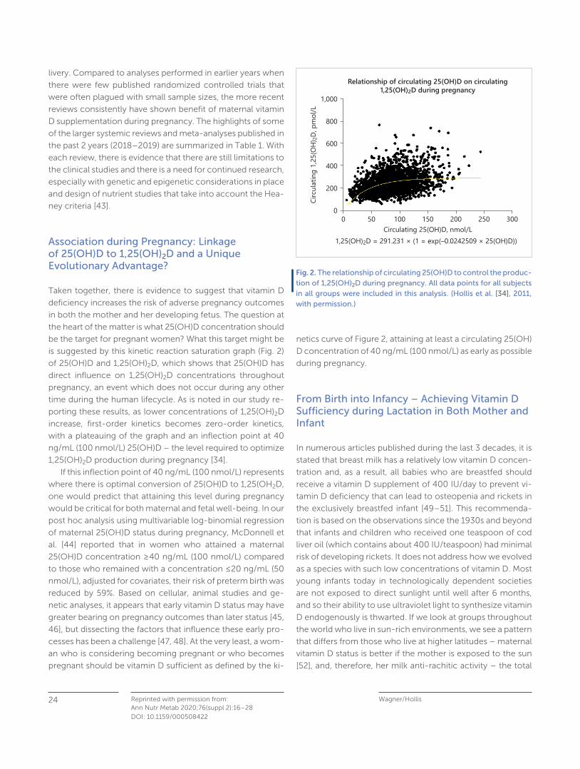
Association during Pregnancy: Linkage of 25(OH)D to 1,25(OH) 2 D and a Unique Evolutionary Advantage?
Taken together, there is evidence to suggest that vitamin D
deficiency increases the risk of adverse pregnancy outcomes
in both the mother and her developing fetus. The question at
the heart of the matter is what 25(OH)D concentration should
be the target for pregnant women? What this target might be
is suggested by this kinetic reaction saturation graph ( Fig. 2 )
of 25(OH)D and 1,25(OH) 2 D, which shows that 25(OH)D has
direct influence on 1,25(OH) 2 D concentrations throughout
pregnancy, an event which does not occur during any other
time during the human lifecycle. As is noted in our study re-
porting these results, as lower concentrations of 1,25(OH) 2 D
increase, first-order kinetics becomes zero-order kinetics,
with a plateauing of the graph and an inflection point at 40
ng/mL (100 nmol/L) 25(OH)D – the level required to optimize
1,25(OH) 2 D production during pregnancy [34] .
If this inflection point of 40 ng/mL (100 nmol/L) represents
where there is optimal conversion of 25(OH)D to 1,25(OH 2 D,
one would predict that attaining this level during pregnancy
would be critical for both maternal and fetal well-being. In our
post hoc analysis using multivariable log-binomial regression
of maternal 25(OH)D status during pregnancy, McDonnell et
al. [44] reported that in women who attained a maternal
25(OH)D concentration ≥ 40 ng/mL (100 nmol/L) compared
to those who remained with a concentration ≤ 20 ng/mL (50
nmol/L), adjusted for covariates, their risk of preterm birth was
reduced by 59%. Based on cellular, animal studies and ge-
netic analyses, it appears that early vitamin D status may have
greater bearing on pregnancy outcomes than later status [45,
46] , but dissecting the factors that influence these early pro-
cesses has been a challenge [47, 48] . At the very least, a wom-
an who is considering becoming pregnant or who becomes
pregnant should be vitamin D sufficient as defined by the ki-
netics curve of Figure 2 , attaining at least a circulating 25(OH)
D concentration of 40 ng/mL (100 nmol/L) as early as possible
during pregnancy.
From Birth into Infancy – Achieving Vitamin D Sufficiency during Lactation in Both Mother and Infant
In numerous articles published during the last 3 decades, it is
stated that breast milk has a relatively low vitamin D concen-
tration and, as a result, all babies who are breastfed should
receive a vitamin D supplement of 400 IU/day to prevent vi-
tamin D deficiency that can lead to osteopenia and rickets in
the exclusively breastfed infant [49–51] . This recommenda-
tion is based on the observations since the 1930s and beyond
that infants and children who received one teaspoon of cod
liver oil (which contains about 400 IU/teaspoon) had minimal
risk of developing rickets. It does not address how we evolved
as a species with such low concentrations of vitamin D. Most
young infants today in technologically dependent societies
are not exposed to direct sunlight until well after 6 months,
and so their ability to use ultraviolet light to synthesize vitamin
D endogenously is thwarted. If we look at groups throughout
the world who live in sun-rich environments, we see a pattern
that differs from those who live at higher latitudes – maternal
vitamin D status is better if the mother is exposed to the sun
[52] , and, therefore, her milk anti-rachitic activity – the total
Circu
lating
1,25
(OH)
2D, p
mol/
L
0 50 100 150Circulating 25(OH)D, nmol/L
1,25(OH)2D = 291.231 × (1 = exp(–0.0242509 × 25(OH)D))
200 250 300
1,000
800
600
200
0
400
Relationship of circulating 25(OH)D on circulating1,25(OH)2D during pregnancy
Fig. 2. The relationship of circulating 25(OH)D to control the produc-tion of 1,25(OH) 2 D during pregnancy. All data points for all subjects in all groups were included in this analysis. (Hollis et al. [34] , 2011, with permission.)

Vitamin D during Pregnancy and Lactation 25Reprinted with permission from:Ann Nutr Metab 2020;76(suppl 2):16–28DOI: 10.1159/000508422
amount of vitamin D moieties in human milk – is better. It is
critical to understand that human milk is deficient in vitamin
D only when the mother herself is deficient [53] . We know that
during pregnancy, maternal vitamin D status is closely linked
with fetal and neonatal vitamin D status. That connection and
relationship continues during lactation.
It was hypothesized by Hollis and Wagner [54] more than
2 decades ago that if a mother had an improved vitamin D
status, then her milk anti-rachitic activity would be improved
and that of her recipient infant, obviating the need for infant
vitamin D supplementation. Maternal vitamin D supplementa-
tion would effectively treat both the mother and her breast-
feeding infant. This was studied in 2 pilot studies by our group
[54, 55] and then in a larger trial sponsored by the National
Institutes of Health [56] that has since been repeated by Da-
wodu et al. [57] in another region of the world – the Middle
East – where there is profound vitamin D deficiency. In the
various trials, mothers at 1 month postpartum were random-
ized to receive 1 of 3 treatments: 400, 2,400, or 6,400 IU vi-
tamin D/day. Infants of mothers in the 400-IU group received
the standard of care of 400 IU/day, while infants of mothers
in the 2,400- and 6,400-IU group received 0 IU/day (place-
bo). Maternal supplementation with 2,400 IU vitamin D/day
with infants on placebo resulted in higher rates of infant insuf-
ficiency and that arm of the study was stopped early-on in the
study. Mothers in the 6,400-IU group had improved vitamin
D status, milk anti-rachitic activity, and their infants had cir-
culating 25(OH)D concentrations that were comparable to in-
fants receiving 400 IU/day direct supplementation [55, 56] .
There were no safety issues noted in these studies except with
the 2,400 IU arm and the higher rates of infant deficiency, but
no issues with toxicity from vitamin D. Similar results were re-
ported by Dawodu et al. [57] .
Oberhelman et al. [58] studied 40 exclusively breastfeed-
ing mothers and infants who were randomized to receive ei-
ther daily maternal vitamin D supplementation of 5,000 IU/
day versus a single large bolus of 150,000 IU once as a higher
bolus vitamin D, with the primary outcome at 28 days being
maternal and infant vitamin D status. The daily versus single
bolus were comparable at 28 days; however, the mother and
infant pair who received the single bolus had a large increase
in their circulating 25(OH)D that rapidly declined but was still
improved compared to baseline.
A systematic review and meta-analysis by O’Callaghan et
al. [59] , reviewing relevant studies on the topic of alternatives
to daily infant vitamin D supplementation through September
2018, identified 28 relevant papers of which 5 were random-
ized clinical trials that met inclusion criteria for the analysis.
The meta-analysis suggests that the results are promising,
with the need for larger studies in diverse groups of women
necessary to be carried out before policy changes can be
made [59] .
While application of alternatives to infant supplementation
are being discussed, a major issue complicating recommen-
dations is that compliance by parents to give their breastfeed-
ing infants daily vitamin D drops is low in many regions of the
world [60, 61] . In the USA, reports of compliance with the
recommendation of infant vitamin D supplementation range
from 9 to 20%, leaving most breastfeeding infants in the USA
deficient, dependent on their mothers who are often them-
selves deficient [62–64] . These are less than satisfying statis-
tics. At the end of the day, where maternal compliance with
taking a vitamin D supplement is much greater than that of
parental adherence with infant supplementation, maternal vi-
tamin D supplementation alone remains as a viable alternative
to infant vitamin D supplementation that safely and effective-
ly treats both the mother and her breastfeeding infant.
Conflict of Interest Statement
The writing of this article was supported by Nestlé Nutrition Institute, and the author declares no other conflicts of interest.
Funding Sources
Funded in part by NIH UL1 TR000062 and Stone Foundation, Chil-dren’s Hospital Fund, Medical University of South Carolina.
That if a mother had an
improved vitamin D status,
then her milk anti-rachitic
activity would be improved
and that of her recipient
infant, obviating the need
for infant vitamin D
supplementation

Wagner/Hollis Reprinted with permission from:Ann Nutr Metab 2020;76(suppl 2):16–28
26
DOI: 10.1159/000508422
References
1 Racicot K, Kwon JY, Aldo P, Silasi M, Mor G. Understanding the complexity of the immune system during pregnancy. Am J Reprod Immunol . 2014 Aug; 72(2): 107–16.
2 Mor G, Cardenas I. The immune system in pregnancy: a unique complexity. Am J Reprod Immunol . 2010 Jun; 63(6): 425–33.
3 Mor G, Cardenas I, Abrahams V, Guller S. Inflammation and preg-nancy: the role of the immune system at the implantation site. Ann N Y Acad Sci . 2011 Mar; 1221(1): 80–7.
4 Bikle DD. Vitamin D: an ancient hormone. Exp Dermatol . 2011 Jan; 20(1): 7–13.
5 Wagner CL, Eidelman AI. The impact of vitamin D on the maternal and infant epigenome: the role of pregnancy and breastfeeding. Breastfeed Med . 2018 Jun; 13(5): 305–6.
6 Bikle DD. Vitamin D regulation of immune function. Vitam Horm . 2011; 86: 1–21.
7 Bikle DD. Vitamin D metabolism and function in the skin. Mol Cell Endocrinol . 2011 Dec; 347(1-2): 80–9.
8 Hollis BW. Comparison of equilibrium and disequilibrium assay conditions for ergocalciferol, cholecalciferol and their major me-tabolites. J Steroid Biochem . 1984 Jul; 21(1): 81–6.
9 Bhattacharyya MH, DeLuca HF. Subcellular location of rat liver calciferol-25-hydroxylase. Arch Biochem Biophys . 1974 Jan; 160(1): 58–62.
10 Hollis BW, Pittard WB 3rd. Evaluation of the total fetomaternal vi-tamin D relationships at term: evidence for racial differences. J Clin Endocrinol Metab . 1984 Oct; 59(4): 652–7.
11 Hollis BW, Wagner CL. Clinical review: The role of the parent com-pound vitamin D with respect to metabolism and function: Why clinical dose intervals can affect clinical outcomes. J Clin Endo-crinol Metab . 2013 Dec; 98(12): 4619–28.
12 Hollis BW, Pittard WB 3rd, Reinhardt TA. Relationships among vi-tamin D, 25-hydroxyvitamin D, and vitamin D-binding protein concentrations in the plasma and milk of human subjects. J Clin Endocrinol Metab . 1986 Jan; 62(1): 41–4.
13 Bikle DD, Gee E, Halloran B, Kowalski MA, Ryzen E, Haddad JG. Assessment of the free fraction of 25-hydroxyvitamin D in serum and its regulation by albumin and the vitamin D-binding protein. J Clin Endocrinol Metab . 1986 Oct; 63(4): 954–9.
14 Bikle DD, Halloran BP, Gee E, Ryzen E, Haddad JG. Free 25-hy-droxyvitamin D levels are normal in subjects with liver disease and reduced total 25-hydroxyvitamin D levels. J Clin Invest . 1986 Sep; 78(3): 748–52.
15 Kumar R, Thompson JR. The regulation of parathyroid hormone secretion and synthesis. J Am Soc Nephrol . 2011 Feb; 22(2): 216–24.
16 Bouillon R, Bikle D. Vitamin D Metabolism Revised: fall of Dogmas. J Bone Miner Res . 2019 Nov; 34(11): 1985–92.
17 Bouillon R, Marcocci C, Carmeliet G, Bikle D, White JH, Dawson-Hughes B, et al. Skeletal and Extraskeletal Actions of Vitamin D: Current Evidence and Outstanding Questions. Endocr Rev . 2019 Aug; 40(4): 1109–51.
18 Hollis BW. Circulating 25-hydroxyvitamin D levels indicative of vi-tamin D sufficiency: implications for establishing a new effective dietary intake recommendation for vitamin D. J Nutr . 2005 Feb; 135(2): 317–22.
19 Nykjaer A, Dragun D, Walther D, Vorum H, Jacobsen C, Herz J, et al. An endocytic pathway essential for renal uptake and activation of the steroid 25-(OH) vitamin D3. Cell . 1999 Feb; 96(4): 507–15.
20 Marzolo MP, Farfán P. New insights into the roles of megalin/LRP2 and the regulation of its functional expression. Biol Res . 2011; 44(1): 89–105.
21 Holick MF. Vitamin D deficiency. N Engl J Med . 2007 Jul; 357(3): 266–81.
22 Bikle DD. Vitamin D metabolism, mechanism of action, and clini-cal applications. Chem Biol . 2014 Mar; 21(3): 319–29.
23 Holick MF. Vitamin D: evolutionary, physiological and health per-spectives. Curr Drug Targets . 2011 Jan; 12(1): 4–18.
24 Gallagher JC, Bikle DD. Vitamin D: Mechanisms of Action and Clinical Applications. Endocrinol Metab Clin North Am . 2017 Dec; 46(4):xvii–xviii.
25 Hollis BW, Wagner CL. New insights into the vitamin D require-ments during pregnancy. Bone Res . 2017 Aug; 5(1): 17030.
26 Bikle DD, Schwartz J. Vitamin D Binding Protein, Total and Free Vitamin D Levels in Different Physiological and Pathophysiological Conditions. Front Endocrinol (Lausanne) . 2019 May; 10: 317.
27 Hacker-Thompson A, Schloetter M, Sellmeyer DE. Validation of a dietary vitamin D questionnaire using multiple diet records and the block 98 health habits and history questionnaire in healthy post-menopausal women in northern California. J Acad Nutr Diet . 2012 Mar; 112(3): 419–23.
28 Kumar R, Cohen WR, Silva P, Epstein FH. Elevated 1,25-dihy-droxyvitamin D plasma levels in normal human pregnancy and lactation. J Clin Invest . 1979 Feb; 63(2): 342–4.
29 Carneiro RM, Prebehalla L, Tedesco MB, Sereika SM, Hugo M, Hol-lis BW, et al. Lactation and bone turnover: a conundrum of marked bone loss in the setting of coupled bone turnover. J Clin Endocri-nol Metab . 2010 Apr; 95(4): 1767–76.
30 Ritchie LD, Fung EB, Halloran BP, Turnlund JR, Van Loan MD, Cann CE, et al. A longitudinal study of calcium homeostasis during hu-man pregnancy and lactation and after resumption of menses. Am J Clin Nutr . 1998 Apr; 67(4): 693–701.
31 Hollis BW, Wagner CL. Assessment of dietary vitamin D require-ments during pregnancy and lactation. Am J Clin Nutr . 2004 May; 79(5): 717–26.
32 Olsen SF, Secher NJ. A possible preventive effect of low-dose fish oil on early delivery and pre-eclampsia: indications from a 50-year-old controlled trial. Br J Nutr . 1990 Nov; 64(3): 599–609.
33 Bikle DD, Gee E, Halloran B, Haddad JG. Free 1,25-dihydroxyvita-min D levels in serum from normal subjects, pregnant subjects, and subjects with liver disease. J Clin Invest . 1984 Dec; 74(6): 1966–71.
34 Hollis BW, Johnson D, Hulsey TC, Ebeling M, Wagner CL. Vitamin D supplementation during pregnancy: double-blind, randomized clinical trial of safety and effectiveness. J Bone Miner Res . 2011 Oct; 26(10): 2341–57.

Vitamin D during Pregnancy and Lactation 27Reprinted with permission from:Ann Nutr Metab 2020;76(suppl 2):16–28DOI: 10.1159/000508422
35 Hollis BW, Wagner CL. Vitamin D and pregnancy: skeletal effects, nonskeletal effects, and birth outcomes. Calcif Tissue Int . 2013 Feb; 92(2): 128–39.
36 Karras SN, Wagner CL, Angeloudi E, Kotsa K. Maternal vitamin D status during pregnancy in Europe: the two sides of the story. Eur J Nutr . 2017 Sep; 56(6): 2207–8.
37 Karras SN, Wagner CL, Castracane VD. Understanding vitamin D metabolism in pregnancy: from physiology to pathophysiology and clinical outcomes. Metabolism . 2018 Sep; 86: 112–23.
38 Al-Garawi A, Carey VJ, Chhabra D, Mirzakhani H, Morrow J, Lasky-Su J, et al. The Role of Vitamin D in the Transcriptional Program of Human Pregnancy. PLoS One . 2016 Oct; 11(10):e0163832.
39 Litonjua AA, Carey VJ, Laranjo N, Harshfield BJ, McElrath TF, O’Connor GT, et al. Effect of Prenatal Supplementation with Vita-min D on Asthma or Recurrent Wheezing in Offspring by Age 3 Years: The VDAART Randomized Clinical Trial. JAMA . 2016 Jan; 315(4): 362–70.
40 Baca KM, Govil M, Zmuda JM, Simhan HN, Marazita ML, Bodnar LM. Vitamin D metabolic loci and vitamin D status in Black and White pregnant women. Eur J Obstet Gynecol Reprod Biol . 2018 Jan; 220: 61–8.
41 Barchitta M, Maugeri A, La Rosa MC, Magnano San Lio R, Favara G, Panella M, et al. Single Nucleotide Polymorphisms in Vitamin D Receptor Gene Affect Birth Weight and the Risk of Preterm Birth: Results From the “Mamma & Bambino” Cohort and A Meta-Anal-ysis. Nutrients . 2018 Aug; 10(9): 10.
42 Newton DA, Baatz JE, Kindy MS, Gattoni-Celli S, Shary JR, Hollis BW, et al. Vitamin D binding protein polymorphisms significantly impact vitamin D status in children. Pediatr Res . 2019 Nov; 86(5): 662–9.
43 Heaney RP. Guidelines for optimizing design and analysis of clin-ical studies of nutrient effects. Nutr Rev . 2014 Jan; 72(1): 48–54.
44 McDonnell SL, Baggerly KA, Baggerly CA, Aliano JL, French CB, Baggerly LL, et al. Maternal 25(OH)D concentrations ≥ 40 ng/mL associated with 60% lower preterm birth risk among general ob-stetrical patients at an urban medical center. PLoS One . 2017 Jul; 12(7):e0180483.
45 Mirzakhani H, Litonjua AA, McElrath TF, O’Connor G, Lee-Parritz A, Iverson R, et al. Early pregnancy vitamin D status and risk of preeclampsia. J Clin Invest . 2016 Dec; 126(12): 4702–15.
46 Weiss ST, Litonjua AA. Vitamin D in Host Defense: Implications for Future Research. Am J Respir Cell Mol Biol . 2017 Jun; 56(6): 692–3.
47 Hewison M. The earlier the better: preconception vitamin D and protection against pregnancy loss. Lancet Diabetes Endocrinol . 2018 Sep; 6(9): 680–1.
48 Hewison M, Wagner CL, Hollis BW. Vitamin D Supplementation in Pregnancy and Lactation and Infant Growth. N Engl J Med . 2018 Nov; 379(19): 1880–1.
49 Hollis BW, Roos BA, Lambert PW. Vitamin D compounds in human and bovine milk. Adv Nutr Res . 1982; 4: 59–75.
50 Wagner CL, Greer FR; American Academy of Pediatrics Section on Breastfeeding; American Academy of Pediatrics Committee on Nutrition. Prevention of rickets and vitamin D deficiency in infants, children, and adolescents. Pediatrics . 2008 Nov; 122(5): 1142–52.
51 Dawodu A, Tsang RC. Maternal vitamin D status: effect on milk vitamin D content and vitamin D status of breastfeeding infants. Adv Nutr . 2012 May; 3(3): 353–61.
52 Luxwolda MF, Kuipers RS, Kema IP, van der Veer E, Dijck-Brouwer DA, Muskiet FA. Vitamin D status indicators in indigenous popula-tions in East Africa. Eur J Nutr . 2013 Apr; 52(3): 1115–25.
53 Wagner C. Current concepts in vitamin D requirements for moth-er and her breastfeeding infant. In: Lawrence R, editor. Interna-tional Society for Research in Human Milk and Lactation. Breast-feeding Medicine. Trieste, Italy; 2013. pp. 561–562.
54 Hollis BW, Wagner CL. Vitamin D requirements during lactation: high-dose maternal supplementation as therapy to prevent hypo-vitaminosis D for both the mother and the nursing infant. Am J Clin Nutr . 2004 Dec; 80(6 Suppl): 1752S–8S.
55 Wagner CL, Hulsey TC, Fanning D, Ebeling M, Hollis BW. High-dose vitamin D3 supplementation in a cohort of breastfeeding mothers and their infants: a 6-month follow-up pilot study. Breastfeed Med . 2006; 1(2): 59–70.
56 Hollis BW, Wagner CL, Howard CR, Ebeling M, Shary JR, Smith PG, et al. Maternal Versus Infant Vitamin D Supplementation During Lactation: A Randomized Controlled Trial. Pediatrics . 2015 Oct; 136(4): 625–34.
57 Dawodu A, Salameh KM, Al-Janahi NS, Bener A, Elkum N. The Ef-fect of High-Dose Postpartum Maternal Vitamin D Supplementa-tion Alone Compared with Maternal Plus Infant Vitamin D Supple-mentation in Breastfeeding Infants in a High-Risk Population. A Randomized Controlled Trial. Nutrients . 2019 Jul; 11(7): 11.
58 Oberhelman SS, Meekins ME, Fischer PR, Lee BR, Singh RJ, Cha SS, et al. Maternal vitamin D supplementation to improve the vita-min D status of breast-fed infants: a randomized controlled trial. Mayo Clin Proc . 2013 Dec; 88(12): 1378–87.
59 O’Callaghan KM, Taghivand M, Zuchniak A, Onoyovwi A, Korsiak J, Leung M, et al. Vitamin D in Breastfed Infants: Systematic Review of Alternatives to Daily Supplementation. Adv Nutr . 2020 Jan; 11(1): 144–59.
60 Oberhelman S, Cozine E, Umaretiya P, Maxson J, Thacher T. Vita-min D and the breastfeeding infant: family medicine clinicians’ knowledge, attitudes, and practices. J Hum Lact . 2018 May; 34(2): 331–6.
61 Wagner CL, Hollis BW. Commentary on “Vitamin D and the Breast-feeding Infant: Family Medicine Clinicians’ Knowledge, Attitudes, and Practices” by Oberhelman et al. J Hum Lact . 2018 May; 34(2): 337–9.
62 Perrine CG, Sharma AJ, Jefferds ME, Serdula MK, Scanlon KS. Ad-herence to vitamin D recommendations among US infants. Pedi-atrics . 2010 Apr; 125(4): 627–32.
63 Weernink MG, van Wijk RM, Groothuis-Oudshoorn CG, Lanting CI, Grant CC, van Vlimmeren LA, et al. Insufficient vitamin D supple-ment use during pregnancy and early childhood: a risk factor for positional skull deformation. Matern Child Nutr . 2016 Jan; 12(1): 177–88.
64 Ahrens K, Rossen L, Simon A. Adherence to vitamin D recommen-dations among US infants aged 0 to 11 Months, NHANES, 2009 to 2012. Clin Pediatr (Phila) . 2016 Jun; 55(6): 555–6.

Wagner/Hollis Reprinted with permission from:Ann Nutr Metab 2020;76(suppl 2):16–28
28
DOI: 10.1159/000508422
65 Amraei M, Mohamadpour S, Sayehmiri K, Mousavi SF, Shirzadpour E, Moayeri A. Effects of Vitamin D Deficiency on Incidence Risk of Gestational Diabetes Mellitus: A Systematic Review and Meta-analysis. Front Endocrinol (Lausanne). 2018 Feb;9:7.
66 Bi WG, Nuyt AM, Weiler H, Leduc L, Santamaria C, Wei SQ. Asso-ciation Between Vitamin D Supplementation During Pregnancy and Offspring Growth, Morbidity, and Mortality: A Systematic Re-view and Meta-analysis. JAMA Pediatr. 2018 Jul;172(7):635–45.
67 Fang K, He Y, Mu M, Liu K. Maternal vitamin D deficiency during pregnancy and low birth weight: a systematic review and meta-analysis. J Matern Fetal Neonatal Med. 2019 Jul 8;1–7.
68 Fogacci S, Fogacci F, Banach M, Michos ED, Hernandez AV, Lip GYH, Blaha MJ, Toth PP, Borghi C, Cicero AFG; Lipid and Blood Pressure Meta-analysis Collaboration (LBPMC) Group. Vitamin D supplementation and incident preeclampsia: A systematic review and meta-analysis of randomized clinical trials. Clin Nutr. 2020 Jun;39(6):1742–52.
69 Gallo S, McDermid JM, Al-Nimr RI, Hakeem R, Moreschi JM, Pari-Keener M, et al. Supplementation during Pregnancy: An Evidence Analysis Center Systematic Review and Meta-Analysis. J Acad Nutr Diet. 2020 May;120(5):898–924.e4.
70 Li W, Qin Z, Gao J, Jiang Z, Chai Y, Guan L, et al. Vitamin D supple-mentation during pregnancy and the risk of wheezing in offspring: a systematic review and dose-response meta-analysis. J Asthma. 2019 Dec;56(12):1266–73.
71 Maugeri A, Barchitta M, Blanco I, Agodi A. Effects of Vitamin D Supplementation During Pregnancy on Birth Size: A Systematic Review and Meta-Analysis of Randomized Controlled Trials. Nu-trients. 2019 Feb;11(2):E442.
72 Ojo O, Weldon SM, Thompson T, Vargo EJ. The Effect of Vitamin D Supplementation on Glycaemic Control in Women with Gesta-tional Diabetes Mellitus: A Systematic Review and Meta-Analysis of Randomised Controlled Trials. Int J Environ Res Public Health. 2019 May;16(10):E1716.
73 Pacheco-González RM, García-Marcos L, Morales E. Prenatal vi-tamin D status and respiratory and allergic outcomes in child-hood: A meta-analysis of observational studies. Pediatr Allergy Immunol. 2018 May;29(3):243–53.
74 Santamaria C, Bi WG, Leduc L, Tabatabaei N, Jantchou P, Luo ZC, et al. Prenatal vitamin D status and offspring’s growth, adiposity and metabolic health: a systematic review and meta-analysis. Br J Nutr. 2018 Feb;119(3):310–9.
75 Shen SY, Xiao WQ, Lu JH, Yuan MY, He JR, Xia HM, et al. Early life vitamin D status and asthma and wheeze: a systematic review and meta-analysis. BMC Pulm Med. 2018 Jul;18(1):120.
76 Shi D, Wang D, Meng Y, Chen J, Mu G, Chen W. Maternal vitamin D intake during pregnancy and risk of asthma and wheeze in chil-dren: a systematic review and meta-analysis of observational studies. J Matern Fetal Neonatal Med. 2019 May 7:1–7.
77 Tous M, Villalobos M, Iglesias L, Fernández-Barrés S, Arija V. Vita-min D status during pregnancy and offspring outcomes: a system-atic review and meta-analysis of observational studies. Eur J Clin Nutr. 2020 Jan;74(1):36-53.
78 Yuan Y, Tai W, Xu P, Fu Z, Wang X, Long W, Guo X, Ji C, Zhang L, Zhang Y, Wen J. Association of maternal serum 25-hydroxyvita-min D concentrations with risk of preeclampsia: a nested case-control study and meta-analysis. J Matern Fetal Neonatal Med. 2019 Jul 17:1–10.
79 Zhang Y, Gong Y, Xue H, Xiong J, Cheng G. Vitamin D and gesta-tional diabetes mellitus: a systematic review based on data free of Hawthorne effect. BJOG. 2018 Jun;125(7):784–93.