east and west separation of rhipicephalus sanguineus ... · short report open access east and west...
TRANSCRIPT

SHORT REPORT Open Access
East and west separation of Rhipicephalussanguineus mitochondrial lineages in theMediterranean BasinSándor Hornok1* , Attila D. Sándor2, Snežana Tomanović3, Relja Beck4, Gianluca D’Amico2, Jenő Kontschán5,Nóra Takács1, Tamás Görföl6, Mohammed Lamine Bendjeddou7, Gábor Földvári1 and Róbert Farkas1
Abstract
Background: Rhipicephalus sanguineus belongs to a complex of hard tick species with high veterinary-medicalsignificance. Recently, new phylogenetic units have been discovered within R. sanguineus, which therefore needstaxonomic revision. The present study was initiated to provide new information on the phylogeography of relevanthaplotypes from less studied regions of Europe and Africa. With this aim, molecular-phylogenetic analyses of twomitochondrial markers were performed on 50 ticks collected in Hungary, the Balkans, countries along theMediterranean Sea, Kenya and Ivory Coast.
Results: In the “temperate lineage” of R. sanguineus, based on cytochrome c oxidase subunit 1 (cox1) and 16S rRNAgenes, Rhipicephalus sp. I was only found in the eastern part of the Mediterranean Basin (with relatively homogenoushaplotypes), whereas Rhipicephalus sp. II occurred in the middle-to-western part of this region (with phylogeneticallydichotomous haplotypes). Ticks identified as R. leporis (based on morphology and cox1 gene) were found in Kenya andIvory Coast. These clustered phylogenetically within R. sanguineus (s.l.) (“tropical lineage”).
Conclusions: In the Mediterranean Basin two mitochondrial lineages of R. sanguineus, i.e. Rhipicephalus sp. I andRhipicephalus sp. II exist, which show different geographical distribution. Therefore, data from this study confirmlimited gene flow between Rhipicephalus sp. I and Rhipicephalus sp. II, but more evidence (analyses of nuclearmarkers, extensive morphological and biological comparison etc.) are necessary to infer if they belong to differentspecies or not. The phylogenetic relationships of eastern and western African ticks, which align with R. leporis,need to be studied further within R. sanguineus (s.l.) (“tropical lineage”).
Keywords: Phylogeography, cox1, 16S rRNA gene, Rhipicephalus sanguineus, Rhipicephalus leporis
BackgroundRhipicephalus sanguineus (sensu lato) (Acari: Ixodidae)belongs to a complex of at least 17 hard tick species,some with high medical and veterinary importance [1].The type-species of this group was formerly called R.sanguineus (sensu stricto) (the brown dog tick), withcosmopolitan distribution owing to its high adaptability(i.e. being able to utilize endophilic and exophilichabitats in both urban and rural environment; havingyear-round activity and up to four generations per year;
exhibiting passive or active host-seeking behaviour) [2].Although typically a dog parasite (in all three develop-mental stages), R. sanguineus may also infest a widerange of domestic and wild animal host species, evenhumans. There are several animal and human pathogens(including zoonotic ones) that are or may be transmittedby R. sanguineus (reviewed in [3]). On account of itspreference of warmer climates, global warming was pre-dicted to induce the expansion of the geographical rangeof R. sanguineus [2].According to current knowledge, R. sanguineus is not
a single species. Molecular phylogeographic studies havefound high intraspecific divergence of mitochondrialDNA between R. sanguineus from Brazil and Argentina,
* Correspondence: [email protected] of Parasitology and Zoology, University of Veterinary Medicine,Budapest, HungaryFull list of author information is available at the end of the article
© The Author(s). 2017 Open Access This article is distributed under the terms of the Creative Commons Attribution 4.0International License (http://creativecommons.org/licenses/by/4.0/), which permits unrestricted use, distribution, andreproduction in any medium, provided you give appropriate credit to the original author(s) and the source, provide a link tothe Creative Commons license, and indicate if changes were made. The Creative Commons Public Domain Dedication waiver(http://creativecommons.org/publicdomain/zero/1.0/) applies to the data made available in this article, unless otherwise stated.
Hornok et al. Parasites & Vectors (2017) 10:39 DOI 10.1186/s13071-017-1985-z

while a strong genetic relationship was detected betweenits European and Argentinean populations [4]. Thedifferences between these strains were also demon-strated with morphological comparisons under scanningelectron microscopy [5] and crossbreeding experiments[4]. Similarly, R. sanguineus collected in the USA andMexico were shown to be genetically different [6]. Alatitude-linked geographical pattern of the two major R.sanguineus groups has been confirmed by further, globalscale studies, which showed with molecular-phylogeneticmethods [7–9] or crossbreeding experiments [10] that(at least) two species might exist under this name, andboth occur in the New World and in the Old World.These two clades have been designated as “tropical spe-cies” or northern lineage and “temperate species” orsouthern lineage [8, 11]. Moreover, a comprehensivemorphological and phylogenetic study drew the atten-tion to the existence of further operational taxonomicunits (Rhipicephalus sp. I-IV) in addition to the “tropicalspecies” [1]. The geographical distribution of thesegroups has recently been shown to be associated withclimate variables, such as temperature [12].In the above studies, certain regions of the globe ap-
pear to be underrepresented, as exemplified by severalcountries in or close to the Mediterranean Basin. Ac-cordingly, it has been stated that further morphologicaland genetic studies of ticks in the R. sanguineus complexare needed from the Old World [1]. Thus, the primaryaim of the present study was to provide relevant datafrom less studied regions, i.e. to report and compare in aphylogeographical context two mitochondrial markers ofR. sanguineus from Hungary (where its occasional emer-gence can be anticipated; [13]), the Balkans and in abroader sense the Mediterranean Basin, as well aswestern and eastern Africa. In this way, representativesof R. sanguineus from both the “temperate” and “trop-ical” lineages have been included. The nomenclature ofthese categories is used sensu Dantas-Torres et al. [1]throughout the text.
MethodsIn this study, 68 ticks were collected (mainly from dogs)in 14 countries between 2010 and 2016. Eighteen ticks,morphologically identified as R. sanguineus and collectedin France, Morocco, Algeria, Tunisia, Serbia and Turkeydid not yield DNA, or their sequencing was not success-ful, therefore these samples were excluded from furtherstudy. Data of the remaining 50 ticks (collected in 11countries) are shown in Table 1.All ticks were stored in 96% ethanol. The morphology
of ticks was preliminarily assessed using a stereo micro-scope (SMZ-2 T, Nikon Instruments, Japan, illuminatedwith model 5000–1, Intralux, Urdorf-Zürich, Switzerland)and standard keys (R. sanguineus: [14]; R. rossicus: [15]).
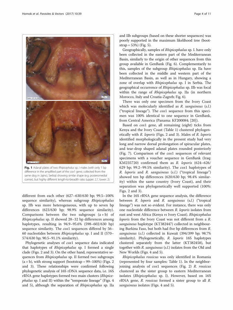
In the category of R. sanguineus the adanal plate length tobreadth ratio (which was reported to provide the only sig-nificant difference between Rhipicephalus sp. I-II: [1])showed extreme variation even between conspecific malescollected from the same dog (Fig. 1), therefore measure-ments were not taken. Identification of R. leporis maleswas based on the adanal and spiracular plates [16].Pictures were made with a VHX-5000 (Keyence Co.,Osaka, Japan) digital microscope.DNA was extracted from ticks using the QIAamp
DNA Mini Kit (Qiagen, Hilden, Germany), according tothe manufacturer’s instruction, and including an over-night digestion in tissue lysis buffer with proteinase K at56 °C. The cytochrome c oxidase subunit 1 (cox1) genewas chosen as the first target for molecular analysis,because of its suitability as a DNA-barcode marker fortick species identification [17]. PCR was modified fromFolmer et al. [18] and amplified approximately 710 bpusing the primers HCO2198 (5′-TAA ACT TCA GGGTGA CCA AAA AAT CA-3′) and LCO1490 (5′-GGTCAA CAA ATC ATA AAG ATA TTG G-3′) in a reac-tion volume of 25 μl, which contained 1 U (0.2 μl)HotStarTaq Plus DNA polymerase, 2.5 μl 10× CoralLoadReaction buffer (including 15 mM MgCl2), 0.5 μl PCRnucleotide Mix (0.2 mM each), 0.5 μl (1 μM finalconcentration) of each primer, 15.8 μl ddH2O and 5 μltemplate DNA. For amplification, an initial denaturationstep at 95 °C for 5 min was followed by 40 cycles ofdenaturation at 94 °C for 40 s, annealing at 48 °C for1 min and extension at 72 °C for 1 min. Final extensionwas performed at 72 °C for 10 min.To confirm the results obtained with cox1, another PCR
was used to amplify approximately 460 bp of 16S rDNAof Ixodidae [19], using the primers 16S + 1 (5′-CTG CTCAAT GAT TTT TTA AAT TGC TGT GG-3′) and 16S-1(5′-CCG GTC TGA ACT CAG ATC AAG T-3′). Otherreaction components, as well as cycling conditionswere the same as above, except for an annealingtemperature of 51 °C.PCR products were visualized in a 1.5% agarose gel.
Purification and sequencing was done by Biomi Inc.(Gödöllő, Hungary). The sequences were submitted to theGenBank database under accession numbers KX757879–KX757917 (cox1) and KX793717–KX793746 (16S) (seeTable 1). The MEGA model selection method was appliedto choose the appropriate model for phylogeneticanalyses. Phylogenetic analyses were conducted usingthe neighbour-joining method (p-distance model) andmaximum likelihood method (Jukes-Cantor model)using MEGA version 6.0.
ResultsOut of the 50 molecularly analysed ticks, 38 were identi-fied as R. sanguineus based on the amplified parts of
Hornok et al. Parasites & Vectors (2017) 10:39 Page 2 of 11

their cox1 and 16S rRNA genes (i.e. corresponding togroups Rhipicephalus sp. I and Rhipicephalus sp. II ofthe “temperate lineage”, and R. sanguineus (s.l.) “tropicallineage”).
The cox1 sequences from Rhipicephalus sp. I had oneto three nucleotides different among them (627–630/630 bp; 99.5–100% sequence similarity). In the subgroupRhipicephalus sp. IIa, one to three nucleotides were
Table 1 Data of Rhipicephalus spp. ticks used in this study. The sex/stage of ticks and date of collection are not shown
Speciesa Country Location Host of origin cox1 sequenceidentityb
cox1 sequenceaccession number
Number of identicalsequences
16S sequenceaccession number
“R. sanguineus” Serbia Kajtasovo dog 630/6302 KX757879 KX793717
Brnjica dog 630/6301 KX757880 KX793718
Jajinci dog 629/6301 KX757881 KX793719
630/6301 KX757882 KX793719
Petnica dog 629/6302 KX757883 KX793720
Lebane dog 629/6301 KX757885 KX793722
Boka dog 628/6301 KX757906 KX793738
629/6301 KX757907 KX793738
629/6302 KX757905 KX793739
Croatia Rovinj unknown 630/6302 KX757887 2 KX793724
Pula dog 629/6302 KX757888 KX793725
630/6302 KX757889 KX793726
Zagreb dog 629/6302 KX757890 1 KX793727
630/6302 KX757896 2 KX793727
628/6303 KX757893 2 KX793730
Zadar dog 629/6302 KX757892 2 KX793729
Sibenik dog 629/6302 KX757895 KX793731
Romania Babadag golden jackal 630/6301 KX757915 KX793746
Histria dog 628/6301 KX757916 KX793746
Hungary Szekszárdc dog 628/6302 KX757901 2 KX793734
Malta Siggiewi dog 630/6302 KX757902 6 KX793735
Italy Piacenza dog 630/6302 KX757904 KX793737
626/6303 KX757903 KX793736
Greece Thessaloniki dog 629/6301 KX757908 2 KX793740
Algeria Kehf Lagareb bat (Myotis punicus) 630/6302 KX757910 KX793742
Morocco Al-Hoceima dog 623/6303 KX757909 KX793741
Ivory Coast Abidjan dog 620/6204 KX757914 KX793745
R. rossicus Romania Caraorman dog 624/6305 KX757897 KX793732
Lazuri dog 628/6305 KX757898 KX793733
630/6305 KX757899 KX793733
Grindul dog 629/6305 KX757900 –
R. leporis Kenya Turkana, Samburu dog, cattle 627/6306 KX757911 4 KX793743
Ivory Coast Bas-Sassandra dog 627/6306 KX757912 1 KX793744
625/6306 KX757913 1 KX793744
625/6306 KX757917 2 KX793744aCurrently species delineation within R. sanguineus (s.l.) requires revision [1]: here species names designating reference sequences in GenBank are usedbNumber of nucleotides identical with reference sequence expressed as bp/bp. Superscript numbers indicate reference sequences (also shown on Figs. 2 and 3)as follows: 1KF219745; 2KU556745; 3AF081829; 4KF200084; 5JX394215; 6KM235720cR. sanguineus is not regarded as indigenous to Hungary; these specimens (collected in 2012 from a dog that has never left the country) exemplify rareautochthonous cases
Hornok et al. Parasites & Vectors (2017) 10:39 Page 3 of 11
different from each other (627–630/630 bp; 99.5–100%sequence similarity), whereas subgroup Rhipicephalussp. IIb was more heterogeneous, with up to seven bpdifferences (623/630 bp; 98.9% sequence similarity).Comparisons between the two subgroups (a + b) ofRhipicephalus sp. II showed 28–32 bp differences amonghaplotypes, resulting in 94.9–95.6% (598–602/630 bp)sequence similarity. The cox1 sequences differed by 56–60 nucleotides between Rhipicephalus sp. I and II (570–574/630 bp; 90.5–91.1% similarity).Phylogenetic analyses of cox1 sequence data indicated
that haplotypes of Rhipicephalus sp. I formed a singleclade (Figs. 2 and 3). On the other hand, representative se-quences from Rhipicephalus sp. II formed two subgroups(a + b), with strong support (bootstrap = 99–100%) (Figs. 2and 3). These relationships were confirmed followingphylogenetic analysis of 16S rDNA sequence data, i.e. 16SrRNA gene haplotypes formed two main clusters (Rhipice-phalus sp. I and II) within the “temperate lineage” (Figs. 4and 5), although the separation of Rhipicephalus sp. IIa
and IIb subgroups (based on these shorter sequences) waspoorly supported in the maximum likelihood tree (boot-strap = 53%) (Fig. 5).Geographically, samples of Rhipicephalus sp. I. have only
been collected in the eastern part of the MediterraneanBasin, similarly to the origin of other sequences from thisgroup available in GenBank (Fig. 6). Complementarily tothis, samples of the subgroup Rhipicephalus sp. IIa havebeen collected in the middle and western part of theMediterranean Basin, as well as in Hungary, showing azone of overlap with Rhipicephalus sp. I in Serbia. Thegeographical occurrence of Rhipicephalus sp. IIb was focalwithin the range of Rhipicephalus sp. IIa (in northernMorocco, Italy and Croatia-Zagreb; Fig. 6).There was only one specimen from the Ivory Coast
which was molecularly identified as R. sanguineus (s.l.)(“tropical lineage”). The cox1 sequence from this speci-men was 100% identical to one sequence in GenBank,from Central America (Panama: KF200084; [20]).Based on cox1 gene, all remaining (eight) ticks from
Kenya and the Ivory Coast (Table 1) clustered phylogen-etically with R. leporis (Figs. 2 and 3). Males of R. leporisidentified morphologically in the present study had verylong and narrow dorsal prolongation of spiracular plates,and tear-drop shaped adanal plates rounded posteriorly(Fig. 7). Comparison of the cox1 sequences of relevantspecimens with a voucher sequence in GenBank (Iraq:KM235720) confirmed them as R. leporis (624–626/629 bp; 99.2–99.5% similarity). The cox1 haplotypes ofR. leporis and R. sanguineus (s.l.) (“tropical lineage”)showed ten bp differences (620/630 bp; 98.4% similar-ity) within the same country (Ivory Coast), and theirseparation was phylogenetically well supported (100%:Figs. 2 and 3).In the 16S rRNA gene sequence analysis, the difference
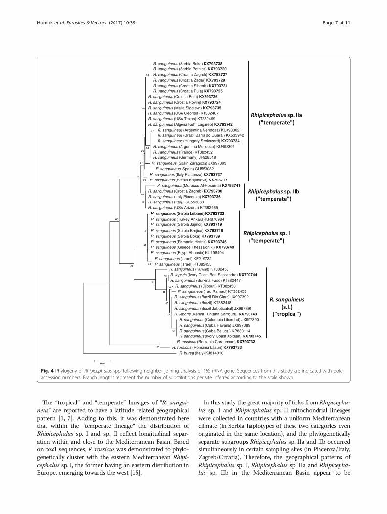
between R. leporis and R. sanguineus (s.l.) (“tropicallineage”) was not so evident. For instance, there was onlyone nucleotide difference between R. leporis isolates fromeast and west Africa (Kenya vs Ivory Coast). Rhipicephalusleporis from the Ivory Coast was not different from a R.sanguineus haplotype (KT382447) collected in neighbour-ing Burkina Faso, but both had five bp differences from R.sanguineus (s.l.) collected in Kuwait (394/399 bp; 98.7%similarity). Phylogenetically, R. leporis 16S haplotypesclustered separately from the latter (KT382458), buttogether with R. sanguineus (s.l.) isolates from the Old andNew Worlds (Figs. 4 and 5).Rhipicephalus rossicus was only identified in Romania
(represented by four samples: Table 1). In the neighbor-joining analysis of cox1 sequences (Fig. 2) R. rossicusclustered as the sister group to eastern Mediterraneanisolates (Rhipicephalus sp. I). However, based on 16SrRNA gene, R. rossicus formed a sister group to all R.sanguineus isolates (Figs. 4 and 5).
Fig. 1 Adanal plates of two Rhipicephalus sp. I males (with only 1 bpdifference in the amplified part of the cox1 gene; collected from thesame dog in Jajinci, Serbia) showing similar shape (e.g. posteromedialcorner), but highly different length-to-breadth ratio (upper: 2.7, lower: 2)
Hornok et al. Parasites & Vectors (2017) 10:39 Page 4 of 11
DiscussionIn a previous comprehensive study on the morphologicaland genetic diversity of R. sanguineus [1] it was shown thattwo groups under the “temperate lineage” (i.e. Rhipicepha-lus sp. I and Rhipicephalus sp. II) diverge molecularly tothe point where they may be considered separate species.However, except for the adanal plate length-to-breadthratio, no consistent morphological differences were ob-served between them [1]. Furthermore, in the present
study the adanal plate length-to-breadth ratio was shownto vary even between almost identical haplotypes of Rhipi-cephalus sp. I. Therefore, for the investigation of theirgeographical distribution, haplotypes were here assignedto specimens of R. sanguineus based on molecular data, in-volving the analysis of two mitochondrial genetic markers.However, it also should be considered that analysis of rela-tively short sequences may cause an over-resolution ofphylogenetic trees based on mitochondrial markers.
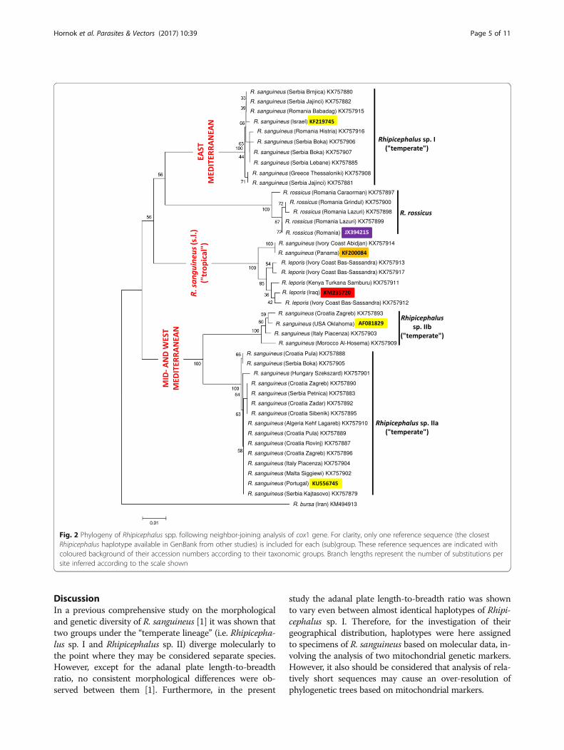
Fig. 2 Phylogeny of Rhipicephalus spp. following neighbor-joining analysis of cox1 gene. For clarity, only one reference sequence (the closestRhipicephalus haplotype available in GenBank from other studies) is included for each (sub)group. These reference sequences are indicated withcoloured background of their accession numbers according to their taxonomic groups. Branch lengths represent the number of substitutions persite inferred according to the scale shown
Hornok et al. Parasites & Vectors (2017) 10:39 Page 5 of 11

Phylogenetic analysis of cox1 sequences from new isolatesof “R. sanguineus” in the present study confirmed the exist-ence of paraphyletic groups previously referred to underthe same species name [1]. Here it was also shown that incontrast to Rhipicephalus sp. I (which is a homogenousgroup), Rhipicephalus sp. II is rather heterogenous, consist-ing of two, phylogenetically well-defined clades (a + b).Phylogenetic analysis of 16S rDNA sequence data verified
that these categories are also represented by formerlyreported sequences from the New World: the clade com-posed of sequences classified as Rhipicephalus sp. IIa, basedon phylogenetic analysis, included samples from Argentina,southernmost Brazil and the USA (Georgia, Texas), whileanother 16S rDNA sequence from the USA (Arizona) (aswell as a cox1 sequence from Oklahoma) belonged toRhipicephalus sp. IIb (Figs. 2, 3, 4 and 5).
Fig. 3 Phylogeny of Rhipicephalus spp. following maximum likelihood analysis of cox1 gene. For clarity, only one reference sequence (the closestRhipicephalus haplotype available in GenBank from other studies) is included for each (sub)group. These reference sequences are indicated withcoloured background of their accession numbers according to their taxonomic groups. Branch lengths represent the number of substitutions persite inferred according to the scale shown
Hornok et al. Parasites & Vectors (2017) 10:39 Page 6 of 11
The “tropical” and “temperate” lineages of “R. sangui-neus” are reported to have a latitude related geographicalpattern [1, 7]. Adding to this, it was demonstrated herethat within the “temperate lineage” the distribution ofRhipicephalus sp. I and sp. II reflect longitudinal separ-ation within and close to the Mediterranean Basin. Basedon cox1 sequences, R. rossicus was demonstrated to phylo-genetically cluster with the eastern Mediterranean Rhipi-cephalus sp. I, the former having an eastern distribution inEurope, emerging towards the west [15].
In this study the great majority of ticks from Rhipicepha-lus sp. I and Rhipicephalus sp. II mitochondrial lineageswere collected in countries with a uniform Mediterraneanclimate (in Serbia haplotypes of these two categories evenoriginated in the same location), and the phylogeneticallyseparate subgroups Rhipicephalus sp. IIa and IIb occurredsimultaneously in certain sampling sites (in Piacenza/Italy,Zagreb/Croatia). Therefore, the geographical patterns ofRhipicephalus sp. I, Rhipicephalus sp. IIa and Rhipicepha-lus sp. IIb in the Mediterranean Basin appear to be
Fig. 4 Phylogeny of Rhipicephalus spp. following neighbor-joining analysis of 16S rRNA gene. Sequences from this study are indicated with boldaccession numbers. Branch lengths represent the number of substitutions per site inferred according to the scale shown
Hornok et al. Parasites & Vectors (2017) 10:39 Page 7 of 11

independent of current climatic conditions (unlike thegeographical distribution of the “tropical” and “temperate”lineages of R. sanguineus: [12]).If not current climatic conditions, then other factors
influencing the tick life-cycle may provide a plausibleexplanation for the parapatric separation of Rhipicephalussp. I and Rhipicephalus sp. II lineages in the MediterraneanBasin. Molecular evidence from a broad range of inverte-brate and vertebrate taxa (i.e. potentially encompassingticks and their hosts) indicate that southern peninsulas of
Europe acted as major refugia during ice age(s), fromwhich genetically distinct clades emerged [21]. Whilerecolonization events to northern parts of Europe mayhave resulted in secondary sympatry for these clades, theirgenetic differences are still maintained and demonstrable.Thus, several (potential) host species of R. sanguineus hadalso been affected by glacial isolation in the same way. Forexample, wolf haplotype lineages and hedgehog speciesdiffer between Italy and the Balkans [21, 22], and genetic-ally distinct populations of bank voles exist in the western
Fig. 5 Phylogeny of Rhipicephalus spp. following maximum likelihood analysis of 16S rRNA gene. Sequences from this study are indicated withbold accession numbers. Branch lengths represent the number of substitutions per site inferred according to the scale shown
Hornok et al. Parasites & Vectors (2017) 10:39 Page 8 of 11

and eastern Balkans [23]. These geographical patterns aresimilar to the one observed for Rhipicephalus sp. I andRhipicephalus sp. II in the present study, suggesting thatduring ice age(s) the Mediterranean range of R. sanguineus(in sympatry with the above hosts) was not confluent,but inhabited by reproductively isolated tick popula-tions. Nevertheless, successful interbreeding betweenticks from Rhipicephalus sp. I and Rhipicephalus sp. IIbpopulations (listed as reference sequences from Israeland USA, Oklahoma on Figs. 2 and 3) had already beendemonstrated [10].Rhipicephalus leporis was hitherto known to occur in
the Middle East and Central Asia [16], but here its spec-imens (identified both morphologically and genetically)are reported from Africa. Apart from a broad range ofwild animals, dogs and goats are among the preferredhosts of this tick species [16]. Consequently, R. leporiscould have been unknowingly transported on these hoststo regions outside its formerly known range, and notnecessarily recently (considering the genetic divergencebetween its isolates from Iraq vs Kenya and the IvoryCoast). If confirmed, a likely explanation for R. leporisnot being discovered in Africa until now is its morpho-logical similarity to R. sanguineus (s.l.).
In the present study R. leporis and R. sanguineus (s.l.)(“tropical lineage”) clustered close to each other phylo-genetically, with their cox1 sequences differing by 1.6%.This sequence divergence is within the range (i.e. 0.2–3%) of reported intraspecific nucleotide variation for thecox1 gene of R. sanguineus (s.l.) [1]. In addition, the cox1sequence/phylogenetic difference between R. leporis andR. sanguineus (s.l.) (“tropical lineage”) was not reprodu-cible with the analysis of 16S rRNA gene. When com-paring 16S rDNA sequences of Rhipicephalus spp. itshould be taken into account that the amplified part ofthe 16S rRNA gene was considerably shorter than cox1,and the average interspecific distance was reported to belower for this gene than for either cox1 or 12S genes[17]. Therefore, the resolution of analysing these shorter16S gene fragments may not suffice to distinguishclosely related species. In addition, the sequence diver-gence between 12S gene sequences of R. leporis and R.sanguineus (s.l.) (“tropical lineage”) was reported to be ofsimilar magnitude than between isolates of R. leporis orR. sanguineus (s.l.) themselves. For instance, sequencesof the 12S gene of ticks from Kuwait, morphologicallyidentified as R. leporis, were 99% similar to sequences ofR. leporis from Iraq and R. sanguineus (s.l.) from South
Fig. 6 Geographical distribution of Rhipicephalus sanguineus cox1 haplotypes in and near the Mediterranean Basin. Coloured circles withoutaccession number indicate samples from this study (yellow, Rhipicephalus sp. I; red, Rhipicephalus sp. IIa; purple, Rhipicephalus sp. IIb). Among these,multiple sequences for the same location are not shown. Haplotypes from other studies (which had 99–100% similarity with those in this study)are marked with GenBank accession numbers, connected with dash line arrows to the relevant location. Overlapping circles indicate the samelocation (where different haplotypes were found); the zigzag arrow marks the direction of location (outside this map) for the sample from Iran(Tehran). The location within a country is accurately indicated for samples of this study, as well as for Egypt, Italy and Romania from other studies,but for the rest of the samples only the country was known
Hornok et al. Parasites & Vectors (2017) 10:39 Page 9 of 11

America [12]. Based on this ambiguity, further andlarger scale studies may be needed to ultimately verifythat R. leporis from east and west Africa are not intra-specific morphological variants of R. sanguineus (s.l.)within the “tropical lineage”.
ConclusionsTwo mitochondrial lineages within the “temperate spe-cies” of R. sanguineus (i.e. Rhipicephalus sp. I and Rhipice-phalus sp. II) show different geographical distribution inthe region of Mediterranean Basin, confirming limitedgene flow between them. However, more evidence (ana-lyses of nuclear markers, extensive morphological andbiological comparison etc.) are necessary to infer if theybelong to different species or not. Similarly, the phylogen-etic relationships of eastern and western African ticks,which align with R. leporis, need to be studied furtherwithin R. sanguineus (s.l.) (“tropical species”).
AcknowledgementsThe survey was organized in the framework of the EurNegVec COST ActionTD1303. The authors thank Elias Papadopoulos for providing ticks from Greece.
FundingMolecular analyses were funded by OTKA 115854 (Hungary). Tick collection wassupported by grants OI173006 (Serbia), PN-II-RU-TE-2014-4-1389 (Romania),GENOTICKTRECK-1957 (Croatia). GF receives János Bolyai Research Scholarshipof the Hungarian Academy of Sciences. The publication of this research wassupported by the 11475-4/2016/FEKUT grant of the Hungarian Ministry of Hu-man Resources.
Availability of data and materialsThe sequences obtained and/or analysed during the current study are depositedin GenBank under accession numbers KX757879–KX757917 (cox1) and KX793717–KX793746 (16S). All other relevant data are included in the manuscript.
Authors’ contributionsSH initiated and supervised the study, extracted the DNA, did part of themorphological and genetic comparisons, wrote the manuscript. ADS, ST, RB, JDAand RF provided samples from multiple locations for the study. JK made themicroscopic pictures and performed phylogenetic analyses. NT performed thePCRs. TG collected the important sample from Hungary, and MLB those fromAlgeria. GF organized part of the sample collection. All authors contributed to,read and approved the final manuscript.
Competing interestsThe authors declare that they have no competing interests.
Consent for publicationNot applicable.
Ethics approval and consent to participateTicks were collected from animals during regular veterinary care, thereforeno permission was needed.
Author details1Department of Parasitology and Zoology, University of Veterinary Medicine,Budapest, Hungary. 2Department of Parasitology and Parasitic Diseases, Universityof Agricultural Sciences and Veterinary Medicine, Cluj-Napoca, Romania.3Laboratory for Medical Entomology, Centre of Excellence for Food andVector-Borne Zoonoses, Institute for Medical Research, University of Belgrade,Belgrade, Serbia. 4Laboratory for Parasitology, Croatian Veterinary Institute, Zagreb,Croatia. 5Plant Protection Institute, Centre for Agricultural Research, HungarianAcademy of Sciences, Budapest, Hungary. 6Department of Zoology, HungarianNatural History Museum, Budapest, Hungary. 7Ecology of Terrestrial and AquaticSystems (EcoSTAq), University of Badji Mokhtar, Annaba, Algeria.
Fig. 7 Diagnostically important structures of R. sanguineus and R. leporis males. a Left spiracular plate of Rhipicephalus sp. I collected in Histria,Romania. b Adanal plates of Rhipicephalus sp. II collected in Pula, Croatia. c Long and narrow dorsal prolongation of spiracular plates in R. leporis.d Tear-drop shaped, posteriorly rounded adanal plates in R. leporis
Hornok et al. Parasites & Vectors (2017) 10:39 Page 10 of 11

Received: 9 September 2016 Accepted: 12 January 2017
References1. Dantas-Torres F, Latrofa MS, Annoscia G, Giannelli A, Parisi A, Otranto D.
Morphological and genetic diversity of Rhipicephalus sanguineus sensulatofrom the New and Old Worlds. Parasit Vectors. 2013;6:213.
2. Dantas-Torres F. Biology and ecology of the brown dog tick, Rhipicephalussanguineus. Parasit Vectors. 2010;3:26.
3. Dantas-Torres F. The brown dog tick, Rhipicephalus sanguineus (Latreille,1806) (Acari: Ixodidae): from taxonomy to control. Vet Parasitol. 2008;152:173–85.
4. Szabó MP, Mangold AJ, João CF, Bechara GH, Guglielmone AA. Biologicaland DNA evidence of two dissimilar populations of the Rhipicephalussanguineus tick group (Acari: Ixodidae) in South America. Vet Parasitol. 2005;130:131–40.
5. Oliveira PR, Bechara GH, Denardi SE, Saito KC, Nunes ET, Szabó MP, MathiasMI. Comparison of the external morphology of Rhipicephalus sanguineus(Latreille, 1806) (Acari: Ixodidae) ticks from Brazil and Argentina. VetParasitol. 2005;129:139–47.
6. Eremeeva ME, Zambrano ML, Anaya L, Beati L, Karpathy SE, Santos-Silva MM,et al. Rickettsia rickettsii in Rhipicephalus ticks, Mexicali, Mexico. J MedEntomol. 2011;48:418–21.
7. Burlini L, Teixeira KR, Szabó MP, Famadas KM. Molecular dissimilarities ofRhipicephalus sanguineus (Acari: Ixodidae) in Brazil and its relation withsamples throughout the world: is there a geographical pattern? Exp ApplAcarol. 2010;50:361–74.
8. Moraes-Filho J, Marcili A, Nieri-Bastos FA, Richtzenhain LJ, Labruna MB.Genetic analysis of ticks belonging to the Rhipicephalus sanguineus group inLatin America. Acta Trop. 2011;117:51–5.
9. Liu GH, Chen F, Chen YZ, Song HQ, Lin RQ, Zhou DH, Zhu XQ. Completemitochondrial genome sequence data provides genetic evidence that thebrown dog tick Rhipicephalus sanguineus (Acari: Ixodidae) represents aspecies complex. Int J Biol Sci. 2007;9:361–9.
10. Levin ML, Studer E, Killmaster L, Zemtsova G, Mumcuoglu KY. Crossbreedingbetween different geographical populations of the brown dog tick,Rhipicephalus sanguineus (Acari: Ixodidae). Exp Appl Acarol. 2012;58:51–68.
11. Nava S, Mastropaolo M, Venzal JM, Mangold AJ, Guglielmone AA.Mitochondrial DNA analysis of Rhipicephalus sanguineus sensu lato (Acari:Ixodidae) in the Southern Cone of South America. Vet Parasitol. 2012;190:547–55.
12. Zemtsova GE, Apanaskevich DA, Reeves WK, Hahn M, Snellgrove A, LevinML. Phylogeography of Rhipicephalus sanguineus sensu lato and itsrelationships with climatic factors. Exp Appl Acarol. 2016;69:191–203.
13. Hornok S, Farkas R. [First autochthonous infestation of dogs withRhipicephalus sanguineus (Acari: Ixodidae) in Hungary: case report andreview of current knowledge on this tick species.] Magyar Állatorvos. 2005;127:623–9 (In Hungarian with English abstract).
14. Estrada-Peña A, Bouattour A, Camicas J-L, Walker AR. Ticks of domesticanimals in the Mediterranean region: a guide to identification of species.Zaragoza: University of Zaragoza Publishing House; 2004.
15. Mihalca AD, Kalmár Z, Dumitrache MO. Rhipicephalus rossicus, a neglectedtick at the margin of Europe: a review of its distribution, ecology andmedical importance. Med Vet Entomol. 2015;29:215–24.
16. Walker JB, Keirans JE, Horak IG. The genus Rhipicephalus (Acari, Ixodidae). Aguide to the brown ticks of the world. Cambridge: Cambridge UniversityPress; 2000.
17. Lv J, Wu S, Zhang Y, Chen Y, Feng C, Yuan X, et al. Assessment of four DNAfragments (COI, 16S rDNA, ITS2, 12S rDNA) for species identification of theIxodida (Acari: Ixodida). Parasit Vectors. 2014;7:93.
18. Folmer O, Black M, Hoeh W, Lutz R, Vrijenhoek R. DNA primers foramplification of mitochondrial cytochrome c oxidase subunit I from diversemetazoan invertebrates. Mel Marine Biol Biot. 1994;3:294–9.
19. Black WC, Piesman J. Phylogeny of hard and soft-tick taxa (Acari: Ixodida)based on mitochondrial 16S rDNA sequences. Proc Natl Acad Sci USA. 1994;91:10034–8.
20. Miller MJ, Esser HJ, Loaiza JR, Herre EA, Aguilar C, Quintero D, et al.Molecular ecological insights into Neotropical bird-tick interactions. PLoSOne. 2016;11:e0155989.
21. Hewitt GM. Post-glacial re-colonization of European biota. Biol J LinneanSoc. 1999;68:87–112.
22. Verginelli F, Capelli C, Coia V, Musiani M, Falchetti M, Ottini L, et al.Mitochondrial DNA from prehistoric canids highlights relationships betweendogs and South-East European wolves. Mol Biol Evol. 2005;22:2541–51.
23. Kotlík P, Deffontaine V, Mascheretti S, Zima J, Michaux JR, Searle JB. Anorthern glacial refugium for bank voles (Clethrionomys glareolus). Proc NatlAcad Sci USA. 2006;103:14860–4.
• We accept pre-submission inquiries
• Our selector tool helps you to find the most relevant journal
• We provide round the clock customer support
• Convenient online submission
• Thorough peer review
• Inclusion in PubMed and all major indexing services
• Maximum visibility for your research
Submit your manuscript atwww.biomedcentral.com/submit
Submit your next manuscript to BioMed Central and we will help you at every step:
Hornok et al. Parasites & Vectors (2017) 10:39 Page 11 of 11