ecg - twosaintspathway.com development/ecg - cci review... · ecg monitor does not provide...
TRANSCRIPT
ECG Interpretation Course
N.O. REVIEW & TEST DAY
HANDOUT

ECG CONVERSION
for HEART RATE
NUMBER OF SMALL
SPACES Rate/Minute
NUMBER OF SMALL
SPACES RATE/MINUTE
5 300 37 41
6 250 38 40
7 214 39 39
8 188 40 38
9 167 41 37
10 150 42 36
11 136 43 35
12 125 44 34
13 115 45 33
14 107 46 33
15 100 47 32
16 94 48 31
17 88 49 31
18 84 50 30
19 79 52 29
20 75 54 28
21 72 56 27
22 68 58 26
23 65 60 25
24 63 62 24
25 60 64 23
26 58 68 22
27 56 72 21
28 54 76 20
29 52 80 19
30 50 84 18
31 48 88 17
32 47 92 16
33 45 98 15
34 44 104 14
35 43 112 13
36 42
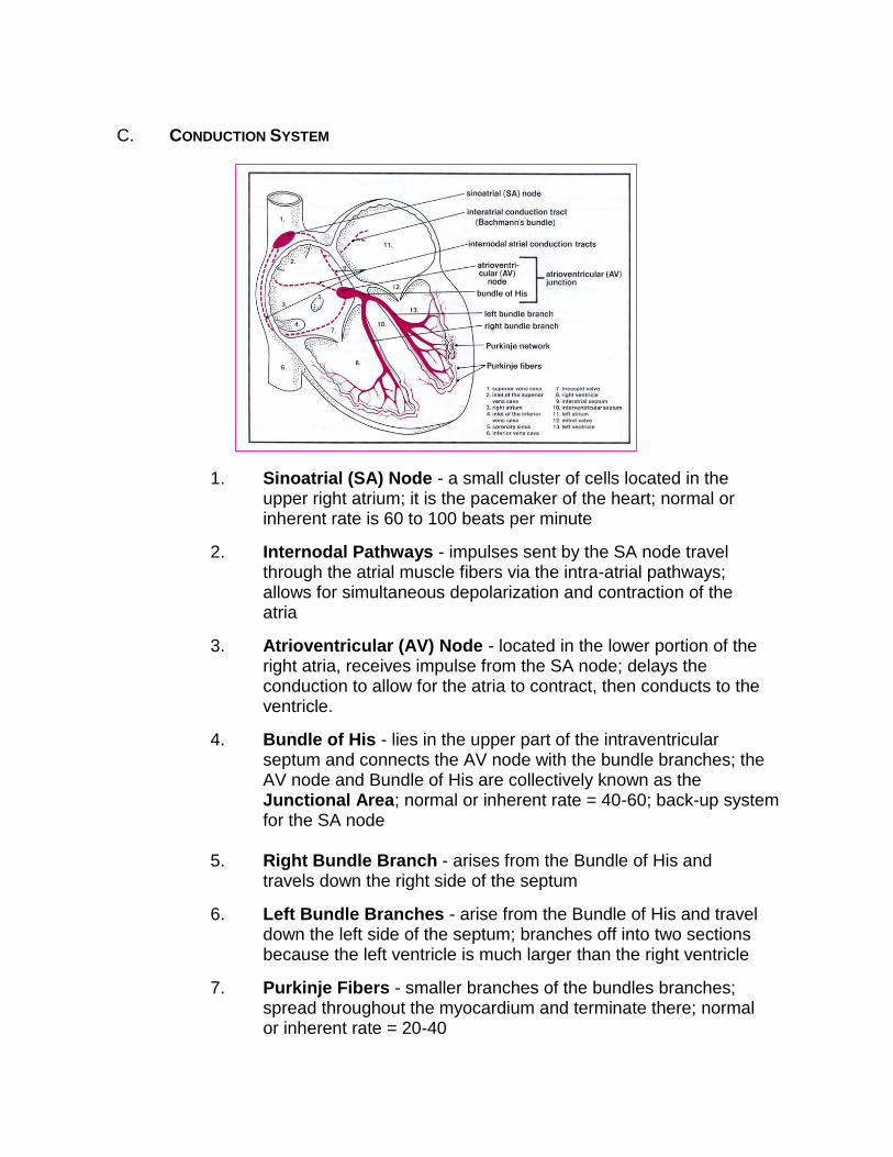
C. CONDUCTION SYSTEM 1. Sinoatrial (SA) Node - a small cluster of cells located in the
upper right atrium; it is the pacemaker of the heart; normal or inherent rate is 60 to 100 beats per minute
2. Internodal Pathways - impulses sent by the SA node travel through the atrial muscle fibers via the intra-atrial pathways; allows for simultaneous depolarization and contraction of the atria
3. Atrioventricular (AV) Node - located in the lower portion of the right atria, receives impulse from the SA node; delays the conduction to allow for the atria to contract, then conducts to the ventricle.
4. Bundle of His - lies in the upper part of the intraventricular septum and connects the AV node with the bundle branches; the AV node and Bundle of His are collectively known as the Junctional Area; normal or inherent rate = 40-60; back-up system for the SA node
5. Right Bundle Branch - arises from the Bundle of His and travels down the right side of the septum
6. Left Bundle Branches - arise from the Bundle of His and travel down the left side of the septum; branches off into two sections because the left ventricle is much larger than the right ventricle
7. Purkinje Fibers - smaller branches of the bundles branches; spread throughout the myocardium and terminate there; normal or inherent rate = 20-40
CCI
ANATOMY AND PHYSIOLOGY
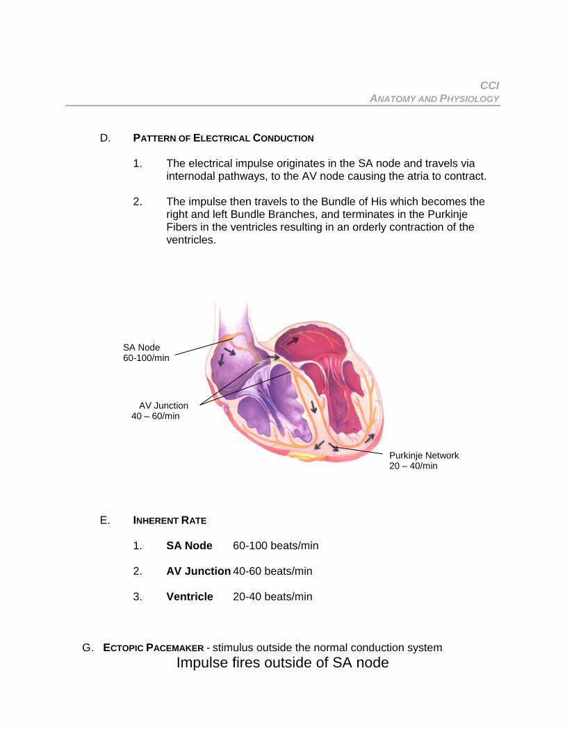
D. PATTERN OF ELECTRICAL CONDUCTION
1. The electrical impulse originates in the SA node and travels via internodal pathways, to the AV node causing the atria to contract.
2. The impulse then travels to the Bundle of His which becomes the
right and left Bundle Branches, and terminates in the Purkinje Fibers in the ventricles resulting in an orderly contraction of the ventricles.
E. INHERENT RATE 1. SA Node 60-100 beats/min 2. AV Junction 40-60 beats/min 3. Ventricle 20-40 beats/min
G. ECTOPIC PACEMAKER - stimulus outside the normal conduction system
Impulse fires outside of SA node
SA Node 60-100/min
AV Junction 40 – 60/min
Purkinje Network 20 – 40/min
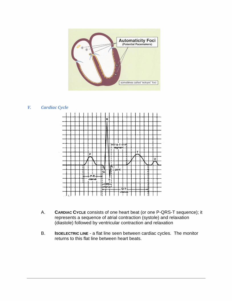
V. Cardiac Cycle
A. CARDIAC CYCLE consists of one heart beat (or one P-QRS-T sequence); it represents a sequence of atrial contraction (systole) and relaxation (diastole) followed by ventricular contraction and relaxation
B. ISOELECTRIC LINE - a flat line seen between cardiac cycles. The monitor
returns to this flat line between heart beats.

CCI ANATOMY AND PHYSIOLOGY
VI. COMPONENTS OF A CARDIAC ELECTRICAL CYCLE A. P WAVE - depolarization of the atria B. PR INTERVAL - time it takes for the electric impulse (which originates in
the SA node) to reach the ventricular conduction system; normal = 0.12 - 0.20 seconds; measured from the beginning of the P wave to the beginning of the QRS complex
C. QRS COMPLEX - depolarization of the ventricles; normal = 0.06 - 0.10
seconds D. ST SEGMENT - early repolarization of the ventricles; ST elevation or
depression (1 mm) indicates cardiac injury/ischemia; normal ST segment is flat (isolelectric)
E. J POINT - junction between QRS complex and ST segment F. T WAVE - ventricular repolarization G. QT INTERVAL - time between onset of ventricular depolarization and end
of ventricular repolarization; normal = varies with heart rate

CCI ANATOMY AND PHYSIOLOGY
1. QT varies with heart rate; at fast rates the QT shortens, at slow rates the QT
lengthens 2. As a rule, the QT Interval should not exceed half the R-R interval at normal heart
rates 3. Causes of lengthened QT interval:
Electrolyte imbalances, antidysrhymics, cocaine & congenital defects.
H. U WAVE - probably represents the final stage of ventricular
repolarization
I. REFRACTORY PERIODS
Absolute Refractory Period Relative Refractory Period
Relative Refractory Period
Period of time during the cardiac cycle when the ventricles are fully depolarized (fully contracted) and unable to respond to even a stronger than normal impulse.
Period of time during the cardiac cycle when the ventricles are repolarizing. If a stronger than normal impulse stimulates the ventricles during this period, the heart may enter into a lethal dysrhythmia (referred to as R of T phenomena) resulting in ventricular tachycardia or ventricular defibrillation.
C. INFORMATION OBTAINED FROM AN ECG
1. The ECG monitor provides information about electrical activity only. The
ECG monitor does not provide information about mechanical
(pumping/pulse) activity.
2. The two basic electrical activities of the heart seen on the ECG waveform
are:
a. Depolarization (P,QRS)
b. Repolarization (ST segment, T wave)
3. Analysis of an ECG tracing can supply information related to:
a. Conduction abnormalities
b. Electrolyte imbalances
c. Myocardial muscle injury/ischemia (ST segment elevation and
depression)
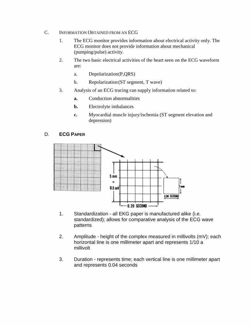
D. ECG PAPER
1. Standardization - all EKG paper is manufactured alike (i.e.
standardized); allows for comparative analysis of the ECG wave patterns
2. Amplitude - height of the complex measured in millivolts (mV); each
horizontal line is one millimeter apart and represents 1/10 a millivolt
3. Duration - represents time; each vertical line is one millimeter apart and represents 0.04 seconds
CCI
ANATOMY AND PHYSIOLOGY
4. Bold Lines - every fifth line, both horizontal and vertical is
inscribed boldly, producing a series of large squares; large squares represent 0.5 millivolts vertically and 0.20 seconds horizontally. Five (5) large blocks equals one second.
IX. INTERPRETATION OF RHYTHM STRIP (FIVE (5) STEPS)
A. REGULARITY - atrial and ventricular
B. RATE - atrial and ventricular; method of determining rate based on
regularity
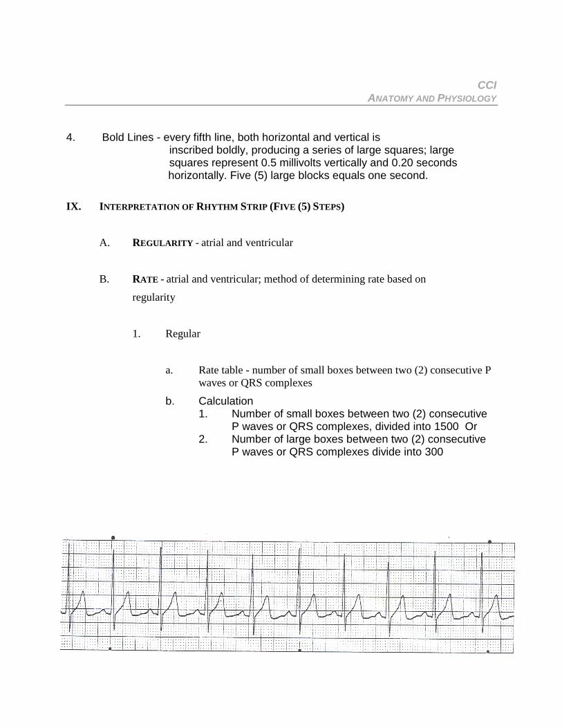
1. Regular
a. Rate table - number of small boxes between two (2) consecutive P
waves or QRS complexes
b. Calculation 1. Number of small boxes between two (2) consecutive
P waves or QRS complexes, divided into 1500 Or 2. Number of large boxes between two (2) consecutive
P waves or QRS complexes divide into 300
CCI
ANATOMY AND PHYSIOLOGY
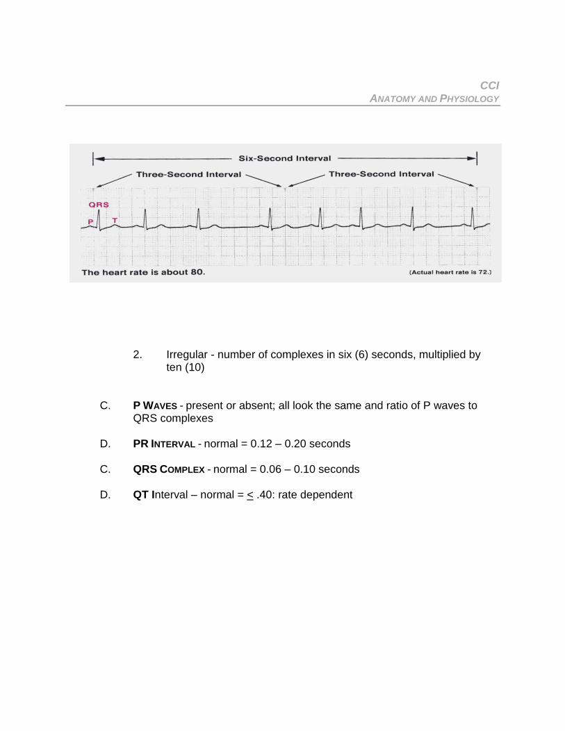
2. Irregular - number of complexes in six (6) seconds, multiplied by ten (10)
C. P WAVES - present or absent; all look the same and ratio of P waves to
QRS complexes D. PR INTERVAL - normal = 0.12 – 0.20 seconds
C. QRS COMPLEX - normal = 0.06 – 0.10 seconds
D. QT Interval – normal = < .40: rate dependent
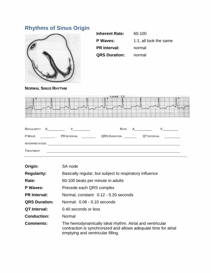
Rhythms of Sinus Origin Inherent Rate: 60-100
P Waves: 1:1, all look the same
PR Interval: normal
QRS Duration: normal
NORMAL SINUS RHYTHM
REGULARITY A__________ V__________ RATE A__________ V_________ P WAVE PR INTERVAL QRS DURATION QT INTERVAL
INTERPRETATION
TREATMENT
Origin: SA node
Regularity: Basically regular, but subject to respiratory influence
Rate: 60-100 beats per minute in adults
P Waves: Precede each QRS complex
PR Interval: Normal, constant: 0.12 - 0.20 seconds
QRS Duration: Normal: 0.06 - 0.10 seconds
QT Interval: 0.40 seconds or less
Conduction: Normal
Comments: The hemodynamically ideal rhythm. Atrial and ventricular contraction is synchronized and allows adequate time for atrial emptying and ventricular filling.
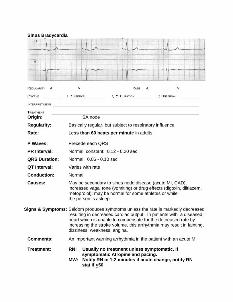
Sinus Bradycardia
REGULARITY A__________ V__________ RATE A__________ V_________ P WAVE PR INTERVAL QRS DURATION QT INTERVAL
INTERPRETATION
TREATMENT
Origin: SA node
Regularity: Basically regular, but subject to respiratory influence
Rate: Less than 60 beats per minute in adults
P Waves: Precede each QRS
PR Interval: Normal, constant: 0.12 - 0.20 sec
QRS Duration: Normal: 0.06 - 0.10 sec
QT Interval: Varies with rate
Conduction: Normal
Causes: May be secondary to sinus node disease (acute MI, CAD), increased vagal tone (vomiting) or drug effects (digoxin, diltiazem, metoprolol); may be normal for some athletes or while
the person is asleep
Signs & Symptoms: Seldom produces symptoms unless the rate is markedly decreased resulting in decreased cardiac output. In patients with a diseased heart which is unable to compensate for the decreased rate by increasing the stroke volume, this arrhythmia may result in fainting, dizziness, weakness, angina.
Comments: An important warning arrhythmia in the patient with an acute MI
Treatment: RN: Usually no treatment unless symptomatic. If symptomatic Atropine and pacing. MW: Notify RN in 1-2 minutes if acute change, notify RN stat if <50
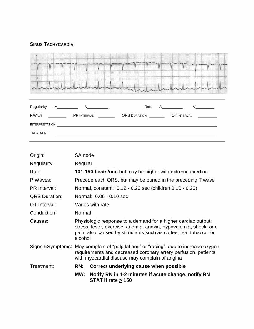
SINUS TACHYCARDIA
Regularity A__________ V__________ Rate A__________ V_________ P WAVE PR INTERVAL QRS DURATION QT INTERVAL
INTERPRETATION
TREATMENT
Origin: SA node
Regularity: Regular
Rate: 101-150 beats/min but may be higher with extreme exertion
P Waves: Precede each QRS, but may be buried in the preceding T wave
PR Interval: Normal, constant: 0.12 - 0.20 sec (children 0.10 - 0.20)
QRS Duration: Normal: 0.06 - 0.10 sec
QT Interval: Varies with rate
Conduction: Normal
Causes: Physiologic response to a demand for a higher cardiac output: stress, fever, exercise, anemia, anoxia, hypovolemia, shock, and pain; also caused by stimulants such as coffee, tea, tobacco, or alcohol
Signs &Symptoms: May complain of “palpitations” or “racing”; due to increase oxygen requirements and decreased coronary artery perfusion, patients with myocardial disease may complain of angina
Treatment: RN: Correct underlying cause when possible
MW: Notify RN in 1-2 minutes if acute change, notify RN STAT if rate > 150
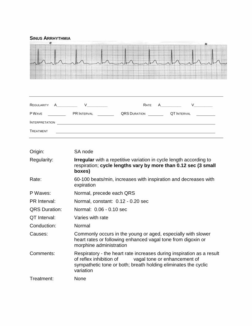
SINUS ARRHYTHMIA
REGULARITY A__________ V__________ RATE A__________ V_________ P WAVE PR INTERVAL QRS DURATION QT INTERVAL
INTERPRETATION
TREATMENT
Origin: SA node
Regularity: Irregular with a repetitive variation in cycle length according to respiration; cycle lengths vary by more than 0.12 sec (3 small boxes)
Rate: 60-100 beats/min, increases with inspiration and decreases with expiration
P Waves: Normal, precede each QRS
PR Interval: Normal, constant: 0.12 - 0.20 sec
QRS Duration: Normal: 0.06 - 0.10 sec
QT Interval: Varies with rate
Conduction: Normal
Causes: Commonly occurs in the young or aged, especially with slower heart rates or following enhanced vagal tone from digoxin or morphine administration
Comments: Respiratory - the heart rate increases during inspiration as a result of reflex inhibition of vagal tone or enhancement of sympathetic tone or both; breath holding eliminates the cyclic variation
Treatment: None
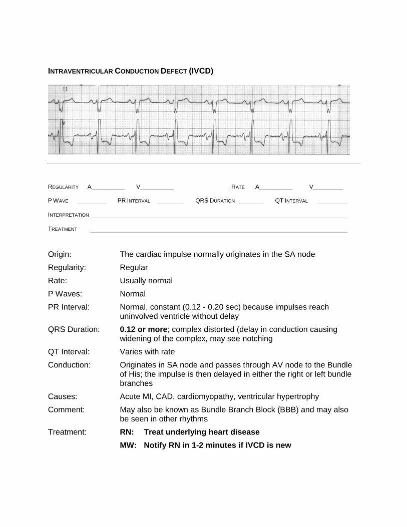
INTRAVENTRICULAR CONDUCTION DEFECT (IVCD)
REGULARITY A__________ V__________ RATE A__________ V_________ P WAVE PR INTERVAL QRS DURATION QT INTERVAL
INTERPRETATION
TREATMENT
Origin: The cardiac impulse normally originates in the SA node
Regularity: Regular
Rate: Usually normal
P Waves: Normal
PR Interval: Normal, constant (0.12 - 0.20 sec) because impulses reach uninvolved ventricle without delay
QRS Duration: 0.12 or more; complex distorted (delay in conduction causing widening of the complex, may see notching
QT Interval: Varies with rate
Conduction: Originates in SA node and passes through AV node to the Bundle of His; the impulse is then delayed in either the right or left bundle branches
Causes: Acute MI, CAD, cardiomyopathy, ventricular hypertrophy
Comment: May also be known as Bundle Branch Block (BBB) and may also be seen in other rhythms
Treatment: RN: Treat underlying heart disease
MW: Notify RN in 1-2 minutes if IVCD is new
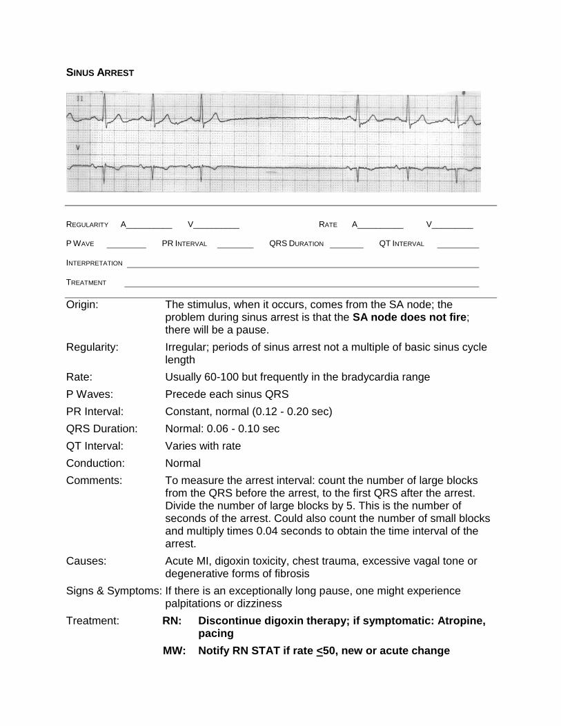
SINUS ARREST
REGULARITY A__________ V__________ RATE A__________ V_________ P WAVE PR INTERVAL QRS DURATION QT INTERVAL
INTERPRETATION
TREATMENT
Origin: The stimulus, when it occurs, comes from the SA node; the problem during sinus arrest is that the SA node does not fire; there will be a pause.
Regularity: Irregular; periods of sinus arrest not a multiple of basic sinus cycle length
Rate: Usually 60-100 but frequently in the bradycardia range
P Waves: Precede each sinus QRS
PR Interval: Constant, normal (0.12 - 0.20 sec)
QRS Duration: Normal: 0.06 - 0.10 sec
QT Interval: Varies with rate
Conduction: Normal
Comments: To measure the arrest interval: count the number of large blocks from the QRS before the arrest, to the first QRS after the arrest. Divide the number of large blocks by 5. This is the number of seconds of the arrest. Could also count the number of small blocks and multiply times 0.04 seconds to obtain the time interval of the arrest.
Causes: Acute MI, digoxin toxicity, chest trauma, excessive vagal tone or degenerative forms of fibrosis
Signs & Symptoms: If there is an exceptionally long pause, one might experience palpitations or dizziness
Treatment: RN: Discontinue digoxin therapy; if symptomatic: Atropine, pacing
MW: Notify RN STAT if rate <50, new or acute change
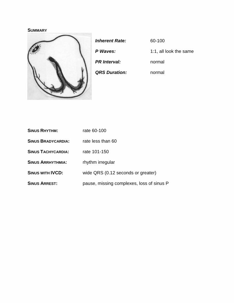
SUMMARY Inherent Rate: 60-100 P Waves: 1:1, all look the same PR Interval: normal QRS Duration: normal
SINUS RHYTHM: rate 60-100 SINUS BRADYCARDIA: rate less than 60 SINUS TACHYCARDIA: rate 101-150 SINUS ARRHYTHMIA: rhythm irregular SINUS WITH IVCD: wide QRS (0.12 seconds or greater) SINUS ARREST: pause, missing complexes, loss of sinus P
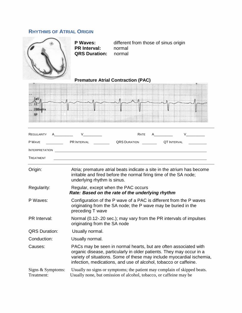
RHYTHMS OF ATRIAL ORIGIN P Waves: different from those of sinus origin PR Interval: normal QRS Duration: normal Premature Atrial Contraction (PAC)
REGULARITY A__________ V__________ RATE A__________ V__________ P WAVE PR INTERVAL QRS DURATION QT INTERVAL
INTERPRETATION
TREATMENT
Origin: Atria; premature atrial beats indicate a site in the atrium has become irritable and fired before the normal firing time of the SA node; underlying rhythm is sinus.
Regularity: Regular, except when the PAC occurs Rate: Based on the rate of the underlying rhythm
P Waves: Configuration of the P wave of a PAC is different from the P waves originating from the SA node; the P wave may be buried in the preceding T wave
PR Interval: Normal (0.12-.20 sec.); may vary from the PR intervals of impulses originating from the SA node
QRS Duration: Usually normal.
Conduction: Usually normal.
Causes: PACs may be seen in normal hearts, but are often associated with organic disease, particularly in older patients. They may occur in a variety of situations. Some of these may include myocardial ischemia, infection, medications, and use of alcohol, tobacco or caffeine.
Signs & Symptoms: Usually no signs or symptoms; the patient may complain of skipped beats. Treatment: Usually none, but omission of alcohol, tobacco, or caffeine may be
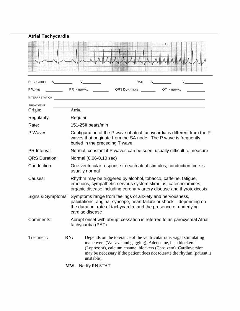
Atrial Tachycardia
REGULARITY A__________ V__________ RATE A__________ V__________ P WAVE PR INTERVAL QRS DURATION QT INTERVAL
INTERPRETATION
TREATMENT
Origin: Atria.
Regularity: Regular
Rate: 151-250 beats/min
P Waves: Configuration of the P wave of atrial tachycardia is different from the P waves that originate from the SA node. The P wave is frequently buried in the preceding T wave.
PR Interval: Normal, constant if P waves can be seen; usually difficult to measure
QRS Duration: Normal (0.06-0.10 sec)
Conduction: One ventricular response to each atrial stimulus; conduction time is usually normal
Causes: Rhythm may be triggered by alcohol, tobacco, caffeine, fatigue, emotions, sympathetic nervous system stimulus, catecholamines, organic disease including coronary artery disease and thyrotoxicosis
Signs & Symptoms: Symptoms range from feelings of anxiety and nervousness, palpitations, angina, syncope, heart failure or shock – depending on the duration, rate of tachycardia, and the presence of underlying cardiac disease
Comments: Abrupt onset with abrupt cessation is referred to as paroxysmal Atrial tachycardia (PAT)
Treatment: RN: Depends on the tolerance of the ventricular rate: vagal stimulating
maneuvers (Valsava and gagging), Adenosine, beta blockers
(Lopressor), calcium channel blockers (Cardizem). Cardioversion
may be necessary if the patient does not tolerate the rhythm (patient is
unstable).
MW: Notify RN STAT
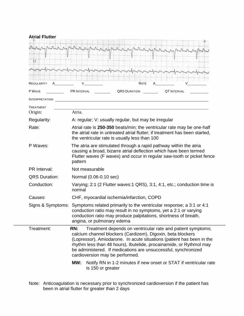
Atrial Flutter
REGULARITY A__________ V__________ RATE A__________ V__________ P WAVE PR INTERVAL QRS DURATION QT INTERVAL
INTERPRETATION
TREATMENT
Origin: Atria.
Regularity: A: regular; V: usually regular, but may be irregular
Rate: Atrial rate is 250-350 beats/min; the ventricular rate may be one-half the atrial rate in untreated atrial flutter; if treatment has been started, the ventricular rate is usually less than 100
P Waves: The atria are stimulated through a rapid pathway within the atria causing a broad, bizarre atrial deflection which have been termed Flutter waves (F waves) and occur in regular saw-tooth or picket fence pattern
PR Interval: Not measurable
QRS Duration: Normal (0.06-0.10 sec)
Conduction: Varying; 2:1 (2 Flutter waves:1 QRS), 3:1, 4:1, etc.; conduction time is normal
Causes: CHF, myocardial ischemia/infarction, COPD
Signs & Symptoms: Symptoms related primarily to the ventricular response; a 3:1 or 4:1 conduction ratio may result in no symptoms, yet a 2:1 or varying conduction ratio may produce palpitations, shortness of breath, angina, or pulmonary edema
Treatment: RN: Treatment depends on ventricular rate and patient symptoms; calcium channel blockers (Cardizem), Digoxin, beta blockers (Lopressor), Amiodarone. In acute situations (patient has been in the rhythm less than 48 hours), Ibutelide, procainamide, or Rythmol may be administered. If medications are unsuccessful, synchronized cardioversion may be performed.
MW: Notify RN in 1-2 minutes if new onset or STAT if ventricular rate is 150 or greater
Note: Anticoagulation is necessary prior to synchronized cardioversion if the patient has been in atrial flutter for greater than 2 days
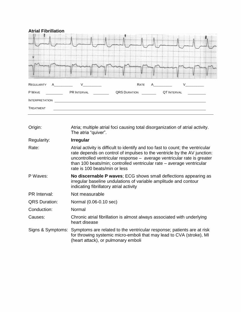
Atrial Fibrillation
REGULARITY A__________ V__________ RATE A__________ V__________ P WAVE PR INTERVAL QRS DURATION QT INTERVAL
INTERPRETATION
TREATMENT
Origin: Atria; multiple atrial foci causing total disorganization of atrial activity. The atria “quiver”.
Regularity: Irregular
Rate: Atrial activity is difficult to identify and too fast to count; the ventricular rate depends on control of impulses to the ventricle by the AV junction: uncontrolled ventricular response – average ventricular rate is greater than 100 beats/min; controlled ventricular rate – average ventricular rate is 100 beats/min or less
P Waves: No discernable P waves; ECG shows small deflections appearing as irregular baseline undulations of variable amplitude and contour indicating fibrillatory atrial activity
PR Interval: Not measurable
QRS Duration: Normal (0.06-0.10 sec)
Conduction: Normal
Causes: Chronic atrial fibrillation is almost always associated with underlying heart disease
Signs & Symptoms: Symptoms are related to the ventricular response; patients are at risk for throwing systemic micro-emboli that may lead to CVA (stroke), MI (heart attack), or pulmonary emboli

Treatment: RN: Treatment depends on ventricular rate, patient symptoms, underlying heart disease and duration of being in the rhythm. Calcium channel blockers (Cardizem), Digoxin, beta blockers (Lopressor), Amiodarone are frequently first line treatment. On occasion Quinidine or procainamide may be used. In acute situations (patient has been in the rhythm less than 48 hours*), Ibutelide, procainamide, or Rythmol may be administered. If medications are unsuccessful, synchronized cardioversion may be performed.
MW: Notify RN in 1-2 minutes if new onset or STAT if ventricular rate is 150 or greater
*Note: Anticoagulation is an intervention that is necessary prior to synchronized
cardioversion if the patient has been in atrial fibrillation for greater than 2 days
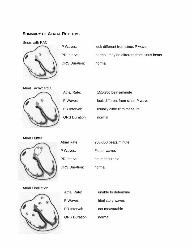
SUMMARY OF ATRIAL RHYTHMS Sinus with PAC
P Waves: look different from sinus P wave PR Interval: normal, may be different from sinus beats QRS Duration: normal
Atrial Tachycardia
Atrial Rate: 151-250 beats/minute P Waves: look different from sinus P wave PR Interval: usually difficult to measure QRS Duration: normal
Atrial Flutter
Atrial Rate: 250-350 beats/minute P Waves: Flutter waves PR Interval: not measurable QRS Duration: normal
Atrial Fibrillation
Atrial Rate: unable to determine P Waves: fibrillatory waves PR Interval: not measurable QRS Duration: normal
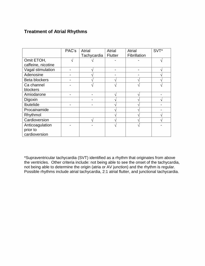
Treatment of Atrial Rhythms
PAC’s Atrial Tachycardia
Atrial Flutter
Atrial Fibrillation
SVT*
Omit ETOH, caffeine, nicotine
- -
Vagal stimulation - - -
Adenosine - - -
Beta blockers -
Ca channel blockers
-
Amiodarone - - -
Digoxin -
Ibutelide - - -
Procainamide -
Rhythmol
Cardioversion
Anticoagulation prior to cardioversion
- - -
*Supraventricular tachycardia (SVT) identified as a rhythm that originates from above the ventricles. Other criteria include: not being able to see the onset of the tachycardia, not being able to determine the origin (atria or AV junction) and the rhythm is regular. Possible rhythms include atrial tachycardia, 2:1 atrial flutter, and junctional tachycardia.
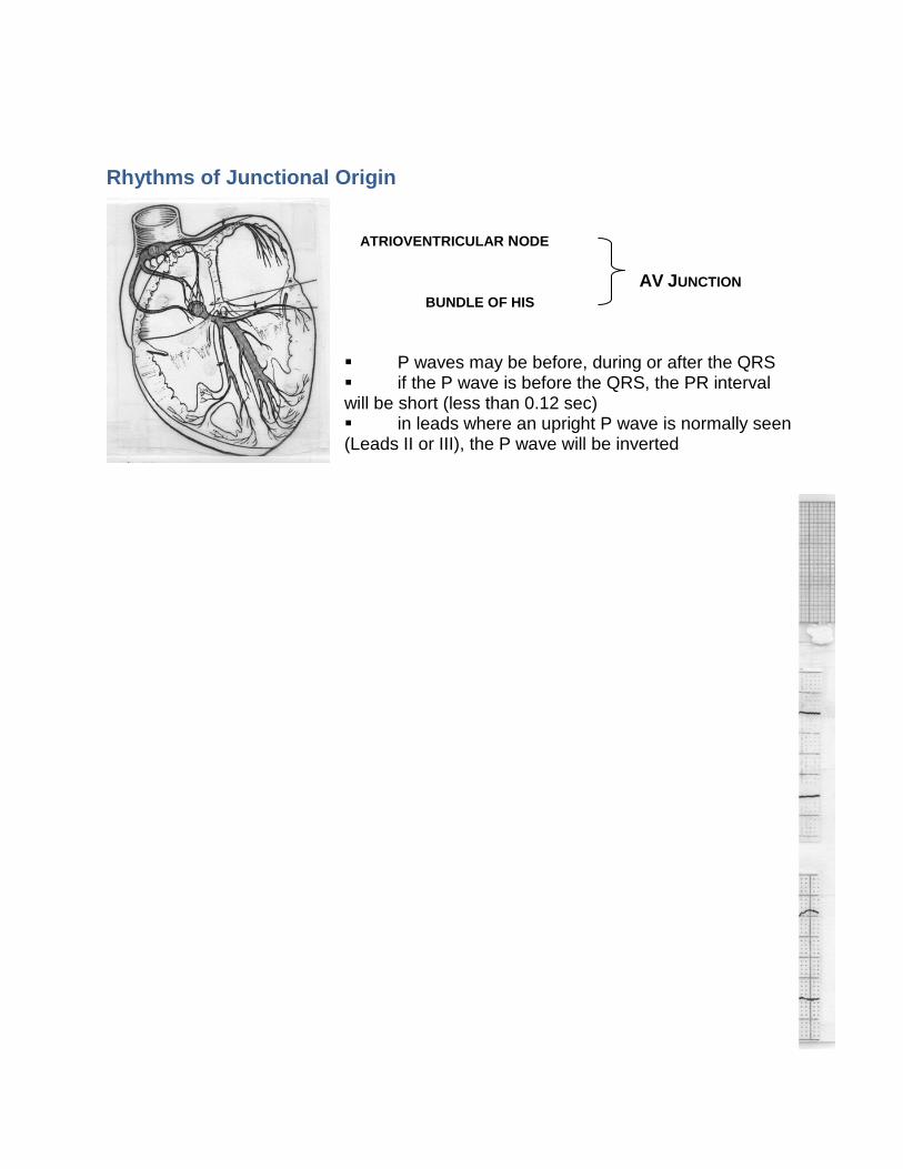
Rhythms of Junctional Origin ATRIOVENTRICULAR NODE
AV JUNCTION BUNDLE OF HIS
P waves may be before, during or after the QRS if the P wave is before the QRS, the PR interval will be short (less than 0.12 sec) in leads where an upright P wave is normally seen (Leads II or III), the P wave will be inverted
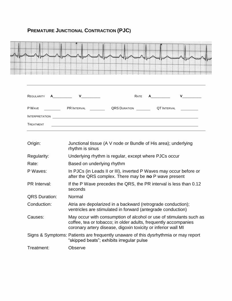
PREMATURE JUNCTIONAL CONTRACTION (PJC)
REGULARITY A__________ V__________ RATE A__________ V__________
P WAVE PR INTERVAL QRS DURATION QT INTERVAL
INTERPRETATION
TREATMENT
Origin: Junctional tissue (A V node or Bundle of His area); underlying rhythm is sinus
Regularity: Underlying rhythm is regular, except where PJCs occur
Rate: Based on underlying rhythm
P Waves: In PJCs (in Leads II or III), inverted P Waves may occur before or after the QRS complex. There may be no P wave present
PR Interval: If the P Wave precedes the QRS, the PR interval is less than 0.12 seconds
QRS Duration: Normal
Conduction: Atria are depolarized in a backward (retrograde conduction); ventricles are stimulated in forward (antegrade conduction)
Causes: May occur with consumption of alcohol or use of stimulants such as coffee, tea or tobacco; in older adults, frequently accompanies coronary artery disease, digoxin toxicity or inferior wall MI
Signs & Symptoms: Patients are frequently unaware of this dysrhythmia or may report “skipped beats”; exhibits irregular pulse
Treatment: Observe
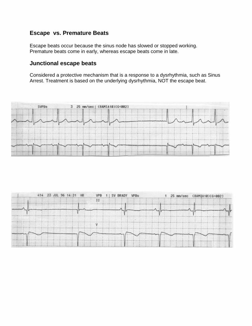
Escape vs. Premature Beats Escape beats occur because the sinus node has slowed or stopped working. Premature beats come in early, whereas escape beats come in late.
Junctional escape beats Considered a protective mechanism that is a response to a dysrhythmia, such as Sinus Arrest. Treatment is based on the underlying dysrhythmia, NOT the escape beat.
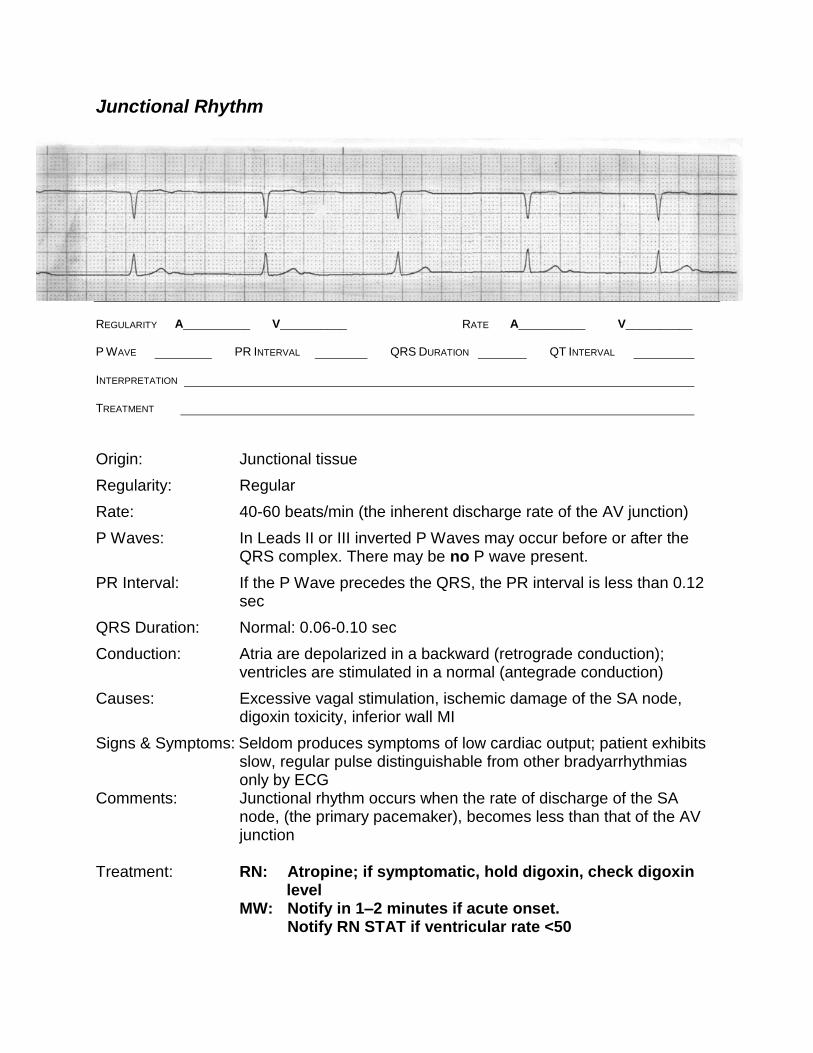
Junctional Rhythm
REGULARITY A__________ V__________ RATE A__________ V__________
P WAVE PR INTERVAL QRS DURATION QT INTERVAL
INTERPRETATION
TREATMENT
Origin: Junctional tissue
Regularity: Regular
Rate: 40-60 beats/min (the inherent discharge rate of the AV junction)
P Waves: In Leads II or III inverted P Waves may occur before or after the QRS complex. There may be no P wave present.
PR Interval: If the P Wave precedes the QRS, the PR interval is less than 0.12 sec
QRS Duration: Normal: 0.06-0.10 sec
Conduction: Atria are depolarized in a backward (retrograde conduction); ventricles are stimulated in a normal (antegrade conduction)
Causes: Excessive vagal stimulation, ischemic damage of the SA node, digoxin toxicity, inferior wall MI
Signs & Symptoms: Seldom produces symptoms of low cardiac output; patient exhibits slow, regular pulse distinguishable from other bradyarrhythmias only by ECG
Comments: Junctional rhythm occurs when the rate of discharge of the SA node, (the primary pacemaker), becomes less than that of the AV junction
Treatment: RN: Atropine; if symptomatic, hold digoxin, check digoxin level MW: Notify in 1–2 minutes if acute onset.
Notify RN STAT if ventricular rate <50
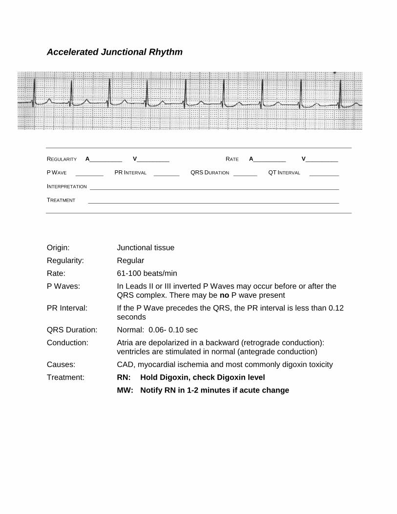
Accelerated Junctional Rhythm
REGULARITY A__________ V__________ RATE A__________ V__________
P WAVE PR INTERVAL QRS DURATION QT INTERVAL
INTERPRETATION
TREATMENT
Origin: Junctional tissue
Regularity: Regular
Rate: 61-100 beats/min
P Waves: In Leads II or III inverted P Waves may occur before or after the QRS complex. There may be no P wave present
PR Interval: If the P Wave precedes the QRS, the PR interval is less than 0.12 seconds
QRS Duration: Normal: 0.06- 0.10 sec
Conduction: Atria are depolarized in a backward (retrograde conduction): ventricles are stimulated in normal (antegrade conduction)
Causes: CAD, myocardial ischemia and most commonly digoxin toxicity
Treatment: RN: Hold Digoxin, check Digoxin level
MW: Notify RN in 1-2 minutes if acute change
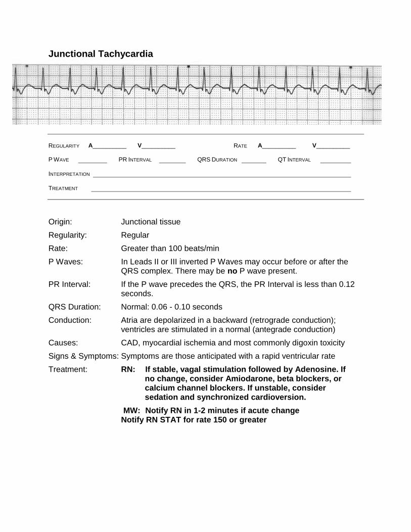
Junctional Tachycardia
REGULARITY A__________ V__________ RATE A__________ V__________
P WAVE PR INTERVAL QRS DURATION QT INTERVAL
INTERPRETATION
TREATMENT
Origin: Junctional tissue
Regularity: Regular
Rate: Greater than 100 beats/min
P Waves: In Leads II or III inverted P Waves may occur before or after the QRS complex. There may be no P wave present.
PR Interval: If the P wave precedes the QRS, the PR Interval is less than 0.12 seconds.
QRS Duration: Normal: 0.06 - 0.10 seconds
Conduction: Atria are depolarized in a backward (retrograde conduction); ventricles are stimulated in a normal (antegrade conduction)
Causes: CAD, myocardial ischemia and most commonly digoxin toxicity
Signs & Symptoms: Symptoms are those anticipated with a rapid ventricular rate
Treatment: RN: If stable, vagal stimulation followed by Adenosine. If no change, consider Amiodarone, beta blockers, or calcium channel blockers. If unstable, consider sedation and synchronized cardioversion.
MW: Notify RN in 1-2 minutes if acute change Notify RN STAT for rate 150 or greater
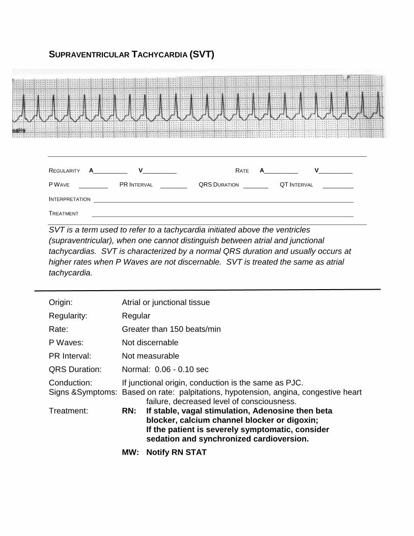
SUPRAVENTRICULAR TACHYCARDIA (SVT)
REGULARITY A__________ V__________ RATE A__________ V__________
P WAVE PR INTERVAL QRS DURATION QT INTERVAL
INTERPRETATION
TREATMENT
SVT is a term used to refer to a tachycardia initiated above the ventricles
(supraventricular), when one cannot distinguish between atrial and junctional
tachycardias. SVT is characterized by a normal QRS duration and usually occurs at
higher rates when P Waves are not discernable. SVT is treated the same as atrial
tachycardia.
Origin: Atrial or junctional tissue
Regularity: Regular
Rate: Greater than 150 beats/min
P Waves: Not discernable
PR Interval: Not measurable
QRS Duration: Normal: 0.06 - 0.10 sec
Conduction: If junctional origin, conduction is the same as PJC. Signs &Symptoms: Based on rate: palpitations, hypotension, angina, congestive heart
failure, decreased level of consciousness. Treatment: RN: If stable, vagal stimulation, Adenosine then beta
blocker, calcium channel blocker or digoxin; If the patient is severely symptomatic, consider sedation and synchronized cardioversion.
MW: Notify RN STAT
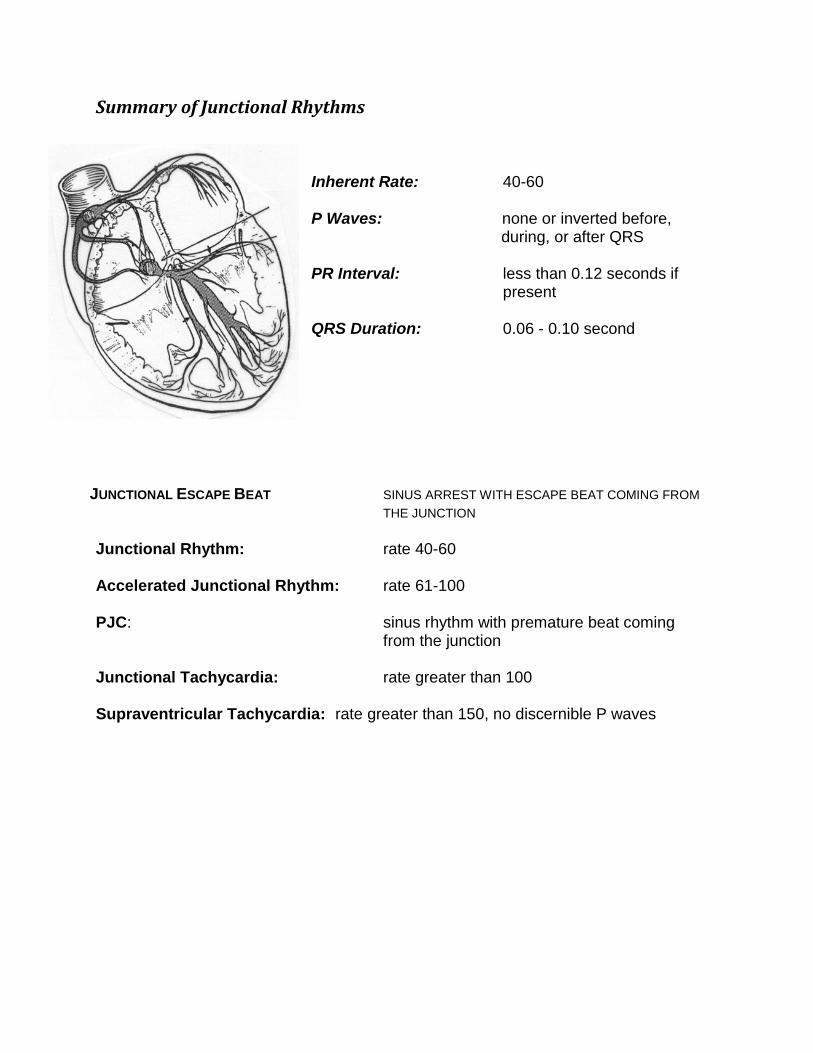
Summary of Junctional Rhythms
Inherent Rate: 40-60 P Waves: none or inverted before,
during, or after QRS PR Interval: less than 0.12 seconds if present QRS Duration: 0.06 - 0.10 second
JUNCTIONAL ESCAPE BEAT SINUS ARREST WITH ESCAPE BEAT COMING FROM
THE JUNCTION Junctional Rhythm: rate 40-60 Accelerated Junctional Rhythm: rate 61-100 PJC: sinus rhythm with premature beat coming
from the junction Junctional Tachycardia: rate greater than 100 Supraventricular Tachycardia: rate greater than 150, no discernible P waves
ECG INTERPRETATION SELF EVALUATION - DAY ONE
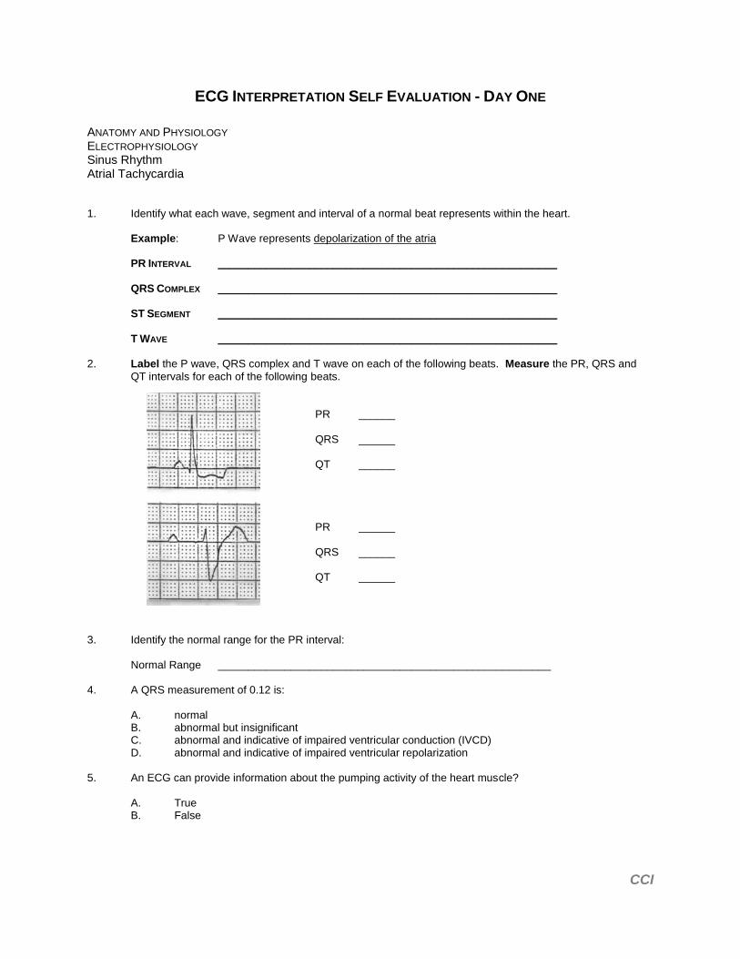
ANATOMY AND PHYSIOLOGY ELECTROPHYSIOLOGY Sinus Rhythm Atrial Tachycardia 1. Identify what each wave, segment and interval of a normal beat represents within the heart. Example: P Wave represents depolarization of the atria
PR INTERVAL ________________________________________________________
QRS COMPLEX ________________________________________________________
ST SEGMENT ________________________________________________________
T WAVE ________________________________________________________
2. Label the P wave, QRS complex and T wave on each of the following beats. Measure the PR, QRS and
QT intervals for each of the following beats. PR ______ QRS ______ QT ______ PR ______ QRS ______ QT ______ 3. Identify the normal range for the PR interval: Normal Range _______________________________________________________ 4. A QRS measurement of 0.12 is: A. normal B. abnormal but insignificant C. abnormal and indicative of impaired ventricular conduction (IVCD) D. abnormal and indicative of impaired ventricular repolarization 5. An ECG can provide information about the pumping activity of the heart muscle? A. True B. False
CCI
SELF- EVALUATION DAY ONE 6. Elevation of the ST Segment may indicate: A. the need to further evaluate the patient’s cardiac status B. myocardial injury C. inadequate blood flow to the heat muscle D. all of the above
E. none of the above 7. A prolonged QT interval can lead to lethal dysrhythmias. A. True B. False
8. Identify the normal, intrinsic/inherent, rates of the SA node, AV node and ventricles. SA NODE ____________________________________________
AV NODE ____________________________________________
VENTRICLES ____________________________________________
9. A regular sinus rhythm with a rate of <60 is called: ________________________.
10. The rate for sinus tachycardia is:
A. 40 – 60 B. 60 - 150
C. 101 - 150 D. 200 - 300
11. Provide the appropriate term for the following characteristics: A. event during which the SA Node does not fire:
______________________________ B. sinus rhythm in which all interval and wave forms are normal except for a QRS measurement of >0.12: ______________________________
B. In Lead II and Lead III the P wave is normally upright. A. True B. False
13. Monitor Watchers: You identify a new onset of sinus bradycardia at a rate of 40.
Notify the RN _______________________
RNs: You go to assess the patient and find that the patient is dizzy.
Drug most commonly used to treat symptomatic sinus bradycardia: ________________
14. Monitor Watchers: You identify a new episode of sinus arrest on the monitor:
Notify the RN _______________________
RNs: You assess the patient and notify the MD of the sinus arrest: Initially, the patient is asymptomatic, but the periods of sinus arrest are becoming longer and more frequent.
Name one intervention the patient may require: ___________________________
CCI
SELF- EVALUATION DAY ONE
15. A premature beat originating from the atria has which of the following characteristics? A. different shaped P Wave is early in the regular cycle B. different shaped P Wave is late in the regular cycle C. normal P Wave is early in the regular cycle
D. normal P Wave is late in the regular cycle
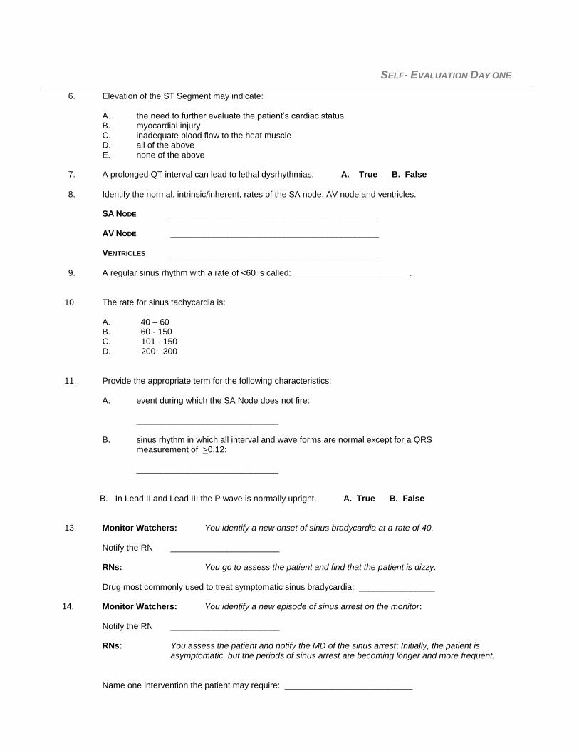
16. Interpret the following rhythms:
Interpretation: ______________________________________________ Intervention: _______________________________________________
17. RNs: Adenosine may be used to slow which of the following rhythms?
A. atrial flutter B. accelerated junctional rhythm C. Atrial Tachycardia D. atrial fibrillation Monitor Watchers: You identify atrial flutter with a ventricle rate of 80:
Notify the RN _______________________ The ventricular rate increases to a rate of 155:
Notify the RN _______________________

18. A beat that originates from the AV Junction will have an _______________ P Wave in a normal Lead II or III; the P Wave may be seen _______________, or _______________ in relation to the QRS Complex. If the P Wave is before the QRS Complex, the PR interval will be _____________.
19. Identify : A. a rhythm originating from the junction with a rate of 45 ____________________ B. a rhythm originating from the junction with a rate of 140 ____________________ C. a rhythm originating from the junction with a rate of 98 ____________________ 20. What is the difference between a junctional escape beat and a premature junctional beat? _____________________________________________________________________________ 21. Both junctional tachycardia and atrial tachycardia are forms of supraventricular tachycardia. A. True B. False 22. Interpret the following rhythms:
INTERPRETATION: ___________________________________ INTERVENTION: ___________________________________
INTERPRETATION: ___________________________________ INTERVENTION: ___________________________________
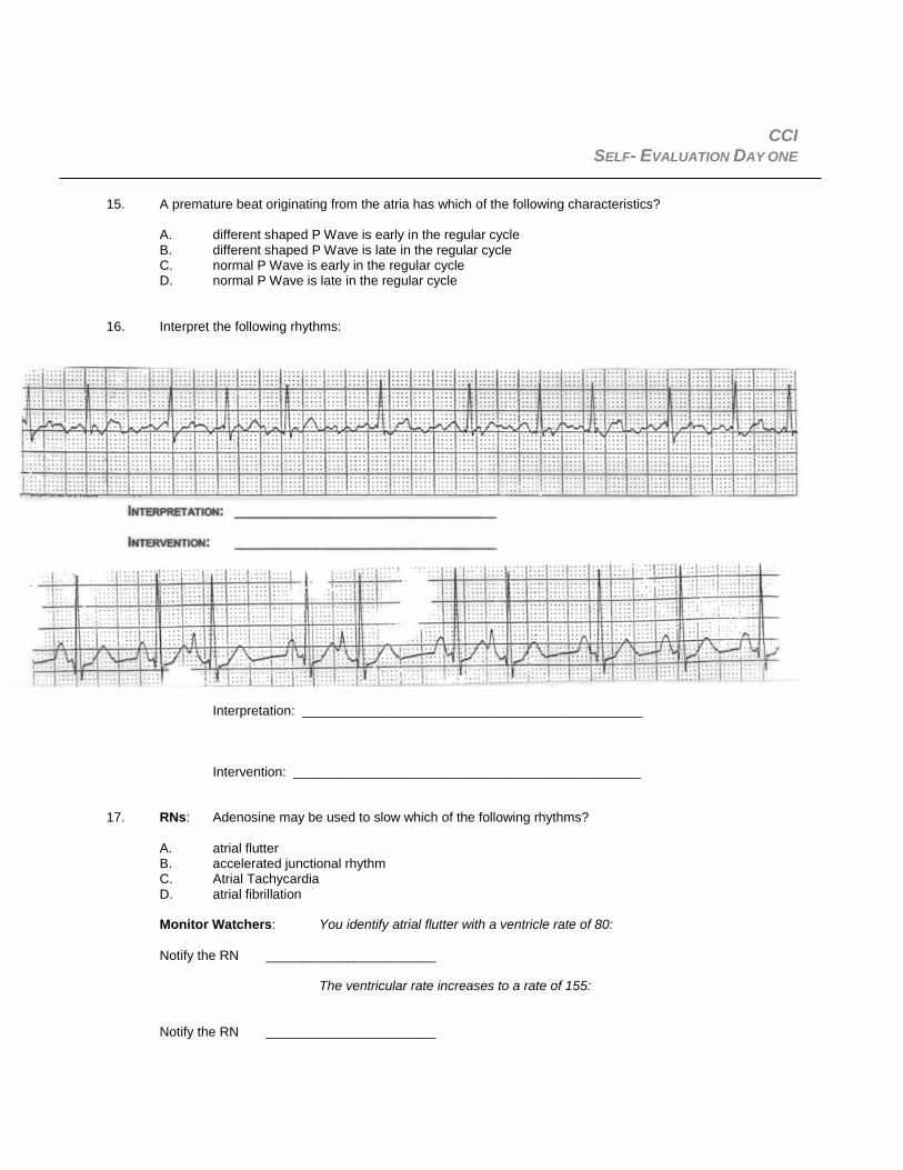
RHYTHMS OF VENTRICULAR ORIGIN
Inherent Rate: 20-40
P Waves: none
PR Interval: none
QRS Duration: wide (0.12 or greater)
IDIOVENTRICULAR RHYTHM (IVR)
REGULARITY A__________ V__________ RATE A__________ V_________ P WAVE PR INTERVAL QRS DURATION QT INTERVAL
INTERPRETATION
TREATMENT
Origin: Ventricle
Regularity: Regular
Rate: 20-40 beats/min
P Waves: None
PR Interval: None
QRS Duration: Wide, bizarre, 0.12 seconds or greater; T wave is directed opposite the QRS
Conduction: Ventricles are stimulated backward or by retrograde conduction
Causes: Slowing of supraventricular pacemakers, acute MI
Signs & Symptoms: Idioventricular rhythm produces dizziness, hypotension or syncope
Comments: Patients in end-stage cardiac disease frequently go into IVR; Treatment and action is based on code status
Treatment: RN: Pacing
MW: Notify Any RN STAT
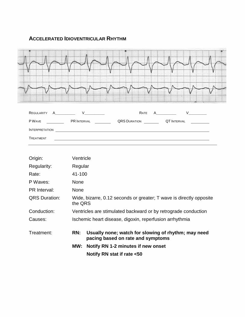
ACCELERATED IDIOVENTRICULAR RHYTHM
REGULARITY A__________ V__________ RATE A__________ V_________ P WAVE PR INTERVAL QRS DURATION QT INTERVAL
INTERPRETATION
TREATMENT
Origin: Ventricle
Regularity: Regular
Rate: 41-100
P Waves: None
PR Interval: None
QRS Duration: Wide, bizarre, 0.12 seconds or greater; T wave is directly opposite the QRS
Conduction: Ventricles are stimulated backward or by retrograde conduction
Causes: Ischemic heart disease, digoxin, reperfusion arrhythmia
Treatment: RN: Usually none; watch for slowing of rhythm; may need pacing based on rate and symptoms
MW: Notify RN 1-2 minutes if new onset
Notify RN stat if rate <50
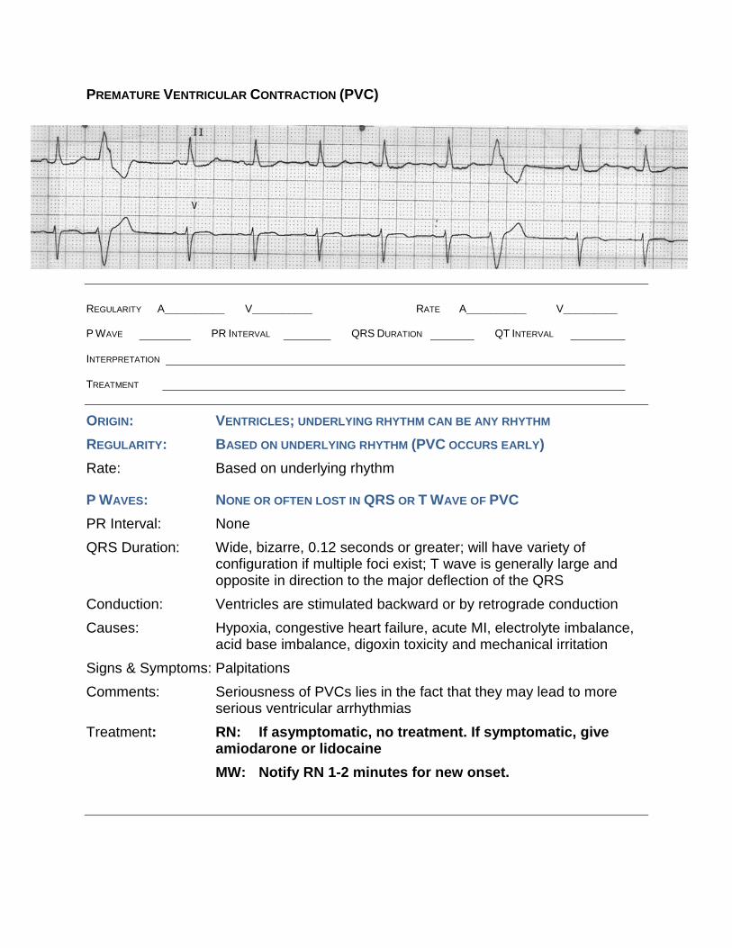
PREMATURE VENTRICULAR CONTRACTION (PVC)
REGULARITY A__________ V__________ RATE A__________ V_________ P WAVE PR INTERVAL QRS DURATION QT INTERVAL
INTERPRETATION
TREATMENT
ORIGIN: VENTRICLES; UNDERLYING RHYTHM CAN BE ANY RHYTHM
REGULARITY: BASED ON UNDERLYING RHYTHM (PVC OCCURS EARLY)
Rate: Based on underlying rhythm
P WAVES: NONE OR OFTEN LOST IN QRS OR T WAVE OF PVC
PR Interval: None
QRS Duration: Wide, bizarre, 0.12 seconds or greater; will have variety of configuration if multiple foci exist; T wave is generally large and opposite in direction to the major deflection of the QRS
Conduction: Ventricles are stimulated backward or by retrograde conduction
Causes: Hypoxia, congestive heart failure, acute MI, electrolyte imbalance, acid base imbalance, digoxin toxicity and mechanical irritation
Signs & Symptoms: Palpitations
Comments: Seriousness of PVCs lies in the fact that they may lead to more serious ventricular arrhythmias
Treatment: RN: If asymptomatic, no treatment. If symptomatic, give amiodarone or lidocaine
MW: Notify RN 1-2 minutes for new onset.
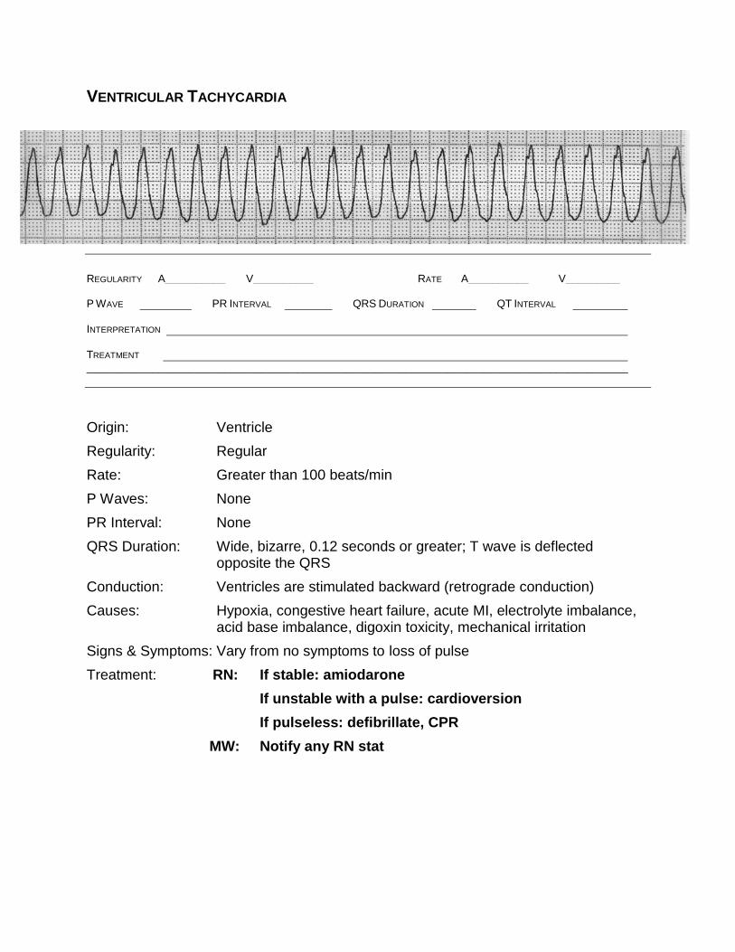
VENTRICULAR TACHYCARDIA
REGULARITY A__________ V__________ RATE A__________ V_________ P WAVE PR INTERVAL QRS DURATION QT INTERVAL
INTERPRETATION
TREATMENT
__________________________________________________________________________________________
Origin: Ventricle
Regularity: Regular
Rate: Greater than 100 beats/min
P Waves: None
PR Interval: None
QRS Duration: Wide, bizarre, 0.12 seconds or greater; T wave is deflected opposite the QRS
Conduction: Ventricles are stimulated backward (retrograde conduction)
Causes: Hypoxia, congestive heart failure, acute MI, electrolyte imbalance, acid base imbalance, digoxin toxicity, mechanical irritation
Signs & Symptoms: Vary from no symptoms to loss of pulse
Treatment: RN: If stable: amiodarone
If unstable with a pulse: cardioversion
If pulseless: defibrillate, CPR
MW: Notify any RN stat
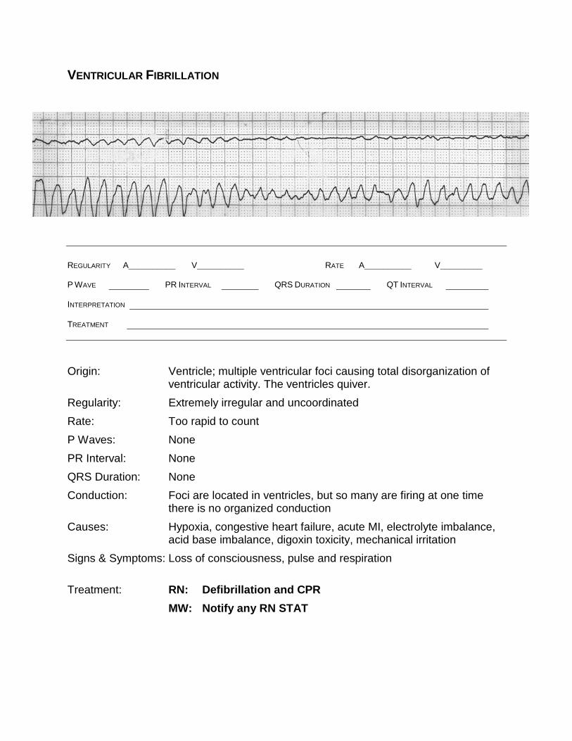
VENTRICULAR FIBRILLATION
REGULARITY A__________ V__________ RATE A__________ V_________ P WAVE PR INTERVAL QRS DURATION QT INTERVAL
INTERPRETATION
TREATMENT
Origin: Ventricle; multiple ventricular foci causing total disorganization of ventricular activity. The ventricles quiver.
Regularity: Extremely irregular and uncoordinated
Rate: Too rapid to count
P Waves: None
PR Interval: None
QRS Duration: None
Conduction: Foci are located in ventricles, but so many are firing at one time there is no organized conduction
Causes: Hypoxia, congestive heart failure, acute MI, electrolyte imbalance, acid base imbalance, digoxin toxicity, mechanical irritation
Signs & Symptoms: Loss of consciousness, pulse and respiration
Treatment: RN: Defibrillation and CPR
MW: Notify any RN STAT

SYNCHRONIZED CARDIOVERSION VS. DEFIBRILLATION SYNCHRONIZED CARDIOVERSION * delivery of electrical energy, synchronized to the R wave of the QRS, to depolarize the
remaining cardiac tissue in hopes the sinus node will assume electrical control of the heart
* used for unstable dysrhythmias of supraventricular tachycardia or ventricular tachycardia with a pulse.
DEFIBRILLATION
* delivery of electrical energy, unsynchronized to heart's activity, to depolarize all of the muscle mass in the hopes the sinus node will assume electrical control of the heart
* used for ventricular fibrillation and pulseless ventricular tachycardia
STERNAL - APEX POSTERIOR - ANTERIOR PADDLE POSITION PADDLE POSITION
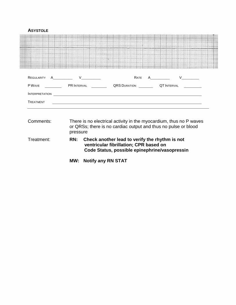
ASYSTOLE
REGULARITY A__________ V__________ RATE A__________ V_________ P WAVE PR INTERVAL QRS DURATION QT INTERVAL
INTERPRETATION
TREATMENT
Comments: There is no electrical activity in the myocardium, thus no P waves or QRSs; there is no cardiac output and thus no pulse or blood pressure
Treatment: RN: Check another lead to verify the rhythm is not ventricular fibrillation; CPR based on
Code Status, possible epinephrine/vasopressin
MW: Notify any RN STAT
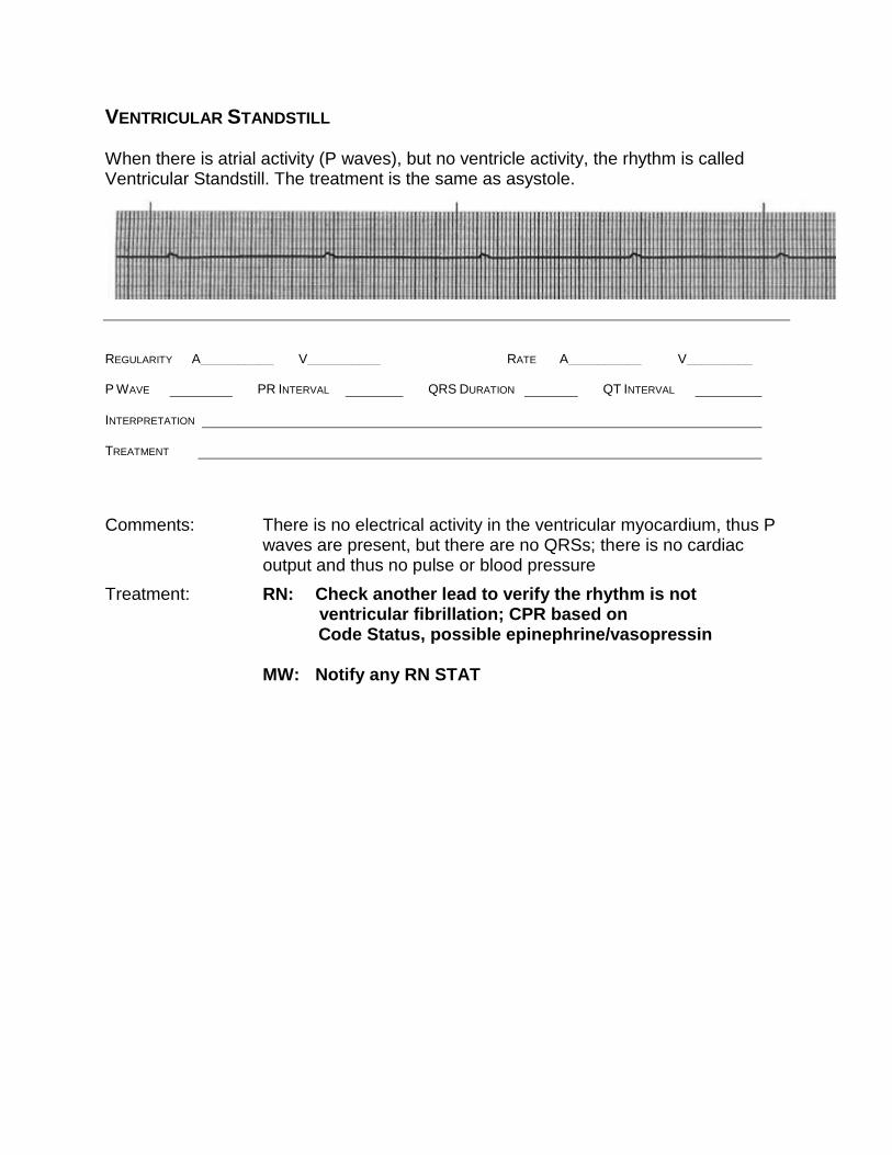
VENTRICULAR STANDSTILL When there is atrial activity (P waves), but no ventricle activity, the rhythm is called Ventricular Standstill. The treatment is the same as asystole.
REGULARITY A__________ V__________ RATE A__________ V_________ P WAVE PR INTERVAL QRS DURATION QT INTERVAL
INTERPRETATION
TREATMENT
Comments: There is no electrical activity in the ventricular myocardium, thus P waves are present, but there are no QRSs; there is no cardiac output and thus no pulse or blood pressure
Treatment: RN: Check another lead to verify the rhythm is not ventricular fibrillation; CPR based on
Code Status, possible epinephrine/vasopressin
MW: Notify any RN STAT
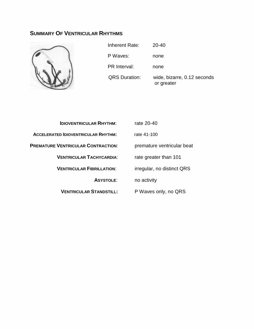
SUMMARY OF VENTRICULAR RHYTHMS
Inherent Rate: 20-40
P Waves: none
PR Interval: none
QRS Duration: wide, bizarre, 0.12 seconds or greater
IDIOVENTRICULAR RHYTHM: rate 20-40
ACCELERATED IDIOVENTRICULAR RHYTHM: rate 41-100
PREMATURE VENTRICULAR CONTRACTION: premature ventricular beat
VENTRICULAR TACHYCARDIA: rate greater than 101
VENTRICULAR FIBRILLATION: irregular, no distinct QRS
ASYSTOLE: no activity
VENTRICULAR STANDSTILL: P Waves only, no QRS
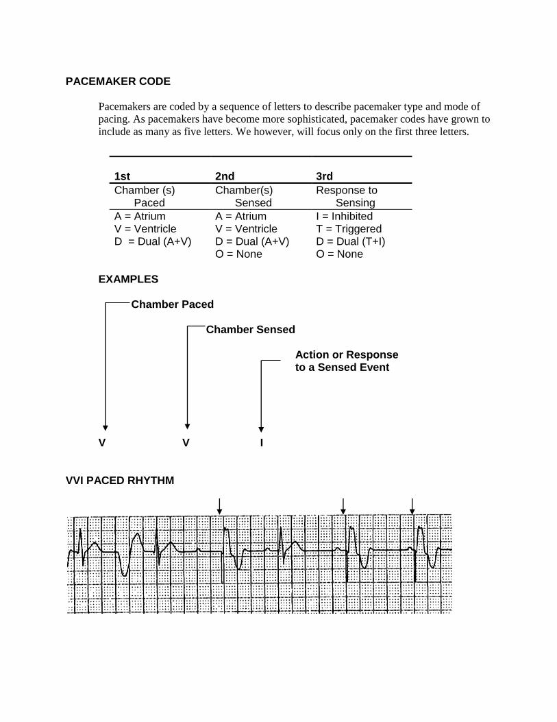
PACEMAKER CODE Pacemakers are coded by a sequence of letters to describe pacemaker type and mode of
pacing. As pacemakers have become more sophisticated, pacemaker codes have grown to
include as many as five letters. We however, will focus only on the first three letters.
1st
2nd
3rd
Chamber (s) Paced
Chamber(s) Sensed
Response to Sensing
A = Atrium A = Atrium I = Inhibited V = Ventricle V = Ventricle T = Triggered D = Dual (A+V) D = Dual (A+V) D = Dual (T+I) O = None O = None
EXAMPLES
Chamber Paced Chamber Sensed Action or Response to a Sensed Event
V V I
VVI PACED RHYTHM
PACEMAKER TERMINOLOGY
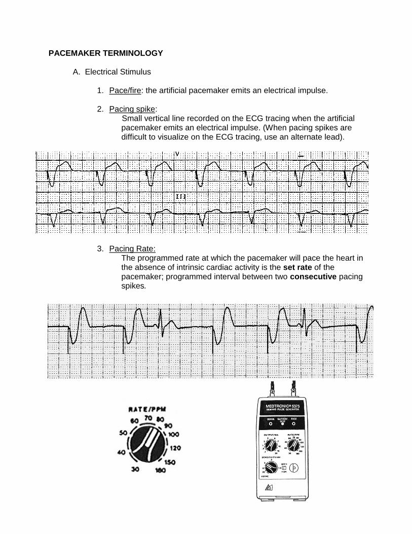
A. Electrical Stimulus
1. Pace/fire: the artificial pacemaker emits an electrical impulse.
2. Pacing spike: Small vertical line recorded on the ECG tracing when the artificial
pacemaker emits an electrical impulse. (When pacing spikes are difficult to visualize on the ECG tracing, use an alternate lead).
3. Pacing Rate: The programmed rate at which the pacemaker will pace the heart in
the absence of intrinsic cardiac activity is the set rate of the pacemaker; programmed interval between two consecutive pacing spikes.
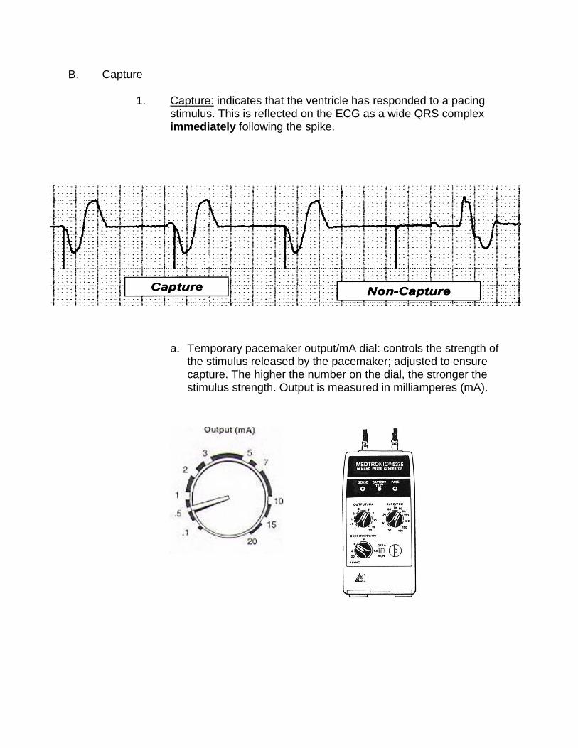
B. Capture
1. Capture: indicates that the ventricle has responded to a pacing stimulus. This is reflected on the ECG as a wide QRS complex immediately following the spike.
a. Temporary pacemaker output/mA dial: controls the strength of
the stimulus released by the pacemaker; adjusted to ensure capture. The higher the number on the dial, the stronger the stimulus strength. Output is measured in milliamperes (mA).
CCI CARDIAC PACING
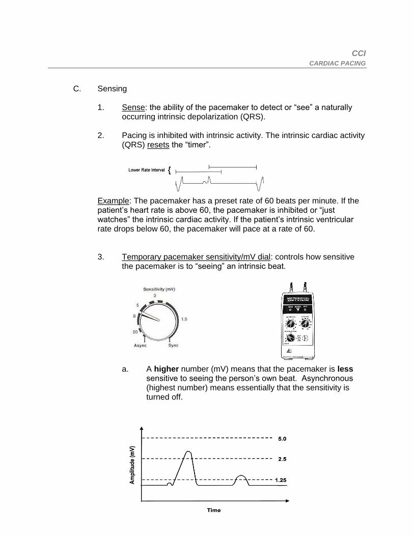
C. Sensing
1. Sense: the ability of the pacemaker to detect or “see” a naturally occurring intrinsic depolarization (QRS).
2. Pacing is inhibited with intrinsic activity. The intrinsic cardiac activity
(QRS) resets the “timer”.
Example: The pacemaker has a preset rate of 60 beats per minute. If the patient’s heart rate is above 60, the pacemaker is inhibited or “just watches” the intrinsic cardiac activity. If the patient’s intrinsic ventricular rate drops below 60, the pacemaker will pace at a rate of 60.
3. Temporary pacemaker sensitivity/mV dial: controls how sensitive the pacemaker is to “seeing” an intrinsic beat.
a. A higher number (mV) means that the pacemaker is less sensitive to seeing the person’s own beat. Asynchronous (highest number) means essentially that the sensitivity is turned off.
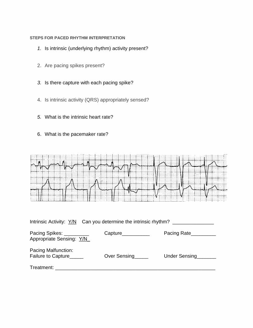
STEPS FOR PACED RHYTHM INTERPRETATION
1. Is intrinsic (underlying rhythm) activity present?
2. Are pacing spikes present?
3. Is there capture with each pacing spike?
4. Is intrinsic activity (QRS) appropriately sensed?
5. What is the intrinsic heart rate?
6. What is the pacemaker rate?
Intrinsic Activity: Y/N Can you determine the intrinsic rhythm? _______________ Pacing Spikes: _________ Capture__________ Pacing Rate_________ Appropriate Sensing: Y/N_ Pacing Malfunction: Failure to Capture_____ Over Sensing_____ Under Sensing_______ Treatment: __________________________________________________________
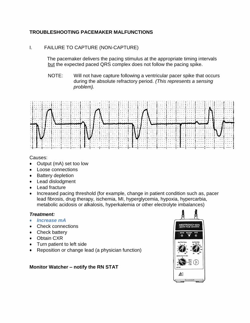
TROUBLESHOOTING PACEMAKER MALFUNCTIONS
I. FAILURE TO CAPTURE (NON-CAPTURE) The pacemaker delivers the pacing stimulus at the appropriate timing intervals but the expected paced QRS complex does not follow the pacing spike.
NOTE: Will not have capture following a ventricular pacer spike that occurs
during the absolute refractory period. (This represents a sensing problem).
Causes:
Output (mA) set too low
Loose connections
Battery depletion
Lead dislodgment
Lead fracture
Increased pacing threshold (for example, change in patient condition such as, pacer lead fibrosis, drug therapy, ischemia, MI, hyperglycemia, hypoxia, hypercarbia, metabolic acidosis or alkalosis, hyperkalemia or other electrolyte imbalances)
Treatment:
Increase mA
Check connections
Check battery
Obtain CXR
Turn patient to left side
Reposition or change lead (a physician function) Monitor Watcher – notify the RN STAT
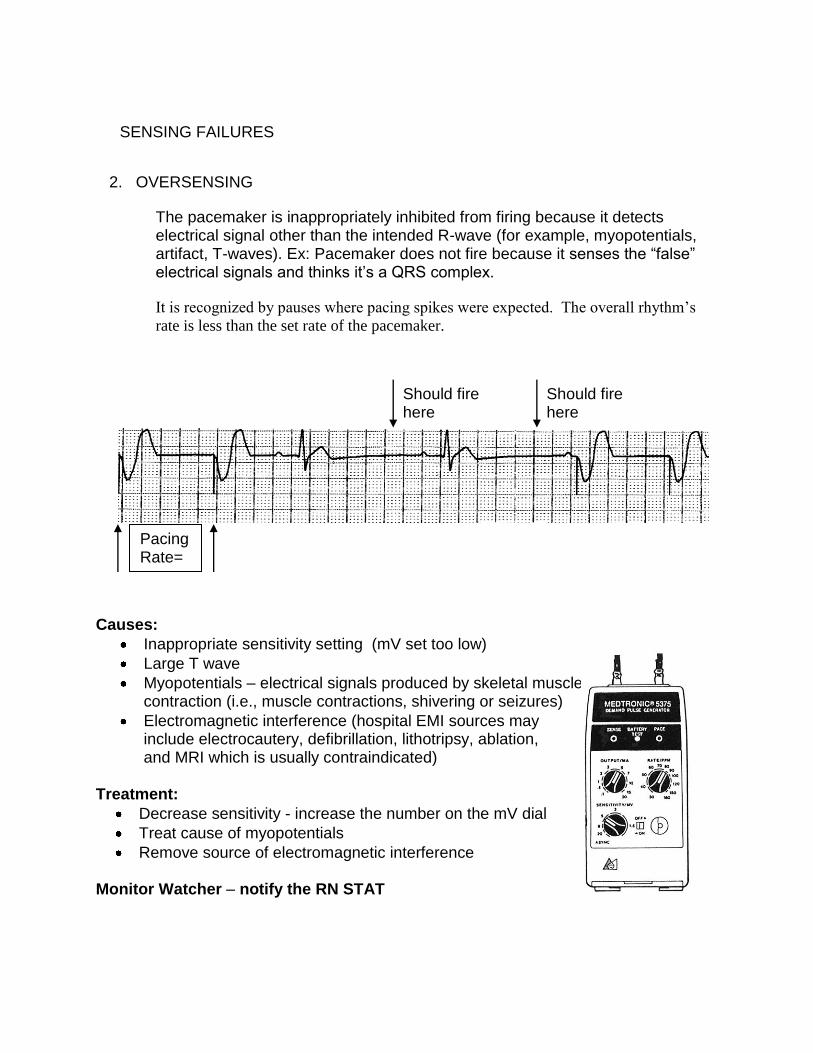
SENSING FAILURES
2. OVERSENSING
The pacemaker is inappropriately inhibited from firing because it detects electrical signal other than the intended R-wave (for example, myopotentials, artifact, T-waves). Ex: Pacemaker does not fire because it senses the “false” electrical signals and thinks it’s a QRS complex.
It is recognized by pauses where pacing spikes were expected. The overall rhythm’s
rate is less than the set rate of the pacemaker.
Causes:
Inappropriate sensitivity setting (mV set too low)
Large T wave
Myopotentials – electrical signals produced by skeletal muscle contraction (i.e., muscle contractions, shivering or seizures)
Electromagnetic interference (hospital EMI sources may include electrocautery, defibrillation, lithotripsy, ablation, and MRI which is usually contraindicated)
Treatment:
Decrease sensitivity - increase the number on the mV dial
Treat cause of myopotentials
Remove source of electromagnetic interference
Monitor Watcher – notify the RN STAT
Should fire here
Should fire here
Pacing Rate=60
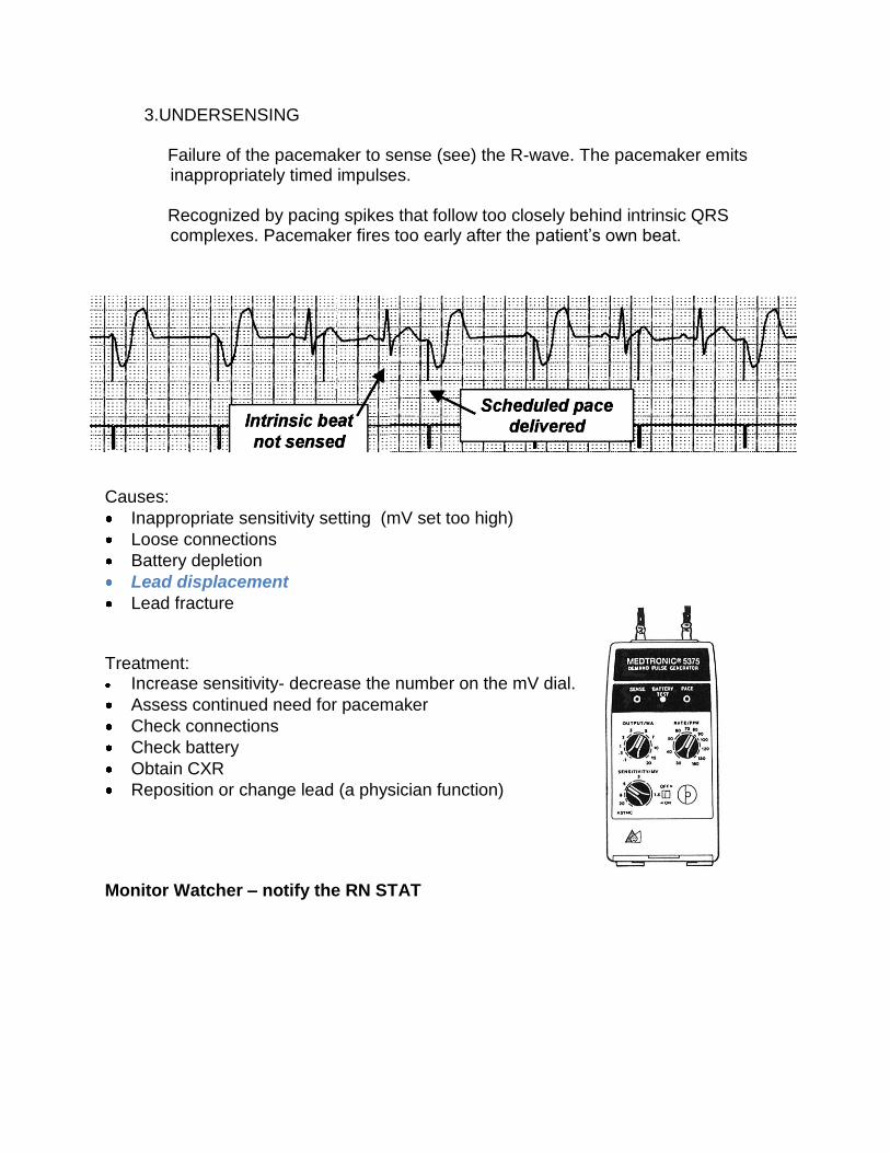
3.UNDERSENSING
Failure of the pacemaker to sense (see) the R-wave. The pacemaker emits inappropriately timed impulses. Recognized by pacing spikes that follow too closely behind intrinsic QRS complexes. Pacemaker fires too early after the patient’s own beat.
Causes:
Inappropriate sensitivity setting (mV set too high)
Loose connections
Battery depletion
Lead displacement
Lead fracture Treatment: Increase sensitivity- decrease the number on the mV dial.
Assess continued need for pacemaker
Check connections
Check battery
Obtain CXR
Reposition or change lead (a physician function) Monitor Watcher – notify the RN STAT
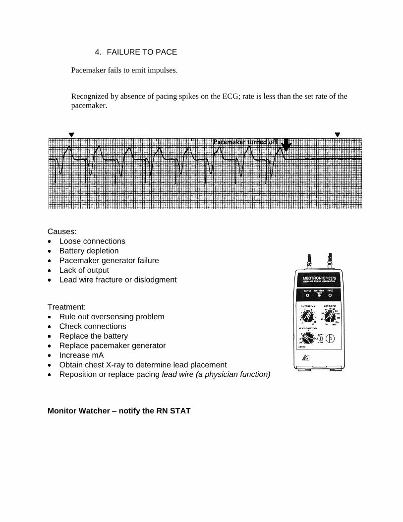
4. FAILURE TO PACE
Pacemaker fails to emit impulses.
Recognized by absence of pacing spikes on the ECG; rate is less than the set rate of the
pacemaker.
Causes:
Loose connections
Battery depletion
Pacemaker generator failure
Lack of output
Lead wire fracture or dislodgment Treatment:
Rule out oversensing problem
Check connections
Replace the battery
Replace pacemaker generator
Increase mA
Obtain chest X-ray to determine lead placement
Reposition or replace pacing lead wire (a physician function)
Monitor Watcher – notify the RN STAT
REGULARITY A__________ V__________ RATE A__________ V__________ P WAVE PR INTERVAL QRS DURATION QT INTERVAL
INTERPRETATION
TREATMENT
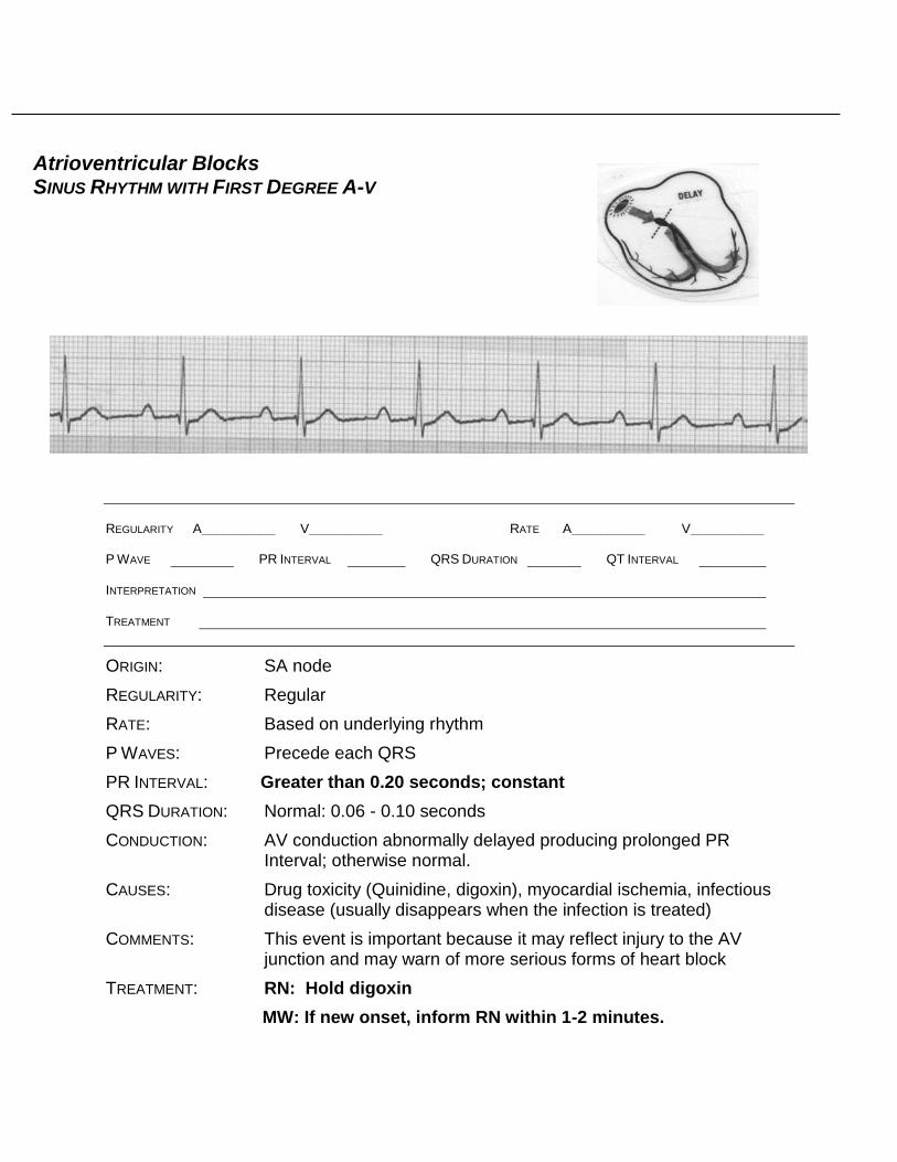
ORIGIN: SA node
REGULARITY: Regular
RATE: Based on underlying rhythm
P WAVES: Precede each QRS
PR INTERVAL: Greater than 0.20 seconds; constant
QRS DURATION: Normal: 0.06 - 0.10 seconds
CONDUCTION: AV conduction abnormally delayed producing prolonged PR Interval; otherwise normal.
CAUSES: Drug toxicity (Quinidine, digoxin), myocardial ischemia, infectious disease (usually disappears when the infection is treated)
COMMENTS: This event is important because it may reflect injury to the AV junction and may warn of more serious forms of heart block
TREATMENT: RN: Hold digoxin
MW: If new onset, inform RN within 1-2 minutes.
Atrioventricular Blocks SINUS RHYTHM WITH FIRST DEGREE A-V
BLOCK
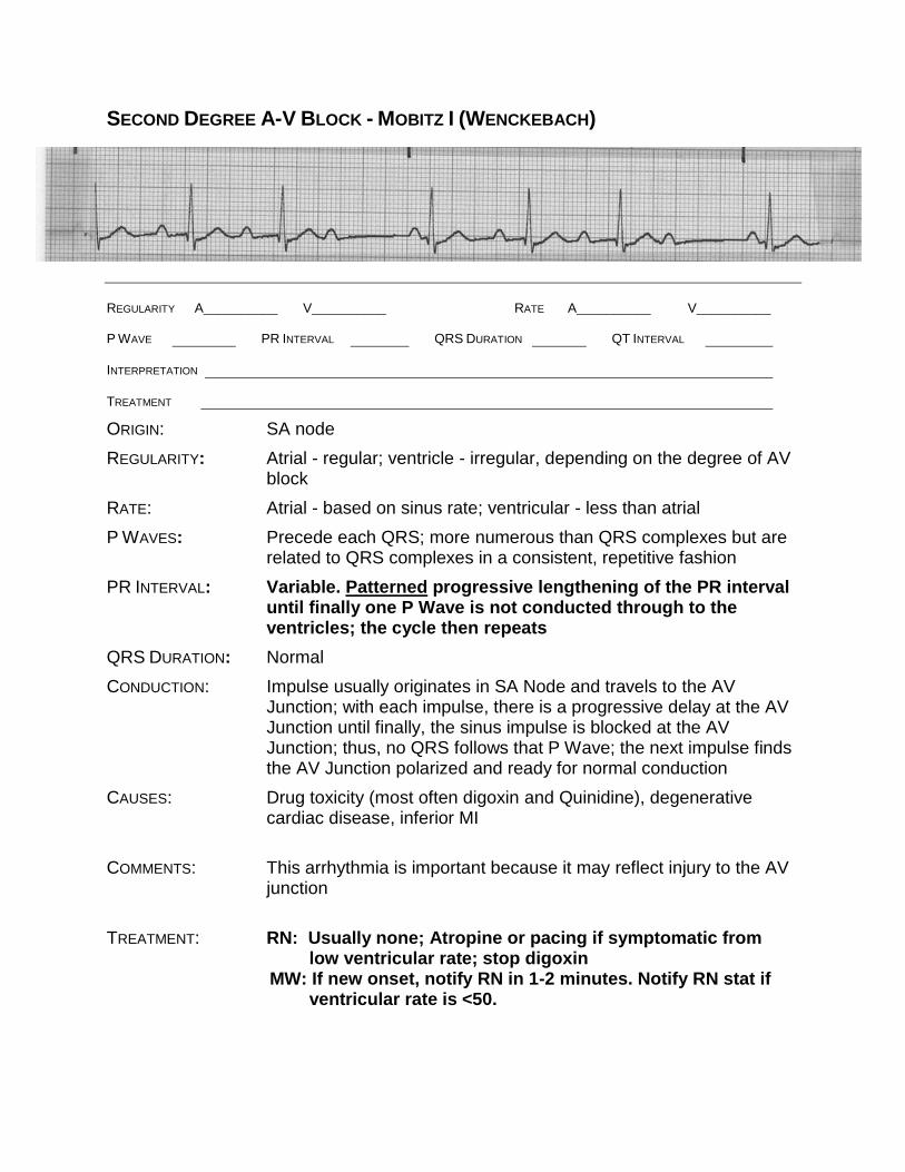
SECOND DEGREE A-V BLOCK - MOBITZ I (WENCKEBACH)
REGULARITY A__________ V__________ RATE A__________ V__________ P WAVE PR INTERVAL QRS DURATION QT INTERVAL
INTERPRETATION
TREATMENT
ORIGIN: SA node
REGULARITY: Atrial - regular; ventricle - irregular, depending on the degree of AV block
RATE: Atrial - based on sinus rate; ventricular - less than atrial
P WAVES: Precede each QRS; more numerous than QRS complexes but are related to QRS complexes in a consistent, repetitive fashion
PR INTERVAL: Variable. Patterned progressive lengthening of the PR interval until finally one P Wave is not conducted through to the ventricles; the cycle then repeats
QRS DURATION: Normal
CONDUCTION: Impulse usually originates in SA Node and travels to the AV Junction; with each impulse, there is a progressive delay at the AV Junction until finally, the sinus impulse is blocked at the AV Junction; thus, no QRS follows that P Wave; the next impulse finds the AV Junction polarized and ready for normal conduction
CAUSES: Drug toxicity (most often digoxin and Quinidine), degenerative cardiac disease, inferior MI
COMMENTS: This arrhythmia is important because it may reflect injury to the AV junction
TREATMENT: RN: Usually none; Atropine or pacing if symptomatic from low ventricular rate; stop digoxin MW: If new onset, notify RN in 1-2 minutes. Notify RN stat if ventricular rate is <50.
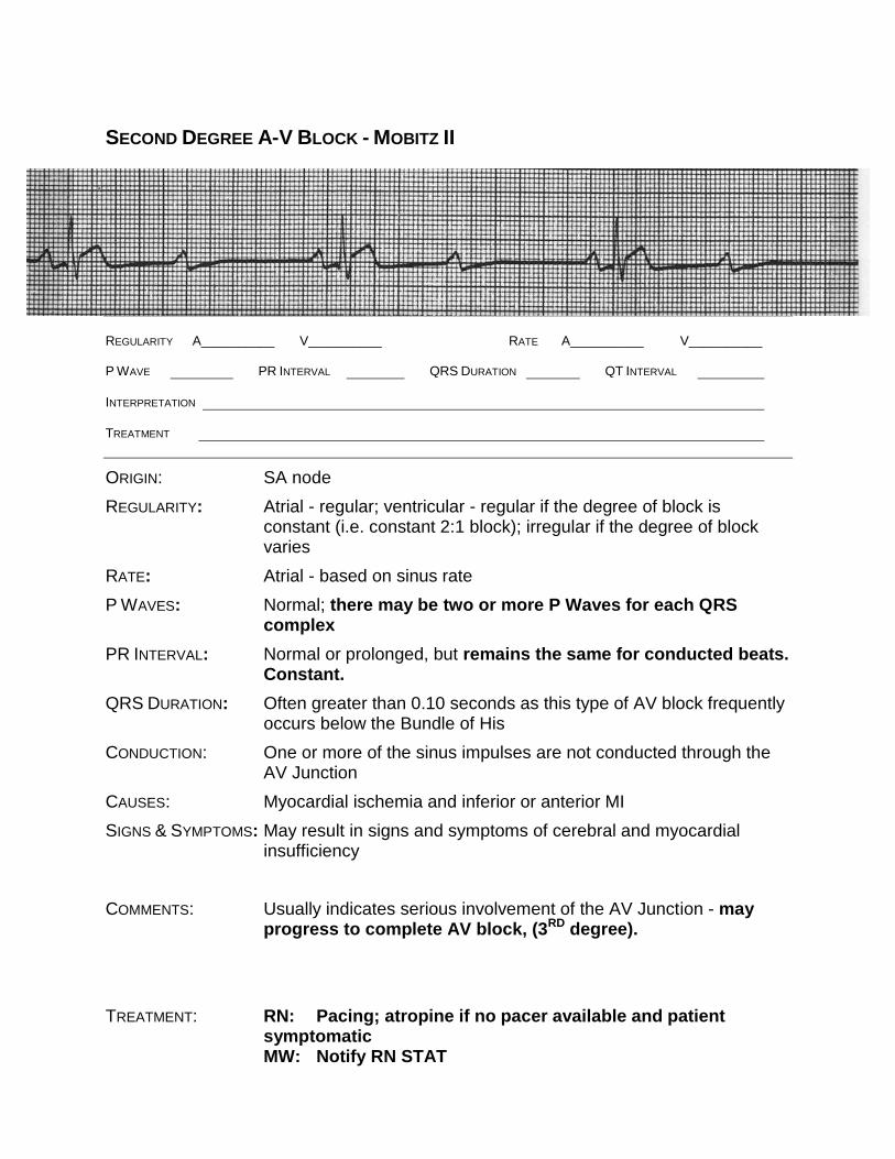
SECOND DEGREE A-V BLOCK - MOBITZ II
REGULARITY A__________ V__________ RATE A__________ V__________ P WAVE PR INTERVAL QRS DURATION QT INTERVAL
INTERPRETATION
TREATMENT
ORIGIN: SA node
REGULARITY: Atrial - regular; ventricular - regular if the degree of block is constant (i.e. constant 2:1 block); irregular if the degree of block varies
RATE: Atrial - based on sinus rate
P WAVES: Normal; there may be two or more P Waves for each QRS complex
PR INTERVAL: Normal or prolonged, but remains the same for conducted beats. Constant.
QRS DURATION: Often greater than 0.10 seconds as this type of AV block frequently occurs below the Bundle of His
CONDUCTION: One or more of the sinus impulses are not conducted through the AV Junction
CAUSES: Myocardial ischemia and inferior or anterior MI
SIGNS & SYMPTOMS: May result in signs and symptoms of cerebral and myocardial insufficiency
COMMENTS: Usually indicates serious involvement of the AV Junction - may progress to complete AV block, (3RD degree).
TREATMENT: RN: Pacing; atropine if no pacer available and patient symptomatic MW: Notify RN STAT
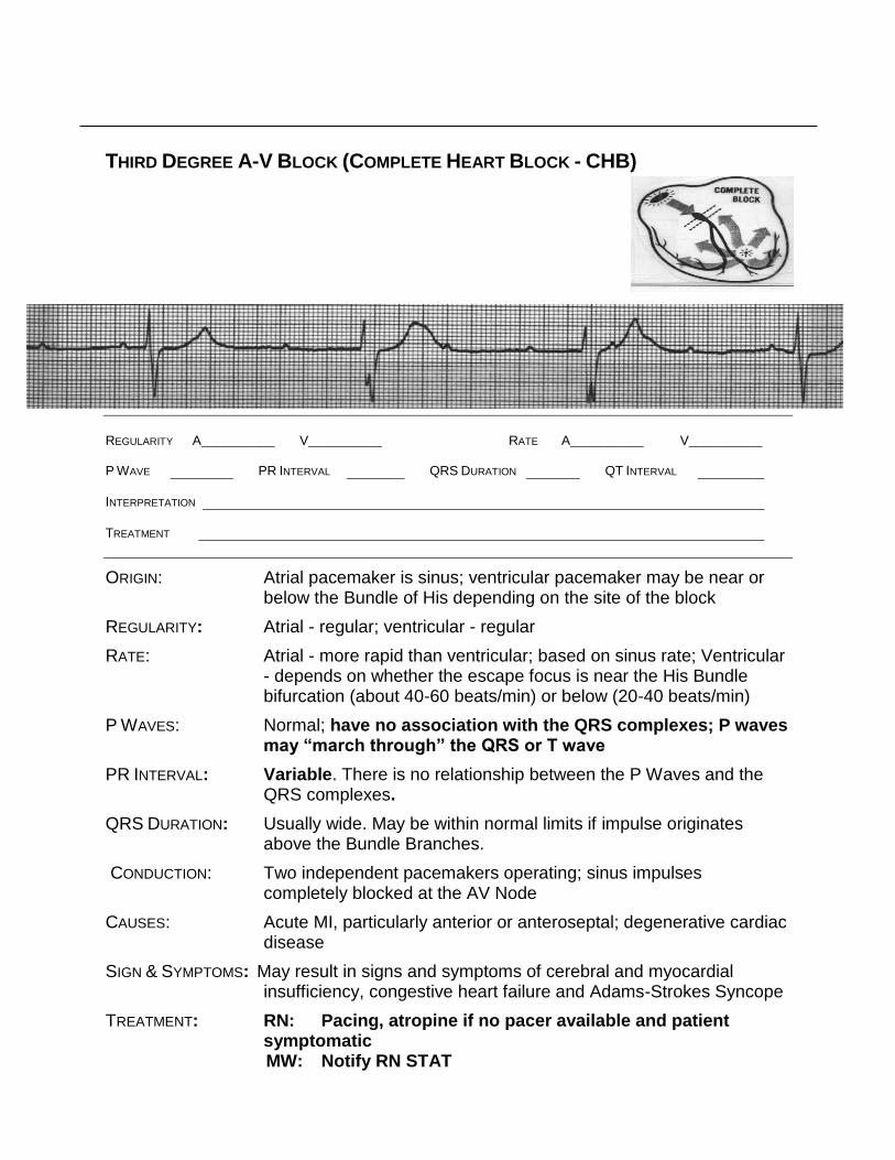
THIRD DEGREE A-V BLOCK (COMPLETE HEART BLOCK - CHB) REGULARITY A__________ V__________ RATE A__________ V__________ P WAVE
PR INTERVAL
QRS DURATION
QT INTERVAL
INTERPRETATION
TREATMENT
ORIGIN: Atrial pacemaker is sinus; ventricular pacemaker may be near or below the Bundle of His depending on the site of the block
REGULARITY: Atrial - regular; ventricular - regular
RATE: Atrial - more rapid than ventricular; based on sinus rate; Ventricular - depends on whether the escape focus is near the His Bundle bifurcation (about 40-60 beats/min) or below (20-40 beats/min)
P WAVES: Normal; have no association with the QRS complexes; P waves may “march through” the QRS or T wave
PR INTERVAL: Variable. There is no relationship between the P Waves and the QRS complexes.
QRS DURATION: Usually wide. May be within normal limits if impulse originates above the Bundle Branches.
CONDUCTION: Two independent pacemakers operating; sinus impulses completely blocked at the AV Node
CAUSES: Acute MI, particularly anterior or anteroseptal; degenerative cardiac disease
SIGN & SYMPTOMS: May result in signs and symptoms of cerebral and myocardial insufficiency, congestive heart failure and Adams-Strokes Syncope
TREATMENT: RN: Pacing, atropine if no pacer available and patient symptomatic
MW: Notify RN STAT
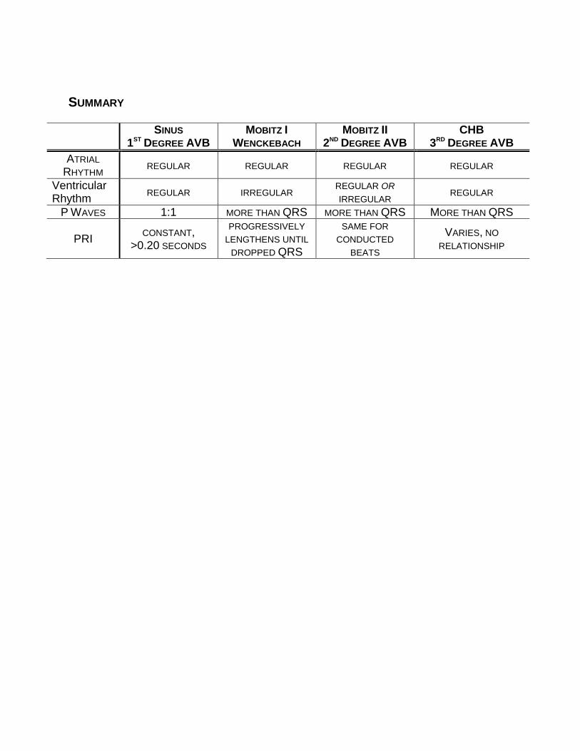
SUMMARY
SINUS
1ST DEGREE AVB
MOBITZ I WENCKEBACH
MOBITZ II 2ND
DEGREE AVB CHB
3RD DEGREE AVB
ATRIAL
RHYTHM REGULAR REGULAR REGULAR REGULAR
Ventricular Rhythm
REGULAR IRREGULAR REGULAR OR
IRREGULAR REGULAR
P WAVES 1:1 MORE THAN QRS MORE THAN QRS MORE THAN QRS
PRI CONSTANT,
>0.20 SECONDS
PROGRESSIVELY
LENGTHENS UNTIL
DROPPED QRS
SAME FOR
CONDUCTED
BEATS
VARIES, NO
RELATIONSHIP
ECG INTERPRETATION SELF EVALUATION - DAY TWO
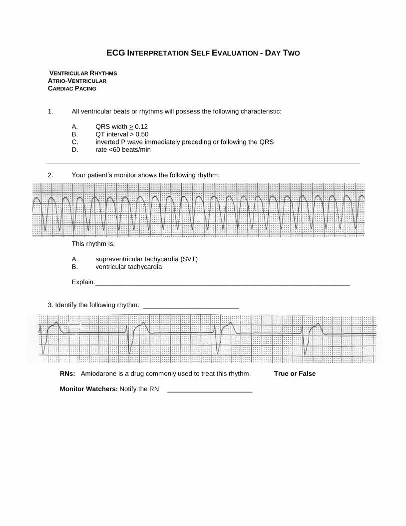
VENTRICULAR RHYTHMS ATRIO-VENTRICULAR CARDIAC PACING 1. All ventricular beats or rhythms will possess the following characteristic: A. QRS width > 0.12 B. QT interval > 0.50 C. inverted P wave immediately preceding or following the QRS D. rate <60 beats/min
2. Your patient’s monitor shows the following rhythm: This rhythm is: A. supraventricular tachycardia (SVT) B. ventricular tachycardia Explain:_____________________________________________________________________ 3. Identify the following rhythm: __________________________
RNs: Amiodarone is a drug commonly used to treat this rhythm. True or False
Monitor Watchers: Notify the RN _______________________
CCI
SELF-EVALUATION DAY TWO
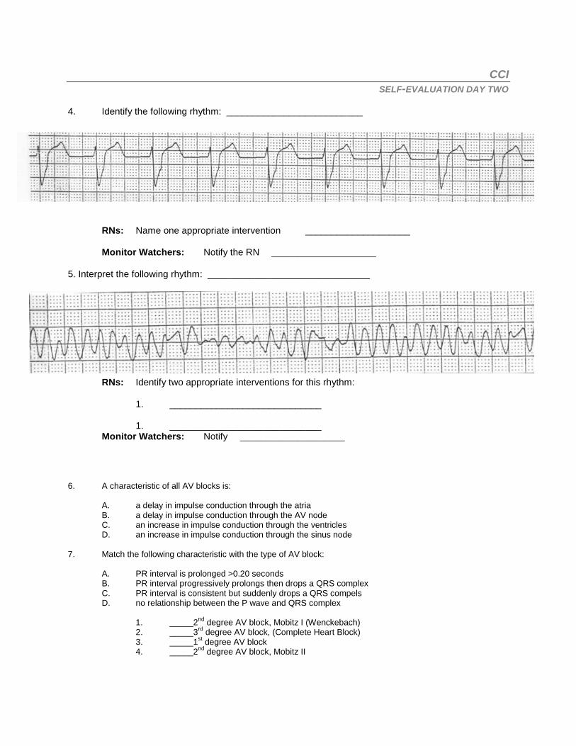
4. Identify the following rhythm: __________________________ RNs: Name one appropriate intervention ____________________ Monitor Watchers: Notify the RN ____________________ 5. Interpret the following rhythm: _______________________________ RNs: Identify two appropriate interventions for this rhythm: 1. _____________________________
1. _____________________________ Monitor Watchers: Notify ____________________
6. A characteristic of all AV blocks is: A. a delay in impulse conduction through the atria B. a delay in impulse conduction through the AV node C. an increase in impulse conduction through the ventricles D. an increase in impulse conduction through the sinus node 7. Match the following characteristic with the type of AV block: A. PR interval is prolonged >0.20 seconds B. PR interval progressively prolongs then drops a QRS complex C. PR interval is consistent but suddenly drops a QRS compels D. no relationship between the P wave and QRS complex 1. _____2
nd degree AV block, Mobitz I (Wenckebach)
2. _____3rd
degree AV block, (Complete Heart Block) 3. _____1
st degree AV block
4. _____2nd
degree AV block, Mobitz II

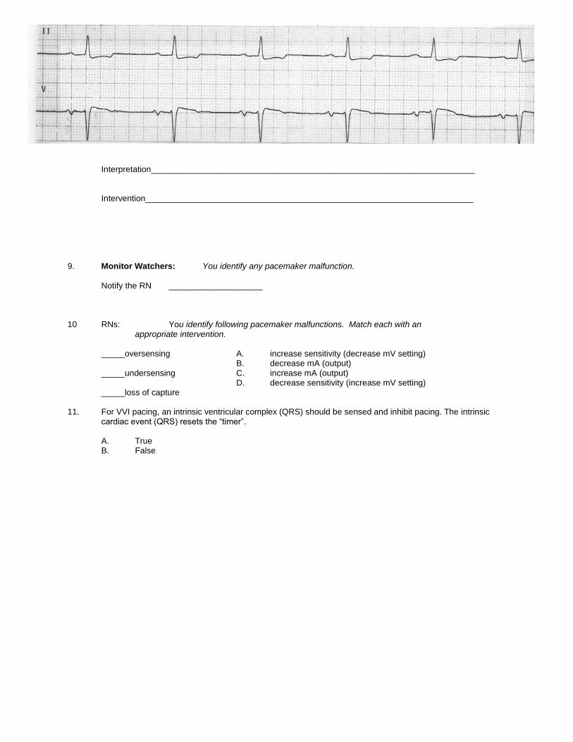
8. Interpret the following rhythms.
Interpretation_____________________________________________________________________
Intervention______________________________________________________________________
Interpretation_____________________________________________________________________
Intervention______________________________________________________________________
Interpretation_____________________________________________________________________
Intervention______________________________________________________________________
Interpretation_____________________________________________________________________
Intervention______________________________________________________________________
9. Monitor Watchers: You identify any pacemaker malfunction.
Notify the RN ____________________ 10 RNs: You identify following pacemaker malfunctions. Match each with an appropriate intervention. _____oversensing A. increase sensitivity (decrease mV setting) B. decrease mA (output) _____undersensing C. increase mA (output) D. decrease sensitivity (increase mV setting) _____loss of capture 11. For VVI pacing, an intrinsic ventricular complex (QRS) should be sensed and inhibit pacing. The intrinsic
cardiac event (QRS) resets the “timer”. A. True B. False
CCI
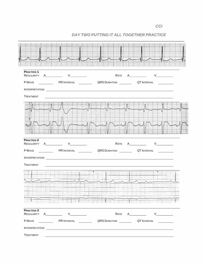
DAY TWO PUTTING IT ALL TOGETHER PRACTICE
PRACTICE-1
REGULARITY A__________ V__________ RATE A__________ V__________ P WAVE PR INTERVAL QRS DURATION QT INTERVAL
INTERPRETATION
TREATMENT
PRACTICE-2
REGULARITY A__________ V__________ RATE A__________ V__________ P WAVE PR INTERVAL QRS DURATION QT INTERVAL
INTERPRETATION
TREATMENT
PRACTICE-3
REGULARITY A__________ V__________ RATE A__________ V__________ P WAVE PR INTERVAL QRS DURATION QT INTERVAL
INTERPRETATION
TREATMENT
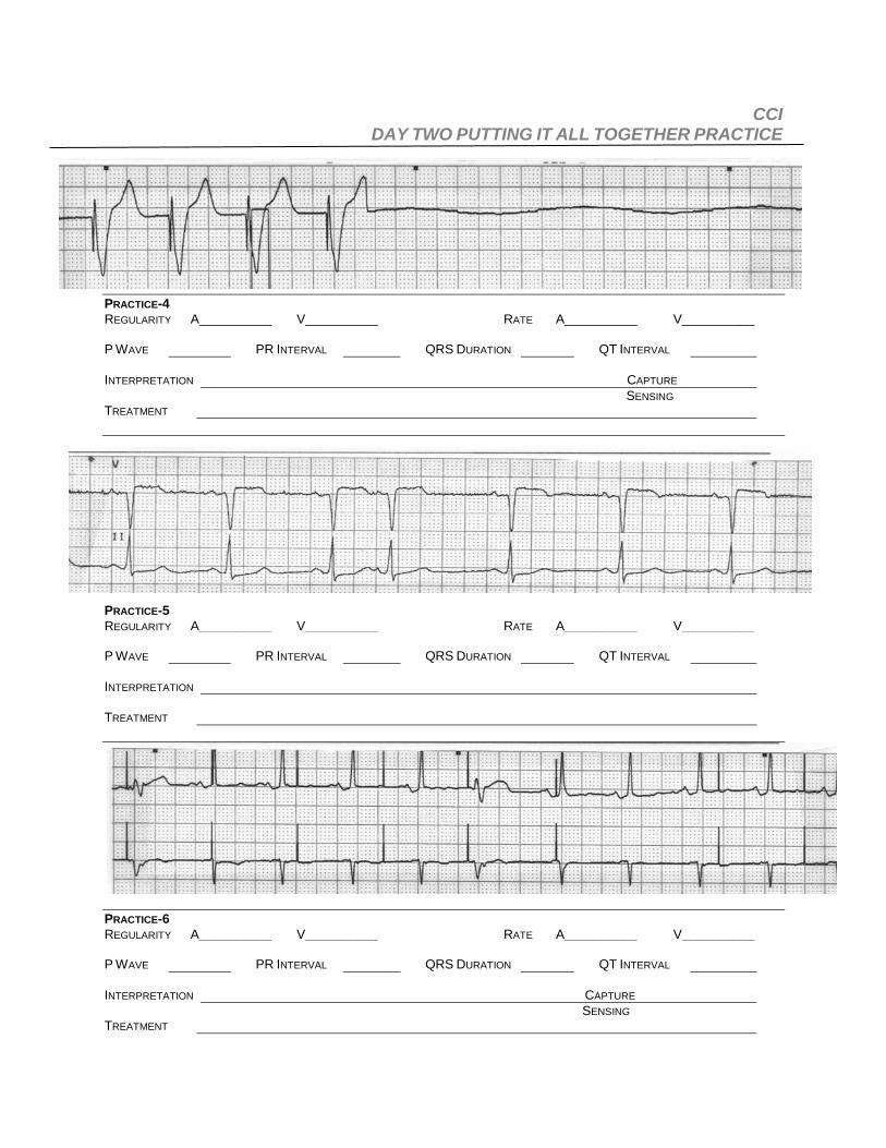
CCI DAY TWO PUTTING IT ALL TOGETHER PRACTICE
PRACTICE-4
REGULARITY A__________ V__________ RATE A__________ V__________ P WAVE PR INTERVAL QRS DURATION QT INTERVAL
INTERPRETATION CAPTURE
SENSING TREATMENT
PRACTICE-5
REGULARITY A__________ V__________ RATE A__________ V__________ P WAVE PR INTERVAL QRS DURATION QT INTERVAL
INTERPRETATION
TREATMENT
PRACTICE-6
REGULARITY A__________ V__________ RATE A__________ V__________ P WAVE PR INTERVAL QRS DURATION QT INTERVAL
INTERPRETATION CAPTURE
SENSING TREATMENT
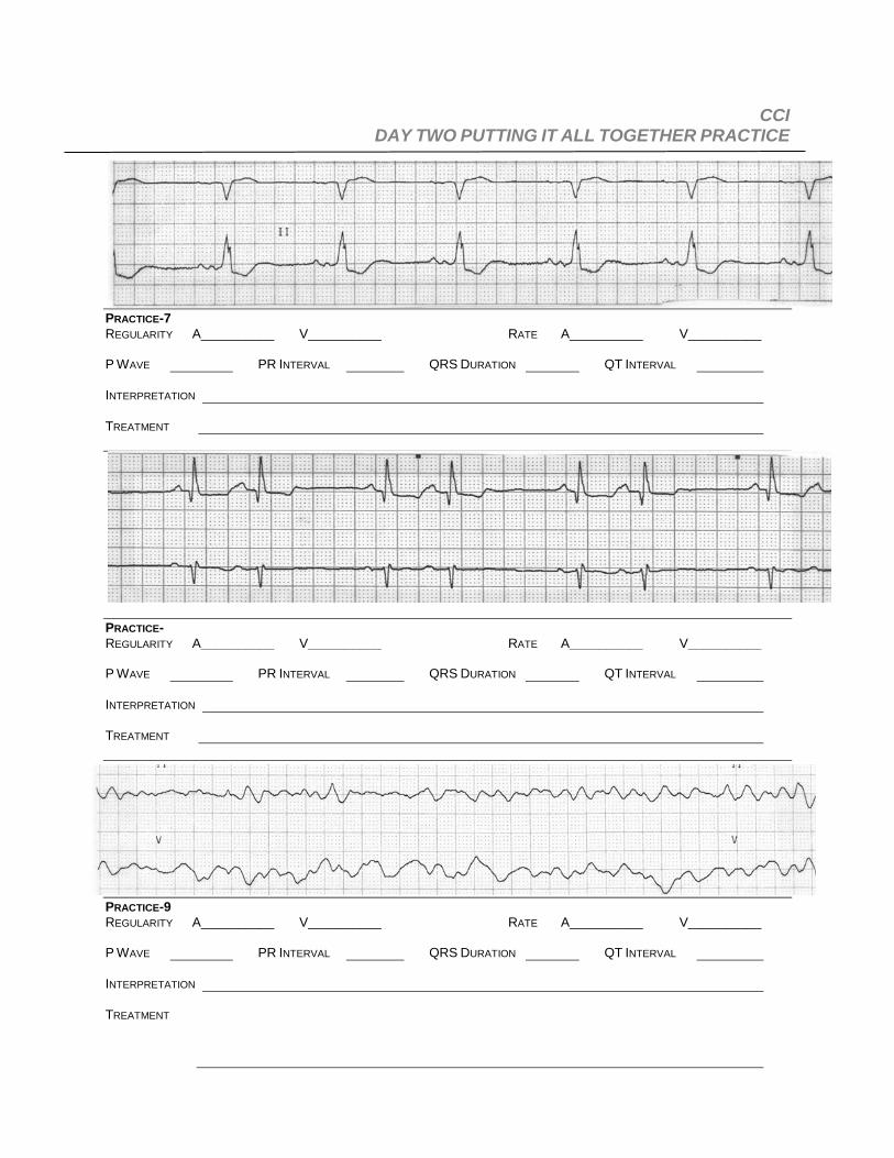
CCI DAY TWO PUTTING IT ALL TOGETHER PRACTICE
PRACTICE-7
REGULARITY A__________ V__________ RATE A__________ V__________ P WAVE PR INTERVAL QRS DURATION QT INTERVAL
INTERPRETATION
TREATMENT
PRACTICE-
REGULARITY A__________ V__________ RATE A__________ V__________ P WAVE PR INTERVAL QRS DURATION QT INTERVAL
INTERPRETATION
TREATMENT
PRACTICE-9
REGULARITY A__________ V__________ RATE A__________ V__________ P WAVE PR INTERVAL QRS DURATION QT INTERVAL
INTERPRETATION
TREATMENT
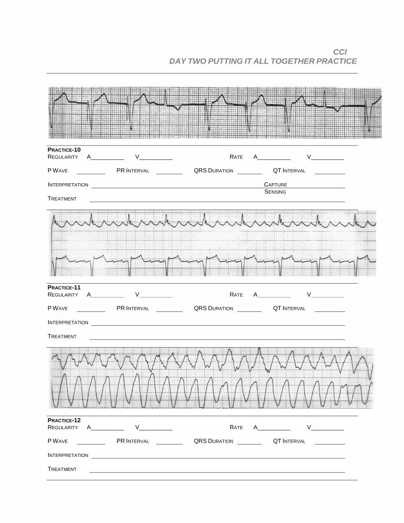
CCI DAY TWO PUTTING IT ALL TOGETHER PRACTICE
PRACTICE-10
REGULARITY A__________ V__________ RATE A__________ V__________ P WAVE PR INTERVAL QRS DURATION QT INTERVAL
INTERPRETATION CAPTURE
SENSING TREATMENT
PRACTICE-11
REGULARITY A__________ V__________ RATE A__________ V__________ P WAVE PR INTERVAL QRS DURATION QT INTERVAL
INTERPRETATION
TREATMENT
PRACTICE-12
REGULARITY A__________ V__________ RATE A__________ V__________ P WAVE PR INTERVAL QRS DURATION QT INTERVAL
INTERPRETATION
TREATMENT
CCI DAY TWO PUTTING IT ALL TOGETHER PRACTICE
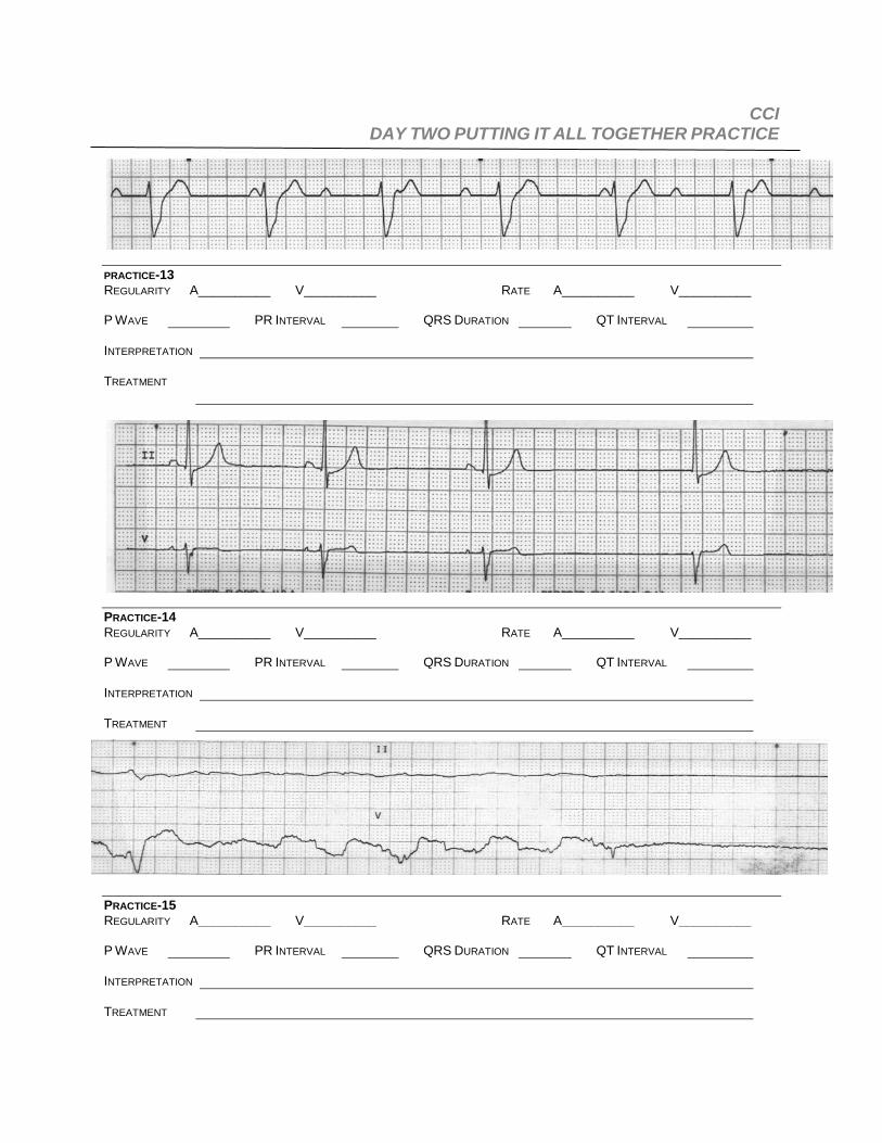
PRACTICE-13
REGULARITY A__________ V__________ RATE A__________ V__________ P WAVE PR INTERVAL QRS DURATION QT INTERVAL
INTERPRETATION
TREATMENT
PRACTICE-14
REGULARITY A__________ V__________ RATE A__________ V__________ P WAVE PR INTERVAL QRS DURATION QT INTERVAL
INTERPRETATION
TREATMENT
PRACTICE-15
REGULARITY A__________ V__________ RATE A__________ V__________ P WAVE PR INTERVAL QRS DURATION QT INTERVAL
INTERPRETATION
TREATMENT
CCI DAY TWO PUTTING IT ALL TOGETHER PRACTICE
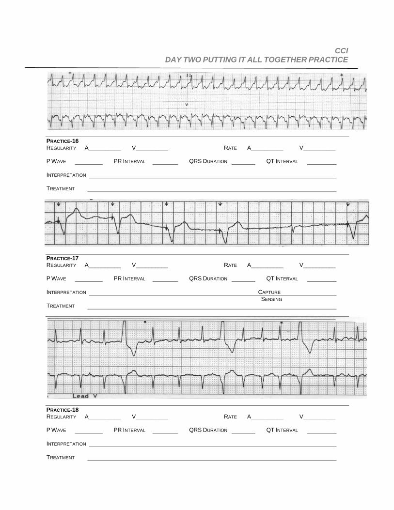
PRACTICE-16
REGULARITY A__________ V__________ RATE A__________ V__________ P WAVE PR INTERVAL QRS DURATION QT INTERVAL
INTERPRETATION
TREATMENT
PRACTICE-17
REGULARITY A__________ V__________ RATE A__________ V__________ P WAVE PR INTERVAL QRS DURATION QT INTERVAL
INTERPRETATION CAPTURE
SENSING TREATMENT
PRACTICE-18
REGULARITY A__________ V__________ RATE A__________ V__________ P WAVE PR INTERVAL QRS DURATION QT INTERVAL
INTERPRETATION
TREATMENT
CCI DAY TWO PUTTING IT ALL TOGETHER PRACTICE
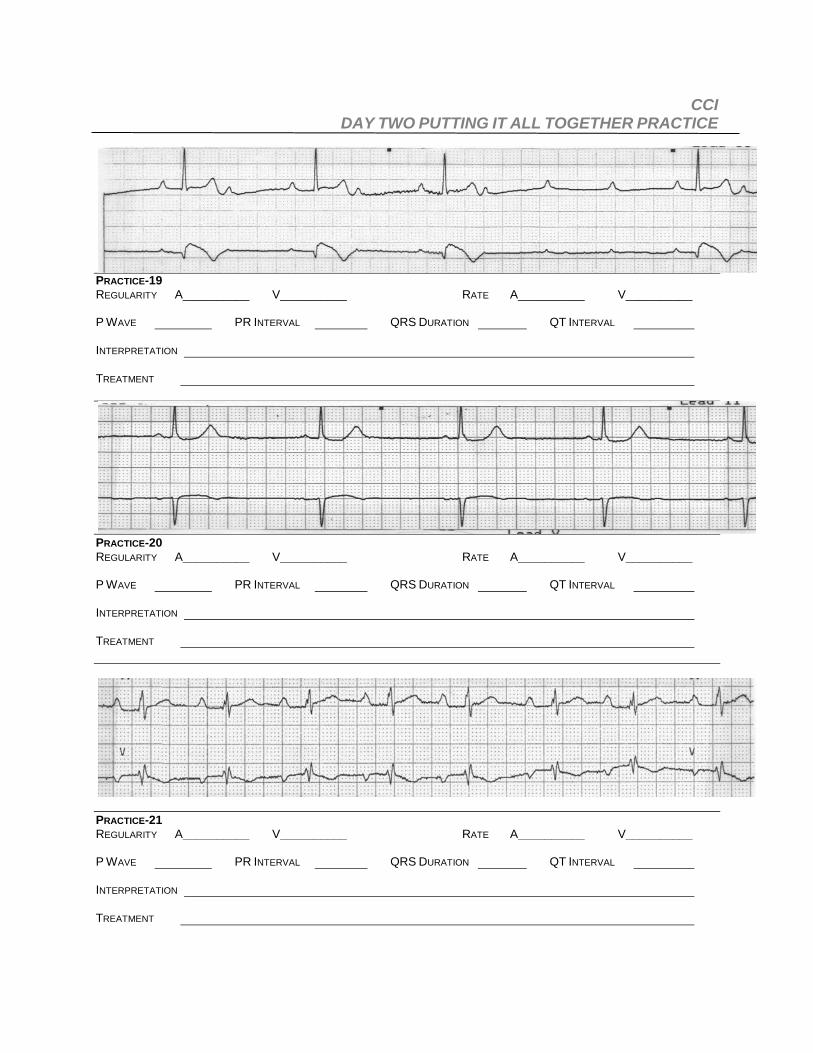
PRACTICE-19
REGULARITY A__________ V__________ RATE A__________ V__________ P WAVE PR INTERVAL QRS DURATION QT INTERVAL
INTERPRETATION
TREATMENT
PRACTICE-20
REGULARITY A__________ V__________ RATE A__________ V__________ P WAVE PR INTERVAL QRS DURATION QT INTERVAL
INTERPRETATION
TREATMENT
PRACTICE-21
REGULARITY A__________ V__________ RATE A__________ V__________ P WAVE PR INTERVAL QRS DURATION QT INTERVAL
INTERPRETATION
TREATMENT
CCI DAY TWO PUTTING IT ALL TOGETHER PRACTICE
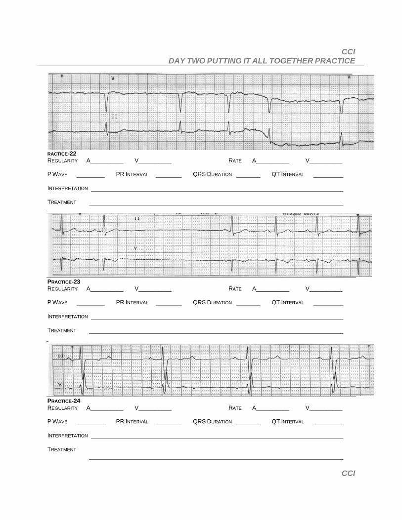
RACTICE-22
REGULARITY A__________ V__________ RATE A__________ V__________ P WAVE PR INTERVAL QRS DURATION QT INTERVAL
INTERPRETATION
TREATMENT
PRACTICE-23
REGULARITY A__________ V__________ RATE A__________ V__________ P WAVE PR INTERVAL QRS DURATION QT INTERVAL
INTERPRETATION
TREATMENT
PRACTICE-24
REGULARITY A__________ V__________ RATE A__________ V__________ P WAVE PR INTERVAL QRS DURATION QT INTERVAL
INTERPRETATION
TREATMENT
CCI
DAY TWO PUTTING IT ALL TOGETHER PRACTICE
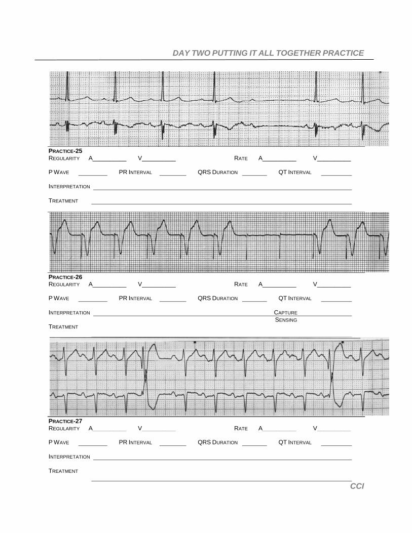
PRACTICE-25
REGULARITY A__________ V__________ RATE A__________ V__________ P WAVE PR INTERVAL QRS DURATION QT INTERVAL
INTERPRETATION
TREATMENT
PRACTICE-26
REGULARITY A__________ V__________ RATE A__________ V__________ P WAVE PR INTERVAL QRS DURATION QT INTERVAL
INTERPRETATION CAPTURE
SENSING TREATMENT
PRACTICE-27
REGULARITY A__________ V__________ RATE A__________ V__________ P WAVE PR INTERVAL QRS DURATION QT INTERVAL
INTERPRETATION
TREATMENT
CCI
DAY TWO PUTTING IT ALL TOGETHER PRACTICE
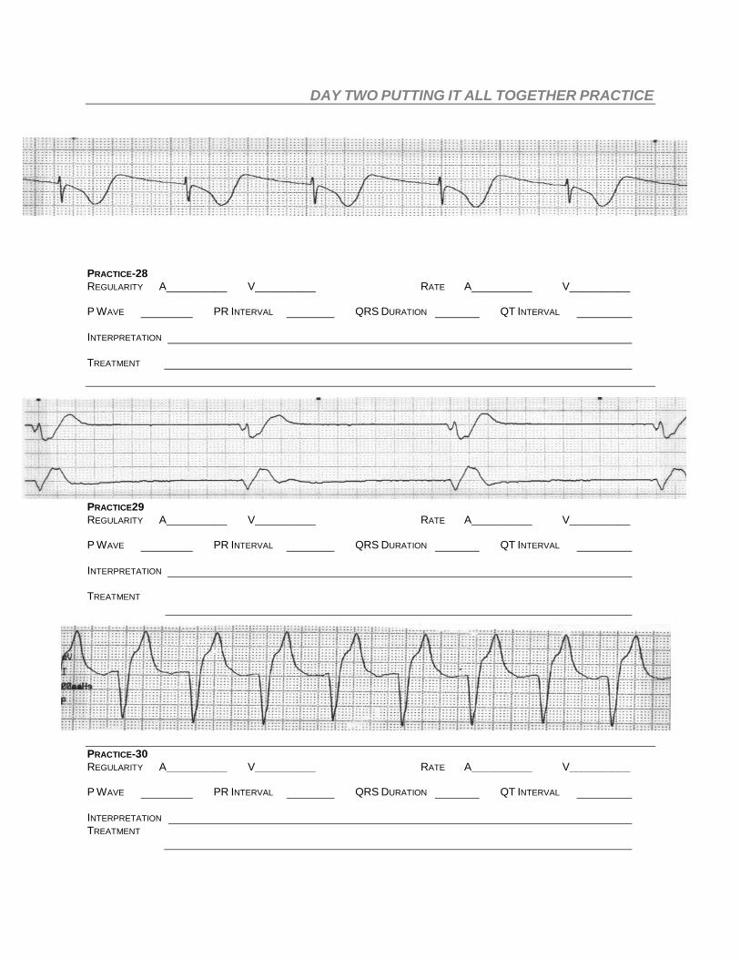
PRACTICE-28
REGULARITY A__________ V__________ RATE A__________ V__________ P WAVE PR INTERVAL QRS DURATION QT INTERVAL
INTERPRETATION
TREATMENT
PRACTICE29
REGULARITY A__________ V__________ RATE A__________ V__________ P WAVE PR INTERVAL QRS DURATION QT INTERVAL
INTERPRETATION
TREATMENT
PRACTICE-30
REGULARITY A__________ V__________ RATE A__________ V__________ P WAVE PR INTERVAL QRS DURATION QT INTERVAL
INTERPRETATION
TREATMENT
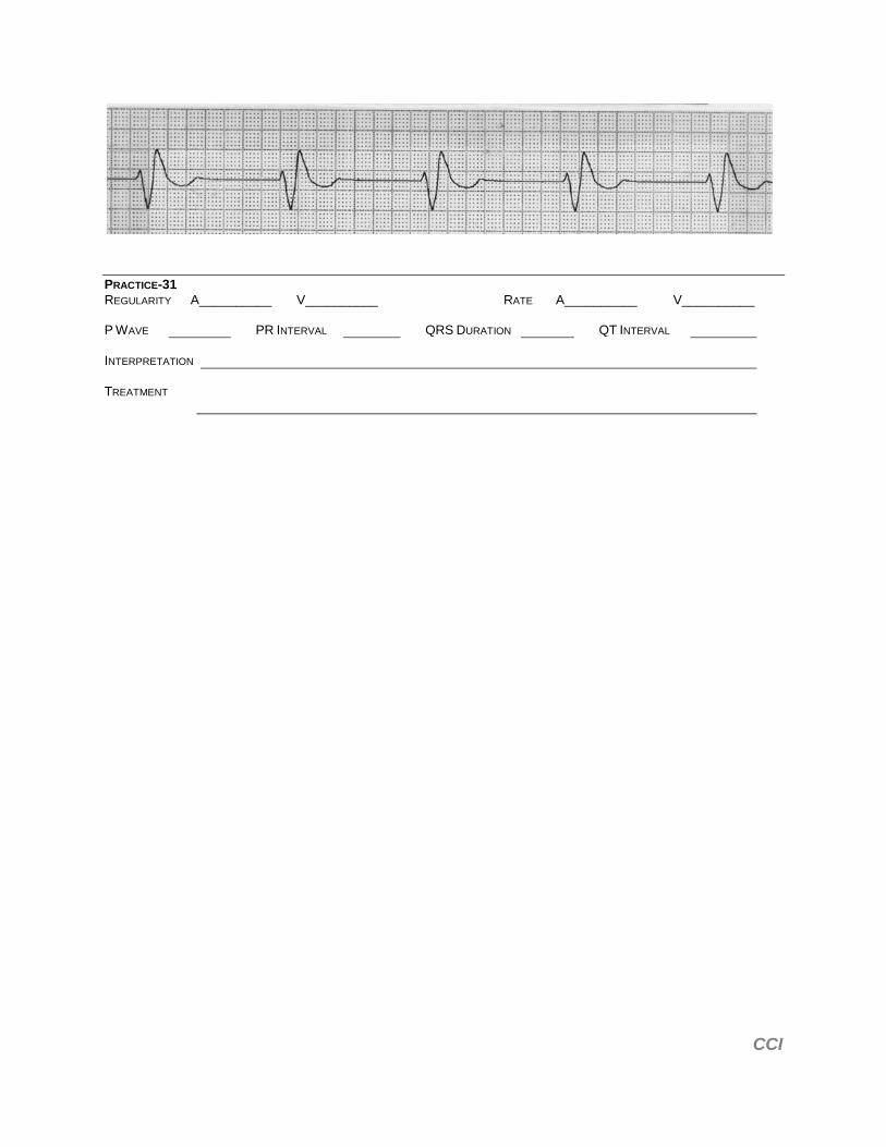
PRACTICE-31
REGULARITY A__________ V__________ RATE A__________ V__________ P WAVE PR INTERVAL QRS DURATION QT INTERVAL
INTERPRETATION
TREATMENT
CCI
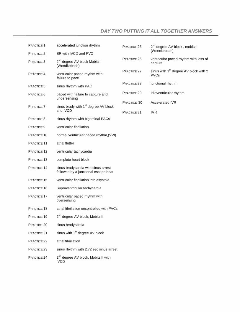
DAY TWO PUTTING IT ALL TOGETHER ANSWERS
PRACTICE 1 accelerated junction rhythm PRACTICE 2 SR with IVCD and PVC PRACTICE 3 2
nd degree AV block Mobitz I
(Wendkebach) PRACTICE 4 ventricular paced rhythm with failure to pace PRACTICE 5 sinus rhythm with PAC PRACTICE 6 paced with failure to capture and
undersensing PRACTICE 7 sinus brady with 1
st degree AV block
and IVCD PRACTICE 8 sinus rhythm with bigeminal PACs PRACTICE 9 ventricular fibrillation PRACTICE 10 normal ventricular paced rhythm,(VVI) PRACTICE 11 atrial flutter PRACTICE 12 ventricular tachycardia PRACTICE 13 complete heart block PRACTICE 14 sinus bradycardia with sinus arrest
followed by a junctional escape beat PRACTICE 15 ventricular fibrillation into asystole PRACTICE 16 Supraventricular tachycardia PRACTICE 17 ventricular paced rhythm with
oversensing PRACTICE 18 atrial fibrillation uncontrolled with PVCs PRACTICE 19 2
nd degree AV block, Mobitz II
PRACTICE 20 sinus bradycardia PRACTICE 21 sinus with 1
st degree AV block
PRACTICE 22 atrial fibrillation PRACTICE 23 sinus rhythm with 2.72 sec sinus arrest PRACTICE 24 2
nd degree AV block, Mobitz II with
IVCD
PRACTICE 25 2
nd degree AV block , mobitz I
(Wenckebach) PRACTICE 26 ventricular paced rhythm with loss of
capture PRACTICE 27 sinus with 1
st degree AV block with 2
PVCs PRACTICE 28 junctional rhythm
PRACTICE 29 Idioventricular rhythm
PRACTICE 30 Accelerated IVR
PRACTICE 31 IVR