edgewood - defense technical information · pdf fileedgewood chemical biological ... h. dupont...
TRANSCRIPT

EDGEWOOD CHEMICAL BIOLOGICAL CENTER
U.S. ARMY RESEARCH, DEVELOPMENT AND ENGINEERING COMMAND
ECBC-TR-700
MERCAPTO-OXIMES AS CAPTURE MOLECULES FOR DETECTION OF NERVE AGENTS
USING SURFACE-ENHANCED RAMAN SPECTROSCOPY
Kato L. Killops
DEPARTMENT OF CHEMISTRY AND BIOCHEMISTRY UNIVERSITY OF CALIFORNIA SANTA BARBARA
Santa Barbara, CA 93106
Roderick A. Fry William R. Creasy
5ML *"W StOTtftf to SOIWWTS
SCIENCE APPLICATIONS INTERNATIONAL CORPORATION
Gunpowder, MD 21010-0068
H. Dupont Durst Jason A. Guicheteau
RESEARCH AND TECHNOLOGY DIRECTORATE
June 2009
20090710285 Approved for public release; distribution is unlimited.
ABERDEEN PROVING GROUND, MD 21010-5424

Disclaimer
The findings in this report are not to be construed as an official Department of the Army position unless so designated by other authorizing documents.
REPORT DOCUMENTATION PAGE Form Approved
OMB No. 0704-0188 Public reporting burden for this collection of information is estimated to average 1 hour per response, including the time for reviewing instructions, searching existing data sources, gathering and maintaining the data needed, and completing and reviewing this collection of information. Send comments regarding this burden estimate or any other aspect of this collection of information, including suggestions for reducing this burden to Department of Defense, Washington Headquarters Services, Directorate for Information Operations and Reports (0704-0188). 1215 Jefferson Davis Highway, Suite 1204. Arlington, VA 22202-4302. Respondents should be aware that notwithstanding any other provision of law, no person shall be subject to any penalty for failing to comply with a collection of information if it does not display a currently valid OMB control number. PLEASE DO NOT RETURN YOUR FORM TO THE ABOVE ADDRESS.
1. REPORT DATE (DD-MM-YYYY)
XX-06-2009 2. REPORT TYPE
Final 3. DATES COVERED (From - To)
Jul 2008 - Sep 2008 4. TITLE AND SUBTITLE
Mercapto-oximes as Capture Molecules for Detection of Nerve Agents Using Surface-Enhanced Raman Spectroscopy
5a. CONTRACT NUMBER
DAAD13-03-D-0017
5b. GRANT NUMBER
5c. PROGRAM ELEMENT NUMBER
6. AUTHOR(S)
Killops, Kato L. (UCSB); Fry, Roderick A.; Creasy, William R. (SAIC); Durst, H. Dupont; and Guicheteau, Jason A. (ECBC)
5d. PROJECT NUMBER
5e. TASK NUMBER
5f. WORK UNIT NUMBER
7. PERFORMING ORGANIZATION NAME(S) AND ADDRESS(ES) Department of Chemistry and Biochemistry, University of California Santa Barbara, CA 93106 SAIC, P.O. Box 68, Gunpowder, MD 21010-0068 DIR, ECBC, ATTN: AMSRD-ECB-RT-PD/AMSRD-ECB-RT-CM/ AMSRD-ECB-RT-DL, APG, MD 21010-5424
8. PERFORMING ORGANIZATION REPORT NUMBER
ECBC-TR-700
9. SPONSORING / MONITORING AGENCY NAME(S) AND ADDRESS(ES) 10. SPONSOR/MONITOR'S ACRONYM(S)
11. SPONSOR/MONITOR'S REPORT NUMBER(S)
12. DISTRIBUTION / AVAILABILITY STATEMENT
Approved for public release; distribution is unlimited.
13. SUPPLEMENTARY NOTES
14. ABSTRACT
Two compounds, 4-methylthiobenzaldehyde oxime and 4-thiobenzaldehyde oxime, were synthesized for use as capture molecules for applications using surface-enhanced Raman as a means of detection. The thioether moiety was successfully attached to the silver nanoparticle substrate as evidenced by the change in the resulting spectrum. The oxime was shown to be reactive toward the nerve agent GD (Soman) in basic solution, as
31 ¥ monitored by P NMR. 15. SUBJECT TERMS
SERS Ag nanoparticle Thiol Nerve agent
16. SECURITY CLASSIFICATION OF:
a. REPORT
U b. ABSTRACT
u c. THIS PAGE
u
17. LIMITATION OF ABSTRACT
UL
18. NUMBER OF PAGES
15
19a. NAME OF RESPONSIBLE PERSON
Sandra J.Johnson
19b. TELEPHONE NUMBER (include area code)
(410)436-2914 Standard Form 298 (Rev. 8- 98)Prescribed by ANSI Std. Z39.18

Blank

PREFACE
The work described in this report was authorized under Contract No. DAAD13- 03-D-0017. The work was started in July 2008 and completed in September 2008.
The use of either trade or manufacturers' names in this report does not constitute an official endorsement of any commercial products. This report may not be cited for purposes of advertisement.
This report has been approved for public release. Registered users should request additional copies from the Defense Technical Information Center; unregistered users should direct such requests to the National Technical Information Service.
Acknowledgments
The authors would like to thank Christine Franklin for assistance in formatting this report. The authors gratefully acknowledge funding from the Science, Mathematics, and Research for Transformation Fellowship sponsored by the Department of Defense.

Blank
CONTENTS
1. INTRODUCTION 7
2. EXPERIMENTAL PROCEDURES 7
2.1 4-Methylthiobenzaldehyde Oxime (2) 7 2.2 4-Thiobenzaldehyde (3) 8 2.3 4-Thiobenzaldehyde Oxime (4) 8 2.4 3,PNMR 8 2.5 Saturation of Ag Nanoparticles 9
3. RESULTS AND DISCUSSION 8
3.1 "PNMR Studies with GD 9 3.2 SERSof2 10 3.3 Saturation of 2 to Silver Colloid 10
4. CONCLUSION 10
LITERATURE CITED 15

FIGURES
1. 31P NMR of (a) GD with 2 and No Base after 20 Min (b) GD with 2 and DIPEA 2 Min after Adding Base (c) GD with DIPEA 30 Min after Addition of Base, Pinacolyl Methylphosphonic Acid(PMPA) 12
2. Normal Raman of Solid 2 and 2 Dissolved in MeOH, and Surface-enhanced Raman of 2 in Ag Colloid Solution 13
3. Preliminary Saturation Curve of 2 onto Ag Nanoparticles 13
SCHEME
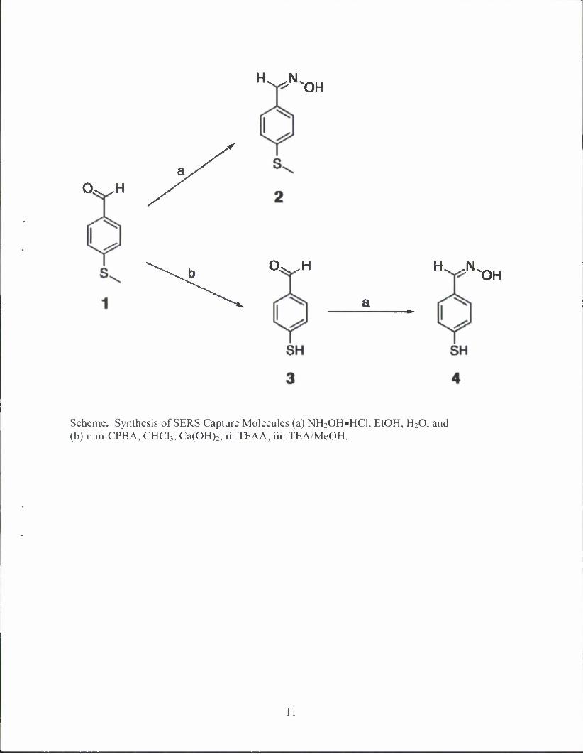
Synthesis of SERS Capture Molecules (a) NH2OH«HCl, EtOH, H20, and (b) i: m-CPBA, CHC13, Ca(OH)2, ii: TFAA, Hi: TEA/MeOH 1

MERCAPTO-OXIMES AS CAPTURE MOLECULES FOR DETECTION OF NERVE AGENTS USING SURFACE-ENHANCED RAMAN SPECTROSCOPY
1. INTRODUCTION
Surface-enhanced Raman spectroscopy (SERS) is emerging as a useful technique for detection and monitoring of potentially hazardous materials. To produce the SERS effect, the analyte of interest must be brought into close proximity of a nanostructured metallic substrate (typically gold or silver). Capture molecules are often used in biological detection to immobilize antibody proteins to the surfaces of SERS active substrates. Capture molecules are dual- functionalized with a thiol moiety for binding to the metal surface and a reactive group for subsequent attachment to the compound of interest or analyte. The reaction between the capture molecule and the analyte produces a unique change in the SER spectrum, which can be used for identification and detection of analytes. The capture molecules prepared for this study were para-substituted benzenes with a thiol or thioether and an oxime. Oximes have been widely used for the treatment of poisoning due to exposure to organophosphorus compounds, such as chemical warfare (CW) agents.2 The capture molecules were characterized using 'H NMR, 13C NMR, and gas chromatography-mass spectrometry (GC-MS). Preliminary data were collected on the reaction between the capture molecule, 4-methylthiobenzaldehyde oxime (2), and the nerve agent GD (Soman). Furthermore, attachment of 2 to the surface of Ag nanoparticles was confirmed with SERS.
2. EXPERIMENTAL PROCEDURES
All chemicals were purchased from Aldrich and used without further purification. The 'H NMR and l3C NMR were recorded on a JEOL ECX 400 MHz spectrometer. The 31P NMR spectra were recorded on a Bruker DPX-300 MHz Ultrashield spectrometer with a 3IP switchable QNP probe. Chemical shifts are reported in parts per million (5) and all spectra were recorded at ambient temperature. The GC-MS was run on an Agilent 6890N network GC system with an Agilent 5975 inert mass selective detector. Liquid chromatography-mass spectrometry (LC-MS) was done with an Agilent 1100 LC/MSD equipped with an electrospray source using a reversed phase gradient from 90% aqueous ammonium acetate buffer to methanol, using a Phenomenex Aqua C18 RP column. SER spectra were recorded using a high resolution (1-2 cm"1) fiber optically coupled (InPhotonics RamanProbe) EIC Echelle spectrograph operating at 785 nm with a laser output of ca. 130mW.
2.1 4-Methylthiobenzaldehyde Oxime (2).
Compound 2 was synthesized according to a literature procedure.3 Specifically, 0.61 g (4.0 mmol) 4-methylthiobenzaldehyde (1) was added to a round bottom flask, dissolved in 15 mL ethanol, and heated to 70 °C. A solution of 7.42 g NH2OHHCl dissolved in 20 mL deionized H20 was added to the reaction flask. The reaction mixture was stirred at 70 °C for 1 hr before cooling to room temperature. The solution was diluted with 50 mL H20, extracted with ethyl acetate (3 * 50 mL), and dried over anhydrous MgS04. The solvent was evaporated

to give the product as a light yellow powder (600 mg, 90%). *H NMR (400 MHz, CDC13) 5 (ppm): 8.08 (s, -C//=N-OH), 7.22-7.48 (m, aromatic), 2.49 (s, -S-C//3), 1.98 (b, =N-07/). 13C NMR (400 MHz, CDCI3) 5 (ppm): 150.0, 141.4, 128.6, 127.4, 126.2, 15.4. Calculated: C8H9NOS m/z = [M]+ 167.04; Found: EI-MS: m/z = [M]+* 167.
2.2 4-Thiobenzaldehyde (3).
Compound 3 was synthesized according to a literature procedure.4 Specifically, 1 (1.26 g, 8.28 mmol) was dissolved in 100 mL of CHC13 and cooled to 0 °C before 2.13 g (12.3 mmol) of m-chloroperoxybenzoic acid was added. The mixture was stirred for 1 hr at 0 °C and warmed to room temperature before 0.92 g Ca(OH)2 (12.4 mmol) was added and stirred for an additional 30 min. The mixture was filtered, and the solvent was evaporated before adding 15 mL of trifluoroacetic anhydride (TFAA,108 mmol) and refluxing for 45 min. The TFAA was removed by evaporation before adding 80 mL of a 50:50 mixture of triethylamine and methanol. The solution was evaporated to dryness before diluting with CHCI3 and extracting with saturated NH4CI solution (2 x 50 mL). The organic fractions were combined and dried over anhydrous MgS04, and the solvent was evaporated to give a yellow solid as the crude product. The compound was purified using column chromatography with a gradient of hexanes:ethyl acetate starting at 4:1 to give 552 mg of light yellow viscous liquid (48% yield). 'H NMR (400 MHz, CDCI3) 5 (ppm): 9.88 (s, 1H, C//=N-OH), 7.40-7.67 (m, 4H, aromatic), 4.57 (s, 1H -SH). 13C NMR (400 MHz, CDC13) 5 (ppm): 191.2, 144.3, 134.4, 130.1, 129.3, 57.4. Calculated: C7H6OS m/z = [M]+ 138.01; Found: EI-MS: m/z = [M-H]+ 137, [Mf 138.
2.3 4-Thiobenzaldehyde Oxime (4).
Compound 4 (250 mg, 0.96 mmol) was dissolved in 10 mL of ethanol and heated to 70 °C. The 3.2 g (46 mmol) of NH20H»HC1 was dissolved in 12 mL of water before adding it to the reaction mixture. The solution was stirred for 1 hr at 70 °C before cooling to room temperature. The reaction mixture was diluted with water (50 mL) and extracted with ethyl acetate (3 x 50 mL). The organic fractions were combined, dried over anhydrous MgSO*4, and evaporated to dryness to give 280 mg (96% yield) of light yellow powder. 'H NMR (400 MHz, c/3-methanol) 5 (ppm): 7.99 (s, 1H, C//=N-OH), 7.22-7.52 (m, 4H, aromatic), 4.85 (s, 1H, SH). 13C NMR (400 MHz, J3-methanol) 8 (ppm): 148.3, 134.0, 130.0, 128.4, 127.1. Calculated: C7H7NOS m/z = [M]+ 153.02; Found: EI-MS: m/z = [M]+* 153.
2.4 31PNMR.
Compound 2 (20 mg, 0.12 mmol) was dissolved in a 50:50 mixture of acetonitrile and water and 1 uL of GD (5.6 x 10"3 mmol) was added. Since no change was observed after 20 min, 20 uL of diisopropylethylamine (DIPEA) was added to make the solution basic. The spectrum of the basic solution was recorded after 2 min. A control experiment was performed using the same conditions as above without the addition of 2, and the spectrum was recorded after 30 min.

2.5 Saturation of Ag Nanoparticles.
The Ag colloid was prepared according to literature procedure.5'6 A solution of 0.25 mg/mL 2 was prepared with 50:50 methanol/water. Added to a 1 cm quartz cuvette was 500 uL of Ag colloid, 10 uL of 1 M NaCl solution, and a blank Raman spectrum was recorded. The solution of 2 was added in 10 \xL aliquots to the cuvette, shaken gently, and allowed to equilibrate for 1 min before each spectrum was recorded.
3. RESULTS AND DISCUSSION
Our approach was to create a simple molecule that contains a nucleophilic moiety, as well as a thiol for attachment to SERS active metal surfaces, such as Ag nanoparticles. An oxime was chosen as the nucleophile because it has been shown to successfully react with the phosphorus center of CWAs.7 The affinity of mercaptans toward SERS active metals like Au or Ag has been well documented.
The synthesis of 2 was achieved by reacting commercially available 4-methyl- thiobenzaldehyde (1) with hydroxylamine hydrochloride in an ethanol/water solution at 70 °C. Oxime formation was verified in the 'H NMR, where the aldehyde proton had shifted from 9.91 ppm to the benzylic region at 8.08 ppm. Compound 2 was used without further purification, as it was shown to be ca. 98% pure by GC-MS. Compound 3, a precursor to 4, was synthesized according to a procedure by Coombs and coworkers, and after removing the methyl group, the oxime 4 was synthesized using the same procedure as compound 2. The reactions are shown in the Scheme. Although compound 4 was not used in these preliminary tests, future work will include reacting 4 with agent and saturation studies with silver colloid.
3.1 31P NMR Studies with GD.
To examine the reaction between the oxime and GD, compound 2 was dissolved in 1 mL of a 50:50 mixture of acetonitrile and water, and 1 uL GD was added to the solution. A 31P NMR spectrum was taken 20 min after the addition of GD, and no reaction had occurred. However, it is well known that oximes require activation, often through the addition of a base, to become nucleophilic enough to react with G-agents.9 Immediately after the addition of 10 uL of the base DIPEA, a signal appeared in the spectrum corresponding to the product of a reaction between GD and 2, and the original GD signal had disappeared (Figure 1). The peak at 24 ppm corresponds to a small amount of the hydrolysis product, pinacolyl methyl-phosphonic acid (PMPA). A control experiment was performed to confirm that the signal in the 'P NMR was indeed due to a reaction between GD and 2 and not a simple product of hydrolysis. For the control, 1 uL of GD was added to the 50:50 acetonitrile/water mixture. The spectrum, which was recorded after a period of 30 min, revealed the presence of residual GD, as well as a new peak from the hydrolysis product PMPA at 24 ppm (Figure 1). By comparing the control spectrum to that of the reaction product of 2 with GD, it is evident that hydrolysis of GD in basic solution is much slower, and suggests that GD reacts preferentially with 2. Further confirmation of the reaction between 2 and GD was provided by running the NMR solution on LC-MS, where both the [M + H]+ and [M + Na]+ peaks were identified at m/z = 330 and m/z = 352, respectively.

3.2 SERSof2.
Ag colloid solutions were prepared according to the modified procedure of Lee and Meisel,5 detailed in the work of Guicheteau and coworkers.6 A normal Raman spectrum was obtained from the solid 2 (Figure 2). To observe the SER effect, a 1 mg/mL solution of 2 in methanol was added to 500 uL of Ag colloid, and a spectrum was recorded. For comparison, a spectrum of 2 dissolved in methanol (without Ag colloid) was also recorded and only a few broad peaks, primarily attributed to methanol, can be observed. In contrast, the SER spectrum of 2 shows many of the same peaks observed in the solid sample, although they appear to be slightly shifted and somewhat broadened. Clearly visible are the aromatic C—C vibrations at ca. 1600 cm"1, the aromatic C—S vibration at ca. 1100 cm"1, and the peak at ca. 1250 cm"1 due to the methyl group, among many others.
3.3 Saturation of 2 to Silver Colloid.
Preliminary saturation experiments were performed to calculate the amount of surface coverage for molecule 2 needed to form a monolayer around the silver nanoparticles. A Langmuir isotherm was constructed using peak area measurements from the 1185 cm"1 mode (Figure 3). A noticeable leveling off is observed between the 28th and 31st additions yielding a surface coverage concentration of approximately 0.13 mg/mL. Additional experiments need to be performed in the lower end of the curve (below 0.05 mg/mL) and in the area of potential saturation.
4. CONCLUSION
Two compounds bearing thiols and oximes were synthesized for use as surface- enhanced Raman spectroscopy (SERS) capture agents. Compound 2 was shown to react quickly with GD under basic conditions, as monitored by 31P NMR. As evidenced by the SER spectrum, compound 2 was also shown to bind with Ag nanoparticles in aqueous solution.
10
H^NV
O^^H a
OH
O^^H HL ^N OH
Scheme. Synthesis of SERS Capture Molecules (a) NH2OH»HCl, EtOH, H20, and (b) i: m-CPBA, CHC13, Ca(OH)2, ii: TFAA, iii: TEA/MeOH.
11

w<iWM*^ (a)
o
CH3
IW|#^^^
(b) /^/M^M-rt^A"**^^
(C)
o
Hcr^o CH3
n—i—i—i—i—i—i—r 30 20
-i—i—|—i—r 10 [ppm]
i i i |—
40
Figure 1. 3IP NMR of (a) GD with 2 and No Base after 20 Min (b) GD with 2 and DIPEA 2 Min after Adding Base (c) GD with DIPEA 30 Min after Addition of Base, Pinacolyl Methylphosphonic Acid (PMPA). All solutions were a 50:50 mixture of acetonitrile and water.
12

*ULJULJLA-J
Normal Raman (solid)
_LA—OV.
Normal Raman (in MeOH)
SERS
500 1000 1500 2000 Raman shift (cm-1)
2500 3000
Figure 2. Normal Raman of Solid 2 (Top) and 2 Dissolved in MeOH (Center), and Surface-Enhanced Raman of 2 in Ag Colloid Solution (Bottom).
Peak at 1185cm1
20000
15000
10000
••
• • »•
» • •
• • .••••••.•..
5000 • . * • •
•
• •
n •
( ) 0.02 0.04 0.06 0.08 0.1 0.12
Concentration (mg/mL)
0.14 0.16 0.18
Figure 3. Preliminary Saturation Curve of 2 onto Ag Nanoparticles.
13

Blank
14
LITERATURE CITED
1. Porter, M.D.; Lipert, R.J.; Siperko, L.M.; Wang, G. SERS as a Bioassay Platform: Fundamentals, Design, and Applications. Chem. Soc. Rev. 2008, 37, 1001-1011.
2. Antonijevic, B.; Stojiljkovic, M. Unequal Efficacy of Pyridinium Oximes in Acute Organophosphate Poisoning, Clin. Med. Res. 2007, 5, 71-82.
3. Cullen, M.D. Synthesis and Biological Evaluation of Alkenyldiarylmethane HIV-1 Non-Nucleoside Reverse Transcriptase Inhibitors that Possess Increased Hydrolytic Stability. J. Med. Chem. 2007, 50, 4854-4867.
4. Young, R.N.; Gauthier, J.Y.; Coombs, W. The Methyl Group as a Protecting Group for Arylthiols: A Mild and Efficient Method for the Conversion of Methyl Aryl Sulfides to Arylthiols. Tet. Lett. 1984,25,1753-1756.
5. Lee, P.C.; Meisel, D. Adsorption and Surface-Enhanced Raman of Dyes on Silver and Gold Sols. J. Phys. Chem. 1982, 86, 3391-3395.
6. Guicheteau, J.; Argue, L.; Emge, D.; Hyre, A.; Jacobson, M.; Christesen, S. Bacillus Spore Classification via Surface-Enhanced Raman Spectroscopy and Principal Component Analysis. Appl. Spectrosc. 2008, 62, 267-272.
7. Lamb, J.C.; Steinberg, G.M.; Solomon, S.; Hackley Jr., B.E. Reaction of 4-Formyl-1 -methylpyridinium Iodide Oxime with Isopropyl Methylphosphonofluoridate. Biochemistry 1965, 4, 2476-2484.
8. Kudelski, A. Characterization of Thiolate-Based Mono- and Bilayers by Vibrational Spectroscopy: A Review. Vib. Spectrosc. 2005, 39, 200-213.
9. Swidler, R.; Plapinger, R.; Steinberg, G.M. The Kinetics of the Reaction of Isopropyl Methylphosphonofluoridate (Sarin) with Substituted Benzohydroxamic Acids. J. Am. Chem. Soc. 1959, 81, 3271-3274.
15