edinburgh research explorer · 312 j. deretic | et al. molecular biology of the cell a rapid...
TRANSCRIPT

Edinburgh Research Explorer
A rapid computational approach identifies SPICE1 as an Aurorakinase substrate
Citation for published version:Deretic, J, Kerr, A & Welburn, JPI 2019, 'A rapid computational approach identifies SPICE1 as an Aurorakinase substrate', Molecular Biology of the Cell, vol. 30, no. 3, pp. 312-323.https://doi.org/10.1091/mbc.E18-08-0495
Digital Object Identifier (DOI):10.1091/mbc.E18-08-0495
Link:Link to publication record in Edinburgh Research Explorer
Document Version:Publisher's PDF, also known as Version of record
Published In:Molecular Biology of the Cell
Publisher Rights Statement:© 2019 Deretic et al. This article is distributed by The American Society for Cell Biology under license from theauthor(s). Two months after publication it is available to the public under an Attribution–Noncommercial–ShareAlike 3.0 Unported Creative Commons License (http://creativecommons.org/licenses/by-nc-sa/3.0).
General rightsCopyright for the publications made accessible via the Edinburgh Research Explorer is retained by the author(s)and / or other copyright owners and it is a condition of accessing these publications that users recognise andabide by the legal requirements associated with these rights.
Take down policyThe University of Edinburgh has made every reasonable effort to ensure that Edinburgh Research Explorercontent complies with UK legislation. If you believe that the public display of this file breaches copyright pleasecontact [email protected] providing details, and we will remove access to the work immediately andinvestigate your claim.
Download date: 25. Dec. 2019

312 | J. Deretic et al. Molecular Biology of the Cell
A rapid computational approach identifies SPICE1 as an Aurora kinase substrate
ABSTRACT Aurora kinases play a major role in mitosis by regulating diverse substrates. Defining their critical downstream targets is important in understanding Aurora kinase func-tion. Here we have developed an unbiased computational approach to identify new Aurora kinase substrates based on phosphorylation site clustering, protein localization, protein struc-ture, and species conservation. We validate the microtubule-associated proteins Clasp2, Elys, tubulin tyrosine ligase-like polyglutamylase residues 330–624 and spindle and centriole as-sociated protein 1, residues 549–855 (SPICE1), as Aurora A and B kinases substrates in vitro. We also demonstrate that SPICE1 localization is regulated by Aurora kinases during mitosis. In the absence of Aurora kinase activity, SPICE1 remains at centrioles but does not target to the spindle. Similarly, a nonphosphorylatable SPICE1 mutant no longer localizes to the spin-dle. Finally, we show that misregulating SPICE1 phosphorylation results in abnormal centriole number, spindle multipolarity, and chromosome alignment defects. Overall, our work indi-cates that temporal and spatial Aurora kinase–mediated regulation of SPICE1 is important for correct chromosome segregation. In addition, our work provides a database-search tool that enables rapid identification of Aurora kinase substrates.
INTRODUCTIONPhosphorylation of hundreds of proteins by a small number of mitotic kinases drives correct chromosome segregation (Dephoure et al., 2008; Olsen et al., 2010). The Aurora kinase family plays an important role in cell division and the maintenance of genomic integrity. In par-ticular, Aurora A and B kinases control mitotic events to ensure ge-nomic stability (reviewed in Welburn and Jeyaprakash, 2018). Aurora kinases are serine/threonine kinases that share a common substrate preference. Consequently, the substrate specificity and the function of Aurora kinases A and B are largely governed by their subcellular localization (Fu et al., 2009; Hans et al., 2009). During mitosis, Aurora A is tethered at centrosomes, where it controls bipolar spindle as-sembly and maintenance. In contrast, Aurora B is bound to chromo-somes and then restricted to centromeres during metaphase before relocating to the spindle midzone during anaphase and midbody and
the cell cortex during cytokinesis. Aurora B promotes chromosome condensation and biorientation, controls the attachment of kineto-chores to microtubules, and regulates abscission during cytokinesis (reviewed in Welburn and Jeyaprakash, 2018).
Proteomic studies indicate that Aurora kinases phosphorylate a large number of substrates during mitosis (Kettenbach et al., 2011; Santamaria et al., 2011). Yet the direct identification of Aurora kinase substrates and their subsequent functional analysis remains difficult. Typically Aurora kinases phosphorylate serines and threonines that fit the consensus site [KR]-[KR]-[S/Tp]-[Φ], although the substrate se-lectivity at P+1 is weaker (Cheeseman et al., 2002; Kettenbach et al., 2011) (Figure 1A). Phosphorylation often occurs in unstructured re-gions of a protein, disrupting the region electrostatically and creat-ing or abolishing binding sites. Some of the known substrates are Kif2c/MCAK (mitotic centromere-associated kinesin), components of the KMN network (Knl1, Mis12 complex, Ndc80 complex) (Ndc80, Dsn1, and Knl1), other kinetochore components (CENP-C, CENP-E, INCENP), and centrosomal proteins such as Tpx2 (Andrews et al., 2004; DeLuca et al., 2006; Okada et al., 2006; Kim et al., 2010; Welburn et al., 2010).
To predict novel physiological substrates of Aurora kinases, we generated a computational tool to identify Aurora kinase substrates based on the consensus sequence for the Aurora kinase (further described under Materials and Methods). This search tool allows
Monitoring EditorLaurent BlanchoinCEA Grenoble
Received: Aug 30, 2018Revised: Nov 8, 2018Accepted: Nov 20, 2018
This article was published online ahead of print in MBoC in Press (http://www .molbiolcell.org/cgi/doi/10.1091/mbc.E18-08-0495) on November 28, 2018.*Address correspondence to: Julie P. I. Welburn ([email protected]).
© 2019 Deretic et al. This article is distributed by The American Society for Cell Biol-ogy under license from the author(s). Two months after publication it is available to the public under an Attribution–Noncommercial–Share Alike 3.0 Unported Creative Commons License (http://creativecommons.org/licenses/by-nc-sa/3.0).“ASCB®,” “The American Society for Cell Biology®,” and “Molecular Biology of the Cell®” are registered trademarks of The American Society for Cell Biology.
Abbreviations used: MCAK, mitotic centromere-associated kinesin; SPICE1, spindle and centriole associated protein 1.
Jovana Deretic, Alastair Kerr, and Julie P. I. Welburn*Wellcome Trust Centre for Cell Biology, School of Biological Sciences, University of Edinburgh, Edinburgh EH9 3JR, Scotland, UK
MBoC | BRIEF REPORT

Volume 30 February 1, 2019 SPICE1 is an Aurora kinase substrate | 313
filtering of potential substrates using various parameters such as the number of these consensus sites clustered within a region, presence and category in the Gene Ontology database, level of conservation across species, and level of disorder in the region containing the patterns. These parameters are controlled by the user interface and offers great search flexibility. We validated a number of predicted substrates in vitro using kinase phosphorylation assays. We also confirmed the spindle and centriole associated protein 1, residues 549–855 (SPICE1), as a novel substrate of Aurora kinases in cells. Overall, this computational tool enables a rapid search of the pro-teome to identify novel Aurora substrates and will be an excellent resource for the research community.
RESULTS AND DISCUSSIONTo search for novel Aurora kinase substrates, we established a species filter so that a potential substrate can be selected de-pending on Aurora kinase sites present in only up to three meta-zoan species (humans, zebrafish, and chickens) (Figure 1, A and B). We also enabled our bioinformatics tool to select data on the basis of predicted structural disorder, number of Aurora consen-sus sites clustered with a small region, tissue specificity, and gene ontology (GO) terms. We designed an interface that allows the search and classification of potential substrates across
species based on any combination of these parameters (Figure 1, B and C).
We tested our approach by identifying known substrates in our search. We applied a species filter so that a potential substrate was only selected if at least three Aurora kinase sites were present in three metazoan species (humans, zebrafish, and chickens). Ndc80 and Kif2c (also known as MCAK) are known to be phosphorylated by Aurora A and B on multiple sites (Andrews et al., 2004; Cheese-man et al., 2006; DeLuca et al., 2006). Searching for substrates that contained at least three consensus sites clustered within a 100-amino-acid stretch, we found Ndc80 contains 11 Aurora con-sensus sites, including multiple sites in its N terminus previously identified (Cheeseman et al., 2006; DeLuca et al., 2006). Human Kif2c/MCAK is predicted to have seven Aurora consensus sites, four of which have been previously identified as Aurora B kinase sites and two as Aurora A kinase sites (Andrews et al., 2004; Zhang et al., 2008). Searching for substrates that contained at least three Aurora consensus sites clustered within a 100-amino-acid stretch, we found 85 and 55 candidates with GO terms mitotic cell cycle and microtubules, respectively. The top candidates are listed in Figure 1C. A number of these proteins have been previously identified as Aurora substrates, validating our approach. We then focused on candidates that were known to localize close to
FIGURE 1: Bionformatics and validation of candidate substrates by in vitro phosphorylation assays. (A) Consensus phosphorylation motif and sequence bias in validating putative phosphorylation sites of Aurora kinases. Motif logo was generated using http://meme-suite.org (Bailey et al., 2009) by submitting 13-amino-acid-long sequences of known Aurora kinase phosphorylation sites. (B) Schematic diagram of bioinformatic design with nonlinear flow of available filters. (C) Example of list of candidates derived from bioinformatics based on the presence of at least three consensus patterns within a 100-amino-acid region of a protein and based on biological process or localization (GO term). Numbers in brackets show predicted phosphorylation sites. (D) Coomassie-stained gels and corresponding autoradiogram showing the protein levels and levels of phosphorylation for the Ndc80 bonsai complex, ELYS1858-2014 (UniProt ID Q8WYP5), CLASP2741-818 isoform X15 (NCBI XP_006713112.1), SPICE1549-855 (Q8N0Z3), TTLL4330-624 (Q14679), and KIAA14681-292 Cra_a isoform (Q96ES0), respectively. Short constructs of candidate substrates containing predicted phosphorylation were mixed with Ipl1 (yeast Aurora B kinase), buffer only, or Aurora A kinase in the presence of 32P-ATP. (E) Schematic diagram of SPICE1 showing the position of the coiled-coil domains (cc, red) and the predicted phosphorylation sites (red disks). (F) Coomassie-stained gel and corresponding autoradiogram showing SPICE1 (fragment 549–855), SPICE15A, and SPICE17A in the presence of Ipl1 and 32P-ATP.

314 | J. Deretic et al. Molecular Biology of the Cell
Aurora A and Aurora B kinases in mitosis but had not so far been identified as Aurora substrates.
To test whether our bioinformatics tool enabled reliable identifi-cation of novel Aurora substrates, we selected five mitotic candi-dates that were predicted as Aurora substrates containing clusters of phosphorylation sites (at least three phosphorylation sites within 100 amino acids) but not previously reported as Aurora kinase substrates. We purified these recombinantly expressed proteins and used in vi-tro kinase assays to test whether they were phosphorylated by Au-rora A and the yeast Aurora B kinase Ipl1. As a positive control, we used the Ndc80 bonsai complex, which is phosphorylated in the N terminus by Aurora kinases (Cheeseman et al., 2006; DeLuca et al., 2006; Ciferri et al., 2008; Ye et al., 2015). Ndc80 was phosphorylated by both Aurora A and Ipl1 (Figure 1D). Then we tested the nucleopo-rin Elys and microtubule binding protein Clasp2, which both localize to kinetochores in mitosis. Elys and Clasp2 were predicted to have 26 and 19 phosphorylation sites in total. We recombinantly ex-pressed and purified domains of Elys (residues 1818–2014) and Clasp2 (residues 741–818), containing multiple predicted phosphor-ylation sites (4 and 5, respectively). Both appeared strongly phos-phorylated by Aurora kinases (Figure 1D). Interestingly, the Clasp2 domain is in the microtubule-binding glutamate-arginine–rich re-gion, containing two EB-binding SXIP motifs and heavily regulated by glycogen synthase kinase 3 (GSK3) phosphorylation (Kumar and Wittmann, 2012). Aurora kinases A and B also phosphorylate tubulin tyrosine ligaselike polyglutamylase residues 330–624 (TTLL4), which regulates microtubule posttranslational modifications (Figure 1D). We found that Aurora kinases A and B also phosphorylate the C terminus of SPICE1 (Figure 1, D and E). Finally, the role of KIAA1468 is not known; however, we found this protein had sequence similari-ties to the microtubule-binding protein tumor overexpressed gene (TOG), comprising a number of TOG-like domains. We found that the N terminus of KIAA1468 (residues 1–292) was phosphorylated by both Ipl1 and Aurora A kinase.
We focused further on SPICE1 as an Aurora substrate and gener-ated a nonphosphorylatable SPICE15A, with the predicted phos-phorylated residues mutated to alanines (residues T557, S738, T780, S797, and S810; Figure 1E). T557 has also previously been found phosphorylated in vivo (Moritz et al., 2010; Hornbeck et al., 2015). Additionally, two sites neighboring the predicted phosphory-lation sites: T798 and S811 also match the Aurora consensus closely and have been reported to be phosphorylated in mass spectrome-try studies (Dephoure et al., 2008; Moritz et al., 2010; Hornbeck et al., 2015). Aurora kinases are capable of phosphorylating con-secutive target residues such as the TSS motif in INCENP (Bishop and Schumacher, 2002; Honda et al., 2003). Thus these sites were included to make a SPICE17A construct (Figure 1E). Phosphorylation of SPICE15A by the yeast Aurora B kinase Ipl1 was strongly reduced, while phosphorylation of SPICE17A was almost abolished, indicating that the seven sites were Aurora kinase phosphorylation sites in vitro (Figure 1F). In total, our bioinformatics approach predicts Aurora kinase substrates in vitro accurately and enables the discovery of new mitotic substrates of Aurora kinases.
We then investigated whether these substrates were regulated by Aurora kinases in vivo and focused on SPICE1. Since the C termi-nus of SPICE1 is phosphorylated in vitro by Aurora kinases (Figure 1F), we examined the role of the C terminus of SPICE1 on its cellular localization in mitosis. Full-length SPICE1 targets to the spindle and centrioles, in agreement with previous work, while SPICE1444-855 failed to localize (Figure 2A) (Archinti et al., 2010). SPICE11-550 local-ized to the spindle and centrioles (Figure 2A) (Archinti et al., 2010), however its spindle localization was weakened when compared with
full-length SPICE1 (Figure 2, B and C). This indicates that the C ter-minus of SPICE1 enhances its targeting to the spindle, despite not targeting to microtubules directly (Figure 2). SPICE11-550 failed to cause centriole amplification whilst the overexpression of full-length SPICE1 caused supernumerary centrioles (Figure 2, C and D), indicating that the C terminus of SPICE1 contributes positively to control of centriole number.
We established functional assays to test how Aurora kinases may regulate SPICE1 function through C-terminal phosphorylation. We first generated an inducible SPICE1 knockout cell line using CRISPR/Cas9-mediated gene targeting (McKinley and Cheeseman, 2014). After 96 h of Cas9 induction, we examined the levels of SPICE1 depletion (Supplemental Figure S1, A and B). SPICE1 depletion was largely successful (Supplemental Figure S1B). In control cells, 82% of untreated cells had a total of four centrioles, as marked by centrin foci staining (Supplemental Figure S1C). The number of centrioles was increasingly abnormal in doxycycline-treated SPICE1-knockout cells with 48% cells having more or less than four centrin foci (Supplemental Figure S1C). Chromosomes showed increased misalignment, 51% misaligned in SPICE1-knockout cells, while only 25% of untreated cells displayed misaligned chromosomes (Supple-mental Figure S1D). There were also spindle organization defects and multipolarity in 31% of SPICE1-knockout cells, while only 7% of untreated cells displayed multipolar spindles (Supplemental Figure S1E).
We then used silencing RNA (siRNA) to deplete SPICE1 and con-firm the knockout phenotype observed for SPICE1 (Supplemental Figure S1, F and G). While 90% of control cells displayed four cen-trioles, only 65% of SPICE1 RNA interference (RNAi)-depleted cells had four centrioles (Supplemental Figure S1H) and 27% of cells had fewer than four centrioles. Respectively, 8 and 48% of control and SPICE1-RNAi-depleted cells displayed misaligned chromosomes (Supplemental Figure S1I). We found that on successful depletion of SPICE1, there was an increase in frequency of multipolar spindles, with 21% of cells displaying a multipolar spindle phenotype (Sup-plemental Figure S1J). Overall, the phenotype of SPICE1 knockout was similar to the published RNAi-based SPICE1 knockdown and our SPICE1 siRNA depletion (Archinti et al., 2010).
Since SPICE1 is phosphorylated in vitro by Aurora kinases, we wanted to test whether SPICE1 localization is regulated by Aurora kinases in cells. Thus we examined the localization of SPICE1 on Aurora kinase inhibition using small molecule Aurora B- and Aurora A-specific inhibitors ZM447439 and MLN8237, respectively (de Groot et al., 2015). Histone H3 is an established Aurora B kinase substrate. First, we confirmed that ZM447439 treatment was suc-cessful in inhibiting Aurora kinase B at a concentration where it has minimal activity on Aurora A, as Histone H3 was no longer phosphorylated after ZM447439 treatment (de Groot et al., 2015; DeLuca et al., 2018). Histone H3 phosphorylation persisted after MLN8237 treatment (Supplemental Figure S2A), indicating that MLN8237 inhibited specifically Aurora A kinase as previously showed (Ye et al., 2015). In the absence of Aurora kinase inhibition, green fluorescent protein (GFP)-SPICE1 localizes to the centrioles and the spindle. After a 1.5 h treatment with ZM447439 and MLN8237, GFP-SPICE1 targeting to the spindle was strongly re-duced (Figure 3, A and B). However, SPICE1 could still localize to centrioles. Similarly, the spindle localization of endogenous SPICE1 was also strongly reduced in the absence of Aurora kinase activity but not its centriole localization (Figure 3, C and D). Next we wanted to test whether the nonphosphorylatable SPICE1SA and phosphomi-metic SPICESE mutants displayed any changes in localization with respect to SPICE1. GFP-SPICE15A and GFP-SPICE17A were strongly

Volume 30 February 1, 2019 SPICE1 is an Aurora kinase substrate | 315
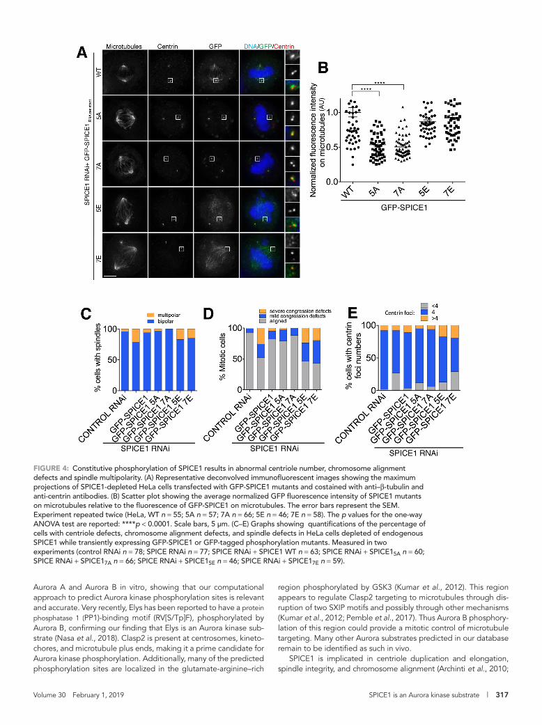
reduced on the spindle as expected, whereas GFP-SPICE15E and GFP-SPICE17E remained present on the spindle with levels similar to GFP-SPICE1 (Figure 4, A and B). In total, these results indicate that Aurora kinase phosphorylation contributes to regulating the target-ing of SPICE1 to the spindle.
SPICE1 is a centriole- and spindle-associated protein implicated in regulating centriole number and length, as well as a role in spin-dle organization (Archinti et al., 2010; Comartin et al., 2013). How-ever, the molecular mechanism underpinning its function is not known. To test whether constitutive phosphorylation could affect SPICE1 function, we examined the spindle morphology, centriole number, and chromosome alignment in the presence of the SPICE1 mutants and in the absence of endogenous SPICE1. First, we estab-lished that siRNA-resistant GFP-SPICE1 could rescue chromosome misalignment defects, multipolar spindles, and centriole number defects (Figure 4 and Supplemental Figure S1F). Of control cells, 96% had bipolar spindles. Similarly, 94, 97, and 100% of cells ex-pressing GFP-SPICE1, GFP-SPICE15A, and GFP-SPICE17A displayed bipolar mitotic spindles (Figure 4C). In the absence of endogenous SPICE1, rates of chromosome alignment in GFP-SPICE15A and GFP-SPICE17A transfected cells were similar to control cells and siRNA-resistant GFP-SPICE1–rescued cells (Figure 4, A and D). The number
of centrin foci observed for control cells was also similar to that of GFP-SPICE1, GFP-SPICE15A, and GFP-SPICE17A, with 90% control cells having four centrioles and 86, 83, and 88% for GFP-SPICE1, GFP-SPICE15A, and GFP-SPICE17A, respectively (Figure 4, A and E). In contrast, GFP-SPICE15E and GFP-SPICE17E transfected cells dis-played an increase in chromosome alignment defects and in multi-polar spindles, similarly to SPICE1-depleted cells. GFP-SPICE15E and GFP-SPICE17E transfected cells displayed more multipolar spin-dles, 15 and 17% of cells, respectively, while 21% of SPICE1-de-pleted cells had multipolar spindles (Figure 4, A and C). There was an accumulation of mitotic cells, and less than 50% of the cells dis-played aligned chromosomes in both GFP-SPICE15E– and GFP-SPICE17E–transfected cells, reminiscent of SPICE1-depleted cells (Figure 4, A and D). GFP-SPICE15E– and GFP-SPICE17E–expressing cells also displayed an increase in the abnormal centriole number similarly to SPICE1-depleted cells, as marked by the number of cen-trin foci (Figure 4E). Only 70 and 52% of GFP-SPICE15E– and GFP-SPICE17E–expressing cells had four centrioles, respectively (Figure 4, A and E). Overall, the phenotypes observed in GFP-SPICE15E– and GFP-SPICE17E–expressing cells were very similar to that of SPICE1-depleted cells. Aurora phosphorylation of SPICE1 plays a role in down-regulating the activity of SPICE1. In total, our results
FIGURE 2: The C terminus of SPICE1 regulates SPICE1 targeting to microtubules and centriole number in cells. (A) Representative live-cell images of HeLa cells transfected with GFP-SPICE1, GFP-SPICE11-550, and GFP-SPICE1444-855 in mitosis and interphase. (B) Quantification of GFP signal intensity on microtubules. Each data point represents one cell for which a background-corrected mean integrated intensity of the GFP signal on MTs was normalized to GFP-SPICE1 fluorescence intensity (GFP-SPICE1 n = 21; GFP-SPICE11-550, n = 34). The error bars represent the SEM. The p value for Student’s t test is ****<0.0001. (C) Representative deconvolved maximum z-projections of the HeLa cells transiently expressing GFP-SPICE1 or GFP-SPICE11-550, immunostained with anti–β-tubulin and anti-centrin antibodies, and stained with Hoechst for DNA. (D) Quantification of centriole number based on centrin staining in HeLa cells transiently expressing GFP-SPICE1 or GFP-SPICE11-550 (control n = 31; GFP-SPICE1 n = 30; GFP-SPICE11-550 n = 34). The error bars represent the SD. The p values for Kolmogorov–Smirnov test are ****<0.0001. Scale bars, 5 μm.

316 | J. Deretic et al. Molecular Biology of the Cell
indicate that the dynamic phosphorylation of SPICE1 is essential for correct centriole number, spindle architecture, and chromosome segregation.
Large proteomic approaches and peptides array studies have identified many mitotic substrates (Dephoure et al., 2008; Dulla et al., 2010; Mok et al., 2010). However, the kinase responsible for the phosphorylation is not necessarily associated with these sites. Additionally, phosphopeptides are often missed, as they are less easily ionized than other peptides. To identify new Aurora kinase substrates, we have used a computational approach and devel-oped a searchable database for Aurora kinase phosphorylation consensus sites, with criteria that the user can select (Figure 1B). Selection factors include both the number of sites and phosphory-lation site clustering: many Aurora kinase phosphorylation sites are found in close proximity to each other as demonstrated for Kif2c/MCAK and the KMN network (Andrews et al., 2004; Welburn et al.,
2010; Zaytsev et al., 2015). Many functionally important phosphory-lation sites are conserved across species and are located in disor-dered regions, and both features can be used here as search parameters. Finally, Gene Ontology is also used to classify the can-didate proteins based on their function and localization, focusing on cell division and the cell cycle due to the mitotic role of Aurora kinases. Applying these criteria to mitotic and spindle associated proteins, we could correctly predict a number of known Aurora kinase substrates such as Ndc80, Numa1, Tpx2, Dsn1, and Kif2c/MCAK (Figure 1C) (Andrews et al., 2004; Cheeseman et al., 2006; DeLuca et al., 2006; Fu et al., 2015; Gallini et al., 2016). From the other candidates identified in our study, we tested whether the nucleopore- and kinetochore-localized Elys, plus-end associ-ated Clasp2, spindle- and centriole-associated SPICE1, tubulin polyglutamylase TTLL4, and an unknown protein KIAA1468 were Aurora kinase substrates. All were successfully phosphorylated by
FIGURE 3: Aurora kinases regulate the cellular localization of SPICE1 to microtubules. (A) Representative deconvolved maximum z-projections of SPICE1-depleted HeLa cells and transiently transfected with GFP-SPICE1 after treatment with dimethyl sulfoxide (DMSO) (control), Aurora A inhibitor MLN8237, or Aurora B inhibitor ZM447439. Cells were immunostained with anti–β-tubulin and anti-centrin antibodies and stained with Hoechst for DNA. (B) Scatter plot showing the average normalized GFP fluorescence intensity on microtubules relative to the GFP fluorescence on microtubules in DMSO-treated metaphase cells. Each data point represents background-corrected integrated intensity of the GFP signal on microtubules (DMSO n = 40; MLN8237 n = 38; ZM447439 n = 30). (C) Representative deconvolved maximum z-projections of HeLa cells treated with DMSO, MLN8237, or ZM447439 and immunostained with anti-SPICE1 and anti–β-tubulin antibodies. (D) Scatter plot showing normalized SPICE1 fluorescence intensity on microtubules relative to the fluorescence intensity of SPICE1 on microtubules in DMSO-treated metaphase cells. Experiment repeated twice (DMSO n = 42; MLN8237 n = 38; ZM447439 n = 27). The error bars represent the SEM. The p values for one-way ANOVA tests are reported: ****p < 0.0001. Scale bars, 5 μm.
Volume 30 February 1, 2019 SPICE1 is an Aurora kinase substrate | 317
Aurora A and Aurora B in vitro, showing that our computational approach to predict Aurora kinase phosphorylation sites is relevant and accurate. Very recently, Elys has been reported to have a protein phosphatase 1 (PP1)-binding motif (RV[S/Tp]F), phosphorylated by Aurora B, confirming our finding that Elys is an Aurora kinase sub-strate (Nasa et al., 2018). Clasp2 is present at centrosomes, kineto-chores, and microtubule plus ends, making it a prime candidate for Aurora kinase phosphorylation. Additionally, many of the predicted phosphorylation sites are localized in the glutamate-arginine–rich
region phosphorylated by GSK3 (Kumar et al., 2012). This region appears to regulate Clasp2 targeting to microtubules through dis-ruption of two SXIP motifs and possibly through other mechanisms (Kumar et al., 2012; Pemble et al., 2017). Thus Aurora B phosphory-lation of this region could provide a mitotic control of microtubule targeting. Many other Aurora substrates predicted in our database remain to be identified as such in vivo.
SPICE1 is implicated in centriole duplication and elongation, spindle integrity, and chromosome alignment (Archinti et al., 2010;
FIGURE 4: Constitutive phosphorylation of SPICE1 results in abnormal centriole number, chromosome alignment defects and spindle multipolarity. (A) Representative deconvolved immunofluorescent images showing the maximum projections of SPICE1-depleted HeLa cells transfected with GFP-SPICE1 mutants and costained with anti–β-tubulin and anti-centrin antibodies. (B) Scatter plot showing the average normalized GFP fluorescence intensity of SPICE1 mutants on microtubules relative to the fluorescence of GFP-SPICE1 on microtubules. The error bars represent the SEM. Experiment repeated twice (HeLa, WT n = 55; 5A n = 57; 7A n = 66; 5E n = 46; 7E n = 58). The p values for the one-way ANOVA test are reported: ****p < 0.0001. Scale bars, 5 μm. (C–E) Graphs showing quantifications of the percentage of cells with centriole defects, chromosome alignment defects, and spindle defects in HeLa cells depleted of endogenous SPICE1 while transiently expressing GFP-SPICE1 or GFP-tagged phosphorylation mutants. Measured in two experiments (control RNAi n = 78; SPICE RNAi n = 77; SPICE RNAi + SPICE1 WT n = 63; SPICE RNAi + SPICE15A n = 60; SPICE RNAi + SPICE17A n = 66; SPICE RNAi + SPICE15E n = 46; SPICE RNAi + SPICE17E n = 59).

318 | J. Deretic et al. Molecular Biology of the Cell
Comartin et al., 2013). While the SPICE1 N terminus contains the microtubule-binding region based on cellular localization (Archinti et al., 2010) (Figure 2A), the C terminus appears as the regulatory region of the protein. Our results indicate that full-length SPICE1 overexpression results in abnormal centriole numbers with an in-crease in supernumerary centrioles (Figure 2, C and D). In the ab-sence of the SPICE1 C terminus, cells overexpressing SPICE1 did not display any extra centrioles (Figure 2, C and D). Our analysis of SPICE1 regulation by Aurora kinases revealed that the C terminus of SPICE1 contains seven Aurora kinase phosphorylation sites. Mu-tants carrying the computationally predicted phosphorylation sites to alanines did not cause any cellular defects. Spindle architecture and centriole number was similar to that in control cells. SPICE1 nonphosphorylatable mutants did not localize to the spindle but remain present at centrioles, suggesting that SPICE1 centriole local-ization rather than spindle localization is important for maintaining centriole number. SPICE1 also promotes centriole elongation. How-ever, it was not possible for us to measure centriole length in the presence of SPICE1 mutants, due to a lack of resolution of centrioles by conventional fluorescence microscopy.
The SPICE1 phosphomimetic mutant localized both to centrioles and spindles. Cells expressing a constitutive SPICE1 phosphomi-metic mutant display spindle architecture defects, modest chromo-some misalignment, and abnormal centriole number, reminiscent of SPICE1 depletion. Thus constitutive phosphorylation appears to in-terfere with SPICE1 function, possibly by allowing the N-terminal microtubule-binding region to associate with microtubules, in a non-controlled manner. While the phosphorylation status of SPICE1 does not seem to regulate its centriole localization, centriole-localized SPICE1 may recruit proteins important to regulate centriole replica-tion in an Aurora kinase-dependent manner. Consequently, the pres-ence of phosphomimetic SPICE1 results in abnormal centriole regu-lation. Both an increase and a reduction in centriole number interfere with force balance in the spindle and can result in multipolarity (McKinley and Cheeseman, 2017). Multipolarity in turn can cause chromosome misalignment (Ganem et al., 2009). Thus we believe that the SPICE1-induced centriole misregulation can cause the ob-served phenotype. Phosphorylation of SPICE1 promotes its associa-tion with the spindle (Figures 3 and 4). This could also interefere with microtubule dynamics, leading to spindle defects and chromosome misalignment. We conclude that dynamic phosphorylation of SPICE1 by Aurora kinases is necessary to maintain correct spindle architec-ture and centriole number. Aurora kinases are tethered to the centro-somes, chromosomes, and kinetochores. Cytoplasmic counteracting phosphatases are likely to dephosphorylate SPICE1 in a manner in-versely proportional to its distance from the Aurora kinases to reduce the levels of SPICE1 on the spindle. We could not observe a localiza-tion gradient of SPICE1 after Aurora kinase inhibition. This could be due to the length of the inhibitor treatment, or the fixing conditions, which do not preserve the localization of weakly bound proteins to microtubules (Welburn and Cheeseman, 2012). Recently, in addition to its centrosome localization, Aurora A has been reported at kineto-chores, which could explain the absence of SPICE1 localization gra-dient (DeLuca et al., 2018; Eot-Houllier et al., 2018). Further, mole-cular studies on SPICE1 are necessary to dissect the mechanism of SPICE1 in centriole and spindle function.
Overall, our computational approach reveals a rapid way to iden-tify new Aurora kinase substrates and represents a user-friendly open-source tool for the community. Using this approach, we have identified multiple new Aurora kinase substrates in vitro and showed that SPICE1 is an Aurora kinase substrate both in vitro and in vivo, validating our approach.
MATERIALS AND METHODSCloning, protein expression, and purificationGenes of interest were PCR amplified from adult male testis or fetus brain cDNA library (Invitrogen) and inserted into pET3aTr and pBabe GFP blasticidin for bacterial expression and for trans-fections into human cells, respectively. Site-directed mutagenesis was performed using QuikChange (Agilent Technologies). Se-quences were verified. A list of constructs and primers is provided in Table 1.
Recombinant proteins were expressed in BL21 (DE3) Codon plus cells with a His-tag preceded by 3C protease cleavage site. Protein expression and purification was performed as described (Talapatra et al., 2015). To isolate the KIAA14681-292 fragment from the inclusion bodies and refold it, 100 ml of the cells expressing KIAA14681-292 was centrifuged, and cell pellets were resuspended in lysis buffer (50 mM Tris–HCl, pH 8.0, 100 mM NaCl, 1 mM EDTA, 0.5% Triton X-100, 10 mM dithiothreitol [DTT]). After repeated freeze–thaw cycles, the cell lysate was sonicated, and insoluble fraction was collected by centrifugation for 20 min at 15,000 rpm and 4°C. Inclusion bodies in pellet were washed three times with wash buffer (50 mM Tris–HCl, pH 8.0, 100 mM NaCl, 1 mM EDTA, 1% Triton X-100, 1 mM DTT), soni-cated briefly, and pelleted at 15,000 rpm for 20 min at 4°C. Inclusion bodies were solubilized into 6 M Gdn–HCl, pH 4.5, and 5 mM β-mercaptoethanol (BME) stirring at room temperature. After solubiliza-tion, the buffer was exchanged with wash buffer (50 mM Tris–HCl, pH 8.0, 100 mM NaCl, 6 M urea, 10 mM imidazole, 5 mM BME) using desalting column (BioRad). The sample was then dialyzed against re-folding buffer (50 mM Tris–HCl, pH 8.0, 100 mM NaCl, 4 M urea, 1 mM EDTA, 10% glycerol, 0.5 mM l-arginine) for 4 h at 4°C. The stepwise refolding continued with dialysis against the buffers with decreasing concentration of urea and finished with buffer without urea. The protein refolded properly without aggregation.
In vitro kinase assay and Western blottingPurified recombinant protein fragments (2 μg) were incubated with 1.1 μg purified yeast recombinant glutathione-S-transferase–Ipl1, 0.2 μg human recombinant His-Aurora A (AMS Biotechnology Ltd.), or water in kinase buffer (150 mM NaCl, 40 mM HEPES, pH 7, 5 mM MgCl, 2 mM DTT, 20% glycerol) for 30 min at 37°C in the presence of 10 mM ATP and 5 μCi [γ-32P] ATP. The kinase reaction mixtures were resuspended in SDS sample buffer and separated with 10% or 15% SDS–PAGE. To detect phosphorylation, dried gels were ex-posed to an x-ray film (Fuji-Film). Simultaneously, same samples were prepared without [γ-32P] ATP and run out with SDS–PAGE, and gels were stained with Coomassie Brilliant Blue. For Western blot-ting, HeLa cells were lysed in SDS sample buffer and equal amount of cell extract per lane were loaded into a SDS–PAGE gel, run out, transferred to a nitrocellulose membrane, and blotted with rabbit anti-SPICE1 (HPA064843; Atlas Antibodies) and mouse anti–β-tubulin (Sigma) as loading control.
Cell culture and microscopyCells were maintained in DMEM (Lonza) supplemented with 10% fetal bovine serum penicillin/streptomycin (Life Technologies) at 37°C in a humidified atmosphere with 5% CO2. Cells were checked monthly for mycoplasma contamination (Mycoalert kit; Lonza). Cells were plated on 18-mm glass coverslips coated with poly-l-lysine (Sigma-Aldrich) for immunostaining. Transient transfections of DNA were conducted using Effectene reagent (Qiagen) according to the manufacturer’s guidelines. RNAi experiments were conducted using jetPRIME transfection reagent (Polyplus) according to the manufac-turer’s protocol. Previously published siRNA oligo was used to

Volume 30 February 1, 2019 SPICE1 is an Aurora kinase substrate | 319
Amplification from cDNA library Primer sequence
Full-length ELYS GTGGAACTCGGTTTTGTCGG
CCTGGACTGAAACAATCACTCC
Full-length TTLL4 CAGACTGACAGACTTCAAGGATGC
AGGCTTTTGGAGAGAGGCCAG
Full-length SPICE1 CTGTGTTTTGAGAGTGCAAGTACG
GAACAAACTCAGTGAGACTTGACTTC
Bacterial expression Primer sequence Vector Enzymes/method Template
ELYS1858-2014-3C prote-ase cleavage site-6xHis
gaaggagatatacatatgACTAAAAAAGAAGTTA-AGGTTTCATC
pET3aTr Ndel/Gibson assembly
ELYS isoform 2 NP_001310271.1
ccaagcttagatctggatccTCAGTGGTGATGAT-GATGATGGCTGCTGCCGGGCCCCTGGAA-CAGAACTTCCAGTGGGGTATTTCTTGTAG-ATCTCC
pET3aTr BamHI/Gibson assembly
ELYS isoform 2 NP_001310271.1
CLASP2741-818-3C prote-ase cleavage site-6xHis
gaaggagatatacatatgCCATCTAGGCTTTCAGTG-GC
pET3aTr NdeI/Gibson assembly
CLASP2 isoform X15 XP_006713112.1
ccaagcttagatctggatccTCAGTGGTGATGAT-GATGATGGCTGCTGCCGGGCCCCTGGAA-CAGAACTTCCAGAGAAGACGACAGTCGACTT-GATTG
pET3aTr BamHI/Gibson assembly
CLASP2 isoform X15 XP_006713112.1
SPICE1549-855-3C prote-ase cleavage site-6xHis
tttaagaaggagatatacatatgCCTCTTCAGGACG-TATTGAG
pET3aTr NdeI/Gibson assembly
SPICE1 isoform a NP_001318007.1
ccaagcttagatctggatccTCAGTGGTGATGAT-GATGATGGCTGCTGCCGGGCCCCTG-GAACAGAACTTCCAGTGATACATGGG-TAGAAAGAGCA
pET3aTr BamHI/Gibson assembly
SPICE1 isoform a NP_001318007.1
TTLL4330-624-3C prote-ase cleavage site-6xHis
tttaagaaggagatatacatatgCCAACTAAGGAGA-TTCGGTTC
pET3aTr NdeI/Gibson assembly
TTLL4 NP_055455.3
ccaagcttagatctggatccTCAGTGGTGATGATGATGA TGGCTGCTGCCGGGCCCCTGGAACAGAACTTC CAGTCCAATGGTCTGCTTGACAATG
pET3aTr BamHI/Gibson assembly
TTLL4 NP_055455.3
KIAA14681-292-3C prote-ase cleavage site-6xHis
tttaagaaggagatatacatatgTTAGTA-CAGAAATTAGAAGATAAAATTAGTTTG
pET3aTr NdeI/Gibson assembly
KIAA1468 isoform CRA_a EAW63120.1
ccaagcttagatctggatccTCAGTGGTGATGATGATG ATGGCTGCTGCCGGGCCCCTGGAACAGAAC TTCCAGTAACATTTGTTGCAACATTGAAAG
pET3aTr BamHI/Gibson assembly
KIAA1468 isoform CRA_a EAW63120.1
Mammalian expression Primer sequence Vector Enzymes/method Template
Full-length GFP-SPICE1 cccgCTCGAGATGTCATTTGTCAGAGTGAAC-CGCT
pBabe blast XhoI Isoform a NP_001318007.1
tcccCCGCGGTTATGATACATGGGTAGAAAGAG-CAAAC
pBabe blast SacII Isoform a NP_001318007.1
GFP-SPICE1444-855 cccCTCGAGATATCACTCACACATGCTATTA-AGAACT
pBabe blast XhoI Isoform a NP_001318007.1
GFP-SPICE11-550 tcccCCGCGGTTAAAGAGGAGAGAATTTTAAGT pBabe blast SacII Isoform a NP_001318007.1
Mutagenesis Primer sequence
GFP-SPICE1 hd 1st 5 bases
CTCCCAGTACTGGGAGATGGGCAG-CAATTAAGGACTAATGAGTCATTAATA-CAAAGAAAGGAC
ContinuesTABLE 1: Details of constructs generated and primer sequences.

320 | J. Deretic et al. Molecular Biology of the Cell
GTCCTTTCTTTGTATTAATGACTCATTAGTCCT-TAATTGCTGCCCATCTCCCAGTACTGGGAG
GFP-SPICE1 hd 2nd 5 bases
CTGGGAGATGGGCAGCAATTAAGGACTA-ACGAAAGTTTAATACAAAGAAAGGACATAAT-GACACG
CGTGTCATTATGTCCTTTCTTTGTAT-TAAACTTTCGTTAGTCCTTAATTGCT-GCCCATCTCCCAG
GFP-SPICE1 T557A AGGACGTATTGAGAAGGGCTGTTCAAACTC-GTCCT
AGGACGAGTTTGAACAGCCCTTCTCAATAC-GTCCT
GFP-SPICE1 S738A TTGCAGAATTGAATCGACAAGCTATGGAG-GCTCGTGGAAAAC
GTTTTCCACGAGCCTCCATAGCTTGTC-GATTCAATTCTGCAA
GFP-SPICE1 T780A GACTGAAGGAGCAAAGAGGGCAATTGAGG-TATCTATTCC
GGAATAGATACCTCAATTGCCCTCTTT-GCTCCTTCAGTC
GFP-SPICE1 S797A CCCAGAAAGCTCAAAATGTGCTACT-GTCTCTCCCGTCAGC
GCTGACGGGAGAGACAGTAGCACATTTT-GAGCTTTCTGGG
GFP-SPICE1 S810A Forward
GCGGGATAAATACAAGAAGAGCTTCCG GGGCTACT
AGTAGCCCCGGAAGCTCTTCTTGTATTTATCCC-GC
GFP-SPICE1 S797A/T798A
GCCCCAGAAAGCTCAAAATGTGCTGCT-GTCTCTCCCGTCAG
CTGACGGGAGAGACAGCAGCACATTTT-GAGCTTTCTGGGGC
GFP-SPICE1 S810A/S811A
TCAGCGGGATAAATACAAGAAGAGCTGCC-GGGGCTACTG
CAGTAGCCCCGGCAGCTCTTCTTGTATT-TATCCCGCTGA
GFP-SPICE1 T557E CTTCAGGACGTATTGAGA-AGGGAAGTTCAAACTCGTCCTGCTCCA
TGGAGCAGGACGAGTTTGAACTTCCCTTCT-CAATACGTCCTGAAG
GFP-SPICE1 S738E GGAACGGATTGCAGAATTGAATCGA-CAAGAAATGGAGGCTCGTGGA
TCCACGAGCCTCCATTTCTTGTC-GATTCAATTCTGCAATCCGTTCC
GFP-SPICE1 T780E GGACTGAAGGAGCAAAGAGGGAAATTGAG-GTATCTATTCCAG
CTGGAATAGATACCTCAATTTCCCTCTTT-GCTCCTTCAGTCC
GFP-SPICE1 S797E GCAGAAGCCCCAGAAAGCTCAAAATGT-GAAACTGTCTCTCCCGT
ACGGGAGAGACAGTTTCACATTTT-GAGCTTTCTGGGGCTTCTGC
ContinuesTABLE 1: Details of constructs generated and primer sequences.

Volume 30 February 1, 2019 SPICE1 is an Aurora kinase substrate | 321
deplete SPICE1 (Archinti et al., 2010). The cells were visualized 48 h after transfection or cotransfection.
The inducible SPICE1 knockout (KO) cell line was generated as previously described (McKinley et al., 2015; Wang et al., 2016) using guide RNA (gRNA) 5′-TTCGGGAGTTGCCCGATGAA-3′, targeting exon 3 of SPICE1. KO was introduced with 1 μg/ml doxycycline (Sigma) treatment of stable HeLa Cas9SPICE1 gRNA cells for 96 h. For Aurora kinase inhibition experiments, HeLa cells were incubated for 1.5 h with small molecule inhibitors at the following concentrations: ZM447439, 2 μM; MLN8237, 150 nM (Selleckchem).
Immunofluorescence in human cells was conducted as previ-ously described (Welburn et al., 2010) using antibodies against mouse anti-tubulin (Sigma), rabbit anti-Centrin (Covance; custom-made), and rabbit α-SPICE1 (HPA064843; Atlas Antibodies). Images were acquired on a DeltaVision Core deconvolution microscope (Applied Precision) equipped with a CoolSnap HQ2 CCD camera. Thirty Z-sections were acquired at 0.2-nm steps using a 100×/1.3 NA Olympus U-PlanApo objective without binning. Equivalent ex-posure conditions were used between controls and drug-treated cells in a single experiment, as well as in case of nontransfected cells and cells transfected with different constructs. On average, experi-ments were repeated three times. To quantitate GFP fluorescence intensity, at least 10 individual MT as polylines of 35–37 pixels in size were selected from projections (selected based on colocalization with β-tubulin) and the integrated intensity was determined after subtracting the average background fluorescence measured from adjacent regions of the cell (two polylines form inside and two from outside the spindle) using Metamorph. To quantify chromosome congression defects in SPICE1 knockdown and KO, the cells were scored as having congressed chromosome, mild congression de-fects, and severe congression defects based on Hoechst staining. The cells with at least five misaligned chromosomes were scored as mild congression defects; the cells with more than five misaligned chromosomes as cells with severe defects. To quantify spindle mor-phology defects, the cells were scored as having bipolar or multipo-lar spindle based on β-tubulin staining and spindle morphology. To quantify defects in centriole numbers, the cells were scored as hav-ing <4, 4, and >4 centrioles based on centrin staining. Integrated intensities of pS10-H3 signals were measured from chromatin sur-
face (based on Hoechst staining) using Image-Pro-Premier software (Media Cybernetics).
Images were stored and vizualised using an OMERO.insight cli-ent (Linkert et al., 2010).
Statistics and reproducibilityStatistical analyses were performed using GraphPad Prism 6.0 or R software. No statistical method was used to predetermine sample size. No samples were excluded from the analyses. All experiments were performed and quantified from at least three independent ex-periments, unless specified, and the representative data are shown.
Statistical analysis of data was performed with GraphPad Prims using Student’s t test, one-way analysis of variance (ANOVA) tests, and nonparametric tests. A Kolmogorov–Smirnov test was used to test variation in centriole number (Figure 2D), which had a non-Gaussian distribution.
Data availabilityAll data supporting the findings of this study are available from the corresponding author on request.
Softwarefuzzpro from the EMBOSS suite (version 6.6.0.0) was used to find matches to patterns in each of the three peptide data sets (Rice et al., 2000). Needle from the same suite was used to calculate the percentage identity between all possible pairwise regions that con-tained pattern matches in orthologous proteins, and the highest identity per pair was recorded. iupred (Dosztanyi et al., 2005) was used to calculate the likelihood for an amino acid regions present in a disordered region. Amino acids that have values larger than 0.5 were counted within regions containing a pattern match. samtools (version 0.1.19) was used to both index and retrieve sequences from the fasta files (Li et al., 2009).
Shiny applicationThe R-Shiny application can be launched in any R session with an installed shiny package using the commands
library (shiny)runGitHub (“DereticWelburnApp,” “AlastairKerr”)
GFP-SPICE1 S810E CCCGTCAGCGGGATAAATACAAGAAGAGA-ATCCGGGGCTACTGG
CCAGTAGCCCCGGATTCTCTTCTTGTATT-TATCCCGCTGACGGG
GFP-SPICE1 S797E/T798E
GGAGCAGAAGCCCCAGAAAGCTCAAAATGT-GAAGAAGTCTCTCCCGTCAGCGGGA
TCCCGCTGACGGGAGAGACTTCTTCACATTTT-GAGCTTTCTGGGGCTTCTGCTCC
GFP-SPICE1 S810E/S811E
AGAAGTCTCTCCCGTCAGCGGGATAAATACAA GAAGAGAAGAGGGGGCTACTGGTAATTCTT
AAGAATTACCAGTAGCCCCCTCTTCTCTTCTTG-TATTTATCCCGCTGACGGGAGAGACTTCT
CRISPR Cas9 Oligo sequence Vector Enzymes/method
guide RNA targeting exon3 in SPICE1 gene
[phospho]caccgTTCGGGAGTTGCCCGATGAA pLentisgRNA BbsI
[phospho]aaacTTCATCGGGCAACTCCCGAAc pLentisgRNA BbsI
TABLE 1: Details of constructs generated and primer sequences. Continued

322 | J. Deretic et al. Molecular Biology of the Cell
The interface produces a table for proteins that match at least one of the prosite patterns in each of the three orthologues.
Prosite patterns used are
pattern 1 “[RK][RK]x[TS][FAST]”
pattern 2 “[RK][RK]x[TS][ILVM]”
pattern 3 “x[RK][RK][TS][FAST]”
pattern 4 “x[RK][RK][TS][ILVM]”
pattern 5 “[RK][RK][RK][TS][ILVM]”
pattern 6 “[RK][RK][RK][TS][FAST]”
Additional filters can be applied to the number of pattern matches in a set protein region, data based on the percentage of amino acids predicted to be in disorder in the region, the maximum identity between two pattern containing regions in orthologous proteins, and annotations to select Gene Ontology terms or mitosis pathways.
The code is available at the links below:https://github.com/AlastairKerr/DereticWelburnPreprocessing
ScriptsAll code that was used for preprocessing the data as well as the
r-shiny application is available on github.Preprocessing: https://github.com/AlastairKerr/DereticWelburn
PreprocessingScriptsR-Shiny application: https://github.com/AlastairKerr/Deretic
WelburnAppThe phosphorylation search database is also found at http://bifx
-rta.bio.ed.ac.uk:3838/Welburn/Patterns_Regions/
ACKNOWLEDGMENTSWe thank members of the Welburn lab for critical reading of the manuscript. J.W. is supported by a Wellcome Trust Senior Research Fellowship (Grant No. 207430). The Wellcome Trust Centre for Cell Biology and A. Kerr are supported by core funding from the Well-come Trust (203149).
REFERENCESAndrews PD, Ovechkina Y, Morrice N, Wagenbach M, Duncan K, Worde-
man L, Swedlow JR (2004). Aurora B regulates MCAK at the mitotic centromere. Dev Cell 6, 253–268.
Archinti M, Lacasa C, Teixido-Travesa N, Luders J (2010). SPICE–a previously uncharacterized protein required for centriole duplication and mitotic chromosome congression. J Cell Sci 123, 3039–3046.
Bailey TL, Boden M, Buske FA, Frith M, Grant CE, Clementi L, Ren J, Li WW, Noble WS (2009). MEME suite: tools for motif discovery and searching. Nucleic Acids Res 37, W202–W208.
Bishop JD, Schumacher JM (2002). Phosphorylation of the carboxyl terminus of inner centromere protein (INCENP) by the Aurora B kinase stimulates Aurora B kinase activity. J Biol Chem 277, 27577–27580.
Cheeseman IM, Anderson S, Jwa M, Green EM, Kang J, Yates JR 3rd, Chan CS, Drubin DG, Barnes G (2002). Phospho-regulation of kinetochore-microtubule attachments by the Aurora kinase Ipl1p. Cell 111, 163–172.
Cheeseman IM, Chappie JS, Wilson-Kubalek EM, Desai A (2006). The conserved KMN network constitutes the core microtubule-binding site of the kinetochore. Cell 127, 983–997.
Ciferri C, Pasqualato S, Screpanti E, Varetti G, Santaguida S, Dos Reis G, Maiolica A, Polka J, De Luca JG, De Wulf P, et al. (2008). Implications for kinetochore-microtubule attachment from the structure of an engineered Ndc80 complex. Cell 133, 427–439.
Comartin D, Gupta GD, Fussner E, Coyaud E, Hasegan M, Archinti M, Cheung SW, Pinchev D, Lawo S, Raught B, et al. (2013). CEP120 and SPICE1 cooperate with CPAP in centriole elongation. Curr Biol 23, 1360–1366.
de Groot CO, Hsia JE, Anzola JV, Motamedi A, Yoon M, Wong YL, Jenkins D, Lee HJ, Martinez MB, Davis RL, et al. (2015). A cell biologist’s field guide to aurora kinase inhibitors. Front Oncol 5, 285.
DeLuca JG, Gall WE, Ciferri C, Cimini D, Musacchio A, Salmon ED (2006). Kinetochore microtubule dynamics and attachment stability are regu-lated by Hec1. Cell 127, 969–982.
DeLuca KF, Meppelink A, Broad AJ, Mick JE, Peersen OB, Pektas S, Lens SMA, DeLuca JG (2018). Aurora A kinase phosphorylates Hec1 to regulate metaphase kinetochore-microtubule dynamics. J Cell Biol 217, 163–177.
Dephoure N, Zhou C, Villen J, Beausoleil SA, Bakalarski CE, Elledge SJ, Gygi SP (2008). A quantitative atlas of mitotic phosphorylation. Proc Natl Acad Sci USA 105, 10762–10767.
Dosztanyi Z, Csizmok V, Tompa P, Simon I (2005). The pairwise energy content estimated from amino acid composition discriminates be-tween folded and intrinsically unstructured proteins. J Mol Biol 347, 827–839.
Dulla K, Daub H, Hornberger R, Nigg EA, Korner R (2010). Quantitative site-specific phosphorylation dynamics of human protein kinases during mitotic progression. Mol Cell Proteomics 9, 1167–1181.
Eot-Houllier G, Magnaghi-Jaulin L, Fulcrand G, Moyroud F-X, Monier S, Jaulin C (2018). Aurora A-dependent CENP-A phosphorylation at in-ner centromeres protects bioriented chromosomes against cohesion fatigue. Nat Commun 9, 1888.
Fu J, Bian M, Liu J, Jiang Q, Zhang C (2009). A single amino acid change converts Aurora-A into Aurora-B-like kinase in terms of partner specificity and cellular function. Proc Natl Acad Sci USA 106, 6939–6944.
Fu J, Bian M, Xin G, Deng Z, Luo J, Guo X, Chen H, Wang Y, Jiang Q, Zhang C (2015). TPX2 phosphorylation maintains metaphase spindle length by regulating microtubule flux. J Cell Biol 210, 373–383.
Gallini S, Carminati M, De Mattia F, Pirovano L, Martini E, Oldani A, Asteriti IA, Guarguaglini G, Mapelli M (2016). NuMA phosphorylation by aurora-A orchestrates spindle orientation. Curr Biol 26, 458–469.
Ganem NJ, Godinho SA, Pellman D (2009). A mechanism linking extra centrosomes to chromosomal instability. Nature 460, 278–282.
Hans F, Skoufias DA, Dimitrov S, Margolis RL (2009). Molecular distinctions between Aurora A and B: a single residue change transforms Aurora A into correctly localized and functional Aurora B. Mol Biol Cell 20, 3491–3502.
Honda R, Korner R, Nigg EA (2003). Exploring the functional interactions between Aurora B, INCENP, and survivin in mitosis. Mol Biol Cell 14, 3325–3341.
Hornbeck PV, Zhang B, Murray B, Kornhauser JM, Latham V, Skrzypek E (2015). PhosphoSitePlus, 2014: mutations, PTMs and recalibrations. Nucleic Acids Res 43, D512–D520.
Kettenbach AN, Schweppe DK, Faherty BK, Pechenick D, Pletnev AA, Gerber SA (2011). Quantitative phosphoproteomics identifies substrates and functional modules of Aurora and Polo-like kinase activities in mitotic cells. Sci Signal 4, rs5.
Kim Y, Holland AJ, Lan W, Cleveland DW (2010). Aurora kinases and protein phosphatase 1 mediate chromosome congression through regulation of CENP-E. Cell 142, 444–455.
Kumar P, Chimenti MS, Pemble H, Schonichen A, Thompson O, Jacobson MP, Wittmann T (2012). Multisite phosphorylation disrupts arginine-glutamate salt bridge networks required for binding of cytoplasmic linker-associated protein 2 (CLASP2) to end-binding protein 1 (EB1). J Biol Chem 287, 17050–17064.
Kumar P, Wittmann T (2012). +TIPs: SxIPping along microtubule ends. Trends Cell Biol 22, 418–428.
Li H, Handsaker B, Wysoker A, Fennell T, Ruan J, Homer N, Marth G, Abecasis G, Durbin R, S. Genome Project Data Processing (2009). The sequence alignment/map format and SAMtools. Bioinformatics 25, 2078–2079.
Linkert M, Rueden CT, Allan C, Burel JM, Moore W, Patterson A, Loranger B, Moore J, Neves C, Macdonald D, et al. (2010). Metadata matters: access to image data in the real world. J Cell Biol 189, 777–782.
McKinley KL, Cheeseman IM (2014). Polo-like kinase 1 licenses CENP-A deposition at centromeres. Cell 158, 397–411.
McKinley KL, Cheeseman IM (2017). Large-scale analysis of CRISPR/Cas9 cell-cycle knockouts reveals the diversity of p53-dependent responses to cell-cycle defects. Dev Cell 40, 405–420 e402.
McKinley KL, Sekulic N, Guo LY, Tsinman T, Black BE, Cheeseman IM (2015). The CENP-L-N complex forms a critical node in an integrated meshwork of interactions at the centromere-kinetochore interface. Mol Cell 60, 886–898.
Mok J, Kim PM, Lam HY, Piccirillo S, Zhou X, Jeschke GR, Sheridan DL, Parker SA, Desai V, Jwa M, et al. (2010). Deciphering protein kinase specificity through large-scale analysis of yeast phosphorylation site motifs. Sci Signal 3, ra12.

Volume 30 February 1, 2019 SPICE1 is an Aurora kinase substrate | 323
Moritz A, Li Y, Guo A, Villen J, Wang Y, MacNeill J, Kornhauser J, Sprott K, Zhou J, Possemato A, et al. (2010). Akt-RSK-S6 kinase signaling networks activated by oncogenic receptor tyrosine kinases. Sci Signal 3, ra64.
Nasa I, Rusin SF, Kettenbach AN, Moorhead GB (2018). Aurora B opposes PP1 function in mitosis by phosphorylating the conserved PP1-binding RVxF motif in PP1 regulatory proteins. Sci Signal 11, eaai8669.
Okada M, Cheeseman IM, Hori T, Okawa K, McLeod IX, Yates JR 3rd, Desai A, Fukagawa T (2006). The CENP-H-I complex is required for the efficient incorporation of newly synthesized CENP-A into centromeres. Nat Cell Biol 8, 446–457.
Olsen JV, Vermeulen M, Santamaria A, Kumar C, Miller ML, Jensen LJ, Gnad F, Cox J, Jensen TS, Nigg EA, et al. (2010). Quantitative phosphopro-teomics reveals widespread full phosphorylation site occupancy during mitosis. Sci Signal 3, ra3.
Pemble H, Kumar P, van Haren J, Wittmann T (2017). GSK3-mediated CLASP2 phosphorylation modulates kinetochore dynamics. J Cell Sci 130, 1404–1412.
Rice P, Longden I, Bleasby A (2000). EMBOSS: the European Molecular Biol-ogy Open Software Suite. Trends Genet 16, 276–277.
Santamaria A, Wang B, Elowe S, Malik R, Zhang F, Bauer M, Schmidt A, Sillje HH, Korner R, Nigg EA (2011). The Plk1-dependent phospho-proteome of the early mitotic spindle. Mol Cell Proteomics 10, M110 004457.
Talapatra SK, Harker B, Welburn JP (2015). The C-terminal region of the mo-tor protein MCAK controls its structure and activity through a conforma-tional switch. Elife 4, e06421.
Wang T, Lander ES, Sabatini DM (2016). Single guide RNA library design and construction. Cold Spring Harb Protoc 2016, prot090803.
Welburn JP, Cheeseman IM (2012). The microtubule-binding protein Cep170 promotes the targeting of the kinesin-13 depolymerase Kif2b to the mitotic spindle. Mol Biol Cell 23, 4786–4795.
Welburn JP, Vleugel M, Liu D, Yates JR 3rd, Lampson MA, Fukagawa T, Cheeseman IM (2010). Aurora B phosphorylates spatially distinct targets to differentially regulate the kinetochore-microtubule interface. Mol Cell 38, 383–392.
Welburn JPI, Jeyaprakash AA (2018). Mechanisms of mitotic kinase regula-tion: a structural perspective. Front Cell Dev Biol 6, 6.
Ye AA, Deretic J, Hoel CM, Hinman AW, Cimini D, Welburn JP, Maresca TJ (2015). Aurora a kinase contributes to a pole-based error correction pathway. Curr Biol 25, 1842–1851.
Zaytsev AV, Mick JE, Maslennikov E, Nikashin B, DeLuca JG, Grishchuk EL (2015). Multisite phosphorylation of the NDC80 complex gradually tunes its microtubule-binding affinity. Mol Biol Cell 26, 1829–1844.
Zhang X, Ems-McClung SC, Walczak CE (2008). Aurora A phosphorylates MCAK to control ran-dependent spindle bipolarity. Mol Biol Cell 19, 2752–2765.