edinburgh research explorer · núria m pastor-soler1,2 ... [email protected]. n m...
TRANSCRIPT

Edinburgh Research Explorer
Aquaporin 9 expression in the developing rat epididymis ismodulated by steroid hormones
Citation for published version:Pastor-Soler, NM, Fisher, JS, Sharpe, R, Hill, E, Van Hoek, A, Brown, D & Breton, S 2010, 'Aquaporin 9expression in the developing rat epididymis is modulated by steroid hormones' Reproduction, vol. 139, no.3, pp. 613-621. DOI: 10.1530/REP-09-0284
Digital Object Identifier (DOI):10.1530/REP-09-0284
Link:Link to publication record in Edinburgh Research Explorer
Document Version:Peer reviewed version
Published In:Reproduction
Publisher Rights Statement:Published in final edited form as:Reproduction. 2010 March; 139(3): 613–621.Published online 2009 November 30. doi: 10.1530/REP-09-0284
General rightsCopyright for the publications made accessible via the Edinburgh Research Explorer is retained by the author(s)and / or other copyright owners and it is a condition of accessing these publications that users recognise andabide by the legal requirements associated with these rights.
Take down policyThe University of Edinburgh has made every reasonable effort to ensure that Edinburgh Research Explorercontent complies with UK legislation. If you believe that the public display of this file breaches copyright pleasecontact [email protected] providing details, and we will remove access to the work immediately andinvestigate your claim.
Download date: 12. Sep. 2018

Aquaporin 9 expression in the developing rat epididymis ismodulated by steroid hormones
Núria M Pastor-Soler1,2, Jane S Fisher3, Richard Sharpe3, Eric Hill1, Alfred Van Hoek1,2,Dennis Brown1,2, and Sylvie Breton1,2
1Program in Membrane Biology and Nephrology Division, Center for Systems Biology,Massachusetts General Hospital, 185 Cambridge Street, Suite CPZN 8.204, Boston,Massachusetts 02114, USA 2Harvard Medical School, Boston, Massachusetts 02115, USA and3Medical Research Council Human Reproductive Sciences Unit, The Queen’s Medical ResearchInstitute, Centre for Reproductive Biology, Edinburgh EH16 4TJ, UK
AbstractFluid and solute transport across the epithelium of the male excurrent duct is important for spermmaturation and storage. Aquaporin 9 (AQP9), which allows permeation of water and neutralsolutes, is abundant throughout the male reproductive tract, where it is expressed at the apicalmembrane of rat epididymal principal cells as early as at 1 week of age. We evaluated the effect ofneonatal exposure to: 1) a GNRH antagonist (GNRHa); 2) diethylstilbestrol (DES); 3) ethinylestradiol (EE); 4) DES plus testosterone (DES+TE); and 5) the anti-androgen flutamide on AQP9expression in the epididymis of peripubertal rats. Control groups received the vehicle alone. In 25-day-old rats, quantification of the mean pixel intensity of immunofluorescence-stained sectionsshowed a significant decrease in AQP9 staining in the apical membrane of epididymal principalcells after treatments with GNRHa, DES, or flutamide, compared to controls. These results wereconfirmed by western blotting. While EE induced a marked decrease in AQP9 levels by westernblotting, the decrease in AQP9-associated fluorescence was not significant compared to controls.DES+TE-treated rats showed levels of AQP9 protein similar to controls, indicating maintenanceof AQP9 expression by testosterone treatment in the presence of DES. Our data show thatexpression of AQP9 in the developing rat epididymis is downregulated by neonatal DES, GNRHa,EE, and flutamide, and that the effects mediated by estrogens can be prevented by testosteroneadministration.
IntroductionThe luminal fluid of the male excurrent duct (efferent ducts, epididymis, and vas deferens)undergoes significant changes in composition as it moves distally (Levine & Marsh 1971,Ilio & Hess 1994, Hinton & Palladino 1995, Robaire & Viger 1995, Turner 1995, Clulow etal. 1998, Robaire et al. 2006, Joseph et al. 2009). Fluid transport across the efferent duct andepididymal epithelium is required to achieve proper sperm concentration, which is in turnimportant for fertility (Levine & Marsh 1971, Johnson & Howards 1977, Wong & Yeung
© 2010 Society for Reproduction and Fertility
Correspondence should be addressed to S Breton at Program in Membrane Biology and Nephrology Division, Center for SystemsBiology, Massachusetts General Hospital; [email protected] M Pastor-Soler is now at the Renal-Electrolyte Division, Department of Medicine, University of Pittsburgh School of Medicine,Pittsburgh, Pennsylvania 15261, USA
Declaration of interestThe authors declare that there is no conflict of interest that could be perceived as prejudicing the impartiality of the research reported.
NIH Public AccessAuthor ManuscriptReproduction. Author manuscript; available in PMC 2013 September 16.
Published in final edited form as:Reproduction. 2010 March ; 139(3): 613–621. doi:10.1530/REP-09-0284.
NIH
-PA Author Manuscript
NIH
-PA Author Manuscript
NIH
-PA Author Manuscript

1978, Hohlbrugger & Pfaller 1983, Turner & Cesarini 1983, Ilio & Hess 1994, Clulow et al.1998, Hansen et al. 2004). In the cauda epididymidis, the activity of an Na/H exchanger andthe Na/K-ATPase has been implicated in net water reabsorption (Wong & Yeung 1977,Bagnis et al. 2001, Leung et al. 2001, Kaunisto & Rajaniemi 2002). In addition, fluidsecretion driven by CFTR-dependent chloride secretion occurs in the distal portion of theepididymis and helps control the viscosity of the luminal content (Wong 1998).
Water channels (aquaporins, AQPs) are involved in fluid transport in a wide variety ofepithelia (Brown et al. 1995, Verkman 2002, King et al. 2004). Two subgroups ofmammalian AQPs have been defined: the ‘aquaporins’, which are highly selective for water,and the ‘aquaglyceroporins’, which can transport solutes in addition to water. Several AQPsare expressed throughout the male excurrent duct (Brown et al. 1993, Stevens et al. 2000,Badran & Hermo 2002, Da Silva et al. 2006a, 2006b, Hermo et al. 2008). However, the mostpredominant AQP in the male reproductive tract is clearly AQP9, which is expressed at highlevels in the apical membrane of nonciliated cells of the efferent ducts, and principal cells allalong the length of the epididymis and vas deferens (Elkjaer et al. 2000, Pastor-Soler et al.2001, Badran & Hermo 2002, Pietrement et al. 2008). Interestingly, one of the solutes thatcan permeate through AQP9 is glycerol (Tsukaguchi et al. 1998), a spermatozoa metabolicsubstrate that accumulates in the lumen of the distal epididymis (Cooper & Brooks 1981).We have shown significant AQP9-dependent glycerol permeability in the apical membraneof epididymal epithelial cells (Pietrement et al. 2008). AQP9 was, thus, identified as a majorapical AQP in the epididymal epithelium, where it could provide a route via whichtransepithelial fluid and solute transport could occur.
In the efferent ducts of the adult male excurrent duct, reabsorption of luminal fluid isregulated by both androgens and estrogens (Wong & Yeung 1978, Hess et al. 1997, Hess2003, Ruz et al. 2006). We have shown that treatment of rodents and primates withdiethylstilbestrol (DES) during the neonatal period strongly inhibits AQP1 expression in theefferent ducts of prepubertal animals (Fisher et al. 1998), indicating that maintaining theestrogen/androgen balance during postnatal development is crucial for the maturation ofthese segments.
In the epididymis, AQP9 regulation is less well documented. We have shown that AQP9permeability is acutely regulated by luminal bradykinin (Belleannee et al. 2009). However,the role of sex hormones in regulating its expression has not been fully characterized. Thepromoter region of AQP9 contains a putative steroid hormone receptor-binding site(Tsukaguchi et al. 1998). In addition, sex-linked differences of AQP9 expression havepreviously been reported in the liver, suggesting that different hormonal profiles during andafter development may play a role in the expression of this protein (Nicchia et al. 2001). Weand others have previously shown that orchidectomy (Pastor-Soler et al. 2002, Oliveira et al.2005), or treatment of adult male rats with the anti-androgen flutamide (Pastor-Soler et al.2002), caused a significant reduction in the level of AQP9 protein expression in the adultepididymis. The downregulation of AQP9 expression in castrated animals was reversed bytestosterone in the cauda epididymidis (Pastor-Soler et al. 2002) and by dihydrotestosteronein the initial segments (Oliveira et al. 2005), indicating that androgens are important forAQP9 expression in the epididymis of adult animals. Androgen and estrogen (α and β)receptors are expressed in the epididymis during development and in adults (McKinnell etal. 2001, Zhou et al. 2002, Hess 2003). Neonatal exposure to the estrogenic compound DESsignificantly affects the morphology of the male excurrent duct, including efferent ducts,epididymis, and vas deferens (McKinnell et al. 2001, Williams et al. 2001, Rivas et al. 2002,2003, Goyal et al. 2003, Atanassova et al. 2005). Disturbances in the estrogen/androgenbalance were proposed to be the key determinant in causing these adverse effects on malereproductive parameters. In addition, our previous study showed a marked reduction in the
Pastor-Soler et al. Page 2
Reproduction. Author manuscript; available in PMC 2013 September 16.
NIH
-PA Author Manuscript
NIH
-PA Author Manuscript
NIH
-PA Author Manuscript

number of clear cells in the epididymis of prepubertal rats after neonatal exposure to DES(Fisher et al. 2002), a finding that further supports the notion that the epididymis is alsosusceptible to changes in the androgen/estrogen balance.
We have shown that AQP9 begins to appear in the apical membrane of epididymal principalcells as early as 1 week of age, and that its expression continues to increase throughout thepostnatal period to reach the highest levels shortly before puberty (Pastor-Soler et al. 2001).This progressive increase in principal cell AQP9 expression during the prepubertal periodwas also confirmed by another laboratory (Badran & Hermo 2002). These findings suggestthat initiation of AQP9 expression in the developing epididymis does not depend exclusivelyon the higher androgen levels that are triggered at puberty, and might require theparticipation of other hormones, including estrogens. The present study, therefore, assesseswhether neonatal treatment with compounds known to affect the androgenic/estrogenicbalance alters the expression of AQP9 in the epididymis in the peripubertal period.Understanding the effect of hormone manipulation during the neonatal period is crucial,because overexposure of young males to exogenous estrogens, including those from variousenvironmental sources, can result in major abnormalities and reproductive dysfunction inadulthood (Atanassova et al. 1999, Goyal et al. 2003).
ResultsHormonal effects on epididymal AQP9 expression
Effect of GNRH antagonist, DES, ethinyl estradiol, and DES+TE treatments—The expression of AQP9 was assessed by immunofluorescence staining of cryostat sectionsof the proximal cauda epididymidis, a region that shows high AQP9 expression. Controlincubations were first performed using anti-AQP9 antibodies that had been preabsorbed withthe AQP9 immunizing peptide. As we have previously described, AQP9 was localized to theapical membrane of principal cells in the epididymis of adult, as well as prepubertal ratepididymis (Fig. 1). This apical staining reached maximum intensity between the third andfourth postnatal weeks. Pre-incubation of the antibody with the AQP9 peptide completelyabolished principal cell apical staining at all time points, demonstrating specificity of theantibody for AQP9.
In a separate cohort of animals, a strong AQP9 staining, comparable to that seen in adultanimals, was observed in the apical membrane of principal cells of control 25-day-old rats(Fig. 2A and F). GNRH antagonist (GNRHa) and DES treatments during the neonatal periodinduced a marked decrease in AQP9 staining intensity, leading to an almost completedisappearance of staining in some tubules (Fig. 2B and C). Ethinyl estradiol (EE) alsoinduced a reduction in AQP9 staining, although less marked than the reduction observedwith GNRHa or DES (Fig. 2D and G). Animals treated with DES together with testosteroneshowed AQP9 levels similar to that of control animals (Fig. 2E).
The level of AQP9 expression was quantified for each treatment by digital quantitativeimmunofluorescence labeling. Images from immunostained sections were acquired with adigital camera using identical parameters, and the mean pixel intensity (MPI) of the apicalAQP9-associated fluorescence was quantified. For each rat, the MPI was normalized relativeto the average control group value. Figure 3 shows the averaged normalized MPI for eachanimal group. A one-way ANOVA analysis revealed a highly significant effect of treatment(P<0.0005). A Tukey’s post hoc test showed marked reductions in AQP9 protein expressionin the GNRHa- and DES-treated groups, compared to the control group (P<0.0005). TheEE-treated rats showed a reduction in AQP9 MPI, although the effect was not statisticallysignificant compared to controls (P=0.149). Animals treated with both DES and testosterone(DES+TE) showed significantly brighter AQP9 epididymal apical membrane staining
Pastor-Soler et al. Page 3
Reproduction. Author manuscript; available in PMC 2013 September 16.
NIH
-PA Author Manuscript
NIH
-PA Author Manuscript
NIH
-PA Author Manuscript
compared to DES-treated animals (P<0.0005), with MPI values that were slightly, but notsignificantly, increased compared to controls (P=0.479).
Effect of flutamide—A separate cohort of rats was treated with the androgen receptorantagonist flutamide or with the vehicle during the neonatal period. Visual inspection ofepididymis sections from 25 day-old rats showed a decrease in AQP9 immunofluorescencein the apical membrane of principal cells of the cauda epididymidis after flutamidetreatment, compared to control (Fig. 4). This impression was confirmed by quantification ofAQP9-associated MPI (Fig. 5), which revealed a significant decrease in AQP9 proteinexpression in the apical membrane of principal cells in the flutamide-treated group,compared to controls (P<0.05).
Immunoblotting (SDS-PAGE and western blotting)Western blots were performed using total epididymal homogenates in order to confirm theeffects of the different hormonal treatment in the expression of AQP9 expression seen byimmunofluorescence labeling (Fig. 6). As we have previously described, the affinity-purified AQP9 antibody detected a main band at an apparent molecular weight of 30–35kDa, as well as additional higher molecular weight bands in total homogenates ofepididymis (caput, corpus, and cauda) of control animals (Pastor-Soler et al. 2001, 2002,Pietrement et al. 2008). No AQP9 bands were detected in lanes containing the same amountof protein from total epididymal extracts of animals treated with GNRHa, DES, and EE. TheDES+TE group showed a stronger signal for AQP9 compared to the DES-treated animals,indicating maintenance of AQP9 protein expression by testosterone in the presence of DES.A very faint AQP9 band was observed in flutamide-treated animals.
DiscussionPrevious studies have shown that AQP9 can be detected in the developing excurrent ducts asearly as 1 week postnatally, indicating that its expression does not strictly depend on thehigh androgen levels that are triggered during puberty. Accordingly, either the low androgenlevels found in prepubertal males or other factors were proposed to be involved in AQP9expression. The present study shows that AQP9 expression in principal cells of peripubertalrats, as quantified by immunofluorescence and visualized by western blotting, is greatlydecreased following perinatal exposure to either the GNRHa, antarelix, or the estrogeniccompound, DES. Exposure of neonatal rats to EE also decreased the level of expression ofAQP9 in epididymal homogenates by western blot. However, the decrease that we observedby immunofluorescence staining failed to reach statistical significance in EE-treatedanimals, probably reflecting the different sensitivities of the two techniques. Quantificationof AQP9 expression by immunofluorescence was restricted to the proximal caudaepididymidis, a region that expresses high levels of AQP9, while assessment of AQP9expression by western blotting was performed on whole epididymis extracts. Thus, itremains possible that other epididymal regions were more strongly affected by EE, leadingto the marked reduction in AQP9 signal observed by western blotting compared to ourimmunofluorescence measurements of the cauda region. The same cohort of animalsshowed a marked reduction in testis weight and plasma testosterone levels after treatmentwith GNRHa, DES, and EE (Fisher et al. 2002). In addition, expression of AR was almostcompletely inhibited by DES, but it was not affected by GNRHa (McKinnell et al. 2001).BothαER(ESR1) and βER (ESR2) expression remained intact following these treatments(McKinnell et al. 2001). Thus, the reduced expression of AQP9 that we observed herefollowing DES exposure could have been attributed to either inhibition of androgenactivation, increase in estrogen stimulation, or both.
Pastor-Soler et al. Page 4
Reproduction. Author manuscript; available in PMC 2013 September 16.
NIH
-PA Author Manuscript
NIH
-PA Author Manuscript
NIH
-PA Author Manuscript

Flutamide alone prevented the induction of AQP9 in neonatal rats (this study) andsignificantly reduced AQP9 levels in adult rats (Pastor-Soler et al. 2002) showing theandrogen requirement of AQP9 expression in the epididymis. Flutamide did not affect testisweight or plasma testosterone levels in the same animals (Fisher et al. 2002), showing that itdid not suppress the hypothalamo-pituitary axis, and further indicating that direct inhibitionof epididymal androgen action is sufficient to inhibit AQP9 expression. In addition, whentestosterone was administered together with DES, AQP9 expression was maintained atcontrol levels. Altogether, these findings suggest that the effects elicited by DES could besolely attributed to a marked inhibition of androgen stimulation secondary to elevation ofestrogen levels.
The fact that normal AQP9 expression necessitates androgen stimulation implies that, duringnormal postnatal development, the lower levels of circulating androgens in prepubertal malerats compared to adults are essential and sufficient to trigger the progressive expression ofAQP9 that was previously observed between postnatal weeks 1 and 4 (Pastor-Soler et al.2001, Badran & Hermo 2002). The developmental pattern of expression of AQP9 inprincipal cells mimics that of V-ATPase in clear cells in the epididymis (Breton et al. 1999).The effects elicited by GNRHa, DES, EE, and flutamide on principal cell AQP9 expressionalso correlate with the marked decrease that we observed in the number of V-ATPase-richclear cells in the epididymis of the same cohort of rats (Fisher et al. 2002). These resultsindicate that similar factors are involved in the steroid hormone regulation of both principalcells (expressing AQP9) and clear cells (expressing V-ATPase) in the epididymis. Incontrast, a previous study has shown different effects of the same treatments on theappearance of basal cells in the cauda epididymis and vas deferens (Atanassova et al. 2005).While the appearance of basal cells was inhibited by neonatal DES treatment, GNRHa orflutamide did not alter basal cell numbers. However, testosterone administration togetherwith DES restored their number to normal levels, and it was concluded that the concomitantexposure to high estrogen and low androgen levels was essential to inhibit basal celldevelopment (Atanassova et al. 2005). Therefore, it appears that different cell types withinthe same tissue might be regulated in slightly different ways by steroid hormones. Whereassuppression of the androgen response is sufficient to reduce AQP9 expression in principalcells (this study) and prevent clear cell development (Fisher et al. 2002), basal cells requireboth androgen and estrogen actions to develop normally (Atanassova et al. 2005). Basalcells have recently been shown to regulate adjacent principal and clear cells via crosstalk(Cheung et al. 2005, Shum et al. 2008), and their regulation by both hormones might havegeneral implications in the overall function of the epididymal epithelium.
The present study suggests that altered expression of AQP9 in the day 25 epididymis afterexposure to compounds known to affect the estrogen/androgen balance is a direct result ofinsufficient androgen action during neonatal development. These results are in agreementwith previous studies from our laboratory and others showing that androgen replacementafter castration of adult rats is sufficient to maintain AQP9 expression in the initial segmentand cauda epididymis (Pastor-Soler et al. 2002, Oliveira et al. 2005), whereas estrogenreplacement failed to retain AQP9 expression in the initial segment (Oliveira et al. 2005).The promoter region of AQP9 contains a putative steroid hormone-binding element(Tsukaguchi et al. 1998), and AQP9 expression in the liver shows gender differences, beinghigher in men than in women (Nicchia et al. 2001), further indicating that AQP9 expressionis under the control of androgens. In contrast in the efferent ducts, estrogen replacement issufficient to maintain AQP9 expression after castration (Oliveira et al. 2005, Picciarelli-Lima et al. 2006). DES administration during the neonatal period increased AQP9 proteinexpression in the efferent ducts, but caused a marked down-regulation of AQP9 in the liver(Wellejus et al. 2008). Altogether, these results show that while AQP9 is definitely under thecontrol of steroid hormones, the role of estrogen, androgen, or an appropriate estrogen/
Pastor-Soler et al. Page 5
Reproduction. Author manuscript; available in PMC 2013 September 16.
NIH
-PA Author Manuscript
NIH
-PA Author Manuscript
NIH
-PA Author Manuscript
androgen balance in its regulation strongly depends upon the cell type in which AQP9 isexpressed. In conclusion, this study emphasizes the role of steroid hormones in theestablishment and maintenance of AQP9 in the epididymis, and further illustrates theimportance of monitoring environmental factors that affect hormonal balance as potentialmediators of male fertility.
Materials and MethodsAnimals and treatments
All the animal studies were performed under license from and in accordance with the legalrequirements of the UK Home Office. Wistar male rats received postnatal treatments (day ofbirth is assigned as day 1). Irrespective of the treatment received, pups were weaned at 22days and fed on standard rat breeding diet no. 3 (SDS, Dundee, Scotland). All animals inthis study were killed on day 25, a time at which maximal AQP9 expression was reached innontreated rats (Pastor-Soler et al. 2001, Da Silva et al. 2006b).
Neonatal rats treated with androgens and/or estrogens—The first group ofanimals had been treated previously with modulators of androgens and estrogens, and werecharacterized for the efficacy of each treatment (Fisher et al. 2002). Archival epididymissections were used in the present study. These animals received one of the following s.c.treatments:
1. Control group (n=6): 20 μl corn oil administered on alternate days (2–12 inclusive)
2. GNRHa group (n=6): 10 mg/kg of a long acting GNRHa (Antarelix; Europeptides,Argenteuil, France) in 5% mannitol administered on postnatal days 2 and 5.
3. DES group (Sigma Chemical Company) (n=4):10 μg in 20 μl corn oil administeredon alternate days (2–12 inclusive).
4. Ethinyl estradiol(EE group; Sigma) (n=8):10 μg in 20 μl corn oil administered onalternate days (2–12 inclusive).
5. DES+testosterone (DES+TE group) (n=5): 10 μg DES co-administered with 200μg testosterone esters (Sustanon, Organon Labs, Cambridge, UK) in 20 μl corn oilon alternate days (2–12 inclusive).
Flutamide-treated neonatal rats—A second series of animals had also been treated andcharacterized previously (Fisher et al. 2002). They received s.c. injections of either:
1. Flutamide (androgen receptor antagonist, Sigma) (n=3): 50 mg/kg in 20 μl of cornoil on alternate days (2–12 inclusive) or
2. Corn oil (20 μl) administered on alternate days from day 2 to 12 inclusive (controlgroup; n=3).
Testosterone levels measured at postnatal day 25 were significantly reduced in the GNRHa,DES, and EE groups (Fisher et al. 2002). In addition, a significant testis weight reduction(>85%) was observed in the GNRHa, DES, EE, and DES+TE groups, demonstrating theeffectiveness of the respective treatments in retarding testicular development (Fisher et al.2002). In contrast, neonatal administration of flutamide was without significant effects onplasma testosterone levels and testicular weight.
Tissue fixation and immunocytochemistryTissue preparation—Rats were killed by inhalation of CO2 followed by cervicaldislocation. The right epididymis from each animal was fixed by immersion in periodate–
Pastor-Soler et al. Page 6
Reproduction. Author manuscript; available in PMC 2013 September 16.
NIH
-PA Author Manuscript
NIH
-PA Author Manuscript
NIH
-PA Author Manuscript
lysine–paraformaldehyde (PLP) containing 2% paraformaldehyde for ~5 h at roomtemperature. The left epididymis was snap frozen and stored at −80°C prior to proteinextraction. The right testis was weighed after necropsy, and their weights are reported in ourprevious publication (Fisher et al. 2002).
Antibodies—An affinity-purified rabbit primary antibody against AQP9 was used forwestern blotting and immunofluorescence labeling. This antibody was raised against a C-terminal peptide from rat AQP9 (PSENNLEKHELSVIM-C) and has been previouslycharacterized (Pastor-Soler et al. 2001, 2002, Da Silva et al. 2006b, Pietrement et al. 2008).
Immunofluorescence labelling—PLP-fixed tissues were cryoprotected in a solution of30% sucrose in PBS. Tissues were embedded in OCT compound (Tissue-Tek; Sakura,Finetek USA, Torrance, CA, USA), mounted on a cutting block, and frozen in a ReichertFrigocut or a Leica 3050 cryotome (spencer Scientific). The tissue was then cut at 4 μmthickness, and sections were placed onto Fisher Superfrost Plus microscope slides (FisherScientific, Pittsburgh, PA, USA). After rehydration in PBS at room temperature, all tissueslides were pretreated with 1% SDS for 4 min, as previously described (Brown et al. 1996).The slides were then washed in PBS (3 × 5 min) and pre-incubated in 1% BSA in PBS/0.02% sodium azide for 15 min at room temperature to block nonspecific staining. The anti-AQP9 antibody was prepared at a dilution of 1:3200 in antibody diluent (DAKO,Carpinteria, CA, USA) and applied for 75 min at room temperature. The slides were thenwashed twice for 5 min in high salt PBS (2.7% NaCl) to reduce nonspecific staining, andonce in normal PBS. Some slides were also incubated with anti-AQP9 antibody that hadbeen premixed with a tenfold excess of the immunizing peptide (PSENNLEKHELSVIM-C).The secondary antibody (a goat anti-rabbit IgG coupled to CY3) was applied for 1 h at roomtemperature ( Jackson Immunologicals, West Grove, PA, USA), followed by three washesas described for the primary antibody. The slides were mounted in Vectashield (VectorLabs, Burlingame, CA, USA) diluted 1:1 in Tris buffer pH 8.5 and examined using a NikonE800 epifluorescence microscope. Digital images were obtained using a Hamamatsu OrcaCCD camera and IPLab Spectrum software (Scanalytics, Vianna, VA, USA). The finalimages were imported into and printed from Adobe Photoshop.
Quantification of AQP9 expression by immunofluorescence labelingImmunofluorescence labeling of each tissue was performed in at least three independentincubations, each incubation including slides from all treatment groups. All slides for aparticular incubation were treated under identical conditions, and digital images wereobtained using identical acquisition parameters. Each image was corrected for its ownluminal unstained background value, and the MPI of apical AQP9-associated fluorescencewas measured using IPLab Spectrum software. The segmentation function of IPLabspectrum was used to set the minimum pixel intensity for inclusion in the quantification sothat the selected pixels corresponded to the brush border membrane area of principal cellsobserved in the initial, non-manipulated image (see Results). We have previously used thismethod to quantify the downregulation of epididymal AQP9 expression induced byflutamide in adult rats (Pastor-Soler et al. 2002).
Statistical analysisGNRHa, DES, EE, and DES+TE treatments—Overall, 28 rats were treated in the firstexperimental setting (control rats and rats treated with GNRHa, DES, EE, and DES+TE). Inorder to minimize procedural variations, 15 epididymis slides including three animals fromeach group were treated per incubation. Thus, the epididymis from one given rat wassampled multiple times. The MPI of AQP9-associated fluorescence for each rat wasnormalized to the average control group value for the corresponding incubation. The
Pastor-Soler et al. Page 7
Reproduction. Author manuscript; available in PMC 2013 September 16.
NIH
-PA Author Manuscript
NIH
-PA Author Manuscript
NIH
-PA Author Manuscript
purpose of this normalization procedure was to correct for any potential variations thatmight occur between incubations. All normalized values for each rat were averaged andanalyzed. A one-way ANOVA was conducted to determine whether the treatment groupsdiffered. A Tukey’s post hoc test was then conducted to determine which group(s) was/weresignificantly different from one another. The mean difference was considered significant atP=0.05.
Flutamide treatment—In this experimental setting, statistical significance of theflutamide effect on the AQP9-associated MPI was compared to its own control group, usinga one-tailed Student’s t-test at the 0.05 level of significance.
Protein extraction and immunoblotting (SDS-PAGE and western blotting)Protein extraction—For each treatment group, whole epididymides of 25-day-old ratswere powdered in a porcelain mortar under liquid nitrogen, and the powder was stored ondry ice. Protein was extracted by the addition of 200 ml cold extraction buffer containing 10mM HEPES, pH 7.9; 0.1 mM EGTA; 1 mM dithiothreitol; 0.5 mM phenylmethylsulphonylfluoride; and Complete protease inhibitors (Roche). The tissue was left on ice for 15 minprior to the addition of 25 ml of 10% Nonidet P-40 (Sigma) to the tube, which was thenvortexed thrice for 10 s each. After centrifugation at 12 000 g for 1 min at 4°C, thesupernatant was decanted, and 100 mg aliquots were frozen on dry ice before being stored at−40°C.
Western blot analysis—Protein samples (from all treatment groups) and protein markers(Bio-Rad) were separated by SDS-PAGE using 4–20% gradient gels (Invitrogen). The gelswere run at 110 V for ~2 h before blotting onto a PVDA membrane (Immobilon-P) at 33 Vfor 180 min in Tris/glycine transfer buffer (Invitrogen). Membranes were blocked with 5%milk in TBS for 1 h at room temperature, and then washed in TBST (Tris-buffered saline; 50mM Tris–HCl, 150 mM NaCl containing 0.05% Tween-20; Sigma). The membranes wereincubated with the AQP9 antibody, diluted 1:2000 in TBS/0.02% sodium azide at 4°Covernight. After extensive washes in TBST, a goat anti-rabbit IgG conjugated to HRP(Sigma) was applied to membranes at a dilution of 1:10 000. After washing, AQP9 wasdetected using the ECL kit (Amersham), and membranes were exposed to film (Kodak) untiloptimal development of the signal was detected.
AcknowledgmentsWe would like to thank Dr Kimberly Cavoto for her help with statistical analysis of the data.
Funding
This work was supported by NIH grants HD45821 (S Breton), DK38452 (D Brown and S Breton). N M Pastor-Soler was supported by an NIH NRSA award HD08684. J S Fisher was supported by contract QLK4-1999-01422from the European Union.
ReferencesAtanassova N, McKinnell C, Walker M, Turner KJ, Fisher JS, Morley M, Millar MR, Groome NP,
Sharpe RM. Permanent effects of neonatal estrogen exposure in rats on reproductive hormonelevels, Sertoli cell number, and the efficiency of spermatogenesis in adulthood. Endocrinology.1999; 140:5364–5373. [PubMed: 10537168]
Atanassova N, McKinnell C, Fisher J, Sharpe RM. Neonatal treatment of rats with diethylstilboestrol(DES) induces stromal–epithelial abnormalities of the vas deferens and cauda epididymis inadulthood following delayed basal cell development. Reproduction. 2005; 129:589–601. [PubMed:15855622]
Pastor-Soler et al. Page 8
Reproduction. Author manuscript; available in PMC 2013 September 16.
NIH
-PA Author Manuscript
NIH
-PA Author Manuscript
NIH
-PA Author Manuscript
Badran HH, Hermo LS. Expression and regulation of aquaporins 1, 8, and 9 in the testis, efferentducts, and epididymis of adult rats and during postnatal development. Journal of Andrology. 2002;23:358–373. [PubMed: 12002438]
Bagnis C, Marsolais M, Biemesderfer D, Laprade R, Breton S. Na+/H+-exchange activity andimmunolocalization of NHE3 in rat epididymis. American Journal of Physiology Renal Physiology.2001; 280:F426–F436. [PubMed: 11181404]
Belleannee C, Da Silva N, Shum WW, Marsolais M, Laprade R, Brown D, Breton S. Segmentalexpression of the bradykinin type 2 receptor in rat efferent ducts and epididymis and its role in theregulation of aquaporin 9. Biology of Reproduction. 2009; 80:134–143. [PubMed: 18829705]
Breton S, Tyszkowski R, Sabolic I, Brown D. Postnatal development of H+ATPase (proton-pump)-rich cells in rat epididymis. Histochemistry and Cell Biology. 1999; 111:97–105. [PubMed:10090570]
Brown D, Verbavatz JM, Valenti G, Lui B, Sabolic I. Localization of the CHIP28 water channel inreabsorptive segments of the rat male reproductive tract. European Journal of Cell Biology. 1993;61:264–273. [PubMed: 8223717]
Brown D, Katsura T, Kawashima M, Verkman AS, Sabolic I. Cellular distribution of the aquaporins: afamily of water channel proteins. Histochemistry and Cell Biology. 1995; 104:1–9. [PubMed:7584554]
Brown D, Lydon J, McLaughlin M, Stuart-Tilley A, Tyszkowski R, Alper S. Antigen retrieval incryostat tissue sections and cultured cells by treatment with sodium dodecyl sulfate (SDS).Histochemistry and Cell Biology. 1996; 105:261–267. [PubMed: 9072183]
Cheung KH, Leung GP, Leung MC, Shum WW, Zhou WL, Wong PY. Cell–cell interaction underliesformation offluid in the male reproductive tract of the rat. Journal of General Physiology. 2005;125:443–454. [PubMed: 15851503]
Clulow J, Jones RC, Hansen LA, Man SY. Fluid and electrolyte reabsorption in the ductuli efferentestestis. Journal of Reproduction and Fertility Supplement. 1998; 53:1–14. [PubMed: 10645261]
Cooper TG, Brooks DE. Entry of glycerol into the rat epididymis and its utilization by epididymalspermatozoa. Journal of Reproduction and Fertility. 1981; 61:163–169. [PubMed: 7452613]
Da Silva N, Pietrement C, Brown D, Breton S. Segmental and cellular expression of aquaporins in themale excurrent duct. Biochimica et Biophysica Acta. 2006a; 1758:1025–1033. [PubMed:16935257]
Da Silva N, Silberstein C, Beaulieu V, Pietrement C, Van Hoek AN, Brown D, Breton S. Postnatalexpression of aquaporins in epithelial cells of the rat epididymis. Biology of Reproduction. 2006b;74:427–438. [PubMed: 16221990]
Elkjaer M, Vajda Z, Nejsum LN, Kwon T, Jensen UB, Amiry-Moghaddam M, Frokiaer J, Nielsen S.Immunolocalization of AQP9 in liver, epididymis, testis, spleen, and brain. Biochemical andBiophysical Research Communications. 2000; 276:1118–1128. [PubMed: 11027599]
Fisher JS, Turner KJ, Fraser HM, Saunders PT, Brown D, Sharpe RM. Immunoexpression ofaquaporin-1 in the efferent ducts of the rat and marmoset monkey during development, itsmodulation by estrogens, and its possible role in fluid resorption. Endocrinology. 1998; 139:3935–3945. [PubMed: 9724049]
Fisher JS, Pastor-Soler N, Sharpe RM, Breton S. Modulation of the onset of postnatal development ofH(+)-ATPase-rich cells by steroid hormones in rat epididymis. Biology of Reproduction. 2002;67:1106–1114. [PubMed: 12297525]
Goyal HO, Robateau A, Braden TD, Williams CS Sr, Srivastava KK, Ali K. Neonatal estrogenexposure of male rats alters reproductive functions at adulthood. Biology of Reproduction. 2003;68:2081–2091. [PubMed: 12606459]
Hansen LA, Dacheux F, Man SY, Clulow J, Jones RC. Fluid reabsorption by the ductuli efferentestestis of the rat is dependent on both sodium and chlorine. Biology of Reproduction. 2004;71:410–416. [PubMed: 15056565]
Hermo L, Schellenberg M, Liu LY, Dayanandan B, Zhang T, Mandato CA, Smith CE. Membranedomain specificity in the spatial distribution of aquaporins 5, 7, 9, and 11 in efferent ducts andepididymis of rats. Journal of Histochemistry and Cytochemistry. 2008; 56:1121–1135. [PubMed:18796408]
Pastor-Soler et al. Page 9
Reproduction. Author manuscript; available in PMC 2013 September 16.
NIH
-PA Author Manuscript
NIH
-PA Author Manuscript
NIH
-PA Author Manuscript
Hess RA. Estrogen in the adult male reproductive tract: a review. Reproductive Biology andEndocrinology. 2003; 1:52. [PubMed: 12904263]
Hess RA, Bunick D, Lee KH, Bahr J, Taylor JA, Korach KS, Lubahn DB. A role for oestrogens in themale reproductive system. Nature. 1997; 390:509–512. [PubMed: 9393999]
Hinton BT, Palladino MA. Epididymal epithelium: its contribution to the formation of a luminal fluidmicroenvironment. Microscopic Research and Technique. 1995; 30:67–81.
Hohlbrugger G, Pfaller K. Post-vasectomy impairment of transepithelial water reabsorption in theinitial segment of the epididymis. Archives of Andrology. 1983; 11:265–270. [PubMed: 6660976]
Ilio KY, Hess RA. Structure and function of the ductuli efferentes: a review. Microscopic Researchand Technique. 1994; 29:432–467.
Johnson AL, Howards SS. Hyperosmolality in intraluminal fluids from hamster testis and epididymis:a micropuncture study. Science. 1977; 195:492–493. [PubMed: 835008]
Joseph A, Yao H, Hinton BT. Development and morphogenesis of the Wolffian/epididymal duct, moretwists and turns. Developmental Biology. 2009; 325:6–14. [PubMed: 18992735]
Kaunisto KM, Rajaniemi HJ. Expression and localization of the Na+/H+exchanger isoform NHE3 inthe rat efferent ducts. Journal of Andrology. 2002; 23:237–241. [PubMed: 11868817]
King LS, Kozono D, Agre P. From structure to disease: the evolving tale of aquaporin biology. NatureReviews Molecular Cell Biology. 2004; 5:687–698.
Leung GP, Tse CM, Chew SB, Wong PY. Expression of multiple Na+/H+exchanger isoforms incultured epithelial cells from rat efferent duct and cauda epididymidis. Biology of Reproduction.2001; 64:482–490. [PubMed: 11159350]
Levine N, Marsh DJ. Micropuncture studies of the electrochemical aspects of fluid and electrolytetransport in individual seminiferous tubules, the epididymis and the vas deferens in rats. Journal ofPhysiology. 1971; 213:557–570. [PubMed: 5551402]
McKinnell C, Atanassova N, Williams K, Fisher JS, Walker M, Turner KJ, Saunders TK, Sharpe RM.Suppression of androgen action and the induction of gross abnormalities of the reproductive tractin male rats treated neonatally with diethylstilbestrol. Journal of Andrology. 2001; 22:323–338.[PubMed: 11229807]
Nicchia GP, Frigeri A, Nico B, Ribatti D, Svelto M. Tissue distribution and membrane localization ofaquaporin-9 water channel: evidence for sex-linked differences in liver. Journal of Histochemistryand Cytochemistry. 2001; 49:1547–1556. [PubMed: 11724902]
Oliveira CA, Carnes K, Franca LR, Hermo L, Hess RA. Aquaporin-1 and -9 are differentiallyregulated by oestrogen in the efferent ductule epithelium and initial segment of the epididymis.Biology of the Cell. 2005; 97:385–395. [PubMed: 15850448]
Pastor-Soler N, Bagnis C, Sabolic I, Tyszkowski R, McKee M, Van Hoek A, Breton S, Brown D.Aquaporin 9 expression along the male reproductive tract. Biology of Reproduction. 2001;65:384–393. [PubMed: 11466204]
Pastor-Soler N, Isnard-Bagnis C, Herak-Kramberger C, Sabolic I, Van Hoek A, Brown D, Breton S.Expression of aquaporin 9 in the adult rat epididymal epithelium is modulated by androgens.Biology of Reproduction. 2002; 66:1716–1722. [PubMed: 12021052]
Picciarelli-Lima P, Oliveira AG, Reis AM, Kalapothakis E, Mahecha GA, Hess RA, Oliveira CA.Effects of 3-beta-diol, an androgen metabolite with intrinsic estrogen-like effects, in modulatingthe aquaporin-9 expression in the rat efferent ductules. Reproductive Biology and Endocrinology.2006; 4:51. [PubMed: 17026757]
Pietrement C, Da Silva N, Silberstein C, James M, Marsolais M, Van Hoek A, Brown D, Pastor-SolerN, Ameen N, Laprade R, et al. Role of NHERF1, cystic fibrosis transmembrane conductanceregulator, and cAMP in the regulation of aquaporin 9. Journal of Biological Chemistry. 2008;283:2986–2996. [PubMed: 18055461]
Rivas A, Fisher JS, McKinnell C, Atanassova N, Sharpe RM. Induction of reproductive tractdevelopmental abnormalities in the male rat by lowering androgen production or action incombination with a low dose of diethylstilbestrol: evidence for importance of the androgen–estrogen balance. Endocrinology. 2002; 143:4797–4808. [PubMed: 12446607]
Pastor-Soler et al. Page 10
Reproduction. Author manuscript; available in PMC 2013 September 16.
NIH
-PA Author Manuscript
NIH
-PA Author Manuscript
NIH
-PA Author Manuscript

Rivas A, McKinnell C, Fisher JS, Atanassova N, Williams K, Sharpe RM. Neonatal coadministrationof testosterone with diethylstilbestrol prevents diethylstilbestrol induction of most reproductivetract abnormalities in male rats. Journal of Andrology. 2003; 24:557–567. [PubMed: 12826695]
Robaire B, Viger RS. Regulation of epididymal epithelial cell functions. Biology of Reproduction.1995; 52:226–236. [PubMed: 7711192]
Robaire, B.; Hinton, BT.; Orgebin-Crist, M-C. The epididymis. In: Neill, JD., editor. Physiology ofReproduction. 3. New York: Elsevier; 2006. p. 1071-1148.
Ruz R, Gregory M, Smith CE, Cyr DG, Lubahn DB, Hess RA, Hermo L. Expression of aquaporins inthe efferent ductules, sperm counts, and sperm motility in estrogen receptor-alpha deficient micefed lab chow versus casein. Molecular Reproduction and Development. 2006; 73:226–237.[PubMed: 16261609]
Shum WW, Da Silva N, McKee M, Smith PJ, Brown D, Breton S. Transepithelial projections frombasal cells are luminal sensors in pseudostratified epithelia. Cell. 2008; 135:1108–1117. [PubMed:19070580]
Stevens AL, Breton S, Gustafson CE, Bouley R, Nelson RD, Kohan DE, Brown D. Aquaporin 2 is avasopressin-independent, constitutive apical membrane protein in rat vas deferens. AmericanJournal of Physiology Cell Physiology. 2000; 278:C791–C802. [PubMed: 10751327]
Tsukaguchi H, Shayakul C, Berger UV, Mackenzie B, Devidas S, Guggino WB, van Hoek AN,Hediger MA. Molecular characterization of a broad selectivity neutral solute channel. Journal ofBiological Chemistry. 1998; 273:24737–24743. [PubMed: 9733774]
Turner TT. On the epididymis and its role in the development of the fertile ejaculate. Journal ofAndrology. 1995; 16:292–298. [PubMed: 8537245]
Turner TT, Cesarini DM. The ability of the rat epididymis to concentrate spermatozoa. Responsivenessto aldosterone. Journal of Andrology. 1983; 4:197–202. [PubMed: 6874561]
Verkman AS. Physiological importance of aquaporin water channels. Annals of Medicine. 2002;34:192–200. [PubMed: 12173689]
Wellejus A, Jensen HE, Loft S, Jonassen TE. Expression of aquaporin 9 in rat liver and efferent ductsof the male reproductive system after neonatal diethylstilbestrol exposure. Journal ofHistochemistry and Cytochemistry. 2008; 56:425–432. [PubMed: 18158284]
Williams K, McKinnell C, Saunders PT, Walker M, Fisher JS, Turner KJ, Atanassova N, Sharpe M.Neonatal exposure to potent and environmental oestrogens and abnormalities of the malereproductive system in the rat: evidence for importance of the androgen–oestrogen balance andassessment of the relevance to man. Human Reproduction Update. 2001; 7:236–247. [PubMed:11392370]
Wong PY. CFTR gene and male fertility. Molecular Human Reproduction. 1998; 4:107–110.[PubMed: 9542966]
Wong PY, Yeung CH. Fluid reabsorption in the isolated duct of the rat cauda epididymidis. Journal ofReproduction and Fertility. 1977; 49:77–81. [PubMed: 833792]
Wong PY, Yeung CH. Absorptive and secretory functions of the perfused rat cauda epididymidis.Journal of Physiology. 1978; 275:13–26. [PubMed: 633097]
Zhou Q, Nie R, Prins GS, Saunders PT, Katzenellenbogen BS, Hess RA. Localization of androgen andestrogen receptors in adult male mouse reproductive tract. Journal of Andrology. 2002; 23:870–881. [PubMed: 12399534]
Pastor-Soler et al. Page 11
Reproduction. Author manuscript; available in PMC 2013 September 16.
NIH
-PA Author Manuscript
NIH
-PA Author Manuscript
NIH
-PA Author Manuscript
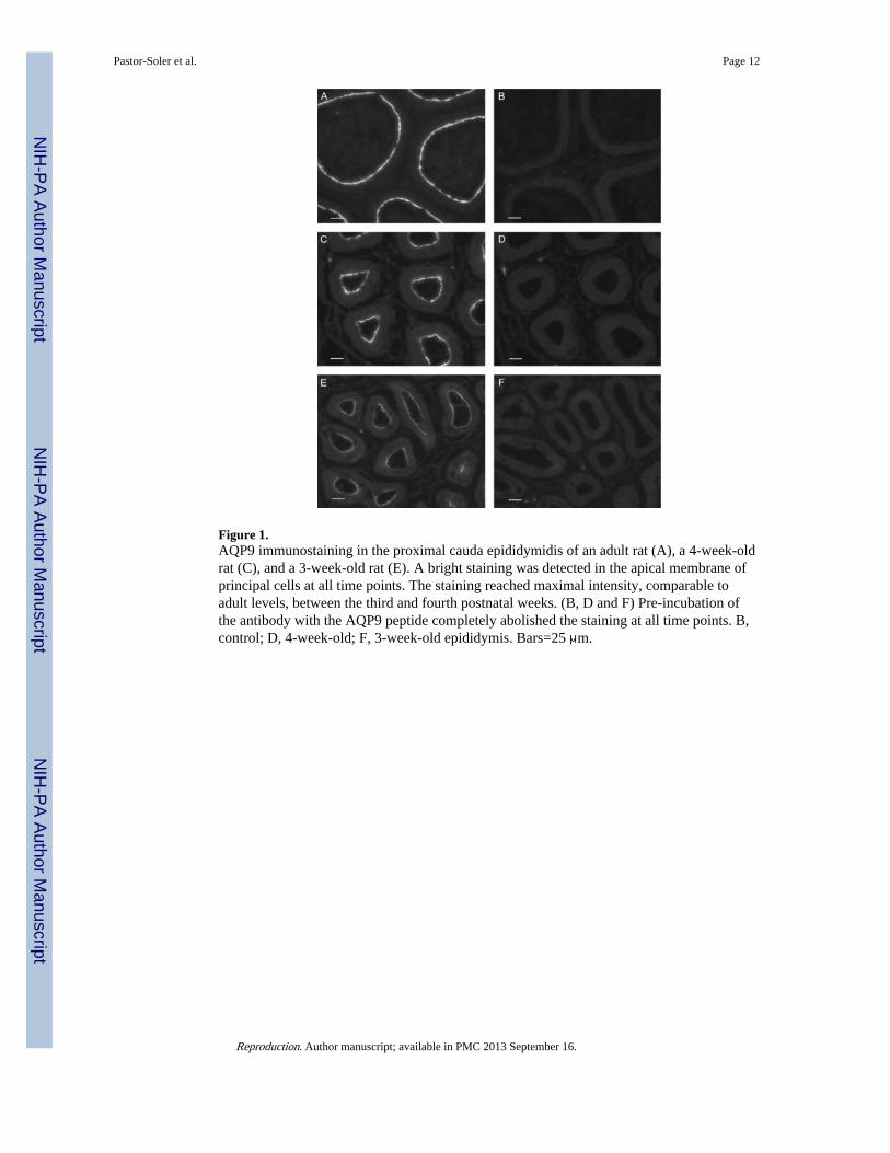
Figure 1.AQP9 immunostaining in the proximal cauda epididymidis of an adult rat (A), a 4-week-oldrat (C), and a 3-week-old rat (E). A bright staining was detected in the apical membrane ofprincipal cells at all time points. The staining reached maximal intensity, comparable toadult levels, between the third and fourth postnatal weeks. (B, D and F) Pre-incubation ofthe antibody with the AQP9 peptide completely abolished the staining at all time points. B,control; D, 4-week-old; F, 3-week-old epididymis. Bars=25 μm.
Pastor-Soler et al. Page 12
Reproduction. Author manuscript; available in PMC 2013 September 16.
NIH
-PA Author Manuscript
NIH
-PA Author Manuscript
NIH
-PA Author Manuscript
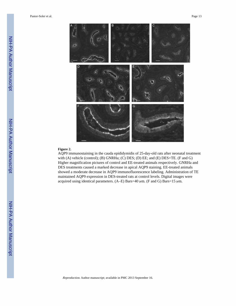
Figure 2.AQP9 immunostaining in the cauda epididymidis of 25-day-old rats after neonatal treatmentwith (A) vehicle (control); (B) GNRHa; (C) DES; (D) EE; and (E) DES+TE. (F and G)Higher magnification pictures of control and EE-treated animals respectively. GNRHa andDES treatments caused a marked decrease in apical AQP9 staining. EE-treated animalsshowed a moderate decrease in AQP9 immunofluorescence labeling. Administration of TEmaintained AQP9 expression in DES-treated rats at control levels. Digital images wereacquired using identical parameters. (A–E) Bars=40 μm. (F and G) Bars=15 μm.
Pastor-Soler et al. Page 13
Reproduction. Author manuscript; available in PMC 2013 September 16.
NIH
-PA Author Manuscript
NIH
-PA Author Manuscript
NIH
-PA Author Manuscript

Figure 3.Quantification of AQP9-associated fluorescence staining in the cauda epididymidis of 25-day-old control and treated rats (groups represented in Fig. 1). Significant decreases inAQP9 staining intensity were induced by GNRHa and DES. EE also induced a reduction inAQP9 staining but the difference was not significant versus control. Co-administration oftestosterone with DES completely restored AQP9 staining compared to DES alone, withAQP9 staining intensity comparable to control. Data are expressed relative to control, andrepresent the means±S.E.M. obtained from 4 to 7 animals per group. **P<0.0005 versuscontrol; ##P<0.005 versus DES; NS, not significant versus control.
Pastor-Soler et al. Page 14
Reproduction. Author manuscript; available in PMC 2013 September 16.
NIH
-PA Author Manuscript
NIH
-PA Author Manuscript
NIH
-PA Author Manuscript

Figure 4.Downregulation of AQP9 expression in the cauda epididymidis of 25-day-old rats by theanti-androgen flutamide. Immunofluorescence for AQP9 in the cauda epididymidis from acontrol rat (A) and a flutamide-treated rat (B) showing a marked decrease in apical AQP9staining in the flutamide-treated animal. Panels C and D are higher magnificationrepresentations of some of the tubules shown in A and B respectively. Digital images wereacquired using identical parameters. Bars=25 μm.
Pastor-Soler et al. Page 15
Reproduction. Author manuscript; available in PMC 2013 September 16.
NIH
-PA Author Manuscript
NIH
-PA Author Manuscript
NIH
-PA Author Manuscript

Figure 5.Quantification of AQP9-associated fluorescence staining in the cauda epididymidis ofcontrol and flutamide-treated 25-day-old rats. The mean pixel intensity of AQP9-associatedfluorescence in the apical pole of principal cells was markedly decreased followingflutamide treatment. Data are expressed relative to control and represent the means±S.E.M.obtained from three animals per group. *P<0.05 versus control.
Pastor-Soler et al. Page 16
Reproduction. Author manuscript; available in PMC 2013 September 16.
NIH
-PA Author Manuscript
NIH
-PA Author Manuscript
NIH
-PA Author Manuscript

Figure 6.Western blot showing downregulation of AQP9 in epididymis of 25-day-old rats bydifferent hormonal treatments. A strong band at ~30–35 kDa is present in total epididymalhomogenate membrane of control animals. Additional, higher molecular weight bandsprobably represent different glycosylation states of AQP9. AQP9 was not detectable in theGNRHa-, DES- and EE-treated groups. In the DES+TE-treated animals, a brighter AQP9band was detected compared to DES treatment alone. A very faint AQP9 band was detectedin the flutamide-treated group. Each well was loaded with the same amount of protein(Fisher et al. 2002).
Pastor-Soler et al. Page 17
Reproduction. Author manuscript; available in PMC 2013 September 16.
NIH
-PA Author Manuscript
NIH
-PA Author Manuscript
NIH
-PA Author Manuscript