editors - centers for disease control and prevention · international editors patrice courvalin...
TRANSCRIPT


EditorsJoseph E. McDade, Editor-in-ChiefAtlanta, Georgia, USAStephen S. Morse, Perspectives EditorNew York, New York, USA
Brian W.J. Mahy, Perspectives EditorAtlanta, Georgia, USAPhillip J. Baker, Synopses EditorBethesda, Maryland, USA
Stephen Ostroff, Dispatches EditorAtlanta, Georgia, USA
Patricia M. Quinlisk, Letters EditorDes Moines, Iowa, USA
Polyxeni Potter, Managing EditorAtlanta, Georgia, USA
International EditorsPatrice CourvalinParis, FranceKeith KlugmanJohannesburg, Republic of South AfricaTakeshi KurataTokyo, JapanS.K. LamKuala Lumpur, MalaysiaJohn S. MacKenzieBrisbane, AustraliaHooman MomenRio de Janeiro, BrazilSergey V. NetesovNovosibirsk Region, Russian FederationV. RamalingaswamiNew Delhi, IndiaDiana WalfordLondon, United Kingdom
Production EditorsMaria T. BritoKevin BurlisonTeresa M. HoodAnne D. MatherAva W. Navin
Retrieve the journal electronically on theWorld Wide Web (WWW) at http://www.cdc.gov/eid or from the CDC homepage (http://www.cdc.gov).
Announcements of new table of contentscan be automatically e-mailed to you. Tosubscribe, send an e-mail [email protected] with the following in thebody of your message: subscribe EID-TOC.
Electronic Access
Emerging Infectious DiseasesEmerging Infectious Diseases is published
six times a year by the National Center forInfectious Diseases, Centers for DiseaseControl and Prevention (CDC), 1600 CliftonRoad, Mailstop D61, Atlanta, GA 30333, USA.Telephone 404-371-5329, fax 404-371-5449,e-mail [email protected].
All material published in EmergingInfectious Diseases is in the public domainand may be used and reprinted withoutspecial permission; proper citation, however,is appreciated.
Use of trade names is for identificationonly and does not imply endorsement by thePublic Health Service or by the U.S. Depart-ment of Health and Human Services.
Emerging Infectious Diseases is printed on acid-free paper that meets the requirements of ANSI/NISO 239.48-1992 (Permanence of Paper).
∞
The journal is distributed electronically and in hard copy and is available at no charge.
YES, I would like to receive Emerging Infectious Diseases.
Please print your name andbusiness address in the box andreturn by fax to 404-371-5449 ormail to EID Editor CDC/NCID/MS D61 1600 Clifton Road, NE Atlanta, GA 30333
In Index Medicus/Medline, Current Contents, Excerpta Medica, and other databases
Moving? Please give us your new address (in the box) and print the number of your old
mailing label here__________
Editorial BoardDennis Alexander, Addlestone Surrey, United Kingdom (2003)Ban Allos, Nashville, Tennesee, USA (2003)Michael Apicella, Iowa City, Iowa, USA (2003)Abdu F. Azad, Baltimore, Maryland, USA (2002)Johan Bakken, Duluth, Minnesota, USA (2001)Ben Beard, Atlanta, Georgia, USA (2003)Barry J. Beaty, Ft. Collins, Colorado, USA (2002)Guthrie Birkhead, Albany, New York, USA (2001)Martin J. Blaser, New York, New York, USA (2002)S.P. Borriello, London, United Kingdom (2002)Donald S. Burke, Baltimore, Maryland, USA (2001)Charles Calisher, Ft. Collins, Colorado, USA (2001)Arturo Casadevall, Bronx, New York, USA (2002)Thomas Cleary, Houston, Texas, USA (2001)Anne DeGroot, Providence, Rhode Island, USA (2003)Vincent Deubel, Lyon, France (2003)J. Stephen Dumler, Baltimore, Maryland, USA (2002)Durland Fish, New Haven, Connecticut, USA (2002)Richard L. Guerrant, Charlottesville, Virginia, USA (2002)Scott Halstead, Arlington, Virginia, USA (2001)Seyed Hasnain, Hyderabad, India (2002)David L. Heymann, Geneva, Switzerland (2001)Dagmar Hulìnskà, Prague, Czech Republic (2001)Sakae Inouye, Tokyo, Japan (2003)Peter B. Jahrling, Frederick, Maryland, USA (2002)Mohamed A. Karmali, Guelph, Ontario, Canada (2002)Charles King, Cleveland, Ohio, USA (2003)Bruce R. Levin, Atlanta, Georgia, USA (2002)Myron Levine, Baltimore, Maryland, USA (2001)Stuart Levy, Boston, Massachusetts, USA (2002)Thomas J. Marrie, Edmonton, Alberta, Canada (2003)John E. McGowan, Jr., Atlanta, Georgia, USA (2002)Patrick S. Moore, New York, New York, USA (2002)Philip P. Mortimer, London, United Kingdom (2002)Fred A. Murphy, Davis, California, USA (2001)Barbara E. Murray, Houston, Texas, USA (2002)P. Keith Murray, Ames, Iowa, USA (2003)James M. Musser, Hamilton, Missouri, USA (2002)Rosanna W. Peeling, Geneva, Switzerland (2001)David H. Persing, Seattle, Washington, USA (2002)Richard Platt, Boston, Massachusetts, USA (2001)Didier Raoult, Marseille, France (2002)Leslie Real, Atlanta, Georgia, USA (2003)David Relman, Palo Alto, California, USA (2002)Rebecca Rico-Hesse, San Antonio, Texas, USA (2001)Pierre Rollin, Atlanta, Georgia, USA (2003)Nancy Rosenstein, Atlanta, Georgia, USA (2003)Connie Schmaljohn, Frederick, Maryland, USA (2001)Robert Shope, Galveston, Texas, USA (2002)Bonnie Smoak, Bethesda, Maryland, USA (2001)Rosemary Soave, New York, New York, USA (2001)P. Frederick Sparling, Chapel Hill, North Carolina, USA (2001)G. Thomas Strickland, Baltimore, Maryland, USA (2001)Jan Svoboda, Prague, Czech Republic (2001)Robert Swanepoel, Sandringham, South Africa (2002)Phillip Tarr, Seattle, Washington, USA (2001)Lucy Tompkins, Stanford, California, USA (2001)Timothy Tucker, Cape Town, South Africa (2003)Elaine Tuomanen, Memphis, Tennessee, USA (2002)David Walker, Galveston, Texas, USA (2002)Mary E. Wilson, Cambridge, Massachusetts, USA (2001)
The opinions expressed by authors contribut-ing to this journal do not necessarily reflect theopinions of the Centers for Disease Controland Prevention or the institutions with whichthe authors are affiliated.
Search EMERGING INFECTIOUS DISEASES at www.cdc.gov/eid

Perspectives
Letters
Seasonal Variation in Host Susceptibility and S.F. DowellCycles of Certain Infectious Diseases ...................................... 369
Cryptococcus neoformans Infection in Organ S. Husain et al.Transplant Recipients: Variables InfluencingClinical Characteristics and Outcome ..................................... 375
PulseNet: The Molecular Subtyping Network for Foodborne B. SwaminathanBacterial Disease Surveillance, United States ....................... 382 et al.
Spoligotype Database of Mycobacterium tuberculosis: C. Sola et al.Biogeographic Distribution of Shared Types andEpidemiologic and Phylogenetic Perspectives ........................ 390
Transmission of an Arenavirus in White-Throated C.H. Calisher et al.Woodrats (Neotoma albigula), SoutheasternColorado, 1995–1999 ................................................................ 397
Geographic Distribution and Genetic Diversity C.F. Fulhorstof Whitewater Arroyo Virus in the et al.Southwestern United States .................................................... 403
Is High Prevalence of Echinococcus multilocularis in Wild B. Gottstein et al.and Domestic Animals Associated with DiseaseIncidence in Humans? .............................................................. 408
Goat-Associated Q Fever: A New Disease T.F. Hatchettein Newfoundland ...................................................................... 413 et al.
Molecular Epidemiology of Serogroup A Meningitis M. Achtman et al.in Moscow, 1969–1997.............................................................. 420
Melioidosis: An Emerging Infection in Taiwan? ..................... 428 P.-R. Hsueh et al.
Outbreak of Human Monkeypox, Democratic Republic Y.J.F. Hutin et al.of Congo, 1996–1997 ................................................................. 434
Nipah Virus Infection in Bats (Order Chiroptera) in M.Y. Johara et al.Peninsular Malaysia ................................................................ 439
The opinions expressed by authorscontributing to this journal do notnecessarily reflect the opinions of theCenters for Disease Control andPrevention or the institutions withwhich the authors are affiliated.
Cover: Detail of La Primavera(ca. 1475–1478) by Sandro Botticelli,used with permission of Uffizi Gallery,Florence, Italy.
Synopses
Research
Dispatches
Candida dubliniensisCandidemia in Australia......................................... 479
D. Marriott et al.
Characterization of a HumanGranulocytic Ehrlichiosis-LikeAgent from Ixodes scapularis,Ontario, Canada ............. 479M.A. Drebot et al.
High Prevalence of Sin NombreVirus in Rodent Populations,Central Utah: A Consequence ofHuman Disturbance? ..... 480R. Mackelprang et al.
Hantavirus Seroconversionof Wild-Caught Peromyscusduring Quarantine .......... 482M. Camaioni et al.
Mycobacterium tuberculosisIsolates of Beijing Genotypein Thailand...................... 483W.M. Prodinger et al.

Dispatches, cont’d.
Third-Generation Cephalosporin Resistance M. Radice et al.in Shigella sonnei, Argentina ................................................... 442
Expanding Drug Resistance through Integron Acquisition A. Carattoli et al.by IncFI Plasmids of Salmonella enterica Typhimurium ...... 444
Decreased Susceptibility to Ciprofloxacin in Salmonella E.J. Threlfall &enterica serotype Typhi, United Kingdom ............................... 448 L.R. Ward
The First Reported Case of California Encephalitis in B.F. EldridgeMore Than 50 Years ................................................................. 451 et al.
Reiter Syndrome Following Protracted Symptoms B.A. Connor et al.of Cyclospora Infection ............................................................. 453
Presence of Class I Integrons in Multidrug-Resistant, A. Nastasi &Low-Prevalence Salmonella Serotypes, Italy .......................... 455 C. Mammina
Risk for Human Tick-Borne Encephalitis, Borrelioses, and E.I. KorenbergDouble Infection in the Pre-Ural Region of Russia ................ 459 et al.
Outbreak of Influenza in Highly Vaccinated K.C. Earhart et al.Crew of U.S. Navy Ship ............................................................ 463
HIV-1 Group O Infection in Cameroon, 1986–1998 ............... 466 A. Ayouba et al.
VEB-1-Like Extended-Spectrum β-Lactamases in L. Poirel et al.Pseudomonas aeruginosa, Kuwait ........................................... 468
Borrelia lonestari DNA in Adult Amblyomma americanum T.R. Burkot et al.Ticks, Alabama ......................................................................... 471
Physicians’ Database Searches as a Tool for V. JormanainenEarly Detection of Epidemics ................................................... 474 et al.
Filamentous Phage Associated with Recent Pandemic T. Iida et al.Strains of Vibrio parahaemolyticus ......................................... 477
Detail from La Primavera (Spring)by Sandro Botticelli (circa 1475–1478) .................................... 492
Letters, cont’d.
Jungle Yellow Fever,Rio de Janeiro ................. 484A.M.B. Filippis et al.
Emergence of Metronidazole-Resistant Bacteroides fragilis,India ................................ 485R. Chaudhry et al.
Proper Nomenclature forthe Human GranulocyticEhrlichiosis Agent .......... 486J.S. Bakken & J.S. Dumler
Single Nucleotide Polymorphismsin Mycobacterium tuberculosisStructural Genes ............ 486J.M. Musser
Response to Dr. Musser .. 487R. Fleischmann
Will Avilamycin ConvertZiracine into Zerocine? ... 488T.R. Shryock
The Antibiotic Food-ChainGang ................................ 489P. Courvalin
Johns Hopkins Graduate SummerInstitute of Epidemiology andBiostatistics, Baltimore,6/8/01 to 7/6/01 ................ 491
Joint Conference of the AmericanSociety for Rickettsiology and theBartonella as an EmergingPathogen Group, Big Sky,8/17/01 to 8/22/01 ............ 491
Errata .............................. 491
News and Notes
The Cover

369Vol. 7, No. 3, May–June 2001 Emerging Infectious Diseases
Perspective
From 1703 onward, the annual rise and fall of measlesdeaths in London was recorded in sufficient detail to allow forcareful mathematical modeling in 1918 (1). Since then,surveillance for a variety of diseases has established thatregular seasonal variation in incidence is the rule, ratherthan the exception, for acute infections. Seasonal variationsshould be distinguished from periodic large epidemics, asobserved every 2 years for measles (2) or at less frequent andmore irregular intervals for meningococcal meningitis (3) andrubella (4). This discussion will focus on the more robustannual cycle, which “locks in” large epidemics to the sametime of year (3,4) and persists even after large epidemics havebeen eliminated by mass vaccination (2). The life cycles ofpathogens spread by insect vectors or maintained in animal orenvironmental reservoirs add complexity because seasonalchanges might influence not only the pathogen or human hostbut also the vector population and animal or environmentalreservoir. Therefore, this discussion will focus on bacterialand viral pathogens maintained primarily by person-to-person spread.
The regular and predictable pattern of seasonaloutbreaks dominates the epidemiology of many exclusivelyhuman pathogens (Figure 1). Different infections peak in eachof the four seasons, but for each pathogen, the timing andcharacteristics of the annual outbreak are remarkablyconsistent from year to year. Other key observations havebeen made on the seasonality of infectious diseases, includingthe simultaneous onset of outbreaks in geographically remoteareas and the persistence of pathogens in the off-season in theabsence of epidemic spread (Table). In fact, latitude has aclear influence on the timing and magnitude of outbreaks ofrotavirus infection (10), influenza (15), and poliomyelitis
Seasonal Variation in Host Susceptibility andCycles of Certain Infectious Diseases
Scott F. DowellCenters for Disease Control and Prevention, Atlanta, Georgia, USA
Address for correspondence: Scott F. Dowell, Centers for DiseaseControl and Prevention, 1600 Clifton Road NE, Mailstop C12, Atlanta,GA 30333, USA; fax: 404-639-3039; e-mail: [email protected]
Seasonal cycles of infectious diseases have been variously attributed tochanges in atmospheric conditions, the prevalence or virulence of the pathogen,or the behavior of the host. Some observations about seasonality are difficult toreconcile with these explanations. These include the simultaneous appearanceof outbreaks across widespread geographic regions of the same latitude; thedetection of pathogens in the off-season without epidemic spread; and theconsistency of seasonal changes, despite wide variations in weather and humanbehavior. In contrast, an increase in susceptibility of the host population,perhaps linked to the annual light/dark cycle and mediated by the pattern ofmelatonin secretion, might account for many heretofore unexplained features ofinfectious disease seasonality. Ample evidence indicates that photoperiod-driven physiologic changes are typical in mammalian species, including some inhumans. If such physiologic changes underlie human resistance to infectiousdiseases for large portions of the year and the changes can be identified andmodified, the therapeutic and preventive implications may be considerable.
Figure 1. Seasonal variation in the occurrence of three humanpathogens in the U.S. A: an annual cycle of rubella activity wasmaintained between larger epidemics, which occurred every 6 to 9years. B: percentage of specimens testing positive for influenza virusesamong specimens tested by World Health Organization and U.S.National Respiratory and Enteric Virus Surveillance Systemcollaborating laboratories. C: a consistent pattern of rotavirusseasonality is evident in the U.S. National Respiratory and EntericVirus Surveillance System. Adapted from references 4-6.

370Emerging Infectious Diseases Vol. 7, No. 3, May–June 2001
Perspective
(Figure 2) (9). Reconciling these observations with theconsistent seasonality of clinical illness is a continuingchallenge.
Explanations of SeasonalityBecause seasonal cycles of infectious diseases are so
universal and no single theory has proved satisfactory,explanations about their cause abound. More than oneexplanation or combination of explanations may be true.Explanations can be grouped into three types: pathogenappearance and disappearance, environmental changes, andhost-behavior changes.
Pathogen Appearance and DisappearancePerhaps the most obvious explanation for the absence of
disease during a period is that the pathogen is also absentduring the period. However, the regular annual migration ofepidemics of influenza, poliomyelitis, and rotavirus infectionfrom northern latitudes across the equator to southern onesand back does not necessarily imply that the pathogensthemselves migrate in this way.
Current theory holds that influenza is maintained onlyby direct spread in a series of chains of transmission from oneill person to another (16). Some evidence suggests thatinfluenza viruses do spread geographically, particularlyduring pandemics, but whether geographic spread accountsfor the patterns observed in annual outbreaks has beenquestioned (11,17,18). The simultaneous onset of geographi-cally widespread outbreaks is difficult to reconcile withchains of person-to-person transmission. One hypothesis isthat earlier “seeding” of the virus throughout the populationmust have occurred (17). During an 1826 influenza epidemic,one observer wrote, “...this epidemic affects a whole region inthe space of a week, nay, a whole continent as large as NorthAmerica, together with all the West Indies, in the course of afew weeks, while the inhabitants could not within so short atime have had any communication or intercourse whateveracross such a vast extent of country” (11). A more recenthypothesis attributes geographic spread to the atmosphericdispersion of virus from Southeast Asia by trans-Pacificwinds across the North American continent (18).
Environmental ChangesEnvironmental changes, particularly changes in weath-
er, are the explanations most often invoked for the seasonalityof infectious diseases. Statistically significant correlationsbetween epidemic cycles and cycles of temperature (19-22),humidity (21-23), rains (24), or winds (24) have beenidentified. However, correlations may be found withconfounders as well as with causes.
In some cases, the association with weather is supported,but the biologic plausibility appears tenuous. Although the
Table. Observations on the seasonal occurrence of infectious diseases
Observation ExamplesPathogens peak at characteristic times in Winter: influenza, pneumococcus, rotavirus all seasons of the year Spring: RSV, measles
Summer: polio, other enterovirusesFall: parainfluenza virus type 1
Timing and duration of peaks for each Measles: regular pattern since 1703 (1) pathogen are similar from year to year Influenza: annual peak varies by only 5 to 10 weeks in the United States (6)
Onset of epidemics often occurs simultaneously Influenza: simultaneous outbreaks across North America, 16 European in areas that are geographically dispersed countries, and 6 Chinese provinces (7) and have different weather conditions and Pneumococcus: simultaneous outbreaks in seven surveillance areas (8) diverse populations
Latitude is a critical determinant of timing An increasing magnitude of seasonal peaks as distance from the equator and magnitude of peaks increases has been documented for polio (9) and rotavirus (10) and
reported for influenza (11).
Pathogens can be detected in the off-season Meningococcus: no decrease in carriage in the off-season, despite despite lower incidence of disease and absence of epidemic disease (12) virtual absence of epidemics RSV: sporadic summer viral isolation but no epidemic spread (13)
Influenza: sporadic summer isolation, occasional clusters of disease without epidemic spread (14)
RSV = respiratory syncytial virus. RSV peaks in the winter or spring in the United States, depending on location. For simplicity, it is listed here as a springpathogen.
Figure 2. Seasonal variation in the incidence of poliomyelitis bylatitude, 1956-57. As distance from the equator increases, a higherproportion of cases are evident in summer and fall months. Adaptedfrom reference 9.
Mon
thly
% o
f cas
es
Latitude

371Vol. 7, No. 3, May–June 2001 Emerging Infectious Diseases
Perspective
seasonal incidence of poliomyelitis correlated quite well withthe summer increase in relative humidity in Boston andHouston from 1942 to 1951 (23), the explanation thataerosolized poliovirus survives for a longer time at higherrelative humidity is difficult to reconcile with the fecal-oralroute of poliovirus transmission.
In other cases, the correlations are supported by biologicplausibility but are not consistently observed. In sub-Saharan Africa, the onset of meningococcal epidemics closelyfollowed the season of dry winds and ended with the onset ofthe rains (25). It has been proposed that drying of mucosalsurfaces increases the probability of bacteremic spread andthat the rains moisten the mucosa or decrease the spread ofthe organism by dust. However, in Oregon and other areas,meningococcal disease peaks during the rainy season (26).Similarly, a significant correlation between the onset of theinvasive pneumococcal disease season and a drop in meandaily temperatures below 24°C in Houston (19) was notconfirmed in seven other areas with more widely varyingweather patterns (8). Respiratory syncytial virus epidemicsoccur in the colder months of winter and spring in the UnitedStates (13) but paradoxically are significantly correlated withthe hotter months in Singapore and Hong Kong (21,22).
Host-Behavior ChangesSeasonal changes in poliomyelitis, measles, and other
seasonal infectious diseases have been attributed to changesin the behavior of the host. Public swimming pools were asource of great concern during the polio epidemics of the1950s, and summer peaks in polio and other enteroviruseswere attributed to swimming (23,27,28). Subsequent studiesdiscounted the importance of swimming in the spread ofenterovirus infections (28).
Crowding of susceptible persons is one of the mostcommon explanations for seasonal infectious diseases, and itcertainly has biologic plausibility. The seasonal patterns ofmeasles in England and Wales have been attributed to thetiming of school holidays (29,30). Although such explanationsare plausible, one must also ask why influenza outbreaks donot occur in crowded international conventions duringsummer, and why measles outbreaks are not common atsummer camps. As one authority noted regarding meningo-coccal seasonality, “The story that African epidemics arecaused by people crowding together at night during the dryseason is a medical myth which is difficult to kill. Villagerssleep inside at the height of the rainy season at least asfrequently as during the cold part of the dry season...” (24).
Comprehensive explanations of seasonal changes ininfectious diseases should identify the means by whichsimilar pathogens peak at different seasons (with character-istic timing and duration) and explain the prompt regionwideepidemics in geographically dispersed populations, thevariation in epidemic patterns by latitude, and thepersistence of the pathogen in the off-season without epidemicdisease (Table).
The Proposed HypothesisRegular annual variations in the incidence of many
infectious diseases may be due to changes in susceptibility ofthe human host to the particular pathogen. Like the seasonalphysiologic cycles of many mammalian species, these changesin susceptibility may be timed to the light/dark cycle,
typically mediated by changes in the duration of the dailymelatonin pulse. The changes in susceptibility may bedistinct for different pathogens and may cover a broad rangeof possibilities, including (but not limited to) changes in thecharacteristic of mucosal surfaces, the expression of epithelialreceptors, the leukocyte numbers or responsiveness, or otherfeatures of specific or nonspecific immunity.
This hypothesis would predict that pathogens do notphysically migrate across the equator and that nationwideepidemics do not necessarily result from chains of person-to-person transmission. Rather, the pathogens may be presentin the population year-round, and epidemics occur when thesusceptibility of the population increases enough to sustainthem. Perhaps the most significant prediction is that peopleare relatively resistant to disease if exposed in the off-seasonand that the specific physiologic process leading to seasonalresistance should be identifiable and perhaps modifiable.
Seasonal Changes in Host PhysiologyMany mammalian species undergo seasonal physiologic
changes. The best characterized are changes in reproductiveorgans and other tissues seen in animals that are seasonalbreeders. Humans are not seasonal breeders, but fertility hasseasonal variations. Seasonal variations have been documentedin other physiologic processes and immunologic features (31,32).
Producing offspring in a season during which food isunavailable and the environment is unsuitable for the youngis an evolutionary dead-end for some species, leading tocarefully regulated breeding seasons for many rodents (33),sheep (34), other ungulates (35), monkeys (36), and primates(37). Seasonal physiologic changes involve not just behaviorbut also the secretion of sex hormones and the size andfunction of reproductive organs. In controlled laboratoryconditions, the duration of the light/dark cycle is the keyparameter governing these seasonal changes, which can becompletely replicated by artificial manipulation of thephotoperiod. Photoperiod is most commonly used rather thantemperature, humidity, food availability, or other seasonallyvarying parameters, presumably because its invariant naturebest prevents accidental breeding at the wrong time of year.Under constant photoperiod, the physiologic changes can alsobe reproduced by controlling the duration of the dailymelatonin pulse.
Seasonal physiologic changes have also been documentedin processes not typically associated with breeding butpotentially related to susceptibility to infectious agents. Forexample, even under constant conditions, red deer havedistinct seasonal changes in digestive features (35), mice haveseasonal changes in seizure threshold (38), and dairy cattlehave seasonal changes in the fat and protein content of theirmilk (39). In recent years, seasonal changes in immunologicfeatures have been documented. For example, Siberianhamsters exposed to short-day photoperiod demonstrateincreased natural killer-cell activity and lymphocyteblastogenesis but decreased phagocytosis and oxidative burstactivity by granulocytes (40); deer mice treated withmelatonin in constant photoperiod exhibit increasedlymphocyte response to mitogen stimulation (41).
A series of studies documented that the death rate in miceexperimentally exposed to pneumococcal infection variedwith the time of day (42-44). Survival patterns were altered bymodifying environmental lighting conditions, rather than

372Emerging Infectious Diseases Vol. 7, No. 3, May–June 2001
Perspective
feeding or activity, and susceptibility appeared related to thedaily cycle of cortisone, although the specific physiologicfeature responsible for increased susceptibility was notidentified. Since these findings, understanding of the role ofmelatonin and its control of circadean and seasonal rhythmshas increased greatly, but further studies of the influence ofphotoperiod on experimental pneumococcal infections in miceappear not to have been pursued.
Seasonal physiologic changes are not as well character-ized for humans as for other mammals, but mounting datasuggest that changes in photoperiod and the melatonin pulsemay also influence human physiology (32). Blind people, wholack the capability for light to cue their biologic clocks, areoften plagued by free-running circadian rhythms. A recentstudy demonstrated that these free-running rhythms can beentrained to a normal cycle by daily administration ofmelatonin (45). Although humans are sexually active year-round, a seasonal distribution in conceptions has consistentlybeen demonstrated, and a variation in the ovulation rate hasbeen postulated as the cause (31). Seasonal affective disorder,a well-characterized depression associated with short daysand specific genetic defects (46), is treatable with extra hoursof exposure to broad-spectrum light (47). Seasonal variationsin heart attacks (48), breast cancer (49), and other seeminglynoninfectious conditions have also been reported.
Recent research has focused on seasonal changes inimmunologic values in humans. Specific melatonin receptorscoupled with G-protein have been identified on lymphocytes(50). As in rodents, seasonal variations in lymphocytemitogenic responses and in the quantity of circulatinglymphocytes, neutrophils, CD4 and CD8 cells, and IL-6 havebeen reported (51-53). Some values, such as lymphocyte arylhydrocarbon hydroxylase activity, peak in summer (54), whileothers, such as number of circulating B cells, peak in winter(52). Although statistically significant, the functionalsignificance of these variations has not yet been established.
Testing the HypothesisThe above observations lend some biologic plausibility to
the proposed hypothesis, but direct testing is needed. Severalobservations support the prediction that the host is lesssusceptible to infection or disease in the off-season.
In a double-blind placebo-controlled trial conducted inthe Soviet Union during different seasons, nonimmunevolunteers were given attenuated live influenza vaccineintranasally (55). Febrile reactions attributable to vaccine(calculated by subtracting the proportion of participants withreactions in the placebo group from the proportion in thevaccine group) were observed in 6.7% of 360 volunteersinoculated in Leningrad in January, compared with 0.8% of197 inoculated in June (p = 0.003). Fourfold rises in antibodytiter were seen in 31% to 40% in Krasnodar in January,depending on the vaccine strain, compared with 4.3% to 4.8%given the same strains in May and October (all p <0.001).Similar trends with less significant differences were seen inthree other cities.
Some years earlier, in a series of experiments on thetransmission of influenza virus from infected to susceptiblemice, <1% of mice exposed from July to October were infected,compared with 22% of those exposed in December or January(p <0.001) (56). One year later, the investigators repeated theexperiment with a different strain of mice, now kept under
constant temperature and humidity, and observed that 34%were infected in May to October, compared with 58% inNovember to April (p <0.001). The photoperiod conditions inthese experiments were not noted.
It is not clear whether attempts were made to replicatethese provocative experiments or if the potential importanceof the observations was fully appreciated. The animalexperiments may be relatively easy to confirm or refute, andthe many live attenuated vaccines currently tested or usedshould provide ample material to evaluate the effects ofseason on immunogenicity or reactogenicity. The season ofadministration influences seroconversion rates to oral poliovaccine (57,58) and protection against polio (59), but much ofthis seasonal variation may be attributable to competition byother enteroviruses during summer (57). Vaccine-associatedparalytic polio among vaccine contacts reflects the seasonalpattern of natural polio (60).
ConclusionPhotoperiod-driven changes in host physiology might
explain certain enigmatic observations about seasonality, butsome observations remain unexplained. For example, thewest-east movement of rotavirus is not easily attributable tohost susceptibility changes timed to the light/dark cycle (5).The increase in hospitalizations coincident with warmweather and El Nino points to temperature rather thanphotoperiod as a key influence on some diarrheal diseasepathogens (20). The sudden appearance and worldwidespread of a new pandemic strain of influenza virus also arguesmore for chains of transmission than for a crop of outbreaksfrom virus already present in the population.
Epidemiologists have long puzzled over why seasonalinfectious disease outbreaks occur when they do. Perhaps themore important question is why they do not occur when theydo not. Is the human population already relatively resistantfor 6 to 9 months each year? If the absence of epidemics ofsummer influenza or winter polio is attributable to climate orweather, we may have little power to influence them. On theother hand, if these annual troughs are due to increased hostresistance, opportunities abound for studying and modifyingthese changes. Such opportunities might include reviews ofexisting databases, careful evaluation of “experiments ofnature,” and studies in laboratory animals.
Databases surely exist that might shed light on thishypothesis. Clinical trials of live attenuated vaccines duringthe usual seasonal peak and seasonal trough for thatparticular disease could be reviewed for seasonal differencesin reactogenicity and immunogenicity. Experiments ofnature, in which groups adapted to summer come into contactwith groups adapted to winter (as in a convention or a cruiseship with passengers from both Southern Hemisphere andNorthern Hemisphere countries) and are exposed to aseasonal pathogen (such as influenza or an enterovirus),could be analyzed for differences in attack rate or clinicalseverity. Laboratory animals housed in photoperiod-controlled rooms could be exposed to seasonal pathogens andevaluated to see if photoperiod or melatonin modifies clinicaland physiologic responses to infection. If differences aredocumented, the specific physiologic feature governingsusceptibility changes could be isolated and identified.
It is time to have a closer look at these possible seasonalchanges in host susceptibility and if they are confirmed,

373Vol. 7, No. 3, May–June 2001 Emerging Infectious Diseases
Perspective
identify and modify the physiologic changes underlyingannual cycles of infectious diseases.
AcknowledgmentsThe author thanks Anne Schuchat, G. Robert Lynch, Marc
Fischer, Stephanie Schrag, Alicia Fry, and David Shay for helpfuldiscussions and suggestions on the manuscript.
Dr. Dowell is acting Associate Director for Global Health, NationalCenter for Infectious Diseases, CDC. His research focuses on the epide-miology of infectious diseases.
References 1. Brownlee J. An investigation into the periodicity of measles
epidemics in London from 1703 to the present day by the method ofthe periodogram. Philosophical Transactions of the Royal Society ofLondon 1918;B 208:225-50.
2. Anderson R, Grenfell BT, May RM. Oscillatory fluctuations in theincidence of infectious disease and the impact of vaccination: timeseries analysis. J Hyg Camb 1984;93:587-608.
3. Riedo F, Plikaytis B, Broome C. Epidemiology and prevention ofmeningococcal disease. Pediatr Infect Dis J 1995;14:643-57.
4. Witte J, Karchmer A, Case M, Herrmann KL, Abrutyn E,Kassanoff I, et al. Epidemiology of rubella. Am J Dis Child1969;118:107-11.
5. Torok TJ, Kilgore PE, Clarke MJ, Holman RC, Bresee JS, Glass RI.Visualizing geographic and temporal trends in rotavirus activity inthe United States, 1991 to 1996. National Respiratory and EntericVirus Surveillance System Collaborating Laboratories. PediatrInfect Dis J 1997;16:941-6.
6. Centers for Disease Control and Prevention. Update: Influenzaactivity—United States, 1999-2000 season. MMWR Morb MortalWkly Rep 2000;49:173-7.
7. Centers for Disease Control and Prevention. Update: Influenzaactivity—United States and worldwide, 1995-96 season, andcomposition of the 1996-97 influenza vaccine. MMWR Morb MortalWkly Rep 1996;45:326-9.
8. Dowell S, Whitney C, Wright C, Schuchat A. Seasonal changes ininvasive pneumococcal disease. Emerg Infect Dis. In press 2001.
9. Paccaud M. World trends in poliomyelitis morbidity and mortality,1951-1975. World Health Stat Quart 1979;32:198-224.
10. Cook S, Glass R, LeBaron C, Ho M-S. Global seasonality ofrotavirus infections. Bull World Health Organ 1990;68:171-7.
11. Hope-Simpson R, Golubev D. A new concept of the epidemic processof influenza A virus. Epidemiol Infect 1987;99:5-54.
12. Blakebrough I, Greenwood B, Whittle H, Bradley A, Gilles H. Theepidemiology of infections due to Neisseria meningitidis andNeisseria lactamica in a northern Nigerian community. J InfectDis 1982;146:626-37.
13. Centers for Disease Control and Prevention. Update: respiratorysyncytial virus activity—United States, 1998-1999 Season.MMWR Morb Mortal Wkly Rep 1999;48:1104-15.
14. Kohn M, Farley T, Sundin D, Tapia R, McFarland L, Arden N.Three summertime outbreaks of influenza type A. J Infect Dis1995;172:246-9.
15. Cox NJ, Fukuda K. Influenza. Infect Dis Clin N Am 1998;12:27-38.16. Cox N, Subbarao K. Influenza. Lancet 1999;354:1277-82.17. Langmuir A, Schoenbaum S. The epidemiology of influenza. Hosp
Pract 1976;11:49-56.18. Hammond G, Raddatz R, Gelskey D. Impact of atmospheric
dispersion and transport of viral aerosols on the epidemiology ofinfluenza. Rev Infect Dis 1989;11:494-7.
19. Kim P, Musher D, Glezen W, Rodriguez-Barradas M, Nahm W,Wright C. Association of invasive pneumococcal disease withseason, atmospheric conditions, air pollution, and the isolation ofrespiratory viruses. Clin Infect Dis 1996;22:100-6.
20. Checkley W, Epstein L, Gilman R, Figueroa D, Cama RI, Patz JA.Effects of El Niño and ambient temperature on hospital admissions fordiarrheal diseases in Peruvian children. Lancet 2000;355:442-50.
21. Chew F, Doraisingham S, Ling A, Kumarasinghe G, Lee B.Seasonal trends of viral respiratory tract infections in the tropics.Epidemiol Infect 1998;121:121-8.
22. Sung R, Murray H, Chan R, Davies D, French G. Seasonal patternsof respiratory syncytial virus infection in Hong Kong: a preliminaryreport. J Infect Dis 1987;156:527-8.
23. Nathanson N, Martin J. The epidemiology of poliomyelitis:enigmas surrounding its appearance, epidemicity, and disappear-ance. Am J Epidemiol 1979;110:672-92.
24. Greenwood B. The epidemiology of acute bacterial meningitis intropical Africa. Bacterial Meningitis. London: Academic Press;1987. p. 61-91.
25. Greenwood B, Blakebrough I, Bradley A, Wali S, Whittle H.Meningococcal disease and season in sub-Saharan Africa. Lancet1984;i:1339-42.
26. Diermayer M, Hedberg K, Hoesly F, Fischer M, Perkins B, Reeves M,et al. Epidemic serogroup B meningococcal disease in Oregon: theevolving epidemiology of the ET-5 strain. JAMA 1999;281:1493-7.
27. D’Alessio D, Minor T, Allen C, Tsiatis A, Nelson D. A study of theproportions of swimmers among well controls and children withenterovirus-like illness shedding or not shedding an enterovirus.Am J Epidemiol 1981;113:533-41.
28. Hawley H, Morin D, Geraghty M, Tomkow J, Phillips C.Coxsackievirus B epidemic at a boys’ summer camp: isolation ofvirus from swimming water. JAMA 1973;226:33-6.
29. Hamer W. Epidemic disease in England—the evidence ofvariability and persistency of type. Lancet 1906;11:733-9.
30. Fine P, Clarkson J. Measles in England and Wales - I: an analysis offactors underlying seasonal patterns. Int J Epidemiol 1982;11:5-14.
31. Rojansky N, Brzezinski A, Schenker J. Seasonality in humanreproduction: an update. Hum Reprod 1992;7:735-45.
32. Wehr T, Moul D, Barbato G, Giesen HA, Seidel JA, Barker C, et al.Conservation of photoperiod-responsive mechanisms in humans.Am J Physiol 1993;265:R846-57.
33. Carlson L, Zimmermann A, Lynch G. Geographic differences fordelay of sexual maturation in Peromyscus leukopus: effects ofphotoperiod, pinealectomy, and melatonin. Biology of Reproduction1989;41:1004-13.
34. Lincoln G. Reproductive seasonality and maturation throughoutthe complete life-cycle in the mouflon ram (ovis musimon). AnimReprod Sci 1998;53:87-105.
35. Rhind S, McMillen S, Duff E, Hirst D, Wright S. Seasonality ofmeal patterns and hormonal correlates in red deer. Physiol Behav1998;65:295-302.
36. Herndon J, Bein M, Nordmeyer D, Turner J. Seasonal testicularfunction in male rhesus monkeys. Horm Behav 1996;30:266-71.
37. Chan P, Hutz R, Dukelow W. Nonhuman primate in vitrofertilization: seasonality, cumulus cells, cyclic nucleotides,ribonucleic acid, and viability assays. Fertil Steril 1982;38:609-15.
38. Loscher W, Fiedler M. The role of technical, biological andpharmacological factors in the laboratory evaluation ofanticonvulsant drugs. VI. Seasonal influences on maximalelectroshock and pentylentetrazol seizure thresholds. Epilepsy Res1996;25:3-10.
39. Sargeant J, Shoukri M, Martin S, Leslie K, Lissemore K.Investigating potential risk factors for seasonal variation: anexample using graphical and spectral analysis methods based onthe production of milk components in dairy cattle. Prev Vet Med1998;36:167-78.
40. Yellon S, Fagoaga O, Nehlsen-Cannarella S. Influence ofphotoperiod on immune cell functions in the male Siberianhamster. Am J Physiol 1999;276:R97-102.
41. Demas G, Nelson R. Exogenous melatonin enhances cell-mediated,but not humoral, immune function in adult male deer mice(Peromyscus maniculatus). J Biol Rhythms 1998;13:245-52.
42. Feigin RD, San Joaquin VH, Haymond MW, Wyatt RG. Dailyperiodicity of susceptibility of mice to pneumococcal infection.Nature 1969;224:379-80.
43. Shackelford PG, Feigin RD. Periodicity of susceptibility topneumococcal infection: influence of light and adrenocorticalsecretions. Science 1973;182:285-7.

374Emerging Infectious Diseases Vol. 7, No. 3, May–June 2001
Perspective
44. Wongwiwat M, Sukapanit S, Triyanond C, Sawyer WD. Circadianrhythm of the resistance of mice to acute pneumococcal infection.Infect Immun 1972;5:442-8.
45. Sack R, Brandes R, Kendall A, Lewy A. Entrainment of free-running circadian rhythms by melatonin in blind people. N Engl JMed 2000;343:1070-7.
46. Sher L, Goldman D, Ozaki N, Rosenthal N. The role of geneticfactors in the etiology of seasonal affective disorder. J Affect Disord1999;53:203-10.
47. Eastman C, Young M, Fogg L, Liu L, Meaden P. Bright lighttreatment of winter depression: a placebo-controlled trial. ArchGen Psychiatry 1998;55:883-9.
48. Pell J, Cobbe S. Seasonal variations in coronary heart disease.QJM 1999;92:689-96.
49. Ownby H, Frederick J, Mortensen R, Ownby D, Russo J. Seasonalvariations in tumor size at diagnosis and immunological responsesin human breast cancer. Invasion Metastasis 1986;6:246-56.
50. Calvo J, Rafil-El-Idrissi M, Pozo D, Guerrero J. Immunomodulato-ry role of melatonin: specific binding sites in human and rodentlymphoid cells. J Pineal Res 1995;18:119-26.
51. Boctor F, Charmy R, Cooper E. Seasonal differences in therhythmicity of human male and female lymphocyte blastogenicresponses. Immunol Invest 1989;18:775-84.
52. Maes M, Stevens W, Scharpe S, Bosmans E, De Meyer F, D’Hondt P,et al. Seasonal variation in peripheral blood leukocyte subsets and inserum interleukin-6, and soluble interleukin-2 and -6 receptorconcentrations in normal volunteers. Experientia 1994;50:821-9.
53. Nelson R, Drazen D. Melatonin mediates seasonal adjustments inimmune function. Reprod Nutr Dev 1999;39:383-98.
54. Paigen B, Ward E, Reilly A, Houten L, Gurtoo HL, Minowada J, etal. Seasonal variation of aryl hydrocarbon hydroxylase activity inhuman lymphocytes. Cancer Res 1981;41:2757-61.
55. Shadrin A, Marinich I, Taros L. Experimental and epidemiologicalestimation of seasonal and climato-geographical features of non-specific resistance of the organism to influenza. J Hyg EpidemiolMicrobiol Immunol 1977;21:155-61.
56. Schulman J, Kilbourne E. Experimental transmission of influenzavirus infection in mice. II. Some factors affecting the incidence oftransmitted infections. J Exp Med 1963;118:267-75.
57. Swartz T, Skalska P, Gerichter C. Routine administration of oralpolio vaccine in a subtropical area. Factors possibly influencingsero-conversion rates. J Hyg Camb 1972;70:719-26.
58. World Health Organization Collaborative Study Group on OralPoliovirus V. Factors affecting the immunogenicity of oralpoliovirus vaccine: a prospective evaluation in Brazil and theGambia. J Infect Dis 1995;171:1097-106.
59. Deming M, Linkins R, Jaitch K, Hull H. The clinical efficacy oftrivalent oral polio vaccine in the Gambia by season of vaccineadministration. J Infect Dis 1997;175 s1:s254-7.
60. Schonberger L, McGowan JJ, Gregg M. Vaccine-associatedpoliomyelitis in the United States, 1961-1972. Am J Epidemiol1976;104:202-11.

Synopses
Vol. 7, No. 3, May–June 2001 Emerging Infectious Diseases375
Invasive fungal infections have been reported in 5% to59% of organ transplant recipients (1-4). Infections due toCryptococcus neoformans, while less common than those dueto Candida and mycelial fungi, are also an importantposttransplant complication. The incidence of invasivecandidiasis has declined in subsets of organ transplantrecipients (e.g., liver transplant patients) as a result offluconazole use and technologic advances in surgery (5).However, the risk factors and pathogenesis of C. neoformansinfection in the transplant setting are poorly understood, andfluconazole prophylaxis is generally not used in the lateposttransplant period when cryptococcal infections usuallyoccur. Thus, the incidence and impact of cryptococcal infectionin organ transplant recipients are unlikely to diminish in theforeseeable future. Indeed, as incidence of C. neoformansinfection in HIV-infected patients has declined, organtransplant recipients have become the group of immunocom-promised patients at highest risk for cryptococcosis. Theoverall death rate in transplant recipients with cryptococcalinfection has been 20% to 100% (6-9). While the predictors ofoutcome in patients with C. neoformans have been welldocumented in nontransplant settings (10-12), predictors intransplant recipients are largely unknown. The uniqueneurotropism and predilection of C. neoformans to cause centralnervous system (CNS) infections are well recognized; CNShas been the most common site for cryptococcal infections.However, 67% of our liver transplant recipients withcryptococcosis who received tacrolimus as primary immuno-suppression had cutaneous or osteoarticular lesions; 17% had
meningitis (8). Small sample size and lack of comparison withpatients on other immunosuppressive regimens, however,precluded meaningful interpretation of these data.
Given the limited number of transplant recipients withC. neoformans infection at individual institutions, accumu-lating a sufficiently large sample was difficult, so we turned toreports and analyses of cases for valuable data. This reviewsummarizes unique epidemiologic and clinical characteristicsof C. neoformans in transplant recipients, as well as variablesinfluencing the outcome of cryptococcal infections aftertransplantation.
MethodsCases of C. neoformans infection in transplant recipients
were identified with a MEDLINE search through 1998 bycross-referencing the keywords “Cryptococcus neoformans”and “transplantation” or “transplant.” Reference lists oforiginal articles and textbooks were reviewed for additionalcases. A patient was considered infected if C. neoformans wascultured from a clinical specimen in the presence of signs orsymptoms of cryptococcus infection. The onset of infectionafter transplantation was determined on the basis of detailedcase studies; summarized data providing only a mean orrange for the group of transplant recipients were excluded.Cryptococcal infection was considered early-onset if itoccurred within 12 months and late-onset if it occurred >12months after transplantation. Predictors or risk factors fordeath were assessed only in detailed cases for which thevariables to be analyzed were explicitly stated.
Statistical AnalysisPatient demographic data were entered into the database
PROPHET Statistics Version 5.0 (BBN Systems and
Cryptococcus neoformans Infection in OrganTransplant Recipients: Variables Influencing
Clinical Characteristics and OutcomeShahid Husain, Marilyn M. Wagener, and Nina Singh
Veterans Affairs Medical Center and University of Pittsburgh,Thomas E. Starzl Transplantation Institute, Pittsburgh, Pennsylvania, USA
Address for correspondence: Nina Singh, VA Medical Center,Infectious Disease Section, University Drive C, Pittsburgh, PA 15240,USA; fax: 412-688-6950; e-mail: [email protected]
Unique clinical characteristics and other variables influencing the outcome ofCryptococcus neoformans infection in organ transplant recipients have not beenwell defined. From a review of published reports, we found that C. neoformansinfection was documented in 2.8% of organ transplant recipients (overall deathrate 42%). The type of primary immunosuppressive agent used intransplantation influenced the predominant clinical manifestation ofcryptococcosis. Patients receiving tacrolimus were significantly less likely tohave central nervous system involvement (78% versus 11%, p =0.001) and morelikely to have skin, soft-tissue, and osteoarticular involvement (66% versus 21%,p = 0.006) than patients receiving nontacrolimus-based immunosuppression.Renal failure at admission was the only independently significant predictor ofdeath in these patients (odds ratio 16.4, 95% CI 1.9–143, p = 0.004).Hypotheses based on these data may elucidate the pathogenesis and mayultimately guide the management of C. neoformans infection in organ transplantrecipients.

Synopses
376Emerging Infectious Diseases Vol. 7, No. 3, May–June 2001
Technologies, Cambridge, MA). The χ3 or the Fisher exact testwas used to compare categorical variables. Continuousvariables (e.g., time of onset) were compared by using theStudent t test or the Mann-Whitney U test. Multiplecomparisons were done by analysis of variance and theKruskal-Wallis test. A multiple regression model was used toexamine the risk factors for death.
ResultsA total of 178 cases of C. neoformans infection in organ
transplant recipients were identified (1,6-9,13-56). Of these,96 cases were individually detailed, and 82 were summarizedin reports containing 2 to 22 cases. Of 178 cases, 145, 20, and10 were in renal, liver, and heart transplant recipients,respectively. Three cases were reported in lung transplantrecipients, and none were described in bowel or pancreastransplant recipients. Patients were 12 to 67 years of age(median 44 years); 78% were male. The mean incidence ofC. neoformans infection was 2.8 per 100 transplants (0.3 to 5.3per 100). The overall incidence was 2.4% in liver, 2.0% in lung,3.0% in heart, and 2.8% in renal transplant recipients.
Of 127 transplant recipients who could be evaluated, 100(79%) had azathioprine as the primary immunosuppressiveagent, 9 (7%) had tacrolimus, 11 (9%) had cyclosporine, and 7(6%) had cyclosporine and azathioprine. Of these 127patients, 78 were also receiving prednisone in variousdosages, 5 were not receiving prednisone, and data onprednisone use were unavailable for 44 patients. Theincidence of cryptococcosis was 4.5 per 100 transplants inpatients who received tacrolimus, 2.4 per 100 transplants inpatients who received cyclosporine, and 3.4 per 100 transplantsin patients who received azathioprine. These rates did not differsignificantly. Rejection episodes preceding cryptococcalinfection were documented in 17 (25%) of 67 patients; rejectionhad occurred a median of 7 months (from 5 days to 49 months)before onset of infection. Eleven (18%) of 62 patients hadreceived augmented immunosuppression (predominantlycorticosteroids) within 6 months of onset of cryptococcosis; twopatients had received antilymphocyte preparations or OKT3monoclonal antibodies for the treatment of allograft rejection.
Time to OnsetCryptococcosis occurred a median of 1.6 years (from 2
days to 12 years) after transplantation. Overall, 14 (15%) of94 cases occurred within 3 months, 10 (11%) of 94 in 3 to 6months, 15 (16%) of 94 in 6 to 12 months, and 55 (59%) of 94>12 months after transplantation.
The time to onset varied significantly for different typesof organ transplant recipients. The median time to onset aftertransplantation was 35 months for kidney, 25 months forheart, 8.8 months for liver, and 3 months for lung transplantrecipients (p = 0.001). Overall, cryptococcosis developed in100% of the lung, 75% of the liver, 33% of the heart, and 30%of the kidney transplant recipients within 12 months oftransplantation (p = 0.002) (Table 1).
C. neoformans infection tended to occur later in patientswho received azathioprine than in patients who receivedtacrolimus or cyclosporine (p = 0.16). The median time toonset was 11.4 months after transplantation in patients whoreceived cyclosporine, 9.2 months in patients who receivedtacrolimus, and 27 months in patients who received onlyazathioprine-based immunosuppression (p = 0.16). Patients
from the northeastern United States were more likely to haveearly-onset cryptococcosis (i.e., infection within 12 months oftransplantation) than other patients (67% versus 31%, p =0.004). Age, cytomegalovirus infection, or prior rejectionepisodes did not correlate with early- versus late-onsetcryptococcal infection (Table 1).
Clinical ManifestationsOf 159 patients, 87 (55%) had C. neoformans infection at
the CNS site only; 20 (13%) had skin, soft tissue, orosteoarticular infection only; and 10 (6%) had pulmonaryinfection only. One patient each had prostate gland infection,myositis, chorioretinitis, and isolated renal allograftinvolvement due to C. neoformans (14,15,19,32). In 38 (24%)of the 159 patients, more than one site of infection wasdocumented: CNS in 115 (72%) of 159; pulmonary in 39 (25%)of 159; and skin, soft tissue, or osteoarticular involvement in34 (21%) of 159 patients.
Patients receiving tacrolimus were significantly lesslikely to have CNS involvement than patients receivingnontacrolimus-based immunosuppression (78% versus 11%,p = 0.013). Skin, soft-tissue, or osteoarticular involvementwas significantly more likely to occur with a tacrolimus- (66%)than with a nontacrolimus-based immunosuppressiveregimen (21%, p = 0.006). When patients who receivedtacrolimus were compared with those who receivedcyclosporine, CNS involvement (1 [11%] of 9 versus 12 [67%]of 18, p = 0.01) was significantly lower, and skin, soft-tissue,or osteoarticular involvement was significantly higher withtacrolimus than with cyclosporine immunosuppressivetherapy (6 [67%] of 9 versus 4 [22%] of 18, p = 0.04).
Table 1. Variables associated with early and late-onset Cryptococcusneoformans infection in organ transplant recipients
Variable (no. Early onsetof patients (within Late onsetfor whom data 12 months) (>12 months)available) (%) (%) p valueMean age in yrs 42.2 44.3 NSa
Type of transplant 0.001 Liver (20) 75 25 Kidney (54) 28 72 Heart (9) 33 67 Lung (2) 100 0Cytomegalovirus (CMV) 50 50 NS infection (2)No CMV infection (6) 67 33Prior rejection (16) 50 50 NSNo prior rejection (45) 36 64U.S. region 0.004b
Northeast (24) 67 33 West (19) 32 68 Midwest (9) 22 78 South (20) 40 60Other countries Europe (7) 28 71 Asia (3) 33 67Site involved Lung (26) 42 58 NS Central nervous 40 60 system (57) Skin/osteoarticular (23) 30 70Death rate (84) 34 41aNS = not significant, p >0.05.bNortheastern United States versus all other regions.

Synopses
Vol. 7, No. 3, May–June 2001 Emerging Infectious Diseases377
Positive blood cultures for C. neoformans weredocumented in 15 (38%) of 39 transplant recipients for whomblood cultures were performed. However, 32 (91%) of 35patients for whom serum cryptococcal antigen was performedhad a positive serum cryptococcal antigen of 1:2 to 1:8192(median 1:256). Leukocytosis was largely absent, the meanperipheral leukocyte count of the patients in this review was6,560/mm3 (range 2,000 to 12,000/mm3). Sixty-eight (74%) of91 patients were febrile.
CNS InfectionOf 125 patients with CNS involvement
(6,7,9,13,14,16,20,22,23,26,30,31,37-39,42,43,45-47,49-51,53,57), 122 (98%) had meningitis. Space-occupying lesions(contrast enhancing mass lesions) due to C. neoformans werepresent in three patients (7,23). Thirty-nine (62%) of 63patients with CNS cryptococcosis had headache, 30 (48%) of62 had confusion or lethargy, and 2 (1%) of 25 had coma onadmission. Serum cryptococcal antigen was positive in 18(86%) of 21 patients with CNS infection (median titer 1:256;range 1:4 to 1:4096). However, 100% of 37 patients had apositive CSF cryptococcal antigen (median titer 1:256; range1:4 to 1:32,768). CSF cultures yielded C. neoformans in 76(93%) of 82 patients, and India ink preparation was positivein 36 (77%) of 47 patients with CNS infection (Table 2).
Pulmonary InfectionUnilateral, nodular, or cavitary infiltrates were the most
frequent radiographic signs of pulmonary cryptococcosis(1,7,9,13,23,26,29,37-40,46,49,50,54-56). Pleural effusionswere documented in 4 of 42 patients. Serum cryptococcalantigen was detectable in 100% of 12 patients withpulmonary lesions (titers of 1:4 to 1:8192).
Skin, Soft Tissue, or Osteoarticular InfectionSeventy-two percent of patients with cutaneous
cryptococcosis (6,9,13,16,17,21,22,25,27-29,35-37,40,44,46,49,54-56,58) had cellulitis; C. neoformans was culturedfrom an aspirate or biopsy in all these cases. Other signsincluded papular or nodular lesions. Septic arthritis andosteomyelitis were documented in five cases. Nineteen (90%)of 21 patients with skin or osteoarticular cryptococcalinfections had positive serum cryptococcal antigen.
Death RateThe overall death rate among organ transplant recipients
with cryptococcal infection was 72 (42%) of 172. The deathrate was 8 (40%) of 20 for liver, 57 (41%) of 139 for kidney, 6(60%) of 10 for heart, and 1 (33%) of 3 for lung transplantrecipients. Death rates did not differ between patients ontacrolimus and patients on other primary immunosuppres-sive regimens (33% versus 38%, p >0.05). CNS infection(p = 0.04), renal failure (defined as serum creatinine >1.5 mg/dL on admission, p = 0.005), and abnormal mental status(p = 0.03) were significant predictors of death in univariateanalysis (Table 3). In logistic regression analysis (with theabove variables in the model), only renal failure on admissionwas predictive of death (odds ratio 16.4; 95% CI 1.9 to 143;p = 0.004). The death rate was 25 (48%) of 52 in patientsreceiving amphotericin B deoxycholate, 29 (38%) of 77 inpatients receiving amphotericin B plus 5 flucytosine, and 3(21%) of 14 in patients receiving fluconazole (p = 0.16).Fluconazole, however, was less likely to be used in patientswith CNS infection; 5% of patients with CNS compared with23% of those with extraneural infection had receivedfluconazole (p = 0.01).
Forty-nine (49%) of 101 patients with CNS cryptococcalinfection died. Of 79 patients with CNS infection who receivedan antifungal agent, 22 had received amphotericin B alone, 52had received amphotericin B plus 5-flucytosine, and 5 hadreceived fluconazole. Death rates did not differ betweenpatients with CNS infection who received amphotericin Balone (59%) and patients with CNS infection who receivedamphotericin B plus flucytosine (44%). Abnormal mental
Table 2. Cerebrospinal fluid (CSF) characteristics in organ transplantrecipients with central nervous system Cryptococcus neoformansinfection
Variable (no. of patientswhom data available) Valuea
Opening pressure, mm H20 (17) 330 (140-700)Leukocytes, mm3 (27) 33 (0-485)Protein, mg/dL (27) 74 (16-715)Glucose, mg/dL (27) 36 (4 – 113)No. with positive India ink 80% (38/47)No. with positive CSF cryptococcal antigen 100% (27/27) Titer, median (range) 1:512 (1:4-1:32,768)No. with positive CSF culture 93% (76/82)No. with positive serum cryptococcal antigen 88% (14/16) Titer, median (range) 1:128 (1:4-1:4096)aMedian and range unless otherwise stated.bNumbers of patients for whom data were available.
Table 3. Variables associated with death in organ transplant recipientswith Cryptococcus neoformans infection
Variable (no of patients Death Survivalfor whom data available) (%) (%) p valueMean age in yrs 43.6 43.4 NSa
Prior rejection (17) 35 65 NSNo rejection (50) 28 72Rejection within 6 months of 33 67 NS onset of cryptococcosis (3)Increased immunosuppression (11) 46 54 NSFever (39) 31 69 NSNo fever (21) 33 67Renal failure (37) 43 57 0.005No renal failure (18) 6 94Mental status Abnormal (22) 54 45 0.03 Normal (53) 28 72Treatment NS AmBb (52) 48 52 (0.16) AmB + 5 FCc (77) 38 62 Fluconazole (14) 21 79Site involved 0.04d
Central nervous system (101) 49 51 Pulmonary (32) 22 78 Skin/osteoarticular (28) 21 79Type of transplant NS Liver (20) 40 60 Kidney (139) 41 59 Heart (10) 60 40 Lung (3) 33 67aNS = not significant, p >0.05.bAmB = amphotericin B deoxycholate.cFC = flucytosine.dp value represents the difference for CNS versus other sites.

Synopses
378Emerging Infectious Diseases Vol. 7, No. 3, May–June 2001
status and absence of headache (p = 0.07) correlated with pooroutcome in patients with CNS cryptococcal infection (Table 4).Presence of fever, CSF pleocytosis, positive blood cultures,and CSF cryptococcal antigen titer did not correlate withoutcome (Table 4).
DiscussionC. neoformans infection was documented in 2.8% of the
organ transplant recipients, with an overall death rate of42%. A number of findings in our study have previously notbeen fully appreciated in the context of cryptococcal infectionsafter transplantation. For example, the type of primaryimmunosuppression after organ transplantation may influ-ence the predominant clinical manifestation. Patientsreceiving tacrolimus were less likely to have CNSinvolvement and more likely to have skin, soft tissue, orosteoarticular involvement due to C. neoformans thanpatients who received nontacrolimus-based immunosuppres-sion. Furthermore, both tacrolimus and cyclosporine wereless likely to be associated with CNS involvement and morelikely to be associated with cutaneous infection thanazathioprine.
A number of biologic plausibilities exist for thisobservation. Tacrolimus is a natural macrolide antifungalproduct (59,60). Although its immunosuppressive effectoutweighs its antifungal action in vivo, tacrolimus is toxic toC. neoformans in vitro by inhibition of calcineurin (59-61).Furthermore, tacrolimus suppresses the growth of C.neoformans at 37oC but not at 24oC, which suggests that thetarget of tacrolimus, calcineurin, is required at higher bodytemperatures (59,61). Thus, temperature-dependent inhibi-tion of cryptococci by tacrolimus may prevent CNS infectionbut allow growth of fungus at cooler body sites, e.g., skin, softtissue, and bone. Cyclosporine also possesses in vitro
antifungal activity by inhibition of calcineurin (60,61).However, cyclosporine does not effectively penetrate the CNS,while tacrolimus crosses the blood-brain barrier (61,62).Thus, the relative rarity of meningitis compared withextraneural manifestations of cryptococcosis in patientsreceiving tacrolimus may merely be due to high cerebrospinalfluid levels of tacrolimus.
Strains of C. neoformans known to be selectivelydermatotropic and rhinotropic have been demostrated inanimal models (63,64). In addition, C. neoformans serotype Dis more likely to be associated with cutaneous lesions (65).However, the precise reason for dermatotropism or thepropensity of these strains to occur in transplant recipientsreceiving calcineurin-inhibiting agents (e.g., cyclosporine andtacrolimus) has not been elucidated.
The immunosuppressive agents (cyclosporine, tacroli-mus, and rapamycin) have in vitro activity against fungi,including C. neoformans (59,61,66,67). The antifungalactivity of cyclosporine and tacrolimus is mediated by fungalhomologs of calcineurin and that of rapamycin throughcomplexes with TOR kinase (61,66). Mutations in calcineurinA and B genes have been shown to confer resistance tocyclosporine and tacrolimus and in FKBP12 gene, to tacrolimusand rapamycin in vitro (66). In addition, TOR I mutants ofcryptococci have been identified that are resistant only torapamycin (66). Despite high seroprevalence of cryptococcalantibodies in early childhood (68), cryptococcal infection israre in transplant recipients. These data suggest that theimmunosuppressive agents currently used may be conferringsome degree of protection against Cryptococcus. WhetherC. neoformans infections in patients receiving these immuno-suppressive agents represent breakthrough infections due toresistant mutants, however, remains to be determined.
Although the susceptibility of transplant recipients toC. neoformans is well recognized, it is not known whethercryptococcal infection in these patients is newly acquired or areactivation of latent infection. That cryptococcal disease maybe due to a reactivation of latent infection is suggested by thefollowing observations in the nontransplant setting: 1)autopsy studies have documented pulmonary granulomascontaining C. neoformans in patients who had no history ofC. neoformans infection (69); 2) molecular typing in Africanpatients residing in Europe indicated that cryptococcosisresulted from a reactivation of latent infection (70); 3)serologic evidence of C. neoformans infection was documentedin most children in New York City in early childhood, eventhough symptomatic infections were rare (68).
We previously reported that transplant recipients fromthe northeastern United States were more likely to havecryptococcosis than transplant recipients from other regionsof the United States (8). This review shows that cryptococcalinfections in patients from the Northeast developedsignificantly earlier after transplantation than in otherpatients. Although, there is incontrovertible evidence ofprimary acquisition of cryptococcosis in isolated case reports(71), our data suggest that C. neoformans may have apredilection for certain geographic areas and that mostcryptococcal infections in transplant recipients may resultfrom a reactivation of latent infection.
Epidemiologic studies of C. neoformans have beenhampered by lack of sensitive and specific immunologic teststo evaluate the prevalence of latent infection. New
Table 4. Variables associated with death in patients with central nervoussystem Cryptococcus neoformans infection
Variable (no. of patients Death Survivalfor whom data available) (%) (%) p valueMean age in yrs 40.6 42.4 NSa
Fever (29) 34 (10/29) 66 (10/29) NSNo fever (7) 43 (3/7) 57 (4/7)Headache (20) 25 (5/20) 75 (15/20) NSNo headache (21) 52 (11/21) 48 (10/21) (0.09)Abnormal mental status (20) 55 (11/20) 45 (9/20) NSNormal mental status (26) 31 (8/26) 69 (18/26)White blood cell >20/mm3 (20) 40 (8/20) 60 (12/20) NSWhite blood cell <20/mm3 (13) 62 (8/13) 38 (5/13)Cryptococcal antigen titer 20 (2/10) 80 (8/10) NS >1,024 (10)Cryptococcal antigen titer 35 (6/17) 65 (11/17) <1,024 (17)Positive blood culture (8) 13 (1/8) 87 (7/8) NSNegative blood culture (16) 50 (8/16) 50 (8/16)Renal failure (22) 54 (12/22) 46 (10/22) 0.011No renal failure (12) 8 (1/12) 92 (11/12)Therapy NS AmBb alone (55) 47 (26/55) 53 (29/55) AmB + 5 FCc (32) 50 (16/32) 50 (16/32) Fluconazole (5) 40 (2/5) 60 (3/5)aNS = not significant, p >0.05.bAmB = Amphotericin B deoxycholate.cFC = flucytosine.

Synopses
Vol. 7, No. 3, May–June 2001 Emerging Infectious Diseases379
immunoblotting assays (68,72), however, have uniqueimplications not only for discerning whether cryptococcalinfections result from reactivation or primary acquisition butalso for identifying patients at high risk for reactivation orpatients never exposed (who may therefore be vulnerable toprimary infection).
The relative rarity of cryptococcal infections in pediatricorgan transplant recipients has been noted (55). However, theprecise reason for this is not known. If cryptococcosisrepresents reactivation of latent infection in a transplantsetting and primary cryptococcal infection is acquiredasymptomatically in childhood, it is plausible that pediatrictransplant recipients may not yet have acquired the infection.C. neoformans infection is also strikingly rare in bone marrowtransplant recipients, possibly because fluconazole prophy-laxis is used widely for candidiasis or because thymicregeneration in bone marrow transplant recipients may render Tcells more efficacious against cryptococci than T cells presentin solid organ transplant recipients (Heitman J, pers. comm.).
Although various clinical manifestations have beendescribed, molluscum contagiosum-like lesions are character-istic of cutaneous cryptococcosis in HIV-infected patients. Inthe transplant setting, cutaneous cryptococcal infection mostfrequently mimicked (and was clinically indistinguishablefrom) bacterial cellulitis. A unique propensity for theextremities to be the site of cutaneous cryptococcosis intransplant recipients was noted in this review; 94% of thepatients with cutaneous C. neoformans infections had lesionson upper or lower extremities. Cutaneous cryptococcosis,however, represents disseminated infection and should betreated with systemic antifungal agents.
Elevated CSF pressure without evidence of obstructivehydrocephalus, believed to result from basilar meningitis andimpaired reabsorption of CSF across arachnoid villi, hasrecently been recognized as an important complication ofcryptococcal meningitis (73). HIV studies have shown thathigh baseline opening pressure in patients with cryptococcalmeningitis correlated inversely and independently withsurvival. CSF opening pressure was recorded infrequently inorgan transplant recipients. However, all 17 patients inwhom such a measurement was conducted had intracranialpressure >140 mm of H2O; the death rate in these patientswas 8 (47%) of 17. These data underscore the need forassessing intracranial pressure in all patients with cryptococcalmeningitis, including organ transplant recipients.
Overall, 72 (42%) of 172 of the transplant recipients withC. neoformans infection died. Preexistent renal failure was anindependently significant predictor of death in transplantrecipients with cryptococcosis. Renal failure has beenproposed to increase the risk for cryptococcosis (62). Uremiadecreased lymphocyte transformation and chemilumines-cence by splenic cells in C. neoformans-infected mice (74).
This review summarizes the overall impact andhighlights the key features of C. neoformans infection inorgan transplant recipients. These include the effect ofprimary immunosuppressive agents on the clinical manifes-tations of cryptococcosis; geographic diversity in the incidenceand onset of infection posttransplantion; and variablesinfluencing outcome, specifically in the transplant setting.More importantly, however, we have identified a number ofoutstanding questions with implications relevant toelucidating the pathogenesis of C. neoformans infection.
These questions involve the biologic basis of tissue tropism,reasons for the predominance of dermatotropic strains inrecipients of tacrolimus, the role or virulence of immunosup-pressive-agent resistant C. neoformans mutants in thetransplant setting, and the relative rarity of cryptococcalinfections in pediatric and bone marrow transplantrecipients. We caution that a retrospective study may carryunknown bias. In this regard, our data may be consideredhypotheses generating.
Dr. Husain is an infectious diseases fellow at the University ofPittsburgh Medical Center. His research interests include infections inimmunocompromised hosts, in particular fungal infections in organtransplant recipients.
References 1. Kanj SS, Welty-Wolf K, Madden J, Tapson V, Baz MA, Davis D, et
al. Fungal infections in lung and heart-lung transplant recipients,report of 9 cases and review of the literature. Medicine1996;75:142-56.
2. Paterson DL, Singh N. Invasive aspergillosis in transplantrecipients. Medicine 1999;78:123-32.
3. Kusne S, Furukawa H, Abu-Elmagd K, Irish W, Rakela J, Fung J,et al. Infectious complications after small bowel transplantation inadults: an update. Transplant Proceed 1996;5:2761-2.
4. Benedetti E, Gruessner A, Troppmann C, Papalois BE, SutherlandDER, Dunn DL, et al. Intra-abdominal fungal infections afterpancreatic transplantation: incidence, treatment, and outcome. JAm Coll Surg 1996;183:307-16.
5. Singh N. Antifungal prophylaxis in organ transplant recipients:seeking clarity amidst controversy. Clin Infect Dis 2000;31:545-53.
6. Chugh KS, Sakhuja V, Jain S, Singh V, Tarafdar A, Joshi K, et al.Fungal infections in renal allograft recipients. Transplant Proc1992;24:1940-2.
7. Jabbour N, Reyes J, Kusne S, Martin M, Fung J. Cryptococcalmeningitis after liver transplantation. Transplantation1996;61:146-67.
8. Singh N, Gayowski T, Wagener MM, Marino IR. Clinical spectrumof invasive cryptococcosis in liver transplant recipients receivingtacrolimus. Clin Transplant 1997;11:66-70.
9. Carlson KC, Mehlmauer M, Evans S, Chandrasoma P.Cryptococcal cellulitis in renal transplant recipients. J Am AcadDermatol 1987;17:469-72.
10. Saag MS, Powderly WG, Cloud GA, Robinson P, Grieco MH,Sharkey PK, et al. Comparison of amphotericin with fluconazole inthe treatment of acute AIDS-associated cryptococcal meningitis. NEngl J Med 1992;326:83-9.
11. Zuger A, Louie E, Holzman RS, Simberkoff MS, Rahal JJ.Cryptococcal disease in patients with the acquired immunodefi-ciency syndrome. Diagnostic features and outcome of treatment.Ann Intern Med 1986;104:234-40.
12. Chuck SL, Sande MA. Infection with Cryptococcus neoformans inthe acquired immunodeficiency syndrome. N Engl J Med1989;321:794-9.
13. Shaariah W, Morad Z, Suleiman AB. Cryptococcosis in renaltransplant recipients. Transplant Proc 1992;24:1898-9.
14. Biswas J, Gopal L, Sharma T, Parikh S, Madhavan HN, BadrinathSS. Recurrent cryptococcal choroiditis in a renal transplantpatient. Retina 1998;18:273-6.
15. Scully RE, Mark EJ, McNeely WF, McNeely BU. Case records ofthe Massachusetts General Hospital. Case 7-1994. N Engl J Med1994;330:490-6.
16. Parisi A, Sacchi P, Filice G. Treatment of cryptococcal meningitis inliver transplantation. Infection 1998;26:314-5.
17. Kaben U. Cryptoccosis of the skin. Hautarzt 1989;40:31-3.18. Conti D, Tolkoff-Rubin NE, Baker GP Jr, Doran M, Cosimi AB,
DelMonico F, et al. Successful treatment of invasive fungalinfection with fluconazole in organ transplant recipients.Transplantation 1989;48:692-5.

Synopses
380Emerging Infectious Diseases Vol. 7, No. 3, May–June 2001
19. O’Neil KM, Ormsby AH, Prayson RA. Cryptococcal myositis: a casereport and review of the literature. Pathology 1998;30:316-7.
20. John GT, Mathew M, Snehaltha E, Anandi V, Date A, Jacob CK, etal. Cryptococcosis in renal allograft recipients. Transplantation1994;58:855-6.
21. Sinott JT, Holt DA. Cryptococcal pyarthrosis complicating goutyarthritis. South Med J 1989;82:1555-6.
22. Leff RD, Smith EJ, Aldo-Benson MA, Arnoff GR. Cryptococcalarthritis after renal transplantation. South Med J 1981;74:1290.
23. Britt RH, Enzmann DR, Remington JS. Intracranial infection incardiac transplant recipients. Ann Neurol 1981;9:107-19.
24. Dauber JH, Paradis IL, Dummer JS. Infectious complication inpulmonary alograft recipients. Clin Chest Med 1990;11:291-308.
25. Hall JC, Brewer JH, Crouch TT, Watson KR. Cryptoccal cellulitiswith multiple sites of involvement. J Am Acad Dermatol1987;17:329-32.
26. Jennings III HS, Bradsher RW, McGee ZA, Johnson HK, AlfordRH. Acute cryptococcal cellulitis in renal transplant recipients.South Med J 1981;74:1150-3.
27. Shrader SK, Watts JC, Dancik JA, Band JD. Disseminatedcryptococcosis presenting as cellulitis with necrotizing vasculitis. JClin Microbiol 1986;24:860-2.
28. Anderson DJ, Schmidt C, Goodman J, Pomeroy C. Cryptococcaldisease presenting as cellulitis. Clin Infect Dis 1992;14:666-72.
29. Gloster HM Jr, Swerlick RA, Solomon AR. Cryptococcal cellulitis ina diabetic, kidney transplant patient. J Am Acad Dermatol1994;30:1025-6.
30. Hoston JR, Pedley TA. The neurological complications of cardiactransplantation. Brain 1976;99:673-94.
31. Kapoor A, Flenchner SM, O’Malley K, Paolone D, File TM Jr,Cutrona AF. Cryptococcal meningitis in renal transplant patientsassociated with environmental exposure. Transplant Infect Dis1999;1:213-7.
32. Ooi HS, Chen BTM, Cheng HL, Khoo OT, Chan KT. Survival of apatient transplanted with a kidney infected with Cryptococcusneoformans. Transplantation 1971;11:428-9.
33. Beine JP, Lontie M, Vandenpitte J. Cryptococcal meningoenceph-alitis and 5-fluorocytosine. BMJ 1971;2:107.
34. Agut H. Puzzles concerning the pathogenicity of humanherpesvirus-6 [Editorial]. N Engl J Med 1994;329:203-4.
35. Mayers DL, Martone WJ, Mandell GL. Cutaneous cryptococcosismimicking Gram-positive cellulitis in a renal transplant patient.South Med J 1981;74:1032-3.
36. Lye WC, Chin NK, Lee YS. Disseminated cryptococcosis presentingwith a pleural effusion in a kidney transplant recipient: earlydiagnosis by pleural biopsy and successful treatment with oralfluconazole. Nephron 1993;65:646.
37. Gallis HA, Berman RA, Cate TR, Hamilton JD, Caullie Gunnells J,Stickel DL. Fungal infection following renal transplantation. ArchIntern Med 1975;135:1163-72.
38. Mishima T, Kobayashi Y, Ohkubo M, Marumo F, Yoshimura H,Uchida H, et al. A case of renal transplant recipient complicatedwith cryptococcosis and amphotericin B induced acute tubularnecrosis. Jpn Circ J 1977;41:1009-13.
39. Bach MC, Sahyoun A, Adler JL, Schlesinger RM, Breman J,Madras P, et al. High incidence of fungus infections in renaltransplantation patients treated with antilymphocyte andconventional immunosuppression. Transplant Proc 1973;5:549-53.
40. Tipple J, Haywood H, Lee HM, Duma RJ. Cryptoccosis in renaltransplant patients. Proc Clin Dial Transplant Forum 1976;6:13-9.
41. Murphy JF, McDonald FD, Dawson M, Reite A, Turcotte J, FeketyR Jr. Factors affecting the frequency of infection in renaltransplant recipients. Arch Intern Med 1976;136:670-7.
42. Duston M, McHenry MC, Braun WE, Fieker DH, Gavan TL,Novick AC. Cryptococcal meningitis causing fever of unknownorigin in renal transplant recipients. Report of two cases initiallydiagnosed by urine cultures. Transplantation 1981;32:334-6.
43. Krajewski S. Cryptococcal meningoencephalitis as a resultof long-term immunosuppression after kidney transplantation. Neuro-pathol Pol 1982;20:495-503.
44. Marcus JR, Hussong JW, Gonzalez C, Dumanian A. Risk factors innecrotizing fascitis: a case involving Cryptococcus neoformans. AnnPlast Surg 1998;40:80-3.
45. Tilney N, Kohler TR, Strom TB. Cerebralmeningitis inimmunosuppressed recipients of renal allografts. Ann Surg1982;195:104-9.
46. Watson AJ, Whelton A, Russell RP. Cure of cryptococcemia andpreservation of graft function in a renal transplant recipient. ArchIntern Med 1984;144:1877-8.
47. Schroter GPJ, Temple DR, Hunsbert BS, Weill R III, Starzl TE.Cryptoccosis after renal transplantation: report of ten cases.Surgery 1976;79:268-77.
48. Plunkett JM, Turner BI, Tallent MB, Johnson K. Cryptococcalsepticemia associated with urologic instrumentation in a renalallograft recipient. J Urol 1981;125:241-2.
49. Nampoory MRN, Khan ZU, Johny KV, Constandi JN, Gupta RK,Al-Muzairi I, et al. Invasive fungal infections in renal transplantrecipients. J Infect 1996;33:95-101.
50. Horrevorts AM, Huysmans FTM, Koopman RJJ, Meis JFGM.Cellulitis as first clinical presentation of disseminatedcryptococcosis in renal transplant recipients. Scand J Infect Dis1994;26:623-6.
51. Hellman RN, Hinrichs J, Sicard G, Hoover R, Golden P, Hoffsten P.Cryptococcal pyelonephritis and disseminated cryptococcosis in arenal transplant recipient. Arch Intern Med 1981;141:128-30.
52. Page B, Thervet E, Legendre C, Kreis H. Cryptococcosis after renaltransplantation. Transplant Proc 1999;27:1732.
53. van den Elshout FJJ, Huysmans FThM, Muytjens HL, Koene RAP.Cryptococcus neoformans meningitis following renal transplanta-tion. Neth J Med 1987;31:183-90.
54. Conces DJ, Vix VA, Tarver RD. Pleural cryptococcosis. J ThoracImag 1990;5:84-6.
55. Singh N, Rihs JD, Gayowski T, Yu VL. Cutaneous cryptococcosismimicking bacterial cellulitis in a liver transplant recipient: casereport and review in solid organ transplant recipients. J ClinTransplant 1994;8:365-8.
56. Case records of the Massachusetts General Hospital. Weeklyclinicopathological exercises. N Engl J Med 1999;340:1981-8.
57. Valero G, Graybill JR. Successful treatment of cryptococcalmeningitis with amhotericin B colloidal dispersion: report of fourcases. Antimicrob Agent Chemother 1995;39:2588-90.
58. Granier F, Kanitakis J, Hermier C, Zhu YY, Thivolet J. Localizedcutaneous cryptococcosis successfully trated with ketoconazole. JAm Acad Dermatol 1987;16:243-9.
59. Odom A, Del Poeta M, Perfect J, Heitman J. Theimmunosuppressant FK506 and its nonimmunosuppressiveanalog L-685, 818 are toxic to Cryptococcus neoformans byinhibition of a common target protein. Antimicrob AgentChemother 1997;41:156-61.
60. Cardenas ME, Muir RS, Breuder T, Heitman J. Targets ofimmunophilin-immunosuppressant complexes are distinct highlyconserved regions of calcineurin A. EMBO J 1995;14:2772-83.
61. Cruz MC, Del Poeta M, Wang P, Wenger R, Zenke G, QuesniauxVFJ, et al. Immunosuppressive and nonimmunosuppressivecyclosporine analogs are toxic to the opportunistic fungal pathogenCryptococcus neoformans via cyclophilin-dependent inhibition ofcalcineurin. Antimicrob Agent Chemother 2000;44:143-9.
62. Perfect JR. Cryptococcosis. Infect Dis Clin North Am 1989;3:77-102.63. Van Custem J, Fransen J, Van Gerven E, Janssen PAJ.
Experimental cryptococcosis: dissemination of Cryptococcusneoformans and dermatropism in guinea pigs. Mykosen1986;29:561-5.
64. Dixon DM, Polak A. In vivo and in vitro studies with atypical,rhinotropic isolate of Cryptococcus neoformans. Mycopathologia1986;96:33-40.
65. Dromer F, Mathoulin S, Dupont B, Letenneur L, Ronin O. Individualand environmental factors associated with infection due toCryptococcus neoformans serotype D. Clin Infect Dis 1996;23:91-6.

Synopses
Vol. 7, No. 3, May–June 2001 Emerging Infectious Diseases381
66. Cruz MC, Cavallo LM, Gorlach JM, Cox G, Perfect JR, CardenasME, et al. Rapamycin antifungal action is mediated via conservedcomplexes with FKBP12 and TOR kinase homologs inCryptococcus neoformans. Mol Cell Biol 1999;19:4101-12.
67. Lorens MC, Heitman J. TOR mutations confer rapamycinresistance by preventing interaction with FKBP12-rapamycin. JBiol Chemother 2000;270:27531-7.
68. Goldman DL, Khine H, Abadi J, Lindenberg DJ, Pirofski L, NiangR, et al. Serologic evidence for Cryptococcus neoformans infection inearly childhood. Pediatrics. In press 2001.
69. Haugen RK, Baker RD. The pulmonary lesions in cryptococcosiswith special reference to subpleural nodules. Am J Clin Pathol1954;37:1381-90.
70. Garcia-Hermoso D, Janbon G, Dromer F. Epidemiological evidencefor dormant Cryptococcus neoformans infection. J Clin Microbiol1999;37:3204-9.
71. Nosanchuk JD, Shoham S, Fries BC, Shapiro DS, Levitz SM,Casadevall A. Evidence of zoonotic transmission of Cryptococcusneoformans from a pet cockatoo to an immunocompromisedpatient. Ann Intern Med 2000;132:205-8.
72. Chen L-C, Goldman DL, Doering T, Pirofski L, Casadevall A.Antibody response to Cryptococcus neoformans proteins in rodentsand humans. Infect Immun 1999;67:2218-24.
73. Graybill JR, Sobel J, Saag M, van der Horst C, Powderly W, CloudG, et al. Diagnosis and management of increased intracranialpressure in patients with AIDS and Cryptococcal meningitis. ClinInfect Dis 2000;30:47-54.
74. Fromtling RA, Fromtling AM, Staib S, Muller S. Effect of uremia onlymphocyte transformation and chemiluminescence by spleen cellsof normal and Cryptococcus neoformans-infected mice. InfectImmun 1981;32:1073-8.

Synopses
382Emerging Infectious Diseases Vol. 7, No. 3, May–June 2001
Molecular subtyping of bacterial isolates by characteriza-tion of proteins or nucleic acids has been successfully appliedto aid epidemiologic investigations of foodborne diseaseoutbreaks since the initial use of plasmid fingerprintingnearly 20 years ago (1,2). Since that time, several methods foridentifying restriction fragment length polymorphisms onchromosomal DNA have been developed, and molecularsubtyping has become an essential component of epidemiologicinvestigations of infectious diseases (3-10).
This widespread use of molecular typing has resulted in aplethora of techniques and protocols for subtyping even thesame species of bacteria (11). Because each laboratory uses itsown protocols for molecular typing and designations ofpatterns, the results cannot be compared with those of anotherlaboratory, even if both laboratories have used essentially thesame methods. This lack of comparability has greatlydiminished the power of molecular subtyping methods.
In 1993, during the investigation of an Escherichia coliO157:H7 outbreak caused by contaminated hamburgersserved in a fast-food restaurant chain in the western UnitedStates, Barrett et al. in our laboratory applied pulsed-field gelelectrophoresis (PFGE) to characterize clinical and foodisolates of E. coli O157:H7 and demonstrated its utility inoutbreak investigations (12). Subsequently, our laboratoryreceived numerous requests from state health departmentsfor subtyping E. coli O157:H7. The demand soon overwhelmedour testing capacity, and delays in subtyping isolates meantthat results were mostly useful only for laboratoryconfirmation of conclusions from epidemiologic investiga-tions. We reasoned that decentralization of subtyping
activities and transfer of standardized molecular subtypingmethodology to public health laboratories should enable moretimely subtyping of clinical and food isolates. One resultwould be information useful to epidemiologists while theywere investigating outbreaks. In addition, routine subtypingof isolates of foodborne pathogenic bacteria received by publichealth laboratories should lead to identification of outbreaksnot readily recognizable by other means. Use of standardizedsubtyping methods would allow isolates to be compared fromdifferent parts of the country, enabling recognition ofnationwide outbreaks attributable to a common source ofinfection, particularly those in which cases are geographicallyseparated.
In 1995, the Centers for Disease Control and Prevention(CDC), with the assistance of the Association of Public HealthLaboratories (APHL), selected the state public healthlaboratories in Massachusetts, Minnesota, Washington, andTexas as area laboratories for a national molecular subtypingnetwork for foodborne bacterial disease surveillance. Thisnetwork later became known as PulseNet (13). StandardizedPFGE typing and pattern analysis technology would betransferred to the area laboratories, which would assumeresponsibility for subtyping foodborne pathogenic bacteriafrom their states and providing subtyping service toneighboring states that requested assistance. At about thesame time, CDC and five state health departments, as part ofa response to emerging infectious disease threats (14),implemented an active foodborne disease surveillanceprogram called FoodNet (15). The objectives of FoodNet wereto accurately estimate the burden of foodborne disease in theUnited States, investigate the sources of infection inoutbreaks and sporadic cases, and build public healthinfrastructure for dealing with emerging foodborne diseaseissues. In 1996, FoodNet included Minnesota, Oregon,
PulseNet: The Molecular Subtyping Networkfor Foodborne Bacterial Disease
Surveillance, United StatesBala Swaminathan, Timothy J. Barrett, Susan B. Hunter,
Robert V. Tauxe, and the CDC PulseNet Task Force1
Centers for Disease Control and Prevention, Atlanta, Georgia USA
Address for correspondence: Bala Swaminathan, Foodborne andDiarrheal Diseases Branch, Centers for Disease Control andPrevention, 1600 Clifton Road, Mailstop C03, Atlanta, GA 30333,USA; fax: 404-639-3333; e-mail: [email protected]
PulseNet, the national molecular subtyping network for foodborne diseasesurveillance, was established by the Centers for Disease Control and Preventionand several state health department laboratories to facilitate subtyping bacterialfoodborne pathogens for epidemiologic purposes. PulseNet, which began in1996 with 10 laboratories typing a single pathogen (Escherichia coli O157:H7),now includes 46 state and 2 local public health laboratories and the food safetylaboratories of the U.S. Food and Drug Administration and the U.S. Departmentof Agriculture. Four foodborne pathogens (E. coli O157:H7; nontyphoidalSalmonella serotypes, Listeria monocytogenes and Shigella) are beingsubtyped, and other bacterial, viral, and parasitic organisms will be added soon.
1The CDC PulseNet Task Force includes John Allan, Michelle Bird, Daniel Cameron, Wallis DeWitt, Thomas Donkar, Collette Fitzgerald, Judy Gaither, LewisGraves, Peggy Hayes, Jared Kerr, Kristy Kubota, Mary Ann Lambert-Fair, Susan Maslanka, Loretta McCroskey, Jeremy Miller, Heather Noll, Efrain Ribot, NicoleTucker, Susan Van Duyne, and the authors.
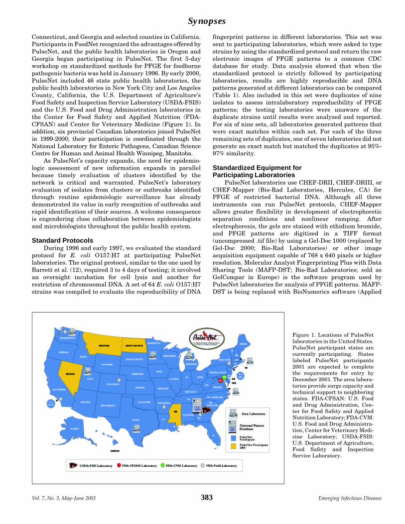
Synopses
Vol. 7, No. 3, May–June 2001 Emerging Infectious Diseases383
Connecticut, and Georgia and selected counties in California.Participants in FoodNet recognized the advantages offered byPulseNet, and the public health laboratories in Oregon andGeorgia began participating in PulseNet. The first 5-dayworkshop on standardized methods for PFGE for foodbornepathogenic bacteria was held in January 1996. By early 2000,PulseNet included 46 state public health laboratories, thepublic health laboratories in New York City and Los AngelesCounty, California, the U.S. Department of Agriculture’sFood Safety and Inspection Service Laboratory (USDA-FSIS)and the U.S. Food and Drug Administration laboratories inthe Center for Food Safety and Applied Nutrition (FDA-CFSAN) and Center for Veterinary Medicine (Figure 1). Inaddition, six provincial Canadian laboratories joined PulseNetin 1999-2000; their participation is coordinated through theNational Laboratory for Enteric Pathogens, Canadian ScienceCentre for Human and Animal Health Winnipeg, Manitoba.
As PulseNet’s capacity expands, the need for epidemio-logic assessment of new information expands in parallelbecause timely evaluation of clusters identified by thenetwork is critical and warranted. PulseNet’s laboratoryevaluation of isolates from clusters or outbreaks identifiedthrough routine epidemiologic surveillance has alreadydemonstrated its value in early recognition of outbreaks andrapid identification of their sources. A welcome consequenceis engendering close collaboration between epidemiologistsand microbiologists throughout the public health system.
Standard ProtocolsDuring 1996 and early 1997, we evaluated the standard
protocol for E. coli O157:H7 at participating PulseNetlaboratories. The original protocol, similar to the one used byBarrett et al. (12), required 3 to 4 days of testing; it involvedan overnight incubation for cell lysis and another forrestriction of chromosomal DNA. A set of 64 E. coli O157:H7strains was compiled to evaluate the reproducibility of DNA
fingerprint patterns in different laboratories. This set wassent to participating laboratories, which were asked to typestrains by using the standardized protocol and return the rawelectronic images of PFGE patterns to a common CDCdatabase for study. Data analysis showed that when thestandardized protocol is strictly followed by participatinglaboratories, results are highly reproducible and DNApatterns generated at different laboratories can be compared(Table 1). Also included in this set were duplicates of nineisolates to assess intralaboratory reproducibility of PFGEpatterns; the testing laboratories were unaware of theduplicate strains until results were analyzed and reported.For six of nine sets, all laboratories generated patterns thatwere exact matches within each set. For each of the threeremaining sets of duplicates, one of seven laboratories did notgenerate an exact match but matched the duplicates at 95%-97% similarity.
Standardized Equipment forParticipating Laboratories
PulseNet laboratories use CHEF-DRII, CHEF-DRIII, orCHEF-Mapper (Bio-Rad Laboratories, Hercules, CA) forPFGE of restricted bacterial DNA. Although all threeinstruments can run PulseNet protocols, CHEF-Mapperallows greater flexibility in development of electrophoreticseparation conditions and nonlinear ramping. Afterelectrophoresis, the gels are stained with ethidium bromide,and PFGE patterns are digitized in a TIFF format(uncompressed .tif file) by using a Gel-Doc 1000 (replaced byGel-Doc 2000; Bio-Rad Laboratories) or other imageacquisition equipment capable of 768 x 640 pixels or higherresolution. Molecular Analyst Fingerprinting Plus with DataSharing Tools (MAFP-DST; Bio-Rad Laboratories; sold asGelCompar in Europe) is the software program used byPulseNet laboratories for analysis of PFGE patterns. MAFP-DST is being replaced with BioNumerics software (Applied
Figure 1. Locations of PulseNetlaboratories in the United States.PulseNet participant states arecurrently participating. Stateslabeled PulseNet participants2001 are expected to completethe requirements for entry byDecember 2001. The area labora-tories provide surge capacity andtechnical support to neighboringstates. FDA-CFSAN: U.S. Foodand Drug Administration, Cen-ter for Food Safety and AppliedNutrition Laboratory; FDA-CVM:U.S. Food and Drug Administra-tion, Center for Veterinary Medi-cine Laboratory; USDA-FSIS:U.S. Department of Agriculture,Food Safety and InspectionService Laboratory.

Synopses
384Emerging Infectious Diseases Vol. 7, No. 3, May–June 2001
Maths, Kortrijk, Belgium); the change-over will be completedin 2001. Each PulseNet laboratory has all the aboveequipment and has the capability to normalize the patterns,compare them with other patterns, and maintain localdatabases of PFGE patterns for each bacterial pathogen ofinterest.
National Database of PFGE Patterns andAssociated Epidemiologic Information
A national database of PFGE patterns is being assembledfor foodborne bacterial pathogens. These databases reside ona PulseNet server at CDC. For each bacterial pathogen, thenormalized PFGE pattern is associated with a patterndatabase and a database of epidemiologic and clinicalinformation for isolates. One isolate may be associated withmore than one PFGE pattern in the database becausePulseNet protocols may call for the use of more than onerestriction enzyme to achieve appropriate discriminationbetween epidemiologically unrelated isolates. The E. coliO157:H7 database is functional; databases for nontyphoidalSalmonella serotypes and Listeria monocytogenes are underconstruction.
Seven PulseNet laboratories (four state public healthlaboratories, FDA-CFSAN, USDA-FSIS, and CDC) havedirect access to the PulseNet database server through theInternet, enabling them to submit normalized PFGE patternsand associated epidemiologic information. (The DST versionof the Molecular Analyst software creates a special “bundle”file for comparison with the national database.) Laboratoriesquery the national database for identical matches or closelyrelated patterns (>95% related under specified conditions). Ifidentical or close matches to the submitted patterns arefound, the submitting laboratory can access epidemiologicinformation associated with those patterns from the textdatabase. When a PulseNet participating laboratory logs onto the PulseNet server, it will display a “recent match”message if two or more laboratories submit identical or closelyrelated patterns within a specified time. This alert providesan early warning to PulseNet laboratories about possiblemultisite foodborne disease outbreaks.
PulseNet laboratories that do not yet have direct onlineaccess to the PulseNet server may still electronically submitraw TIFF images and normalized PFGE patterns (bundlefiles) to the PulseNet database administration team by e-mailor through file transfer protocols (ftp). The team compares thesubmitted patterns with those in the national database and e-mails the results to the submitting laboratory as quickly aspossible. We expect that direct access to the PulseNet serverwill be available to all participating laboratories that havesatisfactorily completed certification requirements by June30, 2001.
Developing Standardized ProtocolsStandardized protocols for foodborne bacterial pathogens
were developed in priority order based on the ability of PFGEto discriminate among strains of the organism and theepidemiologic utility of the resulting data. StandardizedPFGE protocols have been developed for E. coli O157:H7,Salmonella enterica serotype Typhimurium, L. monocytoge-nes, and Shigella species. The S. Typhimurium protocol isapplicable to most other nontyphoidal Salmonella serotypes,including S. Enteritidis. However, neither PFGE nor othermolecular subtyping methods provide acceptable discrimina-tion among strains of this highly clonal serotype. StandardPFGE protocols for Campylobacter jejuni, C. coli, andClostridium perfringens (7) are being developed andvalidated. Although C. jejuni and C. coli infections arecommon, developing a standardized PFGE protocol for theseorganisms was not a high priority because they infrequentlycause outbreaks. On the other hand, although outbreaks of C.perfringens infections are seldom widespread, state and localpublic health laboratories requested a standardizedsubtyping protocol to assist with local outbreak investiga-tions. All PulseNet protocols are 1-day procedures based onthe PFGE protocol developed by the Washington State PublicHealth Laboratory in response to the need for more rapidtechniques (16). All new protocols and modifications ofexisting protocols are evaluated initially at the developinglaboratory, followed by a second evaluation at CDC, alpha-testing at one or two PulseNet laboratories, and beta-testingat several PulseNet laboratories before they are adopted asofficial PulseNet protocols. Evaluation criteria includereproducibility of patterns, appropriateness of the strain usedas the reference standard, and robustness of the procedure.Once a protocol is officially adopted, no changes can be madeexcept by a petition to CDC’s PulseNet Task Force, discussionof the proposed changes, and adoption of the proposal byPulseNet laboratories. The PulseNet Task Force at CDC iscomposed of personnel who carry out PulseNet-relatedactivities. The Task Force members develop and evaluateprotocols, provide technical support for participatinglaboratories, organize and conduct training workshops,administer the certification program and proficiency testingprogram, and maintain the national databases of PFGEpatterns for the bacteria under surveillance in PulseNet.
Quality Control and Assurance ProgramA quality assurance program has been instituted for
PulseNet to ensure the integrity of results obtained with thestandardized PFGE techniques. This program requires strictadherence to the standardized PFGE protocols (17). Inaddition, the quality assurance program consists of standardsfor training, analytical procedures, documentation, and
Table 1. Interlaboratory reproducibility of pulsed-field gel electrophoresis patterns of 64 Escherichia coli isolates by eight laboratories following thePulseNet standardized protocol
Laboratory (%)Result 1 2 3 4 5 6 7 8Matched expected pattern 64/64a 63/63 59/63 62/64 62/64 62/64 61/64 61/64
(100) (100) (93.7) (96.9) (96.9) (96.9) (95.3) (95.3)< 1-band difference from expected pattern 64/64 63/63 62/63 64/64 63/64 64/64 64/64 64/64
(100) (100) (98.4) (100) (98.4) (100) (100) (100)aValues represent no. of patterns fitting in the specified category/no. of isolates tested.

Synopses
Vol. 7, No. 3, May–June 2001 Emerging Infectious Diseases385
equipment; standard operating procedures; an initialcertification set of isolates for each organism; and an ongoingproficiency testing program. The standards detail theminimum requirements a laboratory must meet for trainingpersonnel, analytical procedures, documentation, equipmentcalibration and maintenance, proficiency testing, and reviewof results. The laboratory standard operating proceduresdescribe procedures for record keeping, equipment mainte-nance, gel image acquisition, data analysis, and administra-tive policies. The certification sets consist of isolates withknown patterns, which are sent to each laboratory.Laboratories type the isolates by the standardized protocoland send the gel images to the PulseNet National DatabaseAdministration Team for review. This team, part of thePulseNet Task Force, is responsible for maintaining andupdating the PFGE pattern databases for foodborne disease-causing bacteria. Team members review new patternssubmitted to the databases and verify matches. In addition,this team evaluates the certification data submitted byPulseNet laboratories. Laboratories with DST version of theMAFP software also analyze their gel images and send theresults (bundle files) to the PulseNet National DatabaseAdministration Team for review. This team checks gel imagesand bundle files against the master certification set to ensurethat the laboratory has obtained the correct patterns.Successful completion of the certification set allows PulseNet-affiliated laboratories to compare results with the NationalDatabase. As part of the proficiency testing program,laboratories will be sent a combination of isolates and TIFFfiles on a semiannual basis both to test the laboratory’s abilityto perform the standardized protocol correctly and to ensurethat data analysis is consistent from laboratory to laboratory.
A quality assurance and control manual, beingdeveloped, will describe standardized training, laboratoryand administrative procedures, and policies. A proficiencytesting manual, also in preparation, is designed to maintainthe reproducibility of patterns and consistency in analysis ofpatterns that make PulseNet a valuable ally forepidemiologists.
Laboratories joining PulseNet are sent the standardizedPFGE protocols and certification sets appropriate to theorganism(s) being tested. Appropriate training is scheduledand follow-up is provided by means of the certification setsand the regularly scheduled proficiency testing program. Anannual meeting enables microbiologists from participatingPulseNet laboratories to discuss new protocols and softwareupgrades and exchange information on problems andsolutions.
Standardized Nomenclature for PFGE PatternsA major problem in comparing and interpreting
molecular subtyping information from different laboratorieshas been the lack of a universal naming system for PFGEpatterns. In response, we have developed a standardizednomenclature system for designating PFGE patterns inPulseNet. Each unique pattern in the database is representedby a 10-character code as follows:
XXXYYY.0000The first three characters in the code represent the bacterialpathogen, the next three characters denote the enzyme usedfor DNA restriction, and the last four characters represent thepattern designation. For example, in the pattern designation
EXHA26.0026, EXH represents E. coli O157:H7, A26represents restriction endonuclease AvrII, and 0026 is thepattern number. Because the pattern numbers are assignedsequentially to unique patterns, no evolutionary, phylogenet-ic, or clonal relationships should be implied from the order ofpattern numbers.
A priority order has been developed for inclusion offoodborne bacterial pathogens in PulseNet (Table 2). Theprioritization takes into account the availability of anacceptable molecular subtyping method for a pathogen,severity of disease caused by that pathogen, propensity for thepathogen to cause outbreaks, and the potential forrecognizing outbreaks and taking preventive action byroutine subtyping.
Role of PulseNet in Outbreak InvestigationsPulseNet plays several roles in detecting, investigating,
and controlling outbreaks. Identification by PulseNet of anincrease in a specific subtype of a pathogen may be an earlyindication of an outbreak. PFGE patterns submitted to thenational database by participating laboratories may linkapparently unrelated cases that are geographically dispersed.Once a cluster is detected through PulseNet, an epidemiologicinvestigation is initiated to determine if there is a commonsource. This epidemiologic investigation may be guided by thePFGE subtypes identified through PulseNet. PulseNet canidentify outlier cases in other areas and define the geographicscope of the outbreak. If a common food source is identifiedand the pathogen is isolated from that food, subtyping helpsconfirm it as the outbreak strain. Finally, once controlmeasures are instituted, PulseNet can help confirm that theoutbreak is over by showing a substantial decrease in circulationof the outbreak strain in the affected communities. The followingexamples illustrate these PulseNet functions.
In 1996, epidemiologists at the Washington State andSeattle-King County health departments traced an outbreakof E. coli O157:H7 infections in four states and one Canadianprovince to commercial unpasteurized apple juice. Of 70persons identified as part of this outbreak, 25 requiredhospitalization, 14 had hemolytic uremic syndrome, and onedied. DNA fingerprinting by PFGE at the Washington StatePublic Health Laboratory, a PulseNet area laboratory,showed that isolates from patients and the apple juice werethe same strain. Prompt recognition of the apple juice as thesource of this outbreak resulted in rapid recall of the widelydistributed product (18).
Table 2. Priority order for inclusion of foodborne bacterial pathogensin PulseNet
Pathogen Expected year of inclusionEscherichia coli O157:H7 1997Nontyphoidal Salmonella serotypes 1998Listeria monocytogenes 1999Shigella sonnei 1999Clostridium perfringens 2001Campylobacter jejuni 2001Vibrio parahaemolyticus 2001V. cholerae 2001Clostridium botulinum 2002Other pathogenic E. coli 2002Yersinia enterocolitica 2003

Synopses
386Emerging Infectious Diseases Vol. 7, No. 3, May–June 2001
In 1997, the Colorado State Public Health Laboratory,which had just initiated PFGE typing of E. coli O157:H7,identified a cluster of 14 ill persons whose E. coli O157:H7isolates had matching PFGE patterns. About the same time,the USDA laboratory isolated an E. coli O157:H7 strain froma ground beef patty from the same package as a patty eaten byan ill person. DNA fingerprinting by PFGE on the humanisolate from Colorado and the food isolate from USDA-FSISwere generated by the PulseNet standardized protocol. ThePFGE patterns were transmitted electronically to CDC viathe Internet, where they were found to be indistinguishable.This outbreak pattern was then transmitted to PulseNet sitesand compared with patterns from >300 other recent E. coliO157:H7 isolates. No matching patterns were found otherthan one case in Kentucky, providing strong evidence that theoutbreak was not nationwide.
In May 1998, PulseNet facilitated the investigation oftwo clusters of E. coli O157:H7 infections in the northeasternUnited States. Timely fingerprinting of E. coli O157:H7isolates by the Massachusetts Area Laboratory for PulseNetand other PulseNet laboratories in that region revealed twosimultaneous clusters of E. coli O157:H7 infections (32isolates in four of five states with one PFGE pattern and 25isolates in all of five states with a second PFGE pattern), oneof which could be traced to two supermarkets that receivedground beef from the same distributor. Without assistancefrom PulseNet, epidemiologists would have found it difficultto identify cases associated with each cluster.
Also in May 1998, the state public health departments inboth Illinois and Pennsylvania informed CDC about increasesin Salmonella Agona infections. Serotype-specific surveil-lance data from other states quickly confirmed that 10 stateshad increases in S. Agona infections. A national outbreak ofS. Agona was occurring, with no obvious source. Subsequent-ly, the outbreak was traced to contaminated ready-to-eattoasted oats cereal product from a food-processing facility inMinnesota (19). PulseNet laboratories helped in thisinvestigation by distinguishing cases that were associatedwith the outbreak from those that were not. In addition,timely PFGE typing of S. Agona by PulseNet laboratorieshelped identify outbreak-associated cases in states where thecontaminated product was not initially thought to have beendistributed. PFGE subtyping of S. Agona isolates wasimportant in confirming the successful control of theoutbreak. Not only did the number of reported isolates returnto baseline, but also the outbreak strain disappeared. By thetime this investigation was completed, PulseNet laboratorieshad typed >1,000 isolates of S. Agona. Four hundred ninecases (one fatal) in 23 states were linked to this outbreak(CDC, unpub. data).
From October 20 to November 9, 1998, health officials inConnecticut, New York, Ohio, and Tennessee reportedincreases in Listeria infections in their states (20). PFGEtyping by PulseNet laboratories showed that several caseisolates from different states had indistinguishable DNAfingerprints. On further investigation, 101 Listeria infections(including 15 perinatal infections) with bacteria having thesame or highly similar DNA fingerprints were identified in 22states. Fifteen deaths and six miscarriages or stillbirths werereported among patients who were infected with the outbreakstrain. This outbreak was traced to contaminated hot dogsand sandwich meat produced at a single large meat-processing
plant in Michigan (21). After the company voluntarilyrecalled the implicated lots of product and suspendedproduction, the outbreak rapidly ended.
Surveillance for Foodborne OutbreaksTwenty years ago, most foodborne outbreaks were local
problems that typically resulted from improper food-handlingpractices. Outbreaks were often associated with individualrestaurants or social events and often came to the attention oflocal public health officials through calls from affectedpersons. These persons, who may have known others who hadbecome ill after eating a shared meal or visiting the samerestaurant, provided health officials with much of theinformation needed to begin an investigation.
Today, foodborne disease outbreaks commonly involvewidely distributed food products that are contaminated beforedistribution, resulting in cases that are spread over severalstates or countries. It is less common for ill persons to knowothers who were ill or to be able to identify a likely source oftheir infection. For these reasons, it is becoming increasinglyimportant to be able to identify potential common exposuresthrough DNA fingerprinting of patient isolates.
For foodborne outbreak surveillance to be effective,isolates must be subtyped routinely and the data analyzedpromptly at the local level. Clusters can often be detectedlocally that could not have been identified by traditionalepidemiologic methods alone. This is especially true ofinfections with common pathogens such as S. Typhimurium,which occur so frequently that clusters may be hidden amongsporadic cases. For S. Typhimurium isolates received by themicrobiology laboratory at the Minnesota Department ofHealth from August 14 to September 14, 1995, temporaldistribution did not suggest any obvious clustering, but thedistribution of PFGE subtypes suggested multiple commonsources with continuing exposures (Figure 2). Epidemiologicinvestigation ultimately linked three of the subtypes to localrestaurants, where exposure to S. Typhimurium occurredthroughout the month (Jeffrey B. Bender, pers. comm.). Withoutsubtyping data, it would have been very difficult to associatecases with exposures occurring over such a prolonged period.
In September 1998, the Minnesota state public healthlaboratory informed other PulseNet laboratories that it wasinvestigating two clusters of Shigella sonnei infectionsassociated with restaurants in Minnesota and asked if otherstates had observed increases in S. sonnei infections orS. sonnei isolates with the outbreak PFGE pattern. The LosAngeles County public health department immediatelyresponded that it was also investigating restaurant-associated outbreaks of S. sonnei and that the PFGE patternof their outbreak strain was very similar to the Minnesotapattern. Epidemiologic and laboratory investigations ulti-mately determined that outbreaks in Massachusetts, Florida,and two Canadian provinces were linked to the Minnesotaand Los Angeles outbreaks. With the assistance of the FDA’soutbreak trace-back and coordination group, parsleyimported from Mexico was identified as the common vehicle(22). Mexican and U.S. authorities inspected the parsley farmand recommended changes in growing and harvestingpractices to prevent recurrence of the problem. Rapid sharingof PFGE subtyping data through PulseNet played a criticalrole in linking these apparently unrelated outbreaks andidentifying a common vehicle.

Synopses
Vol. 7, No. 3, May–June 2001 Emerging Infectious Diseases387
The use of molecular subtyping as part of routinesurveillance has benefits beyond outbreak detection.Temporal clustering of unrelated cases is not uncommon, andwithout molecular subtyping, valuable public healthresources can be wasted investigating pseudo-outbreaks. InJune and July 1994, an outbreak of E. coli O157:H7 infectionswas suspected when the New Jersey Department of Healthand Senior Services received reports of 48 culture-confirmedcases; only four were reported during the same period in 1993(23). PFGE subtyping found most isolates to have uniquepatterns, indicating that a large outbreak was unlikely. Theprobable reason for the sudden increase in case reports wasthe concomitant increase in the number of laboratoriesculturing stools for E. coli O157:H7 (Figure 3).
Although PulseNet has proven invaluable in detectingfoodborne disease outbreaks and facilitating their investiga-tion, molecular subtyping is an adjunct to epidemiologicinvestigation and not a replacement for it. The observationthat isolates from two or more persons have indistinguishable
PFGE patterns should not be considered proof that thepersons had a common exposure, merely that the isolates inquestion share a common ancestry. Moreover, outbreaks canbe caused by more than one subtype, so that differences inPFGE pattern alone cannot prove that isolates did not have acommon source (24,25).
Requirements for Effective FunctioningAlthough the area laboratories are set up to assist
neighboring state public health laboratories that are notPulseNet participants, every state must have PFGEsubtyping capacity for optimum performance of the network.A dramatic indication of this was provided during the 1997ground beef-associated E. coli O157:H7 outbreak in Colorado.When the outbreak pattern was posted on PulseNet ListServ,most laboratories that were network participants respondedwithin 48 hours that they had no matching PFGE patternsfrom recent E. coli O157:H7 isolates. In contrast, it took morethan 2 months to identify a case in Kentucky (not a PulseNetparticipant state in 1997) that was related to the Coloradooutbreak. The Association of Public Health Laboratories hasdetermined that PulseNet participation is a core capacity forall state and territorial public health departments in theUnited States.
For the network to work efficiently in detecting foodbornedisease outbreaks through routine surveillance, PulseNetlaboratories must perform, at a minimum, routine PFGEsubtyping of E. coli O157:H7 and L. monocytogenes as soon asisolates are received. In addition, they must perform PFGEsubtyping of other foodborne pathogenic bacteria (Campylo-bacter jejuni and C. coli, Salmonella serotypes, Shigella spp.,Bacillus cereus, Vibrio cholerae, V. parahaemolyticus,V. vulnificus, Clostridium botulinum, C. perfringens, Yersiniaenterocolitica) rapidly when the number of isolates receivedby the laboratory exceeds the expected number for thatperiod. Unfortunately, microbiologists at state and localpublic health laboratories often have responsibilities for allpathogenic bacteria and may not be able to type incomingisolates of foodborne pathogenic bacteria in a timely manner.In addition, like public health surveillance in general, PulseNet
Figure 3. Reported cases of culture-confirmed Escherichia coliO157:H7 infection and percentage of surveyed laboratories routinelytesting all stool specimens for E. coli O157:H7, New Jersey, January1991 through July 1994 (10).
Figure 2. Distribution bydate and pulsed-field gelelectrophoresis subtypeof Salmonella ser.Typhimurium isolatesreceived by the Minne-sota Department ofHealth, August 14-Sep-tember 14, 1995. Dataprovided by JeffreyBender and John Besser,Minnesota Departmentof Health.

Synopses
388Emerging Infectious Diseases Vol. 7, No. 3, May–June 2001
depends on physicians requesting culture of patientspecimens if a bacterial infection is suspected and the clinicaldiagnostic laboratory promptly forwarding isolates to thepublic health laboratory for typing.
PulseNet relies on the cooperation of all participants intyping foodborne pathogenic bacteria by strict adherence tothe standard protocol. Without such a total commitment,PFGE patterns from different PulseNet laboratories could notbe compared to ascertain which cases are associated with aspecific outbreak. The importance of this was underscored bya recent experience. One PulseNet laboratory had decided tochange the PulseNet protocol for S. Typhimurium and wasusing a variation of the standard protocol. A cluster ofS. Typhimurium cases was detected in that state, and PFGEanalysis confirmed that many of the isolates wereindistinguishable. However, when attempts were made todetermine if an increase in S. Typhimurium infections inneighboring states were related to the cluster, the PFGEpatterns could not be compared. This caused a delay of severaldays in the investigation because isolates from the first statehad to be re-typed by the standardized protocol.
Cost-Benefit AnalysisElbasha et al. recently compared the costs and benefits of
PulseNet’s molecular subtyping-based surveillance system,using as an example the Colorado state public healthlaboratory’s investigation of the 1997 E. coli O157:H7outbreak in which contaminated frozen hamburger pattieswere implicated (26). If 15 cases were averted by the recall ofpotentially contaminated ground beef, the PulseNet systemin Colorado would have recovered all costs of start-up and 5years of operation. These authors point out that the systembecomes even more cost-effective if one takes into accountresources that would have been wasted to investigate apparentincreases in sporadic cases of E. coli O157:H7 infections.
The Growth and Future of PulseNetWithin a very short time, PulseNet has grown beyond
expectations and has convincingly demonstrated itseffectiveness as a tool for foodborne disease surveillance. Itbegan with one pathogen (E. coli O157:H7) and 10participating laboratories in 1996 that submitted 191 PFGEpatterns to the PulseNet database during that year. In 1999,four pathogens (E. coli O157:H7, Salmonella, Shigella, andListeria monocytogenes) were tracked through PulseNet and>9,500 patterns were submitted to the PulseNet database.State and local public health laboratories contributed >78% ofthe patterns to the PulseNet database (Figure 4). As the FDAare increasing the number of laboratories that perform PFGEsubtyping using PulseNet protocols, and the USDA aresetting up their own PulseNet-compatible local networks,their contributions to the PulseNet database will no doubtsubstantially increase. In addition, the representation ofPFGE patterns in the PulseNet databases will increase forpathogenic bacteria isolated from foods.
As more public health laboratories at the local and statelevels join PulseNet, the role of the area laboratories ischanging. The area laboratories provide training andconsultation to neighboring PulseNet laboratories, coordinatemultistate outbreak investigations when requested, and
Figure 4. Sources of pulsed-field gel electrophoresis patternssubmitted to PulseNet in 1999.
provide surge capacity for neighboring PulseNet laboratories.Three additional area laboratories, in Michigan, Utah, andVirginia, were designated in 2000, bringing the total numberto seven.
Canada is already an active participant, and internation-al expansion of the network with partners in Mexico, SouthAmerica, and Europe is anticipated. The long-term vision forPulseNet is a global network of public health laboratoriesworking with food regulatory agencies and industry toimprove food safety worldwide.
Finally, we recognize that the methods currently used forsubtyping and data analysis will not always be state-of-the-art. We are working to develop, evaluate, and validate DNAsequencing-based subtyping methods for foodborne patho-gens. These methods will be gradually implemented in thenetwork, and compatibility will be maintained with existingPFGE data. We are also working with software developers toimplement new versions of pattern analysis software andDNA sequence comparison software to improve patternmatching, automate pattern normalization and sequencealignment, and reduce subjectivity in subtype comparisons.
AcknowledgmentsWe thank Mike Hoekstra for suggestions on data presentation
and Susan Van Duyne for information on the quality assurance andcontrol program for PulseNet; we thank personnel in all participatinglaboratories for their enthusiastic participation in PulseNet and forsharing their data and findings in a timely manner; and JeffreyKoplan, James Hughes, Joseph McDade, Mitchell Cohen, andPatrick McConnon for their support of PulseNet.
PulseNet is supported by appropriations to CDC under theNational Food Safety Initiative and the Emerging InfectiousDiseases Program and by appropriations from various states to theirrespective public health departments.
Dr. Swaminathan is chief of the Foodborne and Diarrheal DiseasesLaboratory Section, CDC, and the principal architect of PulseNet.
References 1. Holmberg SD, Wachsmuth IK, Hickman-Brenner FW, Cohen ML.
Comparison of plasmid profile analysis, phage typing, andantimicrobial susceptibility testing in characterizing Salmonellatyphimurium isolates from outbreaks. J Clin Microbiol1984;19:100-4.

Synopses
Vol. 7, No. 3, May–June 2001 Emerging Infectious Diseases389
2. Holmberg SD, Wachsmuth K. Plasmid and chromosomal DNAanalyses in the epidemiology of bacterial diseases. In:Swaminathan B, Prakash G, editors. Nucleic acid and monoclonalantibody probes: applications in diagnostic microbiology. NewYork: Marcel Dekker; 1989. p. 105-29.
3. Ackers ML, Mahon BE, Leahy E, Goode B, Damrow T, Hayes PS,et al. An outbreak of Escherichia coli O157:H7 infections associatedwith leaf lettuce consumption. J Infect Dis 1998;177:1588-93.
4. Barrett TJ. Molecular fingerprinting of foodborne pathogenicbacteria: An introduction to methods, uses and problems. In:Tortorello ML, Gendel SM, editors. Food microbiological analysis:new technologies. New York: Marcel Dekker; 1997. p. 249-64.
5. Graves LM, Swaminathan B, Hunter SB. Subtyping Listeriamonocytogenes. In: Ryser EM, Marth EH, editors. Listeria, listeriosisand food safety. New York: Marcel Dekker; 1999. p. 279-98.
6. Jimenez A, Barros-Velazquez J, Rodriguez J, Villa TG. Restrictionendonuclease analysis, DNA relatedness and phenotypiccharacterization of Campylobacter jejuni and Campylobacter coliisolates invovled in food-borne disease. J Appl Microbiol1997;82:713-21.
7. Maslanka SE, Kerr JG, Williams G, Barbaree JM, Carson LA,Miller JM, et al. Molecular subtyping of Clostridium perfringens bypulsed-field gel electrophoresis to facilitate food-borne-diseaseoutbreak investigations. J Clin Microbiol 1999;37:2209-14.
8. Threlfall EJ, Hampton MD, Ward LR, Rowe B. Application ofpulsed-field gel electrophoresis to an international outbreak ofSalmonella agona. Emerg Infect Dis 1996;2:130-2.
9. Threlfall EJ, Ward LR, Hampton MD, Ridley AM, Rowe B, RobertsD, et al. Molecular fingerprinting defines a strain of Salmonellaenterica serotype Anatum responsible for an internationaloutbreak associated with formula-dried milk. Epidemiol Infect1998;121:289-93.
10. Wachsmuth K. Molecular epidemiology of bacteria infections:Examples of methodology and of investigations of outbreaks. RevInfect Dis 1986;8:682-92.
11. Swaminathan B, Matar GM. Molecular typing methods. In:Persing DH, Smith TF, Tenover FC, White TJ, editors. Diagnosticmolecular microbiology. Washington: American Society forMicrobiology; 1993. p. 26-50.
12. Barrett TJ, Lior H, Green JH, Khakhria R, Wells JG, Bell BP, et al.Laboratory investigation of a multi-state food-borne outbreak ofEscherichia coli O157:H7 by using pulsed-field gel electrophoresisand phage typing. J Clin Microbiol 1994;32:3013-7.
13. Stephenson J. New approaches for detecting and curtailingfoodborne microbial infections. JAMA 1997;277:1337-40.
14. Centers for Disease Control and Prevention. Addressing emerginginfectious disease threats: a prevention strategy for the United States.Atlanta: U.S. Department of Health and Human Services; 1994.
15. Angulo FJ, Voetsch AC, Vugia D, Hadler JL, Farley M, Hedberg C,et al. Determining the burden of human illness from food bornediseases. CDC’s emerging infectious disease program Food BorneDiseases Active Surveillance Network (FoodNet). Vet Clin NorthAm Food Anim Pract 1998;14:165-72.
16. Gautom RK. Rapid pulsed-field gel electrophoresis protocol for E.coli O157:H7 and other Gram-negative organisms in one day. JClin Microbiol 1997;35:2977-80.
17. Centers for Disease Control and Prevention. Standardizedmolecular subtyping of foodborne bacterial pathogens by pulsed-field gel electrophoresis: a manual. Atlanta: National Center forInfectious Diseases; 1996 (updated 2000).
18. Cody SH, Glynn MK, Farrar JA, Cairns KL, Griffin PM, KobayashiJ, et al. An outbreak of Escherichia coli O157:H7 infection fromunpasteurized commercial apple juice. Ann Intern Med1999;130:202-9.
19. Centers for Disease Control and Prevention. Multistate outbreak ofSalmonella serotype Agona infections linked to toasted oatscereal—United States, April-May, 1998. MMWR Morb MortalWkly Rep 1998;47:462-4.
20. Centers for Disease Control and Prevention. Multistate outbreak oflisteriosis—United States, 1998. MMWR Morb Mortal Wkly Rep1998;47:1085-6.
21. Centers for Disease Control and Prevention. Update: Multistateoutbreak of listeriosis—United States, 1998-1999. MMWR MorbMortal Wkly Rep 1999;47:1117-8.
22. Centers for Disease Control and Prevention. Outbreaks of Shigellasonnei infection associated with eating fresh parsley—UnitedStates and Canada, July-August, 1998. MMWR Morb Mortal WklyRep 1999;48:285-9.
23. Mead P, Finelli L, Lambert-Fair MA, Champ D, Townes J,Hutwagner L, et al. Risk factors for sporadic infection withEscherichia coli O157:H7. Arch Intern Med 1997;157:204-8.
24. Keene W, Hedberg K, Herriott DE, Hancock DD, McKay RW,Barrett TJ, et al. A prolonged outbreak of Escherichia coli O157:H7infections caused by commercially distributed milk. J Infect Dis1997;176:815-8.
25. Jackson LA, Keene WE, McAnulty JM, Alexander ER, DiermayerM, Davis MA, et al. Where’s the beef? The role of cross-contamination in four chain restaurant-associated outbreaks ofEscherichia coli O157:H7 in the Pacific Northwest. Ann InternMed 2000;160:2380-5.
26. Elbasha EH, Fitzsimmons TD, Meltzer MI. Costs and benefits of asubtype-specific surveillance system for identifying Escherichiacoli O157:H7 outbreaks. Emerg Infect Dis 200;6:293-7.

Synopses
390Emerging Infectious Diseases Vol. 7, No. 3, May–June 2001
In 1997, 8 million new cases of tuberculosis (TB) werereported worldwide; 3.5 million cases were considered highlycontagious (1). With Africa and some countries having up to20% of their populations infected with HIV, AIDS will have amajor impact on TB in coming years (2). Emergence ofmultidrug-resistant (MDR) strains of Mycobacterium tuber-culosis is also of great epidemiologic concern (3). In thiscontext, molecular fingerprinting of M. tuberculosis complexisolates is a powerful tool that permits detection oftranscontinental spread of TB (4) and outbreaks (5). Ourlaboratory has described a preliminary spoligotypingdatabase that suggested the biogeographic specificity of someof the spoligotypes from the Caribbean (6). The initial aim ofthis work was twofold. First, such an inventory wasmandatory to detect and estimate the relative importance ofTB of foreign origin in the French Caribbean. Although theincidence of TB in Martinique and Guadeloupe is comparablewith that in metropolitan France (approximately 10/100,000new cases each year), this region is part of an area of LatinAmerica and the Caribbean with high TB prevalence. Second,we used spoligotyping results to infer potential phylogeneticrelationships of M. tuberculosis strains in the Caribbeanregion and the history of TB by using molecular markers. Anupdated database could also be helpful in developing newstatistical approaches in the field of population genetics ofcirculating M. tuberculosis clinical isolates.
By systematically analyzing published spoligotypes, wehave now collected 3,319 spoligotyping patterns of various
origins in a single database, essentially from Europe and theUSA (Table 1). This database includes 259 shared typescontaining 2 to 476 patterns (because of the size of thisdatabase, a graphic of it appears online only, at http://www.cdc.gov/ncidod/EID/vol7no3/sola_data.htm). The maindatabase also includes 540 “orphan patterns” (clinicalisolates showing a unique spoligotype), for a current total of799 distinct spoligotype patterns. This article describes thenomenclature and phylogenetic reconstruction of these 259shared types.
The DatabaseSpoligotyping based on the variability of the Direct
Repeat (DR) locus and analysis of a variable number oftandem DNA repeats (VNTR) of M. tuberculosis wereperformed according to the original protocols (7,8). For theconstruction of the database, spoligotyping results wereentered into Excel spreadsheet files in chronological order,according to the availability of results from published articlesand our own investigations. The database was searchedregularly for new shared types, i.e., identical spoligotypesshared by two or more patient isolates. For phylogeneticreconstruction, the spoligotyping results were entered intoRecognizer software of the Taxotron package (Taxolab,Institut Pasteur, Paris), as recommended (9). The “1-Jaccard”Index was calculated for each pairwise comparison of patterns(10), and the neighbor-joining algorithm was used forbuilding trees (11).
The source of the data and its representativeness areshown in Table 1. Of 3,319 individual spoligotypes in ourdatabase, most (2,418 [73%]) were either from Europe (1,142[34%]) or the USA (1,283 [39%]). Spoligotypes shared betweenthe USA and Europe totaled 1,286 isolates distributed among45 shared types (Europe, n=461; USA, n=825). A statistical
Spoligotype Database ofMycobacterium tuberculosis: Biogeographic
Distribution of Shared Types and Epidemiologicand Phylogenetic Perspectives
Christophe Sola,* Ingrid Filliol,* Maria Cristina Gutierrez,† Igor Mokrousov,*Véronique Vincent,† and Nalin Rastogi*
*Institut Pasteur de Guadeloupe, Pointe à Pitre, Guadeloupe; and †Centre Nationalde Référence des Mycobactéries, Institut Pasteur, Paris, France
Address for correspondence: Nalin Rastogi or Christophe Sola, Unitéde la Tuberculose et des Mycobactéries, Institut Pasteur deGuadeloupe, Morne Jolivière, BP 484, 97165 Pointe-à-Pitre, Cedex,Guadeloupe; fax: 590-893880; e-mail: [email protected];[email protected]
We give an update on the worldwide spoligotype database, which now contains3,319 spoligotype patterns of Mycobacterium tuberculosis in 47 countries, with259 shared types, i.e., identical spoligotypes shared by two or more patientisolates. The 259 shared types contained a total of 2,779 (84%) of all theisolates. Seven major genetic groups represented 37% of all clustered isolates.Two types (119 and 137) were found almost exclusively in the USA andaccounted for 9% of clustered isolates. The remaining 1,517 isolates werescattered into 252 different spoligotypes. This database constitutes a tool forpattern comparison of M. tuberculosis clinical isolates for global epidemiologicstudies and phylogenetic purposes.

Synopses
Vol. 7, No. 3, May–June 2001 Emerging Infectious Diseases391
analysis was performed for the 1,286 isolates to evaluate thebiogeographic specificity of the shared types and assesspotential sampling bias by using a sample homogeneity testderived from the chi-square test (see below).
Results and Discussion
Description of DatabaseThe 3,319 spoligotypes were grouped into 259 shared
types containing 2,779 (84%) of the isolates and 540 (16%)orphan spoligotyping patterns (clinical isolates showinga unique spoligotype; results not shown; see onlinegraphic of database, http://www.cdc.gov/ncidod/EID/vol7no3/sola_data.htm). This gives a current total of 799 distinctspoligotype patterns in our database.
The distribution of shared types, their respective sizes,and their relative distribution in different locations (distinctcountries or geographic regions) are summarized in Figure 1.The 24 most frequent shared types totaled 1,804 (65%)isolates (Figure 1A); 7 types were highly frequent,representing 1,250 (45%) isolates. The Beijing type (type 1)was most frequent and represented 18% of isolates. Two types(119 and 137), which were almost exclusively found in theUSA, accounted for 9% of isolates and may be specific forAmerican populations or outbreaks (12). Types 53 and 50accounted for 8% and 6% of isolates and were found in 17 and15 locations, respectively. Two other types (types 42 and 47)
Table 1. Source of data for 3,319 spoligotypes of Mycobacteriumtuberculosis used to generate the database of 259 shared types
No. ofisolates Origina Year Reference136 Denmark 1999 31147 Italy 1999 32157 Cuba 1998 331b Philippines 1997 343 Peru 1998 3518 USA Unpublished R. Frothingham105 France 1997 36167 United Kingdom 1997 37296 France Unpublished This study28 Zimbabwe 1998 3832 Guinea-Bissau 1999 25118 The Netherlands 1997 768 Various countries 1999 1558 France Unpublished J. Maïsetti &
B. Carbonnelle62 Russia Unpublished O. Narvskaya84 West Africa 1999 395 Thailand Unpublished P. Palittapongarnpim14 Romania 1997 4017 Brazil 1999 415b Spain Unpublished S. Semper &
C. Martin1,283 USA 2000 121b United Kingdom 1999 4219 The Netherlands 1998 431b The Netherlands 1999 1969 Far East Asia 1995 4469 Caribbean 1999 6356 Caribbean Unpublished This studyaAlthough a potential sampling bias cannot be excluded, the sampling of isolates and theirrepresentativeness (in order of description) was as follows: Denmark, of 249 isolatesdescribed with a low copy number of IS6110 collected since 1992 (exhaustivity 93%), 24shared types, representing 136 spoligotypes, were retained (9 other shared types,representing 49 isolates that were found exclusively in Denmark(S1,S2,S4,S19,S22,S23,S27,S30,S33), were not included in the present analysis; Italy, of158 isolates from 156 patients in Verona collected during 1996-1997, 147 spoligotypes wereretained; Cuba, of 160 isolates typed (obtained from a pool of 578 smear-positive sputacollected during 1994-1995), 157 spoligotypes described (exhaustivity 36%) were retained;Philippines, no data except for a single spoligotype available; Peru, of 29 strains isolatedduring 1995-1996 from the sputa of patients in Lima and Cuzco, only 3 were retained in thisstudy since the remaining isolates shared spoligotypes with patients in Texas (12) and areincluded in the 1,283 Texan profiles; USA, 18 clinical isolates from the collection of R.Frothingham (representativeness unknown); France, 111 isolates from 105 hospitalizedpatients in Paris obtained during 1993 (patients were from three major hospitals thatrepresented 5% of the total public hospital beds in Paris); United Kingdom, 167 isolatesfrom all the culture-positive tuberculosis (TB) patients from three large hospitals innorthwest London (without any indication of period of recruitment); France, 296 isolatessent for reference purposes during a 3-year period to the Centre National de Référence desMycobactéries, Institut Pasteur, Paris; Zimbabwe, 28 spoligotypes obtained directly fromsputum samples during a 1-month recruitment period (December 1995) of sputum-positiveTB cases representing 20% of all cases; Guinea-Bissau, of 229 spoligotypes obtained fromsamples of 900 patients with suspected TB cases during 1989-1994, only 32 spoligotypeswere fully described by the authors, and were retained for the analysis; the Netherlands,118 isolates of unspecified representativeness from the collection of National Institute ofHealth (RIVM, Bilthoven); international multicenter study, 68 of 90 isolates from 38countries representing the five continents; France, 58 isolates during a 1-year (1999)recruitment in the University Hospital of Angers; Russia, 62 isolates representing the St.Petersburg area collected during 1997-1999; West Africa, 84 isolates from Ivory Coast andaround Dakar, Senegal, collected during 1994-1995; Thailand, 5 isolates from northernThailand (unknown representativeness); Romania, 14 isolates of unknown representative-ness; Brazil, 17 spoligotypes out of 91 isolates from a São Paulo hospital in 1995 (unknownrepresentativeness); Spain, 5 multidrug-resistant isolates (unknown representativeness);USA, 1,429 clinical isolates from 1,283 patients during 1994-1999 that are part of anongoing population-based study in Houston, Texas; United Kingdom, a single spoligotypefrom ancient DNA extracted from a bone sample; the Netherlands, 19 spoligotypes obtainedfrom paraffin-wax embedded tissue samples previously collected during 1983-1993(unknown representativeness); the Netherlands, a single spoligotype from a previous study(unknown representativeness); Far East Asia, 69 isolates from China and Mongoliaobtained during 1992-1994 (unknown representativeness); Caribbean, 425 clinical isolatesfrom a population-based ongoing study that includes all cultures isolated in Guadeloupe,Martinique, and French Guiana since 1994 and covers a 1 million population (exhaustivity100%). Some isolates in this pool came from patients from other countries (essentiallyneighboring countries such as Haiti, Dominican Republic, Brazil, Commonwealth ofDominica, Barbados, and Surinam).bDescription of a given spoligotype without precise number of isolates within this type.
Figure 1. Histograms derived from database (graphic online athttp://www.cdc.gov/ncidod/EID/vol7no3/sola_data.htm) summariz-ing the distribution of shared types (A), their respective sizes (B), andtheir relative distribution in different locations (C).

Synopses
392Emerging Infectious Diseases Vol. 7, No. 3, May–June 2001
accounted for 4% of the isolates and were found in 11countries. The remaining isolates (n=1,517) were scatteredinto 235 types. Figure 1B shows the relative sizes of 259shared types; 109 shared types (42%) contained only twopatients each and 38 shared types contained only threepatients each. Inversely, 24 shared types containing >20patients totaled 1,804 (65%) isolates. Finally, the distribution“unique” versus “ubiquitous” shared types (reported in onelocation versus found in two or more locations) is shown inFigure 1C; 122 (47%) shared types were reported from a singlelocation, 69 (26%) were from two locations, and 25 (10%) werefrom three locations. Inversely, the most ubiquitous types, inincreasing order of distribution, were 33 and 37, 20, 52, 42, 50,and 53. Thus, most M. tuberculosis shared types contained alow number of patient isolates and were confinedgeographically, whereas a minority contained a high numberof patient isolates and were highly disseminated. The findingof identical spoligotypes in distant countries may beexplained either by recent or past transmission events or byphylogenetic convergence. However, the evolution of the DRlocus relies on at least three independent mechanisms,namely, homologous recombination (13), replication slippage(14, 15), and insertion sequence-mediated transposition (16-19), which does not favor a fortuitous convergence.
Geographic Distribution of Shared Types in the DatabaseAnalysis of geographic distribution of the shared types
(see online graphic of database, http://www.cdc.gov/ncidod/EID/vol7no3/sola_data.htm) permitted us to split ourcollection into two broad categories: those reported in a singlearea (n=122, Table 2) and those reported in two or more areas(n=137). In the latter category, matching analysis for 69spoligotypes found in four broad geographic areas, namely,Africa, the Americas (North, Central and Caribbean, andSouth America), Europe, and Asia (Middle East, and Far EastAsia), is shown in Table 3. Contrary to ubiquitousspoligotypes such as type 1, 53, and 50, which have been found
in all regions, this is an attempt to define potential inter-regional and inter-continental flow of M. tuberculosis isolatesso far confined to limited geographic areas. The most frequentmatches were found for clusters in European countries(n=17), followed by Europe and North America (n=8), Europeand Central America and the Caribbean (n=5), and Europeand South America (n=4) (Table 3). These matches mayunderline both recent transcontinental transmission eventsand the history of TB spread in the New World throughEuropean settlers.
A total of 25 shared types were reported in threecountries. Among these, 8 types were exclusively found eitherin Europe (types 10,22,161) or the Americas (types5,67,70,93,130); 10 types were shared between two Europeancountries and a country of another region (types35,49,59,86,115,118,136,138,139,150); 5 types were sharedbetween two countries of the Americas with a country inEurope (types 92,119,168,185,190); 1 type was sharedbetween a European country and two African countries (type125); and 1 type was shared between Asia, Europe, and theUSA (type 124). Finally, 15 types were found in four countries;1 type (type 41) was exclusively found in Europe and may bespecific for this continent. Fourteen other types weredistributed as follows: Europe + Americas, 8 types (types3,7,19,31,40,51,137,152); Europe + Africa, 1 type (type 21);Europe + Asia + Americas, 3 types (type 8,89,167); Europe +Americas + Africa, 1 type (type 64); and Europe + Africa +Asia, 1 type (type 126). Finally, 28 types were reported in fiveor more countries, suggesting that these types are widespreadand may constitute the ubiquitous types such as the Beijingtype (type 1 in our database) or the Haarlem type (type 47).The only exception in this category was type 17, which wasfound in six countries in the Americas and may be specific forthis region. Future population studies should focus on theseubiquitous types to better define their relative prevalence ineach country.
Table 2. Geographic distribution of potentially specific shared types ofMycobacterium tuberculosis reported in a single location (n=122)
No. ofRegion Country types TypesAmericas Guadeloupe 7 12,13,14,15,30,103,259
French Guiana 4 66,76,94,96 Cuba 4 71,74,80,81 USA 46 192,194,197-199,201,202,
205,206,208,210-217,219- 235,237-239,241,243,246, 248,256-258
Europe The Netherlands 4 9,18,28,90United Kingdom 6 16,23,27,38,43,100 France 27 55,57,107-114,116,120,122,
140,141,143-148,170,171, 173,174,184,186
Italy 9 155,157-160,163,165, 166,169
Spain 2 104,106 Russia 3 251,252,253
Africa Zimbabwe 6 79,82-85,87Guinea-Bissau 1 188
Asia Philippines 1 69 Mongolia 2 97, 98
Table 3. Total number of matches found in matching analysis of theshared types (n=69) found at two geographic locations*
Americas Central Asia*
North America/ South Middle FarMatches* Africa America Caribbean America Europe East EastAfrica 3a 3b 2c 1d 5e 0 0North NA+ 6f 4g 8h 0 1 AmericaCentral 2i 4j 5k 0 0 AmericaSouth 3l 4m 0 0 AmericaEurope 17n 1 0Asia 0 0 (Middle East)Asia (Far 0 East)*Indices a to n refer to the designation of the matching types. For fulldescription of the matching shared type, see database (online graphic at http://www.cdc.gov/ncidod/EID/vol7no3/sola_data.htm). Spoligotyping data forisolates from Asia are scarce; hence, only two matches involving the MiddleEast and Far East were found (shared types 127 and 249, respectively). †NA,not applicable (matches were searched only for shared types existing betweentwo countries or regions; as no data were available for Canada, comparison ofisolates within North America was not feasible).

Synopses
Vol. 7, No. 3, May–June 2001 Emerging Infectious Diseases393
Biogeographic Analysis of EuropeanVersus American Spoligotypes
Several possible scenarios could account for theintroduction and spread of TB in the Americas; however,documented contact with Europeans is considered too recentto account for the widespread distribution of the disease byAD 1000 (20). One hypothesis suggests that TB may havepenetrated the Americas through human migration from Asiavia the Bering Strait (21). Another scenario suggests TB’sinitial introduction as a zoonosis that became ananthropozoonosis after cattle were domesticated (20,21). Inthis context, of the 259 shared types in our database, 59 wereexclusively reported in the Americas, whereas 50 were foundonly in Europe (Table 2). This biogeographic dichotomy maysignal the specific history of the disease in each continent. Asenough data were present for the USA and Europe (2,418[73%] isolates), a statistical analysis of distribution of sharedtypes found in those two areas was performed.1 Of 45 sharedtypes in this category, results showed that differences in thedistribution of certain shared types (1,19,20,25,26,37,44,48,50,52,53,118,137) between the USA and Europe were highlysignificant, and sampling bias could not explain thedifferences observed (Table 4). On the other hand, thedifferences observed in the distribution of shared types 2,8,33,34,47,58,62,92,138, and 139 between the USA and Europewere not statistically significant, and in this case samplingbias could not be fully excluded for the differences observed.Finally, our database described 58 isolates of the shared type42 that were present in 11 countries (a ubiquitous type), butnot a single isolate of type 42 was present among the 1,283isolates from Texas (12).
Use of Database for Epidemiologic StudiesEssentially working in a Caribbean setting for last 6
years with systematic typing of all M. tuberculosis isolatesfrom Guadeloupe, Martinique, and French Guiana, weinitially focused on spoligotypes that may be specific to ourregion. Of 259 shared types, 85 types were present in theCaribbean. Of these, 69 were common to the Caribbean andthe rest of the world, and 16 were reported only from theCaribbean (types 5,12,13,14,15,30,63,66,68,72,76,77,94,96,103,259). Although TB has a penchant to be latent foryears or decades, because of an exhaustive (nearly 100%)recruitment of isolates from the French Caribbean for last 6years, finding a previously unreported spoligotype in ourregion may constitute indirect evidence for a newly importedcase of TB in most instances, particularly if an epidemiologicinvestigation does not suggest reactivation of old disease.
As far as global epidemiologic studies are concerned, thisdatabase also emphasizes the existence of highly prevalentfamilies of M. tuberculosis isolates, e.g., the Beijing type,which represents a diverse collection of clones including thenotorious multidrug-resistant strain W and other W-like
drug-sensitive isolates (5,22). Studies focusing onM. tuberculosis isolates from developing countries, where TBis highly prevalent, would improve understanding of theworldwide circulation of tubercle bacilli and provide insightsinto their epidemiology, phylogeny, and virulence.
Phylogenetic Reconstruction of M. tuberculosisFor phylogenetic analysis (23), a neighbor-joining tree
was constructed by calculating the 1-Jaccard Index (10,24).This tree (Figure 2) incorporates the data for 252M. tuberculosis shared types instead of the 259 allele types
Table 4. Analysis of distribution of shared types found in both USAand Europea
Europe USA WorldNo. %b No. % No. %
Type (k1) (p1) (k2) (p2) (p0) d/σd quotientc
1 21 1.8 326 25.5 476 14.4 15.3d
2 6 0.5 2 0.2 28 0.8 1.68 10 0.9 7 0.5 19 0.6 0.919 1 0.1 23 1.8 27 0.8 4.2d
20 8 0.7 2 0.2 20 0.6 2.1d
25 13 1.1 3 0.2 17 0.5 2.7d
26 22 1.9 5 0.4 28 0.8 3.6d
33 13 1.1 10 0.8 38 1.2 0.934 6 0.5 9 0.7 21 0.6 0.637 17 1.5 2 0.2 28 0.8 3.7d
44 12 1.1 1 0.1 15 0.5 3.3d
47 25 2.2 23 1.8 65 2.0 0.748 34 3.0 7 0.5 41 1.2 4.6d
50 56 4.9 32 2.5 155 4.7 3.1d
52 29 2.5 7 0.5 40 1.2 4.0d
53 79 6.9 46 3.6 218 6.6 3.6d
58 4 0.4 7 0.5 17 0.5 0.762 7 0.6 4 0.3 15 0.5 1.192 2 0.2 8 0.6 14 0.4 1.7118 8 0.7 1 0.1 9 0.3 2.5d
119 2 0.2 110 8.6 115 3.5 9.6d
137 10 0.9 134 10.5 146 4.4 9.7d
138 5 1 1 0.1 6 0.2 1.8139 19 1.7 19 1.5 38 1.2 0.3aResults are given for 24 of 45 shared types that contained enough isolates tocompare the results statistically.bPercentages were calculated on the basis of 1,142 (n1), 1,276 (n2), and 3,319individual spoligotypes reported, respectively, for Europe (p1), USA (p2), andthe full database available for the world.cThe quotient d/σd was calculated using the equation d/σd=p1-p2/√p0q0(1/n1+1/n2),where d is the absolute value of the difference between p1 and p2, σd is thestandard deviation of the repartition law of d which follows a normaldistribution and can be calculated by the equation σd =√p0q0 (1/n1+1/n2), andwhere p0 is best estimated by the equation p0=k1+k2/n1+n2=n1p1+n2p2/n1+n2. Inthis equation, individual sampling sizes are n1 and n2, the number ofindividuals within a given shared-type “x” are k1 and k2, and therepresentativeness for the two samples is p1=k1/n1 and p2=k2/n2.dIf the absolute value of the quotient d/σd<2, the variations observed in thedistribution of isolates for a given shared type were not statistically significantand could be due to a sampling bias. Inversely, if d/σd>2, then the differencesobserved in the distribution of isolates for a given shared type were statisticallysignificant and not due to a potential sample bias.
1For this purpose, the independent sampling sizes for Europe and the USA were taken as n1 and n2, the number of individuals within a given shared-type “x” wask1 and k2, and in this case, the representativeness of the two samples was p1=k1/n1 and p2=k2/n2, respectively. To assess if the divergence observed between p1 andp2 was due to sampling bias or the existence of two distinct populations, the percentage of individuals (p0) harboring shared-type “x” in the population studied wasestimated by the equation p0= k1+k2/n1+n2=n1p1+n2p2/n1+n2. The distribution of the percentage of shared-type “x” in the sample sizes n1 and n2 follows a normaldistribution with a mean p0 and a standard deviation of √p0q0/n1 and √p0q0/n2, respectively, and the difference d=p1-p2 follows a normal distribution of mean p0-p0=0and of variance σd
2= σp12+σ p2
2 = p0q0/n1+p0q0/n2 or σd2=p0q0 (1/n1+1/n2). The two samples being independent, the two variances were additive; the standard deviation
σd=√p0q0 (1/n1+1/n2) was calculated, and the homogeneity of the samples tested was assessed using the quotient d/σd=p1-p2/√p0q0(1/n1=1/n2). If the absolute valueof the quotient d/σd<2, the two samples were considered to belong to the same population (CI 95%) and the variation observed in the distribution of isolates for givenshared types could be due to a sampling bias. Inversely, if d/σd>2, then the differences observed in the distribution of isolates for given shared types were statisticallysignificant and not due to potential sample bias.

Synopses
394Emerging Infectious Diseases Vol. 7, No. 3, May–June 2001
described in the online database (types 253 to 259 were addedrecently after the completion of phylogenetic analysis). At anarbitrary distance of 0.2, one may easily distinguish nearly 15branches that may contain significant phylogenetic informa-tion, as seen below for four selected branches (A to D) bycombining results using independent genetic markers (Figure3). As shown in Figure 2 and 3A, the homogeneous branch A(mainly present in Europe, West Africa, and South America)contains 20 types characterized by the absence of spacers 29to 32 and 34. Such a family of isolates was recently describedin Guinea-Bissau and also found to harbor a low copy numberof IS6110 (25). Information concerning katG283-gyrA95 allelecombination was available for 5 of these 20 types and showedthat branch A belonged to the major genotypic group 1 asdefined previously (26) and may represent an ancestral cloneof M. tuberculosis isolates originating in Africa, Asia, or both(27; this work). For this branch, VNTR information wasavailable for 3 of 20 types and showed a high exact tandemrepeat (ETR)-A copy number (between 4 to 7; Figure 3A),which is common both for M. bovis and M. africanum (8,28).
Branch B shared a common root with branch A (Figure 2)but was clearly distinct from the population in branch A, anobservation corroborated both by VNTR and katG283-gyrA95types (Figure 3B). All the isolates in branches A and B were ofthe major genetic group 1, as defined (26), except for a singleisolate of the major genetic group 2 in branch B (type 199); thesignificance of this observation is not clear. Branch C wascomposed of two subbranches, which are likely to be ofdifferent phylogenetic significance (Figure 3C); the upperpart related to the Haarlem family, as previously defined (15),and was highly homogeneous upon VNTR typing (alleles32333), whereas the lower part was quite heterogenous(alleles 42431, 31333, 44553).
Finally, branch D comprised a subfamily of thespoligotypes that all missed spacers 33-36 (Figure 3D). Thisbranch, which contained 30 different shared types, was easily
Figure 2. Phylogenetic tree of shared types of Mycobacteriumtuberculosis constructed by pairwise comparison of patterns using the“1-Jaccard” index and the neighbor-joining algorithm. Approximately15 branches may be visualized at an arbitrary distance of 0.2. Theposition of some reference strains (M. tuberculosis H37Rv, M. bovisBCG) or well-studied spoligotyping families of isolates (Beijing,Haarlem, and the M. africanum group) are also indicated.
Figure 3. Enlargement ofbranches A to D from theMycobacterium tuberculosis phy-logenetic tree (Figure 2). Num-bers in standard charactersrefer to spoligotype numbersaccording to our database; thosein boxes describe both thespoligotype number and variablenumber of tandem DNA repeats(VNTR) allele designations. Itali-cized numbers refer tospoligotype followed by theHouston spoligotype designation(12), and the major geneticgroups 1 to 3 defined on the basisof katG283-gyrA95 allele combi-nation (24). A and B showdistinct branches belonging es-sentially to the major geneticgroup 1 with a high exacttandem repeat (ETR)-A copynumber; C and D show branchesthat include some strains of the“Haarlem family” belonging tothe major genetic group 2 with alow ETR-A copy number.

Synopses
Vol. 7, No. 3, May–June 2001 Emerging Infectious Diseases395
characterized by simultaneous absence of spacers 21-24 and33-36, and constitutes a highly ramified but homogeneousfamily on the basis of its belonging to the major genetic group2 of Sreevatsan et al. (26), and the presence of two copies of theETR-A allele upon VNTR typing. Frequently found insouthern Europe and Central and South America, theancestral type of this family (type 42) may have evolved bystepwise mutation to give, successively, types 20 and 17(Figure 3D). This assumption is corroborated by the positionof the respective types in the tree and their spoligotyping andVNTR patterns; type 42 (all spacers present except 21 to 24and 33 to 36, VNTR 22433), type 20 (identical to type 42 plusa single missing spacer 3, VNTR identical to type 42), andtype 17 (identical to type 20 plus a single missing spacer 13,VNTR 22321).
These results show that branches A and B are likely to beof an older evolutionary origin than branches C and D.Källenius et al. (25) hypothesized that branches A and B couldfind their evolutionary origin in West Africa, whereasbranches C and D could be of European descent. However,since the global evolutionary rate of the DR locus may involvemany independent mechanisms, this tree is likely toincorporate systematic yet unknown errors (6); therefore, adetailed analysis of the robustness of each potentialphylogenetic link is under investigation.
ConclusionWe have presented an update of a database of
M. tuberculosis spoligotypes with a detailed description of 259shared types. This database may help to address majoraspects linked to recent mycobacterial reemergence,evolutionary history, and future epidemiologic studies. Ourresults demonstrate that a few major families of conservedspoligotypes are well distributed throughout the world,whereas others are specific for certain geographic regions.Thus, the current epidemiologic picture of TB appears to bebased both on the persistence of ancestral clones ofM. tuberculosis as well as those emerging more recently, e.g.,the Beijing type (type 1 in our database), which also includesthe MDR strain W from New York City. A future correlationbetween genotyping and resistance data and the respectiveprevalence of various clones region by region may providemore insight into the global circulation of TB and helpestablish priorities in TB control programs. For example,because we have typed all M. tuberculosis clinical isolates inour insular setting for last 6 years, introduction of apreviously unreported clone in Guadeloupe may be detectedand, when placed in epidemiologic context, may either beclassified as a newly imported case of TB or as a reactivation.Simultaneously, an epidemiologic investigation around thecase is immediately initiated by local health authorities. Acomparison of the newly imported clone with those in thedatabase sometimes suggests a probable link to a specificcommunity or, alternatively, regional, national, or interconti-nental importation of the disease.
Concerning the global phylogeny of M. tuberculosis, thepairwise comparison of the 252 shared types by calculation ofthe 1-Jaccard index and the neighbor-joining algorithmunderscored phylogenetic relationships between some of thefamilies of spoligotypes described. Four major families ofspoligotypes (branches A-D) were discussed in detail, and the
results were corroborated by VNTR and katG—gyrApolymorphism data, which support the robustness of thebranchings proposed. Nevertheless, a detailed and moreexhaustive analysis of evolutionary and historical spreadingof the different families of tubercle bacilli is a long-term goalrequiring a never-ending compilation of data. Ideally, thisdatabase could be expanded to incorporate detailed M. bovisand M. africanum results so as to infer the global phylogeny ofall members of the M. tuberculosis complex.
It has been suggested that the evolutionary rate ofM. tuberculosis may be strain dependent (29). In this context,our investigation also pointed out a previously unnoticed linkbetween spoligotypes and the katG—gyrA polymorphism(Figure 3), i.e., the isolates in the spoligotyping-definedbranch A belonged to the major genetic group 1 of Sreevatsanet al. (26), whereas those in branch D belonged to the majorgenetic group 2. Since the isolates in these branches camefrom diverse geographic areas, we suggest that the pace of themolecular clock of the DR locus might be much slower thanthat of other markers, such as IS6110. This assumption issupported by a recent study on the evolutionary origin of theDR locus of M. tuberculosis (19). Finally, by comparingobservations with outcomes of a stepwise mutation model, theinsertion sequences of the tubercle bacilli are far fromequilibrium; indeed, transposition parameters appear to havea much stronger effect on IS6110 copy number distributionthan epidemic parameters and have a direct action onbacterial diversity of the M. tuberculosis complex (30). Newstudies are needed to clarify the complex relationshipsbetween epidemic parameters, selection factors, and genomicevolutionary mechanisms of the tubercle bacilli.
AcknowledgmentsWe thank Olga Narvskaya, Sofia Samper, Carlos Martin,
Bernard Carbonnelle, and Jérôme Maïsetti for permission to usetheir unpublished results in our database and to PrasittPalittapongarnpim and Richard Frothingham for providing some ofthe DNAs used for spoligotyping and permission to use theirunpublished results in our database.
This work was supported through grants by the DélégationGénérale au Réseau International des Instituts Pasteur et InstitutsAssociés, Institut Pasteur, Paris, and Fondation Française RaoulFollereau, Paris, France.
Dr. Sola is a senior scientist at the Pasteur Institute and has beenworking at the Institut Pasteur de Guadeloupe for the last 6 years. Hiscurrent research interest focuses on molecular population genetics oftubercle bacilli for public health and academic purposes.
References 1. World Health Organization. Global tuberculosis control. WHO
report 1999. Geneva: The Organization; 1999. 2. Slutkin G. Global AIDS 1981-1999: the response. Int J Tuberc
Lung Dis 2000;4:S24-S33. 3. Snider DE, Castro KG. The global threat of drug resistant
tuberculosis. N Engl J Med 1998;338:1689-90. 4. Long R, Nobert E, Chomyc S, van Embden J, McNamee C, Duran RR,
et al. Transcontinental spread of multidrug-resistant Mycobacteriumbovis. Am J Respir Crit Care Med 1999;159:2014-17.
5. Bifani PJ, Mathema B, Liu Z, Moghazeh SL, Shopsin B, TempalskiB, et al. Identification of a W variant outbreak of Mycobacteriumtuberculosis via population-based molecular epidemiology. JAMA1999;282:2321-7.
Synopses
396Emerging Infectious Diseases Vol. 7, No. 3, May–June 2001
6. Sola C, Devallois A, Horgen L, Maïsetti J, Filliol I, Legrand E, et al.Tuberculosis in the Carribean: using spacer oligonucleotide typingto understand strain origin and transmission. Emerg Infect Dis1999;5:404-14.
7. Kamerbeek J, Schouls L, Kolk A, van Agterveld M, van SoolingenD, Kuijper S, et al. Simultaneous detection and straindifferentiation of Mycobacterium tuberculosis for diagnosis andepidemiology. J Clin Microbiol 1997;35:907-14.
8. Frothingham R, Meeker-O’Connell WA. Genetic diversity in theMycobacterium tuberculosis complex based on variable numbers oftandem DNA repeats. Microbiology 1998;144:1189-96.
9. Grimont PAD. TAXOTRON instruction manual. Paris: Taxolab,Institut Pasteur; 1996.
10. Jaccard P. Nouvelles recherches sur la distribution florale. Bulletinde la Société Vaudoise de Sciences Naturelles 1908;44:223-70.
11. Saitou N, Nei M. The neighbor-joining method: a new method forreconstructing phylogenetic trees. Mol Biol Evol 1987;4:406-25.
12. Soini H, Pan X, Amin A, Graviss EA, Siddiqui A, Musser JM.Characterization of Mycobacterium tuberculosis isolates frompatients in Houston, Texas, by spoligotyping. J Clin Microbiol2000;38:669-76.
13. Groenen PMA, Bunschoten AE, van Soolingen D, van EmbdenJDA. Nature of DNA polymorphism in the direct repeat cluster ofMycobacterium tuberculosis; application for strain differentiationby a novel typing method. Mol Microbiol 1993;10:1057-65.
14. Hancock JM. The contribution of slippage-like processes to genomeevolution. J Mol Evol 1995;41:1038-47.
15. Kremer K, van Soolingen D, Frothingham R, Haas WH, HermansPWM, Martin C, et al. Comparison of methods based on differentmolecular epidemiological markers for typing of Mycobacteriumtuberculosis strains: interlaboratory study of discriminatory powerand reproducibility. J Clin Microbiol 1999;37:2607-18.
16. Fang Z, Doig C, Kenna DT, Smittipat N, Palittapongarnpim P,Watt B, et al. IS6110-mediated deletions of wild-type chromosomesof Mycobacterium tuberculosis. J Bacteriol 1999;181:1014-20.
17. Fang Z, Morrison N, Watt B, Doig C, Forbes KJ. IS6110transposition and evolutionary scenario of the direct repeat locus ina group of closely related Mycobacterium tuberculosis strains. JBacteriol 1998;180:2102-9.
18. Filliol I, Sola C, Rastogi N. Detection of a previously unamplifiedspacer within the DR locus of Mycobacterium tuberculosis:Epidemiological implications. J Clin Microbiol 2000;38:1231-4.
19. van Embden JDA, van Gorkom T, Kremer K, Jansen R, Van derZeijst BAM, Schouls LM. Genetic variation and evolutionary originof the direct repeat locus of Mycobacterium tuberculosis complexbacteria. J Bacteriol 2000;182:2393-01.
20. Buikstra JE. Paleoepidemiology of tuberculosis in the Americas.In: Palfi G, Dutour O, Deak J, Hutas I, editors. Tuberculosis:past and present. Szeged, Hungary: Golden Book PublisherLtd.; 1999. p. 479-94.
21. Ortner DJ. Paleopathology: implications for the history andevolution of tuberculosis. In: Palfi G, Dutour O, Deak J, Hutas I,editors. Tuberculosis: past and present. Szeged, Hungary: GoldenBook Publisher Ltd; 1999. p. 255-61.
22. Kurepina NE, Sreevatsan S, Plikaytis BB, Bifani PB, Connell ND,Donneelly RJ, et al. Characterization of the phylogeneticdistribution and chromosomal insertion sites of five IS6110elements in Mycobacterium tuberculosis: non random integrationin the dnaA-dnaN region. Tuber Lung Dis 1998;79:31-42.
23. Nei M. Phylogenetic analysis in molecular evolutionary genetics.Annu Rev Genet 1996;30:371-403.
24. Saitou N, Nei M. The neighbor-joining method: a new method forreconstructing phylogenetic trees. Mol Biol Evol 1987;4:406-25.
25. Källenius G, Koivula T, Ghebremichael S, Hoffner SE, Norberg R,Svensson E, et al. Evolution and clonal traits of Mycobacteriumtuberculosis in Guinea-Bissau. J Clin Microbiol 1999;37:3872-8.
26. Sreevatsan S, Pan X, Stockbauer K, Connell N, Kreiswirth B, WhittamT, et al. Restricted structural gene polymorphism in theMycobacterium tuberculosis complex indicates evolutionarily recentglobal dissemination. Proc Natl Acad Sci U S A 1997;97:9869-74.
27. Castets M, Boisvert H, Grumbach F, Brunel M, Rist N. Les bacillestuberculeux de type africain: note préliminaire. Revue de laTuberculose et de Pneumologie 1968;32:179-84.
28. Frothingham R, Strickland PL, Bretzel G, Ramaswamy S, MusserJM, Williams DL. Phenotypic and genotypic characterization ofMycobacterium africanum isolates from West Africa. J ClinMicrobiol 1999;37:1921-6.
29. Warren GM, Richardson M, Sampson S, Bourn W, van der Spuy G,Hide W, et al. RFLP analysis of M. tuberculosis demonstrates strain-dependent evolution. Int J Tuberc Lung Dis 1999;3(Suppl. I);S38.
30. Tanaka MM, Small PM, Salamon H, Feldman MW. The dynamicsof repeated elements: applications to the epidemiology oftuberculosis. Proc Natl Acad Sci U S A 2000;97:3532-7.
31. Bauer J, Andersen AB, Kremer K, Miörner H. Usefulness ofspoligotyping to discriminate IS6110 low-copy-number Mycobacte-rium tuberculosis complex strains cultures in Denmark. J ClinMicrobiol 1999;37:2602-6.
32. Bonora S, Gutierrez MC, Perri GD, Brunello F, Allegranzi B, LigozziM, et al. Comparative evaluation of Ligation-mediated PCR andspoligotyping as screening methods for genotyping of Mycobacteriumtuberculosis strains. J Clin Microbiol 1999;37:3118-23.
33. Diaz R, Kremer K, de Haas PEW, Gomez RI, Marrero A, ValdiviaJA, et al. Molecular epidemiology of tuberculosis in Cuba outside ofHavana, July 1994-June 1995: utility of spoligotyping versusIS6110 restriction fragment length polymorphism. Int J TubercLung Dis 1998;2:743-50.
34. Douglas JT, Qian L, Montoya JC, Sreevatsan S, Musser J, vanSoolingen D, et al. Detection of a novel family of tuberculosisisolates in the Philippines. 97th general meeting of the AmericanSociety for Microbiology. Washington: ASM Press; 1997. p.572.
35. Escalante P, Ramaswamy S, Sanabria H, Soini H, Pan X, Valiente-Castillo O, et al. Genotypic characterization of drug-resistantMycobacterium tuberculosis isolates from Peru. Tuber Lung Dis1998;79:111-18.
36. Goguet dela Salmonière YO, Li HM, Torrea G, Bunschoten A, vanEmbden JDA, Gicquel B. Evaluation of spoligotyping in a study ofthe transmission of Mycobacterium tuberculosis. J Clin Microbiol1997;35:2210-14.
37. Goyal M, Saunders NA, van Embden JDA, Young DB, Shaw RJ.Differentiation of Mycobacterium tuberculosis isolates byspoligotyping and IS6110 restriction fragment length polymor-phism. J Clin Microbiol 1997;35:647-51.
38. Heyderman RS, Goyal M, Roberts P, Ushewokunze S, Zishou S,Marshall BG, et al. Pulmonary tuberculosis in Harare, Zimbabwe:analysis by spoligotyping. Thorax 1998;53:346-50.
39. Niang MN, Goguet de la Salmonière YO, Samb A, Hane AA, CisseMF, Gicquel B, et al. Characterization of M. tuberculosis strainsfrom West-African patients by spoligotyping. Microbes Infect1999;1:1189-92.
40. Popa MI, Goguet de la Salmonière YO, Teodor I, Popa L, Stefan M,Banica D, et al. Genomic profile of Romanian M. tuberculosisstrains appreciated by spoligotyping. Rom Arch MicrobiolImmunol 1997;56:63-75.
41. de C Ramos M, Soini H, Roscanni GC, Jaques M, Villares MC,Musser JM. Extensive cross-contamination of specimens withMycobacterium tuberculosis in a reference laboratory. J ClinMicrobiol 1999;37:916-19.
42. Taylor GM, Goyal M, Legge AJ, Shaw RJ, Young D. Genotypicanalysis of Mycobacterium tuberculosis from medieval humanremains. Microbiology 1999;145:899-904.
43. van der Zanden AG, Hoentgen AH, Heilmann FG, Weltvreden EF,Schouls LM, van Embden JD. Simultaneous detection and straindifferentiation of Mycobacterium tuberculosis complex in paraffinwax embedded tissues and in stained microscopic preparations.Mol Pathol 1998;51:209-14.
44. van Soolingen D, Qian L, de Haas PEW, Douglas JT, Traore H,Portaels F, et al. Predominance of a single genotype ofMycobacterium tuberculosis in countries of East Asia. J ClinMicrobiol 1995;33;3234-8.
Vol. 7, No. 3, May–June 2001 Emerging Infectious Diseases397
Research
Arenaviruses (order Mononegavirales, family Arenaviri-dae, genus Arenavirus) are grouped into two complexes on thebasis of phylogenetic analyses of viral RNA and serology: theOld World or lymphocytic choriomeningitis-Lassa viruscomplex viruses and the New World or Tacaribe complexviruses (1). The principal hosts of the Old World and NewWorld arenaviruses are murine (subfamily Murinae) andsigmodontine (subfamily Sigmodontinae) rodents, respective-ly, of the order Rodentia, family Muridae. Arenavirusesestablish chronic infections in their specific rodent hosts.
In a recent study, antibody to arenaviruses was detectedin Neotoma sp. rodents (2), suggesting that in the southernand western United States an unrecognized arenavirus wasassociated with woodrats, genus Neotoma. Fulhorst et al. (3)then isolated an arenavirus from a white-throated woodrat,Neotoma albigula, captured at Whitewater Arroyo, NewMexico. Whitewater Arroyo virus (WWAV) is antigenicallyand genetically most closely related to Tamiami virus, whichwas isolated from hispid cotton rats (Sigmodon hispidus) inEverglades National Park in south Florida and had been theonly Tacaribe complex arenavirus known to occur in NorthAmerica (4).
We conducted longitudinal mark-recapture studies ofrodents at study sites in Colorado to determine the epizoologyof hantaviruses at those sites. From January 1995 toNovember 1999, we collected blood samples from woodrats,including sequential samples from individual rats, at a short-grass prairie habitat in a canyon in southeastern Colorado.These studies showed that WWAV is highly prevalent in
woodrats. We present data on the dynamics of WWAVinfection and epizoologic and kinetic factors affecting thenatural history of this virus.
Materials and Methods
Study SitesRodents were trapped at the U.S. Army Pinon Canyon
Maneuver Site (PCMA) located in Las Animas County,southeastern Colorado. The site (area >1,040 km2) is a short-grass prairie habitat managed by the Directorate ofEnvironmental Compliance and Management, Fort Carson,Colorado. We established two study sites, MRC (at the mouthof Red Rocks Canyon) and RRC (inside Red Rocks Canyon)(Appendix).
Trapping and ProcessingFor 3 nights approximately every 6 weeks from January
1995 to December 1999, we placed 145 traps in a web patternat the MRC site and 75 traps in an irregular grid at the RRCsite. A Sherman trap, 7.6 cm x 8.9 cm x 22.9 cm, (H.B.Sherman Traps, Inc., Tallahassee, FL) was placed at eachtrap station, and the locations were marked. We conducted14,790 trap-nights at the MRC site and 7,650 at the RRC site,using the mark-recapture-release methods recommended bythe Centers for Disease Control and Prevention (5) asdescribed for Colorado sites (6).
Weight classes were defined for white-throated woodratsas follows: for female rats, 50 g to 100 g = juvenile, 101 g to 150g = subadult, and 151 g to 300 g = adult; and for male rats, 50g to 100 g = juvenile, 101 g to 200 g = subadult, and 201 g to300 g = adult (7).
Blood samples were stored on dry ice and transported tothe Fort Collins laboratory, where they were stored at -70°C
Transmission of an Arenavirus inWhite-Throated Woodrats (Neotoma albigula),
Southeastern Colorado, 1995–1999Charles H. Calisher,* Scott Nabity,† J. Jeffery Root,*
Charles F. Fulhorst,‡ and Barry J. Beaty**Colorado State University, Fort Collins, Colorado, USA; †University of Michigan, Ann Arbor,
Michigan, USA; and ‡University of Texas Medical Branch, Galveston, Texas, USA
Address for correspondence: Charles H. Calisher, Arthropod-Borneand Infectious Diseases Laboratory, Department of Microbiology,Foothills Campus, Colorado State University, Fort Collins, Colorado80523, USA; fax: 970-491-8323; e-mail: [email protected]
From 1995 to 1999, we conducted longitudinal studies of white-throatedwoodrats (Neotoma albigula) in southeastern Colorado. Forty-five (42.9%) of105 female and 15 (26.8%) of 56 male N. albigula had antibodies againstWhitewater Arroyo virus (WWAV). Sixteen female and three male N. albigulaseroconverted during the study period, most of them during July-November,when population densities are highest. Analyses of longevity data, minimumnumbers alive and infected, movements, and weight data suggest that thedominant mode of WWAV transmission among white-throated woodrats inColorado is direct contact. WWAV was recently reported to cause fatal infectionin humans. Our findings will lead to better assessment of the public health threatposed by infected woodrats and may be useful in predicting periods of increasedrisk for human infection.

398Emerging Infectious Diseases Vol. 7, No. 3, May–June 2001
Research
until they were tested for antibody (8). Data were analyzed byEpi-Info, version 5.0 (9).
Antibody AssaysAll samples were tested for immunoglobulin (Ig) G
antibody by indirect immunofluorescence assay (IFA) (10),using a mixture of anti-rat and anti-mouse antibodiesconjugated to fluorescein isothiocyanate, and by enzyme-linked immunosorbent assay (ELISA) (11), using WWAVantigens prepared from Vero E6 cell monolayers infected withWWAV prototype strain AV 9310135 (Table 1).
We calculated relative abundance (minimum numberalive [MNA]), minimum number known to have antibody toWWAV (MNI), estimated standing prevalences of antibody toWWAV (ESP [MNI/MNA]), and incidence of infection(number of new infections/100 rats/month) for N. albigula. Ifa sample had discordant results by the two methods, we didnot consider it positive. Seroconversions were defined asfourfold or greater increases or decreases in IgG antibody titeras determined by ELISA.
ResultsThe first woodrats were captured in May 1995 at each
site. Of 306 samples tested, 207 (67.6%) were negative in bothassays, and 37 (12.1%) were positive at 4+ for IFA and>51,200 for ELISA (Table 1). Antibody, as determined by bothIFA and ELISA, was detected in all three woodrat species(N. albigula; N. mexicana, Mexican woodrat; and N. micropus,southern plains woodrat) captured at PCMS (Table 2). Forty-five (41.7%) of 108 female and 15 (26.8%) of 56 maleN. albigula (60 [36.6%] of 164 seropositive rats) captured atleast once had antibody to WWAV. Of these 164 rodents, 108(65.9%) were female, representing 45 (75%) of allseropositives. These proportions did not differ significantly(Yates corrected chi square 1.19, p = 0.28). Two of 25 Mexicanwoodrats and 2 of 11 southern plains woodrats had antibodyto WWAV (Table 3). Seroconversion was detected in 16N. albigula (13 female and 3 male; Figure 1); mostseroconversions (in 9 female and 3 male rats) occurred fromJuly to November.
The range of MNA for N. albigula during this study was0 to 23 (August 1995), with 11, 17, and 10 later in that year
(Figure 2). Peak MNAs were in August 1995, August toSeptember 1996, July 1997, June and September 1998, andAugust to September 1999, indicating months of peakpopulation density. Highest MNIs were also during August toNovember 1995 (Figure 2). ESPs peaked during November1995 to May 1996 and generally declined thereafter until latesummer 1999 (Figure 2).
Weights varied considerably from capture to capture,with increases being the greatest in younger woodrats, a fewincreasing 20 g to 50 g in a 3- to 4-month period. A few adultwoodrats lost weight, as much as 20 g within a 6-week period.Most large weight losses occurred during late summer to theend of autumn or in mid-winter (data not shown). However,for most of the observation time, the woodrats’ weight eitherincreased 5 g to 15 g or remained stable.
Ten (18.9%) of 53 juvenile female woodrats and none of 17juvenile male woodrats had antibody to WWAV. Thirty-two(45.1%) of 71 subadult female rats and 4 (28.6%) of 14subadult male rats were seropositive. Of adults, 34 (55.7%) of
Table 1. Indirect fluorescent antibodya (IFA) and enzyme-linked immunosorbent assaysb (ELISA) for antibody to Whitewater Arroyo virus (WWAV) in306 blood samples from white-throated woodrats (Neotoma albigula) captured in southeastern Colorado, 1995-1999
ELISAIFA <100 100 200 400 800 1,600 3,200 6,400 12,800 25,600 >51,2000 206 4 1 1 0 0 0 0 0 0 01+ 1 0 0 0 0 0 0 0 0 0 02+ 3 1 1 0 0 0 0 0 0 0 03+ 3 0 1 1 4 1 0 0 1 0 04+ 1 1 2 1 0 2 12 5 7 9 37aTwelve-well IFA spot slides were coated with test antigens consisting of a mixture of Vero E6 cells infected with WWAV prototype strain AV 9310135 and VeroE6 cells infected with Amapari virus prototype strain BeAn 70563; uninfected Vero E6 cells were controls. Blood samples were diluted 1:100 and tested for positivityat that dilution, providing somewhat less sensitivity but greater specificity than using a lower starting dilution. Each sample was scored as a negative (0 or 1+) orpositive (2+, 3+, or 4+) serologic reaction. Antibody-positive samples were titrated in twofold dilutions to determine endpoints.bAll samples were tested for immunoglobulin (Ig) G antibody by ELISA. The infected cells were sonicated and detergent (Triton X-100; Sigma Chemical Co., St.Louis, MO) treated; control antigens were prepared from uninfected Vero E6 cells. The working dilution of the test antigen was determined by box-titration againsta homologous hyperimmune mouse ascitic fluid prepared against WWAV. Test and control antigens were diluted in 0.01 M phosphate-buffered saline, pH 7.4, andcoated onto flat-bottom wells in 96-well polyvinyl chloride assay plates (Becton Dickinson Labware, Oxnard, CA). For screening purposes, 1:100 dilutions of bloodswere tested; positives were titrated in serial twofold dilutions. Bound antibody was detected by using a mixture of goat anti-Rattus rattus IgG peroxidase conjugateand goat anti-Peromyscus leucopus IgG peroxidase conjugate (Kirkegaard & Perry Laboratories, Gaithersburg, MD) and ABTS (Microwell Peroxidase SubstrateSystem, Kirkegaard & Perry) was used as the enzyme indicator. Optical densities (OD) at 410 nm (reference = 490 nm) were measured with a Dynatech MR 5000microplate reader (Dynatech Industries, Inc., McLean, VA). A blood sample was considered positive if the OD with the test antigen was at least 0.300 and at leasttwice that of the same sample with the control antigen, providing the OD with the control antigen was <0.150.
Table 2. Antibody to Whitewater Arroyo virus in woodrats (Neotomaspp.) by sex, Piñon Canyon Maneuver Site, Colorado, 1995-1999
Single samples Multiple samplesNeotoma spp. Female Male Female Malealbigula Pos. 21 (33.3%) 10 (22.7%) 24 (53.3%) 5 (41.7%)
Neg. 42 (66.7%) 34 (77.3%) 21 (46.7%) 7 (58.3%)mexicana Pos. 0 (0) 2 (20%) 0 0
Neg. 9 (100%) 8 (80%) 4 2micropus Pos. 1 (16.7%) 1 0 0
Neg. 5 (83.3%) 1 2 1
Table 3. Prevalencea of antibody to Whitewater Arroyo virus in ratscollected in southeastern Colorado, 1995-1999
Neotoma N. N. SigmodonSite albigula mexicana micropus hispidusMouth of Red 26/64 0 1/2 0/86 Rocks Canyon (40.6%) (50%) (0)
Inside Red 12/36 2/25 1/9 0/46 Rocks Canyon (33.3%) (8.0%) (11.1%) (0)
Total 38/100 2/25 2/11 0/132 (38%) (8.0%) (13.2%) (0)
aNumber of rats seropositive/number of rats sampled (%).

Vol. 7, No. 3, May–June 2001 Emerging Infectious Diseases399
Research
61 female rats and 18 (45%) of 40 male rats were seropositive.In all, 14.3% of juveniles, 42.3% of subadults, and 51.5% ofadult N. albigula had antibody to WWAV. None of eightgravid (as determined by increased weight and palpation)adult female rats and one that delivered a litter in the traphad antibody to WWAV.
Among 54 female and 33 male N. albigula captured onlyonce, mean apparent longevity (number of months from firsttime captured and seropositive to last time captured) of 44seropositive female rats was 7.8 months (2 to 19 months) and10 seropositive male rats was 8.1 months (2 to 22 months).Mean apparent longevity (calculated from first to last timecaptured) of all seronegative female N. albigula was 8.1months (2 to 12 months) and of all seronegative maleN. albigula was 7.9 months (2 to 20 months). Mean longevitiesof uninfected and infected N. albigula did not differsignificantly (Yates corrected chi-square 0.43, p = 0.51).
From 1995 to 1999 at the MRC site, N. albigula werecaptured in 52 of 145 traps. At the RRC site, rats werecaptured in 20 of 25 traps on the north line and 14 of 25 trapson the south line. Incidence was calculated by using themonth in which antibody was first detected in recapturedN. albigula; data for both sites were pooled. In summary,
5-year incidence rates from January to December were 0, 0,3.8, 0, 3.6, 0, 2.0, 5.9, 3.3, 4.4, 9.7, and 0. Overall incidenceswere 1.2 for December to July and 5.8 for August to November.
ConclusionThe characteristics and geographic distribution of
WWAV have been described (2,3). Kosoy et al. (2) detectedanti-arenaviral antibody in 1 of 7 white-throated woodratstrapped near Hesperus, La Plata County, southwesternColorado. Arenaviruses are associated with persistentinfections in the natural vertebrate host, even when the hostis producing antibody (12). WWAV may also be maintainedtransseasonally in long-lived, persistently infected woodrats.
The prototype isolate of WWAV was from an N. albigulacaptured in McKinley County, New Mexico, approximately500 km southwest of our study site. Kosoy et al. (2) did notdetect anti-arenaviral antibody in three other N. albigula,five N. cinerea (bushy-tailed woodrats), two Mexicanwoodrats, or nine southern plains woodrats in Colorado,although they did detect antibody in Mexican woodratscollected in Arizona, New Mexico, and Utah. More than onearenavirus may be occurring in rats of the genus Neotoma andin deer mice and other rodents in southeastern Colorado. Wehave provided the first evidence that Mexican and southernplains woodrats in Colorado are infected with a WWAV-likearenavirus. Kosoy et al. did not detect anti-arenaviralantibody in eight hispid cotton rats from Colorado, and we didnot detect antibody to WWAV in 132 of these rodents fromPCMS. We detected antibody to WWAV in 1 of 2 white-throated woodrats collected in Oklahoma (R. Nisbett et al.,unpub. data). Thus, WWAV or a related virus is distributedwidely in various Neotoma species in the southwesternUnited States, and serologic evidence strongly suggests thatN. albigula is the primary rodent host of WWAV virus insoutheastern Colorado.
Fulhorst (unpub. data) has shown that the arenavirus atPCMS differs genetically from the prototype strain, but it isclearly more similar to WWAV than to either Tamiami orBear Canyon virus, the latter having been isolated fromPeromyscus sp. rodents in California. Furthermore, thearenavirus associated with deer mice that had antibody toWWAV was also confirmed to be WWAV and may representspillover transmission from infected rats.
White-throated woodrats have been described as solitaryanimals, with little social interaction among adults except formating, which occurs mostly during late winter and springuntil mid-June (7). The rate of new infections at PCMS ishighest when the rat populations are largest (in late summerand fall), suggesting that mating may not be central to thetransmission of this virus. Olsen (13) found that shelter-siteselection by these rodents is based on quantity of ground-levelvegetation and debris for cover. As neither the large area atthe MRC site (traps 109 to 143) nor the central area of theRRC site include such flora or much debris, it is not surprisingthat we did not capture white-throated woodrats in theseareas. Nevertheless, if white-throated woodrats gather inplots of preferred habitat, rat-to-rat contact is likely toincrease, particularly when vegetative growth is reducedduring the late fall-spring period. At high densities ofpopulations of competing rat species, the probability ofagonistic encounters increases. The Mexican woodrat,N. mexicana, may compete with N. albigula for den sites in
Figure 2. Minimum number alive (MNA), minimum number infected(MNI) with Whitewater Arroyo virus, and estimated standingprevalence (ESP=MNI/MNA), white-throated woodrats, southeast-ern Colorado, 1995–1999.
Figure 1. Quarter in which white-throated woodrats were first notedto have acquired antibody to Whitewater Arroyo virus in MRC andRRC sites, southeastern Colorado, 1995–1999.

400Emerging Infectious Diseases Vol. 7, No. 3, May–June 2001
Research
some areas (14), although the latter species appearsdominant at the MRC and RRC sites. Such agonisticencounters could be a possible mechanism for interspecificWWAV transmission. Alternatively, another virus antigeni-cally distinct from those used for the IFA (WWAV andAmapari virus) may be occurring in these species, and lack ofsufficient cross-reactivity of antibody to this virus mayexplain the few positives detected.
The duration of this study allowed us to obtain dataregarding the epizoology of WWAV. The high overallprevalence (60/164 = 36.6%) of antibody to WWAV at PCMSand the fact that adults (51.5% seropositive) were at higherrisk for having antibody to WWAV indicate that infectionwith this virus is common and that older rats are more likelyto be infected. Susceptibility of juvenile rats may be delayedas a result of protection by maternal antibody or because thebehavior patterns of juvenile rats do not put them at risk forexposure to a contagious adult. The entrance of a cohort ofyoung nonsusceptible rats may reduce the prevalence ofantibody in the overall population.
Analyses of crude night-to-night movements showed noobvious difference between movement by white-throatedwoodrats with antibody to WWAV and those without, furthersuggesting that infection does not affect behavior. Increasedrates of transmission are more likely during periods of higherpopulation density because of increased overlapping homeranges and, therefore, increased rodent-to-rodent contact andpotential for transmission.
Horizontal modes of transmission by rodent-to-rodentcontact, particularly between mature rats, may be central tothe maintenance of this virus in its rodent reservoir, butcontact associated with mating is unlikely to be an importantmechanism for transmission. The reason for this is unknownbut could be related to sampling bias. The presence of long-lived, virus-infected rodents appears important for transsea-sonal persistence of hantaviruses (15) and may be equallyimportant for arenaviruses. Our observations may supportsuch a hypothesis, given that white-throated woodrats atPCMS lived for as long as 22 months, some of them withantibody living longer than 1 year. Nonetheless, serologicdata alone cannot determine if the woodrats sampled in thisstudy were chronically infected and, if so, what type ofinfection resulted.
Arenaviruses characteristically produce chronic infec-tions and can cause measurable physiologic effects in theirreservoir hosts (12). Persistent infection after intranasalinfection with Junin virus can increase deaths, reducegrowth, and markedly reduce reproductive efficiency in itsreservoir host, Calomys musculinus (16). A similar cyclicalrelationship has been observed between C. callosus andMachupo virus (12). In our study, none of 8 gravid adultN. albigula at PCMS (four each at MRC and RRC sites) hadantibody to WWAV, whereas 34 (60%) of 57 other adult femaleN. albigula that were not apparently gravid wereseropositive. This may indicate that WWAV infectioninfluences breeding in female N. albigula at these sites. Giventhat only 10 (14.3%) of 70 juvenile N. albigula wereseropositive and that WWAV-infected female rats may havereduced reproductive capacity, vertical transmission ofWWAV is unlikely to cause infection rates as high as weobserved. Although persistent infection of reservoir hosts is ahallmark of arenaviruses, not all viruses in this family
establish chronic infections in all vertebrate hosts and suchinfections may be difficult to detect. Tamiami virus can bedetected in kidneys and other organ systems of experimental-ly infected hispid cotton rats and virus can be recoveredreadily from urine (17,18), but a chronic viremic carrier statedoes not develop. Determining whether the long-lived white-throated woodrats with antibody at PCMS continue to shedWWAV will be key to our understanding of the persistence ofthis virus.
If the reproductive capacity of white-throated woodrats iscompromised by infection with WWAV and the virus is morereadily spread among rats when the population is large, wewould expect subsequent generations to be smaller. Thiscould be the result of decreased reproductive capacity in asignificant portion of the mating female population ([65.9% ofthe adult population was female] x [41.7% of adult femalesinfected] = 27.5% of the total adult female populationpotentially, if temporarily, infertile), as adults are culled fromthe population by predators, disease, environmental hazards,and aging. The female preponderance in the N. albigulapopulations at MRC and RRC sites might indicate a sex-biased reduction in male rats resulting from intermittent fooddeprivation, as occurs with eastern woodrats (19).
Two types of observation bias are likely to have occurredin this study design. Most rats included in this longitudinalstudy were captured only once, and approximately 40% of firstcaptures were in the first weight class. Rats captured onlyonce, then, tend to be young. As rats observed for >13 weeksappear to have a greater relative risk for infection, and if ageand season influence behavior, long-term survivors are likelyto differ from those captured only once with respect to bothexposures and presence of antibody. Consequently, theassociations observed between the exposures and presence ofantibody are likely to be biased towards a strongerassociation. Second, recording data subjectively, as indetecting pregnancy in female white-throated woodrats, maylead to misclassification bias. Some female rats thought not tobe gravid may have been in the early stages of pregnancy orfor other reasons may not have been correctly identified asgravid. Since palpation of fetuses was the main criteria fordefining pregnancy, it is less likely that rats were incorrectlyidentified as gravid when they were not.
This study provided further information about thegeographic distribution of arenaviruses associated withNeotoma sp. rodents. The data contribute to our understand-ing of the intraspecific transmission of this virus and themechanisms underlying viral maintenance in naturalpopulations of the white-throated woodrat, the probablerodent reservoir for WWAV in southeastern Colorado. WWAVwas recently associated with fatal hemorrhagic infections inhumans (20). Thus, knowledge of factors influencing WWAVtransmission and changes in prevalence of infection of thisvirus in its natural host, the white-throated woodrat, may beuseful in predicting periods of increased risk for humaninfections.
AcknowledgmentsWe thank Dan Sharps and Max Canestorp for identifying the
plants and for insightful suggestions; Gary Belew, Dale Elliot, and BobHill for their logistical support; and Bill Sweeney, Ted Davis, andGordon Smith for field assistance.

Vol. 7, No. 3, May–June 2001 Emerging Infectious Diseases401
Research
Rodents tested in these investigations were captured under acooperative contract with the Centers for Disease Control andPrevention (U50/ccu809862-3). Dr. Fulhorst’s research was supportedin part by NIH grant AI-41435 (“Ecology of emerging arenaviruses in thesouthwestern United States”).
Dr. Calisher is professor of microbiology, Arthropod-Borne and In-fectious Diseases Laboratory, Department of Microbiology, Colorado StateUniversity. His areas of expertise and focuses of research are arbovi-ruses, hantaviruses and other rodent-borne viruses, and viral evolutionand taxonomy.
References 1. Peters CJ, Buchmeier M, Rollin PE, Ksiazek TG. Arenaviruses. In:
Fields BN, Knipe DM, Howley PM, editors. Fields virology. 3rd ed.Philadelphia: Lippincott-Raven Publishers; 1996. p. 1521-44.
2. Kosoy MY, Elliott LH, Ksiazek TG, Fulhorst CF, Rollin PE, ChildsJE, et al. Prevalence of antibodies to arenaviruses in rodents fromthe southern and western United States: evidence for anarenavirus associated with the genus Neotoma. Am J Trop MedHyg 1996;54:570-6.
3. Fulhorst CF, Bowen MD, Ksiazek TG, Rollin PE, Nichol ST, Kosoy MY,et al. Isolation and characterization of Whitewater Arroyo virus, anovel North American arenavirus. Virology 1996;224:114-20.
4. Calisher CH, Tzianabos T, Lord RD, Coleman PH. Tamiami virus,a new member of the Tacaribe group. Am J Trop Med Hyg1970;19:520-6.
5. Mills JN, Yates TL, Ksiazek TG, Peters CJ, Childs JE. Long-termstudies of hantavirus reservoir populations in the southwesternUnited States: rationale, potential, and methods. Emerg Infect Dis1999;5:95-101.
6. Calisher CH, Sweeney W, Mills JN, Beaty BJ. Natural history ofhantaviruses in western Colorado, 1994-97. Emerg Infect Dis1999;5:126-34.
7. Fitzgerald JP, Meaney CA, Armstrong DM. Mammals of Colorado.Niwot (CO): University Press of Colorado; 1994. 467 pages.
AppendixComplete descriptions of this tract (1,2) have characterized the
area as follows: “Climate is classified as dry continental and elevationranges from 1,300 to 1,700 m. Topography consists of broad,moderately sloping uplands bordered by the Purgatoire RiverCanyon on the east, limestone hills on the west, and a basaltichogback on the south. Vegetation is dominated by short grass prairieand piñon pine (Pinus edulis)-one-seeded juniper (Juniperusmonosperma) woodland” (3). The piñon-juniper plant association isconcentrated along the Purgatoire River Canyon and its sidecanyons, in the limestone hills, and on parts of the basaltic hogback.
Two trapping sites were established in January 1995, one at themouth of Red Rocks Canyon (MRC) and one within Red Rocks Canyon(RRC). The MRC site was a 3.14-ha web, containing twelve 100-mtransects radiating from a central point, resembling the spokes of awheel; additional details have been published (4). The MRC site,located at N 37° 32' 48.8", W 103° 49' 34.3", 1,675 m elevation, is in anarea characterized as recovering grassland, disturbed by previoushuman habitation. The center of the web is essentially flat, but theeastern and western sides are rocky, the eastern side comprisingmostly rock outcroppings and the western side sloping steeply to amesa, which is a piñon-juniper habitat. A natural spring forms apond at the northwest side of the site; the water flows from this pondin a creek that runs southeast, bisecting the site, until it dissipates inthe usually dry soil. From January 1995 until December 1999, therewas water in the pond and for approximately 100 m downstream.
8. Feldmann HA, Sanchez A, Morzunov S, Spiropoulou CF, Rollin PE,Ksiazek TG, et al. Utilization of autopsy RNA for the synthesis ofthe nucleocapsid antigens of a newly recognized virus associatedwith hantaviral pulmonary syndrome. Virus Res 1993;30:351-67.
9. Dean AG, Dean JA, Burton AH, Dicker RC. Epi Info, Version 5: a wordprocessing, database, and statistics program for epidemiology onmicrocomputers. Atlanta: Centers for Disease Control; 1990. p. 1-384.
10. Wulff H, Lange JV, Webb PA. Interrelationships amongarenaviruses measured by indirect immunofluorescence. Intervi-rology 1978;9:344-50.
11. Childs JE, Ksiazek TG, Spiropoulou CF, Krebs JW, Morzunov S,Maupin GO, et al. Serologic and genetic identification of Peromyscusmaniculatus as the primary rodent reservoir for a new hantavirus inthe southwestern United States. J Infect Dis 1994;169:1271-80.
12. Childs JE, Peters CJ. Ecology and epidemiology of arenavirusesand their hosts. In: Salvato MS, editor. The arenaviridae. NewYork: Plenum Press; 1993; p. 331-84.
13. Olsen RW. Shelter-site selection in the white-throated wood rat,Neotoma albigula. J Mammal 1973;54:594-610.
14. Howe R. Agonistic behavior of three sympatric species of wood rats(Neotoma mexicana, N. albigula, and N. stephensi). J Mammal1978;59:780-6.
15. Mills JN, Ksiazek TG, Peters CJ, Childs JE. Long-term studies ofhantavirus reservoir populations in the southwestern UnitedStates: a synthesis. Emerg Infect Dis 1999;5:135-42.
16. Vitullo AD, Hodara VL, Merani MS. Effect of persistent infection withJunin virus on growth and reproduction of its natural reservoir,Calomys musculinus. Am J Trop Med Hyg 1987;37:663-9.
17. Murphy FA, Winn WC Jr, Walker DH, Flemister MR, Whitfield SG.Early lymphoreticular viral tropism and antigen persistence: Tamiamivirus infection in the cotton rat. Lab Invest 1976;34:125-40.
18. Jennings WL, Lewis AL, Sather GE, Pierce LV, Bond JO. Tamiamivirus in the Tampa Bay area. Am J Trop Med Hyg 1970;19:527-36.
19. McClure PA. Sex-biased litter reduction in food-restricted woodrats (Neotoma floridana). Science 1981;211:1058-60.
20. Centers for Disease Control and Prevention. Fatal illnessesassociated with a New World arenavirus—California, 1999-2000.MMWR Morb Mortal Wkly Rep 2000;49:709-11.
Only once, after an exceedingly heavy rain, did we observe waterfurther down the creek bed. The soils at this site range fromsandstone-derived slopes to sandy loam flat areas, with some marshyareas near the pond and its outlet. The slopes are 10%-70% bareground; the western slope, being closer to the piñon-juniper mesa, ispopulated by sparse piñon pines and one-seeded junipers, and theeastern slope is mostly rocky outcroppings with sparse grasses and afew trees. Approximately 70% of the flora of the flatter areas isgrasses, mostly Japanese brome (Bromus japonicus), blue grama(Bouteloua gracilis), and sideoats grama (B. curtipendula); 5%shrubs, mostly Louisiana sagebrush (Artemesia ludoviciana),milkvetch (Astragalus crassicarpus), and other sages (Artemesia sp.);20% forbs, mostly skunkbrush sumac (Rhus trilobata), broomsnakeweed, (Gutierrezia sarothrae), and rabbitbrush (Chrysotham-nus nauseosus); and 5% cacti, mostly soapweed (Yucca glauca) andplains prickly pear (Opuntia polycantha var. polycantha).
Because the canyon at the RRC site is too narrow to install aweb, we placed three parallel east-west lines of traps approximately50 m apart, on the north side, in the middle, and on the south side ofthe canyon. Twenty-five traps were spaced 10 m apart on each line.The center of the middle line was located at N 37° 32' 19.3", W 103°49' 12.5", 1,500 m elevation.
The RRC site is a vacant former homestead that still containsresidences and outbuildings, with an east-west gravel roadtraversing it. An arroyo, dry most of the time, originates west of thesite and bisects the site roughly west to east. The north line of traps

402Emerging Infectious Diseases Vol. 7, No. 3, May–June 2001
Research
was placed on a south-facing slope, which has sandstone-derived soil.This area mostly comprises rocky outcroppings, some piñon pine andone-seeded junipers, very sparse grasses (mostly Japanese bromeand blue grama), forbs (mostly skunkbrush sumac and rabbitbrush),shrubs (mostly sages and skunkbrush sumac), and cacti (essentiallyall tree cholla, O. imbricata); 20% of the ground was bare during mostseasons and most years. The south line (at the base of a north-facingslope), also on sandstone-derived soil, was similar to the north line,with somewhat more rocks and shrubs (mostly skunkbrush sumac).The middle line was placed in an area that had been severelydisturbed from decades of vegetation and water utilization and hoofcompaction but is now recovering. It is primarily deep sandy loambottomland, with alluvial soils. Grasses comprised 10% of the flora,mostly Japanese brome); forbs 55%, mostly pinnate tansymustard(Descurainia pinnata); shrubs 15%, mostly rabbitbrush; and cacti 5%,mostly plains prickly pear); 15% of the middle line area was bare ground.
References 1. Shaw RB, Anderson SL, Schultz KA, Diersing VE. Plant
communities, ecological checklist, and species list for the U.S.Army Piñon Canyon Maneuver Site, Colorado. Fort Collins (CO):Colorado State University, Science Series No. 37; 1989. p. 71.
2. U.S. Department of the Army. Draft environmental impactstatement for training land acquisition. Fort Carson, Colorado,1980.
3. Costello DF. Vegetation zones in Colorado. In: Harrington HD,editor. Manual of the plants of Colorado. Chicago: Swallow Press,Inc.; 1954. p. iii-x.
4. Mills JN, Yates TL, Ksiazek TG, Peters CJ, Childs JE. Long-termstudies of hantavirus reservoir populations in the southwesternUnited States: rationale, potential, and methods. Emerg Infect Dis1999;5:95-101.

Vol. 7, No. 3, May–June 2001 Emerging Infectious Diseases403
Research
The virus family Arenaviridae comprises two serocom-plexes. The lymphocytic choriomeningitis-Lassa (Old World)complex includes lymphocytic choriomeningitis (LCM),Lassa, Mopeia, Mobala, and Ippy viruses. The Tacaribe (NewWorld) complex includes Tamiami (TAM), Whitewater Arroyo(WWA), Pichindé (PIC), Amapari, Flexal, Guanarito, Junin,Latino, Machupo, Oliveros, Parana, Pirital, Sabiá, andTacaribe viruses.
The arenaviruses have bipartite, single-stranded RNAgenomes (1). The large (L) genomic segment encodes the viralRNA-dependent RNA polymerase and a zinc-binding protein.The small (S) genomic segment encodes the nucleocapsid (N)protein and glycoprotein precursor. The most comprehensiveknowledge of the phylogeny of the family Arenaviridae isbased on a fragment of the N protein gene (2-4).
Six arenaviruses are known to cause severe disease inhumans. LCM virus is an agent of acute central nervoussystem disease (5) and congenital malformations (6). Lassa,Junin, Machupo, Guanarito, and Sabiá viruses are etiologicagents of hemorrhagic fever in western Africa, Argentina,Bolivia, Venezuela, and Brazil, respectively (7).
The arenaviruses known to occur in North America areLCM, TAM, and WWA. LCM virus was introduced into theAmericas along with its principal rodent host, Mus musculus(house mouse) (8). TAM virus is known only from Sigmodonhispidus (hispid cotton rat) in southern Florida (9-11). WWAvirus was originally recovered from Neotoma albigula (white-throated woodrat) collected from northwestern New Mexico (12).
In a recent study (13), antibody to an arenavirus wasfound in five Neotoma species in the southwestern UnitedStates: N. albigula in Arizona, Colorado, and New Mexico;N. stephensi (Stephen’s woodrat) in Arizona and New Mexico;N. mexicana (Mexican woodrat) in Arizona and Utah; andN. fuscipes (dusky-footed woodrat) and N. lepida (desertwoodrat) in California. The purpose of the present study wasto extend our knowledge of the geographic distribution andgenetic diversity of the arenavirus(es) associated withNeotoma rodents in the southwestern United States.
Materials and MethodsAll work with rodent tissues and infectious arenavirus
was performed in a biosafety level 3 laboratory at the Centersfor Disease Control and Prevention (Atlanta, GA) orUniversity of Texas Medical Branch, Galveston.
Rodent TissuesFive hundred sixty-six tissue specimens (74 spleen, 225
liver, and 267 kidney) from 425 woodrats were tested forinfectious arenavirus. The specimens were from the Museumof Texas Tech University (Lubbock, TX) or Museum ofSouthwestern Biology (University of New Mexico, Albuquer-que, NM). The specimens from the Museum of SouthwesternBiology were chosen to represent localities in which antibodyto an arenavirus was found in one or more Neotoma species ina previous study (13).
Virus AssayTissue specimens were tested for infectious arenavirus as
described previously (12). Briefly, 0.2 mL of a 10% w/v crudetissue homogenate was inoculated onto a monolayer of VeroE6 cells in a 25-cm2 plastic culture flask (Corning, Inc.,
Geographic Distribution and Genetic Diversityof Whitewater Arroyo Virus in
the Southwestern United StatesCharles F. Fulhorst,* Remi N. Charrel,*† Scott C. Weaver,* Thomas G. Ksiazek,‡Robert D. Bradley,§ Mary L. Milazzo,* Robert B. Tesh,* and Michael D. Bowen‡
*University of Texas Medical Branch, Galveston, Texas, USA; †Faculté de Médecine, Marseille,France; ‡Centers for Disease Control and Prevention, Atlanta, Georgia, USA; and
§Texas Tech University, Lubbock, Texas, USA
Address for correspondence: Charles F. Fulhorst, Department ofPathology, Route 0609, University of Texas Medical Branch,Galveston, Texas 77555-0609, USA; fax: 409-747-2415; e-mail:[email protected]
The purpose of this study was to extend our knowledge of the geographicdistribution and genetic diversity of the arenavirus(es) associated with Neotomaspecies (woodrats) in the southwestern United States. Infectious arenavirus wasrecovered from 14 (3.3%) of 425 woodrats. The virus-positive species includedN. albigula in New Mexico and Oklahoma, N. cinerea in Utah, N. mexicana inNew Mexico and Utah, and N. micropus in Texas. Analyses of viral nucleocapsidprotein gene sequence data indicated that all the isolates were strains of theWhitewater Arroyo virus, an arenavirus previously known only fromnorthwestern New Mexico. Analyses of the sequence data also indicated thatthere can be substantial genetic diversity among strains of Whitewater Arroyovirus from conspecific woodrats collected from different localities and substantialgenetic diversity among strains from different woodrat species collected from thesame locality.

404Emerging Infectious Diseases Vol. 7, No. 3, May–June 2001
Research
Corning, NY). The inoculum was incubated on the cellmonolayer at 37°C for 60 minutes; then the monolayer wasoverlaid with 7.0 mL of a minimum essential mediumcontaining Earle’s salts, 1.5 mg/mL sodium bicarbonate, 2%v/v heat-inactivated (56ºC for 30 minutes) fetal bovine serum,0.29 mg/mL L-glutamine, 100 U/mL penicillin G, 100 µg/mLstreptomycin sulfate, and 100 U/mL nystatin. The cell culturewas maintained at 37°C in a humidified atmosphere of 5%CO2 in air for 13 days. Half the culture medium was replacedwith fresh maintenance medium on day 6 or 7 afterinoculation. Cells were scraped from the monolayer on day 13after inoculation and coated onto 12-well glass microscopeslides (Cel-Line Associates, Inc., Newfield, NJ). The cell spotswere air-dried, fixed in cold acetone, and then tested forarenaviral antigen by using an indirect fluorescent antibodytest (12). In that test, cell spots were stained with ahyperimmune mouse ascitic fluid prepared against the WWAvirus prototype strain AV 9310135, and mouse immunoglobulinG (IgG) bound to cell-associated arenaviral antigen was detectedby using a goat anti-mouse IgG fluorescein isothiocyanateconjugate (Cappel Laboratories, West Chester, PA).
Genetic Characterization of Viral IsolatesThe nucleotide sequence of a fragment of the N protein
gene of each of 12 isolates was determined. Four of the 12isolates were from the spleens and kidneys of two animals,rodents 62425 and 62439 (Table 1). Total RNA was extractedfrom monolayers of infected Vero E6 cells by using TRIzolReagent (Life Technologies, Inc., Grand Island, NY). Reversetranscription of RNA from isolates AV 96010149, AV96010151, AV 96010025, and AV 96010024 was carried out byusing Superscript II RTase (Life Technologies) in conjunctionwith oligonucleotide ARE-3'END (14). This oligonucleotideapparently is complementary to the 19-nt fragment at the 3'terminus of the S genomic segment of all arenaviruses.Polymerase chain reaction (PCR) amplification of the first-strand cDNA was carried out by using Taq DNA polymerase(Promega Corp., Madison, WI) in conjunction witholigonucleotides 1010C and NW1696R (2-3), which flank a616-nt region of the N protein gene of WWA virus prototypestrain AV 9310135 (12). Reverse transcription and PCR (RT-PCR) amplification of a fragment of the N protein gene of eachof the eight other isolates was carried out by using the AccessRT-PCR Kit (Promega Corp.) in conjunction with oligonucle-
otides AVNP1 (5'-CCCTTCTTYTTNYTCTTRATGACTA-3')and AVNP2 (5'-GGKAGRGCNTGGGAYAACAC-3'). AVNP1and AVNP2 flank a 518-nt region in the fragment of the WWAvirus N protein gene that is amplified by usingoligonucleotides 1010C and NW1696R. They were designedbased on N protein gene sequence data for the WWA virusprototype strain AV 9310135 (GenBank Accession No.U52180), WWA virus strains AV 96010149, AV 96010151, AV96010025, and AV 96010024, TAM virus strain W-10777(U43690), and PIC virus strain An 3739 (K02734). Sizeseparation of PCR products was done by agarose gelelectrophoresis; the products of the expected size werepurified from gel slices by using QIAquick Gel Extraction Kit(Qiagen, Inc., Valencia, CA). One strand of each 1010C-NW1696R PCR product was sequenced directly by using thedye termination cycle sequencing technique (AppliedBiosystems, Inc., Foster City, CA) in conjunction witholigonucleotide 1010C. The sequence of the other (i.e.,complementary) strand of each of these products wasdetermined by cloning the PCR product in the TA cloningvector PCRII (Invitrogen Corp., Carlsbad, CA) and then usinga plasmid-specific oligonucleotide (M13) to initiate the cyclesequencing reaction. Both strands of the AVNP1-AVNP2 PCRproducts were sequenced directly by using the sameoligonucleotides that were used to prime the RT-PCR, i.e.,AVNP1 and AVNP2. The 12 nucleotide sequences generatedin this study were deposited with the GenBank nucleotidesequence database under Accession Nos. AY012710-AY012721.
Data AnalysisThe analyses of nucleotide sequence data were restricted
to the 518-nt fragment of the WWA virus N protein gene thatis flanked by oligonucleotides AVNP1 and AVNP2. TheGenBank database sequences included in the analyses wereAccession Nos. U52180 (WWA virus, strain AV 9310135),U43690 (TAM, W-10777), K02734 (PIC, An 3739), U43689(Parana, 12056), U43687 (Flexal, BeAn 293022), U62561(Pirital, VAV-488), U43688 (Latino, 10924), U34248(Oliveros, 3229-1), U70802 (Junin, XJ), X62616 (Machupo,AA288-77), U43686 (Guanarito, INH-95551), U41071 (Sabiá,SPH 114202), U43685 (Amapari, BeAn 70563), M20304(Tacaribe, TRVL 11573), M20869 (LCM, Armstrong), andU80004 (Lassa, LP). The computer software package
Table 1. Recovery of infectious arenavirus from tissues of virus-positive woodrats (Neotoma species)
Date Collected from Virus (strain) recovered froma
Rodent Species collected County State Spleen Kidney Liver1627 N. albigula 07/15/93 McKinley NM AV 9310041 AV 9310135 nt1626 N. albigula 07/15/93 McKinley NM nt AV 9310040 nt62415 N. mexicana 09/24/94 Socorro NM nt AV 96010149 nt62425 N. mexicana 09/24/94 Socorro NM nt AV 96010151 AV 9836001962439 N. mexicana 09/24/94 Socorro NM nt AV 96010154 AV 9836002028731 N. albigula 10/12/85 Cimarron OK nt AV 98490013 Negative28742 N. albigula 10/12/85 Cimarron OK nt AV 97130039 TVP-603884648 N. micropus 07/18/99 Dimmit TX Negative AV A0400098 nt84703 N. micropus 07/19/99 Dimmit TX AV A0400337 AV A0400135 nt84708 N. micropus 07/19/99 Dimmit TX nt AV A0400140 nt84761 N. micropus 07/18/99 Dimmit TX AV A0400373 AV A0400174 nt84816 N. micropus 07/20/99 La Salle TX AV A0400412 AV A0400212 nt36287 N. cinerea 07/06/94 San Juan UT nt AV 96010025 AV 9601020636282 N. mexicana 07/05/94 San Juan UT nt AV 96010024 AV 96010205ant = not tested. The WWA virus prototype strain is bolded. Isolates (strains) included in the (phylo-) genetic analyses are underlined.

Vol. 7, No. 3, May–June 2001 Emerging Infectious Diseases405
Research
CLUSTAL W1.7 (15) was used to construct an alignment ofthe predicted amino acid sequences, and the computerprogram TransAlign (16) was used to generate a multiplenucleotide sequence alignment from the amino acid sequencealignment. Pairwise genetic distances were computed byusing the p distance model as implemented in the computerprogram MEGA, version 1.02 (17). Percent sequenceidentities were calculated by subtracting the geneticdistances from 1.0 and multiplying by 100. Phylogeneticanalysis was carried out on the multiple amino acid sequencealignment by using the neighbor-joining method (gammamodel, alpha = 2) as implemented in MEGA, version 1.02.Bootstrap support (18) for the results of the phylogeneticanalysis was based on 500 pseudoreplicate datasetsgenerated from the original multiple amino acid sequencealignment.
Results
Viral IsolatesTwenty-three arenaviral isolates were recovered from
tissues of 14 (3.3%) of 425 Neotoma rodents (Table 1). The 23isolates included three WWA virus strains (AV 9310135, AV9310041, and AV 9310040) that were reported previously (12).
The virus-positive animals included two N. albigula fromMcKinley County, northwestern New Mexico; two N. albigulafrom Cimarron County, western Oklahoma; three N. mexicanafrom Socorro County, central New Mexico; five N. micropusfrom the Chaparral Wildlife Management Area (Dimmit andLa Salle counties), southern Texas; and one N. mexicana andone N. cinerea from San Juan County, southeastern Utah(Table 2, Figure 1). The virus-positive animals from McKinleyCounty were two (50%) of four woodrats (all N. albigula)collected on July 15, 1993, from Whitewater Arroyo. Thepositive N. albigula from Cimarron County were two (22.2%)of nine woodrats (seven N. albigula and two N. mexicana)collected on October 12, 1985, from a site near Kenton. Thepositive N. mexicana from Socorro County were three (42.9%)of seven woodrats (all N. mexicana) collected on September
24, 1994, from a site in the Magdalena Mountains. Thepositive N. micropus from Dimmit County were 4 (13.8%) of 29woodrats (all N. micropus) collected in a 3-day period (July 17through July 19, 1999) from the western region of theChaparral Wildlife Management Area. The positiveN. micropus from La Salle County was one (25.0%) of fourwoodrats (all N. micropus) collected on July 20, 1999, from theeastern region of the Chaparral Wildlife Management Area.The positive N. mexicana and N. cinerea from San JuanCounty were 2 (12.5%) of 16 woodrats (11 N. mexicana, 2N. cinerea, and 3 N. albigula) collected in an 8-day period(June 29 through July 6, 1994) from Natural Bridges NationalMonument. Information from the Museum of SouthwesternBiology indicated that the positive N. mexicana and N. cinereawere collected from different sites in Natural BridgesNational Monument.
The nucleotide sequences of the isolates from rodent62425 (one isolate each from kidney and liver; strains AV96010151 and AV 98360019, respectively) were identical. Incontrast, the nucleotide sequences of the isolates from rodent62439 (again, one isolate each from kidney and liver; strainsAV 96010154 and AV 98360020, respectively) were 99.6%identical. Further study is needed to determine whether thedifferences between the isolates from rodent 62439 representthe coexistence of multiple virus genotypes (alleles) in thesame rodent. An alternative explanation is that the sequencedifferences are the result of adaptation of the isolates togrowth in cultured (Vero E6) cells or manipulation of viralnucleic acid extracted from cultured cells.
Nucleotide and amino acid sequence identities amongWWA virus prototype strain AV 9310135 and 12 other
Table 2. Results of virus isolation attempts on tissues from 425 woodrats
Neotoma speciesa
Countyb State Nalb Ncin Nflo Nme Nmic Nste TotalApache (1) AZ - - - 0/6 - 0/5 0/11Cochise (2) AZ 0/31 - - - - - 0/31Coconino (2) AZ 0/6 - - - - 0/3 0/9Maricopa (1) AZ 0/23 - - - - - 0/23Navajo (1) AZ 0/29 0/1 - 0/5 - 0/7 0/42Yavapai (2) AZ 0/5 - - 0/2 - - 0/7McKinley (3) NM 2/16 - - - - - 2/16Otero (9) NM 0/33 - - 0/9 0/35 - 0/77Socorro (3) NM 0/31 - - 3/10 0/1 - 3/42Cimarron (4) OK 2/11 - - 0/5 - - 2/16Major (4) OK - - 0/45 - 0/38 - 0/83McIntosh (2) OK - - 0/12 - - - 0/12Pottawatomie (2) OK - - 0/7 - - - 0/7Dimmit (1) TX - - - - 4/29 - 4/29La Salle (1) TX - - - - 1/4 - 1/4San Juan (8) UT 0/3 1/2 - 1/11 - - 2/16Total 4/188 1/3 0/64 4/48 5/107 0/15 14/425aNalb = Neotoma albigula, Ncin = N. cinerea, Nflo = N. floridana, Nmex = N.mexicana, Nmic = N. micropus, Nste = N. stephensi. Values are the numberpositive/number tested; “-” = none tested.bNumber in parentheses indicates the number of sites sampled in the county.
Figure 1. Locations of 14 arenavirus-positive Neotoma rodentcollections. San Juan County, southeastern Utah = N. cinerea and N.mexicana (one virus-positive animal each species); Cimarron County,western Oklahoma = N. albigula (2); McKinley County, northwesternNew Mexico = N. albigula (2); Socorro County, central New Mexico =N. mexicana (3); Dimmit and La Salle counties (Chaparral WildlifeManagement Area), southern Texas = N. micropus (5). The map insetshows the location of study area.

406Emerging Infectious Diseases Vol. 7, No. 3, May–June 2001
Research
isolates from Neotoma rodents ranged from 74.7% to 100.0%and 84.9% to 100.0%, respectively (Table 3). When comparedwith other arenaviruses, the isolates from the Neotomarodents exhibited 69.9% to 73.7% nucleotide sequenceidentity with TAM virus, 61.0% to 63.3% identity with PICvirus, and less than 62.0% sequence identity with all otherarenaviruses.
Phylogenetic analysis of N protein amino acid sequencedata indicated that isolates from Neotoma rodents represent aphylogenetic lineage (viral species) that is in a sisterrelationship to the lineage represented by TAM virus (Figure2). We concluded that all isolates recovered from the Neotomarodents were strains of WWA virus.
DiscussionBefore the present study, WWA virus was known only
from N. albigula in northwestern New Mexico (12). Thepresent work provides unequivocal evidence that the virusalso is naturally associated with N. cinerea, N. mexicana, andN. micropus, and that it occurs in Utah, central New Mexico,Oklahoma, and Texas. The recovery of WWA virus strains AV98490013 and TVP-6083 from N. albigula is the first evidencethat a Tacaribe complex virus occurs in Oklahoma. Likewise,the recovery of strains AV A0400174 and AV A0400212 fromwoodrats collected from southern Texas (Chaparral WildlifeManagement Area) is the first evidence that N. micropus isnaturally associated with a Tacaribe complex virus and thatWWA virus occurs in Texas.
In a previous study (13), antibody to an arenavirus wasfound in N. fuscipes and N. lepida in southern California;N. albigula, N. mexicana, and N. stephensi in Arizona; andN. albigula in southwestern Colorado. Although the results ofthe present study indicate that WWA virus is geographicallywidely distributed in association with Neotoma rodents,further work is needed to determine whether the arenavirusassociated with Neotoma rodents in California, Arizona, andColorado is in fact WWA virus.
The results of the present study indicate that there can besubstantial genetic heterogeneity among strains of WWA
virus from different woodrat species from the same localityand among strains from conspecific woodrats collected fromdifferent localities. For example, nucleotide sequence identitybetween the strains recovered from N. mexicana andN. cinerea from Natural Bridges National Monument (SanJuan County, Utah; strains AV 96010024 and AV 96010025,respectively) was 82.8%, and nucleotide sequence identitybetween strain AV 96010024 and the three strains recoveredfrom N. mexicana collected from the Magdalena Mountains(Socorro County, New Mexico; strains AV 96010149, AV96010151, and AV 96010154) was from 85.1% to 85.5%. Incontrast, nucleotide sequence identity in strains recoveredfrom conspecific rodents collected from the same locality (e.g.,strains AV 9310135 and AV 9310040 from N. albigula fromWhitewater Arroyo, and strains AV A0400174 and AV
Figure 2. Phylogeny of the North American arenaviruses based on aneighbor-joining analysis of nucleocapsid protein amino acid sequencedata. Distances and groupings were determined by using the gammadistance algorithm (alpha = 2). Branch lengths are proportional to thegamma distance between amino acid sequences. Numbers indicate thepercentage of 500 bootstrap replicates that supported each labeledinterior branch. The WWA virus prototype strain AV 9310135 is in boldtype. Nmex = Neotoma mexicana, Nalb = N. albigula, Ncin = N. cinerea,Nmic = N. micropus, and Shisp = Sigmodon hispidus.
Table 3. Nucleotide and amino acid sequence identities among 13 arenavirus isolates recovered from 11 woodrats and Tamiami virusa
Virus or strainAV 93 AV 93 AV 96 AV 96 AV 98 AV 96 AV 98 AV 98 TVP- AV A0 AV A0 AV 96 AV 96 TAM
Virusb Strain 10135 10040 010149 010151 360019 010154 360020 490013 6038 400212 400174 010025 010024
WWA AV 9310135 -- 100.0 86.5 86.5 86.5 85.3 85.3 82.3 82.6 79.1 79.1 83.4 85.1 71.6WWA AV 9310040 100.0 -- 86.5 86.5 86.5 85.3 85.3 82.3 82.6 79.1 79.1 83.4 85.1 71.6WWA AV 96010149 95.3 95.3 -- 100.0 100.0 98.1 98.5 80.1 80.3 79.5 78.8 84.4 85.5 73.7WWA AV 96010151 95.3 95.3 100.0 -- 100.0 98.1 98.5 80.1 80.3 79.5 78.8 84.4 85.5 73.7WWA AV 98360019 95.3 95.3 100.0 100.0 -- 98.1 98.5 80.1 80.3 79.5 78.8 84.4 85.5 73.7WWA AV 96010154 94.8 94.8 98.8 98.8 98.8 -- 99.6 81.3 81.5 79.3 78.6 84.4 85.1 73.2WWA AV 98360020 95.3 95.3 99.4 99.4 99.4 99.4 -- 81.3 81.5 79.5 78.8 84.4 85.1 73.4WWA AV 98490013 91.9 91.9 90.7 90.7 90.7 90.7 91.3 -- 99.4 79.0 77.8 80.9 80.7 72.2WWA TVP-6038 91.9 91.9 90.7 90.7 90.7 90.7 91.3 98.8 -- 79.1 78.0 81.1 80.1 72.4WWA AV A0400212 88.4 88.4 88.9 88.9 88.9 88.9 89.5 86.6 86.6 -- 95.7 77.2 77.0 69.9WWA AV A0400174 88.9 88.9 89.5 89.5 89.5 89.5 88.9 85.5 85.5 98.3 -- 74.7 75.5 69.9WWA AV 96010025 90.7 90.7 91.3 91.3 91.3 91.3 91.3 87.8 87.8 84.9 84.9 -- 82.8 71.6WWA AV 96010024 93.0 93.0 94.2 94.2 94.2 94.2 94.2 88.4 88.4 86.0 86.0 89.5 -- 72.2TAM W-10777 77.9 77.9 78.5 78.5 78.5 78.5 78.5 78.5 78.5 80.2 80.2 77.3 78.5 --aNucleotide and amino acid sequence identities are listed above and below the dashes, respectively.bWWA = Whitewater Arroyo, TAM = Tamiami.
Vol. 7, No. 3, May–June 2001 Emerging Infectious Diseases407
Research
A0400212 from N. micropus from the Chaparral WildlifeManagement Area) was >95.0%.
The results of previous studies (3,19,20) suggested thatthe present-day diversity of the arenaviruses is a product oflong-term coevolution of the various viruses with theirrespective principal rodent hosts. In the present study, WWAviral strains AV 9310135 and AV 9310040 (both fromN. albigula, northwestern New Mexico) appeared to bephylogenetically more closely related to strain AV 96010024(N. mexicana, southeastern Utah) than to strains AV98490013 and TVP-6038 (both from N. albigula, westernOklahoma). This situation suggests that the present-dayassociation of WWA virus with N. albigula and N. mexicanadoes not represent a long-term shared evolutionaryrelationship between virus and rodent species. However, thisconclusion assumes that recovery of WWA virus from a rodentrepresents a principal virus-host relationship. Perhaps someof the virus-positive rodents in the present study wereinfected by contact with other Neotoma species or even non-Neotoma rodent species.
The geographic range of the genus Neotoma extends fromwestern Canada south to Guatemala, Honduras, andNicaragua, and includes 33 states in the contiguous UnitedStates and 26 of the 32 states in Mexico (21). Thus, if thepresent-day association of WWA virus with the genusNeotoma represents a long-term shared evolutionaryrelationship between virus and rodent host, the geographicrange of the virus may extend far beyond the southwesternUnited States. WWA virus recently was associated withseveral human deaths in California (22). Further study isneeded to assess the human health significance of this virusin the southwestern United States and other regions in NorthAmerica in which woodrats are indigenous.
AcknowledgmentsRobert Baker and Terry Yates provided the tissue specimens
that were tested for infectious arenavirus; Wen Li Kang amplifiedand cloned the PCR products generated from isolates AV 96010149,AV 96010151, AV 96010025, and AV 96010024.
This research was supported by the National Institutes ofHealth grant AI-41435 (“Ecology of emerging arenaviruses in thesouthwestern United States”).
Dr. Fulhorst is assistant professor and member of the World HealthOrganization Collaborating Center for Tropical Diseases, University ofTexas Medical Branch, Galveston. His research interests include theepidemiology of the arenaviruses, hantaviruses, and other viral zoonoses.
References 1. Southern PJ. Arenaviridae: the viruses and their replication. In:
Fields BN, Knipe DM, Howley PM, Chanock RM, Melnick JL,Monath TP, et al., editors. Fields virology. 3rd ed. Philadelphia:Lippincott-Raven Publishers; 1996. p. 1505-19.
2. Bowen MD, Peters CJ, Nichol ST. The phylogeny of New World(Tacaribe complex) arenaviruses. Virology 1996;219:285-90.
3. Bowen MD, Peters CJ, Nichol ST. Phylogenetic analysis of theArenaviridae: patterns of virus evolution and evidence forcospeciation between arenaviruses and their rodent hosts. MolPhylogenet Evol 1997;8:301-16.
4. Weaver SC, Salas RA, de Manzione N, Fulhorst CF, Duno G,Utrera A, et al. Guanarito virus (Arenaviridae) isolates fromendemic and outlying localities in Venezuela: sequencecomparisons among and within strains isolated from Venezuelanhemorrhagic fever patients and rodents. Virology 2000;266:189-95.
5. Jahrling PB, Peters CJ. Lymphocytic choriomeningitis virus. Aneglected pathogen of man [editorial]. Arch Pathol Lab Med1992;116:486-8.
6. Barton LL, Budd SC, Morfitt WS, Peters CJ, Ksiazek TG,Schindler RF, et al. Congenital lymphocytic choriomeningitis virusinfection in twins. Pediatr Infect Dis J 1993;12:942-6.
7. Peters CJ, Buchmeier M, Rollin PE, Ksiazek TG. Arenaviruses. In:Fields BN, Knipe DM, Howley PM, Chanock RM, Melnick JL,Monath TP, et al., editors. Fields virology. 3rd ed. Philadelphia:Lippincott-Raven Publishers; 1996. p. 1521-51.
8. Childs JE, Peters CJ. Ecology and epidemiology of arenavirusesand their hosts. In: Salvato MS, editor. The Arenaviridae. NewYork: Plenum Press; 1993. p. 331-84.
9. Berge T, editor. International catalogue of arboviruses and certainother viruses of the world (supplement). Tamiami (TAM), strain W-10777. Am J Trop Med Hyg 1970;19:1157-8.
10. Calisher CH, Tzianabos T, Lord RD, Coleman PH. Tamiami virus,a new member of the Tacaribe group. Am J Trop Med Hyg1970;19:520-6.
11. Jennings WL, Lewis AL, Sather GE, Pierce LV, Bond JO. Tamiamivirus in the Tampa Bay area. Am J Trop Med Hyg 1970;19:527-36.
12. Fulhorst CF, Bowen MD, Ksiazek TG, Rollin PE, Nichol ST, Kosoy MY,et al. Isolation and characterization of Whitewater Arroyo virus, anovel North American arenavirus. Virology 1996;224:114-20.
13. Kosoy MY, Elliot LH, Ksiazek TG, Fulhorst CF, Rollin PE, Childs JE,et al. Prevalence of antibodies to arenaviruses in rodents from thesouthern and western United States: evidence for an arenavirus as-sociated with the genus Neotoma. Am J Trop Med Hyg 1996;54:570-5.
14. Gonzalez JP, Sanchez A, Ricco-Hesse R. Molecular phylogeny ofGuanarito virus, an emerging arenavirus affecting humans. Am JTrop Med Hyg 1995;53:1-6.
15. Thompson JD, Higgins DG, Gibson TJ. CLUSTAL W: improvingthe sensitivity of progressive multiple sequence alignment throughsequence weighting, position-specific gap penalties and weightmatrix choices. Nucleic Acids Res 1994;22:4673-80.
16. Weiller GF. TransAlign, version 1.0. Canberra, Australia:Research School of Biological Sciences; 1999.
17. Kumar S, Tamura K, Nei M. MEGA: Molecular EvolutionaryGenetics Analysis package, version 1.02. University Park (PA):Pennsylvania State University; 1993.
18. Felsentein J. Confidence limits on phylogenies: an approach usingthe bootstrap. Evolution 1985;39:783-91.
19. Gonzalez JP, Georges AJ, Kiley MP, Meunier DM, Peters CJ,McCormick JB. Evolutionary biology of a Lassa virus complex. MedMicrobiol Immunol 1986;175:157-9.
20. Johnson KM, Webb PA, Justines G. Biology of Tacaribe-complexviruses. In: Lehman-Grube F, editor. Lymphocytic choriomeningitisvirus and other arenaviruses. Berlin: Springer-Verlag; 1973. p. 241-58.
21. Musser GG, Carleton MD. Family Muridae. In: Wilson DE, ReederDM, editors. Mammal species of the world. A taxonomic andgeographic reference. Washington: Smithsonian Institution Press;1993. p. 501-755.
22. Centers for Disease Control and Prevention. Fatal illnessesassociated with a New World arenavirus-California, 1999-2000.MMWR Morb Mortal Wkly Rep 2000;49:709-11.

408Emerging Infectious Diseases Vol. 7, No. 3, May–June 2001
Research
Alveolar hydatid disease (AHD) in humans is caused byinfection with the proliferative larval stage of the small foxtapeworm Echinococcus multilocularis. Once the infectionbecomes successfully established, AHD is one of the mostlethal helminthic diseases in humans (1). Infection sources,risk, and rates for humans may be related to prevalence inwild and domestic animals. In a recent study, we describedhighly endemic E. multilocularis in a small area of the cantonof Fribourg, Switzerland (2). An E. multilocularis prevalenceof 47% to 56% per year was found in the fox population.Prevalence in the local Arvicola terrestris populationfluctuated annually between 11% and 39%. The widedistribution of E. multilocularis eggs in the study area,reflected by the high prevalence in rodents, may haverepresented a considerable risk for humans in the denselypopulated periurban regions of Switzerland. Recently, highE. multilocularis prevalence was also found in urban fox androdent populations, which may represent an even higherinfection risk for humans (3). However, in spite of highprevalence in the definitive host in most parts of Switzerland(north of the Alps), disease in humans is relatively rare. Inrecent decades, the annual death rate for AHD inSwitzerland has been 0.18 cases per 100,000 inhabitants
(4). Another study in the United States found no associationbetween high prevalence of E. multilocularis in wild canidsand deaths in trappers from South Dakota (5). A link betweenprevalence in natural definitive and intermediate wildlifehosts and domestic definitive hosts with infection risk andprevalence of disease in humans has not been examined inSwitzerland.
On the basis of our previous findings (2,6,7), we designedthe present study to determine the effect of a naturallyoccurring persistent high prevalence in wildlife hosts ondomestic animals (dogs and cats) and assess (by serologic andclinical tests) the exposure rate of humans living in the area.
MethodsThe study was carried out from 1993 to 1998 in a
periurban area north of the city of Fribourg. The primary (orrodent) study site has been described (2,7). A secondary areafor studying the E. multilocularis infection rate in cats anddogs was delineated by an approximate 5-km radius aroundthe primary site. A tertiary area for studying seroprevalencein human blood donors included approximately 20% of thesurface area of the canton of Fribourg (representingapproximately 400 km2 surrounding the primary site).
Survey in Intermediate Hosts (Rodents)The rodent survey has been ongoing since spring 1993.
The study period assessed in this article is spring 1993 to
Is High Prevalence ofEchinococcus multilocularis in Wildand Domestic Animals Associated
with Disease Incidence in Humans?Bruno Gottstein,* Francis Saucy,† Peter Deplazes,‡ Juerg Reichen,*
Georges Demierre,§ Andre Busato,* Christian Zuercher,* and Paul Pugin¶*University of Bern, Bern, Switzerland; †University of Fribourg, Fribourg, Switzerland;‡University of Zürich, Zürich, Switzerland; §Medecin Cantonal, Fribourg, Switzerland;
and ¶Centre de Transfusion Sanguin, Hôpital Cantonal, Fribourg, Switzerland
Address for correspondence: B. Gottstein, Institute of Parasitology,University of Bern, Länggass Strasse 122, CH-3001 Bern,Switzerland; fax: 41-31-631-2622; e-mail: [email protected]
We investigated a focus of highly endemic Echinococcus multilocularis infectionto assess persistence of high endemicity in rural rodents, explore potential forparasite transmission to domestic carnivores, and assess (serologically)putative exposure versus infection frequency in inhabitants of the region. Fromspring 1993 to spring 1998, the prevalence of E. multilocularis in rodents was 9%to 39% for Arvicola terrestris and 10% to 21% for Microtus arvalis. From June1996 to October 1997, 6 (7%) of 86 feral dogs and 1 of 33 cats living close to theregion tested positive for intestinal E. multilocularis infection. Testing includedegg detection by coproscopy, antigen detection by enzyme-linkedimmunosorbent assay (ELISA), and specific parasite DNA amplification bypolymerase chain reaction. Thus, the presence of infected domestic carnivorescan increase E. multilocularis exposure risk in humans. A seroepidemiologicsurvey of 2,943 blood donors in the area used specific Em2-ELISA. Comparativestatistical analyses of seroprevalence and clinical incidence showed an increasein Em2-seroprevalence from 1986 and 1996-97 but no increase in clinicalincidence of alveolar hydatid disease.

Vol. 7, No. 3, May–June 2001 Emerging Infectious Diseases409
Research
spring 1998. A. terrestris and Microtus arvalis were caught asdescribed (7) by using standardized 100-m trap lines to obtaindensity estimates (8). Each trapped animal was assessed forbiologic variables and E. multilocularis infection status asdescribed (2,7). Liver parts were preserved in 70% ethanoland 4% buffered paraformaldehyde after microscopyexamination to detect metacestode lesions. All preservedlesions were subsequently assessed by immunohistochemis-try or polymerase chain reaction (PCR) (2).
Survey in Definitive Hosts (Dogs and Cats)Three local veterinarians participated in the survey by
distributing information and diagnostic fecal containers todog and cat owners. This part of the study took place fromJune 1996 to October 1997. Fecal samples were examined bythe following methods: Taeniid egg detection by microscopyfollowing a flotation enrichment (9); identification of isolatedE. multilocularis eggs by PCR (10); and detection ofE. multilocularis antigens by sandwich-enzyme-linkedimmunosorbent assay (ELISA) (9). Owners of E. multilocu-laris-positive dogs or cats obtained detailed information andsupport by their veterinarians for treating E. multilocularis-infected animals and taking the necessary safety precautions,which included a recommendation to inform their physiciansabout previous exposure risk and to have all householdmembers undergo serologic testing (procedures described inthe next section).
Survey in HumansThe seroepidemiologic survey was done in collaboration
with the Blood Transfusion Centre, Cantonal Hospital,Fribourg. Serum samples were obtained from 2,943 blooddonors in October 1996 to August 1997. The blood donationcollection area included 38 villages located in the periphery ofthe initial rodent study area. In Switzerland, blood donationis voluntary and not compensated financially. Blood donationstatistics have shown that a constant rate of 15% of thepopulation (independent of geographic area) participates inblood donation (men four times and women three times ayear). The sampling strategy was designed by the blooddonation center to obtain all registered donors of the studyarea and avoid double donations by the same person. Anidentical sampling procedure and strategy had been usedearlier, which also had covered approximately 15% of thestudy area (6). Informed consent was obtained from blooddonors. All sera were stored at -30°C until tested. Em2-ELISA
was performed as described, including the respective datamanagement processing (2,6). For complementary serologicinvestigations, the serum samples were also tested in parallelfor reactivity with an E. granulosus hydatid fluid antigen(EgHF) ELISA (11). If Em2-serologic test results werepositive, a second blood sample was obtained through theresponsible physician to rule out errors in handling the firstsample and confirm the first serologic test result. If the resultwas confirmed, hepatic ultrasonography was performed in aregional imaging center. In cases where sonography showedhepatic lesions, the patient was referred to the UniversityHospital in Bern for further assessment by computedtomography (CT).
Statistical AnalysesComparative statistical analyses of the present
seroepidemiologic data and those of a previous study (6) weredone with SAS v.6.12 and Fisher’s exact test (2-tail); p valueswere <0.005, unless otherwise stated.
ResultsE. multilocularis were repeatedly found in intermediate
hosts (rodents). A. terrestris and M. arvalis were captured inthe same period from 1993 to 1998. The presence ofE. multilocularis metacestodes in the liver was determined bymicroscopy and, if required, was confirmed by immunohis-tochemistry and PCR. Prevalence data (Table 1) indicate thatduring the study period, the area maintained a relativelyconstant high infection rate (prevalence 9% to 39% forA. terrestris and 10% to 23% for M. arvalis). The yearlyfluctuation of prevalence of A. terrestris was different fromthat of M. arvalis. For A. terrestris, a significant interannualtemporal effect (p <0.005) was observed. No significantinterannual effect was observed for M. arvalis.
E. multilocularis in Definitive Hosts (Dogs and Cats)The prevalence of intestinal E. multilocularis infection in
dogs and cats living near the area under investigation isprovided in Table 2. The definitive prevalence of 7% for dogsand 3% for cats may be underestimated as the veterinariansinvolved in the collection of samples informed regional cat anddog owners about the possibility of preventing infection bymonthly administration of praziquantel (5 mg per kg bodyweight). Bias was suggested by the fact that mostE. multilocularis-positive dogs were detected at the beginningof this study in 1996.
Table 1. Prevalence of Echinococcus multilocularis in Arvicola terrestris and Microtus arvalis captured in spring 1993 and 1998
No. of No. positive by No. of No. positive byA. terrestris microscopy/immunochemistrya M. arvalis microscopy/immunohistochemistry
Year trapped PCRb (%) [95% CI] trapped PCR (%) [95% CI]1993 28 11 (39) [21-57] nt -- – --1994 44 5 (11) [ 2-20] 20 2 (10) [ 3-23]1995 67 6 ( 9) [ 2-16] 61 13 (21) [11-32]1996 49 10 (20) [ 9-21] 55 9 (16) [ 7-26]1997 59 4 ( 7) [ 1-13] 52 12 (23) [12-35]1998 46 4 ( 9) [ 1-17] 32 5 (16) [3-28]Totals 293 40 (14) [ 1-18] 220 41 (19) [13-24]aPositivity is based on a primary microscopy lesion detection and subsequent confirmation of E. multilocularis by immunohistochemistry andPCR.bPCR, polymerase chain reaction; nt, not trapped.

410Emerging Infectious Diseases Vol. 7, No. 3, May–June 2001
Research
E. multilocularis in HumansSera from 2,943 healthy blood donors were tested by
Em2-ELISA (Table 3). All six Em2-positive blood donors hada relatively high anti-Em2 antibody concentration (>15antibody units, AU) (11). The donors were referred to theirphysicians for hepatic imaging analyses by ultrasonography.Four of the six donors agreed to this procedure; in three of thefour, no lesions could be detected by the initialultrasonography. The procedure was repeated approximately6 months and 24 months after immunodiagnosis and thenannually. No lesions have been detected in these three donors;one, however, had a small echodense lesion of a fewcentimeters in diameter in the right liver lobe (Figure, A). Thedonor was referred to the outpatient clinic of the UniversityHospital in Bern for an abdominal CT, which confirmed thepresence of a small hypodense lesion assumed to be fullycalcified (Figure, B). No other pathologic changes of the liverwere observed. As the morphologic features of this lesionmatched the criteria for nonviable, so-called “died-out” or“abortive” lesions (1,12,13), the case was classified in thiscategory. Further CT investigations using contrast enhance-ment detected a very small hypodense area in the periphery ofthe calcified herd, which could have harbored a putativelystill viable parasitic zone (Figure, C). The patient wasreexamined by CT at the same time intervals as describedabove for the other seropositive donors. No changes in size andmorphologic features of the calcified lesion and the peripheralhypodense zone have been demonstrated. The patientreceived no treatment at any time. The inert lesion wasdefinitively rated as abortive in 1999.
In parallel, all sera were serologically tested by EgHF-ELISA to compare data with those from a former studyperformed in Switzerland in 1985-86 (2). Of 2,943 sera, 33were seropositive in this EgHF ELISA (including the six Em2-positive sera described above). Ten EgHF ELISA-positive (butEm2-negative) persons were arbitrarily selected for asubsequent hepatic ultrasonography investigation. Ninepersons had negative ultrasonography results. One person,however, had a typical hepatic E. granulosus hydatid cyst of 5cm in diameter and was referred to his physician for furtherclinical testing and treatment.
Table 2. Prevalence of Echinococcus multilocularis in dogs and cats, 1996a
FinalNo. posi- E. multi-
No. of tive for No. posi- No. posi- locularisanimals taeniid tive by tive by positive
Animal tested eggs PCR ELISA diagnosis (%)Feral dogs 86 7b 6bc 6bc 6/86 (7)Cats 33 1 1 1 1/33 (3)aPolymerase chain reaction (PCR) was performed directly on eggs following ataeniid egg isolation (9).bOne dog had a borderline coproantigen reactivity. Subsequent investigationsprovided a negative Echinococcus-PCR by the presence of taeniid eggs. Thus,final test interpretation did not indicate E. multilocularis infection.cPCR- and copro-Ag-positivity refers to all the same animals. The one dogexhibiting a borderline copro-Ag-reactivity is not in these group of animals.
Table 3. Specific seroprevalences for Echinococcus multilocularis inblood donorsa
No. (%) [95%-CI]Blood donors tested (total) 2,943Em2-positive blood donorsb 6b (0.2) [0.04-0.36]EgHF-positive blood donors 33c (1.1) [0.5-2.1] but negative by Em2aAssessed primarily by Em2-enzyme-linked immunosorbent assay (ELISA),complementary serologic data were obtained with the E. granulosus hydatidfluid antigen-ELISA. Blood samples were collected from October 1996 to July1997.bFour of six Em2-positive blood donors received imaging investigations, one ofwhich was computed tomography-positive for an alveolar hydatid disease(AHD) lesion. Two Em2-positive donors refused subsequent imaginginvestigations but have had no signs of AHD.cTen donors with high E. granulosus hydatid fluid antigen-titers but negativein Em2-ELISA were selected for pilot imaging investigations. One had a cystichepatic lesion of 2.5 cm in diameter. Morphologic features were compatiblewith those of E. granulosus cysts.
Figure. Abortive alveolar echinococcosis detected in a 44-year-old blooddonor in Switzerland. A, ultrasonography of the liver demonstrates thepresence of a small echodense lesion (arrow). B, a liver computedtomography scan shows a small, hypodense, apparently fully calcifiedlesion (arrow). C, after contrast enhancement, a very small hypodensearea in the periphery of the calcified herd was detected (arrow).

Vol. 7, No. 3, May–June 2001 Emerging Infectious Diseases411
Research
Retrospective Statistical Comparative AnalysesSeroprevalence and clinical findings obtained in the
present study (Table 3) were directly compared with thoseobtained in a previous study, which had used similarimmunodiagnostic tools and blood donor population (6). Thekey data used from this previous study originated from 17,166blood donors, six Em2-ELISA-positive (two of these withhepatic lesions by CT). Subsequent surgery confirmed thepresence of active E. multilocularis metacestodes. Of 5,166blood donors additionally tested with EgHF-ELISA, 16 Em2-ELISA-negative donors had EgHF seropositivity. Echinococ-cosis tests and standard laboratory tools were the same inboth studies (11): Diagnostic sensitivity of the Em2-ELISAwas 95%, and specificity was 100%. Diagnostic sensitivity ofthe EgHF-ELISA was 96%, and specificity was 97%. By thesekey data and the 2-tailed Fisher’s exact test, a significantincrease of seroprevalence from 1986 to 1996-97 becameapparent for both the Em2-ELISA (p <0.005) and the EgHF-ELISA (p <0.01). Conversely, clinical findings (CT hepaticlesions compatible with E. multilocularis infection) were notsignificantly different between the two studies (p = 0.378).
ConclusionRecent findings have clearly documented a persistently
high prevalence of E. multilocularis in rural and urban foxpopulations of Switzerland (3,9,11,14). These data contrastedto a persistently low annual incidence of AHD (8 to 10 newcases per year) in accidentally infected humans (6,15,16).Until 1996, little information was available about theprevalence of E. multilocularis in the intermediate rodenthost. In 1996 (2), we reported a focus of high E. multilocularisprevalence not only in foxes (annual mean 51%) but also inrodents (annual mean 25%). The latter represented thehighest prevalence ever reported for rodents in centralEurope. The discovery and documentation of an area highlyendemic for E. multilocularis in definitive and intermediatehosts raised the following question: Does high endemicityhave implications for the rate of infection in domestic animalsliving near E. multilocularis-endemic areas and for humanhealth? To address this question, we first had to demonstratepersistence of high endemicity in the rodent population. A6-year follow-up showed that—independent of interannualfluctuations significant for A. terrestris but not forM. arvalis—both species remained infected at an exceptional-ly high level. Consequently, the local dog and cat populationshunting rodents were at a persistently high infection risk.This risk may result in a relatively high exposure risk forhuman populations in the vicinity. The number of dogs andcats in Switzerland is relatively high (approximately 500,000dogs and 1.2 million cats). Local veterinarians assessed thedog and cat populations of our study area as within the Swissaverage (1 dog per 10 inhabitants and 1 cat per 5 inhabitants).Therefore, any person in our study area may have had directcontact with pet dogs or cats or may have been to locationscontaminated by their feces. The veterinarians confirmedthat dog and cat owners in the study area did not exhibit anypeculiarities in comparison to owners in other areas ofSwitzerland, including the area and period covered by anearlier study (6). As a precaution, all pet owners visitingveterinary practices in the study area were given information
on E. multilocularis. The information indicated that carnivoreseating rodents in this specific area could be prophylacticallytreated every 28 days with a therapeutic dose (5 mg per kgbody weight) of praziquantel. Assessment of the effectivenessof this therapeutic regimen will be the topic of a separate study.
When we compared our study to other European studies(17,18), we confirmed (as a consequence of the persistentlyhigh prevalence in intermediate rodent hosts) an exceptional-ly high prevalence of intestinal E. multilocularis infections,especially in dogs. High prevalence among foxes, dogs, cats,and rodents reflects high environmental contamination withE. multilocularis eggs, putatively brought into the householdsby dogs and cats. Thus, we examined the extended exposurerisk of the local human population for increases inseroprevalence, by comparing current data with thosecollected in an earlier study (6). As expected, Em2-ELISAexhibited a significantly higher seroprevalence. (Higherseroprevalence obtained with the EgHF-ELISA will not befurther discussed because the lower specificity of the test maybe due to other nonspecific parameters.) However, the numberof reported clinical cases did not increase. This lack ofincrease in cases was underlined by the unique detection ofone abortive (died-out) case of AHD (first such casedocumented in Switzerland). Earlier, we had postulated thatthe time between infection and clinical manifestation was 5to 15 years (4,6). However, we know that, in experimentalinfections of rodents, seroconversion to the Em2-antigenoccurs as early as 4 to 6 weeks after peroral inoculation of E.multilocularis eggs (unpub. data). As our study covered a 4- to5-year period of high endemicity (1993 to 1996-97) until thehuman population was assessed, a significant increase inclinical cases, including asymptomatic (early) cases, whichare detectable by ultrasonography, should have beenobserved in our study. However, high prevalence ofE. multilocularis in wild (and domestic animals) seemed notto be associated with a higher prevalence of AHD in humansliving in the same region.
Epidemiologic data similar to ours have recently beenreported with regard to a rural community in southwesternGermany where a high prevalence of E. multilocularis (75%)had been observed in foxes (13). Screening of the humanpopulation (2,560 participants) found one case of active AHDand nine cases of seropositivity to specific antibodies withoutdetectable liver lesions.
The human population in our study area exhibited lowsusceptibility to AHD: The relatively high seroprevalenceobserved in the population was associated with thedocumented high exposure rate, but the disease rate did notincrease. (Disease cases included early cases putativelydetectable by ultrasonography.) In addition to lowsusceptibility, the persistently low incidence of AHD in ourstudy area may also be accounted for by increased immunity,which may protect a large proportion of infected persons. Formore detailed and definitive conclusions on disease incidencein humans, we will continue to monitor the affectedpopulation. A long-term assessment of the same humanpopulation is already planned in the Fribourg area over thenext 4 to 10 years. By using the same tools as described in thisproject, it will be possible to document more subtle changes indisease prevalence in humans.

412Emerging Infectious Diseases Vol. 7, No. 3, May–June 2001
Research
AcknowledgmentsWe thank A. Renz, Y. Tingeuely, and M. Berther for excellent
veterinary support; S. Moirandaz, C. Repond, and J.O. Maillard forassisting with blood donations; G. Ammann, F. Detraz, J. Skaggs,and E. Felleisen for organizing blood donations, laboratory samples,and questionnaires; and A. Hemphill for helpful suggestions andcritical comments on the manuscript.
The study was supported by the Swiss National ScienceFoundation (Project No. 31-45575.95) and the Bundesamt furWirtschaft and Arbeit (Interreg II; No. 30.027).
References 1. Gottstein B, Felleisen R. Protective immune mechanisms against
the metacestode of Echinococcus multilocularis. Parasitol Today1995;11:320-6.
2. Gottstein B, Saucy F, Wyss C, Siegenthaler M, Jacquier P, SchmittM, et al. Investigations on a Swiss area highly endemic forEchinococcus multilocularis. Appl Parasitol 1996;37:129-36.
3. Hofer S, Gloor S, Müller U, Mathis A, Hegglin D, Deplazes P. Highprevalence of Echinococcus multilocularis in urban red foxes(Vulpes vulpes) and voles (Arvicola terrestris) in the city of Zürich,Switzerland. Parasitology 2000;120:135-42.
4. Ammann RW, Hoffmann AF, Eckert J. Swiss study of chemotherapy ofalveolar echinococcosis—review of a 20-year clinical research project.Schweiz Med Wochenschr 1999;129:323-32.
5. Hildreth MB, Sriram S, Gottstein B, Wilson M, Schantz PM.Failure to identify alveolar echinococcosis in trappers from SouthDakota in spite of high prevalence of Echinococcus multilocularisin wild canids. J Parasitol 2000;86:75-7.
6. Gottstein B, Lengeler C, Bachmann P, Hagemann P, Kocher P,Brossard M, et al. Sero-epidemiological survey for alveolarechinococcosis (by Em2-ELISA) of blood donors in an endemic areaof Switzerland. Trans R Soc Trop Med Hyg 1987;81:960-4.
7. Schmitt M, Saucy F, Wyborn S, Gottstein B. Befall vonSchermäusen (Arvicola terrestris) mit Metazestoden vonEchinococcus multilocularis im Kanton Freiburg (Schweiz).Schweiz Arch Tierheilkd 1997;139:84-93.
8. Pascal M, Meylan A. L’échanillonage linéaire des populations de laforme fouisseuse du Campagnol terrestre (Arvicola terrestrisscherman (Shaw)). Défense des Végétaux 1986;225:36-44.
9. Deplazes P, Alther P, Tanner I, Thompson RC, Eckert J.Echinococcus multilocularis coproantigen detection by enzyme-linked immunosorbent assay in fox, dog, and cat populations. JParasitol 1999;85:115-21.
10. Mathis A, Deplazes P, Eckert J. An improved test system for PCR-based specific detection of Echinococcus multilocularis eggs. JHelminthol 1996;7:219-22.
11. Gottstein B, Jacquier P, Bresson-Hadni S, Eckert J. Improvedprimary immunodiagnosis of alveolar echinococcosis in humans byan enzyme-linked immunosorbent assay using the Em2plus-antigen. J Clin Microbiol 1993;31:373-6.
12. Rausch RL, Wilson JF, Schantz PM, McMahon BJ. Spontaneousdeath of Echinococcus multilocularis: cases diagnosed serologicallyby Em2-ELISA and clinical significance. Am J Trop Med Hyg1987;36:576-85.
13. Romig T, Kratzer W, Kimmig P, Frosch M, Gaus W, Flegel WA, etal. An epidemiological survey of human alveolar echinococcosis insouthwestern Germany. Am J Trop Med Hyg 1999;6:566-73.
14. Ewald D, Eckert J, Gottstein B, Straub M, Nigg H. Parasitologicaland serological studies on the prevalence of Echinococcusmultilocularis Leuckart, 1863 in red foxes (Vulpes vulpesLinnaeus, 1758) in Switzerland. Rev Sci Techn 1992;11:1057-61.
15. Ammann RW, Ilitsch N, Marincek B, Freiburghaus AU. Effect ofchemotherapy on the larval mass and the long-term course ofalveolar echinococcosis. Swiss Echinococcosis Study Group.Hepatol 1994;19:735-42.
16. Eckert J, Deplazes P. Alveolar echinococcosis in humans: thecurrent situation in Central Europe and the need forcountermeasures. Parasitol Today 1999;15:315-9.
17. Viel JF, Giraudoux P, Abrial V, Bresson-Hadni S. Water vole(Arvicola terrestris scherman) density as risk factor for humanalveolar echinococcosis. Am J Trop Med Hyg 1999;61:559-65.
18. Petavy AF, Deblock S, Walbaum S. Life cycles of Echinococcusmultilocularis in relation to human infection. J Parasitol1991;77:133-7.

Vol. 7, No. 3, May–June 2001 Emerging Infectious Diseases413
Research
Coxiella burnetii is an obligate intracellular pathogenknown to be the causative agent of Q fever, a zoonosis with aworldwide occurrence (1). The organism has been found inmany wild and domestic animals (1-3). The most commonreservoirs of infection in humans are domestic farm animalssuch as cattle, goats, and sheep (4-6). C. burnetii is shed inurine, feces, and milk from infected animals and has aparticularly high concentration in products of conception (7).The organism is highly infectious: Only one organism isrequired to produce infection under experimental conditions(8,9). Inhalation of aerosolized microorganisms is thought tobe the most important route of infection in humans. However,ingestion of raw milk products has also been implicated (6).
Although C. burnetii can cause abortion and stillbirth,most animals have a persistent, relatively asymptomaticsubclinical infection (10). Infection in humans usuallymanifests as a self-limiting febrile illness, pneumonia, orhepatitis (11). Most patients have an uneventful recovery;however, chronic infections such as Q-fever endocarditis andchronic hepatitis are uncommon but well-documentedsequelae (12).
The diagnosis of Q fever is usually established bydemonstrating seroconversion to Coxiella antigens inconjunction with an appropriate clinical history (13).C. burnetii can have two distinct antigenic presentations orphases; animals and humans develop antibody responses toboth phases. In humans, phase II gives rise to thepredominant antibody response in acute infection, while
response to phase I antigen is dominant during chronicinfections (14).
In the spring of 1999, abortions were noted in goats on onefarm belonging to a newly formed cooperative in ruralNewfoundland. Aborted placenta had histologic evidence of C.burnetii infection. At the same time a number of farmers andtheir workers had a nonspecific febrile illness associated withsevere headaches. Serologic testing revealed that thesepersons had recent infection with C. burnetii. No documentedcase of Q fever had previously been reported inNewfoundland. An epidemiologic investigation and serologicsurvey were started in April 1999 to determine the extent ofthe outbreak in animals and humans, the nature of theclinical illness, and risk factors for Q fever associated withthis outbreak.
Methods
Identification of CasesThe cooperative consisted of eight goat farms within a
170-km2 area of rural Newfoundland, with a population ofapproximately 8,000 people (Figure 1). In April 1999,farmers, workers, and contacts (family members of thefarmers or workers and other persons who may have hadcontact with the farms) were interviewed by using a detailedquestionnaire. Workers included persons who were involveddirectly with animal care as well as carpenters and other farmlaborers. Serum samples were drawn to determine the presenceof antibodies to C. burnetii. Family physicians in the areasubmitted serum samples from all patients in their practiceswho had been seen with symptoms compatible with Q fever.
The diagnosis of acute C. burnetii infection inparticipants was based solely on serologic findings as
Goat-Associated Q Fever: A New Diseasein Newfoundland
Todd F. Hatchette,* Robert C. Hudson,† Walter F. Schlech,*Nancy A. Campbell,* Jill E. Hatchette,* Sam Ratnam,‡ Didier Raoult,§
Catherine Donovan,‡ and Thomas J. Marrie¶*Dalhousie University, Halifax, Nova Scotia, Canada; †Newfoundland Department of Forest
Resources and Agrifoods, Clarenville, Newfoundland, Canada; ‡Memorial University ofNewfoundland, St. John’s, Newfoundland, Canada; §Unite des Rickettsies, Marseille, France;
and ¶University of Alberta, Edmonton, Alberta, Canada
Address for correspondence: TJ Marrie, Department of Medicine,University of Alberta, 2F1.30 Walter C. Mackenzie Health SciencesCentre, 8440 – 112 St. Edmonton, Alberta T6G 2R7, Canada; fax: 780-407-3132; email: [email protected]
In the spring of 1999 in rural Newfoundland, abortions in goats were associatedwith illness in goat workers. An epidemiologic investigation and a serologicsurvey were conducted in April 1999 to determine the number of infections,nature of illness, and risk factors for infection. Thirty-seven percent of theoutbreak cohort had antibody titers to phase II Coxiella burnetii antigen >1:64,suggesting recent infection. The predominant clinical manifestation of Q feverwas an acute febrile illness. Independent risk factors for infection includedcontact with goat placenta, smoking tobacco, and eating cheese made frompasteurized goat milk. This outbreak raises questions about management ofsuch outbreaks, interprovincial sale and movement of domestic ungulates, andthe need for discussion between public health practitioners and the dairyindustry on control of this highly infectious organism.

414Emerging Infectious Diseases Vol. 7, No. 3, May–June 2001
Research
described below. In July 1999, follow-up serum samples wereobtained to determine further evidence of seroconversion. Inaddition, 2 weeks earlier, serum was collected from 154volunteers from adjacent communities (community cohort)and a questionnaire was completed for comparison with theoutbreak cohort.
Serum samples were collected in May 1999 from 387random blood donors, primarily from urban areas. Thesesamples were used to determine the background seropreva-lence of C. burnetii infection in Newfoundland.
Source of Animals and Identification ofC. burnetii Infection in Animals
Although a few locally raised goats were present in thecommunity before the cooperative was established, the eightfarms received shipments of goats from Ontario, Nova Scotia,Prince Edward Island, and Maine in the summer and fall of1998. At the time of the outbreak, 174 goats were within thecooperative, with 10 to 38 animals per herd. Serum sampleswere obtained from 147 goats to determine the extent ofC. burnetii infections in the animals.
Serum samples were collected from livestock fromother farms throughout Newfoundland to determine the
background seroprevalence of Q fever in farm animals inNewfoundland.
Laboratory StudiesAntibody titers (immunoglobulin G [IgG]) to C. burnetii
phase I and phase II antigens were determined (15).Antibodies were detected by using indirect immunofluores-cence with whole cells of the Nine-Mile strain of C. burnetii.An IgG antibody titer of >1:8 was considered seropositive,indicating prior exposure to C. burnetii. Acute C. burnetiiinfection was characterized by a phase II IgG titer of 1:64 or afourfold rise in titer between two separate serum samples.
Placenta samples from goats were sent to Dr. D. Raoult inFrance, where they were processed for polymerase chainreaction (PCR) using established protocols (16).
Epidemiologic StudiesA standardized questionnaire was administered to
participants who submitted a serum sample. Demographicdata, a detailed history of exposure to goats, clinical history,and symptoms were collected by direct interview. Whereavailable, charts of patients were reviewed to collectadditional clinical and laboratory data.
To construct epidemic curves, date of onset of symptomswas considered the date of infection. When this date wasunavailable (in asymptomatic cases and participants lackingclinical data), date of infection was based on date of the firstserum sample (if it had a diagnostic titer) or the halfway pointin those who demonstrated a fourfold rise in antibody titerbetween acute- and convalescent-phase serum samples.
Statistical AnalysisDifferences between infected and uninfected participants
were tested for statistical significance by using the chi-squaretest for proportions and Student t test for means. Independentrisk factors for infection were determined by using abackward logistical regression analysis. Variables with ap value of <0.05 on univariate analysis were entered into theregression analysis. All data were analyzed by using SPSS forWindows version 8.0 (SPSS Inc. 1989-1999); results wereconsidered significant when p was <0.05.
Results
Clinical Illness in Goats and HumansKidding began January 6 and ended April 24, 1999.
Although occasionally it was restricted to dedicated pens,most birthing took place in a communal pen on each farm.Coxiella was identified in placental samples examined byusing electron microscopy and light microscopy (Gimenezstain), and C. burnetii DNA was demonstrated in all threeplacental samples with PCR. A total of 30 abortions wererecorded at six of the eight farms. (Some farms had incompleterecords.) The first abortion occurred in December before thekidding season began; the others took place between January14 and April 24, with abortion rates of 16%-22% per farm.There was no relationship between seropositivity in goats andfrequency of abortion.
The epidemic curves differed from farm to farm. Evidenceof a continuous common source of infection was seen at onefarm (Figure 2), while other evidence suggested a point source(Figure 3). The overall epidemic curve suggested a continuous
Figure 1. Map of the Newfoundland goat cooperative showing thefarms (2-9) in relationship to the surrounding communities (A-D, thearea from which the community cohort was derived). (Note: farm 1 isnot directly involved with the cooperative.)

Vol. 7, No. 3, May–June 2001 Emerging Infectious Diseases415
Research
source or reservoir for infection that had a peak during thekidding season (Figure 4).
Illness in goat farmers or their workers was noted inMarch 1999. Serologic data were available for 179 farmers,workers, and contacts (outbreak cohort). Eighty (44.7%)outbreak cohort participants had antibodies against thephase II antigen. Sixty-six (36.9%) had phase II titers of >1:64or had a fourfold rise in titer, suggesting recent infection(Figure 5). The seroprevalence of infected workers (includingfarmers) on each farm ranged from 0 (farm 5) to 87.5% (farm4). In comparison, 35 (22.7%) of 154 community cohortparticipants were seropositive (p<0.001), and 2 (1.3%) hadtiters of antibodies to phase II antigen of >1:64 (Figure 6).Seroprevalence in blood donors (8.3%) (Table 1) wassignificantly lower than that of the control (p<0.001) andoutbreak (p<0.001) cohorts. Five blood donors (1.3%) hadtiters to phase II antigen >1:64.
Questionnaires were completed by 146 (81.6%) farmworkers or contacts who provided blood samples. The
remaining 33 could not be reached for questioning. Of the 146participants, 9 (6.2%) were farmers, 58 (39.7%) were workers,and 79 (54.1%) were contacts. Demographic data werecollected (Table 2). The infected and noninfected groups hadequal numbers of men and women. Infected persons tended tobe slightly older, were more likely to have been ill in the past2 months (odds ratio [OR] 3.53), and to have visited theirdoctor during that time (OR 3.13). Symptoms associated withinfection included sweats, chills, headache, weight loss,malaise, fever, fatigue, myalgias, dyspnea, nausea, anddiarrhea (Table 2).
The incubation period for Q fever was difficult todetermine as most people had many contacts with goats.However, three persons could recall the date of a specific high-risk activity such as assisting with the delivery of a stillbornkid. Incubation periods for these three persons were 21, 31,and 36 days.
A family physician performed clinical laboratory tests on25 of the infected persons. Four (16%) of these hadtransaminase levels >1.5x, the upper limit of normal. Eighthad X rays; one had pneumonia.
Figure 2. Epidemic curve for farm no. 4, showing the timing of humaninfection with kidding and abortions.
Figure 3. Epidemic curve from farm no. 6, showing the timing ofhuman infection with kidding and abortions.
Figure 4. Overall epidemic curve for Q fever outbreak associated withNewfoundland goat cooperative.
Figure 5. Antibody titers to Coxiella burnetii phase II antigen in theoutbreak cohort. Of the infected cohort, 24/66 had a fourfold rise inantibodies to phase II (24/66 of the infected cohort had a fourfold risein antibodies to phase II antigen)

416Emerging Infectious Diseases Vol. 7, No. 3, May–June 2001
Research
Risk Factors for Q FeverRisk factors associated with human infection on
univariate analysis included being a farmer, milking goats,assisting with kidding, handling placentas, shovelingmanure, having direct contact with goats, eating cheese madefrom goat milk, petting goats, feeding goats, being a worker,smoking tobacco, and drinking alcohol (Table 3). When only amultivariate analysis was used, the following weresignificant risk factors for infection with C. burnetii: contactwith the placenta (p<0.001), smoking history (p=0.001),and eating cheese made from goat milk (p= 0.022). Bothinfected persons in the community cohort also had directcontact with goats.
Overall, 82 (55.8%) of the 147 goats were seropositive(range from 10% to 100%, depending on the farm); antibodytiters ranged from 1:8 to >1:4,096. Although 8 (50%) of 16goats from other areas in eastern Newfoundland hadantibodies to C. burnetii, the highest titer was 1:16. Incontrast, titers in goats in the outbreak ranged from 1:8 to>1:4,096. In the goats in the cooperative, 63 (43%) and 30(20%), respectively, had an antibody titer of >1:64 to phase Iand phase II antigen. Correlation between C. burnetiiinfection in goats and the geographic origin of the animals ordetermination of a relationship between seropositivity ofgoats and the number of persons infected on each farm wasnot feasible because of insufficient data.
Figure 6. Antibody titers to Coxiella burnetii phase II antigen in thecommunity cohort.
Table 1. Seroprevalance of antibodies to Coxiella burnetii phase IIantigen in random blood donors from Newfoundland
Region Seropositivity no. (%)St. John’s Center 12/155 (7.7)Cornerbrook 1/31 (3.2)St. John’s 7/57 (12.3)Norman’s Cove 4/40 (10.0)Foxtrap 6/71 (8.5)Conception Bay Central 2/33 (6.1)Total 32/387 (8.3)
Table 2. Demographic features and symptoms associated with humanCoxiella burnetii infection in Newfoundland outbreak
Features and Infected Noninfected Odds ratiosymptoms no. (%) no. (%) (95% CI)Male 31/58 (53.4) 46/88 (52.3) 1.05 (0.154-2.04)Female 27/58 (46.6) 42/88 (47.7)Mean age 38.48±11.76 33.47±19.34 p=0.054Sick in the past 49/60 (81.7) 48/86 (55.8) 3.53 (1.62–7.70) 2 monthsSymptoms Sweats 11/12 (91.7) 3/9 (33.3) 22.0 (1.86–260.5) Chills 12/14 (85.7) 4/10 (40.0) 9.00 (1.27–63.90) Headache 43/60 (71.7) 20/78 (25.6) 7.34 (3.44–15.64) Malaise 44/59 (74.6) 28/77 (36.4) 5.13 (2.43–10.84)Weight loss 8/31 (25.8) 3/43 (7.0) 4.64 (1.12–19.24)Fever 40/60 (66.7) 25/79 (31.6) 4.32 (2.11–8.84)Fatigue 39/58 (67.2) 27/79 (34.2) 3.95 (1.93–8.11)Myalgias 26/58 (44.8) 15/76 (19.7) 3.30 (1.54–7.11)Dyspnea 19/60 (31.7) 12/78 (15.4) 2.55 (1.12–5.79)Nausea 30/60 (50.0) 22/80 (27.5) 2.24 (1.30–5.34)Diarrhea 23/60 (38.3) 18/81 (22.2) 2.18 (1.04–4.55)Cognitivea 14/30 (46.7) 4/15 (26.7) 2.41 (0.62–9.29)Sputum 13/46 (22.0) 11/78 (14.1) 1.72 (0.71–4.18)EOM pain 8/15 (53.3) 4/9 (44.4) 1.43 (0.27–7.52)Neck stiffness 16/22 (72.7) 9/14 (64.3) 1.43 (0.27–7.52)Vomiting 12/60 (20.0) 15/81 (18.5) 1.10 (0.47–2.56)Pleuritic pain 8/57 (14.0) 10/76 (13.2) 1.08 (0.40–2.93)Cough 22/60 (36.7) 28/81 (34.6) 1.10 (0.55–2.20)Loss of libido 13/32 (40.6) 5/12 (41.7) 0.96 (0.25–3.38)aCognitive problems, including changes in concentration, memory, or temper.EOM, extra-ocular eye movement; CI, confidence intervals.
Table 3. Exposure risks associated with Coxiella burnetii infection in theNewfoundland outbreak
Infected Noninfected Odds ratioRisk factors no.(%) no.(%) (95% CI)Visited a barn 57/60 (95.0) 63/86 (73.3) 6.94 (1.98–24.34)Direct contact 54/60 (90.0) 54/86 (62.8) 5.33 (2.06–13.79) with goatsMilking 19/60 (31.7) 3/85 (3.5) 12.67 (3.54–45.29)Assisting with 29/60 (48.3) 6/85 (7.1) 12.32 (4.66–32.57) kiddingHandling placentaa 31/60 (51.6) 7/86 (8.1) 12.06 (4.79–30.39)Shoveling manure 37/60 (61.7) 19/84 (22.6) 5.50 (2.65–11.41)Feeding 39/57 (68.4) 29/85 (34.2) 4.17 (2.06–8.43)Petting goatsa 52/60 (86.7) 51/85 (60.0) 4.33 (1.83–10.26)Farmer 8/60 (13.3) 1/85 (1.2) 12.92 (1.57–106.32)Farm worker 34/60 (56.7) 24/86 (27.9) 3.38 (1.67-6.77)Household contact, 13/60 (21.7) 35/85 (41.2) 0.40 (0.19-0.84) visited farmHousehold contact, 2/60 (3.3) 23/85 (27.1) 0.09 (0.02–0.41) no farm visitAte goat cheesea 17/60 (28.3) 6/86 (7.0) 5.27 (1.94–14.35)Smokeda 36/58 (62.1) 28/84 (33.3) 3.27 (1.63–6.58)Drank alcohol 37/57 (64.9) 38/84 (45.2) 2.08 (1.04–4.13)Have liver 5/53 (9.4) 2/72 (2.8) 3.65 (0.68-19.57) problemsHave cats 25/59 (42.4) 30/86 (34.9) 1.37 (0.70–2.71)Drink goat milk 19/60 (31.7) 27/86 (31.4) 1.01 (0.50–2.06)aBy logistic regression model, the following were statistically significant:Contact with placenta (p <0.001); smoking history (p=0.001); eating goatcheese (p=0.022); and petting goats (p=0.055).

Vol. 7, No. 3, May–June 2001 Emerging Infectious Diseases417
Research
ConclusionGoats have been implicated in outbreaks of Q fever in the
United States, Ontario, Bulgaria, Slovakia, Greece, andAustralia, and have replaced sheep and cattle as the mostcommon source of human infection with C. burnetii inBulgaria (17-19). An estimated 20% of Ontario’s dairy goatpopulation have antibodies to C. burnetii (20).
The incubation period and clinical illness seen in theNewfoundland outbreak were consistent with those reportedfor other outbreaks (5,15,21,22). The most commonmanifestation of C. burnetii infection was an acute febrileillness. Although dyspnea was an associated feature of ouroutbreak, only one of eight patients with X rays hadpneumonia. This is in contrast to what is typically seen inNova Scotia, where C. burnetii pneumonia is common afterexposure to infected parturient cats (15,23).
Although the patients reported here are the firstdocumented cases of Q fever in Newfoundland, serologicresults from blood donors suggest that infection with thisorganism is present elsewhere in this province but goesunrecognized. The seroprevalence of C. burnetii inNewfoundland blood donors (8.3%) is consistent with resultsfrom blood donors in other Atlantic Canadian provinces(24,25). The higher seroprevalence in the population fromcommunities surrounding the outbreak area (22.7%) couldbe due to their close proximity to the outbreak area or mayreflect a difference in prevalence, which is often higher inrural areas (11).
The eight farms in the cooperative house their goats insmall, uninsulated, naturally ventilated barns, many ofwhich have concrete floors. The winter and spring months inNewfoundland can be quite cold, so to provide betterinsulation the hay spread on floors of the pens is packed downinstead of being disposed of regularly. The resulting “manurepack” would be heavily contaminated by C. burnetii in feces,urine, and products of conception. Removing the beddingwould generate aerosols containing C. burnetii. Coxiella isvery hardy and resists desiccation, remaining viable in soil forseveral years (26).
Contaminated hay and manure were also spread on therocky ground to fertilize small pastures next to the barns.This method of disposal represents potential sources ofexposure for surrounding communities. Inhalation ofC. burnetii from contaminated environments is welldocumented, and contaminated fields and roads often serve asreservoirs for airborne spread of C. burnetii (5,18,22,27,28).Studies from Europe demonstrate that wind can spreadC. burnetii >18 km from its source (29). These newly developedpastures in the Newfoundland cooperative may explain thehigher seroprevalence rate in the community cohort comparedwith that in blood donors from across the province.
Kidding took place in isolated pens but also occurred incommunal areas of the barn. Placental tissue and abortedkids were disposed of by incineration or burial. Although theworkers usually did not handle the placenta, they would oftenhelp clean and dry newborn goats covered in amniotic fluidwithout the protection of masks or gloves. Exposure to thebirth products of infected animals has been consistentlyshown to be a risk factor in other Q fever outbreaks (28). Giventhat Coxiella is shed in high numbers in birth products (7) and
aerosolization of the microorganism can persist for days afterparturition, despite immediate removal of the highlyinfectious placenta (30), it is not surprising that exposure tothe placenta was an independent risk factor for infection(p<0.001).
In our study, smoking tobacco was an independent riskfactor for infection (p=0.001). This could be due tocontaminated hands touching cigarettes, resulting iningestion of Coxiella. Smoking does impair pulmonary hostdefenses and thus may have contributed to this finding (31).In addition, some barns did not have running water andwashrooms until late in the spring, contributing to poorhygienic practices in some instances.
The role of drinking unpasteurized milk in C. burnetiiinfection is controversial. C. burnetii has been recovered frommilk from infected cows and goats and from butter (17,32).Epidemiologic studies suggest that ingestion of unpasteur-ized milk has been a source of Coxiella infection for humans(6,17,33). Experimental evidence to support a causalrelationship is sparse. Asymptomatic seroconversion andinfection were noted in inmates fed raw milk from a Q feverinfected herd (33). In another study, volunteers who dranknaturally infected unpasteurized milk did not developsymptoms or an immunologic response to suggest infection(34). These authors suggest that the lack of seroconversion intheir study may have been related to exposure to a differentCoxiella strain than the one that caused infection in theinmate population (33,34). Pasteurization will effectively killCoxiella in raw milk (35). However, in our study, ingestion ofcheese made from pasteurized goat milk was identified as anindependent risk factor for infection (p=0.022) even thoughconsumption of goat milk itself was not associated with anincreased risk of infection (OR 1.07). This is the first time apasteurized dairy product has been implicated in an outbreakof Q fever. However, 21 (14%) of 154 members of thecommunity cohort ate the product but were not infected. Thereason for the association between ingesting goat cheese anddeveloping Q fever is not clear and suggests further study isneeded. At present, this is an epidemiologic association only,as C. burnetii has not been recovered from the goat cheese.
In Canada, C. burnetii infection is not a reportabledisease in animals (36). Serbezov (19) suggests that “goatsmay pose a threat to human health as a source of C. burnetiiinfection in every country in which they are raised extensivelyand are in close contact with humans.” Goats in theNewfoundland cooperative originated from four differentsources—Maine, Nova Scotia, Prince Edward Island, andOntario. Although the sale and movement of infected animalshave been implicated in spreading the disease (4), there wasno relationship between the seroprevalence rate of goatsoriginating from one area compared to another, making itdifficult to determine if one group of imported animals wasresponsible for initiating the outbreak. However, goats tendto remain chronically infected, and once infection isestablished it can spread rapidly through the remainingherds (37). Once C. burnetii infection was identified in theherd, only four goats on one farm in the cooperative weretreated with antibiotics.
These are the first cases of Q fever in Newfoundland. Thesmall barns and poor ventilation created confined conditions

418Emerging Infectious Diseases Vol. 7, No. 3, May–June 2001
Research
and an environment that facilitated infection. Althoughexposure to goats and eating unpasteurized milk have beenimplicated in causing C. burnetii infection in the past, this isthe first time that a product made from pasteurized milk hasbeen associated epidemiologically as a risk factor. Outbreaksof Q fever in research institutions as a result of exposure toinfected parturient sheep and goats has led to number ofrecommendations (38-41). These recommendations includeusing only C. burnetii-seronegative animals in research;vaccinating seronegative animals; using protective clothingand masks while working with animals (especially pregnantones); restricting access to animals; properly decontaminat-ing surfaces with formalin or bleach solutions; properlydisposing of waste by incineration; and using caution, culling,confinement, or chemotherapy in herds with a rate of >20%seropositivity containing animals with titers >1:32.
Some of these measures are difficult to carry out on adairy farm; however, since data suggest that human infectioncan be prevented by vaccination with formalin-inactivatedphase I C. burnetti, persons at risk from occupationalexposure should be offered the vaccine (41).
Our experience raises many questions about manage-ment of C. burnetii outbreaks in the dairy industry, theinterprovincial sale and movement of domestic ungulates,and the need for discussion between public healthpractitioners and the dairy industry on control of this highlyinfectious organism.
AcknowledgmentsWe thank E. Dumka, J. Norman, F. Priddle, M. Hayes, S.
Burbridge, D. Waag, and H. Whitney for their cooperation andassistance.
Dr. Hatchette is a fellow in infectious diseases at Dalhousie Uni-versity.
References1. Kaplan MM, Bertagna P. The geographical distribution of Q fever.
Bull World Health Org 1955;13:829-60.2. Marrie TJ, Embil J, Yates L. Seroepidemiology of Coxiella burnetii
among wildlife in Nova Scotia. Am J Trop Med Hyg 1993;49:613-15.3. Marrie TJ, Van Buren J, Fraser J, Haldane EV, Faulkner RS,
Williams JC, et al. Seroepidemiology of Q fever among domesticanimals in Nova Scotia. Am J Public Health 1985;75:763-6.
4. Luoto L, Pickens EG. A resume of recent research seeking to definethe Q-fever problem. Am J Hyg 1961;74:43-9.
5. Dupuis G, Petite J, Peter O, Vouilloz M. An important outbreak ofhuman Q fever in a Swiss alpine valley. Int J Epidemiol1987;16:282-7.
6. Fishbein DB, Raoult D. A cluster of Coxiella burnetii infectionsassociated with exposure to vaccinated goats and theirunpasteurized dairy products. Am J Trop Med Hyg 1992;47:35-40.
7. Abinanti FR, Lennette EH, Winn JF, Welsh HH. Q fever studiesXVVII. Presence of Coxiella burnetii in the birth fluids of naturallyinfected sheep. Am J Hyg 1953;58:358-88.
8. Tigertt WD, Benenson AS, Gochenour WS. Airborne Q fever.Bacteriol Rev 1961;25:285-93.
9. Ormsbee R, Peacock M, Gerloff R, Tallent G, Wike D. Limits ofrickettsial infectivity. Infect Immun 1978;19:239-45.
10. Moore JD, Barr BC, Daft BM, O’Connor MT. Pathology anddiagnosis of Coxiella burnetii infection in a goat herd. Vet Pathol1991;28:81-4.
11. Tissot Dupont H, Raoult D, Brouqui P, Janbon F, Peyramond D,Weiller PF, et al. Epidemiologic features and clinical presentationof acute Q fever in hospitalised patients: 323 French cases. Am JMed 1992;93:427-34.
12. Raoult D, Tissont-Dupont H, Foucault C, Gouvernet J, FournierPE, Bernit E, et al. Q fever 1985-1998. Clinical and epidemiologicfeatures of 1383 infections. Medicine 2000;79:109-23.
13. Fournier PE, Marrie TJ, Raoult D. Diagnosis of Q fever. J ClinMicrobiol 1998;36:1823-34.
14. Peacock MG, Philip RN, Williams JC, Faulkner RS. Serologicevaluation of Q fever in humans: Enhanced phase I titres ofimmunoglobulins G and A are diagnostic for Q fever endocarditis.Infect Immun 1983;41:1089-98.
15. Marrie TJ, MacDonald A, Durant H, Yates L, McCormick L. Anoutbreak of Q fever probably due to contact with a parturient cat.Chest 1988;93:98-103.
16. Willems HD, Thiele D, Frolich-Ritter R, Krauss H, et al. Detectionof Coxiella in cow’s milk using the polymerase chain reaction. J VetMed 1994;41:580-7.
17. Lennette EH, Clark WH. Observations on the epidemiology of Qfever in northern California. JAMA 1951;145:306-9.
18. Spicer AJ, Crowthier RW, Vella EE, Bengtsson E, Miles R, PitzolisG. Q fever and animal abortion in Cyprus. Trans R Soc Trop MedHyg 1977;71:16-20.
19. Serbezov V, Kazar J, Novkirishki V, Gatcheva N, Kovacova E,Voynova V. Q fever in Bulgaria and Slovakia. Emerg Infect Dis1999;5:388-94.
20. Lang GH. Serosurvey of Coxiella burnetii infection in dairy goatherds in Ontario. A comparison of two methods of enzyme-linkedimmunosorbent assay. Can J Vet Res 1988;52: 37-41.
21. Clark WH, Lennette EH, Railsback OC, Romer MS. Q-fever inCalifornia vii. Clinical features in one hundred eighty cases. ArchIntern Med 1951;88:155-67.
22. Derrick EH. The course of infection with Coxiella burnetii. Med JAust 1973;1:1051-7.
23. Marrie TJ, Durant H, Williams JC, Mintz E, Waag DM, et al.Exposure to parturient cats: A risk factor for acquisition of Q feverin Maritime Canada. J Infect Dis 1988;158:101-8.
24. Marrie TJ, VanBuren J, Faulkner RS, Haldane EV, Williams JC,Kwan C. Seroepidemiology of Q fever in Nova Scotia and PrinceEdward Island. Can J Microbiol 1984;30:129-34.
25. Marrie TJ. Seroepidemiology of Q fever in New Brunswick andManitoba. Can J Microbiol 1988;34:1043-5.
26. Reimer LG. Q fever. Clin Microbiol Rev 1993;6:193-8.27. Tissot-Dupont H, Torres S, Nezri M, Raoult D. Hyperendemic focus of
Q fever related to sheep and wind. Am J Epidemiol 1999;150:67-74.28. Hart RJC. The epidemiology of Q fever. Postgrad Med J
1973;49:535-8.29. Hawker JI, Ayres JG, Blair I, Evans MR, Smith DL, Smith EG, et
al. A large outbreak of Q fever in the West Midlands: windbornespread into a metropolitan area? Commun Dis Public Health1998;1:180-7.
30. Welsh HH, Lennette EH, Abinanti FR, Winn JF. Airbornetransmission of Q fever: The role of parturition in the generation ofinfective aerosols. Ann NY Acad Sci 1958;70:528-40.
31. Daniele RP, Dauber JH, Altose MD, Rowlands DT, Gorenberg DJ.Lymphocyte studies in asymptomatic cigarette smokers: Acomparison between lung and peripheral blood. Am Rev Respir Dis1977;116:997-1005.
32. Biberstein EL, Behymer DE, Bushnell R, Crenshaw G, RiemannHP, Franti CE. A survey of Q fever (Coxiella burnetii) in Californiadairy cows. Am J Vet Res 1974;35:1577-82.
33. Benson WW, Brock D, Mather J. Serologic analysis of apenitentiary group using raw milk from a Q fever infected herd.Public Health Rep 1963;78:707-10.
34. Krumbiegel ER, Wisniewski HJ. Consumption of infected raw milkby human volunteers. Arch Environ Health 1970;21:63-5.

Vol. 7, No. 3, May–June 2001 Emerging Infectious Diseases419
Research
35. Enright HB, Sadler WW, Thomas RC. Pasteurisation of milkcontaining the organism of Q fever. Am J Pub Health1957;47:695-700.
36. Lang GH. Serosurvey of the occurrence of Coxiella burnetii inOntario cattle. Can J Public Health 1988;79:56-9.
37. Wisniewski HJ, Krumbiegel ER. Q fever in the Milwaukee area—I. Q fever in Milwaukee. Arch Environ Health 1970;21:58-65.
38. Simor AE, Brunton JL, Salit IE, Vellend H, Ford-Jones L, SpenceLP. Q fever: hazard from sheep used in research. Can Med Assoc J1984;130:1013-6.
39. Graham CJ, Yamauchi T, Rountree P. Q fever in animal laboratoryworkers: An outbreak and its investigation. Am J Infect Control1989;17:345-8.
40. Perry S, Dennie CJ, Coblentz CL, Cleland S. Minimizing the risk ofQ fever in the hospital setting. Can J Infect Control 1994;9:5-8.
41. Maurin R, Raoult D. Q fever. Clin Microbiol Rev 1999;12:518-53.

420Emerging Infectious Diseases Vol. 7, No. 3, May–June 2001
Research
Until the mid-1970s, Neisseria meningitidis expressingthe serogroup A capsular polysaccharide caused numerousepidemics of meningitis and septicemia, with annualincidence rates >100/100,000 (1). During serogroup Aepidemics, these bacteria can also be isolated from thenasopharynx of a considerable proportion of the healthypopulation (2). After the mid-1970s, large serogroup Aepidemics became rare, except in the so-called MeningitisBelt in the Sahel region of Africa (3,4). In 1997,N. meningitidis caused approximately 500,000 cases ofdisease and 50,000 deaths worldwide, half of them in Africa(5). Serogroup A meningococci are currently isolated onlyrarely in western Europe and the United States, and a rise ineither the healthy carriage rate or the proportion of serogroupA cases would be cause for concern.
Epidemic isolates of N. meningitidis have been assignedto a limited number of clonal groupings, historically bymultilocus enzyme electrophoresis (MLEE) (4) and currentlyby multilocus sequence typing (MLST) (6). For MLST,sequences of housekeeping gene fragments are assigneddifferent numeric allele designations, even if they differ byonly one nucleotide. The combination of alleles from sevenhousekeeping gene fragments is called the sequence type(ST). Serogroup A bacteria have also been assigned to clonalgroupings by random amplification of polymorphic DNA(RAPD) (7). These three methods yield largely concordantresults, except that MLST is the most conservative and RAPDdifferentiates the largest number of isolates. The clonalgroupings among serogroup A meningococci have beendesignated subgroups I through IX according to MLEE (7,8).Some of these subgroups have not been differentiated byRAPD analysis (I vs. II, III vs. VIII) (7) or MLST.
Recent epidemic serogroup A disease in Europe haslargely reflected the pandemic patterns of spread of subgroupIII meningococci. After a large subgroup III epidemic in Chinain the mid-1960s (8), these bacteria caused an outbreak ofmeningococcal disease in 1969 in western Norway (9),followed in the mid-1970s by a major epidemic in Finland(10). During the 1970s, subgroup III meningococci wereisolated throughout western and northern Europe (11). In1987, serogroup A, subgroup III meningococci caused ameningitis epidemic during the Hajj pilgrimage to Mecca,Saudi Arabia (12), and healthy pilgrims carried thesebacteria throughout the world. Localized serogroup A diseasewas associated with former pilgrims for several years in theUnited Kingdom (13) and France (14), but these meningococcihave since largely disappeared from western Europe. Rareendemic serogroup A disease was associated with subgroupVI in East Germany in the 1980s (8) and with subgroup IX inthe Netherlands in the early 1990s (7).
Meningococci are naturally transformable and canimport novel alleles from neisseriae that colonize thenasopharynx (15,16). However, these recombinant strains areusually lost during the bottlenecks caused by limitednumbers of bacteria being transmitted during epidemicspread (17). Occasionally, variants pass through thesebottlenecks first, and all subsequent progeny have a commongenetic background different from that of the parentalbacteria (17). Most subgroup III bacteria isolated from themid-1960s to the mid-1980s (“pre-Mecca”) had particularalleles of the hypervariable opaB and opaD loci encoding Opa(opacity) proteins and the iga locus encoding IgA1 protease. Incontrast, subgroup III bacteria isolated after the 1987 Meccaepidemic (“post-Mecca”) had other alleles at these three loci(17). Exceptional isolates with still other alleles at one ormore of these loci were also found in several countries, butthese variants all disappeared during subsequent epidemicspread (17).
Molecular Epidemiology of Serogroup AMeningitis in Moscow, 1969–1997
Mark Achtman,* Arie van der Ende,† Peixuan Zhu,* Irina S. Koroleva,‡Barica Kusecek,* Giovanna Morelli,* Ilse G.A. Schuurman,† Norbert Brieske,*
Kerstin Zurth,* Natalya N. Kostyukova,§ and Alexander E. Platonov‡*Max-Planck Institut für Molekulare Genetik, Berlin, Germany; †Academic Medical Center,
Amsterdam, the Netherlands; ‡Central Research Institute of Epidemiology, Moscow, Russia; and§N.F. Gamaleya Institute of Epidemiology and Microbiology, Moscow, Russia
Address for correspondence: Mark Achtman, Max-Planck Institut fürInfektionsbiologie, Schumannstr. 21/22, 10117 Berlin, Germany; fax:+49 30 28460 750; e-mail: [email protected]
Molecular analysis of 103 serogroup A Neisseria meningitidis strains isolated inMoscow from 1969 to 1997 showed that four independent clonal groupings wereresponsible for successive waves of meningococcal disease. An epidemic from1969 to the mid-1970s was caused by genocloud 2 of subgroup III, possiblyimported from China. Subsequent endemic disease through the early 1990s wascaused by subgroup X and then by subgroup VI, which has also caused endemicdisease elsewhere in eastern Europe. A 1996 epidemic was part of thepandemic spread from Asia of genocloud 8 of subgroup III. Recent genocloud 8epidemic disease in Moscow may represent an early warning for spread of thesebacteria to other countries in Europe.

Vol. 7, No. 3, May–June 2001 Emerging Infectious Diseases421
Research
Data in the English literature about meningococci fromRussia and other areas of eastern Europe are scarce. In recentdecades, most patients with meningitis in Moscow(population 8.6 million during 1990 to 1999) have beentreated at the Second Moscow Hospital for InfectiousDiseases. Our review of their records shows that from 1980 to1999, the annual incidence rates of meningitis caused byHaemophilus influenzae and Streptococcus pneumoniae wereapproximately 0.2/100,000 and 0.4/100,000, respectively. Incontrast, several epidemics of meningococcal disease withmuch higher incidence rates occurred in Moscow from 1924 to1998 (Figure 1). Large serogroup A epidemics occurred in1931 and 1940, followed by a decline in disease incidence until
the late 1960s. In 1968, a serogroup A epidemic began inLipezk, Russia, where factory workers had recently arrivedfrom Vietnam. These immigrants had traveled by railthrough China, where subgroup III disease was prevalent (8).The incidence rate of meningococcal disease in Moscow andthe proportion of serogroup A isolates remained high until the1980s. In 1996, a new outbreak of serogroup A disease began,again initially associated with the Vietnamese community.During the outbreak peak in early 1996, 50 (34%) of 147patients with meningococcal disease were Vietnamese, andall their disease isolates were serogroup A. During the rest of1996, only 8 (6%) of 145 patients with meningococcal diseasebelonged to the Vietnamese community. During 1997 to 1999,hundreds of thousands of Muscovites were immunized with Apolysaccharide vaccine. The incidence rate and the proportionof serogroup A isolates decreased and have remained stablethrough 1999.
We present data on the molecular epidemiology andgenetic relationships of serogroup A meningococci isolated inMoscow from 1969 to 1997. The data show that four clonal
groupings have been responsible for successive waves ofdisease and that epidemic disease in Moscow since 1994 isassociated with a new subgroup III pandemic.
Materials and Methods
Serogroup A Isolates from MoscowOne hundred three strains of N. meningitidis were
isolated in Moscow from the cerebrospinal fluid or blood ofpatients with systemic meningococcal disease or from throatswabs of their close contacts (seven subgroup VI strains,1988-89). Strains were serogrouped by immune precipitationwith specific sera and lyophilized. The bacteria we describerepresent almost all the viable serogroup A bacteria stillavailable in the laboratory collections at the Central ResearchInstitute of Epidemiology and the Gamaleya Institute ofEpidemiology and Microbiology, with the following excep-tions: Only one third of the bacteria available from 1969 to1972 and half the bacteria from 1996 were tested to avoid biastoward these two periods. The lyophilized bacteria werepurified again by single colony isolation. These bacterialcultures were then serogrouped, serotyped, and subtyped byusing whole-cell enzyme-linked immunosorbent assay(ELISA) with monoclonal antibodies (8). DNA was preparedas described (21) and used for molecular fine typing and MLST.
MLSTBoth strands of fragments of abcZ, adk, aroE, gdh, pdhC,
pgm, and fumC were sequenced as described (http://www.mlst.net), except that primers O1101 (5'TCCGGCT-TGCCGTTTGTCAG) and O1102 (5'TTGTAGGCGGTTTTG-GCGAC) were used for fumC. At the beginning of this project,the MLST database inadequately represented the diversity ofserogroup A meningococci because it contained data for only36 strains, predominantly from MLEE subgroups I, III, andIV-1 (6). To enlarge the database, MLST was performed with66 serogroup A isolates from diverse sources that hadpreviously been assigned to subgroups II through IX byMLEE (7,8) and in some cases by RAPD analysis (7). MLSTwas also performed with 42 serogroup A isolates from Moscowchosen from the most diverse branches of the RAPD tree.Other laboratories had deposited data for eight serogroup Astrains in six novel STs, resulting in 152 serogroup A strainsin 31 STs (Table 1). The results, including strain description,source, and MLEE assignments, are publicly available (http://www.mlst.net).
Molecular Fine Typing of Subgroup IIIAlleles of the four chromosomal opa genes were
determined by restriction analysis of polymerase chainreaction (PCR) products generated by using one primer in theupstream conserved flanking DNA and a second HV2-specificprimer, as described (17). The complete opa genes were alsosequenced from selected representative strains.
A fragment of iga was amplified from chromosomal DNAby using primers O103 and O115 and tested for a BfaI sitethat is present in iga1 but not in iga2 or iga3 (17). A differentfragment of iga was amplified with primers O113 and O151and tested for a DdeI site that is present in iga3 but not iga2or iga1 (17). In addition, the entire iga gene was amplifiedwith primers O103 and O152 and sequenced from one ST5and one ST7 strain.
Figure 1. Annual incidence rates of meningococcal meningitis in Moscowfrom 1924 to 1998. Incidence rates were extracted from the compilationby Bolshakov (18) and data published in annual reports of the RussianMinistry of Public Health or obtained from 1969 to 1998 by the CentralResearch Institute of Epidemiology from epidemiologic investigations inMoscow. The percentages of serogroup A meningococci among allmeningococci isolated from disease were compiled from various sources[number of strains]: 1924-1969, Bolshakov (18); 1962-1971, Kostyukova(19) [300]; 1980-1988, Demina (20) [1,000]; and 1993-1998, Koroleva,laboratory records [500].

422Emerging Infectious Diseases Vol. 7, No. 3, May–June 2001
Research
The IS1106A locus near opcA was amplified by usingprimers in conserved DNA flanking this locus (22). Two sizesof PCR products (4.8 and 3.5 kb) were obtained among thesubgroup III strains. (One strain yielded a 12-kb product thatwas not investigated further.) An internal, polymorphic307-bp stretch was sequenced from one product of each sizeand found to differ by 19 bp, including four restriction sites forHaeIII, ClaI, and BclI. The allele in the 4.8-kb fragment wasnamed IS1106A10, and the allele in the 3.5-kb fragment wasnamed IS1106A7. PCR products from all the subgroup IIIstrains were tested for the four distinctive restriction sites,but no additional diversity was identified. Details on allelesat this locus in subgroup III bacteria from sources other thanMoscow have been described (23).
The 2.1-kb tbpB gene was amplified by using an internalprimer located near the ATG start codon (3’-Met2) and aprimer located in the intergenic region between tbpB and tbpA(5’-Inter1), as described (16). Different alleles of tbpB wererecognized by digesting the 2.1-kb fragment with HaeIII,MspI, and SspI and by sequencing the entire fragment fromtwo ST5 strains and one ST7 strain. The tbpB alleledesignations are congruent with those described for a 600-bp5’-terminal region that has been examined from numerousother strains (16).
To distinguish between pgm3 and pgm19 (6), the pgmlocus was PCR-amplified by using primers O781(5'-CGGCGATGCCGACCGCTTGG) and O782 (5'GGTGAT-GATTTCGGTTGCGCC). The PCR products were tested for anHinfI site that is absent in pgm3 and present in pgm19.
Results
Genetic Relationships of Serogroup A Strains fromMoscow
One hundred three serogroup A N. meningitidis strainsisolated in Moscow during 1969 to 1998 were investigated byRAPD analysis to elucidate their genetic relationships. Thedata (Figure 2) revealed three primary clusters of isolates,two of which (subgroups III and VI) were assigned to knownclonal groupings by comparison with previous data (7). Thethird cluster (24 isolates) differs from previously definedsubgroups and was therefore tentatively designated subgroupX. Two singleton strains were labeled IV (a strain formerlyassigned to subgroup IV-2 by MLEE) and ST79.
Forty-two strains that represent the diversity revealed byRAPD were analyzed by MLST. These data were combinedwith data from other strains as described in Materials andMethods, resulting in a database containing 152 serogroup Aisolates (Table 1, Figure 3). MLST did not distinguish severalsubgroups defined by MLEE (I and II, IV-1 and IV-2, III andVIII, V and VII). Otherwise, the subgroup structure in aneighbor-joining (NJ) tree of the fraction of alleles differingbetween STs (Figure 3) was largely concordant with priorsubgroup assignments from MLEE (8) or RAPD (7). Only STs1 and 57 clustered inappropriately in the NJ tree (Figure 3).
The MLST results confirmed the assignments of isolatesfrom Moscow to subgroups III and VI. They also demonstratedthat subgroup X is distinct from previously defined subgroupsand is closely related to subgroups I and II. Individual STs of
Table 1. Multilocus sequence typing analysis of serogroup A meningococci
STa MLEE/ RAPD Sources No. fumC abcZ adk aroE gdh pdhC pgm21 other UK (1941) 1 1 1 5 1 2 16 171 I, II Pandemic (1963-80) 15 1 1 3 1 1 1 375 X Moscow (1983-94) 3 1 2 3 25 1 1 378 X Moscow (1997) 1 1 2 3 25 1 1 1776 X Moscow (1983-85) 4 17 2 3 1 1 1 377 X Moscow (1984-85) 3 1 2 3 1 1 1 369 VI Moscow (1970) 1 28 1 3 4 1 1 32 VI Moscow, GDR (1980-95) 17 7 1 3 4 1 1 368 VI Moscow (1994-96) 2 7 1 3 4 21 1 1770 VI Moscow (1984-86) 2 7 1 3 4 22 21 373 VI Moscow (1995) 1 7 1 3 4 1 21 358 VI GDR (1986) 1 7 1 3 21 1 1 371 VI Moscow (1985) 1 7 1 3 10 1 1 372 VI Moscow (1987) 1 7 1 3 7 1 1 357 II USA (1930-43) 2 1 1 3 1 1 2 34 IV-1,IV-2 Pandemic (1917-90) 37 1 1 3 3 4 2 380 Gambia (1983) 1 31 1 3 15 4 2 3203 Gambia (1996) 1 1 1 3 2 3 2 35 III,VIII Pandemic (1963-98) 24 1 1 1 2 3 2 36 III China (1966) 1 1 1 1 2 3 2 117 III Pandemic (1992-96) 9 1 1 1 2 3 2 193 V,VII China (1963-87) 11 1 1 3 1 1 23 1359 V China (1980) 1 22 1 14 1 1 23 13103 Greece (1996) 1 17 8 4 6 5 18 260 IX Holland (1989-92) 4 17 17 5 19 3 26 261 IX Holland (1992) 1 17 17 10 19 3 26 279 other Moscow (1995) 1 26 12 5 6 9 22 8299 Czechoslovakia (1972) 1 26 2 29 36 26 18 20300 Czechoslovakia (1972) 1 26 2 29 9 26 18 20388 Czechoslovakia (1972) 2 35 57 2 2 6 68 12400 Czechoslovakia (1972) 1 43 12 16 2 3 37 7aST = sequence type; GDR = German Democratic Republic (East Germany); MLEE = multilocus enzyme electrophoresis; RAPD = random amplification ofpolymorphic DNA.

Vol. 7, No. 3, May–June 2001 Emerging Infectious Diseases423
Research
these three subgroups share identical alleles for up to six ofthe seven gene fragments sequenced (Table 1).
Inconsistent Fine Typing by RAPD and MLSTSubgroup III isolates from Moscow were isolated during
more than one epidemic wave, raising the question whetherthese isolates shared a common source. However, theconcordance between RAPD and MLST did not extend belowthe subgroup level. The two major RAPD branches insubgroup III initially seemed to correspond to MLST ST5 andST7. These STs are identical except for their pgm alleles (ST5,pgm3; ST7, pgm19,) which differ by 19 (4%) of 450 bp,including a distinctive restriction site. Genotyping of pgmfrom all subgroup III isolates by PCR-restriction fragmentlength polymorphism (PCR-RFLP) showed that both majorRAPD branches contained both pgm alleles (Figure 2).Therefore, additional powerful fine typing methods, such as
Figure 2. UPGMA tree of band differences between random amplifiedpolymorphic DNA (RAPD) patterns from 103 serogroup A strains fromMoscow. RAPD tests were performed with primers 1254, 1281, NM03,and NM04 as described (7), generating four patterns for each strain. Thecombination of the differences between these patterns was used togenerate a UPGMA tree with the program GelCompar (Applied Maths,Kortrijk, Belgium), as described (7). Subgroup designations areindicated within the tree. The strain designations and the sequence type(ST) assignments from MLST analysis are shown at the right. Forsubgroup III bacteria, dark gray rectangles indicate pgm3 and light grayrectangles indicate pgm19. Strain Z4714 is indicated as belonging tosubgroup IV because RAPD does not distinguish between subgroups IV-1 and IV-2.
Figure 3. Midpoint rooted neighbor-joining (NJ) tree of the proportion ofseven housekeeping gene fragments that differed between individualsequence types (STs) among 152 serogroup A isolates. The STdesignations are indicated at the right of each twig, and the subgroupdesignations are shown in bold print in the tree. A scale bar showing thedistance of 0.1 is at the lower left. STs 1 and 57, containing six subgroupII strains, are widely separated in this tree although they differ only atthe pdhC locus (Table 1). These two STs were on adjacent twigs in aUPGMA tree (data not shown), but that tree separated STs 3 and 59(subgroup V), that differ only at two of the seven loci (Table 1) and are onneighboring twigs in the NJ tree. Given the low number of allelicdifferences between STs 1 and 57 and STs 3 and 59, their aberrantrelative positions in one or the other tree were ignored and their formersubgroup assignments have been retained.

424Emerging Infectious Diseases Vol. 7, No. 3, May–June 2001
Research
multilocus PCR-RFLP, were needed to resolve whether allbacteria within a subgroup originated from the same sources.
Serologic PropertiesIn some cases, serologic serotyping and serosubtyping
can be used to enable rapid recognition of clonal groupings.The serologic properties of all the serogroup A bacteria fromMoscow were determined by ELISA with monoclonal typingantibodies. The results confirmed previous results forsubgroups III and VI (8) and showed that subgroup X is4,21:P1.10 or 21:P1.10 (Table 2). Subgroup X is therefore notdistinguishable by serologic testing from serogroup Asubgroups I, II, IV-2 and VI, which are also 4,21:P1.10. Thesubgroup III strains uniformly expressed the class I and IIapilin epitopes (8); however, the pilin epitopes of subgroups VIand X were not uniform: half of each group of strains expressedclass I and IIa epitopes and the other half the class IIb epitope.Thus, except for subgroup III, serologic typing would not beuseful for distinguishing these various subgroups.
Successive Waves Caused by Different Clonal GroupingsThe three subgroups were isolated in Moscow during four
successive waves of disease caused, respectively, by ST5 ofsubgroup III (1969 to 1977, 17 of 19 isolates), subgroup X(1983 to 1985; 21 of 27), subgroup VI (1986 to 1995; 32 of 36)and ST7 of subgroup III (1996-97; 19 of 21) (Table 3). Theisolation of subgroup III bacteria correlated with epidemicpeaks of disease in 1970 and 1996 (Figure 1); in both cases,subgroup III bacteria were also isolated in Moscow 1 to 2 yearsbefore the peak epidemic year (Table 3). In contrast, theisolation of subgroup X or subgroup VI did not correlate withan increase in the incidence rate for serogroup A disease. Theslight increase in disease incidence in the mid-1980s wasassociated with serogroup B meningococci.
Molecular Fine Typing of Subgroup III IsolatesBoth pre-Mecca and post-Mecca isolates are ST5, while
the subgroup III meningococci isolated in Moscow since themid-1990s are ST7. PCR-RFLP of the opaB, opaD and iga locidistinguished pre-Mecca from post-Mecca bacteria (17) butdid not allow unambiguous assignment of the isolates fromMoscow to either of these two waves. To resolve the finestructure of subgroup III, we identified three additionaldistinctive molecular markers, tbpB, IS1106A, and pgm,which were tested by PCR-RFLP plus sequencing fornumerous subgroup III isolates from other countries (23).PCR-RFLP of these six variable loci subdivides subgroup IIIbacteria into nine so-called genoclouds, each containing aspectrum of related genotypes, and shows that a third wave ofpandemic disease caused by genocloud 8 began in eastern
Asia in the early 1990s. Genocloud 8 bacteria differ from othersubgroup III meningococci in having the tbpB55, pgm19, andIS1106A7 alleles (Table 4). All 21 subgroup III bacteriaisolated in Moscow from 1994 to 1997 also have these alleles(Table 4) and thus represent an extension of the thirdpandemic wave.
The subgroup III ST5 meningococci isolated in Moscowfrom 1969 to 1977 are distinct from all other subgroup IIImeningococci and define genocloud 2 (Table 4). The mostclosely related genocloud is genocloud 1, isolated in China inthe mid-1960s, which contains the same IS1106A10 elementbut differs at tbpB and opaB. The genocloud 3 bacteria thatcaused outbreaks in northern Europe concurrently with theepidemic in Moscow differed in IS1106A (IS1106A4) as well astbpB and opaB. Other subgroup III genoclouds, includinggenocloud 5 (post-Mecca), differed at even more loci (Table 4).
Phylogenetic Ancestry of Genocloud 2The unique tbpB allele in ST5 subgroup III bacteria from
Moscow is not informative for phylogenetic descent: It was notfound in any other genocloud and was probably imported byDNA transformation (16) shortly before or after thesebacteria arrived in Moscow. However, the phylogenetichistory of the unique opaB alleles in the subgroup III bacteriafrom Moscow can be reconstructed from their sequences.Genocloud 1 bacteria from China have the opa132, opa92,opa131, and opa101 alleles at the opaA, B, D, and J loci,respectively. The novel alleles at the opaB and opaD loci ingenocloud 2 bacteria from Moscow probably represent geneconversion with the opaJ101 and opaA132 alleles (Figure 4).The opaB140 allele, which was present in all but one strainfrom Moscow (Table 4), differs from opaJ101 by only onenucleotide (Figure 4). The opaB113 allele (one strain) can beexplained by gene conversion of opaB140 with DNA from theopaA132 allele. opaD131 is present in genocloud 1 and was
Table 3. Years of isolation of individual subgroups of serogroup ANeisseria meningitidis in Moscow
Number of strainsYear III (ST5a) X VI III (ST7) other Total1969 1 11970 9 1 101971 4 1 51973 1 11977 2 21983 4 41984 14 3 171985 3 3 61986 3 31987 3 31988 8 81991 8 81993 2 21994 1 4 1 61995 4 1 1 61996 1 17 181997 1 2 3Total 17 23 40 21 2 103aST = sequence typeSubgroup assignments are according to data from random amplification ofpolymorphic DNA (Figure 2). For strains of subgroup III where a completemultilocus sequence typing analysis was not performed, bacteria wereassigned to ST5 and ST7 on the basis of results with pgm polymerase chainreaction/restriction analysis. Other: 1971, subgroup IV-2; 1995, ST79.
Table 2. Serologic properties of serogroup A meningococci from Moscow
Subgroup Proportion (%) Serotype SerosubtypeIII 34/38 (89) 21 P1.20,9VI 6/39 (15) 21 P1.10VI 12/39 (31) 4,21 P1.10VI 17/39 (44) 4,21 P1.5,10X 17/24 (71) 4,21 P1.10X 6/24 (25) 21 P1.10

Vol. 7, No. 3, May–June 2001 Emerging Infectious Diseases425
Research
also found in all but two genocloud 2 strains from Moscow. Thetwo exceptional strains contain the opaD109 and opaD110alleles, which could easily have arisen by gene conversion of partof the opaD131 locus with DNA from the opaJ101 locus.
DiscussionSince the 1920s, serogroup A meningococci have been
responsible for a large proportion of the bacterial meningitisin Moscow (Figure 1). Serogroup A meningococci isolated inMoscow from 1969 to 1997 (103 isolates) were analyzed byRAPD. Strains representing the diversity revealed by theRAPD data were analyzed by MLST (44 isolates), and all 38subgroup III strains were subjected to molecular fine typingby multilocus PCR-RFLP. The results illustrate thelimitations of these methods and yield important informationabout the phylogenetic relationships among serogroup Ameningococci. They also demonstrate that serogroup Ameningococcal disease in Moscow since 1969 has been causedby four successive, independent waves of bacterialcolonization, most recently by the spread of a third subgroupIII pandemic wave from East Asia.
Typing MethodsRAPD analysis was used to provide a quick overview of
the genetic diversity in the 103 strains investigated(Figure 2). This information was used to select a smallergroup of bacteria to be tested by more expensive and labor-intensive methods such as MLST. As in previous comparisons
between RAPD and MLEE (7), the subgroup assignmentswere concordant between MLST and RAPD. The RAPD dataallowed the initial recognition of a new subgroup, subgroup X,and the MLST data confirmed that subgroup IX is notgenetically related to the epidemic serogroup A subgroups asoriginally demonstrated by RAPD data (7). Thus, RAPDanalysis can be a highly reliable, quick method for recognizinggenetically related bacterial groups.
Finer subdivisions in subgroups III and X, suggested bythe RAPD data, were contradicted by MLST results. ST76 wasdistributed among both primary RAPD subdivisions of thesubgroup X cluster, and the two primary RAPD subdivisionsof the subgroup III cluster did not correlate with presence ofpgm3 versus pgm19 (Figure 2) or with epidemiologic data.Such random noise will probably be most extreme for bacteriasuch as N. meningitidis, which frequently import DNA fromunrelated neisseriae (15). Thus, putative subdivisions withinphylogenetically related groups suggested by RAPD datashould be confirmed by independent methods.
MLST is based on sequences of conserved housekeepinggenes and sequence variants are rarer than with most othermethods used in molecular epidemiology, thus facilitatingglobal, long-term epidemiology (6). For subgroup III, thedistinction between STs 5 and 7 based on pgm is a very usefulmolecular marker for recognizing bacteria from the thirdpandemic wave. However, the epidemiologic data do notsuggest that other STs are useful markers for genetic variantsthat have spread extensively, and MLST does not necessarilyprovide the discrimination desired for fine typing.
Unfortunately, the fine structure of bacteria thatfrequently import foreign DNA is very difficult to resolve, andwe know of no method that is eminently suitable for this task.Our data argue against the use of RAPD and MLST. Althoughpulsed-field gel electrophoresis is highly suitable fordifferentiating almost identical bacteria in limited numbersof strains (17,24,25), it is poorly suited for elucidating therelationships among large numbers of less strongly relatedbacteria. Fine typing was therefore attempted based onmultilocus RFLP of PCR fragments of pgm plus the variablegenes opa, iga, and tbpB and the IS1106A element. MultilocusPCR-RFLP of variable genes allowed molecular fine typingwithin subgroup III meningococci, but the data are alsolimited by considerable random noise, resulting in genocloudsof related variants (23). Alternative strategies involving IS
Table 4. Hypervariable alleles among subgroup III meningococci isolated in Moscow
LocusStrains No. tbpB iga pgm IS1106 opaA opaB opaD opaJagenocloud 1 1 2 3 A10 132 92 131 101genocloud 3 1 2 3 A4 132 92 131 101genocloud 5 1 3 3 A4 132 94 100 101genocloud 8 55 2 19 A7 132 92 131 101Moscow genocloud 2 1969-1973 14 38 2 3 A10 132 140 131 101 1970 1 38 2 3 A10 132 113 131 101 1977 1 38 2 3 A10 132 140 110 101 1977 1 38 2 3 ndb 132 140 109 101Moscow genocloud 8 1994-1997 21 55 2 19 A7 132 92 131 101aData for the genoclouds at the top of the table are from a separate manuscript (23). Sources of genoclouds: 1, China, 1966; 3, pre-Mecca isolates (1969-1984) fromEurope, Brazil and China; 5, post-Mecca isolates (1987-1998) from Africa and pilgrims returning from Mecca in 1987 to various countries; and 8, China, Mongolia,and Africa, 1993-2000. All serogroup A isolates from Moscow that were assigned to subgroup III by random amplification of polymorphic DNA are included in thistable. The opaA and opaJ alleles have not been tested in all bacteria from the various genoclouds because they are otherwise so uniform.bnd = not determined because this strain yields an unusual 12-kb polymerase chain reaction product for the IS1106A region.
Figure 4. Sequence differences among opa alleles found in genocloud2 subgroup III serogroup A meningococci from Moscow. Each verticalline represents a nucleotide different from the consensus sequence.Hatched rectangles indicate identical stretches that are inferred torepresent gene conversion.

426Emerging Infectious Diseases Vol. 7, No. 3, May–June 2001
Research
elements (26), amplified fragment-length polymorphism (27),or variable nucleotide repeat sequences (28) have been usedsuccessfully for fine typing of highly uniform microorganisms.Such methods have not yet been extensively tested withN. meningitidis but seem likely to be limited by at least asmuch random noise as the methods we used. Thus, reliablefine typing remains difficult to achieve.
Phylogeny of Serogroup A MeningococciThe MLST data on 152 serogroup A strains allow
inferences about the evolution of these bacteria. The STs insubgroups I through VIII and X (the epidemic subgroups)probably stem from a common ancestor. They form amonophyletic clade in phylogenetic trees (Figure 3), and manyof these STs share the fumC1, abcZ1, adk3, gdh1, pdhC1, andpgm3 alleles (Table 1). The epidemic subgroups includemeningococci isolated since the beginning of this century(ST4, subgroup IV-2, 1917; ST57, subgroup II, 1930) (8) andmust have evolved before then.
Older isolates of subgroups III (1960s), IV-1 (1960s), andIV-2 (1917) had identical or nearly identical opa, iga, andtbpB alleles (16,17), whereas more recent isolates haveimported novel alleles at these loci. These data indicate thatthe common ancestor of these three subgroups existedapproximately 100 years ago (17).
The branch lengths (Figure 3) and common allelesindicate that the epidemic subgroups are probably not mucholder than the split between subgroups III, IV-1, and IV-2.The epidemic serogroup A subgroups may therefore havedescended from a common ancestor that existed within thelast few hundred years. Epidemic meningitis was firstobserved in Geneva in 1805 (29), and that epidemic may havemarked the evolution of serogroup A after the import of thesacA-D genes encoding the A polysaccharide. Horizontalexchange between serogroups B and C of genes encodingcapsular polysaccharides has been documented (30), andmore than one capsular polysaccharide serogroup is presentin several of the otherwise uniform STs in the complete MLSTdatabase. Furthermore, the sacA-D genes are characterizedby an unusually low GC content (31), as if these genes wererecently imported from an unrelated species. If thisspeculation is correct, the other serogroup A bacteria (Figure3, bottom) may represent unrelated N. meningitidis that haveimported sacA-D genes from the epidemic subgroups.Alternatively, the sacA-D genes may have been imported froman unrelated species more than once, or the epidemicsubgroups may have imported the sacA-D from one of thenonepidemic STs (Figure 3, bottom). Analysis of the sequencediversity of the sacA-D genes in various STs might resolvethese alternative explanations.
Molecular EpidemiologyEvaluation of the long-term molecular epidemiology of
bacterial pathogens within a global context is complex,requiring large, representative strain collections and reliable,preferably phylogenetically based, typing schemes. Ourresults illustrate both strengths and weaknesses in ourknowledge of serogroup A epidemiology.
The assignment of genocloud 8 bacteria isolated in themid-1990s in Moscow to the third subgroup III pandemicwave is straightforward and indisputable. It rests on three
unusual markers (pgm19, tbpB55, and IS1106A7), and nodifferences in these three markers were detected amongrecent subgroup III isolates from China, Mongolia, Africa,and Russia. The initial association of genocloud 8 disease inMoscow with the Vietnamese community remains unex-plained.
The genocloud 2 subgroup III bacteria from the early1970s differ at the opaB and tbpB loci from all knownsubgroup III meningococci. We argue that genocloud 2 isprobably derived from genocloud 1 in China because of theshared IS1106A10 allele, which was not found in any othersubgroup III bacteria. In support of this interpretation basedon molecular data, this epidemic first broke out amongVietnamese immigrants who had traveled through China enroute to Russia. However, few subgroup III strains exist thatwere isolated before 1969, and our knowledge about thediversity and patterns of serogroup A disease before 1969 isonly fragmentary.
Subgroup VI bacteria of serogroups A, B, and C wereoccasionally isolated from endemic disease in East Germanyin the 1980s (8). Our data show that most of the serogroup Ameningococci isolated during 1986 to 1994 in Moscow werealso subgroup VI, and these bacteria may have been widelydistributed in eastern Europe in the 1980s. In contrast,subgroup X, common in Moscow during 1983 to 1985, has notbeen described previously. Various STs (Figure 3, bottom)have also been isolated only from single countries (subgroupIX in the Netherlands during 1989 to 1993 [7]; STs 299, 300,388, and 400 in Czechoslovakia in 1972 [K. Jolley and P. Kriz,pers. comm.]; ST79 in Moscow in 1995; and ST103 in Greece in1996 [R. Urwin, pers. comm.]). These bacteria haveapparently not spread extensively, unlike the epidemicsubgroups. In this respect, they resemble most endemicserogroup B meningococci, such as those identified by MLEEamong endemic disease isolates from the Netherlands (32)and carrier isolates from Norway (33). Additional serogroup ASTs specific to certain countries or regions probably remain tobe identified. Current meningococcal strain collectionscontaining a few thousand isolates are probably one or twoorders of magnitude too small to yield reliable epidemiologicdata for unusual STs that have not caused larger diseaseoutbreaks.
Subgroup III has caused large disease outbreaks, and itsmolecular epidemiology has been extensively investigated(11,17,23). As a result, the available information on geneticchanges during pandemic spread of these bacteria is amongthe most extensive for any bacterial pathogen. Our data showthat the extensive pandemic spread of these bacteria is likelyto continue. Although Moscow and western Europe differsomewhat in epidemiologic patterns, the recent extension ofthis third pandemic wave to Moscow warns us that we shouldbe prepared for new, large serogroup A outbreaks in countriesin both eastern and western Europe.
AcknowledgmentsWe gratefully acknowledge information on sources of strains in
the MLST database and permission to cite these data from KeithJolley and Rachel Urwin.
This work was partly supported by grants Ac 36/9-2 from theDeutsche Forschungsgemeinschaft and INTAS.

Vol. 7, No. 3, May–June 2001 Emerging Infectious Diseases427
Research
Dr. Achtman is a scientific group leader at the Max-Planck Institutfür Infektionsbiologie, Berlin. His group investigates the population struc-tures of bacterial pathogens, including N. meningitidis, Helicobacterpylori, and Yersinia pestis. These analyses integrate results from epide-miologic studies of global disease patterns.
References 1. Peltola H. Meningococcal disease: still with us. Rev Infect Dis
1983;5:71-91. 2. Crowe BA, Wall RA, Kusecek B, Neumann B, Olyhoek T, Abdillahi
H, et al. Clonal and variable properties of Neisseria meningitidisisolated from cases and carriers during and after an epidemic in theGambia, West Africa. J Infect Dis 1989;159:686-700.
3. Achtman M. Molecular epidemiology of epidemic bacterialmeningitis. Rev Med Microbiol 1990;1:29-38.
4. Achtman M. Global epidemiology of meningococcal disease. In:Cartwright K, editor. Meningococcal disease. Chichester, England:John Wiley & Sons, Ltd; 1995:159-75.
5. Tikhomirov E, Santamaria M, Esteves K. Meningococcal disease:public health burden and control. World Health StatisticalQuarterly 1997;50:170-6.
6. Maiden MCJ, Bygraves JA, Feil E, Morelli G, Russell JE, Urwin R,et al. Multilocus sequence typing: a portable approach to theidentification of clones within populations of pathogenicmicroorganisms. Proc Natl Acad Sci U S A 1998;95:3140-5.
7. Bart A, Schuurman IGA, Achtman M, Caugant DA, Dankert J, vander Ende A. Random amplified polymorphic DNA (RAPD)genotyping of serogroup A meningococci yields similar results tomultilocus enzyme electrophoresis (MEE) and reveals newgenotypes. J Clin Microbiol 1998;36:1746-9.
8. Wang J-F, Caugant DA, Li X, Hu X, Poolman JT, Crowe BA, et al.Clonal and antigenic analysis of serogroup A Neisseriameningitidis with particular reference to epidemiological featuresof epidemic meningitis in China. Infect Immun 1992;60:5267-82.
9. Hjetland R, Caugant DA, Hofstad T, Frøholm LO, Selander RK.Serogroup A Neisseria meningitidis of clone III-1 in westernNorway, 1969-73. Scand J Infect Dis 1990;22:241-2.
10. Mäkelä PH, Peltola H, Käyhty H, Jousimies H, Pettay O, RuoslahtiE, et al. Polysaccharide vaccines to group A Neisseria meningitidisand Haemophilus influenzae type b: a field trial in Finland. J InfectDis 1977;136:43-50.
11. Achtman M, Kusecek B, Morelli G, Eickmann K, Wang J, Crowe B,et al. A comparison of the variable antigens expressed by clone IV-1 and subgroup III of Neisseria meningitidis serogroup A. J InfectDis 1992;165:53-68.
12. Moore PS, Reeves MW, Schwartz B, Gellin BG, Broome CV.Intercontinental spread of an epidemic group A Neisseriameningitidis strain. Lancet 1989;ii:260-3.
13. Jones DM, Sutcliffe EM. Group A meningococcal disease inEngland associated with the Haj. J Infect 1990;21:21-5.
14. Riou JY, Caugant DA, Selander RK, Poolman JT, GuibourdencheM, Collatz E. Characterization of Neisseria meningitidis serogroupA strains from an outbreak in France by serotype, serosubtype,multilocus enzyme genotype and outer membrane protein pattern.Eur J Clin Microbiol Infect Dis 1991;10:405-9.
15. Maiden MCJ, Malorny B, Achtman M. A global gene pool in theneisseriae. Mol Microbiol 1996;21:1297-8.
16. Linz B, Schenker M, Zhu P, Achtman M. Frequent interspecificgenetic exchange between commensal neisseriae and Neisseriameningitidis. Mol Microbiol 2000;36:1049-58.
17. Morelli G, Malorny B, Müller K, Seiler A, Wang J, del Valle J, et al.Clonal descent and microevolution of Neisseria meningitidis during 30years of epidemic spread. Mol Microbiol 1997;25:1047-64.
18. Bolshakov KN. Epidemiological and geographical aspects ofmeningococcal infection [dissertation]. Moscow: Moscow Univ.; 1973.
19. Kostyukova NN, Mironova TK, Leschinskaya EV. Microbiologicalcharacteristics of meningococcus infection in Moscow during aperiodic rise in morbidity (1968-1971). Zh Mikrobiol EpidemiolImmunobiol 1974;5:29-32.
20. Demina AA, Martinov JV. An analysis of the epidemic cycle ofmeningococcal infection in Russia (1969-1993). Zh MikrobiolEpidemiol Immunobiol 1996;4:57-61.
21. Sarkari J, Pandit N, Moxon ER, Achtman M. Variable expressionof the Opc outer membrane protein in Neisseria meningitidis iscaused by size variation of a promoter containing poly-cytidine. MolMicrobiol 1994;13:207-17.
22. Zhu P, Morelli G, Achtman M. The opcA and opcB regions inNeisseria: genes, pseudogenes, deletions, insertion elements andDNA islands. Mol Microbiol 1999;33:635-50.
23. Zhu P, van der Ende A, Falush D, Brieske N, Morelli G, Linz B, etal. Fit genotypes and escape variants of subgroup III Neisseriameningitidis during three pandemics of epidemic meningitis. ProcNatl Acad Sci U S A. In press 2001.
24. Kwara A, Adegbola RA, Corrah PT, Weber M, Achtman M, MorelliG, et al. Meningitis caused by a serogroup W135 clone of the ET-37complex of Neisseria meningitidis in West Africa. Trop Med IntHealth 1998;3:742-6.
25. Vogel U, Morelli G, Zurth K, Claus H, Kriener E, Achtman M, et al.Necessity of molecular techniques to distinguish between Neisseriameningitidis isolated from meningococcal disease and from theirhealthy contacts. J Clin Microbiol 1998;36:2465-70.
26. Van Embden JDA, Cave MD, Crawford JT, Dale JW, Eisenach KD,Gicquel B, et al. Strain identification of Mycobacteriumtuberculosis by DNA fingerprinting: recommendation for astandardized methodology. J Clin Microbiol 1993;31:406-9.
27. Keim P, Kalif A, Schupp J, Hill K, Travis SE, Richmond K, et al.Molecular evolution and diversity in Bacillus anthracis as detectedby amplified fragment length polymorphism markers. J Bacteriol1997;179:818-24.
28. Keim P, Klevytska AM, Price LB, Schupp JM, Zinser G, Smith KL,et al. Molecular diversity in Bacillus anthracis. J Appl Microbiol1999;87:215-7.
29. Vieusseux M. Mémoire sur le maladie qui a régné à Genève auprintemps de 1805. J Med Chirurg Pharm 1806;11:163-82.
30. Swartley JS, Marfin AA, Edupuganti S, Liu LJ, Cieslak P, PerkinsB, et al. Capsule switching of Neisseria meningitidis. Proc NatlAcad Sci U S A 1997;94:271-6.
31. Parkhill J, Achtman M, James KD, Bentley SD, Churcher C, KleeSR, et al. Complete DNA sequence of a serogroup A strain ofNeisseria meningitidis Z2491. Nature 2000;404:502-6.
32. Scholten RJPM, Bijlmer HA, Poolman JT, Kuipers B, Caugant DA,van Alphen L, et al. Meningococcal disease in the Netherlands,1958-1990: a steady increase in the incidence since 1982 partiallycaused by new serotypes and subtypes of Neisseria meningitidis. JInfect Dis 1993;16:237-46.
33. Caugant DA, Høiby EA, Magnus P, Scheel O, Hoel T, Bjune G, et al.Asymptomatic carriage of Neisseria meningitidis in a randomlysampled population. J Clin Microbiol 1994;32:323-30.

428Emerging Infectious Diseases Vol. 7, No. 3, May–June 2001
Research
Melioidosis is an infectious disease of humans andanimals caused by Burkholderia pseudomallei (1). Thisorganism is widely distributed in rice field soil and instagnant water throughout the tropics (1-8). Although a majordisease in Southeast Asia and northern Australia,melioidosis occurs sporadically throughout the world (often inpatients with a history of residence in these disease-endemicareas) (1). Humans are usually infected by traumaticinoculation of the organism from the soil or, rarely, byinhalation or ingestion (1,7). Clinical manifestations areprotean, ranging from benign and localized abscess, to severe,community-acquired pneumonia to fatal septicemia (1,3,4,7).The two biotypes of B. pseudomallei are categorized by theirability to assimilate L-arabinose (9). The arabinose non-assimilators (Ara-) are virulent and can be isolated from bothclinical specimens and the environment, whereas thearabinose assimilators (Ara+) are usually avirulent andmainly found in the environment (9,10). Relapse, recrudes-cence, or reinfection may occur in immunocompromisedpatients under inappropriate antimicrobial treatment orafter resolution of primary infection (1,7,11,12).
In Taiwan in 1985, melioidosis was first reported in apreviously healthy patient with multilobar pneumonia, whichdeveloped secondary to his near drowning in the Philippines(13). Since then, only six other cases have been reported(14-18). In our study, an additional eight patients withmelioidosis found from 1996 to 2000 by National TaiwanUniversity Hospital (NTUH) were reported and microbio-logic characteristics (including biotyping, cellular fattyacid chromatograms, antimicrobial susceptibilities, andgenotyping) of the strains isolated from these patients wereevaluated.
Materials and Methods
Patients and Bacterial IsolatesFrom January 1980 to May 2000, eight patients with
melioidosis were treated at NTUH, a tertiary-care referralcenter with 2,000 beds in northern Taiwan, and sevenpatients reported to have melioidosis were treated at otherhospitals in Taiwan. Data on demographics, underlyingdiseases, travel history, type of infection, antimicrobialtreatment, and outcome of these patients were analyzed.Infections were considered indigenous if patients had nohistory of residence or travel to China, Australia, or SoutheastAsia. A total of 17 isolates of B. pseudomallei from variousclinical specimens of the eight patients treated at NTUH werestudied. These isolates were identified by conventionalbiochemical methods and confirmed by the API 20NEidentification system (Biomerieux, Basingstoke, UK) (19,20).
Susceptibility and AntibiotypesThe following antimicrobial agents were provided by
their manufacturers for use in this study: amoxicillin-clavulanate (SmithKline Beecham, Welwyn Garden City,UK); cefotaxime (Marion Merrell Dow, Cincinnati, OH);ceftazidime (Glaxo, Greenford, UK); amoxicillin, cefepime,aztreonam, and amikacin (Bristol-Meyer Squibb, Princeton,NJ); imipenem (Merck Sharp & Dohme, Rahway, NJ);meropenem (Sumitomo Pharmaceuticals, Osaka, Japan);piperacillin-tazobactam (Wyeth-Ayerst Laboratories, PearlRiver, NY); flomoxef (Shionogi, Osaka, Japan); trovafloxacin(Pfizer Inc., New York, NY); and ciprofloxacin andmoxifloxacin (Farbenfabriken Bayer GmbH, Leverkrusen,Germany). MICs of these antimicrobial agents weredetermined by the agar dilution method according toguidelines established by the National Committee for ClinicalLaboratory Standards (21). Isolates were grown overnight ontrypticase soy agar plates supplemented with 5% sheep blood(BBL Microbiology Systems, Cockeysville, MD) at 37°C.
Melioidosis: An Emerging Infection in Taiwan?Po-Ren Hsueh,* Lee-Jene Teng,* Li-Na Lee,*† Chong-Jen Yu,*
Pan-Chyr Yang,* Shen-Wu Ho,*† and Kwen-Tay Luh,**National Taiwan University Hospital, Taipei, Taiwan; and
†National Taiwan University College of Medicine, Taipei, Taiwan
Address for correspondence: Kwen-Tay Luh, Department ofLaboratory Medicine, National Taiwan University Hospital, No. 7Chung-Shan South Road, Taipei, Taiwan; fax: 886-2-23224263; e-mail: [email protected]
From January 1982 to May 2000, 17 infections caused by Burkholderiapseudomallei were diagnosed in 15 patients in Taiwan; almost all the infectionswere diagnosed from 1994 to May 2000. Of the 15 patients, 9 (60%) hadunderlying diseases, and 10 (67%) had bacteremic pneumonia. Thirteen (76%)episodes of infection were considered indigenous. Four patients died ofmelioidosis. Seventeen B. pseudomallei isolates, recovered from eight patientsfrom November 1996 to May 2000, were analyzed to determine their in vitrosusceptibilities to 14 antimicrobial agents, cellular fatty acid and biochemicalreaction profiles, and randomly amplified polymorphic DNA patterns. Eightstrains (highly related isolates) were identified. All isolates were arabinose non-assimilators and were susceptible to amoxicillin-clavulanate, piperacillin-tazobactam, imipenem, and meropenem. No spread of the strain wasdocumented.

Vol. 7, No. 3, May–June 2001 Emerging Infectious Diseases429
Research
Bacterial inocula were prepared by suspending the freshlygrown bacteria in sterile normal saline and adjusted to a 0.5McFarland standard. Mueller-Hinton agar (BBL Microbiolo-gy Systems) was used for susceptibility testing. With a Steersreplicator, an organism density of 104 CFU/spot wasinoculated onto the appropriate plate with variousconcentrations of antimicrobial agents. Escherichia coliATCC 25922 and Pseudomonas aeruginosa ATCC 27853 wereincluded as control strains. Antibiotypes were consideredidentical if all MICs tested were identical or within a twofolddilution discrepancy (22).
Cellular Fatty Acid Chromatogram and BiotypesBiotypes of these isolates were identified according to the
reaction profiles obtained by API 20NE. Arabinose utilizationwas determined by growth on minimal salt agar containingL-arabinose (0.2%) (9). Fatty acid methyl esters (FAMEs) ofthese isolates were analyzed by gas-liquid chromatographyusing a Hewlett-Packard 5890A (Hewlett-Packard; Avondale,PA) as described previously (22). The software library used toidentify the B. pseudomallei was TSA, version 3.9 (MicrobialID Inc., Newark, DE).
Strain TypingExtraction of genomic DNA and polymerase chain
reaction (PCR) for determining random amplified polymor-phic DNA (RAPD) patterns generated by arbitrarily primedPCR of the 17 isolates of B. pseudomallei were performed aspreviously described (22). Three oligonucleotide primers usedwere: M13 (5'-TTATGTAAAACGACGGCCAG-3' (Gibco BRLproducts, Gaithersburg, MD), ERIC1 (5'-GTGAATCCCCAG-GAGCTTACAT-3' (Gibco Bethesda Research LaboratoriesProducts), and OPH-03 (5'-AGACGTCCAC-3') (OperonTechnologies, Inc., Alameda, CA). To interpret RAPDpatterns, both faint and intense bands were included (22).The entire procedure, from bacterial growth to interpretationof RAPD pattern, was repeated three times for each isolate toconfirm results. Patterns were considered identical only ifthey differed by no more than one band. Isolates were definedas being of the same strain (highly related isolates) if they hadidentical antibiotypes, biotypes, and RAPD patterns.
Results
Clinical Characteristics of PatientsFrom January 1982 to May 2000, 17 episodes of infection
caused by B. pseudomallei were diagnosed in 15 patients inTaiwan. All but one episode occurred between 1994 and 2000(Figure 1; Table 1), indicating that cases have increasedsubstantially in recent years. In these 15 patients, 13 weremale; mean age was 64 years (range, 40 to 76 years). Patient1 acquired pneumonia secondary to his near drowning in thePhilippines. Patient 4 had had a fever during his stay inmainland China. Patient 6 had fever and left upperabdominal pain on his arrival in Taiwan after a 4-day trip inRangoon, Burma. Patient 8 had septicemic melioidosis 5years after travel to Thailand. The 13 other episodes (76%)were considered indigenous. Of the 11 patients withindigenous melioidosis, occupation was known for 7 patients(patients 9 to 15); none were rice farmers. Of the 15 infectedpatients, 9 (60%) had underlying diseases (6 had diabetesmellitus, and 3 had chronic obstructive pulmonary disease),
12 patients (80%) had pneumonia (including 10 withbacteremia and 1 with concomitant peritonitis), 2 (13%) hadsoft-tissue abscess, and 1 (7%) had mycotic aneurysm. Twopatients each had two episodes of infection, separated by 8and 10 months, respectively. Four patients (27%) died ofmelioidosis. One patient (no. 15), who had pneumonia causedby an organism initially identified as P. aeruginosa andtreated at another hospital with ceftazidime and amikacin for1 month, died on the third day in our hospital of refractorypneumonia complicated by empyema and acute respiratorydistress syndrome. Two sets of blood cultures collected uponadmission grew B. pseudomallei. Susceptibility results by theroutine disk susceptibility test showed the organism wasresistant to ceftazidime and amikacin. MICs of the organismto ceftazidime and amikacin were 32 µg/mL and >256 µg/mL,respectively (Table 2).
Bacterial Isolates and BiotypesAll B. pseudomallei isolates had characteristic colonial
morphology on trypticase soy agar supplemented with 5%sheep blood (BBL Becton Dickinson, Microbiology, Cock-eysville, MD), positive oxidase reaction, and resistance tocolistin and gentamicin. Of the 17 isolates (from eightpatients), two biochemical profiles based on the results ofidentification by the API 20NE system were 1156576 (biotypeI; citrate negative) and 1156577 (biotype II; citrate positive).All isolates tested were Ara-. The probability of identificationof each B. pseudomallei biochemical profiles was 99.9%.
All 17 isolates (from eight patients) studied had similarFAME profiles, and all had five major FAMEs (eachpresenting >3% of the total): 14:0 (tetradecanoic acid), 3-OH-14:0 (3-hydroxytetradecanoic acid), 16:0 (hexadecanoic acid),17:0cyc (-cis-9, 10-methylenehexadecanoic acid), 2-OH-16:0(2-hydroxyhexadecanoic acid), 3-OH-16:0 (3-hydroxyhexade-canoic acid), 18:1w7c (cis-11-octadecenoic acid), and 19:0cyc(-cis-11, 12-methyleneoctadecanoic acid). FAME profiles ofthese isolates were consistent with the identification ofB. pseudomallei.
Antimicrobial Susceptibilities and AntibiotypesMICs of 14 antimicrobial agents for the 17 isolates of
B. pseudomallei were determined (Table 2). When MICbreakpoints for susceptibility and resistance used for non-Enterobacteriaceae were applied to B. pseudomallei (21),all isolates were susceptible to amoxicillin-clavulanate,
Figure 1. Cases of melioidosis in Taiwan, by year of diagnosis.Number above each bar indicates number of episodes withindigenous infection.

430Emerging Infectious Diseases Vol. 7, No. 3, May–June 2001
Research
piperacillin-tazobactam, imipenem, and meropenem. Mostisolates were intermediate or resistant to ampicillin,flomoxef, cefepime, aztreonam, amikacin, and ciprofloxacin.Ceftazidime had in vitro activity equal to or greater than thatof cefotaxime against B. pseudomallei. Two isolates (both frompatient 12) showed high resistance to ceftazidime and cefepimeand intermediate resistance to cefotaxime. Five antibiotypes(antibiotypes I to V) were identified in the 17 isolates.
RAPD Patterns and IdentificationEight RAPD patterns were identified by use of the three
primers (Figure 2). RAPD patterns of multiple isolates fromthe same patients were identical. The two isolates (recovered10 months apart) from patient 10 belonged to strain 5; two ofthe three isolates (recovered 8 months apart) from patient 11were also identical (strain 6; Table 3). Isolates recovered fromdifferent patients had distinct RAPD patterns.
Table 1. Clinical characteristics of 15 patients in Taiwan with invasive infections caused by Burkholderia pseudomallei
Underlying diseases/ Isolate
Patient Age/ Travel associated Clinical sourceno./ref sex history conditions diagnosis (date) Treatment Outcome1 (13) 46/M Philippines Near drowning Cavitary Blood (9/82) Cephalothin, amikacin (30 d) Surv
pneumonia2 (14) 75/F None Liver cirrhosis, Pneumonia, Blood, ascites Cefazolin, gentamicin (1 d) Died
uremia, DM peritonitis (8/94)3 (14) 70/M None None Pneumonia Blood (8/94) Ceftazidime (30 d), amoxicillin- Surv
clavulanate (3 mo)4 (15) 70/M China None Mycotic Blood (11/94); Ceftazidime (35 d); amoxicillin- Surv
aneurysm aortic tissue clavulanate (6 mo) (12/94)
5 (16) 75/M None DM Pneumonia Blood (11/96) Ceftazidime (14 d); amoxicillin- Surv clavulanate (2 mo)
6 (17) 51/M Burma DM Pneumonia, adrenal Blood (7/97) Ceftazidime (2 d); cotrimoxazole Surv gland abscess (60 d)
7 (18) 40/M None DM Pneumonia, ARDS Blood (7/97) Moxalactam (9 d); netilmicin (9 d); Died erythromycin (9 d)
8 (PR) 56/M Thailand None Pneumonia Blood-A (11/96) Ceftazidime, gentamicin (1 d) Died9 (PR) 67/M None COPD Pneumonia Blood-B (1/97) Ceftazidime (14 d); amoxicillin- Surv
clavulanate (2 d)10 (PR) 73/M None Prostate cancer Cavitary Lung aspirate- Ciprofloxacin (5 mo) Surv
with bone pneumonia C1 7/97); metastasis; RAPDa DM; cypro- patterns terone and sputum- leuprolide C2 (7/97) acetate use
11 (PR) 76/F None None Septic arthritis, Blood-D1; Ciprofloxacin (4 mo); Surv subcutaneous blood-D2; surgical drainage abscess synovial
fluid-D3; abscess fluid- D4; wound drainage-D5; all 9/98
12 (PR) 58/M None None Subcutaneous Abscess fluid-E1 Piperacillin (10 d); Surv abscess (4/99); abscess ciprofloxacin (14 d)
fluid-E2 (2/2000)
13 (PR) 66/M None DM Pneumonia, Lung aspirate- Imipenem (10 d); Surv arthritis, sub- F1 (5/99); ciprofloxacin (2 mo); cutaneous synovial fluid- meropenem (1 mo); abscess F2 (5/99); amoxicillin-
abscess clavulanate (4 mo) aspirate-F3 (1/2000)
14 (PR) 74/M None COPD Pneumonia Blood-G (9/99) Ceftazidime, amikacin Surv (14 mo); ciprofloxacin (2 mo)
15 (PR) 70/M None COPD, TB Pneumonia, Blood-H1 Ceftazidime, amikacin (1 mo) Died empyema; Blood-H2 ARDS (1/2000)
Abbreviations used in this table: ARDS, adult respiratory distress syndrome; COPD, chronic obstructive pulmonary disease; d, day; DM, diabetes mellitus; TB,tuberculosis; mo, month; PR, present report; surv, survived.

Vol. 7, No. 3, May–June 2001 Emerging Infectious Diseases431
Research
ConclusionBetween January 1982 and 1994, one episode of
melioidosis was identified in Taiwan. From 1994 to May 2000,16 more cases occurred. Whether these figures represent anactual increase in B. pseudomallei infections in Taiwan orbetter recognition of this organism by microbiologylaboratories is difficult to clarify. In NTUH, the firstclinical isolate of B. pseudomallei was recognized in 1980(the medical record and isolate are now unavailable). Sincethen, improved laboratory procedures and increasingalertness of laboratory staffs permit more accurateidentification of this organism. However, no B. pseudomalleiisolate was identified in our laboratory from 1981 to 1995.Thus, from our vantage point, the observed increase incases of melioidosis is indeed an emerging problem of thelast 5 years.
Most of these infections were indigenous. All strains,whether imported or indigenous, were genetically distinct.Different patients were infected with different strains,indicating that spread of strains (intercontinental or withinthis island), as with Penicillium marneffei (another emergingpathogen in Taiwan), did not occur (23). Our data suggest thatTaiwan should be considered a melioidosis-endemic area, inaddition to China, Australia, and Southeast Asia.
Melioidosis has been called the great mimicker because ofits protean clinical features (1,7). The most common clinicalsign is an acute pulmonary infection (as in our study), thoughits chronic pulmonary form often resembles tuberculosis(3,16). When localized, melioidosis may cause abscessformation in skin, soft tissue, joints, and visceral organs (1).Melioidosis can become a latent infection that later (as muchas 26 years after initial exposure) reactivates into a full-blownillness (even with acute septicemia) (1,12). The content ofAra- B. pseudomallei in the soil of a geographic area has beendocumented to correlate directly with the incidence ofmelioidosis in that area (5,6). In our study, all but fourpatients had no prior exposure to well-known disease-endemic areas. Therefore, the strains of B. pseudomallei theyacquired might have originated in Taiwanese soil. Unlikeother reports (24), most (87%) of our patients were male. Also,all but one patient with indigenous infection were >65 years,and none were rice farmers or obviously had heavy exposureto soil. Environmental surveys for the presence of thisorganism in the soil of Taiwan, especially in rice fields, areongoing; thus far, B. pseudomallei has been isolated from twosoil samples (data not shown). Although Ara+ B. pseudomalleihas been reported to cause severe infection (10), all isolates
Table 2. Susceptibilities and antibiotypes of 17 isolates of Burkholderia pseudomallei isolated in Taiwan from November 1996 to January 2000
Patient no./ MIC (mg/mL)a Antibioticisolate AM AMC PZP CTX CAZ FEP FLO ATM IPM MEM AN CIP TRO MOX type8/A 64 8 0.5 4 4 16 128 16 0.5 1 64 2 2 2 I9/B 64 8 1 4 4 16 128 16 0.5 1 128 2 2 2 I10/C1, 2 64 8 0.5 4 4 16 128 16 0.5 1 32 1 2 2 II11/D1, 2, 3, 4, 5 64 8 0.5 2 1 16 128 16 0.5 1 32 2 4 1 III12/E1, 2 64 8 0.5 2 1 8 128 16 0.5 1 32 2 4 1 III13/F1, 2, 3 64 8 0.5 2 1 16 128 16 0.5 1 16 2 4 1 III14/G 16 2 0.12 2 1 8 64 8 0.25 0.5 64 1 1 1 IV15/H1, 2 64 8 1 16 32 64 128 32 0.5 2 >256 4 8 4 VaAM, amoxicillin; AMC, amoxicillin-clavulanate; PZP, piperacillin-tazobactam; CTX, cefotaxime; CAZ, ceftazidime; FEP, cefepime; FLO, flomoxef; ATM,aztreonam; IPM, imipenem; MEM, meropenem; AN, amikacin; CIP, ciprofloxacin; TRO, trovafloxacin; MOX, moxifloxacin.
Figure 2. Random amplified polymorphic DNA (RAPD) patterns of 16isolates of Burkholderia pseudomallei generated by arbitrarilyprimed polymerase chain reaction with the primers M13 (upperpanel) and ERIC1 (lower panel). Lanes: M, molecular size marker; 1,isolate A; 2, isolate B; 3 and 4, isolates C1 and C2; respectively; 5 to9, isolates D1 to D5; 10 and 11, isolates E1 and E2, respectively; 12,isolates G; 13 and 14, isolates F1 and F2, respectively; and 15 and 16,isolates H1 and H2, respectively. (See Table 3 for designation ofisolates).
Table 3. Phenotypic and genotypic characteristics of 17 isolates ofBurkholderia pseudomallei
RAPDa patternsIsolate(s)/ Anti- M13/ERIC1/patient no. biotype Biotype OPH-3 StrainA/8 I A a 1B/9 I A b 2C1, C2/10 II A c 3D1, D2, D3, D4, D5/11 III A d 4E1, E2/12 III A e 5F1, F2, F3/13 III A f 6G/14 IV B g 7H1, H2/15 V A h 8aRAPD = Random amplified polymorphic DNA.

432Emerging Infectious Diseases Vol. 7, No. 3, May–June 2001
Research
causing melioidosis in our study were Ara-. Our findingssupport previous observations (1,5,6).
B. pseudomallei are frequently intrinsically resistant tomany antibiotics, including aminoglycosides and first- orsecond-generation cephalosporins (25,26). Current recom-mendations for therapy of severe melioidosis includeintravenous ceftazidime or imipenem for 10 days to 4 weeks(25,26), followed by maintenance therapy with oralamoxicillin-clavulanate or a combination of trimethoprim-sulfamethoxazole and doxycycline for 10 to 18 weeks (27-31).Cefotaxime and ceftriaxone are both less active thanceftazidime against B. pseudomallei in vivo and in vitro (28).However, the observation of ceftazidime resistance’semerging during treatment has been previously reported (30).Its occurrence in our patient 15 might be related to thepresence of an empyema. In areas in which melioidosis isendemic, empirical regimens that contain cefotaxime orceftriaxone for the treatment of severe community-acquiredpneumonia or septicemia may not be appropriate.
Some investigators suggest that melioidosis is afacultative intracellular infection (25). The failure of beta-lactam antibiotics to penetrate intracellular sites and killnonmultiplying dormant bacteria may explain the frequentrelapses of melioidosis after treatment with these drugs (25).On the other hand, relapse is documented to be less common(10% versus 30%) in patients who complete a full course ofantibiotic treatment (32). Our two patients who had recurrentinfections both received oral ciprofloxacin for 2 or 8 weeks.Nearly all isolates had MICs ranging from 1 µg/mL to 4 µg/mLfor the three newer fluoroquinolones tested. Further studiesare needed to determine the clinical efficacy of these newerfluoroquinolones for treating melioidosis and their role inpreventing future relapse.
Several molecular typing methods have been applied toB. pseudomallei to evaluate the strain relationship in isolatesrecovered from humans and environment (33-37). Amongthese methods, RAPD typing has also been documented to beuseful for analyzing isolates that cause recurrent infection orreinfection (35). In our study, RAPD typing using threeprimers clearly demonstrated the genetic diversity of isolatesfrom different patients (with either imported or indigenousinfections). In addition, this method showed that multipleisolates from the same patient and isolates causing recurrentinfections were genetically identical. The microbial identifi-cation system, based on cellular FAME analysis by use of gaschromatography, is an established method for identifyingspecies of bacteria and fungi and showing clustering inbacterial and fungal strains (22,23). Although all our isolatesof B. pseudomallei had identical FAME profiles, clusteranalysis of these esters (data not shown) failed to provideacceptable discriminatory power for typing the isolates.
In conclusion, Taiwan should be included as an endemicarea of melioidosis, and physicians managing patients inTaiwan should be alert to the possibility that this organismmight cause community-acquired pneumonia and sepsis.
Dr. Hsueh is assistant professor, departments of Laboratory Med-icine and Internal Medicine, National Taiwan University College ofMedicine, Taipei, Taiwan. His research interests include epidemiologyof emerging and nosocomial infections and mechanisms of antimicrobi-al drug resistance. He is actively involved in developing a national re-search program for antimicrobial drug resistance (Surveillance for Mul-ticenter Antimicrobial Resistance in Taiwan).
References 1. Dance DAB. Melioidosis: the tip of the iceberg? Clin Microbiol Rev
1991;4:52-60. 2. Kanaphun P, Thirawattanasuk N, Suputtamongkol Y, Naigowit P,
Dance DAB, Smith MD, et al. Serology and carriage ofPseudomonas pseudomallei: a prospective study in 1000hospitalized children in Northern Thailand. Clin Infect Dis1993;167:230-3.
3. Everett ED, Nelson RA. Pulmonary melioidosis: observation inthirty-nine cases. Am Rev Respir Dis 1975;112:331-40.
4. Chaowagul W, White NJ, Dance DAB, Wattanagoon Y, Naigowit P,Davis TME, et al. Melioidosis: a major cause of community-acquired septicemia in northeastern Thailand. J Infect Dis1989;159:890-9.
5. Smith MD, Wuthiekanun V, Walsh AL, White NJ. Quantitativerecovery of Burkholderia pseudomallei from soil in Thailand.Trans R Soc Trop Med Hyg 1995;89:488-90.
6. Parry CM, Wuthiekanun V, Hoa NTT, Diep TS, Thao LTT, Loc PV,et al. Melioidosis in southern Vietnam: clinical surveillance andenvironmental sampling. Clin Infect Dis 1999;29:1323-6.
7. Yee KC, Lee MK, Chua CT, Puthucheary SD. Melioidosis, the greatmimicker: a report of 10 cases from Malaysia. J Trop Med Hyg1988;91:249-54.
8. Wilks D, Jacobson SK, Lever AML, Farrington M. Fatalmelioidosis in a tourist returning from Thailand. J Infect1994;29:87-90.
9. Smith MD, Angus BJ, Wuthiekanun V, White NJ. Arabinoseassimilation defines a nonvirulent biotype of Burkholderiapseudomallei. Infect Immun 1997;65:4319-21.
10. Lertpatanauwun N, Sermsri K, Petkaseam A, Trakulsomboon S,Thamlikitkul V, Suputtamongkol Y. Arabinose-positive Burkhold-eria pseudomallei infection in humans. Clin Infect Dis1999;28:927-8.
11. Silbermann MH, Gyssens IC, Endtz HP, Kuijper EJ, van der MeerJTM. Two patients with recurrent melioidosis after prolongedantibiotic therapy. Scand J Infect Dis 1997;29:199-201.
12. Mays EE, Ricketts EA. Melioidosis: recrudescence associated withbronchogenic carcinoma twenty-six years following initialgeographic exposure. Chest 1975;68:261-3.
13. Lee N, Wu JL, Lee CH, Tsai WC. Pseudomonas pseudomalleiinfection from drowning: the first reported case in Taiwan. J ClinMicrobiol 1985;23:352-4.
14. Lee SSJ, Liu YC, Chen YS, Wann SR, Wang JH, Yen MY, et al.Melioidosis: two indigenous cases in Taiwan. J Formos Med Assoc1996;95:562-6.
15. Lee SSJ, Liu YC, Wang JH, Wann SR. Mycotic aneurysm due toBurkholderia pseudomallei. Clin Infect Dis 1998;26:1013-4.
16. Tsai WC, Liu YC, Yen MY, Wang JH, Chen YS, Wang JH, et al.Septicemic melioidosis in southern Taiwan: a case report. JMicrobiol Immunol Infect 1998;31:137-40.
17. Lee SC, Ling TS, Chen JC, Huang BY, Sheih WB. Melioidosis withadrenal gland abscess. Am J Trop Med Hyg 1999;61:34-6.
18. Chen YH, Peng CF, Hwang KP, Tsai JJ, Lu PL, Chen TP. Anindigenous melioidosis: a case report. Kaohsiung Journal ofMedical Sciences 1999;15:292-6.
19. Dance DAB, Wuthiekanun V, Naigowit P, White NJ. Identificationof Pseudomonas pseudomallei in clinical practice: use of simplescreening tests and API 20NE. J Clin Pathol 1989;42:645-8.
20. Gilligan PH, Whittier S. Burkholderia, Stenotrophomonas,Ralstonia, Brevundimonas, Comamonas, and Acidovorax. In:Murray PR, Baron EJ, Pfaller MA, Tenover FC, Yolken RH,editors. Manual of clinical microbiology. 7th ed. Washington:American Society for Microbiology; 1999.
21. National Committee for Clinical Laboratory Standards. Perfor-mance standards for antimicrobial susceptibility testing: ninthinformational supplement M100-S10. Wayne (PA): The Commit-tee; 2000.
22. Hsueh PR, Teng LJ, Pan HJ, Chen YC, Sun CC, Ho SW, et al.Outbreak of Pseudomonas fluorescens bacteremia among oncologypatients. J Clin Microbiol 1998;36:2914-7.

Vol. 7, No. 3, May–June 2001 Emerging Infectious Diseases433
Research
23. Hsueh PR, Teng LJ, Hung CC, Hsu JH, Yang PC, Ho SW, et al.Molecular evidence for strain dissemination of Penicilliummarneffei: an emerging pathogen in Taiwan. J Infect Dis2000;181:1706-12.
24. Suputtamongkol Y, Chaowagul P, Lertpatanauwun N, IntaranongpaiS, Ruchutrakool T, Budhsarawong D, et al. Risk factors for melioidosisand bacteremic melioidosis. Clin Infect Dis 1999;29:408-13.
25. Mceniry DW, Gillespie SH, Felmingham D. Susceptibility ofPseudomonas pseudomallei to new ß-lactam and aminoglycosideantibiotics. J Antimicrob Chemother 1988;21:171-5.
26. Smith MD, Wuthiekanun V, Walsh AL, White NJ. In-vitro activityof carbapenem antibiotics against beta-lactam susceptible andresistant strains of Burkholderia pseudomallei. J AntimicrobChemother 1996;37:611-5.
27. White NJ, Dance DAB, Chaowagul W, Wattanagoon Y,Wuthiekanun V, Pitakwatchara N. Halving of mortality of severemelioidosis by ceftazidime. Lancet 1989;2:697-701.
28. Chaowagul P, Simpson AJH, Suputtamongkol Y, White NJ.Empirical cephalosporin treatment of melioidosis. Clin Infect Dis1999;28:1328.
29. Simpson AJH, Suputtamongkol Y, Smith MD, Angus BJ,Rajamuwong A, Wuthiekanun V, et al. Comparison of imipenemand ceftazidime as therapy for severe melioidosis. Clin Infect Dis1999;29:381-7.
30. Dance DAB, Wuthiekanun V, Chaowagul W, Suputtamongkol Y,White NJ. Development of resistance to ceftazidime and co-amoxiclav in Pseudomonas pseudomallei. J Antimicrob Chemother1991;29:408-13.
31. Chaowagul W, Simpson AJH, Suputtamongkol Y, Smith MD,Angus BJ, White NJ. A comparison of chloramphenicol,trimethoprim-sulfamethoxazole, and doxycycline with doxycyclinealone as maintenance therapy for melioidosis. Clin Infect Dis1999;29:375-80.
32. Chaowagul W, Suputtamongkol Y, Dance DA, Rajchanuvong A,Pattara-Arechachai J, White NJ. Relapse in melioidosis: incidenceand risk factors. J Infect Dis 1993;168:1181-5.
33. Desmarchelier PM, Dance DAB, Chaowagul W, SuputtamongkolY, White NJ, Pitt TL. Relationships among Pseudomonaspseudomallei isolates from patients with recurrent melioidosis. JClin Microbiol 1993;31:1592-6.
34. Vadivelu J, Puthucheary SD, Drasar BS, Dance DAB, Pitt TL.Stability of strain genotypes of Burkholderia pseudomallei frompatients with single and recurrent episodes of melioidosis. TropMed Int Health 1998;3:518-21.
35. Haase A, Melder A, Smith-Vaughan H, Kemp D, Currie B. RAPDanalysis of isolates of Burkholderia pseudomallei from patientswith recurrent melioidosis. Epidemiol Infect 1995;115:115-21.
36. Trakulsomboon S, Dance DAB, Smith MD, White NJ, Pitt TL.Ribotype differences between clinical and environmental isolates ofBurkholderia pseudomallei. J Med Microbiol 1997;46:565-70.
37. van Phung L, Chi TTB, Hotta H, Yabuuchi E, Yano I. Cellular lipidand fatty compositions of Burkholderia pseudomallei strainsisolated from human and environment in Viet Nam. MicrobiolImmunol 1995;39:105-16.

434Emerging Infectious Diseases Vol. 7, No. 3, May–June 2001
Research
Human monkeypox, a sporadic smallpox-like zoonoticviral exanthema that occurs in the rain forests of Central andWest Africa, was discovered in 1970 (1-3). The illness iscaused by an orthopoxvirus, monkeypox virus, which was firstisolated from primate tissues (4). Animal antibody surveys inthe Democratic Republic of Congo (DRC; former Zaire)suggested that squirrels play a major role as a reservoir of thevirus and that humans are sporadically infected (3,5,6).Human-to-human transmission occurs with an incubationperiod of 12 days (range 7-21 days) (3).
After smallpox eradication, surveillance for humanmonkeypox from 1981 to 1986 in the DRC identified 338 cases(67% confirmed by virus culture). The case-fatality rate was9.8% for persons not vaccinated with vaccinia (smallpox)vaccine, which was about 85% efficacious in preventinghuman monkeypox (3,7). The secondary attack rate inunvaccinated household members was 9.3%, and 28% of case-patients reported an exposure to another case-patient duringthe incubation period. Transmission chains beyond secondarywere rare (8,9). A mathematical model to assess the potentialfor monkeypox to spread in susceptible populations aftercessation of vaccinia vaccination indicated that person-to-person transmission would not sustain monkeypox inhumans without repeated reintroduction of the virus from thewild (7).
In 1996, 71 suspected human monkeypox cases werereported from the Katako-Kombe Health Zone, KasaiOriental, DRC. These initial reports suggested predominant
person-to-person transmission and prolonged chains oftransmission. Two cases were confirmed by monkeypox virusisolation from lesion material (10). In February 1997, aninvestigation was initiated (11). Our report describesepidemiologic observations and laboratory results supportingthe conclusion that repeated animal reintroduction ofmonkeypox virus is needed to sustain the disease in the localhuman population.
Methods
Epidemiologic and Clinical StudiesBefore civil unrest in the area curtailed the study, a
dwelling-to-dwelling search was conducted for cases thatoccurred from February 1996 and February 1997 in 12villages (total population 4,057) in the Katako-Kombe HealthZone, located around Akungula, a village reported to be theepicenter of the current outbreak. A clinical case ofmonkeypox was defined as the occurrence of fever with a rashrecognized as being similar to that in a reference photoprovided by the World Health Organization. Monkeypox caseswere classified as active until desquamation of the rash. Afterdesquamation, cases were identified retrospectively byinterview and examination for residual scars. Onset dateswere estimated by using local event calendars.
Patients (or their adult respondent) who agreed toparticipate were queried by using a standardized datacollection instrument to obtain information on demograph-ics, signs and symptoms of disease, exposures to wildanimals, presence of a smallpox vaccination scar, andexposure to another patient. Consenting participantsunderwent a physical examination, and a blood sample forserum was obtained.
Outbreak of Human Monkeypox,Democratic Republic of Congo, 1996–1997
Yvan J.F. Hutin,* R. Joel Williams,* Philippe Malfait,† Richard Pebody,†Vladamir N. Loparev,* Susan L. Ropp,* Mariangelli Rodriguez,*
Janice C Knight,* Florimont K. Tshioko,‡ Ali S Khan,*Mark V. Szczeniowski,‡ and Joseph J. Esposito*
*Centers for Disease Control and Prevention, Atlanta, Georgia, USA;†European Programme for Intervention Epidemiology Training, Brussels, Belgium;
and ‡World Health Organization, Geneva, Switzerland
Address for correspondence: Joseph J. Esposito, Director, WHOCollaborating Center for Smallpox and Other Poxvirus Infections,Centers for Disease Control and Prevention, 1600 Clifton Road, MS G18,Atlanta, GA 30333, USA; fax: 404-639-3111; e-mail: [email protected]
Human monkeypox is a zoonotic smallpox-like disease caused by anorthopoxvirus of interhuman transmissibility too low to sustain spread insusceptible populations. In February 1997, 88 cases of febrile pustular rashwere identified for the previous 12 months in 12 villages of the Katako-KombeHealth Zone, Democratic Republic of Congo (attack rate = 22 per 1,000; case-fatality rate = 3.7%). Seven were active cases confirmed by virus isolation.Orthopoxvirus-neutralizing antibodies were detected in 54% of 72 patients whoprovided serum and 25% of 59 wild-caught animals, mainly squirrels.Hemagglutination-inhibition assays and Western blotting detected antibodies in68% and 73% of patients, respectively. Vaccinia vaccination, which protectsagainst monkeypox, ceased by 1983 after global smallpox eradication, leadingto an increase in the proportion of susceptible people.
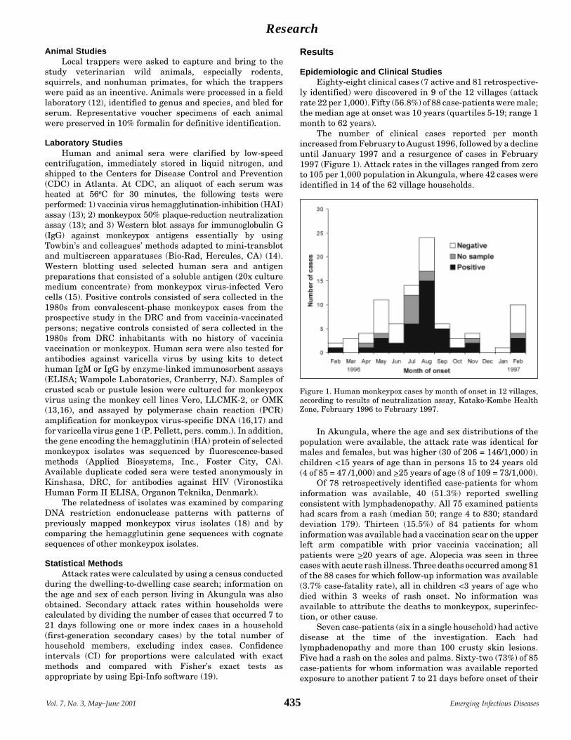
Vol. 7, No. 3, May–June 2001 Emerging Infectious Diseases435
Research
Animal StudiesLocal trappers were asked to capture and bring to the
study veterinarian wild animals, especially rodents,squirrels, and nonhuman primates, for which the trapperswere paid as an incentive. Animals were processed in a fieldlaboratory (12), identified to genus and species, and bled forserum. Representative voucher specimens of each animalwere preserved in 10% formalin for definitive identification.
Laboratory StudiesHuman and animal sera were clarified by low-speed
centrifugation, immediately stored in liquid nitrogen, andshipped to the Centers for Disease Control and Prevention(CDC) in Atlanta. At CDC, an aliquot of each serum washeated at 56oC for 30 minutes, the following tests wereperformed: 1) vaccinia virus hemagglutination-inhibition (HAI)assay (13); 2) monkeypox 50% plaque-reduction neutralizationassay (13); and 3) Western blot assays for immunoglobulin G(IgG) against monkeypox antigens essentially by usingTowbin’s and colleagues’ methods adapted to mini-transblotand multiscreen apparatuses (Bio-Rad, Hercules, CA) (14).Western blotting used selected human sera and antigenpreparations that consisted of a soluble antigen (20x culturemedium concentrate) from monkeypox virus-infected Verocells (15). Positive controls consisted of sera collected in the1980s from convalescent-phase monkeypox cases from theprospective study in the DRC and from vaccinia-vaccinatedpersons; negative controls consisted of sera collected in the1980s from DRC inhabitants with no history of vacciniavaccination or monkeypox. Human sera were also tested forantibodies against varicella virus by using kits to detecthuman IgM or IgG by enzyme-linked immunosorbent assays(ELISA; Wampole Laboratories, Cranberry, NJ). Samples ofcrusted scab or pustule lesion were cultured for monkeypoxvirus using the monkey cell lines Vero, LLCMK-2, or OMK(13,16), and assayed by polymerase chain reaction (PCR)amplification for monkeypox virus-specific DNA (16,17) andfor varicella virus gene 1 (P. Pellett, pers. comm.). In addition,the gene encoding the hemagglutinin (HA) protein of selectedmonkeypox isolates was sequenced by fluorescence-basedmethods (Applied Biosystems, Inc., Foster City, CA).Available duplicate coded sera were tested anonymously inKinshasa, DRC, for antibodies against HIV (VironostikaHuman Form II ELISA, Organon Teknika, Denmark).
The relatedness of isolates was examined by comparingDNA restriction endonuclease patterns with patterns ofpreviously mapped monkeypox virus isolates (18) and bycomparing the hemagglutinin gene sequences with cognatesequences of other monkeypox isolates.
Statistical MethodsAttack rates were calculated by using a census conducted
during the dwelling-to-dwelling case search; information onthe age and sex of each person living in Akungula was alsoobtained. Secondary attack rates within households werecalculated by dividing the number of cases that occurred 7 to21 days following one or more index cases in a household(first-generation secondary cases) by the total number ofhousehold members, excluding index cases. Confidenceintervals (CI) for proportions were calculated with exactmethods and compared with Fisher’s exact tests asappropriate by using Epi-Info software (19).
Results
Epidemiologic and Clinical StudiesEighty-eight clinical cases (7 active and 81 retrospective-
ly identified) were discovered in 9 of the 12 villages (attackrate 22 per 1,000). Fifty (56.8%) of 88 case-patients were male;the median age at onset was 10 years (quartiles 5-19; range 1month to 62 years).
The number of clinical cases reported per monthincreased from February to August 1996, followed by a declineuntil January 1997 and a resurgence of cases in February1997 (Figure 1). Attack rates in the villages ranged from zeroto 105 per 1,000 population in Akungula, where 42 cases wereidentified in 14 of the 62 village households.
In Akungula, where the age and sex distributions of thepopulation were available, the attack rate was identical formales and females, but was higher (30 of 206 = 146/1,000) inchildren <15 years of age than in persons 15 to 24 years old(4 of 85 = 47 /1,000) and >25 years of age (8 of 109 = 73/1,000).
Of 78 retrospectively identified case-patients for whominformation was available, 40 (51.3%) reported swellingconsistent with lymphadenopathy. All 75 examined patientshad scars from a rash (median 50; range 4 to 830; standarddeviation 179). Thirteen (15.5%) of 84 patients for whominformation was available had a vaccination scar on the upperleft arm compatible with prior vaccinia vaccination; allpatients were >20 years of age. Alopecia was seen in threecases with acute rash illness. Three deaths occurred among 81of the 88 cases for which follow-up information was available(3.7% case-fatality rate), all in children <3 years of age whodied within 3 weeks of rash onset. No information wasavailable to attribute the deaths to monkeypox, superinfec-tion, or other cause.
Seven case-patients (six in a single household) had activedisease at the time of the investigation. Each hadlymphadenopathy and more than 100 crusty skin lesions.Five had a rash on the soles and palms. Sixty-two (73%) of 85case-patients for whom information was available reportedexposure to another patient 7 to 21 days before onset of their
Figure 1. Human monkeypox cases by month of onset in 12 villages,according to results of neutralization assay, Katako-Kombe HealthZone, February 1996 to February 1997.
436Emerging Infectious Diseases Vol. 7, No. 3, May–June 2001
Research
illness. The remaining 23 (27%) patients reported either noexposure to other cases (n=13; 15%) or an exposure to anothercase from 0 to 6 days before onset of illness (n=10; 12%). Datesof onset for patients who reported no exposure to other casesduring the incubation period were distributed throughout thestudy period (11). Exposures during the incubation periodthat were reported by all six patients who resided in a singlehousehold and who had active disease at the time of thisinvestigation included exposure to a patient within thehousehold and eating monkey, gazelle, pig, and rat.
The 88 clinical cases were identified for 39 households, inwhich 297 persons resided (overall attack rate in affectedhouseholds = 30%). For 240 household members for whominformation was available, rash that met the clinical casedefinition developed in 20 in 7 to 21 days after exposure to >1index cases in a household (household secondary attack rate8.3 per 100; 95% CI 5.2% to 12.6%).
Eating wild animals was common for all patients.However, patients who had no exposure to other case-patientsduring the incubation period were more likely to eatporcupine at least once a month (Table 1). All queriedparticipants reported trapping animals less frequently.
Laboratory StudiesHuman monkeypox was confirmed in all seven active
cases by virus isolation and monkeypox virus-specific DNAamplification from skin lesion samples; antibodies againstorthopoxvirus were detected in two by neutralization assay,in three by HAI assay, and in six by Western blotting. Inaddition, IgM antibodies against varicella virus were detectedin five active cases in patients who lived in the samehousehold. Varicella virus DNA was detected in lesionmaterial from two of these patients.
Seventy-two of the 81 retrospectively identified casesprovided a serum sample, although 2 were low in volume.Orthopoxvirus antibodies were detected by neutralizationassay in 39 (54%, Figure 1) of the 72 sera; by HAI in 51 (73%)of 70 sera, and by a new, nonvalidated Western blot test in 49(68%) of 72 sera. Thirty-eight (54%) of 70 available sera werepositive by all three tests, 45 (64%) were positive by two tests,and 62 (89%) were positive by at least one test. Westernblotting and HAI test results were concordant for 79% of thesera; Western blotting and neutralization test results agreedfor 83% of the sera; and HAI and neutralization test resultsagreed for 79% of the sera. Fifty-five (76%) of the 72 patientswho provided serum had detectable varicella IgG antibodies,1 had varicella IgM, and none showed detectable HIV antibodies.
Based on DNA assays (16-18), monkeypox virus isolatesfrom the seven active cases appeared virtually identical toeach other and very closely related to two isolates fromhumans in 1996 and an isolate from a squirrel trapped in theEquateur Province of the DRC in 1985. Analysis of sequencescoding for the viral HA protein (Figure 2) indicated thatviruses isolated in the DRC since 1970 comprised a cladedistinguishable from a clade comprising isolates from otherAfrican countries and from outbreaks in primate-holdingfacilities in Europe and the United States. The group of nineisolates from the 1996-97 outbreak, which showed identicalHA sequences, and from the squirrel from 1985 constituted asubset of the clade of examined viruses previously isolated inthe DRC.
Animal StudiesFifty-nine captured animals representing 14 species were
tested. Sera from 42% of the various squirrel species, from 16%of Gambian rats, and from an elephant shrew and a domestic pigshowed orthopoxvirus neutralizing antibodies (Table 2).
Table 1. Exposure to wild animals reported by monkeypox case-patientsaccording to history of exposure to another case-patient, Katako-KombeHealth zone, February 1996 to February 1997
Cases without Cases withexposure to exposure toother case- other case-
patients during patients duringExposures reported incubation incubationto occur usually (N=23) (N=62)on a monthly basis (#) (%) (#) (%) p valueExposure to squirrels Trapping 2 8.7 9 14.8 0.79 Exposure to raw meat 11 47.8 33 54.1 0.78 Eating 19 86.4 52 85.2 1.00Exposure to monkeys Trapping 1 4.3 4 6.6 1.00 Exposure to raw meat 16 69.6 34 55.7 0.36 Eating 21 91.3 58 95.1 0.61Exposure to rats Trapping 5 21.7 18 29.5 0.66 Exposure to raw meat 13 56.5 40 65.6 0.60 Eating 22 95.7 55 90.2 0.66Exposure to porcupines Trapping 1 4.3 3 4.9 1.00 Exposure to raw meat 12 52.2 24 39.3 0.41 Eating 22 95.7 42 68.9 0.02Exposure to gazelles Trapping 1 4.3 2 3.3 1.00 Exposure to raw meat 14 56.5 28 45.9 0.53 Eating 21 91.3 47 77.0 0.21
Figure 2. Phylogenetic inference relationships of the open readingframes encoding the viral hemagglutinin protein of variousmonkeypox virus isolates and selected strains of vaccinia, variola,and cowpox viruses. Nucleotide sequences of polymerase chainreaction-generated amplicons were analyzed using PAUP parsimonyanalysis software version 3.1.1, as described (10). Parsimonyanalysis used 5,000 bootstraps and weighted the sequences for atransition-transversion ratio of 2 (bootstrap confidence intervalsshown on branches).

Vol. 7, No. 3, May–June 2001 Emerging Infectious Diseases437
Research
ConclusionThe epidemiologic and laboratory evidence presented
here suggests that this monkeypox outbreak is the largestreported. Epidemiologic features of monkeypox that differedfrom those described during an intensified prospective studyin the DRC in the 1980s included an increase in the proportionof patients reporting an exposure to another case during theincubation period (28% in the 1980s, 73% this study) and theclustering of successive cases within households, with asmany as eight consecutive cases in one instance (3,10).
Prospective surveillance in the 1980s in the DRCprovided opportunities to observe most patients with active-stage monkeypox; thus, skin lesion samples were available forvirus isolation to confirm clinical diagnosis. In contrast, thepresent study identified most patients retrospectively andrelied on serologic testing for confirmation of the diagnosis;only seven clinically active cases could be confirmed by virusisolation and PCR (17) for monkeypox virus. Becausemonkeypox is the only known indigenous orthopoxvirus ofAfrica that infects humans systemically, seropositive casesshowing genus-specific antibodies can be reasonablyinterpreted as cases that had a monkeypox virus infection(20). Of 72 retrospectively identified cases tested, 54% hadantibodies by plaque-reduction assay, a preferred test forverifying orthopoxvirus infections during smallpox eradica-tion (20,21). However, this neutralization test was only 83%sensitive for the detection of vaccinia vaccine-inducedantibodies and may have been negative (3) in some patientswith monkeypox virus infection during this study interval. Ahigher proportion of sera from retrospectively identified cases(73%) tested positive by the HAI test, which had a highersensitivity (96%) than the neutralization test for theidentification of vaccinia-induced antibodies (22). However,
the HAI test may be less specific, and antibody titers detectedby it decrease rapidly after infection and would be unlikely tobe residual from prior vaccination in the small percentage ofpatients who had been vaccinated (3,21,23). Finally, 72% ofsera from retrospectively identified cases tested positive in anew, nonvalidated Western blot orthopoxvirus antibody testfor which sensitivity and specificity have not beendetermined. Chickenpox, which can be mistaken formonkeypox (24), was reported in the area during the studyperiod. Some patients may have had dual infection bymonkeypox and varicella viruses. Within one household, fivepatients from whom monkeypox virus was isolated showedserologic evidence of recent varicella virus infection, and twoof those had detectable varicella virus DNA in skin lesionsamples. Thus, a substantial percentage of cases may havebeen chickenpox, thereby explaining why the case-fatalityrate from this study (3.7%) was lower than the 9.6%previously reported (3,24). However, the presence ofantibodies against orthopoxviruses by three different assaysin 54% of patients suggests that varicella virus was notresponsible for most cases during the study period.
The 1996-97 crude secondary attack rate of about 8% inhouseholds cannot be compared with the 3.7% risk of attackfor household contacts from the 1981-1986 study (3) becausethe proportion of susceptible (unvaccinated) persons inhousehold members increased markedly from 1981-1986 to1996-97. However, the 1996-97 secondary attack rate withinhouseholds was similar to the 1981-1986 rates (3) of 9.3% forunvaccinated household members. This similarity isconsistent with the high degree of similarity noted incomparing current monkeypox virus isolate DNAs with DNAsof isolates from the DRC in the 1970s, including the HAsequences (Figure 2). While the virus and its person-to-persontransmission potential have not changed substantially, theproportion of susceptible persons among exposed householdmembers increased most likely because vaccinia vaccinationceased after smallpox eradication. The reduced herdimmunity probably also caused a higher proportion of cases tobe attributable to person-to-person transmission, as theincrease in the proportion of patients who reported exposureto another patient during the incubation interval wouldsuggest. HIV was not a cofactor of this outbreak because noHIV antibodies were detected in patients’ sera.
Exposure to an animal reservoir might have been thesource of infection for at least 27% of patients who reported noexposure to other patients during the incubation period. Pastanimal studies pointed to several species of squirrels asanimal reservoirs (5,6). Although the possibility of infection ofanimals with another unknown indigenous orthopoxviruscannot be ruled out in animals with neutralizing antibodies,the results of our investigation suggest that, in addition tosquirrels, Gambian rats may play a role in monkeypox viruscirculation. Villagers ate a number of different animals, and itwas impossible to draw any conclusions as to whether any onespecies was a greater risk factor than any other. However,antibody surveys could be used to evaluate porcupines as apossible reservoir (Table 1), which was not done during ourinvestigation because no porcupines were captured by trappers.
Our study had several limitations, some of which relatedto the brevity imposed by the civil unrest. Most cases wereidentified retrospectively and without a case-control group;thus, they could not be confirmed by a serologic test for which
Table 2. Species of animals caught in the wild and monkeypox virusplaque reduction neutralization antibody assay results, Katako-KombeHealth Zone, February 23-27, 1997
No. ProportionAnimal species Common name Tested Pos. pos.Cercopithecus Spot-nosed monkey 2 0 0.0% ascaniusCercopithecus Debrazza monkey 1 0 0.0% neglectusPerodicticus Bosman’s potto 1 0 0.0% pottoGalogoides Demidoff galago 2 0 0.0% demidoviGenetta Genet 1 0 0.0% rubiginosaSus scrofa Domestic pig 1 1 100.0%Praomys Forest rat 1 0 0.0% jacksoniCricetomys Gambian rat 19 3 15.8% eminiCavia sp. Guinea pig 3 0 0.0%Petrodromus Elephant shrew 3 1 33.3% tetradactylusDendrohyrax Tree hyrax 1 0 0.0% arboreusFunisciurus Thomas’s tree squirrel 4 2 50.0% anerythrusFunisciurus Kuhl’s tree squirrel 18 7 38.9% congicusHeliosciurus Sun squirrel 2 1 50.0% rufobrachiumTotal 59 15 25.4%

438Emerging Infectious Diseases Vol. 7, No. 3, May–June 2001
Research
the sensitivity and the specificity had been measured using astandard technique and case-control sera. Because informa-tion was not available regarding the vaccination status ofother members of the patients’ households, secondary attackrates within households could not be calculated according tothe vaccination status. We did not assess the environmentalchanges that could have facilitated this outbreak, includingincreases in household sizes, rates of monkeypox virusinfection in the animal population, and a change in rate andtype of exposure to wild animals among residents. We wereunable to attribute infections in patients who reported anexposure to another patient during the incubation period toperson-to-person transmission because some of these patientsmay also have been exposed to infected animals. Finally,patients with a history of exposure to a patient during theincubation period were the only controls available to generatehypotheses regarding the type of animal exposure possiblyassociated with infection in patients not reporting anexposure to other patients during the incubation period. Thisaspect may have caused bias because cases clustered withinhouseholds where exposures to animals may have beenidentical and because cases may have been misclassified as totheir purported source of infection.
There is no evidence to date that person-to-persontransmission alone can sustain monkeypox in the localpopulation. However, our study suggests that in a populationwith low herd immunity, person-to-person transmission andrepeated introduction of virus from the animal reservoir maylead to more and larger clusters of human monkeypox cases inAfrican rain forest areas (25). Further studies are needed tomeasure the sensitivity and the specificity of the serologictests currently available so they can be used in the future tobetter confirm retrospectively identified cases, identify thetype of animal or patient exposures associated withacquisition of illness, and evaluate the respective roles ofperson-to-person and animal-to-human transmission. How-ever, access to the Kasai region has been limited since ourstudy, and little information has become available to betterdocument the current dynamics of monkeypox occurrences orimprove World Health Organization recommendations toprevent human monkeypox more substantially.
AcknowledgmentsThe authors thank T.P. Muamba, G. Mwema, D. Messinger, K.
Kilundu, V.B. Mukinda, L.V. Okito, and V.L. Mangindula for assistancein data collection; L. Otshudi for assistance in setup of the investigation;J.J. Muyembe-Tamfum for HIV testing; A. Moudi; J.A. Stewart and J.Patton for varicella virus testing; A. Moren, W.C. Reeves, C.J. Peters,and D.A. Henderson for critically reading the manuscript; and J.O’Connor for editorial assistance.
The investigation was partly funded by the U.S. Agency forInternational Development.
Dr. Yvan Hutin was an Epidemic Intelligence Service officer withthe Centers for Disease Control and Prevention at the time of this in-vestigation. He is now at the World Health Organization.
References 1. Marennikova SS, Seluhina EM, Mal’ceva NN, Cimiskjan KL,
Macevic GR. Isolation and properties of the causal agent of a newvariola-like disease (monkeypox) in man. Bull World Health Organ1972;46:599-611.
2. Arita I, Jezek Z, Khodakevich L, Ruti K. Human monkeypox: anewly emerged orthopoxvirus zoonosis in the tropical rain forests ofAfrica. Am J Trop Med Hyg 1985;34:781-9.
3. Jezek Z, Fenner F. Human monkeypox. In: JL Melnick, editor.Monographs in virology. Volume 17. Basel: Karger; 1988.
4. von Magnus P, Andersen EK, Petersen KB, Birch-Andersen A. Apox-like disease in cynomolgus monkeys. Acta Pathol MicrobiolScand 1959;46:156-76.
5. Khodakevich L, Jezek Z, Messinger D. Monkeypox virus: ecology andpublic health significance. Bull World Health Organ 1988;66:747-52.
6. Khodakevich L, Szczeniowski M, Manbu-ma-Disu, Jezek Z,Marennikova S, Nakano J, et al. The role of squirrels in sustainingmonkeypox virus transmission. Trop Geogr Med 1987;39:115-22.
7. Fine PE, Jezek Z, Grab B, Dixon H. The transmission potential ofmonkeypox virus in human populations. Int J Epidemiol1988;17:643-50.
8. Jezek Z, Grab B, Szczeniowski MV, Paluku KM, Mutombo M.Human monkeypox: secondary attack rates. Bull World HealthOrgan 1988;66:465-70.
9. Jezek Z, Arita I, Mutombo M, Dunn C, Nakano JH, SzezeniowskiM. Four generations of probable person-to-person transmission ofhuman monkeypox. Am J Epidemiol 1986;123:1004-12.
10. Mukinda VB, Mwema G, Kilundu M, Heymann DL, Khan AS,Esposito JJ. Re-emergence of human monkeypox in Zaire in 1996.Lancet 1997;349:1449-50.
11. Centers for Disease Control and Prevention. Human monkeypox-Zaire, 1996-1997. MMWR Morb Mortal Wkly Rep 1997;46:304-7.
12. Mills JN, Childs JE, Ksiazek TG, Peters CJ, Velleca WM. Methodsfor trapping and sampling small mammals for virologic testing.Atlanta: Centers for Disease Control and Prevention; 1995.
13. Esposito JJ, Massung RF. Poxvirus infections in humans. In:Murray PR, Tenover F, Baron EJ, Pfaller MA, Yolken RH, editors.Manual of clinical microbiology. 6th ed. Washington: ASMPress;1995. p. 1131-8.
14. Towbin H, Staehelin T, Gordon J. Electrophoretic transfer of proteinsfrom polyacrylamide gels to nitrocellulose sheets: procedure and someapplications. Proc Natl Acad Sci U S A 1979;76:4350-4.
15. Loparev VN, Parsons JM, Knight JC, Panus JF, Ray CA, BullerRM, et al. A third distinct tumor necrosis factor receptor oforthopoxviruses. Proc Natl Acad Sci U S A 1998;95:3786-91.
16. Ropp SL, Esposito JJ, Loparev VN, Palumbo G. Poxvirusesinfecting humans. In: Murray PR, Barron CJ, Pfaller MA, TenoverFC, Yolken RH, editors. Manual of clinical microbiology. 7th ed.Washington: ASM Press; 1999. p. 1137-44.
17. Ropp SL, Jin Q, Knight JC, Massung RF, Esposito JJ. PCRstrategy for identification and differentiation of smallpox and otherorthopoxviruses. J Clin Microbiol 1995;33:2069-76.
18. Esposito JJ, Knight JC. Orthopoxvirus DNA: A comparison ofrestriction profiles and maps. Virology 1985;143:230-51.
19. Dean AG, Dean JA, Coulombier D, Burton AH, Brendel KA, SmithDC. Epi Info, version 6: A word-processing, database, and statisticsprogram for public health on IBM-compatible microcomputers.Atlanta: Centers for Disease Control and Prevention; 1994.
20. Fenner F, Wittek R, Dumbell KR. The Orthopoxviruses. San Diego:Academic Press; 1989. p. 162-5, 312-15.
21. Ziegler DW, Hutchinson HD, Koplan JP, Nakano JH. Detection byradioimmunoassay of antibodies in human smallpox patients andvaccinees. J Clin Microbiol 1975;1:311-7.
22. Cherry JD, McIntosh K, Connor JD, Benenson AS, Alling DW,Rolfe UT, et al. Primary percutaneous vaccination. J Infect Dis1977;135:145-54.
23. McIntosh K, Cherry JD, Benenson AS, Connor JD, Alling DW,Rolfe UT, et al. Clinical and serologic study of four smallpoxvaccines comparing variations of dose and route of administration.Standard percutaneous revaccination of children who receiveprimary percutaneous vaccination. J Infect Dis 1977;135:155-66.
24. Jezek Z, Szczeniowski M, Paluku KM, Mutombo M, Grab B. Humanmonkeypox: confusion with chickenpox. Acta Trop 1988;45:297-307.
25. Centers for Disease Control and Prevention. Human monkeypox —Kasai Oriental, Democratic Republic of Congo, February 1996-
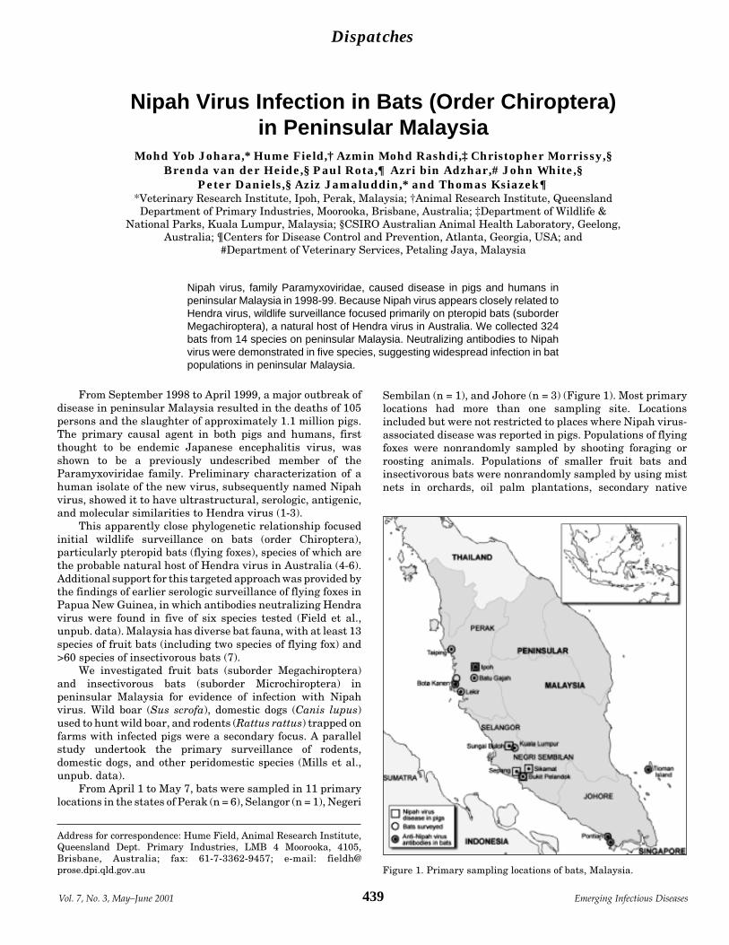
Dispatches
439Vol. 7, No. 3, May–June 2001 Emerging Infectious Diseases
From September 1998 to April 1999, a major outbreak ofdisease in peninsular Malaysia resulted in the deaths of 105persons and the slaughter of approximately 1.1 million pigs.The primary causal agent in both pigs and humans, firstthought to be endemic Japanese encephalitis virus, wasshown to be a previously undescribed member of theParamyxoviridae family. Preliminary characterization of ahuman isolate of the new virus, subsequently named Nipahvirus, showed it to have ultrastructural, serologic, antigenic,and molecular similarities to Hendra virus (1-3).
This apparently close phylogenetic relationship focusedinitial wildlife surveillance on bats (order Chiroptera),particularly pteropid bats (flying foxes), species of which arethe probable natural host of Hendra virus in Australia (4-6).Additional support for this targeted approach was provided bythe findings of earlier serologic surveillance of flying foxes inPapua New Guinea, in which antibodies neutralizing Hendravirus were found in five of six species tested (Field et al.,unpub. data). Malaysia has diverse bat fauna, with at least 13species of fruit bats (including two species of flying fox) and>60 species of insectivorous bats (7).
We investigated fruit bats (suborder Megachiroptera)and insectivorous bats (suborder Microchiroptera) inpeninsular Malaysia for evidence of infection with Nipahvirus. Wild boar (Sus scrofa), domestic dogs (Canis lupus)used to hunt wild boar, and rodents (Rattus rattus) trapped onfarms with infected pigs were a secondary focus. A parallelstudy undertook the primary surveillance of rodents,domestic dogs, and other peridomestic species (Mills et al.,unpub. data).
From April 1 to May 7, bats were sampled in 11 primarylocations in the states of Perak (n = 6), Selangor (n = 1), Negeri
Sembilan (n = 1), and Johore (n = 3) (Figure 1). Most primarylocations had more than one sampling site. Locationsincluded but were not restricted to places where Nipah virus-associated disease was reported in pigs. Populations of flyingfoxes were nonrandomly sampled by shooting foraging orroosting animals. Populations of smaller fruit bats andinsectivorous bats were nonrandomly sampled by using mistnets in orchards, oil palm plantations, secondary native
Nipah Virus Infection in Bats (Order Chiroptera)in Peninsular Malaysia
Mohd Yob Johara,* Hume Field,† Azmin Mohd Rashdi,‡ Christopher Morrissy,§Brenda van der Heide,§ Paul Rota,¶ Azri bin Adzhar,# John White,§
Peter Daniels,§ Aziz Jamaluddin,* and Thomas Ksiazek¶*Veterinary Research Institute, Ipoh, Perak, Malaysia; †Animal Research Institute, QueenslandDepartment of Primary Industries, Moorooka, Brisbane, Australia; ‡Department of Wildlife &
National Parks, Kuala Lumpur, Malaysia; §CSIRO Australian Animal Health Laboratory, Geelong,Australia; ¶Centers for Disease Control and Prevention, Atlanta, Georgia, USA; and
#Department of Veterinary Services, Petaling Jaya, Malaysia
Address for correspondence: Hume Field, Animal Research Institute,Queensland Dept. Primary Industries, LMB 4 Moorooka, 4105,Brisbane, Australia; fax: 61-7-3362-9457; e-mail: [email protected]
Nipah virus, family Paramyxoviridae, caused disease in pigs and humans inpeninsular Malaysia in 1998-99. Because Nipah virus appears closely related toHendra virus, wildlife surveillance focused primarily on pteropid bats (suborderMegachiroptera), a natural host of Hendra virus in Australia. We collected 324bats from 14 species on peninsular Malaysia. Neutralizing antibodies to Nipahvirus were demonstrated in five species, suggesting widespread infection in batpopulations in peninsular Malaysia.
Figure 1. Primary sampling locations of bats, Malaysia.

Dispatches
Emerging Infectious Diseases Vol. 7, No. 3, May–June 2001440
vegetation, and residential areas, where bats were reported orobserved, where flowering or fruiting trees were observed,and near known or possible roosts. A target of 30 animals perspecies was set, providing 95% statistical confidence ofdetecting infection at a minimum population prevalence of10%, assuming homogeneity of infection across overlappingpopulations and a test sensitivity and specificity of 100% (8).Blood for serologic examination was also collected from twocaptive colonies of flying foxes in zoos. In addition to blood,fresh tissue samples of liver, lung, kidney, spleen, heart, andfetus were taken from wild-caught animals, and the carcasseswere stored in 10% buffered formalin for reference. Virusisolation was attempted by using Vero E6 cells in a biosafetylevel 4 laboratory as described (9). All cell harvests werechecked for Nipah virus antigens by indirect fluorescencewith Nipah hyperimmune ascitic fluid. Reverse tran-scriptase-polymerase chain reaction (RT-PCR) was per-formed on the same tissues, from which RNA was extracted.Virus isolation and RT-PCR were performed on both kidneyand spleen from each animal from which tissues had beencollected. Additionally, all tissues were blind passaged twicemore, and each harvest was tested for viral antigen. RT-PCRused forward and reverse primers designed to amplify a228-bp region of the N gene.
A total of 324 animals from 14 species of bat weresampled, with the target sample size being achieved for fivespecies. Sera were either forwarded directly to the AustralianAnimal Health Laboratory (AAHL) in Geelong, Australia, orscreened by indirect enzyme-linked immunofluorescent assay(ELISA) using Nipah virus antigen at the VeterinaryResearch Institute in Ipoh, Malaysia, before being forwardedto AAHL. At AAHL, sera were tested by indirect ELISA usingNipah virus antigen and by serum neutralization test (SNT).As the latter is the recognized standard, we used these data inour analysis. Serum neutralization results were obtained for265 sera, the balance producing toxic reactions (attributedto poor serum sample quality) at a dilution of >1:10.Neutralizing antibodies to Nipah virus were detected in thesera of 21 wild-caught animals from five species (Table):Pteropus hypomelanus (island flying fox), P. vampyrus(Malayan flying fox), Eonycteris spelaea (cave bat),
Cynopterus brachyotis (lesser dog-faced fruit bat), andScotophilus kuhli (house bat). Antibody prevalence amongthese species was 31%, 17%, 5%, 4%, and 3%, respectively.Titers ranged from 1:5 (n = 2), the lowest dilution tested, to1:40 (n = 1), median 1:10 (Figure 2). The Nipah virusneutralization titer of the positive control rabbit anti-Hendravirus serum was 1:20. Of the 21 sera neutralizing Nipahvirus, only one neutralized Hendra virus, with a titer fourfoldless than the corresponding Nipah virus titer. The Hendravirus neutralization titer of the positive control anti-Hendravirus serum was 1:160. All culture harvests were negative forNipah virus antigen. Two of the tissues yielded cytopathicagents that do not react with either Nipah or Hendraantibodies; these agents are being characterized. All attemptsto amplify Nipah virus RNA were also negative.
Wild boar, hunting dogs, and rodents were sampled inPerak state from April 1 to May 7. Wild boar (n = 18) werenonrandomly sampled by shooting in oil palm plantations,secondary native vegetation, national parks, and ruralresidential areas. Blood samples were also collected from dogs(n = 16) used to hunt wild boar. Rodents (n = 25) were trappedon several farms where pigs were infected. None of the serafrom wild boar, hunting dogs, or rodents were positive byindirect ELISA using Nipah virus antigen.
We interpret the presence of neutralizing antibodies toNipah virus in the identified bat species as evidence ofinfection with this virus or a cross-neutralizing virus. Cross-neutralization of Nipah antigen by antibodies to Hendra viruswas excluded as the cause of the reactivity, and otherparamyxoviruses have not demonstrated cross-neutraliza-tion with either Hendra (10) or Nipah virus (2). We believethat the presence of anti-Nipah antibodies in a population ofP. hypomelanus on the east coast island of Tioman (Figure 1),geographically remote from the west coast foci of Nipah viraldisease in pigs, indicates that Nipah virus infection iswidespread in flying fox populations in peninsular Malaysia.Ecologically, P. hypomelanus is an island specialist whosemainland foraging is limited to nearby coastal areas (11).
The low neutralizing antibody titers in the positiveMalaysian bats were unexpected. In Australia, anti-Hendravirus titers of >1:640 in wild-caught flying foxes (Field et al.,unpub. data) and 1:80 in experimentally infected flying foxes(12) have been observed. The absence of high titers in thesampled animals could be explained in several ways: thesample may not be representative of the population; Nipahvirus may bind inefficiently to Vero cells used in the
Figure 2. Frequency distribution of antibody titers to Nipah virus in21 Malaysian bats testing positive by serum neutralization test.
Table. Species description of 237 wild-caught Malaysian bats of knownneutralizing antibody status to Nipah virusa surveyed April 1–May 7, 1999
Species No. of bats No. positive (%)Megachiroptera (fruit bats) Cynopterus brachyotis 56 2 (4) Eonycteris spelaea 38 2 (5) Pteropus hypomelanus 35 11 (31) Pteropus vampyrus 29 5 (17) Cynopterus horsfieldi 24 0 Ballionycterus maculata 4 0 Macroglossus sobrinus 4 0 Megaerops ecaudatus 1 0Microchiroptera (Insectivorous bats) Scotophilus kuhlii 33 1 (3) Rhinolophus affinis 6 0 Taphozous melanopogon 4 0 Taphozous saccolaimus 1 0 Hipperosiderus bicolor 1 0 Rhinolophus refulgens 1 0Total 237 21aSera from 324 bats were tested: 59 sera that gave toxic results at dilutions1:10 were excluded from analysis, as were sera from 28 captive P. vampyrus.

Dispatches
441Vol. 7, No. 3, May–June 2001 Emerging Infectious Diseases
neutralization assays; bats’ immune response to Nipah virusmay be muted as a result of high-level adaptation of the virusto these species; or the antigenic structure of the virus in pigsand humans may differ from that in bats, resulting in lesseffective neutralization of a test antigen derived from ahuman isolate. Alternatively, the antibodies detected may becross-neutralizing antibodies to another related but as yetunidentified virus in bats.
The detection of anti-Nipah virus antibodies in non-pteropid species is notable, although the significance of thefinding remains unclear. Limited surveillance of non-pteropid species in Australia for anti-Hendra virus antibodieshas not found evidence of infection in these species. Furtherwork is needed to clarify any role of non-pteropid species inthe natural history of both viruses.
Isolation of Nipah virus from bats is essential tocorroborate the serologic findings and enable comparison ofbat isolates with human and pig isolates. However, cellculture of fresh tissue samples from antibody-positive and-negative bat species forwarded to the Centers for DiseaseControl and Prevention did not produce an isolate reactivewith anti-Nipah virus antibodies. All PCR attempts on thesetissues were also negative. The tissues submitted (heart,liver, lung, kidney, spleen, fetal) were considered appropriate,as these tissues, as well as white cells, have yielded Hendravirus isolates in naturally infected (13) or experimentallyinfected (12) flying foxes in Australia. In Malaysia, the periodof sampling did not overlap the seasonal gestation of eitherP. vampyrus or P. hypomelanus. Fetal tissues submittedwere from S. kuhli, E. spelaea, C. brachyotis, Taphozousmelanopogon, T. saccolaimus, and Rhinolophus affinis.
The wild boar and hunting dog serologic results need to beinterpreted in light of the limited sample size, nonrandomsampling, and test methods. Nonetheless, as the behavioraland foraging patterns of wild boar promote contact within andbetween neighboring populations, the absence of anti-Nipahvirus antibodies in the sample supports the absence ofestablished infection in wild boar populations in the areassurveyed. The absence of anti-Nipah virus antibodies inhunting dogs is also consistent with lack of exposure to Nipahvirus. A Nipah virus antibody prevalence of 42 (46%) of 92 wasidentified in domestic dogs sampled near infected pig farms(Mills et al., unpub. data), and if hunting dogs, which haveregular contact with the blood, urine, and oronasal secretionsof wild boar, were exposed, similar antibody prevalences couldreasonably be expected. The negative findings in the rodentsample are consistent with those of the comprehensiveparallel survey of rodents (Mills et al., unpub. data).
We report evidence of infection with Nipah virus in fourfruit bat species and one insectivorous bat species inpeninsular Malaysia. A proposed second phase will describethe occurrence and frequency of infection in the identifiedNipah antibody-positive species at additional locations inpeninsular Malaysia and in Sabah and Sarawak, Borneo. Inaddition to successful virus isolation from bats, other
proposed research includes retrospective studies of archivalspecimens, experimental infections of fruit bats, and serologicsurveys of other arboreal mammalian species.
AcknowledgmentsWe thank the Malaysian Department of Veterinary Services
Veterinary Research Institute at Ipoh for field and laboratoryassistance and hospitality, the Malaysian Department of Wildlifeand National Parks for field expertise, Craig Smith for technical andlaboratory support, Greer Meehan for assistance with serology, LimBoo Liat for his interest and advice, and village leaders and propertyowners for their cooperation.
Dr. Johara is a senior veterinarian with the Malaysian Depart-ment of Veterinary Services. Her research interests include infectiousdiseases of livestock, infectious zoonotic diseases, animal welfare andwildlife conservation. During the Nipah virus outbreak, she played akey role in the investigation of wildlife species for the origin of Nipahvirus.
References 1. Centers for Disease Control and Prevention. Outbreak of Hendra-
like virus—Malaysia and Singapore, 1998-1999. MMWR MorbMortal Wkly Rep 1999;48:265-9.
2. Chua KB, Bellini WJ, Rota PA, Harcourt BH, Tamin A, Lam SK, etal. Nipah virus: a recently emergent deadly paramyxovirus.Science 2000;288:1432-5.
3. Mohd Nor M, Gan C, Ong B. Nipah virus infection of pigs inpeninsular Malaysia. Rev Sci Tech 2000;19:160-5.
4. Young PL, Halpin K, Selleck PW, Field HE, Gravel JL, Kelly MA,et al. Serologic evidence for the presence in Pteropus bats of aparamyxovirus related to equine morbillivirus. Emerg Infect Dis1996;2:239-40.
5. Field H, Halpin K, Young P. An overview of equine morbillivirusand bat paramyxovirus in Australia. The OIE/FAVA EpidemiologyProgramme: special session on emerging diseases, Cairns,Australia; 1997 Aug 28; Office International des Epizooties (OIE).
6. Williamson MM, Hooper PT, Selleck PW, Gleeson LJ, Daniels PW,Westbury HA, et al. Transmission studies of Hendra virus (equinemorbillivirus) in fruit bats, horses and cats. Aust Vet J1998;76:813-8.
7. Medway L. The wild mammals of Malaya (Peninsular Malaysia)and Singapore. Kuala Lumpur: Oxford University Press; 1978.
8. Cannon R. Livestock disease surveys: a field manual forveterinarians. Canberra (Australia): Australian Bureau of AnimalHealth; 1982.
9. Leirs H, Mills JN, Krebs JW, Childs JE, Dudu A, Woollen N, et al.Search for the Ebola virus reservoir in Kikwit, DemocraticRepublic of the Congo: reflections on a vertebrate collection. JInfect Dis 1999;179:155-63.
10. Murray K, Selleck P, Hooper P, Hyatt A, Gould A, Gleeson L, et al.A morbillivirus that caused fatal disease in horses and humans.Science 1995;268:94-7.
11. Mickleburg S, Hutson A, Racey P. Old world fruit bats: an action planfor their conservation. Gland (Switzerland): International Union forthe Conservation of Nature and Natural Resources; 1992.
12. Williamson M, Hooper P, Selleck P, Westbury H, Slocombe R. Theeffect of Hendra virus on pregnancy in fruit bats and guinea pigs.J Comp Pathol 2000;122:201-7.
13. Halpin K, Young P, Field H, Mackenzie J. Isolation of Hendra virusfrom pteropid bats: a natural reservoir of Hendra virus. J Gen Virol2000;81:1927-32.

Dispatches
Emerging Infectious Diseases Vol. 7, No. 3, May–June 2001442
Shigellosis is an important public health problem,especially in developing countries. Antibiotic treatment ofbacterial dysentery, aimed at resolving diarrhea or reducingits duration, is especially indicated whenever malnutrition isinvolved. First-line drugs include ampicillin and trimethoprim-sulfamethoxazole (TMP-SMX); however, multidrug-resistant(i.e., resistant to ampicillin, TMP-SMX, and chloramphenicol)Shigella isolates are ubiquitous (1-3). When epidemiologicdata indicate a rise in resistance, fluoroquinolones may beused in adults and oral third-generation cephalosporins inchildren. Except for a single strain in Calcutta (4), Shigellahave not obtained any extended-spectrum beta-lactamase(ESBL)(4), even though multidrug-resistance plasmids weredescribed in this genus as early as the late 1950s (5,6).
A new family of ESBLs, displaying greater affinity forcefotaxime than ceftazidime (CTX-M), has appeared invarious countries. In Argentina, CTX-M-2 is the mostprevalent ESBL (7) and has been reported in severalenterobacteria (8-10). Although very common in nosocomialpathogens, it has also been recovered from other entericmicroorganisms (11). We describe our analysis of the firstShigella sonnei isolate resistant to cefotaxime (CTX) but notto ceftazidime.
The StudyThe isolate was from stool samples from a 6-month-old
girl, seen in the outpatient clinic of Hospital SAMIC Oberá innorthern Argentina. The child had vomiting and laboratory-confirmed bloody diarrhea approximately 20 days after a 1-week hospital stay for diarrhea and primary malnutrition.During the hospital stay, she received gentamicin for 1 week,but no bacterial enteropathogen was isolated.
The child lived in Oberá, a subtropical area in Misionesthat has no running water. In this region, Shigella spp. isthree times more prevalent than Salmonella spp. and otherenteropathogens as the cause of pediatric diarrhea, andS. sonnei resistance to ampicillin and TMP-SMX isapproximately 43% and 74%, respectively.
S. sonnei was isolated on eosin-methylene blue agarplates. The isolate did not produce gas and was hydrogensulfide-negative and nonmotile. In triple sugar iron agar, itwas negative for citrate, phenylalanine deaminase, and indolproduction. It was methyl-red positive, Voges-Proskauernegative, ornithine decarboxylase positive, arginine dihydro-lase negative, lysine decarboxylase negative, and urease(Christensen) negative. (All media were from Britania,Argentina.) Confirmatory serotyping was carried out withantisera from the Instituto Nacional de Microbiología Dr.Carlos Malbran.
Confirmatory susceptibility tests followed conventionalmethods (12,13). Briefly, MICs were determined by the agardilution method, using Mueller-Hinton agar (Britania) andinoculums of 104 CFU per spot; plates were incubated18 hours at 35°C. Escherichia coli ATCC 25922 and E. coliATCC 35218 were included as quality controls. Antibioticstested were ampicillin, ampicillin + clavulanate (CLA),cefoxitin, cefotaxime (CTX), CTX + CLA (CTX/CLA),ceftazidime, and ceftazidime + CLA (ceftazidime/CLA); afixed concentration of 4 µg/mL lithium CLA was used whencombined with beta-lactam drugs. Antimicrobial drugs wereprovided by Argentia, Argentina (Ampicillin, CTX), SigmaChemical Co., St. Louis, MO, USA (ceftazidime), Roemmers,Argentina (CLA) and Merck Sharp & Dohme, Argentina(cefoxitin). MICs were as follows: ampicillin, >1,024 µg/mL;ampicillin + CLA 16 µg/mL; cefoxitin, 2 µg/mL; CTX, 16 µg/mL;CTX/CLA, < 0.063 µg/mL; ceftazidime, 1 µg/mL; andceftazidime/CLA, < 0.063 µg/mL.
Resistance to gentamicin, amikacin, and TMP-SMX wasdetected by agar diffusion (13); quality controls also includedE. faecalis ATCC 29212.
ConclusionsThe presence of an ESBL was confirmed by microbiologic
and biochemical tests using different third-generationcephalosporins as substrates for the enzymes present inbacterial sonicates and an iodometric detection system (9). Amicrobiologic confirmation test for ESBLs was performedaccording to recommendations of the National Committee forClinical Laboratory Standards for E. coli and Klebsiella spp.(13). After isoelectric focusing (9), crude bacterial extractsrendered two bands that hydrolyzed 500 µg/mL ampicillin
Third-Generation Cephalosporin Resistance inShigella sonnei, Argentina
Marcela Radice,* Cristina González,† Pablo Power,*María del Carmen Vidal,† and Gabriel Gutkind*
*Universidad de Buenos Aires, Buenos Aires, Argentina, and†Hospital SAMIC Oberá, Misiones, Argentina
Address for correspondence: Gabriel O. Gutkind, Cátedra deMicrobiología, Facultad de Farmacia y Bioquímica, Universidad deBuenos Aires, Junín 954, 1113-Buenos Aires, Argentina; fax: +54 11 4964 8274; e-mail: [email protected]
Shigella sonnei resistant to cefotaxime (but not to ceftazidime) was isolated forthe first time in stool samples from a pediatric patient with vomiting and bloodydiarrhea in northern Argentina. Microbiologic and biochemical tests confirmedthe presence of an extended spectrum beta-lactamase displaying an apparentisoelectric point value of 8.2.

Dispatches
443Vol. 7, No. 3, May–June 2001 Emerging Infectious Diseases
(isoelectric points 5.4 and 8.2), the latter also active on 1,000µg/mL ceftriaxone. As this enzyme was likely CTX-M-2 (theband comigrated with authentic CTX-M-2 samples) and thefirst probably corresponded to TEM-1 (or another relatedenzyme), plasmid DNA obtained by the method of Birnboinand Doly (14) was used as the template for polymerasechain reaction amplification, with specific primersfor CTX-M-2 (blaCTX-M-2 I: 5'-TTAATGATGACTCAGAGCATTC-3'; blaCTX-M-2 II: 5'-GATACCTCGCTCCATTTATTG-3') andTEM-1 (blaTEM-1 I: 5'-ATAAAATTCTTGAAGACGAAA-3';bla TEM-1 II 5'-GACAGTTACCAATGCTTAATCA-3'). Twofragments of 0.9 kbp and 1.2 kbp were obtained; they showed100% agreement with theoretical and experimentallyobtained fragments from bona fide CTX-M-2 and TEM-1producing strains, respectively (8,10).
No resistant Shigella was isolated from the other 124pediatric patients admitted that month for nonsurgical (85patients) or surgical reasons (29) nor was a nosocomialoutbreak caused by a third-generation cephalosporin-resistant enterobacteria detected. The patient recovered. Theisolated microorganism was not the likely cause for thepatient’s first hospitalization as it could not be isolated at thattime. Likely alternatives for its acquisition are 1) intestinalselection or acquisition of a resistant enterobacterium and invivo transference to a freshly acquired Shigella or 2) directacquisition of the resistant strain from contaminated water(not sustained, as no other resistant Shigella was obtainedfrom the same community in the following year).
These findings, which may portend the spread of seriousresistance in Shigella throughout Argentina and beyond,suggest the need for susceptibility testing of all Shigella spp.whenever financially feasible.
This work was supported, in part, by grant TB 39 fromUniversidad de Buenos Aires to Gabriel Gutkind, who is a member ofCarrera del Investigador Científico, CONICET (Argentina).
Dr. Radice is a teaching assistant and researcher at the Universidadde Buenos Aires. Her main area of research is bacterial resistance tobeta-lactam antibiotics mediated by beta-lactamases, especially in en-terobacteria.
References 1. Mates A, Eyny D, Philo S. Antimicrobial resistance trends in
Shigella serogroups isolated in Israel, 1990-1995. Eur J ClinMicrobiol Infect Dis 2000;19:108-11.
2. Prats G, Mirelis B, Llovet T, Muñoz C, Miro E, Navarro F.Antibiotics resistance trends in enteropathogenic bacteria isolatedin 1985-1987 and 1995-1998 in Barcelona. Antimicrob AgentsChemother 2000;44:1140-5.
3. Replogle ML, Fleming DW, Cieslak PR. Emergence ofantimicrobial-resistant shigellosis in Oregon. Clin Infect Dis2000;30:515-9.
4. Ahamed J, Kundu M. Molecular characterization of the SHV-11 ß-lactamase of Shigella dysenteriae. Antimicrob Agents Chemother1999;43:2081-3.
5. Watanabe T, Fukasawa T. Episome-mediated transfer of drugresistance in Enterobacteriaeceae. J Bacteriol 1960;81:669-78.
6. Akiba T, Koyama T, Isshiki Y, Kimura S, Fukushima T. Studies onthe mechanism of development of multiple drug-resistant Shigellastrains. Nihon Iji Shimpo 1960;1866:45-50.
7. Radice M. Mecanismos de resistencia a antibióticos ß-lactámicos:Análisis de las ß-lactamasas de espectro expandido en Escherichiacoli [dissertation]. Buenos Aires: University of Buenos Aires; 1998.
8. Bauernfeind A, Casellas JM, Goldberg M, Holley M, Jungwirth R,Mangold P, et al. A new plasmidic cefotaximase from patientsinfected with Salmonella typhimurium. Infection 1992;20:158-63.
9. Rossi A, Lopardo H, Woloj M, Picandet AM, Mariño M, Galas M, etal. Non-typhoid Salmonella spp. resistant to cefotaxime. JAntimicrob Chemother 1995;36:697-702.
10. Power P, Radice M, Barberis C, de Mier C, Mollerach M, MaltagliattiM, et al. Cefotaxime-hydrolysing ß-lactamases in Morganellamorganii. Eur J Clin Microbiol Infect Dis 1999;18:743-7.
11. Rossi A, Galas M, Binztein N, Rivas M, Caffer MI, Corso A, et al.Unusual multiresistant Vibrio cholerae O1 El Tor in Argentina.Lancet 1993;342:1172-3.
12. National Committee for Clinical Laboratory Standards. Methodsfor dilution antimicrobial susceptibility tests for bacteria that growaerobically. Approved Standard M7-A4, 4th edition, Vol. 19, No. 1.Wayne (PA): NCCLS; 1999.
13. National Committee for Clinical Laboratory Standards. Zonediameter interpretative standards and equivalent minimuminhibitory concentration (MIC) breakpoints for Enterobacteriaceae.Approved Standard M-2-A6, 6th edition, Vol. 19, No. 1. Wayne(PA): NCCLS; 1999.
14. Birnboin HC, Doly J. A rapid alkaline extraction procedure forscreening recombinant plasmid DNA. Nucleic Acids Res1979;7:1513.

Dispatches
Emerging Infectious Diseases Vol. 7, No. 3, May–June 2001444
Multidrug resistance (MDR) in Salmonella entericaserotype Typhimurium has substantial public health andeconomic impact. Recent epidemiologic data highlightedbroadening of antibiotic resistance spectra in this serotype(1), along with worldwide distribution of MDR S. Typhimuriumclones (2). These clones may have been selected byindiscriminate use of antimicrobial agents in clinical practiceand animal husbandry. Understanding the geneticmechanism(s) responsible for acquisition and spread ofantibiotic resistance could facilitate development of effectiveprevention and control strategies.
Acquisition of mobile genetic elements encoding multipleantibiotic resistance genes is the main mechanism for short-term accumulation of resistance determinants in bacterialgenomes. Resistance (R-) plasmids of the IncFI incompatibil-ity group have been associated with emergence of MDRS. enterica serotypes (3). IncFI plasmids were found mostoften in Middle Eastern isolates of S. Typhimurium, butsimilar plasmids were also identified in Salmonella serotypesfrom Africa, Europe, and North America (3). Convincingepidemiologic and genetic data support the view thatacquisition of IncFI plasmids contributed in the mid-1970s toepidemic spread of S. enterica serotype Wien. The IncFI-carrying salmonellae were responsible for protractedoutbreaks of severe pediatric infections that were difficult totreat because of the wide spectrum of antimicrobial resistance(4). Most of IncFI plasmids isolated in the early 1970s fromepidemic S. Typhimurium and S. Wien strains conferredresistance to ampicillin (Ap), chloramphenicol (Cm),streptomycin (Sm), spectinomycin (Sp), sulphonamides (Su),tetracycline (Tc), mercuric ions (Hg), and occasionallykanamicin (Km) (3). A few IncFI plasmids, particularly thosefrom Mediterranean S. Wien isolates, lacked R-SmSpSudeterminants (5). After the S. Wien epidemics waned in theearly 1980s, outbreaks linked to IncFI-carrying Salmonellastrains have occasionally been reported (6).
We have recently shown that 37 MDR S. Typhimuriumstrains isolated from 1996 to 1997 from sporadic cases ofpediatric gastroenteritis harbored conjugative IncFI plas-mids of around 120 kilobases (kb) with the commonprofile R-ApCmKmSmSuTp (7). Conjugal transfer of aprototypic IncFI plasmid (designated IncFI/97) from the typestrain ST366 to Escherichia coli K-12 was associated withtransmission of the entire resistance cluster. We demonstrat-ed that most of the antibiotic-resistance genes in this plasmidwere located in integrons (7). These genetic elements encode asite-specific recombinase (integrase) capable of capturingresistance genes, which are assembled as gene cassettescontrolled by the strong promoter Pant. Gene cassettes areflanked by two conserved segments, the 5'CS and 3'CS,encoding the integrase (intI1) and the sulphonamideresistance determinant (sul1), respectively (8).
Two integrons were identified in IncFI/97: In-t1(R-CmKmSu), carrying the aadB and catB3 genes, and In-t2(R-ApSmSu), carrying the oxa1 and aadA1 genes (7).Integrons are now recognized as the main genetic vehicles ofantibiotic resistance in gram-negative bacteria (9), but itremains unclear to what extent these elements contributed tothe expansion of the antibiotic resistance repertoire of IncFIplasmids.
The StudyTo gain insight into the evolution of the resistance
determinants on IncFI plasmids, we compared the R-profileand the integron content of IncFI/97 with that of five IncFIplasmids representative of those identified in epidemicS. Typhimurium and S. Wien from human sources during1970-1978 (Table). Prototypic plasmids were transferred toE. coli K-12 either by conjugation (11) or conventionalelectrotransformation before antibiotic-susceptibility testingby the disc-diffusion method (12). All but one plasmid showeda common core of antibiotic resistance (R-ApCmTcHgKm),while additional resistance to Sm, Sp, and Su was observed inplasmids IncFI/97, NTP101, pZM3, and TP181.
The IncFI collection was screened for the presence of theintegrase gene intI1. Six IncFI plasmids were purified from
Expanding Drug Resistance throughIntegron Acquisition by IncFI Plasmids
of Salmonella enterica TyphimuriumAlessandra Carattoli,* Laura Villa,* Cristina Pezzella,*
Eugenio Bordi,† and Paolo Visca†‡*Istituto Superiore di Sanità, Rome, Italy; †National Institute for Infectious Diseases
Lazzaro Spallanzani, Rome, Italy; and ‡Università di Roma Tre, Rome, Italy
Address for correspondence: Alessandra Carattoli, Laboratory ofBacteriology and Medical Mycology, Istituto Superiore di Sanità,Rome, Viale Regina Elena 299, 00161 Rome, Italy; phone: 39-06-4990-3128; fax: 39-06-4938-7112; e-mail: [email protected]
We conducted a 30-year retrospective analysis of IncFI plasmids fromSalmonella enterica serotype Typhimurium. These plasmids have beenassociated with the emergence of epidemic clones of multidrug-resistantSalmonella. Molecular and genetic evidence indicates that IncFI plasmids areevolving through sequential acquisition of integrons carrying different arrays ofantibiotic-resistance genes.
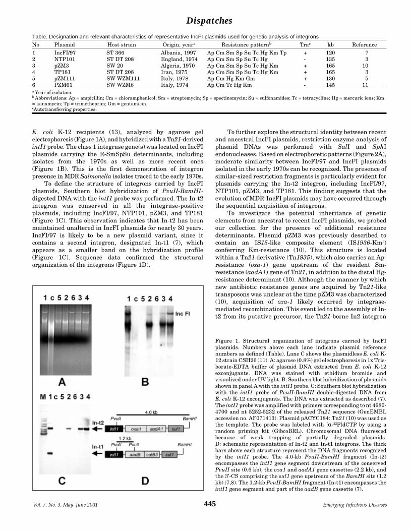
Dispatches
445Vol. 7, No. 3, May–June 2001 Emerging Infectious Diseases
E. coli K-12 recipients (13), analyzed by agarose gelelectrophoresis (Figure 1A), and hybridized with a Tn21-derivedintI1 probe. The class 1 integrase gene(s) was located on IncFIplasmids carrying the R-SmSpSu determinants, includingisolates from the 1970s as well as more recent ones(Figure 1B). This is the first demonstration of integronpresence in MDR Salmonella isolates traced to the early 1970s.
To define the structure of integrons carried by IncFIplasmids, Southern blot hybridization of PvuII-BamHI-digested DNA with the intI1 probe was performed. The In-t2integron was conserved in all the integrase-positiveplasmids, including IncFI/97, NTP101, pZM3, and TP181(Figure 1C). This observation indicates that In-t2 has beenmaintained unaltered in IncFI plasmids for nearly 30 years.IncFI/97 is likely to be a new plasmid variant, since itcontains a second integron, designated In-t1 (7), whichappears as a smaller band on the hybridization profile(Figure 1C). Sequence data confirmed the structuralorganization of the integrons (Figure 1D).
To further explore the structural identity between recentand ancestral IncFI plasmids, restriction enzyme analysis ofplasmid DNAs was performed with SalI and SphIendonucleases. Based on electrophoretic patterns (Figure 2A),moderate similarity between IncFI/97 and IncFI plasmidsisolated in the early 1970s can be recognized. The presence ofsimilar-sized restriction fragments is particularly evident forplasmids carrying the In-t2 integron, including IncFI/97,NTP101, pZM3, and TP181. This finding suggests that theevolution of MDR-IncFI plasmids may have occurred throughthe sequential acquisition of integrons.
To investigate the potential inheritance of geneticelements from ancestral to recent IncFI plasmids, we probedour collection for the presence of additional resistancedeterminants. Plasmid pZM3 was previously described tocontain an IS15-like composite element (IS1936-Kmr)conferring Km-resistance (10). This structure is locatedwithin a Tn21 derivative (Tn1935), which also carries an Ap-resistance (oxa-1) gene upstream of the resident Sm-resistance (aadA1) gene of Tn21, in addition to the distal Hg-resistance determinant (10). Although the manner by whichnew antibiotic resistance genes are acquired by Tn21-liketransposons was unclear at the time pZM3 was characterized(10), acquisition of oxa-1 likely occurred by integrase-mediated recombination. This event led to the assembly of In-t2 from its putative precursor, the Tn21-borne In2 integron
Table. Designation and relevant characteristics of representative IncFI plasmids used for genetic analysis of integrons
No. Plasmid Host strain Origin, yeara Resistance patternb Trac kb Reference1 IncFI/97 ST 366 Albania, 1997 Ap Cm Sm Sp Su Tc Hg Km Tp + 120 72 NTP101 ST DT 208 England, 1974 Ap Cm Sm Sp Su Tc Hg - 135 33 pZM3 SW 20 Algeria, 1970 Ap Cm Sm Sp Su Tc Hg Km + 165 104 TP181 ST DT 208 Iran, 1975 Ap Cm Sm Sp Su Tc Hg Km + 165 35 pZM111 SW WZM111 Italy, 1978 Ap Cm Hg Km Gm + 130 56 PZM61 SW WZM6 Italy, 1974 Ap Cm Tc Hg Km - 145 11a Year of isolation.b Abbreviations: Ap = ampicillin; Cm = chloramphenicol; Sm = streptomycin; Sp = spectinomycin; Su = sulfonamides; Tc = tetracycline; Hg = mercuric ions; Km= kanamycin; Tp = trimethoprim; Gm = gentamicin.cAutotransferring properties.
Figure 1. Structural organization of integrons carried by IncFIplasmids. Numbers above each lane indicate plasmid referencenumbers as defined (Table). Lane C shows the plasmidless E. coli K-12 strain CSH26 (11). A: agarose (0.8%) gel electrophoresis in 1x Tris-borate-EDTA buffer of plasmid DNA extracted from E. coli K-12exconjugants. DNA was stained with ethidium bromide andvisualized under UV light. B: Southern blot hybridization of plasmidsshown in panel A with the intI1 probe. C: Southern blot hybridizationwith the intI1 probe of PvuII-BamHI double-digested DNA fromE. coli K-12 exconjugants. The DNA was extracted as described (7).The intI1 probe was amplified with primers corresponding to nt 4680-4700 and nt 5252-5232 of the released Tn21 sequence (GenEMBLaccession no. AF071413). Plasmid pACYC184::Tn21 (10) was used asthe template. The probe was labeled with [α-32P]dCTP by using arandom priming kit (GibcoBRL). Chromosomal DNA fluorescedbecause of weak trapping of partially degraded plasmids.D: schematic representation of In-t2 and In-t1 integrons. The thickbars above each structure represent the DNA fragments recognizedby the intI1 probe. The 4.0-kb PvuII-BamHI fragment (In-t2)encompasses the intI1 gene segment downstream of the conservedPvuII site (0.6 kb), the oxa1 and aadA1 gene cassettes (2.2 kb), andthe 3’-CS comprising the sul1 gene upstream of the BamHI site (1.2kb) (7,8). The 1.2-kb PvuII-BamHI fragment (In-t1) encompasses theintI1 gene segment and part of the aadB gene cassette (7).

Dispatches
Emerging Infectious Diseases Vol. 7, No. 3, May–June 2001446
(9). Therefore, we tested IncFI plasmids for the presence of theIS1936-Kmr element, the transposase (tnp) machinery, andthe Hg-resistance (mer) locus. All but one of the IncFIplasmids carry the IS1936-Kmr element (Figure 2B). NTP101is the only exception, consistent with the Km-susceptiblephenotype conferred by this plasmid. Additional bands, someof which were shared by different IncFI plasmids, were alsodetected, likely as a result of cross-hybridization of the probewith IS15-like elements located on these replicons. PlasmidsIncFI/97, NTP101, PZM3, and TP181 were also positive forthe presence of the 4.4-kb SphI band (Figure 2C), which ispredicted to contain the tnpM (regulator), the tnpR(resolvase), and part of the tnpA (transposase) genes of Tn21-like transposons (GenEMBL accession no. AF071413).
Furthermore, the IncFI plasmids recognized by the merAprobe also showed a common SphI band of 2.1 kb, providingevidence for a structural conservation of the mer operon(Figure 2D). Plasmid pZM61 showed hybridization patternsindistinguishable from those of pZM111 (data not shown).
Taken together, the above data indicate that IncFI/97shares antibiotic resistance determinants with the ancestorplasmids pZM3, NTP101, and TP181, suggesting that MDRdeveloped through independent acquisition of integronswithin the structurally conserved IncFI plasmid scaffold. Theevolutionary story of these plasmids combines themaintenance of indigenous antibiotic resistance genes withthe acquisition of new determinants, leading to accumulationof multiple resistance mechanisms. For example, the oxa1gene cassette was acquired despite the presence of the TEM-type beta-lactamase encoded by the preexisting TnAtransposon (11). Likewise, acquisition of In-t1 introduced athird aminoglycoside-resistance determinant (aadB) inaddition to the resident IS1936-Kmr (aph) element (10) and
the In-t2-encoded aadA1 gene. Along the same lines,resistance to chloramphenicol was determined by both thecatB3 gene cassette of In-t1 and by the resident cmlA gene.Besides increasing the level of antibiotic resistance due to theexpression of multiple detoxification mechanisms, acquisi-tion of new resistance genes is also expected to enlarge thespectrum of resistance. Our results (not shown) indicate thatIn-t2 serves as a vehicle for the oxa-1 gene, extending beta-lactam resistance to ureidopenicillins, and of the aadA1 gene,conferring resistance to streptomycin and spectinomycin.Likewise, the In-t1-carried aadB gene broadens theaminoglycoside resistance to tobramycin and gentamicin.
ConclusionsOur retrospective investigation provides novel evidence
for the involvement of integrons in the development of MDRthrough sequential acquisition of new resistance determi-nants within IncFI plasmids. This family of replicons couldplay a role in the future development of resistance inS. Typhimurium and, more generally, in Enterobacteriaceae.A dramatic outcome of this evolutionary pathway may bepredicted from the intrinsic structural and functionalproperties of mobile DNA elements found on theseR-plasmids. Integrons provide a unique mechanism for therecruitment of additional resistance genes. Moreover,spreading resistance genes among different replicons isfavored by the location of integrons within transposableelements. Finally, horizontal transmission is expectedbecause of the conjugative or mobilizable properties of IncFIcarriers, which enables them to traverse species and genusboundaries. Future success in controlling resistance depends,at least in part, on a complete understanding of themechanisms involved in dissemination. A systematic search
Figure 2. Restriction enzyme analysis anddetection of common genetic elements inIncFI plasmids. Numbers above each laneindicate plasmid reference numbers asdefined (Table). M and M' are HindIII-digested lambda DNA and 1-kb plus DNAladder (GibcoBRL), respectively. A: agarose(0.8%) gel electrophoresis in 1x Tris-borate-EDTA buffer of plasmids digested withrestriction endonucleases SalI and SphI.DNA was stained with ethidium bromideand visualized under UV light. B: Southernblot hybridization of SalI-digested plas-mids with the IS1936-Kmr probe purified as1.8-kb SalI fragment from plasmidpACYC184::Tn1935 (10). C and D: South-ern blot hybridizations of SphI-digestedplasmids with the tnpM and merA probes,respectively. The tnpM probe was amplifiedwith primers corresponding to nt 3689-3709and nt 4070-4050 of the released Tn21sequence (GenEMBL accession no.AF071413). For synthesis of the merA probe,primers annealing to nt 17224-17249 andnt 17931-17907 of the Tn21 sequence wereused. Plasmid pACYC184::Tn21 (10) wasused as the template. Probes were labeledwith [α-32P]dCTP by using a randompriming kit (GibcoBRL, Rockville, MD).The arrows point to predicted hybridizationfragments.

Dispatches
447Vol. 7, No. 3, May–June 2001 Emerging Infectious Diseases
for integrons and transposable elements could provide auseful genetic basis for MDR in human and animalS. Typhimurium isolates.
AcknowledgmentsWe thank B. Colonna, M.A. Casalino, and M. Nicoletti for the
encouraging discussion and the generous gift of strains and plasmidsand F. Angulo, S. Fanning, and R. Helmuth for critical reading of themanuscript and helpful suggestions.
This investigation was supported by grants from the ItalianMinistry of Health to AC and PV.
Dr. Carattoli is a senior scientist at the Reference Center for Patho-genic Enterobacteria of Istituto Superiore di Sanità. Her research inter-est focuses on the genetic basis of multidrug resistance and pathogen-esis in gram-negative bacteria.
References 1. Glynn MK, Bopp C, Dewitt W, Dabney P, Mokhtar M, Angulo FJ.
Emergence of multidrug-resistant Salmonella enterica serotypeTyphimurium DT104 infections in the United States. N Engl J Med1998;338:1333-8.
2. Prager R, Liesegang A, Rabsch W, Gericke B, Thiel W, Voigt W, etal. Clonal relationship of Salmonella enterica serovar Typhimuri-um phage type DT104 in Germany and Austria. Zentralb Bakteriol1999;289:399-414.
3. Anderson ES, Threlfall EJ, Carr JM, McConnell MM, Smith HR.Clonal distribution of resistance plasmid-carrying Salmonellatyphimurium, mainly in the Middle East. J Hyg (Lond)1977;79:425-48.
4. Domart A, Robineau M, Stroh A, Dubertret LM, Modai J.Septicemia due to Salmonella wien. Diagnostic, therapeutic andepidemiologic problems. Ann Med Interne (Paris) 1974;125:915-8.
5. Casalino MA, Comanducci A, Nicoletti M, Maimone F. Stability ofplasmid content in Salmonella wien in late phases of the epidemichistory. Antimicrob Agents Chemother 1984;25:499-501.
6. Frost JA, Rowe B, Ward LR, Threlfall EJ. Characterization ofresistance plasmids and carried phages in an epidemic clone ofmulti-resistant Salmonella typhimurium in India. J Hyg (Lond)1982;88:193-204.
7. Tosini F, Visca P, Luzzi I, Dionisi AM, Pezzella C, Petrucca A,Carattoli A. Class 1 integron-borne multiple-antibiotic resistancecarried by IncFI and IncL/M plasmids in Salmonella entericaserotype Typhimurium. Antimicrob Agents Chemother1998;42:3053-8.
8. Hall RM, Collis CM. Mobile gene cassettes and integrons: captureand spread of genes by site-specific recombination. Mol Microbiol1995;15:593-600.
9. Liebert CA, Hall RM, Summers AO. Transposon Tn21, a flagship ofthe floating genome. Microbiol Mol Biol Rev 1999;63:507-22.
10. Colonna B, Bernardini M, Micheli G, Maimone F, Nicoletti M,Casalino M. The Salmonella wien virulence plasmid pZM3 carriesTn1935, a multiresistance transposon containing a compositeIS1936-kanamycin resistance element. Plasmid 1988;20:221-31.
11. Maimone F, Colonna B, Bazzicalupo P, Oliva B, Nicoletti M,Casalino MA. Plasmids and transposable elements in Salmonellawien. J Bacteriol 1979;139:369-75.
12. National Committee for Clinical Laboratory Standards. Perfor-mance standards for antimicrobial disc susceptibility tests. M2A2.Villanova (PA): The Committee; 1992.
13. Kado CI, Liu S. Rapid procedure for detection and isolation of largeand small plasmids. J Bacteriol 1981;145:1365-73.

Dispatches
Emerging Infectious Diseases Vol. 7, No. 3, May–June 2001448
Salmonella enterica serotype Typhi is endemic indeveloping countries in Africa, South and Central America,and the Indian subcontinent, with an estimated incidence of33 million cases each year (1). By contrast, in developedcountries such as the United Kingdom or the USA, incidenceis much lower, and most cases are in travelers returning fromendemic areas. For example, 150 to 300 cases occur each yearin the U.K., at least 70% in patients with a history of recentforeign travel.
For patients with typhoid fever, administration of aneffective antibiotic should begin as soon as clinical diagnosisis made, without recourse to results of antimicrobialsensitivity tests. From 1948 to the mid-1970s, chlorampheni-col was the first-line drug of choice, and in developed countriesits use resulted in a reduction in mortality rates from 10% to<2%. After extensive outbreaks of typhoid fever occurred inMexico and India in the early and mid-1970s, in whichepidemic strains were resistant to chloramphenicol (2,3), theefficacy of this antimicrobial agent was in doubt.
Alternative drugs for typhoid fever are ampicillin andtrimethoprim. However, following outbreaks in the Indiansubcontinent, the Arabian Gulf, the Philippines, and SouthAfrica in the late 1980s and early 1990s, in which causativestrains were resistant to ampicillin and trimethoprim inaddition to chloramphenicol, the efficacy of these antimicrobi-al agents has also been impaired (4).
The Laboratory of Enteric Pathogens of the Public HealthLaboratory Service of England and Wales is the referencecenter in the U.K. for strains of S. Typhi. Strains areidentified by Vi-phage typing; all strains are tested with anagar dilution method for resistance to a panel of antimicrobialdrugs. The final plate concentrations for selected antimicrobi-al drugs were chloramphenicol 8 mg/L, ampicillin 8 mg/L,trimethoprim 2 mg/L, nalidixic acid 16 mg/L, ciprofloxacin0.125 mg/L, ceftriaxone 1 mg/L, and cefotaxime 1 mg/L.
For isolates resistant to ciprofloxacin at 0.125 mg/L, fullMICs are determined either by incorporating doubled
concentrations of the antimicrobial agent into the agarsubstrate or by E-test. All isolates resistant to ciprofloxacin at0.125 mg/L were also resistant to nalidixic acid at 16 mg/L. Incontrast, isolates sensitive to nalidixic acid at 16 mg/L hadMICs to ciprofloxacin of <0.025 mg/L. All strains withresistance to chloramphenicol, ampicillin, trimethoprim, ornalidixic acid/ciprofloxacin were tested for the ability totransfer these resistances to a drug-sensitive strain ofEscherichia coli K12. Resultant resistance plasmids werecharacterized by incompatibility grouping and agarose gelelectrophoresis after extraction of plasmid DNA from donorstrains of S. Typhi and recipient strains of E. coli K12.
From 1978 to 1985, resistance to chloramphenicol wasidentified in 11 (0.47%) of 2,356 strains studied (5); therefore,chloramphenicol remained the first-line drug for typhoidfever before results of laboratory sensitivity tests becameavailable. From 1986 to 1989, chloramphenicol resistanceincreased threefold: 12 (1.5%) of 790 isolates were resistant.However, this increase was not considered sufficient tochange recommendations about therapy. In 1990, there was adramatic change, with 20% of 248 isolates resistant tochloramphenicol; most were also resistant to ampicillin andtrimethoprim (6). In 1991, because of this increasedchloramphenicol resistance, ciprofloxacin was recommendedas an alternative for patients with a history of recent travel toepidemic areas (7).
From 1990 to 1999, 151 to 291 (mean 210) patients peryear in the U.K. had typhoid fever. The incidence of multidrugresistance (MDR) to chloramphenicol, ampicillin, andtrimethoprim increased from 21% in 1991 to 36% in 1994,declined to 13% in 1997, and then increased to 26% in 1999(Table 1). More than 90% of patients infected with MDRstrains had recently returned from the Indian subcontinent,particularly Pakistan and India. The reasons for the declinein MDR strains in 1997 followed by the return to 1996 levelsin 1998 and 1999 are not known but may be related to changesin climatic conditions in the Indian subcontinent in the mid-1990s, followed by reestablishment of MDR strains indifferent parts of India in the late 1990s. Epidemiologicinvestigations to test these hypotheses are in progress.
In the early 1990s, the most common Vi-phage type inMDR strains was phage type M1, and almost all patients
Decreased Susceptibility to Ciprofloxacinin Salmonella enterica serotype Typhi,
United KingdomE. John Threlfall and Linda R. Ward
Central Public Health Laboratory, London, United Kingdom
Address for correspondence: E. John Threlfall, Laboratory of EntericPathogens, Central Public Health Laboratory, 61 Colindale Avenue,London NW9 5HT, U.K.; fax: + 44 0208 905 9929; e-mail:[email protected]
In 1999, 23% of Salmonella enterica serotype Typhi isolates from patients inthe United Kingdom exhibited decreased susceptibility to ciprofloxacin (MIC0.25-1.0 mg/L); more than half were also resistant to chloramphenicol,ampicillin, and trimethoprim. Increasing numbers of treatment failures havebeen noted. Most infections have been in patients with a recent history of travelto India and Pakistan.

Dispatches
449Vol. 7, No. 3, May–June 2001 Emerging Infectious Diseases
infected with strains of this phage type had acquired theinfections in Pakistan. The last isolations of MDR phage typeM1 in the U.K. were in 1994 (4). Since 1993, the most commonMDR phage type has been E1. Most patients infected withMDR strains of phage type E1 had acquired the infections inIndia or Pakistan. However, infections were also recorded inpatients returning from Bangladesh, Sri Lanka, andAfghanistan. Regardless of phage type, in all MDR strainsresistance to chloramphenicol, ampicillin, and trimethoprimhas been encoded by plasmids of approximately 100megadaltons belonging to the H1 incompatibility group.
In 1991, a strain of S. Typhi with plasmid-encodedresistance to chloramphenicol, ampicillin, and trimethoprimand with chromosomally encoded resistance to nalidixic acid(MIC 512 mg/L) was isolated from a 1-year-old child who hadrecently returned from India. The strain also showed amarked decrease in sensitivity to ciprofloxacin (MIC 0.6 mg/L).The patient did not respond to treatment with ciprofloxacindespite serum levels of 1.5 mg/L. In 1995, 8 (3%) of 291isolates showed decreased sensitivity to ciprofloxacin (MICs0.38–0.75 mg/L by E-test); 5 were also resistant tochloramphenicol, ampicillin, and trimethoprim. In 1998, 32(21%) of 151 strains exhibited decreased susceptibility tociprofloxacin. One patient, a 65-year-old woman who returnedfrom India infected with a strain of phage type E1 (MIC tociprofloxacin of 1.0 mg/L) did not respond to twice a day
treatment with ciprofloxacin, 400 mg intravenously. After 5days, treatment was changed to amoxicillin and ceftriaxone.Within 3 days, the patient’s condition improved, and after afurther 5 days she was apyrexial (8).
S. Typhi with decreased susceptibility to ciprofloxacinincreased to 23% in the U.K. in 1999 (Table 1). All strains withdecreased sensitivity to ciprofloxacin were also resistant tonalidixic acid (MIC 512 mg/L). The predominant phage typeshave been E1 (81% of cases) and E9 (4%). However, strains ofphage types C2, E7, M1, untypeable Vi (UVS), and Vi-negative have also been identified. Most patients had recentlyreturned from India or Pakistan. However, in 1998 and 1999,strains with decreased susceptibility to ciprofloxacin werealso isolated from travelers returning from Sri Lanka, Nepal,Bangladesh, and Thailand (Table 2). Furthermore, in bothyears >50% of isolates with decreased susceptibility tociprofloxacin were also MDR (Table 1). In 1999, at least 10patients infected with strains with decreased susceptibility tociprofloxacin did not respond to treatment with fluoroquinoloneantimicrobials. In such cases, ceftriaxone was the mostfrequently used alternative. In contrast to resistance tochloramphenicol, ampicillin, and trimethoprim, resistance tociprofloxacin has been chromosomally encoded in all isolateswith decreased sensitivity to this antimicrobial agent.
Since 1993, strains of S. Typhi with decreasedsusceptibility to ciprofloxacin have been isolated withincreasing frequency in Vietnam (9). In 1997, >6,000 casesoccurred in an extensive epidemic in Tajikistan of nalidixicacid-resistant S. Typhi with decreased susceptibility tociprofloxacin (10). The epidemic strain was untypeable withthe Vi typing phages but had a pulsed-field profileindistinguishable from that of isolates of MDR Vi-phage typeE1 from patients infected in India (11). In both Vietnam andTajikistan, treatment failures with fluoroquinolone antibiot-ics have been noted.
The accepted British Society for Antimicrobial Chemo-therapy and National Committee for Clinical LaboratoryStandards’ zone size equivalents for resistance to ciprofloxa-cin in disc diffusion tests are 2 mg/L and 4 mg/L, respectively,for Enterobacteriaceae. However, testing for resistance atthese levels could result in decreased susceptibilities notbeing detected. As all strains with decreased susceptibility tociprofloxacin have also been resistant to nalidixic acid, wesuggest that the latter antimicrobial agent be included in thepanel of drugs used for sensitivity testing. If resistance tonalidixic acid is detected, full MICs to ciprofloxacin should beperformed in the event of treatment failure.
Table 2. Phage types in Salmonella Typhi isolates with decreased susceptibility to ciprofloxacin (CpL), United Kingdom, 1991–1999
Phage typesYear CpL E1 M1 E9 Others Country of origin (no.)1991 2 2 0 0 0 India (1), Nepal (1)1992 1 0 0 0 1 (B2) India (1)1993 1 0 0 0 1 (E14) Bangladesh (1)1994 5 5 0 0 0 India (3), Nepal (1), Bangladesh (1)1995 8 6 1 0 1 (D1) India (4), Pakistan (3), NSa (1)1996 11 11 0 0 0 India (9), Pakistan (2)1997 9 8 0 0 1(A) India (5), Pakistan (2), Nepal (1), NS (1)1998 32 23 0 6 3 (G2,2;O,1) Pakistan (14), India (9), Sri Lanka (1), Bangladesh (1), NS (7)1999 42 27 1 3 11 (UVS, 7; E7, 1; C2, 1; India (31), Pakistan (6), Bangladesh (1), Thailand (1), NS (3)
Vi-negative, 1)aNS = details of travel itinerary not provided; UVS = untypeable with the Vi typing phages.
Table 1. Incidence of multidrug resistance and decreased susceptibilityto ciprofloxacin in Salmonella enterica serotype Typhi, U.K., 1990–1999
Decreased suscepti- CPL strains
MDRb bility to CPL also resistant to:Year Strainsa no. (%) no. (%) C A Tm Ct Cf1990 248 50 (20) 0 (0) 0 0 0 0 01991 226 48 (21) 2 (0.9) 1 1 1 0 01992 204 49 (24) 1 (0.5) 0 0 0 0 01993 194 49 (25) 1 (0.5) 1 1 1 0 01994 259 94 (36) 5 (2) 5 5 5 0 01995 291 100 (34) 8 (3) 5 5 5 0 01996 210 52 (25) 11 (7) 7 7 7 0 01997 174 22 (13) 9 (5) 6 6 6 0 01998 151 34 (23) 32 (21) 19 19 19 0 01999 179 47 (26) 42 (23) 25 25 25 0 0aStrains referred to Laboratory of Enteric Pathogens.bMDR = multidrug resistant (to chloramphenicol, ampicillin, and trimethoprim).Resistance symbols: C, chloramphenicol; A, ampicillin; Tm, trimethoprim;CPL, ciprofloxacin (MIC 0.25-1.0 mg/L); Ct, ceftriaxone; Cf, cefotaxime.Percentages of total isolates in parentheses.

Dispatches
Emerging Infectious Diseases Vol. 7, No. 3, May–June 2001450
Our findings suggest that strains of S. Typhi withdecreased sensitivity to ciprofloxacin are now endemic inseveral countries in the Indian subcontinent and that suchstrains are increasing in travelers returning to the U.K.Despite the low level of resistance, treatment failures arebeing increasingly noted. In such cases, possible alternativessuch as ceftriaxone or cefotaxime could be considered. In thisrespect, it is reassuring that all strains of S. Typhi so fartested were sensitive to these antimicrobial drugs.
Dr. Threlfall is head of the Antibiotic Resistance and MolecularEpidemiology Laboratory at the Central Public Health Laboratory ofthe Public Health Laboratory Service of England and Wales.
Mrs. Ward is head of the Salmonella Reference Unit in the PublicHealth Laboratory Service of England and Wales.
References 1. Ivanoff B. Typhoid fever: global situation and WHO recommendations.
Southeast Asian J Trop Med Public Health 1995; 26(Suppl 2):1-6. 2. Anderson ES, Smith HR. Chloramphenicol resistance in the
typhoid bacillus. BMJ 1972;3:329-31.
3. Paniker CKJ, Vilma KN. Transferable chloramphenicol resistancein Salmonella typhi. Nature 1972;239:109-10.
4. Rowe B, Ward LR, Threlfall EJ. Multidrug-resistant Salmonella typhi:a worldwide epidemic. Clin Infect Dis 1997;24(Suppl 1): S106-9.
5. Rowe B, Threlfall EJ, Ward LR. Does chloramphenicol remain thedrug of choice for typhoid? Epidemiol Infect 1987;98:379-83.
6. Rowe B, Ward LR, Threlfall EJ. Spread of multiresistantSalmonella typhi. Lancet 1990;336:1065-6.
7. Rowe B, Ward LR, Threlfall EJ. Treatment of multiresistanttyphoid fever. Lancet 1991;337:1422.
8. Threlfall EJ, Ward LR, Skinner JA, Smith HR, Lacey S.Ciprofloxacin-resistant Salmonella typhi and treatment failure.Lancet 1999;353:1590-1.
9. Parry C, Wain J, Chinh NT, Vinh Ha, Farrar JJ. Quinolone-resistant Salmonella typhi in Vietnam. Lancet 1998;351:1289.
10. Murdoch DA, Banatvala NA, Bone A, Shoismatulloev BI, Ward LR,Threlfall EJ, et al. Epidemic ciprofloxacin-resistant Salmonellatyphi in Tajikistan. Lancet 1998;351:339.
11. Hampton MD, Ward LR, Rowe B, Threlfall EJ. Molecularfingerprinting of multidrug-resistant Salmonella enterica serotypeTyphi. Emerg Infect Dis 1998;4:317-20.

Dispatches
451Vol. 7, No. 3, May–June 2001 Emerging Infectious Diseases
The arbovirus California encephalitis virus was firstisolated in 1943 from mosquitoes collected in Kern County,California (1). Two years later, three human cases ofencephalitis were attributed to this new virus (2); all threecases were in residents of Kern County in the Central Valleyof California. The best-documented case occurred in a2-month-old Hispanic boy who had encephalitis and resultantdevelopmental delay. There was strong laboratory evidenceconfirming infection from the presence of neutralizingantibodies to California encephalitis, but not to St. Louisencephalitis virus or western equine encephalomyelitis virus.Serum from a 7-year-old boy hospitalized with encephalitisalso had neutralizing antibodies to California encephalitis.Serologic tests were inconclusive in a third possible case in a22-year-old agricultural worker with mild encephalitis;neutralizing antibodies against both California encephalitisand St. Louis encephalitis were detected.
Since the original virus was isolated, other viruses havebeen isolated that are closely related to Californiaencephalitis. This group of related viruses is now classified asthe California serogroup, one of 16 serogroups within thegenus Bunyavirus, family Bunyaviridae. Several otherhuman pathogens (e.g., Jamestown Canyon virus, La Crossevirus, and Tahyna virus) also belong to the Californiaserogroup. Little human disease was associated with theseviruses until 1960, but now California serogroup virusinfections are the most commonly reported cause of arboviralencephalitis in the United States. From 1996 to 1998,approximately three times as many reported human cases ofarboviral encephalitis were caused by California serogroupviruses as were reported for western equine encephalomyeli-tis virus, St. Louis encephalitis, and eastern equineencephalomyelitis viruses combined (3). However, since thethree original cases from California, no further cases ofhuman disease caused by the prototype Californiaencephalitis had been reported (4). Campbell et al.summarized results of surveys for human antibodies toCalifornia serogroup viruses in California (5).
Case ReportIn June 1996, a 65-year-old man who lived in Marin
County, California, became ill with blurred vision anddizziness. Eight days after the onset of symptoms, he visitedhis primary physician. A physical examination wasremarkable only for nystagmus. Laboratory studies includedleukocytes 8.2x10³, hematocrit 45.8%, and a chemistry panelthat was normal except for phosphorus 4.5 mg/dL (normalrange 2.7-4.4 mg/dL), cholesterol 265 mg/dL (normal range125-200 mg/dL), and high-density lipoprotein cholesterol 67mg/dL (normal range 35-60 mg/dL). A magnetic resonanceimage and arteriogram were normal. One month after theinitial visit, the patient no longer complained of blurred visionor vertigo, and nystagmus had disappeared. Two years afterthe episode, he had no neurologic sequelae.
The patient lived in a suburban area of Marin County,approximately 1 km from a large brackish marsh borderingSan Francisco Bay. He had traveled outside the United Statesthe previous February, when he had visited Egypt and severalCaribbean islands. He had not traveled outside Californiabetween this time and the onset of his illness in June, 4months later.
An acute-phase serum specimen was sent to the Viral andRickettsial Disease Laboratory, California Department ofHealth Services. Indirect immunofluorescence antibody testswere negative for St. Louis encephalitis and Western equineencephalomyelitis virus, as were serum samples taken 8 and16 days after onset of illness. However, when this series ofsamples was tested by neutralization test 2 years later at theArbovirus Research Unit of the University of CaliforniaCenter for Vector-Borne Disease Research as part of aretrospective study of arboviruses, the 8-day sample showedan antibody titer of 1:80 and the 16-day sample an antibodytiter < 1:320 to California encephalitis (Table).
California encephalitis-related arboviruses included inthe tests were snowshoe hare, La Crosse, Jamestown Canyon,Morro Bay, and Tahyna. Tahyna, an important Californiaserogroup virus widely distributed in Europe and Asia, wasincluded because of the patient’s travel history. Northway,Main Drain, and Lokern viruses, members of theBunyamwera serogroup occurring in California, were alsoincluded, as were western equine encephalomyelitis virus andSt. Louis encephalitis.
The First Reported Case of CaliforniaEncephalitis in More Than 50 Years
Bruce F. Eldridge,* Carol Glaser,† Robert E. Pedrin,‡ and Robert E. Chiles**University of California, Davis, California, USA; †California Department of Health Services,
Berkeley, California, USA; and ‡Family Practice, Greenbrae, California, USA
Address for correspondence: Bruce F. Eldridge, Department ofEntomology, University of California, Davis, CA 95616, USA; fax: 530-752-1537; e-mail: [email protected]
A recent case of California encephalitis, a rare mosquito-borne viral disease,represents only the fourth ever reported and the first since the initial three casesin 1945. This case was diagnosed retrospectively on the basis of a rise inantibody titer between acute- and convalescent-phase serum samples.

Dispatches
Emerging Infectious Diseases Vol. 7, No. 3, May–June 2001452
The sera were tested by plaque reduction-serum dilutionneutralization with African green monkey kidney (Vero) cellcultures, according to the protocol of Campbell et al. (6). Anincrease in titer between the acute- and convalescent-phasesamples was found only for California encephalitis andsnowshoe hare virus. The titers for the convalescent-phaseserum were 1:320 for California encephalitis and 1:180 for theclosely related snowshoe hare virus (Table), which has neverbeen reported in California. The patient had not traveledrecently to alpine areas of the state, where snowshoe harevirus might be expected to occur.
ConclusionsBecause the patient’s travel occurred several months
before his illness, exposure to mosquitoes near his home is themost likely route of infection. The most common mosquitospecies in salt marshes in Marin County are Aedes washinoiand Ae. squamiger (7). Bloodsucking adult females of boththese species are usually present in late winter to earlyspring. Ae. dorsalis also occurs in this area, but later in theyear. Culiseta inornata occurs frequently near salt marshes.California encephalitis has not been recovered from any ofthese species in California; most isolates have come from Ae.melanimon in the Central Valley (8).
Campbell et al. (5) found that most seropositive samplesfrom humans, mostly from high elevations (>1,000 m) inCalifornia, were apparently infected with Jamestown Canyonvirus. However, these investigators also reported thatapproximately 35% of samples from horses in low elevations(<1,000 m) in California showed evidence of prior infectionwith California encephalitis (9), but they did not test samplesfrom the San Francisco Bay area. Fulhorst et al. (10) reported
that 57% of the horses sampled in Marin County showedevidence of prior infection with Jamestown Canyon virus, butnone with California encephalitis.
Awareness of arboviruses in the United States has beenheightened as a result of the recent outbreak of humanillnesses caused by West Nile virus. That outbreak wasoriginally thought to be due to St. Louis encephalitis (11). Thecase we describe is a further reminder that clinicians shouldconsider several causative agents when a patient has acentral nervous system infection, especially if mosquitoexposure has occurred.
Further studies are needed to assess the risk for humaninfection by California encephalitis in coastal California andthe role of various mosquito species in transmission.
AcknowledgmentWe thank William C. Reeves for his careful review of the
manuscript.
Dr. Eldridge is Emeritus Professor of Entomology at the Univer-sity of California, Davis, and continues to serve as director of the UCMosquito Research Program. His area of specialization is the ecology ofmosquitoes and mosquito-borne viral diseases of humans.
References 1. Hammon WM, Reeves WC, Sather G. California encephalitis virus,
a newly described agent. II. Isolations and attempts to identify andcharacterize the agent. J Immunol 1952;69:493-510.
2. Hammon WM, Reeves WC. California encephalitis virus, a newlydescribed agent. I. Evidence of infection in man and other animals.California Medicine 1952;77:303-9.
3. Centers for Disease Control and Prevention. Summary of notifiablediseases, United States, 1998. MMWR Morb Mortal Wkly Rep1998;47:22-5.
4. McJunkin JE, Khan RR, Tsai T. California-La Crosse encephalitis.Infect Dis Clin North Am 1998;47:83-93.
5. Campbell GL, Reeves WC, Hardy JL, Eldridge BF. Seroepidemiol-ogy of California and Bunyamwera serogroup viruses in humans inCalifornia. Am J Epidemiol 1992;136:308-19.
6. Campbell GL, Eldridge BF, Hardy JL, Reeves WC, Jessup DA, PresserSB. Prevalence of neutralizing antibodies against California andBunyamwera serogroup viruses in deer from mountainous areas ofCalifornia. Am J Trop Med Hyg 1989;40:428-37.
7. Lanzaro GC, Eldridge BF. A classical and population geneticdescription of two new sibling species of Aedes (Ochlerotatus)increpitus Dyar. Mosquito Systematics 1992;24:85-101.
8. Reeves WC, editor. Epidemiology and control of mosquito-bornearboviruses in California, 1943-1987. Sacramento: Calif MosqVector Control Assoc; 1990. p. 508.
9. Campbell GL, Reeves WC, Hardy JL, Eldridge BF. Distribution ofneutralizing antibodies to California and Bunyamwera serogroupviruses in horses and rodents in California. Am J Trop Med Hyg1990;42:282-90.
10. Fulhorst CF. The epidemiology and ecology of mosquito-borneviruses in coastal areas of California [dissertation]. Berkeley (CA):University of California, Berkeley; 1994.
11. Centers for Disease Control and Prevention. Outbreak of WestNile-like viral encephalitis—New York, 1999. MMWR MorbMortal Wkly Rep 1999;48:845-9.
Table. Neutralizing antibody titers to 10 arboviruses in paired serumsamples of a patient with neurologic symptoms, California, 1996
Acute-phase Convalescent-Virus (strain) seruma phase serumb
California encephalitis 1:80 1:320 (BFS-283)Snowshoe hare (original) 1:80 1:160La Crosse (prototype) 1:40 1:40Jamestown Canyon 1:20 1:20 (BFS 4474)Morro Bay (DAV 457) 1:80 1:40Tahyna (Bardos 92) 1:40 1:20Northway (BFN 2654) 1:20 <1:20Main Drain (BFS 5015) <1:20 <1:20Lokern (BFS 5183) <1:20 <1:20Western equine encephalo- <1:20 <1:20 myelitis (BFS 1703)St. Louis encephalitis <1:20 <1:20 (BFS 1750)aSample taken 8 days after onset of symptoms.bSample taken 16 days after onset of symptoms.

Dispatches
453Vol. 7, No. 3, May–June 2001 Emerging Infectious Diseases
Cyclospora, a protozoan pathogen that causes a syndromeof diarrhea and fatigue, was responsible for two large-scaleoutbreaks in North America in 1996 and 1997 (1,2). Theseoutbreaks, along with studies in Cyclospora-endemic areas(Nepal and Peru) and treatment of travelers returning fromthese areas, have increased our understanding of the clinicalillness associated with this pathogen. Chronic diarrhea is notuncommon, especially in patients whose condition isuntreated or partially treated. Recently, Guillain-Barrésyndrome has been reported as an extraintestinalcomplication of Cyclospora infection (3). We describe Reitersyndrome diagnosed in a patient after prolonged gastrointes-tinal illness associated with Cyclospora infection.
The StudyA 31-year-old man had onset of gastrointestinal illness on
May 11, 1997, 7 days after attending a dinner at a countryclub. His symptoms (extreme fatigue, dizziness, fever of101°F, intermittent vomiting and diarrhea) were similar tothose of the 10 other guests at this party and caused him tomiss work for 2 weeks. He visited his internist on May 22,1997, and was hospitalized with predominant symptoms offatigue and dehydration; he also reported constipation. Fluidsand gentamicin were administered intravenously. Stoolcultures and microscopy were negative; however, Cyclosporawas not searched for specifically. After the patient wasdischarged from the hospital, diarrhea resumed, and a stoolspecimen was examined by our laboratory by a concentrationtechnique and modified acid-fast staining. This examinationidentified Cyclospora oocysts. Standard therapy, trimethop-rim/sulfamethoxazole, could not be administered because ofthe patient’s allergy to sulfa, and as no effective alternativeantibiotic therapy for Cyclospora infections exists, antibiotictreatment was not offered. Symptoms of epigastric pain anddiscomfort, bloating, and alternating diarrhea and constipa-tion predominated, along with intermittent anorexia. An8-pound weight loss (5% of body weight) was noted. By mid-June 1997, the patient’s symptoms had abated slightly butwere not completely resolved. He was enrolled at our institutionin an open-label trial of albendazole, 400 mg 2 times a day for 14
days, and reported no change in his symptoms during thistime. Cyclospora was present in stool after treatment withalbendazole. Soft stool, intermittent diarrhea, and abdominalcramping persisted, along with proctalgia. Results of bloodtests, including a complete blood count, biochemical profile,and liver function tests, were normal.
The patient’s medical history included mild asthma withflare-ups approximately monthly, for which he had usedtheophylline. A diagnosis of prostatitis was made on clinicalgrounds in December 1996, 6 months before the onset ofdiarrheal illness and 10 months before the diagnosis of Reitersyndrome. These symptoms resolved with antibiotic therapyand did not recur.
When the patient first came to our institution on July 29,1997, he was not in acute distress. His abdomen was soft andtender only to deep palpation in the left and right lowerquadrants. There were no masses or enlarged organs. Stoolspecimens were negative for occult blood. As part of anongoing study of small-bowel histopathologic changesassociated with Cyclospora infections, the patient agreed toendoscopic evaluation. Upper gastrointestinal endoscopy wasperformed on August 15, 1997, with examination to thedescending duodenum. This examination revealed inflamma-tion of the distal esophagus and mild erythema of the gastriccardia and antrum, as well as erythema of the duodenal bulband descending duodenum. Flexible fiber-optic sigmoidoscopyto the mid-descending colon was unremarkable. NoCyclospora oocysts were identified on either duodenal or stoolaspirates. Biopsies of the duodenum revealed partial villousatrophy and moderate crypt hyperplasia with increasedintraepithelial lymphocytes and rare intraepithelial neutro-phils. Biopsies of the gastric antrum and cardia were normal.A rapid urease test performed on the gastric biopsy wasnegative for Helicobacter pylori. Electron microscopyevaluation of the small bowel biopsies revealed acute andchronic inflammation evidenced by focally intense epithelialinjury, including vacuolization, lipid accumulation, andabundant interstitial and epithelial reactive elements.Sigmoid and rectal biopsies revealed no histopathologicchanges.
Gastrointestinal symptoms persisted, including abdomi-nal bloating and cramping and intermittent soft stool, alongwith fatigue. In September 1997, the patient noted pain and
Reiter Syndrome Following ProtractedSymptoms of Cyclospora InfectionBradley A. Connor, Erik Johnson, and Rosemary Soave
New York-Presbyterian Hospital and Weill Medical College ofCornell University, New York, New York, USA
Address for correspondence: Bradley A. Connor, 50 East 69th Street,New York, NY 10021; fax: 212-734-4200; e-mail: [email protected]
Two large outbreaks of diarrheal illness associated with Cyclosporacayetanensis, a coccidian parasite, provided an opportunity to evaluate clinicalsyndromes associated with this enteric pathogen. Reiter syndrome, a triad ofocular inflammation, inflammatory oligoarthritis, and sterile urethritis, has beenassociated with enteric infections. We describe the first case of Reiter syndromefollowing protracted symptoms of Cyclospora infection.
Dispatches
Emerging Infectious Diseases Vol. 7, No. 3, May–June 2001454
soreness in knees, ankles, right first toe, and plantar heels.During early October 1997, he noted a constellation ofsymptoms, including left eye pain, dysuria, arthralgias, and apainful ulcer of the right buccal mucosa. An ophthalmologistconfirmed that the left eye pain resulted from iritis andepiscleritis. A rheumatologist suspected the diagnosis ofReiter syndrome on the basis of the constellation of findingsand negative serologic tests for Lyme disease and Chlamydia.A urinalysis was negative for leukocytes and bacteria,although no attempts were made to recover Chlamydia fromthe urine. Stool microscopy was negative for parasites, and astool culture was negative for bacterial pathogens. Thepatient was noted to be negative for HLA-B27. Oralcorticosteroids were begun for the iritis symptoms, and he wastreated with doxycycline (100 mg twice a day) for itsantiinflammatory effects. The arthritis symptoms improvedpromptly and dramatically. Four days after beginningdoxycycline therapy, the patient complained of progressivelysevere odynophagia, and a clinical diagnosis of pill-inducedesophagitis or ulcer was made. The symptoms resolved with acombination of sucralfate suspension and omeprazole, andthe patient has remained clinically well.
ConclusionsReiter syndrome, characterized by the triad of ocular
inflammation (conjunctivitis, iritis, episcleritis), inflammato-ry oligoarthritis, and sterile urethritis, usually occurs severalweeks after a triggering infection (4). Infections associatedwith Reiter syndrome include genitourinary Chlamydia orenteric infections with Salmonella, Shigella, Yersinia,Campylobacter, Clostridium difficile, and Cryptosporidium,although the incidence of Reiter syndrome with each of theseinfections is suspected to be low. This is the first reported caseof Reiter syndrome following Cyclospora infection. AlthoughReiter syndrome in this patient could have been coincidental,we propose Cyclospora as another infectious trigger for Reitersyndrome.
This patient’s history is unique in several respects.Because of sulfa allergy, he was inadequately treated for theCyclospora infection. An experimental trial of albendazole,which is used to treat other parasitic infections, did notshorten his illness. Chronic gastrointestinal symptoms
persisted for 12 weeks before endoscopy, when variable crypthyperplasia and villous atrophy were noted—characteristicfindings in small bowel biopsies of patients with Cyclosporainfection (5). No Cyclospora oocysts were noted on stoolexamination at the time of endoscopy. The pathogenesis ofReiter syndrome may involve molecular mimicry betweenmicrobial fragments in synovial fluid and the HLA-B27molecule; however, it is unknown whether microbialfragments of Cyclospora could be found in synovial fluid, andin this case the patient was HLA-B27 negative (6).Inflammatory lesions in the small intestine may allowantigen priming of receptive T lymphocytes and trigger aninflammatory response in affected organs. Depending on thelength of this process, it can be hypothesized that prompt,effective treatment of Cyclospora infections may minimize therisk for postinfectious complications such as Reiter syndrome.
Dr. Connor is clinical associate professor of medicine at Weill Medi-cal College of Cornell University, associate attending physician at NewYork-Presbyterian Hospital, and adjunct faculty member at RockefellerUniversity. He was part of the Kathmandu, Nepal, team that first de-scribed the illness associated with Cyclospora and has done research onthe pathogenesis, clinical illness, epidemiology, and treatment ofCyclospora infections.
References 1. Herwaldt BL, Ackers ML, and the Cyclospora Working Group. An
outbreak in 1996 of Cyclosporiasis associated with importedraspberries. N Engl J Med 1997;336:1548-56.
2. Herwaldt BL, Beach MJ, and the Cyclospora Working Group. Thereturn of Cyclospora in 1997: Another outbreak of cyclosporiasis inNorth America associated with imported raspberries. Ann InternMed 1999;130:210-20.
3. Richardson RF, Remler BF, Katirji B, Murad MH. Guillain-Barrésyndrome after Cyclospora infection. Muscle Nerve 1998;21:669-71.
4. Konttinen YT, Nordstrom DC, Bergroth V, Leirisalo-Repo M,Santavirta S. Occurrence of different ensuing triggering infectionspreceding reactive arthritis: a follow-up study. BMJ 1988;296:1644-5.
5. Connor BA, Shlim DR, Scholes JB, Rayburn JL, Reidy J, Rajah R.Pathologic changes in the small bowel in nine patients withdiarrhea associated with a coccidia-like body. Ann Intern Med1993;119:377-82.
6. Hermann E, Yu DT, Meyer zum Buschenfelde KH, Fleischer B.HLA-B27 restricted CD8 T-cells derived from synovial fluids ofpatients with reactive arthritis and ankylosing spondylitis. Lancet1993;342:646-50.

Dispatches
455Vol. 7, No. 3, May–June 2001 Emerging Infectious Diseases
Bacterial resistance to antimicrobial agents is a seriousproblem worldwide. Of particular concern is the increasingfrequency of multidrug resistance within Salmonella strainsisolated from zoonotic foodborne infections (1,2). This aspecthas been extensively investigated in Salmonella entericaserovar Typhimurium in relation to the worldwide spread ofmultidrug-resistant (MDR) strains of definitive phage type(DT) 104, with chromosomally integrated genes coding forresistance to ampicillin, chloramphenicol, streptomycin,sulfonamides, and tetracycline (3,4).
Recently, a basic role in dissemination and evolution ofantimicrobial resistance in MDR S. Typhimurium DT104(MDR-DT104) and many other organisms has been attributedto integrons, gene expression elements that potentiallyaccount for rapid and efficient transmission of drug resistancebecause of their mobility and ability to collect resistance genecassettes (5,6). These elements have been described in a widerange of pathogenic organisms (7), including S. Typhimuriumand S. Enteritidis (8,9); reports of these integrons in otherSalmonella serotypes are anecdotal (10).
Although S. Enteritidis and S. Typhimurium account forapproximately 45% and 25%, respectively, of the strains ofSalmonella identified at the Centre for Enteric Pathogens ofsouthern Italy, other serotypes, such as Brandenburg, Derby,Livingstone, and Thompson, are frequently identified fromvarious sources, exhibiting sometimes unusually widepatterns of antibiotic resistance. We investigated the presence ofclass I integrons in MDR strains of Salmonella serotypesother than S. Typhimurium and S. Enteritidis, identified in1997 to 1999, to obtain information on the presence of theseelements in low-prevalence serotypes and to determine theirassociation with multidrug-resistance phenotypes.
The StudySeventy-four strains of Salmonella (of serotypes other
than S. Enteritidis and S. Typhimurium) resistant to three ormore antibacterial drugs were identified from January 1997to December 1999. Isolates were from human and nonhumansources. Sixty-two isolates were available for furtherinvestigation. Identification was performed by the API 20E
system (Biomerieux, Marcy l’Etoile, France) and serotyping(11) by commercially obtained antisera (Sanofi DiagnosticsPasteur, Marnes-La Coquette, France).
Susceptibility to ampicillin, amoxicillin-clavulanic acid,cefotaxime, chloramphenicol, ciprofloxacin, gentamicin,nalidixic acid, nitrofurantoin, sulfonamides, streptomycin,tetracycline, and trimethoprim was tested by disk-diffusionassay, according to National Committee for ClinicalLaboratory Standard Guidelines (12).
The rifampin-resistant strain of Escherichia coli K12J5Rifr was used as the recipient in conjugation experiments (13).Transconjugants were selected on Luria-Bertani agarcontaining 250 µg/mL of rifampin plus 50 µg/mL of ampicillinor sulfonamides or 30 µg/mL of chloramphenicol, streptomy-cin, tetracycline, or trimethoprim.
Plasmid DNA was extracted by the procedure of Birnboimand Doly (14), electrophoresed on 0.7% agarose, and stainedwith ethidium bromide simultaneously with reference sizeplasmids (39R861, MIP 233, R27, and R477).
Screening of isolates for presence of class I integrons wasperformed by a high-stringency protocol with oligonucleotideprimers specific for the sequence of the published 5'-CS and 3'-CSregions adjacent to the site-specific recombinational insertionsequence (15). Primer sequences were: 5'-CS, GGCATCCAAG-CAGCAAG and 3'-CS, AAGCAGACTTGACCTGA (15).
Further polymerase chain reaction (PCR) analysis wasperformed on the 26 isolates harboring class I integrons tobetter characterize the antibiotic resistance genes associatedwith the integron structure. This was done by using primerslocated at the beginning extremities of the inserted resistancegenes in combination with that specific for the 5'-CSconserved segment. The following sequences were tested:sulfonamide resistance gene sulI; beta-lactam resistancegenes oxa2, pse1, and tem; aminoglycoside resistance genesaac(3)-Ia, aac(3)-IIa, aac(6')-Ib, ant(3")-Ia, aadA2 [alsonamed ant(3")-Ib]; and trimethoprim resistance gene dhfrI(15). The presence of the pasppflo-like (flor) and tetG genes,conferring resistance to chloramphenicol, florfenicol, andtetracycline in MDR-DT104, was also investigated by usingPCR primers specific for these sequences (16).
From 1997 to 1999, 18 Salmonella serotypes wereidentified, including isolates resistant to three or moreantibacterial drugs: Anatum, Blockley, Brandenburg,Bredeney, Derby, Hadar, Heidelberg, Livingstone, Muenchen,
Presence of Class I Integrons inMultidrug-Resistant, Low-Prevalence
Salmonella Serotypes, ItalyAntonino Nastasi* and Caterina Mammina†
*University of Florence, Florence, Italy, and †University of Palermo, Palermo, Italy
Address for correspondence: Caterina Mammina, Via del Vespro 133,I-90127 Palermo, Italy; fax: 3909-1655-3641; e-mail:[email protected]
In 1997 to 1999, we detected class I integrons in multidrug-resistant isolates ofSalmonella enterica serovars Anatum, Blockley, Brandenburg, Bredeney,Derby, Heidelberg, Livingstone, Newport, Ohio, Panama, Paratyphi B,Saintpaul, Sandiego, and Stanley.

Dispatches
Emerging Infectious Diseases Vol. 7, No. 3, May–June 2001456
Newport, Ohio, Panama, Paratyphi B, Saintpaul, Sandiego,Stanley, Thompson, and Virchow. Seventy-four multidrug-resistant isolates were identified, which accounted for 10.0%of the strains belonging to the serotypes under study. Theproportion of isolates with a pattern of resistance to three ormore drugs is summarized (Table 1); 26 isolates belonging to14 serotypes contained class I integrons (Table 2). Screeningfor the presence of plasmid DNA detected no plasmids in 14strains and plasmids, between 35 and 140 megadaltons insize, in the remaining 12. Three isolates of serotypeBrandenburg clustered as an epidemic, according toepidemiologic data and shared identical plasmid DNA andintegron profiles. Transfer of plasmids was associated withtransmission to E. coli of the complete or partial resistancepattern (Table 2). In all but one case, PCR analysis with the
5'CS and 3'CS primers confirmed the presence of integrons inthe recipient cells.
Heterogeneous integron-associated resistance geneswere present in the isolates under study, despite the extensivesimilarities of the antibiotic resistance phenotypes (Table 3).Strains belonging to serotypes Ohio, Panama, and Saintpaulcarried the ant(3")-Ia and pseI gene cassettes previouslydescribed in two different chromosome-located integrons inMDR-DT104 but inserted in a single integron transferable byconjugation. The integron-associated aminoglycosideresistance genes aac(3)-IIa and aac(6')-Ib were not detected inthe strains tested.
Both tetG and flor resistance determinants, known tocharacterize MDR-DT104 strains (16), were found in onestrain of Paratyphi B isolated from tropical fish importedfrom Singapore.
ConclusionsThe emergence of multidrug resistance in Salmonella
serotypes is causing growing concern because of the highpotential of human involvement through food and animalcontact. We have detected integrons in MDR-resistantisolates of Salmonella identified in southern Italy in the last3 years. Our findings confirmed not only that integrons arenot confined to S. Typhimurium DT104 but also that they canbe found in many less- prevalent serotypes with extensivereservoirs, encompassing animal species (swine, poultry,domestic pets) and environmental sites (rivers, sewageeffluents). A further concern is the presumed location ofintegrons on the chromosome, detected in isolates of ninedifferent serotypes. This resistance gene location has provedto be very efficient in acquiring and establishing resistancetraits and in supporting spread of S. Typhimurium DT104through the food chain in western countries (17).
Table 1. Proportion of low-prevalence Salmonella serotypes resistant tothree or more antibacterial drugs
Serotype No (% of isolates)Sandiego 3 (33.3)Blockley 22 (31.9)Heidelberg 6 (21.4)Thompson 11 (20.4)Stanley 2 (16.7)Saintpaul 2 (12.5)Muenchen 1 (11.1)Brandenburg 6 (10.7)Anatum 5 (10.0)Hadar 1 (7.7)Ohio 2 (7.1)Bredeney 2 (5.7)Paratyphi B 1 (5.0)Newport 1 (4.5)Panama 1 (4.3)Virchow 3 (3.3)Livingstone 2 (2.9)
Table 2. Phenotypic and molecular characteristics of multidrug-resistant and class I integron carrying strains of SalmonellaIntegron
Year and Plasmid Integron Resistance pattern sizes source of Resistance pattern sizes of recipient (kb) of
Serotype isolation pattern (mDa) (kb) Escherichia coli E. coliDerby 1997 Human Ap Cm Sm Su Tc Tp Gm 120b 2.0 Ap Cm Sm Su Tc Tp 2.0Newport 1997 Human Ap Cm Sm Su Tc Tp Gm 120 2.0 Ap Cm Sm Su Tc Tp 2.0Paratyphi B 1997 Tropical fish Ap Cm Sm Su Tc 1.2, 1.0Saintpaul 1997 Poultry Ap Cm Sm Su Tc Tp 120 1.8 Ap Cm Sm Su Tc Tp 1.8Sandiego 1997 Poultry Ap Cm Sm Su Tc Tp Kf 1.4Anatum 1998 Food (not specified) Ap Sm Su Tc Tp F 1.8Blockley 1998 River water Cm Sm Su Tc Tp F Na 1.8, 1.0Brandenburga 1998 Human Cm Sm Su Tc Tp 120 1.0 Cm Sm Su Tc Tp 1.0Livingstone 1998 River water Ap Cm Sm Su Tc Tp 120 1.8 Ap Cm Sm Su Tc Tp 1.8Ohio 1998 River water Ap Cm Sm Su Tc Tp 120 1.8 Ap Cm Sm Su Tc Tp 1.8Ohio 1998 Swine Ap Sm Su Tc Tp 120 1.6 Ap Sm Su Tc Tp 1.6Panama 1998 Swine Ap Cm Sm Su Tc Tp 120 1.8 Ap Cm Sm Su Tc Tp 1.8Saintpaul 1998 Human Ap Cm Sm Su Tc Tp Kf 140 1.8 Ap Cm Sm Su Tc Tp 1.8Anatum 1999 Sewage Ap Sm Su Tc Tp 1.8Anatum 1999 River water Ap Su Tc Tp F 1.8Anatum 1999 River water Su Tc Tp Na < 0.1Blockley 1999 Human Cm Sm Su Tc F Na < 0.1Brandenburg 1999 Tropical fish Cm Su Tc Tp 0.8, 0.2, <0.1Brandenburg 1999 Tropical fish Cm Sm Su Tc Tp 1.8Brandenburg 1999 Poultry Cm Sm Su Tc Tp Gm F Na 120 1.8 Cm Sm Su Tc Tp 1.8Bredeney 1999 Sewage Sm Su Tc 1.8, 1.0Derby 1999 Sewage Ap Sm Su Tc 60, 35 1.0 ApHeidelberg 1999 Human Ap Sm Su Tc 1.8, 1.0, 0.2Stanley 1999 Tropical fish Cm Sm Su Tc Tp 1.8, 1.0aOutbreak strain.bNumbers in bold indicate the approximate molecular size of self-transferable resistance plasmids.

Dispatches
457Vol. 7, No. 3, May–June 2001 Emerging Infectious Diseases
We also recognized in different serotypes a pattern ofresistance similar to the five-drug pattern typical of DT104, aphenomenon reported by Glynn et al. (10). The heterogeneousdistribution and organization of resistance genes within severallow-prevalence serotypes of Salmonella suggest the possibleemergence of MDR-DT104-like patterns in serotypes otherthan S. Typhimurium that share a similar selective pressurebecause of intensive use of antimicrobial agents in farming.Moreover, tetG and flor resistance sequences in one S. ParatyphiB isolate from Singapore tropical fish suggest that the use ofantimicrobial agents in aquaculture in Asia is contributing tothe emergence and spread of multidrug resistance within fishpathogens and, subsequently, MDR-DT 104 strains (18).
The association between emergence of MDR Salmonellastrains and excessive use of antibiotics in animal husbandry(as growth promoters and for disease prevention and therapy)is receiving increasing attention in developed countries. Thepresence of integrons in zoonotic serotypes such as Blockley,Brandenburg, Derby, or Saintpaul, which in southern Italyare epidemiologically linked to farming practices, underscoresthe public health problem of antibiotic resistance diffusion.
Surveillance and monitoring of antimicrobial-drugresistance, including screening for class I integrons as likelyindicators of evolution of drug resistance mechanisms andacquisition of new resistance traits, are necessary steps inplanning effective strategies for containing this phenomenonwithin foodborne infectious organisms.
Dr. Nastasi is professor of hygiene at the Department of PublicHealth of the University of Florence, Italy. His research interests in-clude epidemiology and surveillance of infectious diseases.
Dr. Mammina is a physician at the Department of Hygiene andMicrobiology of the University of Palermo, Italy. Her work focuses onepidemiologic investigation of infectious diseases by molecular typing.
References 1. Threlfall EJ, Rowe B, Ward LR. A comparison of multiple drug
resistance in salmonellas from humans and food animals inEngland and Wales, 1981 and 1990. Epidemiol Infect1993;111:189-97.
2. Tassios PT, Chadjichristodoulou C, Lambiri M, Kansouzidou-Kanakoudii A, Sarandopoulou Z, Kourea-Kremastinou J, et al.Molecular typing of multidrug-resistant Salmonella blockleyoutbreak isolates from Greece. Emerg Infect Dis 2000;6:604.
3. Glynn MK, Bopp C, Dewitt W, Dabney P, Mokhtar M, Angulo FJ.Emergence of multidrug-resistant Salmonella enterica serotypeTyphimurium DT104 infections in the United States. N Engl J Med1998;338:1333-8.
4. Mølbak K, Baggesen DL, Aarestrup FM, Ebbesen JM, Engberg J,Frydendahl K, et al. An outbreak of multidrug-resistant,quinolone-resistant Salmonella enterica serotype TyphimuriumDT104. N Engl J Med 1999;341:1420-5.
5. Recchia GD, Hall RM. Gene cassettes: a new class of mobileelements. Microbiology 1995;141:3015-27.
6. Tosini F, Visca P, Luzzi I, Dionisi AM, Pezzella C, Petrucca A, et al.Class I integron-borne multiple-antibiotic resistance carried byIncF1 and IncL/M plasmids in Salmonella enterica serotypeTyphimurium. Antimicrob Agents Chemother 1998;42:3053-8.
7. Jones ME, Peters E, Weersink AM, Fluit A, Verhoef J. Widespreadoccurrence of integrons causing multiple antibiotic resistance inbacteria [letter]. Lancet 1997;349:1742-3.
8. Rankin SC, Coyne MJ. Multiple antibiotic resistance in Salmonellaenterica serotype enteritidis [letter]. Lancet 1998;351:1740.
9. Brown AW, Rankin SC, Platt DJ. Detection and characterization ofintegron in Salmonella enterica serotype Enteritidis. FEMSMicrobiol Lett 2000;191:145-9.
10. Glynn MK, Ribot EM, Barrett TJ. Multidrug-resistant Salmonellaenterica serotype Typhimurium infections [letter]. N Engl J Med1998;339:922.
11. Kauffman F. Serological diagnosis of Salmonella species.Copenhagen (Denmark): Munksgaard; 1972.
12. National Committee for Clinical Laboratory Standards. Perfor-mance standards for antimicrobial disk susceptibility tests forbacteria that grow aerobically. Approved standard M7-A4.Villanova (PA): The Committee; 1997.
Table 3. Resistance genetic sequences identified in class I integron-carrying multidrug-resistant strains of SalmonellaIntegron
Serotype sulI pse1 tem oxa2 aadA2 ant(3")-Ia aac(3)-IA dhfrI tetG pasppflo-like (flor)Derby + +Newport + +Paratyphi B + + + +Saintpaul + + + +Sandiego + + +Anatum + +Blockley + +Brandenburga + + +Livingstone + +Ohio + + +Ohio + + + +Panama + + + +Saintpaul + + + +Anatum + + +Anatum + +Anatum +Blockley +Brandenburg +Brandenburg + +Brandenburg + + +Bredeney +Derby + + +Heidelberg + +Stanley + +aOutbreak strain.

Dispatches
Emerging Infectious Diseases Vol. 7, No. 3, May–June 2001458
13. Datta N. Transmissible drug-resistance in an epidemic strain ofSalmonella typhimurium. J Hyg 1997;78:297-300.
14. Birnboim HC, Doly J. A rapid alkaline extraction procedure forscreening recombinant plasmid DNA. Nucleic Acids Res1979;7:1513-23.
15. Lévesque C, Piché L, Larose C, Roy PH. PCR mapping of integronsreveals several novel combinations of resistance genes. AntimicrobAgents Chemother 1995;39:185-91.
16. Ng LK, Mulvey MR, Martin I, Peters GA, Johnson W. Geneticcharacterization of antimicrobial resistance in Canadian isolates ofSalmonella serovar Typhimurium DT104. Antimicrob AgentsChemother 1999;43:3018-21.
17. Casin I, Breuil J, Brisabois A, Moury F, Grimont F, Collatz E.Multidrug-resistant human and animal Salmonella typhimuriumisolates in France belong predominantly to a DT104 clone with thechromosome- and integron-encoded beta-lactamase PSE-1. J InfectDis 1999;179:1173-82.
18. Angulo FJ, Griffin PM. Changes in antimcrobial resistance inSalmonella enterica serovar Typhimurium [letter]. Emerg InfectDis 2000;6:436-7.

Dispatches
459Vol. 7, No. 3, May–June 2001 Emerging Infectious Diseases
Tick-borne encephalitis (TBE) and infections of the Lymeborreliosis group, or ixodid tick-borne borrelioses (ITBB) (1),are widespread in Russia. In 1996, 1997, and 1998, cases ofthese diseases totaled approximately 16,650, 11,350, and14,700, respectively. Most cases were acquired from the bite ofan adult Ixodes persulcatus tick. In some regions west of theVolga River, the I. ricinus tick may also transmit theseinfections. The geographic distribution and epidemiology ofTBE and ITBB in Eurasia are almost identical (2).
These data suggest that double infection by TBE virusand Borrelia may result from transmission of both pathogensfrom double-infected ticks to humans (2). Such ticks arepresent in natural foci, and the occurrence of TBE virus andBorrelia is independent in ticks rather than mutuallyexclusive. The prevalence of Borrelia in unfed I. persulcatusticks with and without TBE virus is virtually identical, andthe same is true for TBE virus prevalence in ticks with andwithout Borrelia infection. Concentrations of virus andBorrelia in double-infected ticks are not correlated. In thenatural mixed foci of TBE and ITBB, interannual changes inthe prevalence of virus and spirochetes in ticks are virtuallyparallel. The coexistence of these microorganisms in theirprincipal vectors, which promotes simultaneous infection inhumans bitten by ticks, is apparently an importantprecondition for the relative autonomy of conjugate parasiticsystems formed by TBE virus and Borrelia (3). However,prevalence of both pathogens in ticks collected fromvegetation has not previously been compared with theirprevalence in ticks removed from humans.
Cases of double infection were identified for the first timeduring 1986 to 1988 in Austria and northwestern EuropeanRussia (4,5). They have been described in the species range ofthe I. ricinus tick (6) but occur more frequently in areas whereI. persulcatus ticks are abundant (7,8). We describe long-term
studies assessing the risk for human double infectioncompared with that for separate TBE and ITBB infection in aregion with a consistently high level of both diseases (1).
The StudyThe study was performed from 1994 to 1998 in the city of
Perm, located in the Pre-Ural region, which has among thehighest rates of reported cases in Russia. In this area, theI. persulcatus tick is the only vector transmitting theseinfections to humans (1,2,9,10). Only two Borrelia species(B. garinii and B. afzelii) circulate in this area, but thefrequency of B. garinii in ticks, rodents, and patients issubstantially higher (11). Each year, unfed adult ticks werecollected from vegetation in several areas where people areexposed to ticks. In most cases (>98%), people are bitten byadult I. persulcatus ticks, and only rarely by nymphs (12).Adult ticks were removed from patients who sought medicaltreatment at a city laboratory designated for the diagnosisand prophylaxis of TBE and ITBB. We studied 66,289 adultI. persulcatus ticks, including 3,473 collected from vegetationand 62,816 removed from humans. Some ticks remainedattached 1 to 6 days before being removed, but in most cases(~90%) people came to the laboratory <1 day after the tickattached (12), virtually before it began to feed.
Only living ticks were studied for Borrelia infection bydark-field microscopic analysis of tick gut contents instandard vital preparations at a magnification of X600. Twohundred fifty microscopic fields per preparation were studied,in 1,797 unfed ticks collected from vegetation and 7,442 ticksremoved from patients. For virologic analysis, 1,676 unfedticks were individually homogenized in 1 mL of medium 199with Earle’s salts (Sigma M3769, St. Louis, MO) containing25% inactivated normal calf serum. Virus was isolated fromthe supernatant in cultures of an established pig kidney cellline and identified by the direct fluorescent antibody methodand in hemagglutination inhibition and complement fixationtests with specific antibodies from ascitic fluid (13). In addition,all ticks removed from humans were studied on the same dayto determine TBE virus by indirect immunofluorescent assay
Risk for Human Tick-Borne Encephalitis,Borrelioses, and Double Infection in
the Pre-Ural Region of RussiaEdward I. Korenberg,* Lidiya Ya. Gorban’,† Yurii V. Kovalevskii,*
Vladimir I. Frizen,† and Andrei S. Karavanov‡*Gamaleya Research Institute for Epidemiology and Microbiology, Russian Academy
of Medical Sciences, Moscow, Russia; †Perm Center of State Sanitary-Epidemiologic Inspection,Perm, Russia; and ‡Chumakov Institute of Poliomyelitis and Viral Encephalites,
Russian Academy of Medical Sciences, Moscow, Russia
Address for correspondence: E.I. Korenberg, Gamaleya ResearchInstitute for Epidemiology and Microbiology, Gamaleya Str. 18,Moscow, 123098 Russia; fax: 7-095-115-1255; e-mail:[email protected]
We assessed the risk for human tick-borne encephalitis (TBE), ixodid tick-borneborrelioses, and double infection from 1994 to 1998 in Perm, which has amongthe highest rates of reported cases in Russia. We studied 3,473 unfed adultIxodes persulcatus ticks collected from vegetation in natural foci and 62,816ticks removed from humans. TBE virus and Borrelia may coexist in ticks.

Dispatches
Emerging Infectious Diseases Vol. 7, No. 3, May–June 2001460
(14); 7,442 of these ticks were concurrently analyzed forinfection with Borrelia.
Both TBE and ITBB are reportable diseases for officialstatistics throughout Russia. Diagnosis is established on thebasis of clinical signs and serologic evidence. We used thesedata for the total population of Perm (slightly over 1 million)on the number of TBE and ITBB cases and requests formedical aid to remove attached ticks.
Calculated percentages were analyzed by using aconfidence interval based on double sampling error (±2mp).Significance of differences between mean values (p<0.05) wasassessed by the Student t test. The correlation coefficient rwas determined by Kendall rank correlation analysis. Thedegree of independence of phenomena under study wasestimated by 2x2 contingency analysis for calculating chisquare at p<0.05 and df = 1.
Borrelia prevalence in I. persulcatus ticks removed fromhumans varied from 25% to 35% in different seasons(Table 1). A 5-year series of data on the proportion of Borrelia-infected ticks from vegetation in natural foci compared withthose removed from people showed good correlation (r = 0.71),indicating that changes in prevalence occurred in parallel.
Pairwise comparison for each year (1994, 1997, and 1998)of Borrelia prevalence in ticks removed from patients andcollected from vegetation demonstrated that they were almostsimilar (t<2), whereas in 1995 and 1996, as well as over thewhole study period, the proportions of specimens withBorrelia among ticks that attached to people were slightlylower than in nature.
As in the case of Borrelia, the lowest incidence ofspecimens with TBE antigen among ticks removed frompatients was recorded in 1995, and the highest was observedin 1996 (Table 2). As the virologic analysis of ticks collectedfrom vegetation and removed from patients was performed bydifferent methods, direct pairwise comparison, as in the caseof Borrelia infection (Table 1), would not be valid. However, inany year, the proportion of infected ticks from humans did notexceed that in ticks collected in nature (Table 2).
The degree of independence of TBE virus and Borreliaprevalence in concurrently studied ticks removed fromhumans was estimated by 2x2 contingency analysis. Theresulting chi-square values, ranging from 0.22 to 1.01,provided evidence for lack of any dependence between theprevalence of TBE virus and Borrelia in ticks.
Among ticks removed from patients, proportions withdouble infection from 1994 to 1998 were as follows: 2.2 ±1.0%,1.2 ±0.7%, 3.3 ±0.7%, 2.3 ±0.8%, and 2.2 ±0.7%, respectively.In general, annual changes in TBE, ITBB, and doubleinfection illness occur in parallel (Figure). Coefficients ofcorrelation (r) between prevalence of these infections are asfollows: TBE and ITBB, r = 0.73; ITBB and double infection, r= 0.53; TBE and double infection, r = 0.94; and TBE + ITBBand double infection, r = 0.84.
Based on the number of reported requests for medicaltreatment after tick bites and the pathogen prevalences inticks, the probable frequency of contact with infected tickswas estimated (Table 3). These estimates, characterizing onlythe study group, substantially exceed the actual prevalence ofillness for the population of Perm (Figure).
ConclusionsOur data provide evidence for a distinct correlation
between prevalence of TBE virus and Borrelia inI. persulcatus ticks collected from vegetation in natural fociand removed from humans. However, we found no evidencethat ticks infected by TBE virus attach to people more oftenthan uninfected ticks (15). Borrelia prevalence in tickscollected from vegetation and from patients is virtuallyidentical. A trend toward increasing prevalence of spirochetesin unfed ticks in some years may be explained by technicaldifficulties in visualizing Borrelia under a microscope, whichincrease with engorgement (16).
Table 1. Prevalence of infection by Borrelia burgdorferi sensu lato (%) inunfed adult Ixodes persulcatus ticks collected from vegetation andremoved from humans
Ticks collected Ticks removedfrom vegetation from humans
Prevalence of Prevalence ofYear No. infection(p ± 2mp) No. infection (p ± 2mp) t1994 220 31.3 ± 6.2 878 27.3 ± 3.0 1.21995 232 34.9 ± 6.3 833 25.2 ± 3.0 2.71996 500 46.8 ± 4.5 2,388 35.5 ± 2.0 4.51997 451 25.0 ± 5.7 1,535 27.9 ± 2.3 0.91998 394 27.4 ± 4.5 1,808 28.9 ± 2.1 0.6Total 1,797 35.1 ± 2.2 7,442 30.0 ± 1.1 4.1±2mp = confidence interval based on double sampling error.
Table 2. Prevalence of infection by tick-borne encephalitis virus (%) inunfed adult Ixodes persulcatus ticks collected from vegetation andremoved from humans
Ticks collected Ticks removedfrom vegetation from humans
Prevalence of Prevalence ofYear No. infection(p ± 2mp) No. infection(p ± 2mp)1994 664 21.5 ± 3.2 9,815 9.5 ± 0.61995 265 10.9 ± 3.8 8,523 5.5 ± 0.51996 167 35.3 ± 7.4 17,905 11.0 ± 0.51997 336 38.7 ± 5.3 12,443 8.8 ± 0.51998 244 15.2 ± 4.6 14,129 8.4 ± 0.5Total 1,676 23.7 ± 2.1 62,816 9.0 ± 0.2±2mp = confidence interval based on double sampling error; Coefficient ofcorrelation was r = 0.7.
Figure. Rates of disease in Perm, Russia (per 100,000 residents), of 1)tick-borne encephalitis (TBE), 2) Ixodid tick-borne borreliosis(ITBB), and 3) double infection.

Dispatches
461Vol. 7, No. 3, May–June 2001 Emerging Infectious Diseases
The results of our microbiologic and virologic testing ofindividual ticks removed from patients, as well as those ofunfed I. persulcatus ticks from a natural focus (3), showedthat the prevalences of their infection by TBE virus andBorrelia are independent. Our data do not support the conceptthat, in double-infected ticks, Borrelia circulation restrictsTBE virus circulation; at the same time, the presence of virusin ticks may even promote Borrelia transmission (15,17,18).In fact, in double-infected I. persulcatus ticks, Borrelia andTBE virus do not interfere with one another.
This is why the prevalences of TBE, ITBB, and doubleinfection illness correlate well, and human cases of doubleinfection are more frequent in the regions where the incidenceof both TBE and borrelioses is especially high. Hence, thehypothesis that TBE is replaced by borreliosis in conjugatefoci (18) appears to be erroneous.
Knowing the frequency of human contact with ticks andthe prevalence of both TBE virus and Borrelia in ticks, theprobable annual frequency of combined exposure to thesepathogens may be calculated for residents of a certain area.Our data show, however, that such calculations substantiallyexceed the current level of illness and the actual risk foracquiring not only double infection, but also each infectionseparately. This discrepancy results because not all bitesfrom infected ticks transmit a dose of pathogen sufficient tocause clinical illness. In TBE, for example, most bites result inasymptomatic infection and immune response, and the meanratio of clinically manifested to asymptomatic cases is 1:60(19). However, this ratio varies widely by regions and year.Because most people remove ticks promptly (within <1 day[12]), a physiologic phenomenon called reactivation, which isrequired for some microorganisms before infectivity isattained, does not have time to develop. The risk for clinicalillness of TBE and borreliosis is largely determined by highlyinfected ticks (20), which always account for only a few of allinfected ticks in a population (21). In addition, amongI. persulcatus ticks infected by B. garinii and B. afzelii, nomore than half contain spirochetes in the salivary glandsafter a blood meal and are capable of transmitting them tobitten people (16). At the same time, only a fraction of bittenpeople seek medical help in removing an attached tick, andthe actual number of people bitten by ticks is many timesgreater than reported (1). Therefore, prevalences of risk forhuman TBE, ITBB, and double infection may be no more thanestimates. Nevertheless, the epidemiologic and clinicalaspects of widespread tick-borne coinfections merit closeattention from researchers and clinicians.
This study was supported in part by the Russian Foundation forFundamental Research, grant no. 98-04-48127a.
Prof. Korenberg is chief of Laboratory of Vectors of Infections,Gamaleya Research Institute for Epidemiology and Microbiology, Rus-sian Academy of Medical Sciences. He is also head of the Centre onBorrelioses for Russia and chairman of the committee of the RussianAcademy of Medical Sciences on human infections with natural focality.His research interests include epidemiology and epizootiology of zoonoses,especially tick-borne infections.
References 1. Korenberg EI. Ixodid tick-borne borrelioses (ITBBs), infections of
the Lyme borreliosis group, in Russia: country report. In: Report ofWHO Workshop on Lyme Borreliosis Diagnosis and Surveillance.Warsaw: Sanitati; 1995. p. 128-36.
2. Korenberg EI. Comparative ecology and epidemiology of Lymedisease and tick-borne encephalitis in the former Soviet Union.Parasitol Today 1994;4:157-60.
3. Korenberg EI, Kovalevskii YuV, Karavanov AS, Moskvitina GG.Mixed infection by tick-borne encephalitis virus and Borrelia inticks. Med Vet Entomol 1999;13:204-8.
4. Kristoferitsch W, Stanek G, Kunz Ch. Doppelinfection mitFruhsommermeningoenzephalitis- (FSME-) und Borrelia burgdor-feri. Dtsch Med Wochenscrift 1986;111:861-4.
5. Mebel’ BD, Beitrishvili GA, Zhivich MB, Osetrov BA, Kuznetsova RI,Korenberg EI, et al. Clinical progression of Lyme disease in the acuteperiod. Med Parazitol Parazit Bolezni 1988;3:30-3 (in Russian).
6. Cimperman J, Maraspin V, Lotricfurlan S, Ruzicsabljic E, Avsic-Zupanc T, Picken RN, et al. Concomitant infection with tick-borneencephalitis virus and Borrelia burgdorferi sensu lato in patientswith acute meningitis or meningoencephalitis. Infection1998;26:160-4.
7. Korenberg EI, Kuznetsova RI, Kovalevskii YuV, Vasilenko ZE,Mebel’ BD. Basic epidemiological features of Lyme disease in thenorthwestern Soviet Union. Med Parazitol Parazit Bolezni1991;3:14-7 (in Russian).
8. Laikovskaya EE, Lesnyak OM, Volkova LI, Turova EL, SokolovaZI, Khodyrev VN. Mixed infection by Lyme borreliosis and tick-borne encephalitis agents. In: Korenberg EI, editor. Problems oftick-borne borrelioses. Moscow; 1993. p. 93-8 (in Russian).
9. Ustinova OYu, Volegova GM, Devyatkov MYu, Gusmanova AG.Clinical-epidemiological features of tick-borne encephalitis in thePerm region. Zh Microbiol Epidemiol Immunobiol 1997;3:33-6 (inRussian).
10. Vorobyeva NN. Clinical picture, treatment and prevention of ixodidtick-borne borrelioses. Perm; 1998. p. 1-132 (in Russian).
11. Postic D, Korenberg E, Gorelova N, Kovalevskii Yu, Bellenger E,Baranton G. Borrelia burgdorferi sensu lato in Russia andneighbouring countries: high incidence of mixed isolates. ResMicrobiol 1997;148:691-702.
12. Korenberg EI, Moskvitina GG, Vorobyeva NN. Prevention ofhuman borreliosis after infected tick’s bite. In: Cevenini R, SambriV, La Placa M, editors. Advances in Lyme borreliosis research.Proceedings of the VI International Conference on LymeBorreliosis. Bologna: Esculapio; 1994. p. 209-11.
13. Korenberg EI, Horakova M, Kovalevskii YuV, Hubalek Z,Karavanov AS. Probability models of the rate of infection with tick-borne encephalitis virus in Ixodes persulcatus ticks. Folia Parasitol1992;39:85-92.
14. Matveev LE, Karavanov AS, Rubin SG, Semashko IV,Tsekhanovskaya NA, Presman EK. Comparative estimation of twoimmunofermantal test systems for determining antibodies to TBEvirus. Vopr Virusol 1989;4:488-491 (in Russian).
15. Alekseev AN. Symbiotic relationships in the complex vector-pathogen system. Dokl Akad Nauk SSSR 1994;338:259-61 (inRussian).
16. Korenberg EI, Moskvitina GG. Interrelationships betweendifferent Borrelia genospecies and their principal vectors. J VectorEcol 1996;21:178-85.
Table 3. Frequency of possible contacts of people with ticks infected bypathogens of tick-borne encephalitis, ixodid tick-borne borrelioses, andboth (double infection)
No. who had probable contact No. seeking with ticks infected with: medical Both treatment (double
Year after tick bite TBEa ITBB infection)1994 1,260 120 345 271995 970 55 240 111996 2,070 230 735 681997 1,340 110 370 301998 1,280 115 370 28aTBE = tick-borne encephalitis; ITBB = ixodid tick-borne borreliosis.

Dispatches
Emerging Infectious Diseases Vol. 7, No. 3, May–June 2001462
17. Alekseev AN, Burenkova LA, Vasil’eva IS, Dubinina EV,Chunikhin SP. The functioning of the foci of mixed tick-borneinfections on the Russian territory. Med Parazitol Parazit Bolezni1996;4:9-16 (in Russian).
18. Alekseev AN, Dubinina EV. Epidemiological aspects of mixed(encephalitis and borreliosis) tick-borne infections. Zh InfektsPatol 1996;3:5-9 (in Russian).
19. L’vov DK, Pokrovsky VI. Encephalites. In: Pokrovsky VI, editor. Amanual of zoonoses. Leningrad: Meditsina; 1983. p. 7-10 (inRussian).
20. Korenberg EI, Kovalevskii YuV. Main features of tick-borneencephalitis eco-epidemiology in Russia. Zentralbl Bacteriol1999;289:525-39.
21. Korenberg EI, Kovalevskii YuV. Variation in parameters affectingrisk for human disease due to TBE virus. Folia Parasitol1995;42:307-12.

Dispatches
463Vol. 7, No. 3, May–June 2001 Emerging Infectious Diseases
Influenza is a highly contagious virus transmitted by therespiratory route by inhalation of aerosols as well as by directcontact with animate or inanimate objects (1). Because it canspread rapidly in persons in semiclosed or crowdedenvironments, influenza epidemics have posed major risks tomilitary populations (2). The impact of influenza on the U.S.Navy and Marine Corps has been greatly reduced since anannual immunization program began in 1954 (3,4). Thismandatory program uses the influenza vaccine reformulatedannually by the Vaccine and Related Biological ProductsAdvisory Committee, Food and Drug Administration (5). The1995/1996 influenza vaccination program contained A/Texas/36/91(H1N1), A/Johannesburg/33/94(H3N2), and B/Harbin/7/94 (6).
Data from the Centers for Disease Control andPrevention (CDC) indicate that the earliest specimencollection date for a new variant, A/Wuhan/359/95(H3N2),was July 1995. The variant was later identified in China(during August, September, and October 1995), Hong Kong(October 1995), and Guam (November 1995). During thewinter months of 1996, A/Wuhan/359/95-like viruses wereisolated in Asia, Europe, and North America (7). In February1996, this virus caused an outbreak onboard a U.S. Navy shipwhose crew had received the 1995-96 influenza vaccine. Wedescribe the details of this outbreak.
The USS Arkansas is a nuclear-powered, guided missilecruiser with a complement of >500 men. It was in its homeport of Bremerton, Washington, in January 1996. OnFebruary 1, this cruiser and her sister ship, the USSCalifornia (crew >550 men), departed for a 3-week trainingexercise in the waters off southern California. Up to departuredate, the primary contact the two crews had was a shareddining facility. On February 5, the USS Arkansas contactedthe Navy Environmental and Preventive Medicine Unit inSan Diego, California (NEPMU-5), to report the onset of anacute febrile respiratory disease in many crew members.
Subsequently, high rates of an incapacitating illness forcedthe ship to dock in San Diego, where it remained for 2 days.
The StudyOn February 6, >60 crew members reported to the USS
Arkansas medical department with respiratory symptoms;the 16 most symptomatic patients underwent a completemedical examination and additional laboratory testing.Pharyngeal swabs were collected from 50 ill crew membersand submitted for viral culture. After influenza A wasdiagnosed, amantadine was flown to the ship and offered, 100mg twice a day, to unvaccinated persons and those in the firstdays of illness. Follow-up surveys and interviews wereconducted in early March after the ship returned from itstraining exercises. The crew’s medical, berth, work site, andimmunization records were reviewed.
Case FindingSeveral case-finding methods were used in this outbreak.
Initially, cases were identified by the ship’s medical officerand corpsmen. Their case definition included crew membersseeking medical evaluation with symptoms of fever,headache, sore throat, and/or cough along with nonspecificsymptoms of fatigue and malaise. NEPMU-5, notified of theoutbreak, sent an epidemiology team to meet the ship. Thisteam used a symptom-based questionnaire, administered tothe entire crew at the outbreak peak (February 6), to seekadditional cases.
On February 23, the ship returned to Bremerton. Afollow-up investigation then took place, which includedreview of sick-call logs and medical department reports,interviews with select crew members, and a follow-upquestionnaire given to all crew members onboard during theoutbreak. This questionnaire sought to identify the impact ofthe outbreak on the ship’s function and the effects ofintervention with amantadine. Those persons who reported ainfluenza-like illness on the follow-up questionnaire and hadnot yet completed the initial questionnaire were asked to do so.
A person was considered a case if he had a documentedillness consistent with influenza or if he had indicated on the
Outbreak of Influenza in Highly VaccinatedCrew of U.S. Navy Ship
Kenneth C. Earhart,* Christine Beadle,† Larry K. Miller,* Martin W. Pruss,*Gregory C. Gray,‡ Elizabeth K. Ledbetter,† and Mark R. Wallace*
*Naval Medical Center San Diego, San Diego, California, USA;†Navy Environmental and Preventive Medicine Unit 5, San Diego, California, USA;
and ‡Naval Health Research Center, San Diego, California, USA
Address for correspondence: Kenneth Earhart, Division of InfectiousDiseases, Naval Medical Center San Diego, San Diego, CA 92134,USA;fax: 619-532-7478; e-mail: [email protected]
An outbreak of influenza A (H3N2) occurred aboard a U.S. Navy ship in February1996, despite 95% of the crew’s having been appropriately vaccinated. Virusisolated from ill crew members was antigenically distinct from the vaccinationstrain. With an attack rate of 42%, this outbreak demonstrates the potential forrapid spread of influenza in a confined population and the impact subsequentillness may have upon the workplace.

Dispatches
Emerging Infectious Diseases Vol. 7, No. 3, May–June 2001464
initial questionnaire that he had had an influenza-like illnessand associated symptoms in the first 3 weeks of February. Apossible case was a person who had an influenza-like illnessbut either did not complete the symptom questionnaire orcompleted it before becoming ill.
Fifty pharyngeal swabs collected in viral transport mediawere submitted for culture to the virology lab of GreenHospital at Scripps Clinic in La Jolla, California. Influenza Avirus was identified by immunofluorescent staining ofinfected cell cultures with commercially available monoclonalantibodies. Successfully cultured viruses were forwarded toSan Diego County’s public health department laboratory foridentification of influenza A subtype. The laboratoryforwarded five of these isolates to CDC for antigeniccharacterization by hemagglutination-inhibition reactionswith ferret antisera directed against a reference battery ofinfluenza viral antigens, including currently circulatingstrains received from the global World Health Organization(WHO) influenza network.
ConclusionsA total of 548 Navy crew members and 3 civilians were
onboard February 1-23; 440 crew members (80%) completedthe first questionnaire and 509 (93%) completed the follow-upquestionnaire. All respondents were male, age 18 to 48 years(mean 26 ± 6 years). Some 523 crew members (95%) hadreceived the 1995-96 influenza vaccine in December 1995.Administration had been uniform, and the cold chain wasmaintained. The vaccine lot number and administration timewere the same as those used for the crew of the companionship, the USS California.
A total of 232 USS Arkansas crew members wereidentified with an influenza-like illness during the outbreak(attack rate = 42%); 158 cases were identified by the medicaldepartment; 74 patients did not seek medical attention butmet case criteria. An additional 63 crew members (11%)reported having some influenza-like symptoms (probablecases) but did not meet the definite case criteria. The outbreakpeaked on February 6; the last case was identified onFebruary 20 (Figure 1). Review of medical logs and interviewswith the medical department and crew of the USS Californiarevealed that no one aboard that ship had sought medicalattention for influenza-like illness during the trainingexercise.
Ill members of the Arkansas crew missed 106 workingdays (Figure 2). One patient with severe chest pain as a resultof infection was medically evacuated by helicopter. Anadditional 8 work days were lost by probable cases (N = 63).
After influenza A was identified in the crew, amantadine,100 mg twice a day for 5 days, was offered to all unvaccinatedpersons (until the outbreak subsided) and to those in the first2 days of illness. A total of 59 persons chose to take theantiviral drug, 49 for therapy and 10 for prophylaxis; 11 of 59had side effects (insomnia, 4; headache, 4; nausea, 2;dizziness, 1; and bad dreams, 1). One person discontinuedtherapy because of headaches. A total of 28 unvaccinated crewmembers were onboard; 11 (61%) of the 18 unvaccinatedpersons who did not take amantadine met case criteria forinfluenza, but none of 10 unvaccinated persons who tookamantadine prophylaxis had influenza-like symptoms (p-value <0.03, Fisher’s exact test, 1-tailed).
Interviews and record reviews yielded a possibleexplanation for an outbreak’s occurring on the Arkansas butnot on the California, a ship with the same home port andvaccination schedule and a crew in close proximity until theday of departure. A sailor returned by plane to Bremertonfrom vacation in North Carolina on January 27 and became illon January 28. He reported to the USS Arkansas on the day ofdeparture (February 1). On January 28, a second sailorvisited the first sailor in his home. The second sailor becameill on January 31 and was immediately placed on the sick listupon reporting to the Arkansas on February 1. On February 3,members of the crew who worked and lived in the same partsof the ship as the second sailor became ill. The outbreakpeaked 3 days later. This correlated with a 3-day incubationperiod and efficient transmission. Four of six members of theinvestigating team (all Navy personnel vaccinated with the1995-96 influenza vaccine) had an influenza-like illness thatappeared to respond to amantadine therapy.
Influenza A was isolated from 30 of 50 throat culturespecimens. They were confirmed as influenza A (H3N2) by theSan Diego County Public Health Laboratory. All five isolatessubmitted to CDC were antigenically characterized asA/Wuhan/359/95-like.
This outbreak demonstrates the potential for rapidspread of influenza A throughout a confined populationdespite appropriate vaccination. The efficiency of human-to-human transmission is emphasized by the fact that there wasno discernible difference in attack rates between variousareas of the ship by the end of the outbreak. Although over95% of the Arkansas crew were appropriately immunizedwith the 1995-96 influenza vaccine, at least 42% became illFigure 1. Influenza outbreak aboard USS Arkansas, February 1996.
Figure 2. Number of lost workdays during February 1–20, 1996. Alost workday = day a patient is placed at bed rest with influenza.

Dispatches
465Vol. 7, No. 3, May–June 2001 Emerging Infectious Diseases
with influenza; when definite and probable cases wereincluded, the attack rate was 54%, for an estimated 46%efficacy of the 1995-96 influenza vaccine against the Wuhanstrain.
The outbreak reiterates that optimal prevention ofinfluenza by vaccination depends on the antigenic fit betweenthe vaccine strain and the infecting virus, which, in turn, isdependent on the early identification of drift variants. Theidentification of the drift variant, A/Wuhan/359/95(H3N2), asthe causative pathogen underscores the need for increasingthe number as well as the capabilities of surveillancelaboratories worldwide to rapidly isolate and identify strainsof currently circulating influenza viruses. In recent years, theDepartment of Defense has augmented CDC and WHOsurveillance efforts through establishing a collaborativenetwork of >30 domestic and international influenzasurveillance sites (8,9). Influenza isolates from thissurveillance have proven valuable in decisions about vaccinecontent.
AcknowledgmentsWe thank the medical department of the USS Arkansas for
assistance with data collection; the crews of the USS Arkansas andUSS California for their cooperation during the investigation; HelenRegnery and the Influenza Branch, Strain Surveillance Section,CDC, for antigenic characterization; and Capt. Patrick E. Olson, forcritical review of the manuscript.
This research was conducted in compliance with all applicablefederal regulations governing the protection of human subjects inresearch. This represents report no. 00-20, supported by Departmentof Defense’s Global Emerging Infections System and the NavyBureau of Medicine and Surgery, Department of the Navy,Washington, DC, under work unit no. reimbursable-6609.
Dr. Earhart is an infectious disease attending physician at NavalMedical Center San Diego. His academic interests include emergingdiseases with military relevance.
References 1. Cox NJ, Fukuda K. Influenza. Infect Dis Clin North Am
1998;2:27-38. 2. White DC. A shipboard epidemic of Asian influenza. US Armed
Forces Medical Journal 1957;8:1717-25. 3. Ninety-first annual report of the Surgeon General, US Navy:
Statistics of diseases and injuries in the United States Navy,calendar year 1955. Washington: Department of the Navy, Bureauof Medicine and Surgery; 1956.
4. Seal JR. Reactions to influenza vaccine. US Armed Forces MedicalJournal 1955;6:1559-63.
5. Immunizations and chemoprophylaxis. Bureau of Medicine andSurgery Instruction 6230.15. Washington: Department of theNavy, Bureau of Medicine and Surgery;1995.
6. Marwick C. Antigenically different subtype in new flu vaccine.JAMA 1996;275:825.
7. Centers for Disease Control and Prevention. Update: influenzaactivity—worldwide, 1996. MMWR Morb Mortal Wkly Rep1996;45:816-9.
8. Gambel J, Shlim D, Canas LC, Cox NJ, Regnery HL, Scott RM, etal. Partnerships for detecting emerging infectious diseases: Nepaland global influenza surveillance. Emerg Infect Dis 1998; 4:128-30.
9. Gray GC, Callahan JD, Hawksworth AW, Fisher CA, Gaydos JC.Respiratory diseases among U.S. military personnel: counteringemerging threats. Emerg Infect Dis 1999; 5:379-85.

Dispatches
Emerging Infectious Diseases Vol. 7, No. 3, May–June 2001466
HIV-1 group O (HIV-1/O) was first identified inCameroon in 1994 (1), raising concern about the emergence ofa new HIV-1 variant, with implications for public health andblood safety. Anti-HIV-1/O antibodies are weakly detected bysome HIV-1 screening tests (2), and the natural resistance ofmost HIV-1/O strains to nonnucleoside inhibitors of reversetranscriptase limits their use in preventing verticaltransmission (3).
The StudyAll HIV-1-positive samples stored at the Centre Pasteur
from 1986 to 1991, from January 1994 to December 1995, andfrom July 1997 to June 1998 were systematically tested todifferentiate HIV-1/M and HIV-1/O infections by using agroup-specific, synthetic peptide-based enzyme immunoas-say with high reliability (4). The Centre Pasteur is the publichealth national reference laboratory and a reference centerfor HIV confirmation in Cameroon.
We compared data from two main regions (encompassingmore than 47% of the country’s population) on opposite sidesof Cameroon. We chose these regions because they differ inenvironment (rain forest in the central region and grasslandsin the north) and sociocultural characteristics (mostlyChristians in the central region and Muslims in the north).Data from other regions were insufficient, especially for thetotal number of samples tested, to be included in our analysis.
We tested all samples (18,921) collected in 1994-95 and1997-98 in the north and central regions and referred byphysicians to our laboratory for HIV screening or sent bycollaborating laboratories for confirmation of HIV-positiveresults. The 4,722 samples confirmed as HIV-1 positive werefurther differentiated into groups M and O (Table 1).
ELAVIA Mixt (Sanofi Diagnostics Pasteur, Marnes LaCoquette, France) was used as the screening enzyme-linkedimmunosorbent assay (ELISA). Positive samples werefurther tested with Enzygnost HIV1/2 (Behring, Marburg,Germany), and subsequently, Western blot (LAV blot, SanofiDiagnostics Pasteur) was systematically performed. AfterJanuary 1992, GeneElavia Mixt from Sanofi Diagnostics
Pasteur was used as the screening ELISA, followed by theabove-mentioned assays until July 1993, when WellcozymeHIV-1 recombinant (Murex, Dartford, UK) began to be usedas the confirmatory second reagent. Samples givingdiscordant results were further tested by Enzygnost HIV1/2and Western blot.
Data from stored samples indicate a substantial drop inthe relative frequency of HIV-1/O (Table 2). The apparentrelative frequencies of HIV-1/M and HIV-1/O variedsignificantly from 1986 to 1998, with an increase in HIV-1/Mand decrease in HIV-1/O infections (chi square for trend =297.52; p<10-8). To determine whether the observed decreaseof HIV-1/O is absolute or relative, we averaged the number ofpositive samples by the total number of sera tested.
Pooled results from the two regions show that theprevalence of HIV-1/M was significantly higher in 1997-98than in 1994-95 (2,550 [31.5%] of 8,095] versus 2,074 [19.2%]of 10,826). The odds ratio (OR) (1.94; 95% confidence intervals[CI] 1.81-2.08) is highly significant (p<10-7). The prevalence of
HIV-1 Group O Infection inCameroon, 1986–1998
Ahidjo Ayouba,* Philippe Mauclère,* Paul M.V. Martin,*Patrick Cunin,* Jermie Mfoupouendoun,* Bernadette Njinku,*
Sandrine Souquières,† and François Simon†*Centre Pasteur du Cameroun, Yaoundé, Cameroon; and
†Centre International de Recherches Médicales de Franceville, Gabon
We report a survey of HIV-1 group O infection in Cameroon during 1986 to 1998.The prevalence of HIV-1/O decreased from 0.6% to 0.4%, while HIV-1/Mincreased from 19.2% to 31.5% from 1994 to 1998. We concluded that HIV-1/Oinfection is stable in Cameroon and may be declining slightly.
Address for correspondence: Ahidjo Ayouba, Centre Pasteur duCameroun B.P. 1274 Yaounde, Cameroon; fax: 237-23-1564; e-mail:[email protected]
Table 1. Trends in HIV-1/O and /M positivity in two major regions ofCameroon, 1994–1998
TotalPeriods tested Positive (%) HIV-1/M (%) HIV-1/O (%)Central region1994-95 6,008 908 (15.1) 859 (14.3) 49 (0.8)1997-98 5,470 1,823 (33.3) 1,800 (32.9) 23 (0.4)Total 11,478 2,731 (23.8) 2,659 (23.2) 72 (0.6)
Northern region1994-95 4,818 1,231 (25.6) 1,215 (25.2) 16 (0.3)1997-98 2,625 760 (29.0) 750 (28.6) 10 (0.4)Total 7,443 1,991 (26.7) 1,965 (26.4) 26 (0.3)
Table 2. Differentiation into group M and O of HIV-1 positive samples,1986–1998
No. ofPeriods samples HIV-1/M (%) HIV-1/O (%)1986-1988 301 239 (79.4) 62 (20.6)1989-1991 625 571 (91.4) 54 (8.6)1994-1995 2,458 2,376 (96.7) 82 (3.3)1997-1998 4,160 4,100 (98.6) 60 (1.4)Total 7,544 7,286 (96.6) 258 (3.4)

Dispatches
467Vol. 7, No. 3, May–June 2001 Emerging Infectious Diseases
HIV-1/O was lower in the second period, but not significantly:33 (0.4%) of 8,095 versus 65 (3%) of 2,139 (OR 0.68; 95% CI[0.44-1.05]; p=0.07). Data were then analyzed separately byregion. In the central region, HIV-1/O infections declinedsignificantly, from 49 (0.8%) of 6,008 to 23 (0.4%) of 5,470(OR 0.51; p=0.008) (Table 1).
HIV-1/O infections did not change significantly in thenorth from 1994-95 to 1997-98: 16 (0.3%) of 4,818 to 10 (0.4%)of 2,625 (OR 1.15; p=0.7) (Table 1). In both regions, asignificant increase of HIV-1/M was observed from 1994 to1998. The prevalence of HIV-1/M infections almost doubled inthe central region during that period (OR 2.94; p<10-7).
ConclusionsOur data show a decline of HIV-1/O in HIV-1-positive
samples from 1986 to 1998. Refined analysis for a definedtime (1994 to 1998) in two major Cameroonian regions showsstable prevalence of HIV-1/O infections in the north and adecrease in the central region, contrasting with a concomitantdramatic increase in HIV-1/M in both regions. These trendssuggest that HIV-1/O may have decreased fitness comparedwith group M HIV. Experimental studies are in progress tocorroborate these epidemiologic observations.
We checked for the possibility of sampling biases,especially for the period 1986 to 1988, when the apparentrelative frequency of HIV-1/O was the highest. We found nodifferences in recruitment. Moreover, the same diagnosticalgorithm was used from 1986 to December 1991.
Our 12-year survey demonstrates that HIV-1/O infectionwas not emerging at the time it was first described (1).Moreover, as the first well-documented case of HIV infection
in the world was in a patient infected in 1966 with an HIV-1/O virus (5) and HIV-1/O viruses seem as pathogenic as HIV-1/M (Saimot AG et al., unpubl. data) understanding why thisvariant remains stable or is even declining merits further study.
AcknowledgmentsThe authors thank Antoine Griffet and Bernard Poste for their
help.
This work was supported by a grant from the Agence Nationalede Recherche sur le SIDA (France). Dr. Ayouba was supported by aFrench Cooperation Office fellowship.
Dr. Ayouba conducts research in the virology laboratory of the Cen-tre Pasteur du Cameroun. His work focuses on the biodiversity of HIVand the mechanisms of mother-to-child transmission of HIV.
References 1. Gurtler LG, Hauser PH, Eberle J, von Brunn A, Knapp S, Zekeng
L, et al. A new subtype of human immunodeficiency virus type 1(MVP-5180) from Cameroon. J Virol 1994;68:1581-5.
2. Loussert-Ajaka I, Ly TD, Chaix ML, Ingrand D, Saragosti S,Courouce AM, et al. HIV-1/HIV-2 seronegativity in HIV-1 subtypeO infected patients. Lancet 1994;343:1393-4.
3. Descamps D, Collin G, Letourneur F, Apetrei C, Damond F,Loussert-Ajaka I, et al. Susceptibility of HIV-1 group O isolates toantiretroviral agents: in vitro phenotypic and genotypic analyses. JVirol 1997;71:8893-8.
4. Mauclere P, Damond F, Apetrei C, Loussert-Ajaka I, Souquiere S,Buzelay L, et al. Synthetic peptide ELISAs for detection of anddiscrimination between group M and group O HIV type 1 infection.AIDS Res Hum Retroviruses 1997;13:987-93.
5. Jonassen TO, Stene-Johansen K, Berg ES, Hungnes O, LindboeCF, Froland SS, et al. Sequence analysis of HIV-1 group O fromNorwegian patients infected in the 1960s. Virology 1997;231:43-7.
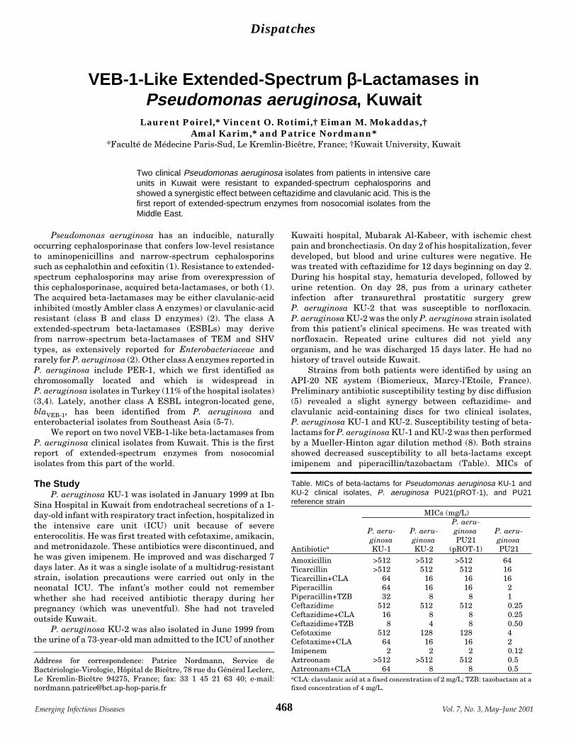
Dispatches
Emerging Infectious Diseases Vol. 7, No. 3, May–June 2001468
Pseudomonas aeruginosa has an inducible, naturallyoccurring cephalosporinase that confers low-level resistanceto aminopenicillins and narrow-spectrum cephalosporinssuch as cephalothin and cefoxitin (1). Resistance to extended-spectrum cephalosporins may arise from overexpression ofthis cephalosporinase, acquired beta-lactamases, or both (1).The acquired beta-lactamases may be either clavulanic-acidinhibited (mostly Ambler class A enzymes) or clavulanic-acidresistant (class B and class D enzymes) (2). The class Aextended-spectrum beta-lactamases (ESBLs) may derivefrom narrow-spectrum beta-lactamases of TEM and SHVtypes, as extensively reported for Enterobacteriaceae andrarely for P. aeruginosa (2). Other class A enzymes reported inP. aeruginosa include PER-1, which we first identified aschromosomally located and which is widespread inP. aeruginosa isolates in Turkey (11% of the hospital isolates)(3,4). Lately, another class A ESBL integron-located gene,blaVEB-1, has been identified from P. aeruginosa andenterobacterial isolates from Southeast Asia (5-7).
We report on two novel VEB-1-like beta-lactamases fromP. aeruginosa clinical isolates from Kuwait. This is the firstreport of extended-spectrum enzymes from nosocomialisolates from this part of the world.
The StudyP. aeruginosa KU-1 was isolated in January 1999 at Ibn
Sina Hospital in Kuwait from endotracheal secretions of a 1-day-old infant with respiratory tract infection, hospitalized inthe intensive care unit (ICU) unit because of severeenterocolitis. He was first treated with cefotaxime, amikacin,and metronidazole. These antibiotics were discontinued, andhe was given imipenem. He improved and was discharged 7days later. As it was a single isolate of a multidrug-resistantstrain, isolation precautions were carried out only in theneonatal ICU. The infant’s mother could not rememberwhether she had received antibiotic therapy during herpregnancy (which was uneventful). She had not traveledoutside Kuwait.
P. aeruginosa KU-2 was also isolated in June 1999 fromthe urine of a 73-year-old man admitted to the ICU of another
Kuwaiti hospital, Mubarak Al-Kabeer, with ischemic chestpain and bronchectiasis. On day 2 of his hospitalization, feverdeveloped, but blood and urine cultures were negative. Hewas treated with ceftazidime for 12 days beginning on day 2.During his hospital stay, hematuria developed, followed byurine retention. On day 28, pus from a urinary catheterinfection after transurethral prostatitic surgery grewP. aeruginosa KU-2 that was susceptible to norfloxacin.P. aeruginosa KU-2 was the only P. aeruginosa strain isolatedfrom this patient’s clinical specimens. He was treated withnorfloxacin. Repeated urine cultures did not yield anyorganism, and he was discharged 15 days later. He had nohistory of travel outside Kuwait.
Strains from both patients were identified by using anAPI-20 NE system (Biomerieux, Marcy-l’Etoile, France).Preliminary antibiotic susceptibility testing by disc diffusion(5) revealed a slight synergy between ceftazidime- andclavulanic acid-containing discs for two clinical isolates,P. aeruginosa KU-1 and KU-2. Susceptibility testing of beta-lactams for P. aeruginosa KU-1 and KU-2 was then performedby a Mueller-Hinton agar dilution method (8). Both strainsshowed decreased susceptibility to all beta-lactams exceptimipenem and piperacillin/tazobactam (Table). MICs of
VEB-1-Like Extended-Spectrum βββββ-Lactamases inPseudomonas aeruginosa, Kuwait
Laurent Poirel,* Vincent O. Rotimi,† Eiman M. Mokaddas,†Amal Karim,* and Patrice Nordmann*
*Faculté de Médecine Paris-Sud, Le Kremlin-Bicêtre, France; †Kuwait University, Kuwait
Address for correspondence: Patrice Nordmann, Service deBactériologie-Virologie, Hôpital de Bicêtre, 78 rue du Général Leclerc,Le Kremlin-Bicêtre 94275, France; fax: 33 1 45 21 63 40; e-mail:[email protected]
Two clinical Pseudomonas aeruginosa isolates from patients in intensive careunits in Kuwait were resistant to expanded-spectrum cephalosporins andshowed a synergistic effect between ceftazidime and clavulanic acid. This is thefirst report of extended-spectrum enzymes from nosocomial isolates from theMiddle East.
Table. MICs of beta-lactams for Pseudomonas aeruginosa KU-1 andKU-2 clinical isolates, P. aeruginosa PU21(pROT-1), and PU21reference strain
MICs (mg/L) P. aeru-
P. aeru- P. aeru- ginosa P. aeru- ginosa ginosa PU21 ginosa
Antibiotica KU-1 KU-2 (pROT-1) PU21Amoxicillin >512 >512 >512 64Ticarcillin >512 512 512 16Ticarcillin+CLA 64 16 16 16Piperacillin 64 16 16 2Piperacillin+TZB 32 8 8 1Ceftazidime 512 512 512 0.25Ceftazidime+CLA 16 8 8 0.25Ceftazidime+TZB 8 4 8 0.50Cefotaxime 512 128 128 4Cefotaxime+CLA 64 16 16 2Imipenem 2 2 2 0.12Aztreonam >512 >512 512 0.5Aztreonam+CLA 64 8 8 0.5aCLA: clavulanic acid at a fixed concentration of 2 mg/L; TZB: tazobactam at afixed concentration of 4 mg/L.

Dispatches
469Vol. 7, No. 3, May–June 2001 Emerging Infectious Diseases
ceftazidime were decreased by addition of clavulanic acid andtazobactam, indicating the presence of a clavulanic-acidinhibited ESBL (Table). According to antibiotic susceptibilitytesting by Mueller-Hinton agar disc diffusion, P. aeruginosaKU-1 and KU-2 were also resistant to aminoglycosides(amikacin, gentamicin, netilmicin, tobramycin), chloram-phenicol, tetracyclines, sulfonamides, and fluoroquinolones(except for KU-2).
Beta-lactamase extracts from cultures of P. aeruginosaKU-1 and KU-2 were obtained (8). Isoelectric focusinganalysis (8) revealed beta-lactamases with isoelectric pointsof 7.4 and 8-8.4, the latter likely corresponding toP. aeruginosa AmpC cephalosporinases. Whole-cell DNAs ofP. aeruginosa KU-1 and KU-2 were then obtained (3).Preliminary polymerase chain reaction (PCR) experimentswere performed with DNAs of P. aeruginosa KU-1 and KU-2as templates and primers specific for the following class Abeta-lactamases: TEM, SHV, CARB (PSE-1), GES-1, PER-1,and VEB-1 (3,5,9-11). Only PCR using internal primers forblaVEB-1 gave a positive result with an identical 642-bpfragment. External blaVEB-1 specific primers gave 1,070-bpPCR fragments with DNAs of both P. aeruginosa strains astemplates that were sequenced on both strands (6). Thededuced amino acid sequences, obtained over the internet (8),identified VEB-1-like sequences that shared 99% amino acididentity with VEB-1 (Figure). Compared with VEB-1, theamino acid changes in VEB-1a and VEB-1b fromP. aeruginosa KU-1 and KU-2, respectively, occurred in theputative leader peptide sequence (Figure). Thus, thehydrolytic activity of VEB-1a and VEB-1b should be identicalto that reported for VEB-1 beta-lactamase (5).
The genetic background of blaVEB-1a and blaVEB-1b wasfurther characterized. Plasmid extraction, conjugation, andelectroporation experiments were performed (8). A plasmid(pROT-1) of ca. 70 kb carrying blaVEB-1a gene was identifiedaccording to hybridization results by using an internal PCR-obtained probe for blaVEB-1 (5). Plasmid pROT-1 was self-conjugative from P. aeruginosa KU-1 to in vitro obtainedrifampin-resistant P. aeruginosa PU21 reference strain afterselection of transconjugants onto Mueller-Hinton agar plateseach containing 150 µg/mL rifampin and 200 µg/mLticarcillin. As assessed by antibiotic susceptibility testing by
disc diffusion, plasmid pROT-1 conferred additionalresistance to gentamicin, netilmicin, sulfonamides, andtobramycin. MICs of beta-lactams for P. aeruginosa PU21(pROT-1) mirrored those obtained for P. aeruginosa KU-1(Table). While the plasmid location of blaVEB-1 gene is knownonly in Enterobacteriaceae (5-7), its report in P. aeruginosamay signal the evolution of its spread. The blaVEB-1b gene wasnot plasmid located, but a PCR-obtained 642-bp internalprobe for blaVEB-1 hybridized at chromosomal position ofwhole-cell DNA of P. aeruginosa KU-2.
The blaVEB-1 gene is located on different structures ofclass 1 integrons (5-7). Integrons comprise two conservedregions (5'-CS and 3'-CS) flanking an internal variable regionusually containing several gene cassettes (13). Integrons arein fact expression vectors for antibiotic resistance genes thatare included as gene cassettes and are neighbored (13). Byusing primers located either in the 5'-CS sequence and the 5'end of blaVEB-1 or in the 3' end of blaVEB-1 and the 3'-CSsequence (5,14), PCR amplification experiments wereperformed with whole-cell DNAs of P. aeruginosa KU-1 andKU-2 as templates. In one case (strain KU-1), a PCR fragmentwas obtained by using blaVEB-1 and 5'-CS primers, indicatingthat blaVEB-1a was located downstream of a class 1 integrasegene. In this case, the 4-kb PCR fragment differed from thoseof known blaVEB-1 containing integrons identified inEscherichia coli and P. aeruginosa isolates (5-7). Amplimersof 1 kb were obtained for both strains using blaVEB-1 and 3'-CSprimers, showing that the 3'-CS end was present in both casesand that the blaVEB-1-like sequences were located next to the3'-CS end within class 1 integrons. The attC (59-be)recombination sites (15) located downstream of gene cassetteswere identical for blaVEB-1a and blaVEB-1b to those described forblaVEB-1 in P. aeruginosa and enterobacterial isolatesidentified so far from Southeast Asia (5-7). Therefore, anidentical blaVEB-1-like gene cassette may be located ondifferent class 1 integrons. Using 5'-CS and 3'-CS primers,two additional PCR fragments were obtained foreach P. aeruginosa strain, showing that both strainscontained another blaVEB-negative class 1 integron.For P. aeruginosa KU-1, a 950-bp PCR fragment for anaadA1a gene coding for an aminoglycoside modifying enzymewas found to be plasmid- and integron-located in Salmonellaenterica serotype Typhimurium (16). For P. aeruginosa KU-2,a 500-bp PCR fragment encoding a putative 95 amino acidprotein of unknown function was PCR amplified. It shared71% amino acid identity with an amino acid sequence from agene that was Tn1696 transposon-located and In4 integron-located in P. aeruginosa (17).
Finally, P. aeruginosa KU-1 and KU-2 isolates containingVEB-1-like beta-lactamases were compared with VEB-1positive P. aerugionosa strain JES from Thailand by usingrandom amplified polymorphic DNA technique (10,18). Theisolates were not clonally related (data not shown). Althoughthe patients had not traveled outside Kuwait, introduction ofP. aeruginosa into Kuwaiti hospitals by travelers or patientsfrom Southeast Asia cannot be ruled out.
ConclusionsThe presence of clavulanic-acid inhibited ESBLs in
P. aeruginosa isolates may account for part of the 50%resistance to ceftazidime of P. aeruginosa strains isolatesfrom ICUs in Kuwait (19). ESBLs in P. aeruginosa in Kuwait
Figure. Comparison of the amino acid sequence of VEB-1, VEB-1a,and VEB-1b beta-lactamases from Pseudomonas aeruginosa JESfrom Thailand and KU-1 and KU-2 from Kuwait. Dashed linesindicate identical amino acids. Arrow indicates position of theputative cleavage site of the peptide leader. Numbering is accordingto Ambler designation (12).

Dispatches
Emerging Infectious Diseases Vol. 7, No. 3, May–June 2001470
and other Middle Eastern hospitals may be underestimatedbecause routine detection with a double disc synergy test maybe difficult. Identification of ESBLs is of interest since theyconfer resistance to all extended-spectrum cephalosporinsand aztreonam, whatever their MICs. This has beenconfirmed by experimental data using a model of pneumoniain rats with the Ambler class A ESBL, PER-1 (20).
This work underscores that very similar ESBLs may beidentified in different parts of the world. It is the first reportof ESBL genes characterized from P. aeruginosa isolates fromthe Middle East.
This work was supported by a grant from the Ministères del’Education Nationale et de la Recherche (UPRES, grant JE-2227),Université Paris XI, Paris, France.
Dr. Poirel is a researcher at Hôpital de Bicêtre, Le Kremlin-Bicêtre,France. He studies the biochemical and genetic mechanisms in beta-lactam resistance.
References 1. Chen HY, Yuan M, Livermore DM. Mechanisms of resistance to ß-
lactam antibiotics amongst Pseudomonas aeruginosa isolatescollected in the UK in 1993. J Med Microbiol 1995;43:300-9.
2. Nordmann P, Guibert M. Extended-spectrum ß-lactamases inPseudomonas aeruginosa. J Antimicrob Chemother 1998;42:128-31.
3. Nordmann P, Naas T. Sequence analysis of PER-1 extended-spectrum ß-lactamase from Pseudomonas aeruginosa andcomparison with class A ß-lactamases. Antimicrob AgentsChemother 1994;38:104-14.
4. Vahaboglu H, Ozturk R, Aygun G, Coskunkan F, Yaman A,Kaygusuz A, et al. Widespread detection of PER-type extended-spectrum ß-lactamases among nosocomial Acinetobacter andPseudomonas aeruginosa isolates in Turkey: a nationwidemulticenter study. Antimicrob Agents Chemother 1997;41:2265-9.
5. Poirel L, Naas T, Guibert M, Chaibi EB, Labia R, Nordmann P.Molecular and biochemical characterization of VEB-1, a novel classA extended-spectrum ß-lactamase encoded by an Escherichia coliintegron gene. Antimicrob Agents Chemother 1999;43:573-81.
6. Naas T, Poirel L, Karim A, Nordmann P. Molecularcharacterization of In50, a class 1 integron encoding the gene forthe extended-spectrum ß-lactamase VEB-1 in Pseudomonasaeruginosa. FEMS Microbiol Lett 1999;176:411-9.
7. Tribuddharat C, Fennewald M. Integron-mediated rifampinresistance in Pseudomonas aeruginosa. Antimicrob AgentsChemother 1999;43:960-2.
8. Poirel L, Naas T, Nicolas D, Collet L, Bellais S, Cavallo JD, et al.Characterization of VIM-2, a carbapenem-hydrolyzing metallo-ß-lactamase and its plasmid- and integron-borne gene from aPseudomonas aeruginosa clinical isolate in France. AntimicrobAgents Chemother 2000;44:891-7.
9. Mercier J, Lévesque RC. Cloning of SHV-2, OHIO-1 and OXA-6 ß-lactamase and cloning and sequencing of SHV-1 ß-lactamase.Antimicrob Agents Chemother 1990;34:1577-83.
10. Poirel L, Guibert M, Bellais S, Naas T, Nordmann P. Integron-andcarbenicillinase-mediated reduced susceptibility to amoxicillin-clavulanic-acid in isolates of multidrug-resistant Salmonellaenterica serotype typhimurium DT104 from French patients.Antimicrob Agents Chemother 1999;43:1098-2004.
11. Poirel L, Le Thomas I, Naas T, Karim A, Nordmann P. Biochemical-sequence analyses of GES-1, a novel class A extended-spectrum ß-lactamase, and the class 1 integron In52 from Klebsiella pneumoniae.Antimicrob Agents Chemother 2000;44:622-32.
12. Ambler RP. The structure of ß-lactamases. Philos Trans R SocLond B Biol Sci 1980;289:321-31.
13. Fluit AC, Schmitz FJ. Class 1 integrons, gene cassettes, mobility,and epidemiology. Eur J Clin Microbiol Infect Dis 1999;18:761-70.
14. Lévesque C, Piché L, Larose C, Roy PH. PCR mapping of integronsreveals several novel combinations of resistance genes. AntimicrobAgents Chemother 1995;39:185-91.
15. Stokes HW, O’Gorman DB, Recchia GD, Parsekhian M, Hall RM.Structure and function of 59-base element recombination sitesassociated with mobile gene cassettes. Mol Microbiol 1997;26:731-45.
16. Tosini F, Visca P, Luzzi I, Dionisi AM, Pezella C, Petrucca A, et al.Class 1 integron-borne multiple-antibiotic resistance carried byIncFI and IncL/M plasmids in Salmonella enterica serotypetyphimurium. Antimicrob Agents Chemother 1998;42:3053-8.
17. Hall RM, Brown HJ, Brookes DE, Stokes HW. Integrons found indifferent locations have identical 5' ends but variable 3' ends. JBacteriol 1994;176:6286-94.
18. Williams JG, Kubelik AR, Livak KJ, Rafalski JA, Tingey SV. DNApolymorphisms amplified by arbitrary primers are useful asgenetic markers. Nucleic Acids Res 1990;18:6531-5.
19. Jamal WY, El Din K, Rotimi VO, Chugh TD. An analysis ofhospital-acquired bacteraemia in intensive care unit patients in auniversity hospital in Kuwait. J Hosp Infect 1999;43:49-56.
20. Mimoz O, Elhelali N, Léotard S, Jacolot A, Laurent F, Samii K, etal. Treatment of experimental pneumonia in rats caused by a PER-1 extended-spectrum ß-lactamase-producing strain of Pseudomo-nas aeruginosa. J Antimicrob Chemother 1999;44:91-7.

Dispatches
471Vol. 7, No. 3, May–June 2001 Emerging Infectious Diseases
Lyme disease is the most commonly reported vector-borne disease in the United States. In the northeastern,midwestern, and western coastal states, the pathogenBorrelia burgdorferi sensu lato is well established and ismaintained by Ixodes scapularis and I. pacificus in a varietyof rodent reservoirs.
In the southeastern states, where I. scapularis iswidespread but is less commonly found infected withB. burgdorferi or attaching to humans (1,2), isolations fromhumans are uncommon (3). However, a clinical conditionsimilar to Lyme disease, termed southern tick-associatedrash illness (STARI), has been described in humans in thesoutheastern region of the United States associated with thebite of Amblyomma americanum ticks (1,4,5). Moreover, anew spirochete, B. lonestari, was described from A. americanumon the basis of polymerase chain reaction (PCR) amplificationof the flagellin and 16s rRNA genes (6,7). Virtually identicalsequences have been found in ticks from geographic regions asdisparate as New Jersey and Texas (6), suggesting thisorganism is widely distributed. Likewise, Borrelia spiro-chetes have been detected in A. americanum and I. scapularisin Alabama (8,9).
As part of an epidemiologic investigation of a reportedcluster of STARI cases in Choctaw County, Alabama, wecollected both A. americanum and A. maculatum ticksadjacent to the houses of suspected patients and in controlareas, and the B. lonestari flagellin gene sequence wasamplified from DNA extracted from A. americanum.
The StudyTicks were collected with drag cloths in areas around the
homes of persons with suspected cases, as well as in controlareas. Clinical cases were defined as illness characterized byacute onset of an annular, expanding erythema migrans-likerash at least 5 cm in diameter, when no alterative explanationfor the rash can be found; and there is a history of tick bite at
the rash site or potential exposure to ticks within 14 daysbefore rash onset. Ticks were identified to species by usingstandard taxonomic keys.
DNA was isolated from Amblyomma sp. by using anextraction procedure reported previously (10). Briefly,individual ticks were frozen in liquid nitrogen, maceratedbetween metal plates, and then homogenized by adding 1 mLof DNA STAT-60 (Tel-Test, Friendswood, TX). The tickhomogenate was then incubated with chloroform for phaseseparation of DNA, which was precipitated with 100%isopropanol. To ensure PCR-quality DNA, all tick extractswere tested for the presence of tick mitochondrial DNA (11).Amblyomma DNA was then subjected to a nested PCRprocedure for B. lonestari by using primers FlaLL/FlaRL,then FlaLS/FlaRS (6). Flagellin-positive samples werefurther analyzed by using OspA primers as a control forpossible flagellin false-positive samples. Subsequent DNAsequencing of positive samples for Borrelia sequenceidentification was done with a Taq Dyedeoxy terminator cyclekit (Applied Biosystems, Foster City, CA) and run on anApplied Biosystems 377 automated sequencer. The derivedsequences were aligned with MegAlign (DNASTAR, Inc.,Madison, WI) by using the clustal algorithm. Alignedsequences were transferred to PAUP (Sinauer Associates Inc.,Sutherland, MA) for phylogentic analysis. Accession numbersD88295 (B. afzelii), X75201 (B. anserina), D82857(B. bissettii), Y15097 (B. burgdorferi), D63372 (B. garinii),AF228034 (B. hermsii), D43777 (B. miyamotoi), D82863(B. parkeri), U26705 (B. lonestari, New Jersey isolate), andU26704 (B. lonestari, Texas isolate) were used in thiscomparative genetic analysis.
Two hundred four A. americanum (21 adults and 183nymphs) were collected: 13 adults and 44 nymphs from theproperties of controls, and 8 adults and 139 nymphs nearhomes of persons meeting the STARI case definition. Twenty-nine A. maculatum adults were collected from control andSTARI case areas. All but five ticks yielded PCR-qualityDNA, as determined by PCR amplification of tickmitochondrial DNA (11).
Two (11%) of 19 of adult A. americanum ticks analyzedwere positive for B. lonestari flagellin gene DNA (Table).
Borrelia lonestari DNA in AdultAmblyomma americanum Ticks, Alabama
Thomas R. Burkot,* Gary R. Mullen,† Renee Anderson,† Bradley S. Schneider,*Christine M. Happ,* and Nordin S. Zeidner*
*Centers for Disease Control and Prevention, Fort Collins, Colorado, USA;†Auburn University, Auburn, Alabama, USA
Address for correspondence: Nordin Zeidner, Division of Vector-BorneInfectious Diseases, Centers for Disease Control and Prevention, POBox 2087, Rampart Road, Foothills Campus, Fort Collins, CO 80522-2087; fax: 970-221-6476; e-mail: [email protected]
Polymerase chain reaction analysis of 204 Amblyomma americanum and 28 A.maculatum ticks collected in August 1999 near the homes of patients withsouthern tick-associated rash illness and in control areas in Choctaw County,Alabama, showed Borrelia lonestari flagellin gene sequence from two adult A.americanum. The presence of B. lonestari in A. americanum ticks from Alabamasuggests that this suspected pathogen may be widespread in the southeasternUnited States.

Dispatches
Emerging Infectious Diseases Vol. 7, No. 3, May–June 2001472
Positive results for B. lonestari were confirmed byamplification of the 16s rRNA gene as described by Barbour etal. (6). All 183 nymphs and all 26 adult A. maculatum werePCR negative for B. lonestari flagellin gene DNA. Moreover,all tick DNA samples were PCR negative when analyzed forthe B. burgdorferi OspA gene. Sequence analyses for bothpositive samples showed >99% homology with the publishedB. lonestari sequences from New Jersey and Texas. Alabamaisolates numbers 1 and 2 were 100% homologous toB. lonestari NJ and differed by 1 bp when compared withpublished sequences of the Texas isolate of B. lonestari.Phylogenetic analysis, using maximum likelihood andbootstrap analysis with 500 replications of derived sequences(Figure), illustrated that both isolates clustered with reportedsequences of the B. lonestari NJ and TX strains anddemonstrated considerable divergence from B. burgdorferisensu stricto, the only genospecies shown to cause disease inthe United States.
The nucleotide sequences of the B. lonestari flagellin genehave been submitted to GenBank and assigned accessionnumbers AF298653 (Alabama isolate 1) and AF298654(Alabama isolate 2).
ConclusionsSpirochetes have been reported in A. americanum ticks
from New York, New Jersey, Virginia, North Carolina,Alabama, Missouri, and Texas. In contrast to B. burgdorferispirochetes, attempts to propagate these spirochetes fromA. americanum in Barbour-Stoenner-Kelly (BSK-H) culturehave been unsuccessful. Likewise, attempts to coculture A.americanum samples with an I. scapularis cell line (IDE2)failed to propogate B. lonestari (B. Johnson, pers. comm.).Thus, we used genetic analysis to determine the presence ofthis organism.
Erythema migrans associated with the bites ofA. americanum has been reported from the southeasternUnited States, including Missouri (4) and North Carolina (5).Serum samples from these patients did not recognizeB. burgdorferi antigens (4,5), and spirochetes from thesecases have not been successfully cultured in BSK-H medium.Hence, these cases have been diagnosed as STARI. Recently,novel DNA sequences, amplified by PCR with primer setsrecognizing the flagellin and 16s rRNA genes, have identifieda new spirochete in A. americanum and B. lonestari (6).Nearly identical sequences have been amplified from A.americanum collected from Texas and New Jersey (2) andMaryland (1). B. lonestari is the suspected pathogenresponsible for the STARI-related erythema migransassociated with bites by A. americanum (1). Moreover,B. lonestari was isolated from a patient in WestchesterCounty, New York, who had traveled to Maryland and NorthCarolina and had an attached A. americanum at the site of anerythema migrans rash. This isolate differed only slightlyfrom B. lonestari isolates reported in New Jersey and Texas(A. James et al., unpub. data).
Although the numbers of ticks we analyzed were small,our results suggest that adult A. americanum may transmitB. lonestari to persons in this area. Finding B. lonestarisequences in A. americanum from Alabama suggests that thisspirochete is widely distributed in the United States. Thesefirst sequences from the southeastern United States arenoteworthy because this region is a focus for numerousreports of erythema migrans associated with the bites ofA. americanum. Further investigation is needed to formallyisolate and propagate B. lonestari, as well as to determine itshost reservoir.
AcknowledgmentsThe authors thank Katherine Hensleigh for initially recognizing
and reporting these cases and Jennifer Foster, William Johnson,Barbara Shoemaker, Rhonda Stephens, David Dennis, VirginiaHinrichsen, and Sarah Lathrop for identifying case-patient sites andfor their invaluable assistance in these field studies.
Dr. Burkot is a senior research entomologist in the bacterialZoonoses Branch, Division of Vector-Borne Infectious Diseases, Centersfor Disease Control and Prevention. His research interests include vec-tor ecology, epidemiology, and the control of vector-borne bacterial, vi-ral, and protozoal diseases.
References 1. Felz MW, Durden LA. Attachment sites of four tick species
(Acari:Ixodidae) parasitizing humans in Georgia and SouthCarolina. J Med Entomol 1999;36:361-4.
2. Merten HA, Durden LA. A state-by-state survey of ticks recordedfrom humans in the United States. J Vector Ecol 2000;25:102-13.
Table. Ticks collected in Choctaw County, Alabama
Amblyomma americanum A. maculatumTicks Adult Adult Adult Adultanalyzed male female Nymphs male femaleControlsa 11b 2 44 6 12Casesc 1 5b 139 7 1Total 12 7 183 13 13aDenotes ticks collected near the homes of persons not diagnosed with southerntick-associated rash illness (STARI).bPositive results as demonstrated by amplification of the flagellin gene ofBorrelia lonestari. A single positive tick was found among 11 adult male A.americanum males collected near the homes of controls and 1 of 5 adult femalescollected near the homes of STARI controls was positive.cDenotes ticks collected near the homes of persons diagnosed with STARI.
Figure. Maximum parsimony phylogenetic tree generated by usingPAUP. Numbers indicate parsimony bootstrap scores for the branch.Only bootstrap scores >70 are included in the phylogenetic tree.

Dispatches
473Vol. 7, No. 3, May–June 2001 Emerging Infectious Diseases
3. Oliver JH Jr, Clark KL, Chandler FW, Tao L, James AM, BanksCW, et al. Isolation, cultivation and characterization of Borreliaburgdorferi from rodents and ticks in the Charleston area of SouthCarolina. J Clin Microbiol 2000;38:120-4.
4. Campbell GL, Paul WS, Schriefer ME, Craven RB, Robbins KE,Dennis DT. Epidemiologic and diagnostic studies of patients withsuspected early Lyme disease, Missouri, 1990-1993. J Infect Dis1995;172:470-80.
5. Kirkland KB, Klimbo TB, Meriwether RA, Schriefer M, Levin M,Levine J, et al. Erythema migrans-like rash illness at a camp inNorth Carolina: a new tick-borne disease? Arch Intern Med1997;157:2635-41.
6. Barbour AG, Maupin GO, Teltow GJ, Carter CJ, Piesman J.Identification of an uncultivatable Borrelia species in the hard tickAmblyomma americanum: possible agent of a Lyme disease–likeillness. J Infect Dis 1996;173:403-9.
7. Armstrong PM, Rich SM, Smith RD, Hart DL, Spielman A, TelfordSR III. A new Borrelia infecting lone star ticks. Lancet 1996;347:67-8.
8. Luckhart SG, Mullen GR, Wright JC. Etiologic agent of Lymedisease, Borrelia burgdorferi, detected in ticks (Acari:Ixodidae)collected at a focus in Alabama. J Med Entomol 1991;28:652-7.
9. Luckhart SG, Mullen GR, Durden LA, Wright JC. Borrelia sp. inticks recovered from white-tailed deer in Alabama. J Wildl Dis1992;28:449-52.
10. Zeidner NS, Burkot TR, Massung R, Nicholson WL, Dolan MC,Rutherford JS, et al. Transmission of the agent of humangranulocytic ehrlichiosis by Ixodes spinipalpis ticks: evidence of anenzootic cycle of dual infection with Borrelia burgdorferi innorthern Colorado. J Infect Dis 2000;182:616-9.
11. Black WC, Piesman J. Phylogeny of hard- and soft-tick taxa (Acari:Ixodida) based on mitochondrial 16S rDNA sequences. Proc NatlAcad Sci U S A 1994;91:10034-8.

Dispatches
Emerging Infectious Diseases Vol. 7, No. 3, May–June 2001474
Epidemics are conventionally recognized through theobservation of spatial or temporal clustering of patients withsimilar illnesses or through laboratory findings indicatingunusually high incidence of a specific disease. Surveillancesystems based on passive physician reports frequently havelow sensitivity and may not be timely. Recognizing anepidemic by detecting clusters of microbiologically verifiedcases may also involve delays, depending on such factors asthe severity and familiarity of the clinical illness, time fromthe appearance of the first clinical cases of an epidemic to thefirst appropriate samples taken and transported, and timerequired for laboratory testing. Samples from geographicallyscattered cases from the same epidemic may be sent todifferent laboratories, reducing the sensitivity of detection. Arecent study in infectious disease surveillance used as anindex the number of cases per week per sentinel medicalinstitution in the area covered by a health center in Japan (1).
Physicians may consult electronic databases for selectingappropriate diagnostic measures or treatment for specificinfectious diseases. A computerized set of primary-careguidelines, the Physicians’ Desk Reference and Database(PDRD), has been available in Finland since 1989 (2). Thisdatabase contains structured information on diseases andconditions that are common or important to recognize inprimary care. Physicians often seek information aboutinfectious diseases from the PDRD guidelines (3).
We hypothesized that the frequency of physician searchesin a popular database could be useful as a complementary toolin early recognition of infectious disease epidemics. In a pilotstudy, we analyzed retrospectively the feasibility of usingsurveillance of database searches in the PDRD to identifyepidemics of four specific infectious diseases, as recorded in arecently revised National Infectious Diseases Register (NIDR).
The StudyThe PDRD computerized guidelines on CD-ROM are
updated three times a year. We collected the frequency of
infection guideline-specific searches by producing a log file ofall searches. This data-collecting version was piloted inspring 1994 and mailed to all registered PDRD subscribers in1995 (3). The data for searches were collected from thecomputer hard disks of individual users during each updatingof the program and were mailed to the PDRD maintenance team.
The NIDR in Finland, which was thoroughly revised in1994, consists of mandatory laboratory reporting ofdiagnostic findings for more than 70 pathogens or pathogengroups and mandatory physician reporting of 32 microbiolog-ically confirmed infectious diseases (4). Microbiologiclaboratories reported approximately 41,000 cases in 1995.From NIDR, we recorded the sampling dates of cases or thedate of the report if the sampling date was not available.
We chose four infectious diseases for comparison betweenPDRD searches and NIDR laboratory reports: tularemia,ICD-10 A21, caused by Francisella tularensis; epidemicnephropathy, ICD-10 A98.5, caused by a Puumala virus (ahantavirus); Pogosta disease, ICD-10 A92.8, caused by theSindbis virus (an arbovirus); and Lyme disease, ICD-10A69.2, caused by Borrelia burgdorferi. In Finland, epidemicnephropathy (758 to 1,305 laboratory-reported infections peryear during 1995-1999) and Lyme disease (346 to 538laboratory reports of B. burgdorferi infections per year) areendemic, with pronounced seasonal variation. Laboratory-confirmed cases of tularemia, mostly of the glandular type,and Pogosta disease, an acute syndrome of fever with rashand self-limiting arthritis, are usually rare, but epidemicsoccur at intervals of several years.
We compared distributions of PDRD searches and NIDRlaboratory reports by disease and month for 1995. Becausethe number of observations was small, we did not teststatistical significance between distributions but based ourobservations on time-frequency graphs produced by calculat-ing each month’s proportion of the total number of cases.
PDRD had 477 subscribers in 1995: 48% of the usersreturned 306 log files on 15,267 searches; 23,083 specificguidelines were read. The five most popular subject areaswere dermatology (9% of searches), infectious diseases (8%),cardiology (6%), gastroenterology (6%), and pediatrics (5%).The Lyme disease guideline was the third most frequently
Physicians’ Database Searches as a Toolfor Early Detection of Epidemics
Vesa Jormanainen,*† Jukkapekka Jousimaa,‡Ilkka Kunnamo,§ and Petri Ruutu¶
*Finnish Defence Forces, Helsinki, Finland; †University of Helsinki, Helsinki, Finland;‡University of Kuopio, Kuopio, Finland; §Finnish Medical Society Duodecim,Helsinki, Finland; and ¶National Public Health Institute, Helsinki, Finland
We analyzed retrospectively the use of Physician Desk Reference Databasesearches to identify epidemics of tularemia, nephropathy, Pogosta disease,and Lyme disease and compared the searches with mandatory laboratoryreports to the National Infectious Diseases Register in Finland during 1995.Continuous recording of such searches may be a tool for early detection ofepidemics.
Address for correspondence: Jukkapekka Jousimaa, Silmukuja 4,80710 LEHMO, Finland; fax: 358-13-2674370; e-mail:[email protected]

Dispatches
475Vol. 7, No. 3, May–June 2001 Emerging Infectious Diseases
read, with 144 readings; Pogosta disease was fifth, with 91;epidemic nephropathy eighth, with 87; and tularemianineteenth, with 68 readings.
Three hundred forty-six laboratory-confirmed B. burg-dorferi cases were reported to NIDR, distributed throughoutthe year (incidence 0.68 per 100,000; 95% confidence interval[CI] 0.60-0.75), but peaking in August. A large epidemic ofPogosta disease took place in late summer and autumn 1995.Cases were distributed throughout the country, with 1,310laboratory-confirmed (incidence 2.56 per 100,000; 95% CI2.42-2.70). The 888 laboratory-confirmed cases of Puumalavirus infection (1.74; 95% CI 1.62-1.85) peaked in late 1995and occurred throughout Finland. A major epidemic oftularemia, involving 467 laboratory-reported cases (0.91; 95%CI 0.83-1.00) and distributed over central and southernFinland, began in July 1995 and continued until late autumn.
The distributions of PDRD readings and laboratory-reported cases of the four infectious diseases fell into twopatterns (Figure). For tularemia (Figure, panel D) andPogosta disease (Figure, panel C), the PDRD searches and thecases in NIDR rose from a low baseline level, peaked sharply,and then declined in parallel. For epidemic nephropathy(Figure, panel B), the curves followed the same pattern onlypartly, with PDRD searches peaking twice, the major peakoccurring earlier than in NIDR, in the latter half of 1995. ForLyme disease (Figure, panel A), PDRD search data had a peakwell before that of NIDR reports during June to September.
ConclusionsThe temporal correlation observed between the distribu-
tion of database searches and laboratory reports for Pogostadisease and tularemia supports the concept that continuousmonitoring of database searches for specific infections couldbe a novel tool for surveillance and detection of epidemics. Weare not aware of previous reports of this application forelectronic desk reference database searches. For thisinvestigation, we used retrospectively collected logs of CD-ROM–based searches from computers of physicians who usedthe widely distributed and popular electronic guidelinedatabase. The statistics on searches were created automati-cally into log files without active input by the physicians, andthe users had free access to the data they were providing.Although only a few users expressed negative attitudestowards data collection, fewer than half of subscribersreturned the data.
Monitoring database searches has the potential toprovide timely recognition of an epidemic, as physicians aremore likely to use searches to seek guidance for thediagnostics and management of the first patients of anunusual cluster, even before an order for laboratory work isgiven. Another potential benefit of monitoring databasesearches at a central facility is that it consolidatesconsultations from different geographic areas, making itpossible to detect subtle changes in widely distributed cases.
Access through the Internet to databases such as thePDRD provides an opportunity to monitor the searches forspecific topics. In 1999, the PDRD had 3,500 subscribersthroughout Finland, a sevenfold increase from 1995, the year
Figure. Laboratory reports to the National Infectious DiseasesRegister compared with searches on the Physicians’ Desk Referenceand Database, Finland, 1995. Panel A, Borrelia burgdorferi andLyme disease; panel B, Puumala virus and epidemic nephropathy;panel C, Sindbis virus and Pogosta disease; panel D, Francisellatularensis and tularemia.

Dispatches
Emerging Infectious Diseases Vol. 7, No. 3, May–June 2001476
of this study. An Internet version of the PDRD guidelines(now renamed Evidence-Based Medicine Guidelines) (5),which collects a log file of all searches to the database, wasintroduced in October 2000. In the future, log files willautomatically be sent from CD-ROM users, with theirpermission. As no patient data are transmitted, no concernsabout confidentiality can arise.
Reference databases are likely to be used by cliniciansmore frequently for syndromes or suspected infections if thephysician encounters the specific problem infrequently, e.g.,infectious diseases with low transmission levels betweenepidemics. This observation is supported by the closecorrelation between the distributions in PDRD searches andNIDR reports of tularemia, epidemics of which have notoccurred annually, and Pogosta disease, for which theinterepidemic interval has been long (previous epidemics in1981 and 1988). Search-frequency-based surveillance cannever achieve the specificity of laboratory reporting.However, it can provide an effective early warning forinfectious diseases in which the clinical syndromes arespecific enough to prompt the clinician to search specificguidelines.
For Lyme disease, the increase and peak distribution ofthe database searches substantially preceded the increaseand peak in the laboratory reports of B. burgdorferi to theNIDR. The diagnosis of early Lyme disease, with itscharacteristic skin rash (6), is entirely clinical; serologicdiagnosis can be made no earlier than 3 weeks after a tick bite.
This delay could explain why the database searches peakedwell before the NIDR reports. The earlier peak in databasesearches could also reflect a search for preventive measuresduring summer outdoor activities or for tick bite managementbefore symptoms appear.
Dr. Jormanainen works as a consulting physician at the HealthCare Division of the General Staff of Finnish Defense Forces, where hisresearch interests include infectious disease epidemiology, surveillance,and public health issues.
References 1. Hashimoto S, Murakami Y, Taniguchi K, Nagai M. Detection of
epidemics in their early stage through infectious diseasesurveillance. Int J Epidemiol 2000;29:905-10.
2. Jousimaa J, Kunnamo I. PDRD-a computer-based primary caredecision support system. Med Inform 1993;18:103-12.
3. Jousimaa J, Kunnamo I, Mäkelä M. Physicians’ patterns of usinga computerized collection of guidelines for primary health care. IntJ Technol Assess Health Care 1998;14:484-93.
4. National Public Health Institute. Infectious diseases in Finland in1995. Publications of the National Public Health Institute, KTLB8/1996. Helsinki: 1997.
5. Finnish Medical Society Duodecim. Available at: URL: Http://www.ebm-guidelines.com. [Note: The English version of theguidelines will be available for registered Internet users in 2001.]
6. Benenson AS, editor. Control of communicable diseases manual.16th ed. Washington, DC: American Public Health Association;1995.

Dispatches
477Vol. 7, No. 3, May–June 2001 Emerging Infectious Diseases
Vibrio parahaemolyticus, a halophilic gram-negative rod,causes seafood-borne gastroenteritis in humans. Infectionscaused by this organism have been associated with diverseserovars: 13 O serotypes and 75 K serotypes have beenidentified. Recent studies, however, have revealed theemergence and pandemic spread of a single serovar, O3:K6 (1-6). Strains belonging to the O3:K6 serovar abruptly appearedin India in 1996 and have since been isolated in SoutheastAsian countries, from travelers at quarantine stations inJapan, and from foodborne outbreaks in the United States (1-5). This serovar accounts for more than half of the V.parahaemolyticus isolates from diarrheal patients in Japan(6). Such widespread occurrence of a single serovar of V.parahaemolyticus had not previously been reported. Since1998, V. parahaemolyticus strains belonging to other twoserovars, O4:K68 and O1:K untypeable (KUT), have also beenisolated with increasing frequency from diarrheal patients(2,6,7). The genetic background of the O4:K68 and O1:KUTisolates is almost indistinguishable from that of the recentO3:K6 strains, suggesting a common origin (2,7).
In a previous study, we reported on a filamentous phagethat is specifically associated with the recent O3:K6 serovarstrains of V. parahaemolyticus (8). This phage, f237, hasseveral genes in common with and a similar genomicstructure to another filamentous phage, CTX (9), which isknown to carry the genes for cholera enterotoxin (ctxAB), themost important virulence factor of V. cholerae. Instead ofctxAB, f237 possesses a unique open reading frame, ORF8,which has no homology with other sequences in DNAdatabases (8). In this study, we examined the distribution off237 in recent clinical isolates of V. parahaemolyticus.
The StudyWe studied 96 strains of V. parahaemolyticus isolated
from diarrheal patients during January to May 1999 at theKansai International Airport quarantine station, Osaka,Japan. Detection of f237 was by colony hybridization using adigoxigenin-labeled DNA probe targeted for ORF8 (8) andprepared as described (8). In brief, after amplification of apartial DNA sequence of ORF8 (746 bp in size) by polymerase
chain reaction (PCR) with the genomic DNA ofV. parahaemolyticus KXV237 strain (RIMD2210633) (8) as atemplate, the amplified DNA was labeled with digoxigeninusing a PCR DIG labeling kit (Boehringer GmbH, Mannheim,Germany). PCR primers for ORF8 were 8S (5'-GTTCGCATA-CAGTTGAGG-3') and 8A (5'-AAGTACAGCAGGAGTGAG-3').The conditions for PCR were as follows: After heating at 94°Cfor 3 minutes, a cycle of 94°C for 30 seconds, 57°C for 30seconds, and 72°C for 1 minute was repeated 30 times,followed by end extension step at 72°C for 5 minutes. PCR wasdone on a Perkin-Elmer (Foster City, CA) thermal cycler type9700. Pulsed-field gel electrophoresis (PFGE) and Southernhybridization were performed as described (10). For PFGE,the V. parahaemolyticus genomic DNA in agarose blocks wasdigested with restriction enzymes NotI or SfiI. A contour-clamped homogeneous electric field method was used forPFGE on a CHEF Mapper System (Bio-Rad Laboratories,Richmond, CA). The conditions for PFGE were 1% agarose gelin 0.5x Tris-borate-EDTA buffer at 6 V cm-1, with pulse timesincreasing linearly from 1 to 60 seconds within 24 hours.
Of the 96 strains examined, 53 tested positive for ORF8,suggesting the presence of phage f237 (Table). In addition toO3:K6, the ORF8-positive strains were found in strains withserotypes O4:K68 and O1:KUT. None of the strains with otherserovars showed evidence of ORF8. When the genomic DNA ofthe strains was digested with NotI or SfiI endonuclease,PFGE showed very similar restriction patterns for thebacterial genomes of the ORF8-positive strains, irrespective
Filamentous Phage Associated with RecentPandemic Strains of Vibrio parahaemolyticus
Tetsuya Iida, Akiko Hattori, Kenichi Tagomori,Hatsumi Nasu, Rochman Naim, and Takeshi Honda
Osaka University, Suita, Osaka, Japan
Address for correspondence: Tetsuya Iida, Department of BacterialInfections, Research Institute for Microbial Diseases, OsakaUniversity, 3-1 Yamadaoka, Suita, Osaka 565-0871, Japan; fax: 81-6-6879-8277; e-mail: [email protected]
A group of pandemic strains of Vibrio parahaemolyticus has recently appearedin Asia and North America. We demonstrate that a filamentous phage isspecifically associated with the pandemic V. parahaemolyticus strains. Anopen reading frame unique to the phage is a useful genetic marker to identifythese strains.
Table. Distribution of ORF8 in clinical isolates of Vibrio parahaemolyticus
Serovar ORF8a PFGE genotypeb No. of strainsO3:K6 + A 34
- A 1O4:K68 + A 11
- - 0O1:KUT + A 8
- A 1- B 8
Others + - 0- B 33
Total 96aPossession of ORF8: (+) denotes presence of ORF8; (-) denotes absence ofORF8.bPulsed-field gel electrophoresis (PFGE) pattern of the genomic DNA ofstrains; A, genotype closely related to recent O3:K6 strains (7); B, genotypedistinct from that of recent O3:K6 strains.

Dispatches
Emerging Infectious Diseases Vol. 7, No. 3, May–June 2001478
of their serovars (data not shown). Southern hybridizationwith a probe for ORF8 after PFGE demonstrated that, in allthe ORF8-positive strains, the largest NotI fragment (sizeapproximately 1,080 kb) reacted with the probe, suggestingthat f237 integrated into the bacterial chromosome.
ConclusionsOur study shows that the filamentous phage f237 is
associated not only with O3:K6 serovar but also with otherrecently emerging serovars of V. parahaemolyticus. In the 55strains showing PFGE genotypes closely related to those ofthe recent O3:K6 strains, >96% was positive for the ORF8(Table). Such high prevalence of the phage f237 in theV. parahaemolyticus strains showing pandemic spreadsuggests that the phage might confer a so-far-unknownphenotype to the bacterium. The phenotype might, in turn,protect the organism against selective pressure in a certainenvironment before it infects humans. Whether phage f237has played a part in the recently increasing pandemic potencyof strains of V. parahaemolyticus is a subject for further study.
The virulence of V. parahaemolyticus extends beyondacute gastroenteritis: it can also cause wound infections andsepticemia (5). Physicians everywhere need to be alert to thepossibility of infection from these recently emergent strains.To distinguish the new and increasingly commonV. parahaemolyticus strains from classic ones, ORF8 is auseful genetic marker.
AcknowledgmentsThe authors thank the staff at the Kansai International Airport
quarantine station for providing the V. parahaemolyticus strainsused in this study.
This work was supported by a grant-in-aid for scientific researchfrom the Ministry of Education, Science, Sports, and Culture ofJapan and the “Research for the Future” Program of the JapanSociety for the Promotion of Sciences (JSPS-RFTF 97L00704).
Dr. Iida is an associate professor in the Department of BacterialInfections, Research Institute for Microbial Diseases, Osaka Univer-sity, Japan. His research interests include the mechanism and evolu-tion of bacterial pathogenesis, especially of enteropathogens such asvibrios and Escherichia coli.
References 1. Okuda J, Ishibashi M, Hayakawa E, Nishino T, Takeda Y,
Mukhopadhyay AK, et al. Emergence of a unique O3:K6 clone ofVibrio parahaemolyticus in Calcutta, India, and isolation of strainsfrom the same clonal group from Southeast Asian travelersarriving in Japan. J Clin Microbiol 1997;35:3150-5.
2. Matsumoto C, Okuda J, Ishibashi M, Iwanaga M, Garg P,Rammamurthy T, et al. Pandemic spread of an O3:K6 clone ofVibrio parahaemolyticus and emergence of related strainsevidenced by arbitrarily primed PCR and toxRS sequence analyses.J Clin Microbiol 2000;38:578–85.
3. Centers for Disease Control and Prevention. Outbreak of Vibrioparahaemolyticus infections associated with eating raw oysters—Pacific Northwest, 1997. MMWR Morb Mortal Wkly Rep1998;47:457-62.
4. Centers for Disease Control and Prevention. Outbreak of Vibrioparahaemolyticus infection associated with eating raw oysters andclams harvested from Long Island Sound—Connecticut, NewJersey, and New York, 1998. MMWR Morb Mortal Wkly Rep1999;48:48-51.
5. Daniels NA, MacKinnon L, Bishop R, Altekruse S, Ray B,Hammond RM, et al. Vibrio parahaemolyticus infections in theUnited States, 1973–1998. J Infect Dis 2000;181:1661–6.
6. World Health Organization. Vibrio parahaemolyticus, Japan,1996-1998. Wkly Epidemiol Rec 1999;74:361-3.
7. Chowdhury NR, Chakraborty S, Eampokalap B, Chaicumpa W,Chongsa-Nguan M, Moolasart P, et al. Clonal dissemination ofVibrio parahaemolyticus displaying similar DNA fingerprint butbelonging to two different serovars (O3:K6 and O4:K68) inThailand and India. Epidemiol Infect 2000;125:17-25.
8. Nasu H, Iida T, Sugahara T, Yamaichi Y, Park K-S, Yokoyama K, et al.A filamentous phage associated with recent pandemic Vibrioparahaemolyticus O3:K6 strains. J Clin Microbiol 2000;38:2156–61.
9. Waldor MK, Mekalanos JJ. Lysogenic conversion by a filamentousphage encoding cholera toxin. Science 1996;272:1910–4.
10. Yamaichi Y, Iida T, Park K-S, Yamamoto K, Honda T. Physical andgenetic map of the genome of Vibrio parahaemolyticus: presence of twochromosomes in Vibrio species. Mol Microbiol 1999;31:1513-21.
Letters
479Vol. 7, No. 3, May–June 2001 Emerging Infectious Diseases
Candida dubliniensis Candidemia in Australia
Letter to the Editor: We read with interest the recentpublications of Meis et al. (1) and Brandt et al. (2) describ-ing the first reported cases of Candida dubliniensisfungemia from Europe and North America, respectively. Tocontribute to the growing recognition of the pathogenic roleof this organism, we report the first case of Candidadubliniensis fungemia from Australia.
A 68-year-old Caucasian woman with a long history ofalcohol abuse visited the emergency department witha 5-week history of progressive weakness. On physicalexamination she was afebrile, cachectic, and confused. Poororal hygiene with gingivitis and tooth decay was noted, butno oral candidiasis was seen. Stigmata of chronic liverdisease, including palmar erythema and spider nevi, werepresent. Neurologic examination showed generalized muscleweakness, mild cerebellar ataxia, and peripheral neuropa-thy.
Abnormal laboratory tests included hemoglobin8.7 g/dL with macrocytosis, neutrophils 0.2 x 10⁹/L, plate-lets 98 x 109/L, alkaline phosphatase 229 U/L, gammaglutamyltransferase 128 U/L, calcium 1.62 mmol/L, andalbumin 2.4 g/dL. Coagulopathy was identified, with aprothrombin time of 23 seconds and an automated partialthromboplastin time of 44 seconds. The HIV 1 and 2antibody test was negative.
The patient was admitted to the hospital, where shewas rehydrated through a peripheral venous cannula,which was removed on day 5. Ticarcillin-clavulanic acid andgentamicin were administered for 4 days until her neutro-phil count increased to >1.0 x 10⁹/L. The cause of thetransient neutropenia was not identified. On day 7, thepatient was increasingly unwell, with confusion, posturalhypotension, and a temperature of 38°C. Yeasts wereisolated from a blood culture taken on day 9. Oral andvaginal cultures collected on day 10 did not grow yeast.Treatment with intravenous fluconazole (400 mg/day) wasbegun. The patient’s fever resolved within 48 hours, and herclinical condition improved gradually. Fluconazole wasended on day 37. There was no evidence of metastaticcandidemia.
Positive cultures were detected at 31 hours by theBacTAlert (Organon Teknika Corp., Durham, NC). bloodculture system, and yeast were present on Gram stain.Subculture on ChromAgar (ChromAgar Candida, Paris,France) grew apple-green colonies, which were germ tubepositive. The Analytical Profile Index 20C profile at 48 and72 hours was 6 1 5 2 0 1 4 (C. dubliniensis - 99.9% certain-ty) and the API 32C profile was 7 1 4 2 1 4 0 0 1 5, whichmatches the reported profile for C. dubliniensis (3). Theisolate grew well at 35°C but did not grow at 42°C. Identifi-cation was confirmed by Professor T. Patterson, Universityof Texas (San Antonio) by Ca3 probe, contour clampedhomogeneous electric field (CHEF) karyotype, and CHEFpulsed-field gel electrophoresis. The fluconazole MIC was<0.125 mg/mL by the National Committee for ClinicalLaboratory Standards broth microdilution method (4).
The initial description by Meis et al. (1) of C. dublinien-sis fungemia included three patients with chemotherapy-induced immunosuppression and hematologic malignancy.The subsequent publication of Brandt et al. (2) broadens the
spectrum of susceptible patients, with two of four patientshaving end-stage liver disease and one with HIV infection,although the CD4+ count was in the normal range. Ourpatient also had advanced liver disease but had few otherrecognizable risk factors for candidemia, with transientneutropenia and a short course of broad-spectrum antimi-crobial therapy the only apparent predisposing factors.
Identification of C. dubliniensis as the causativepathogen in cases of candida fungemia is important, asconcerns have been expressed that resistance may developrapidly in oral isolates of C. dubliniensis in HIV-infectedpatients treated with fluconazole (5). However, all eightbloodstream isolates reported to date have been susceptibleto fluconazole, and treatment with this agent was initiatedin every patient. Our patient had a rapid and completeclinical response to fluconazole therapy. Fluconazoleappears to be appropriate empiric therapy in patients withno prior azole exposure. However, susceptibility testingshould be performed on each isolate to confirm azolesensitivity.
Deborah Marriott, Martin Laxton,and Jock Harkness
Microbiology Department, St. Vincent’s Hospital, Sydney, Australia
References 1. Meis JFGM, Ruhnke M, DePauw BE, Odds FC, Siegert W,
Verweij PE. Candida dubliniensis candidemia in patients withchemotherapy-induced neutropenia and bone marrow transplan-tation. Emerg Infect Dis 1999;5:150-3.
2. Brandt ME, Harrison LH, Pass M, Sofair AN, Huie S, Li R, et al.Candida dubliniensis fungemia: the first four cases in NorthAmerica. Emerg Infect Dis 2000;6:46-9.
3. Sullivan DJ, Westerneng TJ, Haynes KA, Bennett DE, ColemanDC. Candida dublinensis sp. nov.: phenotypic and molecularcharacterisation of a novel species associated with oral candido-sis in HIV-infected individuals. Microbiology 1995;141:1507-152.
4. National Committee for Clinical Laboratory Standards. Refer-ence method for broth dilution antifungal susceptibility testing ofyeasts. Approved standard M27-A, vol. 17, no. 9. Wayne (PA):The Committee; 1997.
5. Moran GP, Sullivan DJ, Henman MC, McCreary CE, HarringtonBJ, Shanley DB, et al. Antifungal drug susceptibilities of oralCandida dubliniensis isolates from human immunodeficiencyvirus (HIV)-infected and non-HIV-infected subjects and genera-tion of stable fluconazole-resistant derivatives in vitro. Antimi-crob Agents Chemother 1997;41:616-23.
Characterization of a Human GranulocyticEhrlichiosis-like Agent from Ixodes scapularis,Ontario, Canada
To the Editor: Human granulocytic ehrlichiosis (HGE), atick-associated febrile illness first described in Minnesotaand Wisconsin in 1994 (1), has recently been reported ina number of European countries (2,3). Molecular andserologic characterization has shown that the HGE agent isclosely related or identical to Ehrlichia equi and E. phagocy-tophila (4,5). In the United States, human cases of HGEoverlap the range of the blacklegged tick, Ixodes scapularis,and the detection of HGE agent DNA in this species

Letters
480Emerging Infectious Diseases Vol. 7, No. 3, May–June 2001
provides direct evidence that this arthropod is a competentvector (4). We report the first identification and character-ization of an HGE-like agent in a blacklegged tick collectedin a tick-endemic area of Canada (6).
Sixty male and 60 female I. scapularis were collectedfrom five white-tailed deer shot on Long Point Peninsula,Ontario, during November 1999. Live ticks were cutlongitudinally into halves, and half of each specimen wasplaced in lysis buffer from a QIAamp DNA Mini Kit (QiagenInc., Canada); DNA was extracted per manufacturer’sinstructions. Five microliters of extracted tick DNA wasthen added to a polymerase chain reaction (PCR) mixturecontaining primers Ehr 521 and Ehr 790 (7), and theresulting amplification products were run on agarose gels.Extracted DNA from one male tick generated the expected293-bp HGE agent amplicon. Preliminary DNA sequencinganalysis of the putative granulocytic Ehrlichia PCR productindicated a high sequence identity with HGE agent 16SrDNA. To further genotype this HGE-like agent, an 894-bpportion of 16S rDNA was amplified by using primers ge3a,ge9f, ge10r (8), and primer mdge9r (5' ATGTCAAGGAGTG-GTAAGGT) in a nested PCR reaction.
Genetic characterization of the Long Point HGE-likeagent (designated here as L3H) was carried out by sequenc-ing an 849-bp portion of the rDNA gene and comparing itwith other HGE-like agents in GenBank. Within the rDNAportion sequenced, L3H shares 99.6% identity with theHGE agent and E. equi/E. phagocytophila. In the 849-bpportion of the rDNA gene amplified and sequenced, the L3Hstrain differed from the HGE agent by three nucleotides.Comparison of L3H with HGE-like agents from the UnitedStates, Europe, and China suggests a high degree ofsequence identity at the rDNA level; however, a number ofnucleotide positions did show variation. (GenBank accessionnumber for L3H is AF311343.)
This study documents for the first time (by rDNAsequence comparisons) that I. scapularis from a tick-endemic site in Canada can harbor an ehrlichia of theE. equi genogroup and is closely related to the HGE agent.The taxonomic significance of HGE-like agents that varysomewhat in their rDNA sequence is still unclear. HGE-likeagents from diverse geographic locations and various hostscan exhibit nucleotide differences at a number of positionsand still be >99% similar at the level of rDNA sequenceidentity. Recently it has been shown that sequencing of amore variable genomic region, such as the ank gene ofgranulocytic ehrlichia, can group these agents into differentNorth American and European genetic clades or geno-groups (9). Whether all HGE-like “variants” that differsomewhat in their rDNA or ank gene sequences can causehuman or animal disease remains to be determined.
The identification of an HGE-like agent further high-lights the concern that I. scapularis may transmit a numberof pathogens to humans or other animals in Canada. Publichealth officials and veterinarians should be aware of thisfinding and consider HGE in the differential diagnosis ofpatients or clients with relevant clinical presentations.Further studies documenting the prevalence of the HGE-like agent(s) in ticks from Canada and characterization ofany agents identified are warranted to better definepotential human and animal health risks.
Michael A. Drebot,* Robbin Lindsay,* Ian K. Barker,† and Harvey Artsob*
*Health Canada, Canadian Science Centre for Human and Animal Health, National Microbiology Laboratory, Winnipeg,
Manitoba, Canada; and †Ontario Veterinary College,University of Guelph, Guelph, Ontario, Canada
References 1. Bakken JS, Dumler JS, Chen SM, Eckman MR, Van Etta LL,
Walker DH. Human granulocytic ehrlichiosis in the upper midwestUnited States. A new species emerging? JAMA 1994;272:212-8.
2. Petrovec M, Furlan SL, Zupanc TA, Strle F, Brouqui P, Roux V,et al. Human disease in Europe caused by a granulocyticEhrlichia species. J Clin Microbiol 1997;35:1556-9.
3. Laferl H, Hogrefe W, Kock T, Pichler H. A further case of acutehuman granulocytic ehrlichiosis in Slovenia. Eur J ClinMicrobiol Infect Dis 1999;18:385-92.
4. Walker DS, Dumler JS. Emergence of the ehrlichioses as humanhealth problems. Emerg Infect Dis 1996;2:18-28.
5. Chen SM, Dumler JS, Bakken JS, Walker DS. Identification of agranulocytotropic Ehrlichia species as the etiological agent ofhuman disease. J Clin Microbiol 1994;32:589-95.
6. Watson TG, Anderson RC. Ixodes scapularis Say on white-taileddeer (Odocoileus viginianus) from Long Point, Ontario. J WildlDis 1976;12:66-77.
7. Kolbert C. Detection of the agent of human granulocyticehrlichiosis by PCR. In: Persing D, editor. PCR protocols foremerging infectious diseases. Washington: American Society forMicrobiology Press; 1996. p. 106-11.
8. Massung RF, Slater K, Owens JH, Nicholson WL, Mather TN,Solberg VB, et al. Nested PCR assay for detection of granulocyticehrlichia. J Clin Microbiol 1998;36:1090-5.
9. Massung RF, Owens JH, Ross D, Reed KD, Petrovec M,Bjoersdorff A, et al. Sequence analysis of the ank gene ofgranulocytic ehrlichiae. J Clin Microbiol 2000;38:2917-22.
High Prevalence of Sin Nombre Virus in RodentPopulations, Central Utah: A Consequenceof Human Disturbance?
To the Editor: Sin Nombre virus (SNV) (Bunyaviridae) is anewly discovered hantavirus responsible for hantaviruspulmonary syndrome (HPS) in humans. The deer mouse,Peromyscus maniculatus, is its primary reservoir (1,2). Toaddress a gap in our understanding of the temporaldynamics of SNV, we conducted a longitudinal study inthe Great Basin.
Our study site was the West Tintic Mountains, Jericho,Utah, 39°57' N, 112°22' W. We trapped on May 29 to 31,July 10 to 12, and October 7 to 9, 1999. Previous research(3) indicated woodrats (Neotoma lepida) were reservoirs forSNV; therefore, we concentrated our trapping efforts atwoodrat middens. Middens (2 m diam) are structures ofthousands of sticks built by woodrats and serve as nestingsites for a variety of small rodents, including deer mice(4,5). Each night of the 3-night trapping session, we set ~3live traps baited with oats, peanut butter, and cotton ateach of ~40 middens.
Captured rodents were collected each morning andanesthetized with Metafane (methoxyflurane). Animalswere identified to species and ear-clipped for future identifi-cation. Scarring, body mass, sex, tail length, and reproduc-tive status were recorded (data available by request).

Letters
481Vol. 7, No. 3, May–June 2001 Emerging Infectious Diseases
Animals were bled via the retroorbital sinus. We performedan enzyme-linked immunosorbent assay for detection ofhantavirus antibody (3).
We trapped six species of rodents; P. maniculatus wasthe most common, followed by P. truei and N. lepida. Otherspecies captured infrequently were Dipodomys ordii,Largurus curtatus, and Chaetodipus sp.
Over three trapping periods, we captured 212P. maniculatus; 63 were antibody-positive (29.7%). Preva-lence of SNV was greater in males than in females (chi-square = 3.8, p = 0.05), and it varied little among samplingperiods. Most of the variation was due to changes inprevalence in males, which was 28% to 42%; prevalenceamong female deer mice was 17% to 20%. P. truei alsotested positive for SNV. Of 37 P. truei tested, 4 wereantibody-positive (10.8%).
We found a high and relatively stable level of SNVprevalence in a population of deer mice in Central Utah.Mean antibody prevalence (29.7%) across 3 periods was upto 3 times higher than that of other locations. Prevalence ofSNV in this population was comparable with that duringthe 1993 outbreak of HPS in the Four Corners region.
We propose that the high level of SNV prevalence couldbe due to disturbance by humans, primarily intensive use ofall-terrain vehicles at the study site. Little Sahara Recre-ation Area, ~4 km from the study site, recorded 180,000visitors during 1999, mostly all-terrain vehicle users(Bureau of Land Management statistics). Many visitors toLittle Sahara camp and recreate on land in our study area.This heavy recreational use has produced numerous dirtroads and campsites. Vehicle movement has denuded thearea of vegetation other than large (>1 m tall) shrubs andtrees and has removed cryptogamic crust, resulting incompaction of sandy soil into roads, trails, and large openspaces. Open spaces caused by disturbance reduce habitatsuitable for species such as Peromyscus (6,7) and may causeanimal density to increase within a microhabitat. Increasedintra- and interspecific interactions would favor the trans-mission of SNV. Thus, fragmentation of the landscape mayalter behavior of deer mice in a manner that enhancestransmission of SNV.
Four pieces of evidence corroborate our speculation thathabitat disturbance increases prevalence of SNV. First, inexperimentally fragmented habitats, the density of deermice increased dramatically. In one study, density of deermice in small patches (4 m x 8 m) was consistently 3 timeshigher during 7 years of the study than that of deer mice inlarger patches (10 x 50 m) (6). These small patches aresimilar in size to patches created by vehicle movement atour study site. Second, deer mice in fragmented habitatstravel much longer distances, on average 2 times as far, asdeer mice in less fragmented habitats (6). Third, immuno-competence of small rodents may decline as populationdensity increases, making rodents more susceptible toinfection than at lower densities (8). These three factorstaken together should enhance transmission of SNV byincreasing interactions among deer mice with loweredimmunocompetence. Finally, prevalence of SNV in deermice is lower in populations from habitats less impactedbut similar to our study site. Across four other sites in theGreat Basin, prevalence of SNV was 11% (9). Although wehave not quantified the disturbance in these other areas,
their general locations suggest they are not as disturbed byhumans as the site near Little Sahara Recreational Area.
Further investigation of the effect of human distur-bance on SNV prevalence is needed. We have presentedseveral possible mechanisms that may be involved in acausal relationship between these two factors. Given thatmost HPS cases are contracted in areas where there hasbeen human alteration to the landscape, future investiga-tion of this hypothesis is warranted.
The prevalence and total numbers of infected rodentswere much lower in P. truei than P. maniculatus. AdultP. truei are larger than P. maniculatus and tend to competewith P. maniculatus for food and nesting sites. Interspecificcompetition could lead to aggressive contact between thesetwo species that could result in interspecific transmissionof SNV. P. truei were regularly captured at the samemiddens on the same nights with P. maniculatus.
We suggest that the high level of disturbance at thisstudy site could increase the probability of SNV transmis-sion between species through the same mechanismssuggested for the high levels of prevalence within deer mice.Rodents at our study site may be living at higher densitiesthan in other areas. The increased contact between species,especially when SNV prevalence is high in deer mice, couldpromote transmission to species other than P. maniculatus.
AcknowledgmentsWe thank Holly Nelson, Jennifer Sorensen, Aaron Knudson,
Erica Smith, Karena Kelly, Cory Heidelberger, Mike Bastiani,Sara Al-Timimi, Anna Skorut, Robert Rose, Jaclyn Holden, MaryLombardi, Amber Bilak, and John Boone.
Funding for this study was provided by the National Insti-tutes of Health (AI36518, AI39808, AI45059) to Stephen St. Jeor.
Rachel Mackelprang,* M. Denise Dearing,*and Stephen St. Jeor†
*University of Utah, Salt Lake City, Utah, USA;and †University of Nevada, Reno, Nevada, USA
References 1. Hjelle B, Torrez-Martinez N, Koster FT, Jay M, Ascher MS,
Brown T, et al. Epidemiologic linkage of rodent and humanhantavirus genomic sequences in case investigations of hantavi-rus pulmonary syndrome. J Infect Dis 1996;173:781-6.
2. Childs JE, Ksiazek TG, Spiropoulou CF, Krebs JW, Morzunov S,Maupin GO, et al. Serologic and genetic identification ofPeromyscus maniculatus as the primary rodent reservoir for anew hantavirus in the southwestern United States. J Infect Dis1994;169:1271-80.
3. Dearing MD, Mangione AM, Karasov WH, Morzunov S, Otteson E,St. Jeor S. Prevalence of hantavirus in four species of Neotoma fromArizona and Utah. Journal of Mammalogy 1998;79:1254-9.
4. Egoscue HJ. The desert woodrat: a laboratory colony. Journal ofMammalogy 1957;38:472-81.
5. Stones RC, Hayward CL. Natural history of the desert woodrat,Neotoma lepida. The American Midland Naturalist 1968;80:458-76.
6. Diffendorfer JE, Gaines MS, Holt RD. Habitat fragmentationand movements of three small mammals (Sigmodon, microtus,and Peromyscus). Ecology 1995;76:827-39.
7. Mader HJ. Animal habitat isolation by roads and agriculturalfields. Biological Conservation 1984;29:81-96.
8. Moshkin MP, Dobrotvorsky AK, Mak VV, Panov VV, Dobrotvor-skaya EA. Variability of immune response to heterologouserythrocytes during population cycles of red (Clethrinomysrutilus) and bank (C. glareolus) voles. Oikos 1998;82:131-8.

Letters
482Emerging Infectious Diseases Vol. 7, No. 3, May–June 2001
9. Mills JN, Ksiazek TG, Ellis BA, Rollin PE, Nichol ST, Yates TL,et al. Patterns of association with host and habitat: antibodyreactive with Sin Nombre virus in small mammals in the majorbiotic communities of the southwestern United States. Am JTrop Med Hyg 1997;56:273-84.
Hantavirus Seroconversion of Wild-CaughtPeromyscus During Quarantine
To the Editor: In 1993 an outbreak of unexplained respira-tory deaths in the Four Corners region of the United Statesled to the discovery of Sin Nombre (SN) hantavirus and theassociated human disease, hantavirus cardiopulmonarysyndrome (HCPS). Numerous studies have shown that aseries of hantaviruses similar to SN virus are maintainedin natural reservoirs composed of Sigmodontine rodents,including deer mice (Peromyscus maniculatus), white-footedmice (P. leucopus), cotton rats (Sigmodon hispidus) andwestern harvest mice (Reithrodontomys megalotis) (1). Deermice, however, are the principal reservoir of SN virus, theprimary etiologic agent of HCPS in North America.
Some hantaviruses, thus far not including SN virus,have been transmitted in indoor animal-care facilitiesthrough the airborne route (2). The high case-fatality ratioof HCPS (40%), coupled with its airborne transmission bycaptive rodents, has led to classification of the agents ofHCPS as biosafety level 3 (BSL-3) in tissue culture andBSL-4 in reservoir host rodents. Although deer mice mountan antibody response and develop chronic infection, thevirus does not harm them. Deer mice are believed to shedSN virus in urine, feces, and saliva. Infection in humansoccurs primarily by inhalation of aerosols from driedexcreta containing infectious virus, particularly in closedspaces with poor ventilation (3).
Handling mice infected with SN virus in a laboratoryrequires BSL-4 conditions (4). However, outdoor standardsgreatly reduce costs and difficulties associated with han-dling infected rodents safely, since workers wearing respira-tors and protective clothing may handle infected miceoutdoors (5). Thus, we have constructed outdoor quarantinefacilities for the temporary housing of potentially infectedmice (6). These facilities consist of a series of individualnest boxes enclosed by a partially buried steel plate fence.Mice are placed into individual nest boxes spaced 3 mapart, which prevents transmission of hantavirus amongmice during quarantine (J. Botten and B. Hjelle, unpub.data). Each nest box is composed of an artificial burrowenclosed within a small steel container, which serves as abarrier to contain each mouse. These facilities allow safehandling of wild rodents at much lower cost than thatassociated with BSL-4 laboratories. Very few, if any,patients with HCPS contracted the virus in an open,outdoor environment (3).
Viral infections are characterized by a window periodduring which the host is infected but diagnostic test (e.g.,antibody) results are negative. To detect infections reliably,it is important to conduct antibody tests after the hostanimal has been given sufficient time to mount a detectableimmune response. Mills et al. (5) recommend testingcaptured rodents for hantavirus antibodies at the beginning
and end of a 5-week quarantine period whenever potentialreservoir species are used to establish laboratory colonies.Only upon completion of the second test can an animal beconsidered truly uninfected by a hantavirus.
We describe two cases of seroconversion in Peromyscusspp. that were undergoing such quarantine. These resultssupport the use of a quarantine period in combination withhantavirus antibody testing to clear mice for indoor use.
We collected 132 white-footed mice from one southernand two northern areas of Illinois that have not previouslybeen examined for the presence of hantavirus. The averageseroprevalence among these populations was 1.5%.Forty-six of these mice were quarantined for 5 weeks (6),and one mouse underwent seroconversion as detected bystrip immunoblot assay. The presence of viral RNA in thismouse was confirmed by reverse transcriptase-polymerasechain reaction (RT-PCR) from lung tissue. In addition, wecollected 69 deer mice from an area of New Mexico that hadan overall seroprevalence of approximately 20% and placedthem in quarantine (6).
One deer mouse delivered four pups while in quaran-tine and seroconverted 19 days after delivery (6,7). Whileall four pups were seropositive, viral RNA was detected inthe dam by using RT-PCR for lung tissue and immunohis-tochemistry for heart, lung, and liver tissue (data notshown). Infectiousness of the virus from this mouse wasdemonstrated by successful passage through uninfecteddeer mice (7). The fact that the New Mexico pups had notbecome infected when they were euthanized at 21 dayssupports other epidemiologic data that suggest that deermice do not transmit the virus vertically (9-11). Theseresults strongly support the recommendations promulgatedby Mills et al. (5) and the Centers for Disease Control andPrevention that wild rodents be used as colony foundersonly if they remain seronegative for hantavirus after a5-week quarantine period.
Working in outdoor quarantine facilities is labor-intensive and requires routine maintenance and occasionalrepair. Building costs depend on the number of nest boxes,but the material cost of a substantial quarantine facility is$10,000 to $20,000. However, safety concerns and thedifficulties of maintaining mice alive outdoors withoutbringing them indoors necessitate their use. A possibleexception could be made for very temperate climates, whereoutdoor cages might be used temporarily.
Our finding that even a recently infected dam, oneknown to be infectious by horizontal route, did not transmitvirus to her pups supports lack of vertical transmission ofSN virus as argued previously by workers using less directmethods (9-11).
Matthew Camaioni*, Jason Botten,†Brian Hjelle,† and Sabine S. Loew*
*Illinois State University, Normal, Illinois, USA; †University ofNew Mexico School of Medicine, Albuquerque, New Mexico, USA
AcknowledgmentsWe thank J. Ohnemus, S.B. Pasquale, D. Guan, and especially
J. Biser, for their assistance during field work and quarantinemaintenance and K. Mirowsky, K. Krause, J. Berger, M. Saavedra,and I. Wortman for their technical assistance.

Letters
483Vol. 7, No. 3, May–June 2001 Emerging Infectious Diseases
This work was supported by grants from the NationalInstitutes of Health (NIH; 1 R15 ES10121-01, 1 R01 AI41692), theDefense Advanced Research Projects Agency, the Illinois StateUniversity (ISU) University Research Office, and funds from theISU Department of Biological Sciences.
References 1. Schmaljohn C, Hjelle B. Hantaviruses: a global disease problem.
Emerg Infect Dis 1997;3:95-104. 2. Tsai T. Hemorrhagic fever with renal syndrome: mode of
transmission to humans. Lab Anim Sci 1987;37:428-30. 3. Hjelle B, Glass GE. Outbreak of hantavirus infection in the Four
Corners region of the United States in the wake of the 1997-98El Niño-Southern oscillation. J Infect Dis 2000;181:1569-73.
4. Centers for Disease Control and Prevention. Laboratorymanagement of agents associated with hantavirus pulmonarysyndrome: interim biosafety guidelines. MMWR Morb MortalWkly Rep 1994;43(RR-7):1-7.
5. Mills JN, Yates TL, Childs JE, Parmenter RR, Ksiazek TG, RollinPE, et al. Guidelines for working with rodents potentially infectedwith hantavirus. Journal of Mammalogy 1995;76:716-22.
6. Botten J, Nofchissey R, Kirkendoll-Ahern H, Rodriguez-Moran P,Wortman IA, Goade D, et al. Outdoor facility for quarantine ofwild rodents infected with hantavirus. Journal of Mammalogy2000;81:250-9.
7. Botten J, Mirowsky K, Kusewitt D, Bharadwaj M, Yee J, Ricci R,et al. Experimental infection model for Sin Nombre hantavirusin the deer mouse (Peromyscus maniculatus). Proc Natl Acad SciU S A 2000;97:10578-83.
8. Green W, Feddersen R, Yousef O, Behr M, Smith K, Nestler J, etal. Tissue distribution of hantavirus antigen in naturally infectedhumans and deer mice. J Infect Dis 1998;177:1696-700.
9. Mills JN, Ksiazek TG, Ellis BA, Rollin PE, Nichol ST, Yates TL,et al. Patterns of association with host and habitat: antibodyreactive with Sin Nombre virus in small mammals in the majorbiotic communities of the southwestern United States. Am JTrop Med Hyg 1997;56:273-84.
10. Bennett SG, Webb JP Jr, Madon MB, Childs JE, Ksiazek TG,Torrez-Martinez, et al. Hantavirus (Bunyaviridae) infections inrodents from Orange and San Diego counties, California. Am JTrop Med Hyg 1999;60:75-84.
11. Borucki MK, Boone JD, Rowe JE, Bohlman MC, Kuhn EA,DeBaca R, et al. Role of maternal antibody in natural infection ofPeromyscus maniculatus with Sin Nombre virus. J Virol2000;74:2426-29.
Mycobacterium tuberculosis Isolatesof Beijing Genotype in Thailand
To the Editor: Anh and colleagues recently reported that innew tuberculosis (TB) cases from Vietnam, Mycobacteriumtuberculosis isolates of the Beijing genotype are associatedwith younger age and, in isolates from Ho Chi Minh City,with resistance against isoniazid and streptomycin (1).However, occurrence of Beijing genotype M. tuberculosisstrains may be different in other Southeast Asian countriessuch as Thailand.
From May 1999 to June 2000, we obtained 244M. tuberculosis isolates from TB patients at RamathibodiHospital, Bangkok, Thailand (the hospital treats approxi-mately 625 TB patients annually). The isolates have beenprospectively analyzed by DNA-fingerprinting with thespoligotyping method (2). Drug-resistance testing andrecording of patients’ data (sex, age, geographic origin)were completed for 204 of 244 patients. The 204 patients
originated from all six regions of Thailand, although thecentral region (comprising the Bangkok area) and thenortheast region predominated (59% and 23.5%, respective-ly). Altogether 111 male and 93 female patients with amedian age of 34 years (1 to 89 years) were included. Statusof BCG vaccination or HIV infection was not assessed.
The Beijing genotype was found in 90 (44.1%) of the204 isolates analyzed in detail, without significant differ-ences regarding the respective patients’ geographic origin orsex. Thus in Thailand, the frequency of the Beijing type issomewhere between the frequency in Vietnam (53%) (1) andin peninsular Malaysia (estimated at 24%) (3). Using thesame age groups as Anh and colleagues, we did not find anassociation of Beijing genotype with young age (p = 0.41;chi-square test for trend). Although Beijing type isolateswere more frequent among patients <25 years (18 [56%] of32) than among those >25 years (69 [43%] of 161), this wasnot significant (p = 0.13). This association remained notsignificant, if only isolates from the central or the northeastregion were analyzed.
Of 204 isolates, 62 (30%) showed resistance to >1 of 4drugs tested (isoniazid, 8.8%; rifampicin, 6.4%; streptomycin,19.6%; ethambutol, 4.9%). However, overall drug resistance,resistance to single drugs, and multidrug resistance werenot associated with the Beijing genotype. The frequency ofresistance was similar in distribution but overall lowerthan reported for the Ho Chi Minh City isolates (isoniazid,24%; rifampicin, 2%; streptomycin, 31%; ethambutol, 2%) (1).
In both studies, the highest percentage of drug resis-tance was found for streptomycin. In our sample, this wasnot associated with particular spoligotypes or with geo-graphic origin of the patient. Furthermore, streptomycin-resistant isolates were not more frequent in older agegroups, although there was a nonsignificant trend (p = 0.12;chi-squaretrend). Streptomycin is still used for standardquadritherapy in Thailand, and occurrence of resistantstrains can reflect selection or transmission recently or inthe past. This differs from occurrence of streptomycinresistance in countries where streptomycin is no longerused in standard therapy (4).
In the original description of the Beijing family ofstrains in 1995, Beijing genotype isolates were found in 7(37%) of 19 Thai isolates (5). In a subsequent IS6110restriction fragment length polymorphism analysis, 80(38%) of 211 isolates from central Thailand collected in1994 to 1995 belonged to the Beijing family (6). Whether 90(44%) of 204 among our recent isolates reflect a reliableincrease in Beijing type transmission over the last 5 years,is difficult to state. However, the fact that no correlation ofBeijing type with (young) age of the patient was observed inthe previous analysis (6) supports the notion that increas-ing incidence of the Beijing strain in Thai cases is not dueto recent transmission.
The M. tuberculosis population appears to be consider-ably more heterogeneous in Thailand than in the largeurban areas of Vietnam. In our study, the three mostcommon spoligopatterns besides Beijing, S156, S153, andS22 (according to the nomenclature of Soini et al. [7]),together comprised 20% of 244 isolates. However, thesecond most frequent spoligopattern, the “Vietnam geno-type” (S10 according to Soini), reportedly shared by 27% ofthe Vietnamese isolates, was not found in our sample of

Letters
484Emerging Infectious Diseases Vol. 7, No. 3, May–June 2001
Thai isolates. Only 20 different spoligopatterns were foundamong 499 isolates in the Vietnam study, compared to 60among 244 isolates in our study.
Although the Beijing genotype of M. tuberculosis has beenrecognized in settings of emerging drug resistance around theworld, the situation in Southeast Asian countries with a highfrequency of Beijing type isolates appears to be nonuniform.
Wolfgang M. Prodinger,* Porntip Bunyaratvej,†Roongnapa Prachaktam,† and Marion Pavlic*
*University of Innsbruck, Innsbruck, Austria; and†Faculty of Medicine, Ramathibodi Hospital,
Mahidol University, Bangkok, Thailand
References 1. Anh DD, Borgdorff MW, Van LN, Lan NTN, van Gorkom T,
Kremer K, et al. Mycobacterium tuberculosis genotype Beijingemerging in Vietnam. Emerg Infect Dis 2000;6:302-5.
2. Kamerbeek J, Schouls L, Kolk A, van Agterveld M, van Soolin-gen D, Kuijper S, et al. Simultaneous detection and straindifferentiation of Mycobacterium tuberculosis for diagnosis andepidemiology. J Clin Microbiol 1997;35:907-14.
3. Dale JW, Nor RM, Ramayah S, Tang TH, Zainuddin ZF.Molecular epidemiology of tuberculosis in Malaysia. J ClinMicrobiol 1999;37:1265-8.
4. Niemann S, Richter E, Rusch-Gerdes S, Thielen H, Heykes-UdenH. Outbreak of rifampin and streptomycin-resistant tuberculosisamong homeless in Germany. Int J Tuberc Lung Dis1999;3:1146-7.
5. van Soolingen D, Qian L, de Haas PE, Douglas JT, Traore H,Portaels F, et al. Predominance of a single genotype of Mycobac-terium tuberculosis in countries of east Asia. J Clin Microbiol1995;33:3234-8.
6. Palittapongarnpim P, Luangsook P, Tansuphaswadikul S,Chuchottaworn C, Prachaktam R, Sathapatayavongs B.Restriction fragment length polymorphism study of Mycobacteri-um tuberculosis in Thailand using IS6110 as probe. Int J TubercLung Dis 1997;1:370-6.
7. Soini H, Pan X, Amin A, Graviss EA, Siddiqui A, Musser JM.Characterization of Mycobacterium tuberculosis isolates frompatients in Houston, Texas, by spoligotyping. J Clin Microbiol2000;38:669-76.
Jungle Yellow Fever, Rio de Janeiro
To the Editor: Yellow fever control in Brazil throughvaccination campaigns began in 1937. However, cases ofjungle yellow fever still occur despite the existence of apotent vaccine and immunization campaigns focused onareas endemic for the jungle form of the disease (1).Most of these cases are in men in rural areas.
In Brazil from 1980 to 1998, 376 cases of jungle yellowfever were laboratory confirmed (by virus isolation, with orwithout immunoglobulin [Ig]M-capture enzyme-linkedimmunosorbent assay [MAC-ELISA] and immunoperoxi-dase stain), with 216 deaths (case-fatality rate 57.4%). Mostcases were from Maranhão and Goiás States, with 99 and41 cases, respectively; Goiás, in midwestern Brazil, reporteda case-fatality rate of 95%.
During 1998 to 1999, 106 cases of jungle yellow fever wereconfirmed, with 40 deaths (37.7%). During 1999, 75 caseswere confirmed, compared with 34 cases in 1998 and a meanof 20 cases per year from 1980 to 1998 (2). In 2000, 84 cases
were confirmed, with 40 deaths (47.6% case-fatality rate).During 2000, the probable site of infection for nearly allcases was in Goiás, with 53 confirmed cases and 23 deaths,suggesting epizootic circulation of the virus (2). These caseswere in unvaccinated persons who became ill in their homestates after traveling to endemic areas for tourism or work.
In Brazil, almost two thirds of the territory is consid-ered an enzootic area (3). Rio de Janeiro State is notendemic for jungle yellow fever, but in January 2000, theOswaldo Cruz Institute confirmed a case of yellow fever in a24-year-old woman who had traveled to a national park inGoiás State on December 28, 1999, with a group of 17 persons.Yellow fever infections were also confirmed in tourists fromother states who visited this park in late 1999.
The young woman became ill on January 3 with fever,headache, retroocular pain, prostration, anorexia, andnausea. She returned to Rio de Janeiro on January 5 andvisited a private clinic on January 7, when a complete bloodcount, platelet count, urea, creatinine, liver function tests,and dengue serologic testing were performed. The patienthad leukopenia (1,730 leukocytes/mm3), 100,000 platelets/mm3, AST 911 U/L and ALT 680 U/L, creatinine 0.90 mg/dL, urea 10 mg/dL, and normal bilirubin and protein. Anti-dengue IgM serology was negative. A blood sample wascollected January 11 for yellow fever diagnosis. Reversetranscription-polymerase chain reaction (RT-PCR) test wasperformed on RNA extracted from the serum (4), and virusisolation was attempted on C6/36 cells, both with negativeresults. A MAC-ELISA test was positive for yellow fever,with a serum IgM titer 1/80,000 8 days after onset ofsymptoms. The patient recovered within a week. Afterconfirmation of this case in the only person who became illin the travel group, yellow fever IgM serologic testing wasperformed on the other group members, all of whom testednegative. RT-PCR and virus isolation were not attemptedbecause the sera were taken after the viremia period.
Control measures for the Aedes aegypti vector werepromptly taken for a radius of 300 m around the patient’shome. A vaccination campaign was carried out, during which735 neighbors were vaccinated. An epidemiologic survey wasconducted in the area by using active surveillance for allsymptomatic cases of fever during the period of yellow fevertransmissibility. Blood samples from patients with feverwere assessed for yellow fever virus and antibodies. Surveil-lance was intensified immediately in Rio de Janeiro State,and our laboratory examined 54 sera from patients who hadtraveled recently to endemic areas and who had compatiblesigns and symptoms (in accordance with a nationwideprotocol). All these persons tested negative for yellow fever.
From January to July 2000, >16.9 million people werevaccinated against yellow fever (2); however, cases continueto occur. Unvaccinated persons who visit yellow fever-endemic areas pose a high risk of introducing jungle yellowfever cases into nonendemic areas.
Ana M.B. Filippis,* Hermann G. Schatzmayr,* Cecília Nicolai,† Mary Baran,†
Marize P. Miagostovich,* Patrícia C. Sequeira,* and Rita M.R. Nogueira*
*Oswaldo Cruz Institute, Oswaldo Cruz Foundation-FIOCRUZ,Rio de Janeiro, Brazil; and †Municipal Health Department,
Rio de Janeiro, Brazil
Letters
485Vol. 7, No. 3, May–June 2001 Emerging Infectious Diseases
References 1. Barros MLB, Boecken G. Jungle yellow fever in central Amazon.
Lancet 1996;348:969. 2. Ministry of Health. Notified cases of yellow fever by state, Brazil.
Brasilia: National Epidemiology Center; 2000. 3. Vasconcelos PFC, Rodrigues SG, Degalier N, Moraes MAP, Rosa
JFST, Rosa EST, et al. An epidemic of sylvatic yellow fever in theSoutheast Region of Maranhão State, Brazil, 1993-1994:epidemiological and entomological findings. Am J Trop Med Hyg1997;57:132-7.
4. Deubel V, Huerre M, Cathomas G, Drouet M-T, Wuscher N, LeGuenno B, et al. Molecular detection and characterization ofyellow fever in blood and liver specimens of a non-vaccinatedfatal human case. J Med Virol 1997;53:212-7.
Emergence of Metronidazole-ResistantBacteroides fragilis, India
To the Editor: Members of the Bacteroides fragilis group arethe most commonly isolated anaerobic pathogens inhumans. Metronidazole has been the drug of choice forpreventing and treating such infections for 40 years.Although B. fragilis exhibits the broadest spectrum ofrecognized resistance to antimicrobial agents amonganaerobes, the worldwide rate of metronidazole resistanceremains low, <5% (1,2). We report here the first metronida-zole-resistant strain of B. fragilis from India.
A 34-year-old man with myelodysplastic syndrome wasadmitted to our hospital with a short history of myalgia,general malaise, and bleeding gums. Bone marrow exami-nation showed evidence of severe aplastic anaemia, forwhich he was treated with cyclophosphamide and bloodtransfusions. Ceftazidime and amikacin were also adminis-tered empirically for febrile neutropenia. The patientremained in the intensive care unit of our medical oncologyward and was given repeated courses of chemotherapy andblood transfusions. He also had repeated episodes of febrileneutropenia, which resolved with a combination of vanco-mycin, aminoglycosides, and third-generation cephalospor-in. After 4 months in the hospital, during an episode offebrile neutropenia, the patient’s condition started todeteriorate, and high-grade fever developed. Physicalexamination showed temperature of 38°C, heartrate 80/min, blood pressure 100/70 mmHg, and markedpallor. Laboratory investigations showed a hemoglobin levelof 4g/dL and marked neutropenia (absolute neutrophil count320/mm3). Liver and renal function test results were withinnormal limits. Peripheral blood smears were negative formalarial parasites. Culture of urine revealed no growth,and the Widal test was negative. Two blood samples werecollected in Wampole isolator tubes (Wampole Laboratories,Cranbury, NJ), for isolation of aerobic and anaerobicbacteria. Subsequently, intravenous antimicrobial therapywith vancomycin, metronidazole, and ceftazidime was started.The patient died a day after collection of blood for culture.
Antemortem blood cultures grew Pseudomonas aerugi-nosa and B. fragilis. The isolate of B. fragilis was identifiedby conventional tests and Rap ID ANA II system (InnovativeDiagnostic System, Norcross, GA). P. aeruginosa wassensitive to piperacillin but resistant to amikacin, ceftazi-
dime, cefotaxime, and ciprofloxacin. B. fragilis was resis-tant to metronidazole (MICs, 256 µg/mL) by both standardbroth dilution method and E-test (AB Biodisk, Solne,Sweden). The isolate was also resistant to cefotaxime andceftazidime. However, it was sensitive to chloramphenicol,clindamycin, and imipenem.
Primary bacteremia caused by anaerobic organismsaccounts for <5% of septicemia in cancer patients (3).Chemotherapy is a known predisposing factor for anaerobicbacteremia because it causes gastrointestinal ulceration,which permits anaerobes to enter circulation (4).
Anaerobic bacteremia is usually polymicrobial inetiology and has a high death rate (4). In this case, bothbacterial isolates were resistant to the empirical treatment.Delay in initiating appropriate therapy was perhaps amajor contributor to the patient’s death.
Metronidazole is the drug of choice for empiricalcoverage of anaerobic infections. The precise incidence ofresistance to metronidazole in B. fragilis isolates is difficultto estimate (5), since routine antimicrobial sensitivitytesting of anaerobes is not being done by most laboratoriesin the world. Published articles reveal only a few reportedcases of B. fragilis that were resistant to metronida-zole (6-10). Although the incidence of resistance to penicil-lin, cephalosporins, and clindamycin is increasing dramati-cally, no resistance to metronidazole in B. fragilis was found insome large-scale studies done throughout the world (11,12).
The true incidence of metronidazole resistance in Indiatoo is possibly underestimated since antimicrobial sensitivi-ty testing is not being done routinely. However, we areconducting antimicrobial susceptibility testing of allanaerobic isolates in our institute. In a previous study weconducted (13), contrary to this report, none of 32 clinicalisolates belonging to the family Bacteroidaceae obtainedover a 5-year period were resistant to metronidazole.
Recently, the anaerobic reference unit in the UK noteda possible increase in the incidence of metronidazoleresistance in B. fragilis, an observation that would havemajor implications for clinical microbiology laboratories, aswell as for prophylactic and treatment regimens (5).
There is now a growing debate whether in vitro suscep-tibility testing should be performed for all Bacteroidesisolates to guide antimicrobial therapy. The acquisition ofmetronidazole resistance by B. fragilis reported here fromIndia emphasizes the need for a study to assess more accurate-ly the susceptibilities of clinical isolates of Bacteroides spp.
Diagnostic microbiology laboratories and cliniciansshould be aware that the incidence of metronidazoleresistance in clinically significant anaerobes may beincreasing (5). Since antimicrobial resistance in anaerobesvaries from one hospital to another and between differentgeographic locations, all hospitals should survey theirsensitivity patterns and report any emerging resistance.
Rama Chaudhry, Purva Mathur,Benu Dhawan, and Lalit Kumar
All India Institute of Medical Sciences, New Delhi, India
References 1. Finegold SM, Wexler HM. Present status of therapy for anaero-
bic infections. Clin Infect Dis 1996;23(Suppl 1):S9-14.
Letters
486Emerging Infectious Diseases Vol. 7, No. 3, May–June 2001
2. Falagas ME, Siakavella S. Bacteroides, Prevotella, and Porphy-romonas species. A review of antibiotic resistance and therapeu-tic options. Int J Antimicrob Agents 2000;15:1-9.
3. Freifeld AG, Walsh TJ, Philip A. Infections in the cancer patient.In: Devita VT Jr, Hellman S, Rosenberg SA, editors. Cancer.Principles and practice of oncology. 5th ed. New York: Lippin-cott-Raven; 1997. p. 2659-04.
4. Bodey GP. Infections in patients with cancer. In: Holland JF,Frei F, editors. Cancer medicine. 2nd ed. Philadelphia: Lea andFebiger; 1982. p. 1339-72.
5. Brazier JS, Stubbs SL, Duerden BI. Metronidazole resistanceamong clinical isolates belonging to the Bacteroides fragilisgroup: time to be concerned? J Antimicrob Chemother1999;44:580-1.
6. O’Donoghue MA, Potter J, Allen KD. Metronidazole-resistantBacteroides fragilis infection. J Infect 1992;25:211-4.
7. Hickey MM, Davies UM, Dave J, Vogler M, Wall R. Metronida-zole resistant Bacteroides fragilis infection of a prosthetic hipjoint. J Infect 1990;20:129-33.
8. Brogan O, Garnett PA, Brow R. Bacteroides fragilis resistant tometronidazole, clindamycin and cefoxitin. J AntimicrobChemother 1989;23:660-2.
9. Lamothe F, Fijalkowski C, Malouin F, Bourgault AM, DelornelL. B. fragilis resistant to both metronidazole and imipenem. JAntimicrob Chemother 1986;18:642-3.
10. Turner P, Edward SR, Weston V, Gazes A, Ispaham P, Green-wood D. Simultaneous resistance to metronidazole, co-amoxiclav,and imipenem in clinical isolate of Bacteroides fragilis. Lancet1995;345:1275-7.
11. Snydman DR, Jocubus NV, Dermott LA, Supran S, CuchuralCG, Finegold S. Multicentric study of in vitro susceptibility of theBacteroides fragilis group 1995 to 1996 with comparison ofresistance trends from 1990 to 1996. Antimicrob AgentsChemother 1999;2417-22.
12. Aldridge KE, Gelfand M, Reller LB, Ayers CW, Pierson CL,Schoenknecht F, et al. A five year multicentre study of thesusceptibility of the Bacteroides fragilis group isolates tocephalosporins, cephamicins, penicillin, clindamycin andmetronidazole in the United States. Diagn Microbiol Infect Dis1994;18:235-41.
13. Chaudhry R, Mishra B, Dhawan B, Sharma N. Anaerobicinfections in an Indian tertiary care hospital with specialreference to Bacteroidaceae. J Infect 1999;38:54-5.
Proper Nomenclature for the HumanGranulocytic Ehrlichiosis Agent
To the Editor: In their recent article, “Antigenic variationsin vector-borne pathogens,” Barbour and Restrepo discussthe outer membrane protein components of Anaplasmamarginale and related bacteria (1). Citing a reference by Zhiet al. (2), they state that Ehrlichia granulocytophila is theagent of human granulocytic ehrlichiosis (HGE).
The use of new names and combinations not widelyrecognized for genera and species lends increasing confu-sion to a group of bacteria already in taxonomic disarray.Several other species names have been suggested for theHGE agent since the initial description of the clinicalillness caused by this agent and the in vitro technique usedto isolate the agent in blood samples (3,4). Both E. phagocy-tophila and E. equi are genetically nearly identical to theHGE agent, and the three are probably conspecific. Thus,most scientists in the field today would support use of thename Ehrlichia phagocytophila to describe these bacteria.
Recent phylogenetic analyses show that E. phagocyto-phila strains align into a clade that includes Anaplasmamarginale, the historical precedent in this grouping. Suchphylogenetic analyses, which are also supported by compar-ative antigenic and biological studies, have resulted in aproposal for reclassification of several Ehrlichia spp.,including E. phagocytophila, into the genus Anaplasma (5).Until a cogent reclassification based on objective criteria isfirmly accepted, the creation and use of new scientific namecombinations for a single bacterium yield clinical andlaboratory confusion and should be avoided.
Johan S. Bakken* and J. Stephen Dumler†*St. Mary’s Duluth Clinic, Duluth, Minnesota, USA; and
†Johns Hopkins Medical Institutions, Baltimore, Maryland, USA
References 1. Barbour AG, Restrepo BI. Antigenic variations in vector-borne
pathogens. Emerg Infect Dis 2000;6:449-57. 2. Zhi N, Ohashi N, Rikihisa Y. Multiple p44 genes encoding major
outer membrane proteins are expressed in the human granulo-cytic ehrlichiosis agent. J Biol Chem 1999;274:17828-36.
3. Bakken JS, Dumler JS, Chen S-M, Eckman MR, VanEtta LL,Walker DH. Human granulocytic ehrlichiosis in the uppermidwest United States: A new species emerging? JAMA1994;272:212-8.
4. Goodman JL, Nelson C, Vitale B, Madigan JE, Dumler JS,Munderloh UG. Direct cultivation of the causative agent ofhuman granulocytic ehrlichiosis. N Engl J Med 1996;334:209-15.
5. Dumler JS, Rihihisa Y, Dasch GA, Barbet AF, Palmer GH, RaySC. Proposal for taxonomic reorganization of the order Rickettsi-ales, family Rickettsiaceae, and tribe Ehrlichieae. [abstract 75].In: Program and Abstracts of the 15th Sesqui-Annual Meeting ofthe American Society for Rickettsiology; April 30-May 5, 2000;Captiva Island, Florida. American Society for Rickettsiology; 2000.
Single Nucleotide Polymorphisms inMycobacterium tuberculosis Structural Genes
To the Editor: A recent article by Fraser et al. (1) discussedthe frequency of single nucleotide polymorphisms (SNPs) intwo genomes of Mycobacterium tuberculosis, strains H37Rv(2) and CDC1551 (unpublished). The article contains aninaccurate representation of our published M. tuberculosisdata on SNP frequency. The authors state that “detailedcomparison of strains H37Rv and CDC1551 indicates ahigher frequency of polymorphism, approximately 1 in3,000 bp, with approximately half the polymorphism [sic]occurring in the intergenic regions. In other words, 50% ofthe polymorphisms are in 10% of the genome. While thisrate is higher than that suggested (3), it still represents alower nucleotide diversity than found in limited compari-sons from other pathogens.”
On the basis of comparative sequence analysis of eightM. tuberculosis structural gene loci (open reading frames[orf]), we initially published an estimated average numberof synonymous substitutions per synonymous site (Ks value)that indicated that this pathogen had, on average, approxi-mately 1 synonymous difference per 10,000 synonymoussites (4). This finding was unexpected given the relativelylarge population size of M. tuberculosis and paleopathologic

Letters
487Vol. 7, No. 3, May–June 2001 Emerging Infectious Diseases
evidence suggesting its presence in humans as early as3700 B.C. Subsequent sequence analysis of two megabasesin 26 structural genes or loci in strains recovered globallyconfirmed the striking reduction of silent (synonymous)nucleotide substitutions compared with other humanbacterial pathogens (3). A large study (approximately 2 Mbof comparative sequence data) of 12 genes potentiallyinvolved in ethambutol resistance (5) and 24 genes encodingprotein targets of the host immune system (6) provided dataconsistent with the original estimate of 1 synonymousnucleotide change per 10,000 synonymous sites in structur-al genes in this pathogen. Our estimate did not includeSNPs located in putative regulatory regions of structuralgenes (intergenic regions), nor did it include nonsynony-mous nucleotide changes in structural genes. These classesof polymorphisms were not included in our estimatesbecause of difficulties in ruling out the possibility that theyarose as a consequence of selective pressure due to antimi-crobial agent treatment or perhaps extensive in vitropassage. Synonymous nucleotide changes (neutral muta-tions) are commonly used to estimate many values ofinterest to evolutionary biologists and population geneticists.
The estimate provided by Fraser et al. is based on agenomewide frequency of SNPs (1/3,000 nucleotide sites),50% of which presumably are located in intergenic regionsand 50% in structural genes. On the basis of a genome sizeof roughly 4.4 Mb, there would be roughly 1,500 total SNPs,with approximately 750 in orfs (90% of genome = 3,960,000bp) and 750 in intergenic regions (10% of genome = 440,000bp). On the basis of these estimates, the frequency of allSNPs located in structural genes would be roughly 1/5,280 bp.(An estimate of 1,300 total SNPs [translating to 1/6,000 bp]was presented by the group at a meeting held at the BanburyCenter last December.) As expected, these numbers differfrom our estimate (1/10,000), in part because they contain bothsynonymous and nonsynonymous nucleotide polymorphisms.
We analyzed orfs (available in public databases)dispersed around the chromosome of M. tuberculosis strainsCDC1551 and H37Rv. Surprisingly, the number of nonsyn-onymous SNPs exceeded the number of synonymous SNPs.We found only approximately 323 synonymous SNPs, yieldinga synonymous SNP frequency of roughly 1/12,260 bp in orfs.
M. tuberculosis, a pathogen that infects one third ofhumans, clearly has an unusual if not unique molecularevolution history. Precise data on the frequency of its trueSNPs genomewide are critical. At this point, data (3-6) areconsistent with our original estimate of 1 synonymousnucleotide change per 10,000 synonymous sites in structur-al genes in natural populations of this pathogen.
James M. MusserNational Institutes of Health, Hamilton, Montana
References 1. Fraser CM, Eisen J, Fleischmann RD, Ketchum KA, Peterson S.
Comparative genomics and understanding of microbial biology.Emerg Infect Dis 2000;6:505-12.
2. Cole ST, Brosch R, Parkhill J, Garnier T, Churcher C, Harris D,et al. Deciphering the biology of Mycobacterium tuberculosis fromthe complete genome sequence. Nature 1998; 393:537-44.
3. Kapur V, Whittam TS, Musser JM. Is Mycobacterium tuberculo-sis 15,000 years old? J Infect Dis 1994;170:1348-9.
4. Sreevatsan S, Pan X, Stockbauer KE, Connell ND, Kreiswirth BN,Whittam TS, et al. Restricted structural gene polymorphism in theMycobacterium tuberculosis complex indicates evolutionarily recentglobal dissemination. Proc Natl Acad Sci U S A 1997;94:9869-74.
5. Ramaswamy SV, Amin AG, Goksel S, Stager CE, Dou S-J, ElSahly H, et al. Molecular genetic analysis of nucleotide polymor-phisms associated with ethambutol resistance in human isolatesof Mycobacterium tuberculosis. Antimicrob Agents Chemother2000;44:326-36.
6. Musser JM, Amin A, Ramaswamy S. Negligible genetic diversityof Mycobacterium tuberculosis host immune system proteintargets: evidence of limited selective pressure. Genetics2000;155:7-16.
Single Nucleotide Polymorphisms inMycobacterium tuberculosis Structural Genes—Response to Dr. Musser
To the Editor: In his letter on single nucleotide polymor-phisms in Mycobacterium tuberculosis, Dr. Musser indicatesthat genome strain CDC1551 has not been published. Coleet al. (1) described some of the biology of M. tuberculosisbased on the genome sequence data. The actual sequence,while not published, is in GenBank (Accession NC00962),the sequence data are available at www.sanger.ac.uk, andthe annotation is available at http://genolist.pasteur.fr/TubercuList/ . We have a manuscript in preparation using amethod of whole genome comparison (2) to evaluate thesequence diversity of strains H37Rv and CDC1551 andapplying the information to the analysis of >150 clinicalisolates. The complete sequence data and annotation forstrain CDC1551 have been available for over a year atwww.tigr.org and www.tigr.org/CMR, and periodic updatesare provided. In addition, we are preparing to submit thestrain CDC1551 sequence and annotation to GenBank(Accession AE000516).
We agree that sequencing accuracy in assessingcomparative single nucleotide polymorphism (SNP) data isimportant. The error frequency suggested by Dr. Weinstock(“Error frequency in a finished sequence has never beenprecisely measured but is thought to be one error [frame-shift or base substitution] in 103 to 105 bases” [3]) is notsupported by any evidence. The whole-genome shotgunsequencing method developed by The Institute for GenomicResearch (TIGR) (4) and adopted by many others is highlyaccurate because of the following qualities: 1) high redun-dancy in shotgun sequencing (average 7.9-fold for the strainCDC1551 project with a minimum of 2-fold coverage for anynucleotide); 2) assignment of quality values to each nucle-otide base; 3) adoption of assembly programs that usequality values for consensus building; and 4) manualediting of electropherograms as necessary.
These methods were applied to the M. tuberculosisgenome sequencing project. In comparing the CDC1551 andH37Rv strains, it is reasonable to suspect that the SNPsalso have the potential to be results of sequencing errors.The sequence differences were verified by two independentmethods. One hundred SNPs were chosen at random, andthe base calls were independently verified by inspection ofthe original electropherograms at TIGR (CDC1551) and theSanger Center (H37Rv). A second method, independent of

Letters
488Emerging Infectious Diseases Vol. 7, No. 3, May–June 2001
sequencing, was also used to confirm the base calls of these100 SNPs. The visual inspection of the electropherogramsand the sequencing independent method were in goodagreement and indicated that 80 (91%) of 88 successfulassays of the nucleotide differences were genuine.
Since our initial report, we have improved our methodsfor overlaying the annotation of open reading frame coordi-nates onto our analysis of the coordinates of nucleotidesubstitutions. Approximately 7% of the genome is noncod-ing, and approximately 15% of the substitutions are inthese regions.
Dr. Musser is correct in pointing out that the substitu-tion frequency expressed in Fraser et al. (5), based on ourpreliminary annotation of our M. tuberculosis sequencedata, is not an equivalent comparison to the synonymoussubstitution frequency derived by his method of sequencinga select set of genes over a wide range of M. tuberculosisstrains. He uses the methods of Li et al. (6), among the mostwidely accepted, for the calculation of nucleotide substitu-tion frequencies and derives a Ds value of <0.01 synonymoussubstitutions per 100 synonymous sites. Our preliminarydata presented the frequency of total nucleotide substitu-tions at all positions (coding [synonymous and nonsynony-mous] and noncoding) of the two recently sequenced strains,H37Rv and CDC1551. Our manuscript in preparationcomparing the two M. tuberculosis strains will contain ananalysis of synonymous substitutions. However, while Dr.Musser compared a select group of genes over perhapsseveral hundred strains, our frequency will be based on agenome-wide comparison between two strains.
Robert FleischmannThe Institute for Genomic Research
Rockville, Maryland, USA
References 1. Cole ST, Brosch R, Parkhill J, Garnier T, Churcher C, Harris D,
et al. Deciphering the biology of Mycobacterium tuberculosis fromthe complete genome sequence. Nature 1998;393:537-44.
2. Delcher AL, Kasif S, Fleischmann RD, Peterson J, White O,Salzberg SL. Alignment of whole genomes. Nucleic Acids Res1999;27:2369-76.
3. Weinstock GM. Genomics and bacterial pathogenesis. EmergInfect Dis 2000;6:496-504.
4. Fleischmann RD, Adams MD, White O, Clayton RA, Kirkness EF,Kerlavage AR, et al. Whole-genome random sequencing andassembly of Haemophilus influenzae Rd. Science 1995;269:496-512.
5. Fraser CM, Eisen J, Fleischmann RD, Ketchum KA, Peterson S.Comparative genomics and understanding of microbial biology.Emerg Infect Dis 2000;6:505-12.
6. Li WH, Wu CI, Luo CC. A new method for estimating synony-mous and nonsynonymous rates of nucleotide substitutionconsidering the relative likelihood of nucleotide and codonchanges. Mol Biol Evol 2000;2:150-512.
Will Avilamycin Convert Ziracine into Zerocine?
To the Editor: Dr. Courvalin urges that avilamycin beprospectively banned as an antibiotic growth promoter toprevent the development of bacteria cross-resistant to thepotential human-use product evernimicin (1). ElancoAnimal Health, the manufacturer of avilamycin, would like
to clarify the situation with respect to avilamycin andeverninomicin. It should be noted that there is incompletecross-resistance in that enterococci resistant to avilamycinexhibit only decreased susceptibility, not complete resis-tance, to everninomicin (2). Dr. Courvalin’s recommenda-tion has become moot, since Schering-Plough has discontin-ued clinical development of Ziracin, as announced in earlyMay 2000, “because the balance between efficacy and safetydid not justify further development of the product” (http://www.sch-plough.com/news/research/2000/050500.html).Thus, avilamycin actually remains in compliance with theSwann principles. In addition, the Scientific Committee onAnimal Nutrition, which advises the European UnionCommission, released its assessment of the potentialimpact from cross-resistance in late April 2000 (http://www.europa.eu.int/comm/food/fs/sc/scan/out48_en.pdf) andconcluded that, although transfer of resistant bacteria–andpresumably resistance genes–from animal to humanbacteria is possible, the magnitude of the transfer withavilamycin resistance was not possible to predict. In part,this conclusion reflected the early developmental status ofZiracin and a few reports of clinical experience. An exten-sive survey of Ziracin showed that 100% of 4,208 enterococ-cal isolates from patients in 27 European countries weresusceptible (3). Another survey of Ziracin showed that99.5%-100% of 6,030 isolates of methicillin-resistantStaphylococcus aureus/epidermidis, enterococci, streptococ-ci, and pneumococci from 33 laboratories around the worldwere susceptible (4). Avilamycin has been used in animalproduction in many of the countries from which theseclinical isolates originated. To fairly balance a preemptiveprecautionary action against a currently marketed animaluse product and a human clinical candidate, the WorldHealth Organization Global Principles recommended thatsuch an action be initiated only when the human clinicalcandidate dossier is submitted for regulatory approval, toensure that the candidate will indeed enter the market-place. (Use of antimicrobial growth promoters that belong toclasses of antimicrobial agents used or submitted forapproval in humans and animals should be terminated orrapidly phased out in the absence of risk-based evalua-tions.) [http://www.who.int/emc/diseases/zoo/who_global_principles.html#Purpose]). This recommenda-tion also acknowledges, in accordance with the SwannPrinciples, that antimicrobial agents intended for nonhu-man use can be used in animal production. The modifica-tion by the pharmaceutical industry of older classes ofantimicrobials for human clinical use, with counterpartspreviously developed by animal health companies for use asgrowth promoters, has become common. Dr. Aarestrup ofthe Danish Veterinary Laboratory commented that “it willbe necessary in the future to either totally avoid the use ofantimicrobials for growth promotion or, once antimicrobialshave been approved for growth promotion, to reserve theseclasses for growth promotion and search for therapeuticoptions among other classes” (2). With respect to avilamy-cin, this latter option is the better one, now that evernino-micin (and perhaps the entire orthosomycin class byextension) has been demonstrated to be unsafe for parenter-al or injectable use in humans, because it allows animalproducers to use a product that poses no resistance threat topublic health. Finally, other unique antibiotics for treatment

Letters
489Vol. 7, No. 3, May–June 2001 Emerging Infectious Diseases
of serious gram-positive infections in humans (with noanimal use counterparts) are in the pharmaceutical pipe-line (e.g., LY333328 and daptomycin) or have recently beenapproved (e.g., linezolid). We hope that a fair balance can beachieved by the human medical and the animal health andproduction communities with regard to the types of antimi-crobial agents that can be used in each sector.
Thomas R. ShryockElanco Animal Health, Greenfield, Indiana, USA
References 1. Courvalin P. Will avilamycin convert ziracine into zerocine?
Emerg Infect Dis 2000;6:558. 2. Aarestrup FM. Association between decreased susceptibility to a
new antibiotic for treatment of human disease, everninomicin(SCH 27899), and resistance to an antibiotic used for growthpromotion in animals, avilamycin. Microb Drug Resist1998;4:137-41.
3. Schouten MA, Voss A, Hoogkamp-Korstanje JAA, and theEuropean VRE Study Group. Antimicrobial susceptibilitypatterns of enterococci causing infections in Europe. AntimicrobAgents Chemother 1999;43:2542-6.
4. Jones RN, Hare RS, Sabatelli FJ, Ziracin Susceptibility TestingGroup. In vitro gram-positive antimicrobial activity of evernimi-cin (SCH 27899), a novel oligosaccharide, compared with otherantimicrobials: a multicentre international trial. J AntimicrobChemother 2001;47:15-25.
The Antibiotic Food-Chain Gang
To the Editor: In his reply to my letter (1), Dr. Shryockstates that use of the growth promoter avilamycin, whichconfers cross-resistance to other members of the evernino-mycin class of drugs, was in compliance with the Swannprinciples. The Swann report, issued in 1969, recommendsthat antibiotics used to treat infections in humans not beused as animal-food additives (2). The combined efforts ofmany scientists were needed to bring about the 1999 ban inEurope of spiramycin, tylosin, virginiamycin, and bacitra-cin, each of which confers resistance to antibiotics used inclinical settings. It appears that more than 30 years was
necessary for the animal-food industry to act in accordancewith the Swann report.
The reasoning in terms of drug structures can bemisleading. The implication is that drugs that are chemical-ly closely related have the same target of action and aretherefore subject to cross-resistance, and vice versa. Forexample, because it has an unusual structure, apramycin (a4-substituted-2-deoxystreptamin) was used exclusively inanimals in the hope that it would not be recognized by anyof the known aminoglycoside-modifying enzymes (3).However, enterobacteria of animal origin were resistant toapramycin by synthesis of a plasmid-mediated 3-N-ami-noglycoside acetyltransferase type IV, which also confersresistance to gentamicin (4). Following spread in animalstrains (5), the plasmid was later found in clinical isolatesfrom hospitalized patients (6).
The use of antibiotics in general should be based on themechanisms of resistance in bacteria, rather than on theirchemical makeup. In particular, the concept that resistancewas a class phenomenon rapidly lost favor because of theextension of the concept of cross-resistance and the in-creased occurrence of co-resistance.
In classical cross-resistance, a single biochemicalmechanism confers resistance to a single class of drugs: useof a given antibiotic can select resistance to other membersof the group but not to drugs belonging to other classes.However, cross-resistance between drug classes can occurby two mechanisms: overlapping targets and drug efflux. Anexample of target overlap is provided by the macrolides,lincosamides, and streptogramins (MLS), which are chemi-cally distantly related. However, constitutive methylation ofa single adenine residue in ribosomal RNA confers high-level resistance to the three classes of antibiotics. Thisresistance phenotype is due to the fact that all theseantibiotics have overlapping targets on the ribosome (7).Active efflux of the drugs outside bacteria has recently beenrecognized as a common resistance mechanism (8,9). Thisenergy-dependent export confers low-level resistance to awide variety of antibiotics. The broad substrate specificitiesof the pumps account for decreased susceptibility to beta-lactams, aminoglycosides, tetracyclines, chloramphenicol,trimethoprim, sulfonamides, fluoroquinolones, and MLS,among others (9).
In contrast to cross-resistance, co-resistance is due tothe presence in the same host of several mechanisms, eachconferring resistance to a given class of drugs. In addition,the corresponding genes are often adjacent (physicallylinked) and expressed in a coordinated fashion. One of themost efficient system of this type is represented by theintegrons (10) first described in gram-negative bacilli(11,12) and more recently found in gram-positive bacteria(13). Because of the genetic organization resulting in co-expression of the various genes, use of any antibiotic that isa substrate for one of the resistance mechanism will co-select for resistance to the others and thus for maintenanceof the entire gene set. Since cross-resistance means cross-selection and co-resistance implies co-selection, the use ofany antimicrobial agent is de facto rendered inadequate asa growth promoter.
I also disagree with the notion that because a memberof an antibiotic class has been misused as a growth promot-er the class should not be used in the future for human
Cartoon from poster of annual meeting of Société Française deMicrobiologie, Section des Agents Antimicrobiens. Used withpermission and courtesy of P. Courvalin.

Letters
490Emerging Infectious Diseases Vol. 7, No. 3, May–June 2001
therapy; the hierarchy could conceivably be humans first,animals second, rather than the opposite. For variousreasons, the development of daptomycin and ramoplaninhas been suspended for several years. If, during this period,these agents had been used as growth promoters, theywould not now be under development for humans. I wouldrather see ramoplanin used for the microbial modulation ofthe intestinal tract in immunocompromised patients thanas an animal-food additive.
During the last 30 years, thanks to molecular biology,enormous progress has been made in understanding thegenetics and biochemistry of resistance. Incorporating thisknowledge for decision-making in problems of public healthimportance is timely. I hope that it will not take 30 yearsfor the pharmaceutical industry to act in agreement.
Patrice CourvalinInstitut Pasteur, Paris, France
References 1. Courvalin P. Will avilamycin convert ziracine into zerocine? Emerg
Infect Dis 2000;6:558. 2. Swann MM. Report of the Joint Committee on the use of antibiotics
in animal husbandry and veterinary medicine. London: HerMajesty’s Stationery Office; 1969.
3. Price KE, Godfrey JC, Kawaguchi H. Effect of structuralmodifications on the biological properties of aminoglycosideantibiotics containing 2-deoxystreptamine. In: Perlman D, editor.Structure-activity relationships among the semisynthetic antibiot-ics. New York: Academic Press; 1977. p. 272-4.
4. Chaslus-Dancla E, Martel JL, Carlier C, Lafont JL, Courvalin P.Emergence of 3-N-acetyltransferase IV in Escherichia coli andSalmonella typhimurium isolated from animals in France.Antimicrob Agents Chemother 1986;29:239-43.
5. Chaslus-Dancla E, Gerbaud G, Lafont JP, Martel JL, Courvalin P.Nucleic acid hybridization with a probe specific for 3-aminoglycoside acetyltransferase type IV: a survey of resistance toapramycin and gentamicin in animal strains of Escherichia coli.FEMS Microbiol Lett 1986;34:265-8.
6. Chaslus-Dancla E, Pohl P, Meurisse M, Marin M, Lafont JL. Highgenetic homology between plasmids of human and animal originsconferring resistance to the aminoglycosides gentamicin andapramycin. Antimicrob Agents Chemother 1991;35:590-3.
7. Fernandez-Munoz R, Monro RE, Torres-Pinedo R, Vasquez D.Substrate- and antibiotic-binding sites at the peptidyl-transferasecentre of Escherichia coli ribosomes. Studies on the chlorampheni-col, lincomycin and erythromycin sites. Eur J Biochem1971;23:185-93.
8. Nikaido H. Multidrug efflux pumps of gram-negative bacteria. JBacteriol 1996;178:5853-9.
9. Paulsen IT, Brown MH, Skurray RA. Proton-dependent multidrugefflux systems. FEMS Microbiol Rev 1996;60:575-608.
10. Rowe-Magnus DA, Mazel D. Resistance gene capture. Curr OpinMicrobiol 1999;2:483-8.
11. Hall RM. Mobile gene cassettes and integrons: moving antibioticresistance genes in gram-negative bacteria. Ciba FoundationSymposium 1997;207:192-202.
12. Hall RM, Stokes HW. Integrons: novel DNA elements which capturegenes by site-specific recombination. Genetica 1993;90:115-32.
13. Nesvera J, Hochmannova J, Patek M. An integron of class 1 ispresent on the plasmid pCG4 from gram-positive bacteriumCorynebacterium glutamicum. FEMS Microbiol Letters1998;169:391-5.

News and Notes
491Vol. 7, No. 3, May–June 2001 Emerging Infectious Diseases
Upcoming Events
Johns Hopkins 19th Annual Graduate SummerInstitute of Epidemiology and BiostatisticsBaltimore, MDJune 18-July 6, 2001
The Johns Hopkins University School of Public Healthand School of Medicine Department of Epidemiology andDepartment of Biostatistics are sponsoring the GraduateSummer Institute of Epidemiology and Biostatistics. TheInstitute provides an opportunity for physicians, nurses, orhealth professionals to enroll in courses for either academicor continuing education credit. The deadline for applica-tions is June 1, 2001.
For more information, contact Ayesha Khan, Depart-ment of Epidemiology, School of Public Health, JohnsHopkins University, 615 North Wolfe Street, Baltimore, MD21205, USA. Telephone: 410-955-7158; fax: 410-955-0863; e-mail: [email protected]; or visit http://www.jhsph.edu/summerepi
2001 Joint Conference of the American Society forRickettsiology and the Bartonella as an EmergingPathogen GroupBig Sky Resort, Big Sky, Montana,just outside Yellowstone National ParkAugust 17-22, 2001
Topics will include epidemiology, clinical aspects,immunity, molecular pathogenesis, and genomics of Ehrli-chia, Bartonella, Coxiella, and Rickettsia.
For more information, contact Michael F. Minnick bytelephone: 406-243-5972; fax: 406-243-4184; or e-mail:[email protected]. Detailed information is alsoavailable at the conference website: http://www.cas.umt.edu/asr/.
Errata Vol. 6, No. 5
In the article, “Prevalence of Non-O157:H7 Shiga Toxin-Producing Escherichia coli in Diarrheal Stool Samples fromNebraska,” by Paul D. Fey et al., an error occurred inreporting a primer used for amplifying the shiga-toxin gene.The first complete sentence at the top of column 1, page 531,should read, “The following set of primers, which detects bothstx1 and stx2, was used: 5' TTTACGATAGACTTCTCGAC 3'and 5' CACATATAAATTATTTCGCTC 3'.” We regret anyconfusion this error may have caused.
Vol. 7, No. 1In the International Update “Emerging Infectious
Diseases in Russia, 1990-1999,” by Sergey V. Netesov and J.Lyle Conrad, the area of Russia is given incorrectly. The areashould be approximately 17,075,000 km2. We regret thiserror.
Vol. 7, No. 1In the article “Quinolone and Macrolide Resistance in
Campylobacter jejuni and C. coli: Resistance Mechanismsand Trends in Human Isolates,” Figure 1, page 25, the genesshould have been referred to as follows: gyrA and parC. Inaddition, Thr-90 should have been Asp-90. To view thecorrect figure, see URL: http://www.cdc.gov/ncidod/EID/vol7no3/erratum.htm. We regret any confusion this errormay have caused.

492Emerging Infectious Diseases Vol. 7, No. 3, May–June 2001
The Cover
Detail from La Primavera (Spring)(circa 1475-1478).Chloris transformed into Flora, mother of flowersTempera on panel; 315 cm x 205 cmSandro Botticelli
La Primavera was painted for the Villa di Castello,which was owned by a branch of the Medici family. In1815, the painting was moved to the Uffizi, where it isnow. Botticelli’s sophisticated understanding of perspec-tive, anatomy, and the humanist debate of the Medicicourt never overshadowed the poetry of his vision. Anallegory of life, beauty, and knowledge united by love, LaPrimavera captures the freshness of an early springmorning, with the light shining through the tall, straighttrees, already laden with their golden fruit.
The meaning of La Primavera has been interpreted inmany ways but remains enigmatic. One persistinginterpretation suggests that the mythological figures inthe painting undergo transformation to show a progres-sive sublimation of sensual love in intellectual contem-plation. This sublimation is in accordance with theharmony that governs the cosmos and is evoked in thefigure of Venus.
The transformation of the mythological figures, whichis of neoplatonic inspiration, uses symbols and episodesfrom classical literature, from Hesiod to Ovid, as well asinterpretations of classical themes by Botticelli’s contem-poraries Leon Battista Alberti and Marsilio Ficino. Onthe right of the painting, Zephyrus (the warm wind ofspring) seizes Chloris, the nymph who will be trans-formed into Flora, mother of flowers. At the center of thepainting, through the mediation of Eros and Venus(Ficino’s Venus Humanitas, who arouses passion but alsoconverts it into contemplation), the transformationmoves on to the Three Graces. The Graces, an ancientsymbol of liberality (Aglaia who gives, Euphrosyne whoreceives, Thalia who returns), in the most properlyplatonic sense, allude to the relationship between thedivine element and the human. On the left, Mercurydispersing clouds marks the definitive arrival of thespiritual moment. Put in these terms, the theme of thepainting may appear pedantic; however, Botticelli leavesthe extrinsic meaning unresolved and controversial.
Abstracted from Great Museums of the World, Uffizi Florence.Milan: Newsweek, Inc., and Arnold Mondadori Editore; 1968.
The next issue ofEmerging Infectious Diseases
Coming up in July-August 2001collected West Nile virus
perspectives and dispatches:
Serologic Evidence of West Nile Virus Infection inBirds, New York City, 1999
Virus Isolates from Mosquitoes in New York and NewJersey during the 1999 West Nile Virus Outbreak
Exposure of Domestic Mammals to West Nile Virus,New York City, During an Outbreak of HumanEncephalitis, 1999
For a more complete list of articles included in theJuly–August issue, and for articles published
online ahead of print publication, seehttp://www.cdc.gov/ncidod/eid/upcoming.htm

Instructions to Authors
Manuscript PreparationFollow “Uniform Requirements for Manuscripts Submitted to
Biomedical Journals” (Ann Intern Med 1997:126[1]36-47) (http://www.acponline.org/journals/annals/01jan97/unifreqr.htm).
Begin each of the following sections on a new page and in this order:title page, abstract, text, acknowledgments, references, tables, figurelegends, and figures.
Title page. Give complete information about each author (i.e., fullname, graduate degree(s), affiliation, and the name of the institution inwhich the work was done). Also provide address for correspondence(include fax number and e-mail address).
Abstract and key words. Avoid citing references in the abstract.Include up to 10 key words; use terms listed in the Medical SubjectHeadings from Index Medicus (http://www.nlm.nih.gov/mesh/meshhome.html).
Text. Double-space everything, including the title page, abstract,references, tables, and figure legends. Type only on one side of the paperand number all pages, beginning with the title page. Indent paragraphs5 spaces; leave no extra space between paragraphs. After a period, leaveonly one space before beginning the next sentence. Use Courier font size10 and ragged right margins. Italicize (rather than underline) scientificnames when needed.
Electronic formats. For word processing, use WordPerfect or MSWord. Send graphics in native format or convert to .TIF (Tagged ImageFile), or .EPS (Encapsulated Postscript) formats. The preferred font forgraphics files is Helvetica. Convert Macintosh files into one of thesuggested formats. Submit slides or photographs in glossy, camera-ready photographic prints.
References. Follow the Uniform Requirements style. Place referencenumbers in parentheses, not in superscripts. Number citations in order ofappearance (including in text, figures, and tables). Cite personalcommunications, unpublished data, and manuscripts in preparation orsubmitted for publication in parentheses in text. Consult List of JournalsIndexed in Index Medicus for accepted journal abbreviations; if a journalis not listed, spell out the journal title in full. List the first six authorsfollowed by “et al.”
Tables and figures. Create tables within the word processingprogram’s table feature (not columns and tabs within the wordprocessing program). For figures, use color as needed; send files, slides,photographs, or prints. Figures, symbols, lettering, and numberingshould be clear and large enough to remain legible when reduced. Placefigure keys within the figure.
Access the journal’s style guide at http://www.cdc.gov/ncidod/EID/style_guide.htm
Manuscript SubmissionInclude a cover letter verifying that the final manuscript has been
seen and approved by all authors.Submit three copies of the original manuscript with three sets of
original figures and an electronic copy (on diskette or by e-mail) to theEditor, Emerging Infectious Diseases, Centers for Disease Control andPrevention, 1600 Clifton Rd., MS D 61, Atlanta, GA 30333, USA; [email protected].
Types of Articles
Perspectives, Synopses, Research Studies, and Policy Reviews:Articles should be approximately 3,500 words and should include refer-ences, not to exceed 40. Use of subheadings in the main body of the textis recommended. Photographs and illustrations are encouraged. Pro-vide a short abstract (150 words) and a brief biographical sketch.Perspectives: Articles in this section should provide insightfulanalysis and commentary about new and reemerging infectious diseasesor related issues. Perspectives may also address factors known toinfluence the emergence of diseases, including microbial adaptation andchange; human demographics and behavior; technology and industry;economic development and land use; international travel andcommerce; and the breakdown of public health measures. If detailedmethods are included, a separate section on experimental proceduresshould immediately follow the body of the text.Synopses: This section comprises concise reviews of infectious diseasesor closely related topics. Preference is given to reviews of new andemerging diseases; however, timely updates of other diseases or topicsare also welcome. Use of subheadings in the main body of the text isrecommended. If detailed methods are included, a separate section onexperimental procedures should immediately follow the body of the text.Photographs and illustrations are encouraged.Research Studies: These articles report laboratory and epidemiologicresults within a public health perspective. Although these reports maybe written in the style of traditional research articles, they shouldexplain the value of the research in public health terms and place thefindings in a larger perspective (e.g., “Here is what we found, and hereis what the findings mean”).Policy Reviews: Articles in this section report public health policiesthat are based on research and analysis of emerging disease issues.Dispatches: These brief articles are updates on infectious diseasetrends and research. The articles include descriptions of new methodsfor detecting, characterizing, or subtyping new or reemergingpathogens. Developments in antimicrobial drugs, vaccines, or infectiousdisease prevention or elimination programs are appropriate. Casereports are also welcome. Dispatches (1,000 to 1,500 words) need not bedivided into sections. Provide a short abstract (50 words); references, notto exceed 10; figures or illustrations, not to exceed two; and a briefbiographical sketch.Another Dimension: Thoughtful esays on philosophical issues relatedto science and human health.Book Reviews: Short reviews (250 to 500 words) of recently publishedbooks on emerging disease issues are welcome.Letters: This section includes letters that give preliminary data orcomment on published articles. Letters (500 to 1,000 words) should notbe divided into sections, nor should they contain figures or tables.References (not more than 10) may be included.News and Notes: We welcome brief announcements (50 to 150 words)of timely events of interest to our readers. (Announcements can be postedon the journal web page only, depending on the event date.) In thissection, we also include summaries (500 to 1,500 words) of conferencesfocusing on emerging infectious diseases. Summaries may provide ref-erences to a full report of conference activities and should focus on themeeting’s content.
Editorial Policy and Call for Articles
Emerging Infectious Diseases is a peer-reviewed journal established expressly to promote the recognition of new and reemerging infectiousdiseases around the world and improve the understanding of factors involved in disease emergence, prevention, and elimination.
The journal has an international scope and is intended for professionals in infectious diseases and related sciences. We welcome contributionsfrom infectious disease specialists in academia, industry, clinical practice, and public health, as well as from specialists in economics, demography,sociology, and other disciplines. Inquiries about the suitability of proposed articles may be directed to the Editor at 404-371-5329 (tel), 404-371-5449(fax), or [email protected] (e-mail).
Emerging Infectious Diseases is published in English and features the following types of articles: Perspectives, Synopses, Research Studies, PolicyReviews, and Dispatches. The purpose and requirements of each type of article are described in detail below. To expedite publication of information,we post journal articles on the Internet as soon as they are cleared and edited.
Chinese, French, and Spanish translations of some articles can be accessed through the journal’s homepage at www.cdc.gov/eid. Articles byauthors from non-English-speaking countries can be made simultaneously available in English and in the author’s native language (electronic versionof the journal only).