efavirenz binding to hiv-1 reverse transcriptase monomers and dimers
TRANSCRIPT

pubs.acs.org/BiochemistryPublished on Web 12/29/2009r 2009 American Chemical Society
Biochemistry 2010, 49, 601–610 601
DOI: 10.1021/bi901579y
Efavirenz Binding to HIV-1 Reverse Transcriptase Monomers and Dimers†
Valerie A. Braz, Leslie A. Holladay,‡ and Mary D. Barkley*
Department of Chemistry, Case Western Reserve University, 10900 Euclid Avenue, Cleveland, Ohio 44106. ‡Mailing address:P.O. Box 244, Townsend, TN 37882. E-mail: [email protected].
Received September 9, 2009; Revised Manuscript Received November 4, 2009
ABSTRACT: Efavirenz (EFV) is a nonnucleoside reverse transcriptase inhibitor (NNRTI) of HIV-1 reversetranscriptase (RT) used for the treatment of AIDS. RT is a heterodimer composed of p66 and p51 subunits;p51 is produced from p66 by C-terminal truncation by HIV protease. The monomers can form p66/p66 andp51/p51 homodimers as well as the p66/p51 heterodimer. Dimerization and efavirenz binding are coupledprocesses. In the crystal structure of the p66/p51-EFV complex, the drug is bound to the p66 subunit. Thebinding of efavirenz to wild-type and dimerization-defective RT proteins was studied by equilibrium dialysis,tryptophan fluorescence, and native gel electrophoresis. A 1:1 binding stoichiometry was determined for bothmonomers and homodimers. Equilibrium dissociation constants are ∼2.5 μM for both p66- and p51-EFVcomplexes, 250 nM for the p66/p66-EFV complex, and 7 nM for the p51/p51-EFV complex. An equilibriumdissociation constant of 92 nM for the p66/p51-EFV complex was calculated from the thermodynamiclinkage between dimerization and inhibitor binding. Binding and unbinding kinetics monitored byfluorescence were slow. Progress curve analyses revealed a one-step, direct binding mechanism withassociation rate constants k1 of ∼13.5 M-1 s-1 for monomers and heterodimer and dissociation rateconstants k-1 of ∼9 � 10-5 s-1 for monomers. A conformational selection mechanism is proposed toaccount for the slow association rate. These results show that efavirenz is a slow, tight-binding inhibitorcapable of binding all forms of RT and suggest that the NNRTI binding site in monomers and dimers issimilar.
HIV-11 RT converts single-stranded viral RNA into double-stranded proviral DNA. The enzyme has two activities, DNApolymerase and RNase H. The biologically relevant form is aheterodimer composed of two subunits, p66 and p51 (1). Thesubunits are products of the same gene and have identicalN-terminal amino acid sequences; p51 lacks the C-terminalRNase H domain (2-4). The individual subunits can also formhomodimers. The p66 subunit in the heterodimer has bothpolymerase and RNase H active sites (5). The monomeric speciesare devoid of enzymatic activity (3, 4). Because of its essential rolein the HIV life cycle, RT is a major target of antiretroviraldrugs (6). Two classes of inhibitors have been developed andapproved for clinical use,NRTIs andNNRTIs. TheNNRTIs arehighly effective and relatively noncytotoxic (7). These small,
amphiphilic, noncompetitive inhibitors nestle into a hydrophobicpocket ∼10 A from the polymerase active site in the p66 subunitof RT (8, 9). NNRTIs primarily interfere with reverse transcrip-tion, but they also affect late stages of HIV replication inGag-Polpolyprotein processing (10-12).
NNRTIs have diverse effects on RT subunit dimerization.Efavirenz (EFV) and nevirapine (NVP) enhance subunit inter-actions (13, 14); delavirdine has little or no effect (13), andTSAOe3T, BBNH, and BBSH weaken subunit interactions (15,16). The evidence for these results derives from multiple techni-ques, including yeast two-hybrid, pull-down assays, urea-induceddissociation, size exclusion chromatography, and sedimentationequilibrium studies. To explain the contrasting effects of NNRTIbinding on RT, we previously proposed a thermodynamiccycle (14). In Scheme 1, P denotes p66 or p51 monomer, P/P isp66/p51 heterodimer, p66/p66 homodimer, or p51/p51 homo-dimer, and I is NNRTI. The thermodynamic linkage betweenNNRTI binding and RT subunit dimerization makes the follow-ing predictions. (1) NNRTIs bind to both monomeric anddimeric forms of RT proteins. The crystal structures of RT-NNRTI complexes show one drug bound per heterodimer (8, 9).In solution, the stoichiometry of drug binding to the dimer is notknown. (2)NNRTIs that enhance dimerization bindmore tightlyto dimers. Conversely, NNRTIs that weaken dimerization bindmore tightly to monomers. Identifying and quantifying thevarious protein-ligand interactions are essential for a thoroughunderstanding of the inhibition mechanism of NNRTIs. Theprevious thermodynamic cycle (Scheme 1 in ref 14) makes theadditional prediction that low concentrations of inhibitor will
†This work was supported by National Institutes of Health GrantGM071267.*To whom correspondence should be addressed. Telephone: (216)
368-0602. Fax: (216) 368-0604. E-mail: [email protected]: BBNH, N-(4-tert-butylbenzoyl)-2-hydroxy-1-naph-
thaldehyde hydrazone; BBSH, (4-tert-butylbenzoyl)-2-hydroxy-1-sali-cylyl hydrazone; BN-PAGE, Blue Native polyacrylamide gel electro-phoresis; DMF, dimethylformamide; DMSO, dimethyl sulfoxide;EDTA, ethylenediaminetetraacetic acid; EFV, efavirenz;HIV-1, humanimmunodeficiency virus type 1; ITC, isothermal titration calori-metry; NATA, N-acetyltryptophanamide; Ni-NTA, nitriloacetic acid;NNRTI, nonnucleoside reverse transcriptase inhibitor; NRTI, nucleo-side reverse transcriptase inhibitor; NVP, nevirapine; PDB, ProteinData Bank; PR, HIV-1 protease; RT, reverse transcriptase; SPR,surface plasmon resonance; Tris, tris(hydroxymethyl)aminomethane;TCEP, tris(2-carboxyethyl)phosphine; TSAOe3T, 1-(spiro{40 0-amino-20 0,20 0-dioxo-10 0,20 0-oxathiole-50 0,30-[20,50-bis-O-(tert-butyldimethylsilyl)-β-D-ribofuranosyl]})-3-ethylthymine.

602 Biochemistry, Vol. 49, No. 3, 2010 Braz et al.
promote dimerization ifKd(1) <Kd(3).2 However, eventually Le
Chatelier’s principle will shift the equilibrium toward the forma-tion of P-I at high concentrations of inhibitor.
Previous sedimentation equilibrium studies showed that efa-virenz enhances the formation of p66/p51, p66/p66, and p51/p51by 25-, 50-, and 600-fold, respectively (14). Here we assess thebinding of efavirenz to p66 and p51 monomers in wild-type anddimerization-defective mutant RTs and determine the bindingstoichiometry of monomers and homodimers. Binding stoichi-ometry and equilibrium dissociation constants for binding ofdrug to dimer and monomer, Kd(1) and Kd(3), respectively, weredetermined by equilibrium dialysis. The kinetics of binding ofdrug to monomers and heterodimer were monitored by intrinsicprotein fluorescence. Finally, the binding of [14C]efavirenz to p66monomer and p66/p66 homodimer was visualized byBlueNativegel electrophoresis.
EXPERIMENTAL PROCEDURES
Materials. Efavirenz was obtained from the NIH AIDSResearch and Reference Reagent Program (Germantown,MD). [14C]Efavirenz (specific activity of 52 mCi/mmol) waspurchased from Vitrax (Placentia, CA). Dialysis tubing waspurchased from Spectrum Laboratories (Rancho Dominguez,CA). Rapid equilibrium dialysis (RED) devices and TCEP werepurchased from Pierce (Rockford, IL). Econo-Safe scintillationfluid was purchased fromAtlantic Nuclear Corp. (Canton,MA).Oligodeoxynucleotide primers, 5% Coomassie blue G-250 sam-ple additive, and NativePAGE Novex Bis-Tris gel systems werepurchased from Invitrogen Corp. (Carlsbad, CA). EZ-RunProtein Gel Staining solution was purchased from Fisher Scien-tific (Fair Lawn, NJ). Biochemical reagents were purchased fromRoche Applied Science (Indianapolis, IN). Other chemicals werefrom Sigma Chemicals (St. Louis, MO). RT buffer D consists of0.05MTris (pH 7.0), 25mMNaCl, 1mMEDTA, and 10% (v/v)glycerol (molecular biology grade redistilled).Protein Preparation. HIV-1 RT proteins with N-terminal
hexahistidine extensions were expressed in Escherichia coli M15strains containing plasmid p6H RT for p66, p6H RT51 for p51,or p6H RT-PR for p66/p51 heterodimer and purified by Ni-NTA, S-Sepharose, and DEAE chromatography as previouslydescribed (14, 17). The protein concentration is determined fromthe absorbance at 280 nm and is expressed in monomer units (14,18). Protein stock solutions were dialyzed overnight into RTbuffer D containing 1 mM TCEP3 prior to use.
Dimerization-defective RT proteins were prepared fromplasmids p6H RT and p6H RT51 containing the W401A
mutation (19). The W401A mutation was introduced by oneround of mutagenesis using the QuickChange site-directedmutagenesis kit (Stratagene, La Jolla, CA). The oligonucleotideprimer sequences were as follows: forward, 50-GGGAAACA-GCGTGGCCAGAGTATTGGCAAGCCACCTG-30; reverse,50-CAGGTGGCTTGCCAATACTCTGTCCACGCTGTTT-CCC-30. All mutations were confirmed by DNA sequencing atAgencourt Bioscience (Beverly, MA).EquilibriumDialysis. Equilibrium dialysis experiments were
conducted using 1.5 mL RNase/DNase free amber microcentri-fuge tubes and 4 mm dialysis tubing with 3500 molecular weightcutoff or RED devices. A 1 mM stock solution of [14C]efavirenzin DMF was prepared. A 250 μL aliquot of the RT solution wasloaded into dialysis tubing or one chamber of the RED device.RT concentrations were 0.1-10 μM p51, 1-10 μM p51W401A,2-4 μM p51L234A, 0.4-5 μM p66, and 0.8-7.5 μM p66W401A.RT buffer D containing 1 mM TCEP and 0.2-20 μM [14C]-efavirenzwas used as dialysate buffer. Formicrocentrifuge tubes,the dialysis bag and 1 mL of dialysate buffer were placed in thetube and the tube was capped. For RED devices, 0.4 mL ofdialysate buffer was placed in the other chamber. The sampleswere set up in triplicate, secured to a benchtop rotator, anddialyzed at 4 �C. Wild-type RT proteins were dialyzed for up to5 days; W401A mutant proteins were dialyzed for 30 h. Equili-bration of efavirenz across the membrane occurred by 20 h.
Efavirenz binding was quantified by counting three 50 μLaliquots of the inside protein solution and outside dialysatesolution in 5 mL of scintillation fluid using a Beckman CoulterLS6500 multipurpose scintillation counter. A buffer blank and50 μL aliquots of the initial dialysate solution were also counted.The bound ligand concentration was calculated from
½Ibound� ¼ ½Iin�-½Iout� ð1Þwhere [Iin] is the total concentration of free and bound efavirenzinside the dialysis tubing or RED chamber and [Iout] =[I] is theconcentration of free efavirenz in the outside dialysate. Scintilla-tion counting data were converted to molarity and fit tomathematical models in the Dialfit program as described in theAppendix. The value ofKa(4) was fixed in the data analysis usinga lnKa value of 8.3 for the p51/p51 homodimer and a lnKa valueof 12.4 for the p66/p66 homodimer (14).Isothermal Titration Calorimetry. ITC experiments were
performed on a Microcal VP-ITC microcalorimeter. Wild-typep51 solutions (1.5 and 3.0 μM) were titrated with efavirenz (200μM) in RT buffer D containing 3% DMF at 5 �C. Prior to thereaction, p51 was dialyzed into RT buffer D containing 3%DMF to eliminate any solvent effects. Aliquots of 5, 10, and 15μL of the efavirenz solution were added over 60 min to a finalconcentration of 40 μM. The amounts of heat released after eachaddition of efavirenz into the p51 solution and the buffer blankwere identical, indicating that (1) the binding event is too slow tomeasure by this technique or (2) ΔH = 0.Fluorescence. Absorbance was measured on a Cary 3E
UV-vis spectrophotometer. Fluorescence was measured on aPC1 photon counting spectrofluorometer (ISS, Champaign, IL)in ratiomode undermagic angle conditions using 4 nm excitationand 16 nm emission bandwidths at 5 �C. The sample compart-mentwas flushedwith nitrogen to prevent condensation. Sampleswere placed in 45 μL quartz cells with a path length of 3 mm(Starna Cells, Inc., Atascadero, CA). Absorbance at 280 nm was<0.3 to avoid inner filter effects. Fluorescence quantum yieldsΦwere measured at an excitation wavelength of 295 nm relative to
Scheme 1: Thermodynamic Linkage of NNRTI Binding andSubunit Dimerization
2In the previous thermodynamic cycle, reaction 2 for dimerization inthe presence of NNRTI is written as P/P-I þ I = 2P-I. The dissocia-tion constant for this reaction is a composite equilibrium constant equalto Kd(2)/Kd(3).
3Additionof 1mMTCEP toRTbufferD lowered the pH from7.0 to 6.5.

Article Biochemistry, Vol. 49, No. 3, 2010 603
NATA in water with a Φ of 0.23 at 5 �C. The quantum yield ofNATA at 5 �Cwas determined relative to tryptophan in water atan excitation wavelength of 295 nm and 25 �C, with a Φ of0.14 (20).
Association and dissociation kinetics of RT proteins andefavirenz were monitored by fluorescence using Vinci 1.6.SP7(ISS). The intrinsic tryptophan fluorescence was measured at anexcitation wavelength of 295 nm and an emission wavelength of340 nm using NATA in water as a reference. Slow kineticintensity data were collected from samples and NATA every30 s (signal averaged over 5 s) for 4-5 h, and then every 5 min(signal averaged over 10 s) for 27 h. Fluorescence intensityF = Is/Ir was calculated from the ratio of sample intensity Is toreference intensity Ir to correct for instrumental drift.
Association reactions were started via addition of 2 μL of adiluted efavirenz stock solution (250 mM in DMF) to 80 μL of2.5 μM p66W401A or p51W401A, 4.5 μM wild-type p51, or 10-folddilution of 20 μMp66/p51 (85% dimer). The solution was mixedin the cell for 5 s and immediately placed in the fluorometer. Finalefavirenz concentrations were 4-40 μM. Dissociation reactionswere started by 100-fold dilution of 20 μMp66W401A or p51W401A
equilibrated with 35 μM efavirenz. The change in intrinsictryptophan fluorescence due to binding or unbinding of efavirenzwas fit to a single-exponential function.
½FðtÞ-F0�=ðF¥ -F0Þ ¼ C½1-expð-kobstÞ� ð2aÞ
½FðtÞ-F¥�=ðF0 -F¥Þ ¼ C1 expð-kdisstÞþC2 ð2bÞwhere F(t) is the intensity at time t, F0 is the intensity at time zero,F¥ is the intensity of the last time point, and the C terms areconstants.Native Gel Electrophoresis. BN-PAGE was conducted
using the Novex Bis-Tris gel system as described previously (21).A 5-10 μL aliquot of 2 μM p66W401A and 0.8-5 μM p66 in theabsence or presence of NNRTI was mixed with 0.3 μL ofCoomassie G-250 sample additive, 2.5 μL of NativePAGESample Buffer, and water to a final volume of 15 μL. Gels werestainedwith EZ-RunProteinGel Staining solution and destainedin water. For gels containing [14C]efavirenz, p66 was incubatedfor 2 h or 1 week and subjected to BN-PAGE. Gels were imagedby a PhosphorImager (Amersham Biosciences, Piscataway, NJ),viewed with ImageQuant, and then stained in EZ-Run ProteinGel Staining solution and destained in water.
RESULTS
Equilibrium Dialysis. Binding of efavirenz to p66 and p51 iscoupled to formation of homo- and heterodimers (Scheme 1).Dimerization constants in the absence and presence of NNRTIare characterized byKd(4) andKd(2), while inhibitor dissociationconstants of dimer andmonomer complexes areKd(1) andKd(3).Dimerization constants for p66/p66 and p51/p51 homodimers inthe absence and presence of efavirenzwere previously determinedby sedimentation equilibrium (14). Equilibrium dialysis was usedto determine inhibitor dissociation constants Kd(1) and Kd(3).
Equilibriumbinding experimentswere initially set upwith p51,because the dimerization constants in the absence and presence ofefavirenz,Kd(4)=230μMfor p51/p51, andKd(2)=0.37 μMforthe p51/p51-I complex, respectively, provide access to bothmonomer and homodimer. The first binding experiments used 10μM p51 (7.5% homodimer) and 20 μM [14C]efavirenz. Dialysiswas terminated, and samples were analyzed at 30 h and at 3, 5,
and 7 days. After 30 h, the ratio of efavirenz to p51 was∼0.84:1,indicating a binding stoichiometry of either one inhibitor per p51monomer or two inhibitors per p51/p51 homodimer. The ratio ofefavirenz to p51 decreased to 0.68:1 after 3 days, 0.52:1 after5 days, and 0.49:1 after 7 days. A ratio of one efavirenz per p51/p51 homodimer is consistent with the stoichiometry in the crystalstructure of the p66/p51-EFV complex (9). Because of the slowdimerization, all experiments examining binding of efavirenz todimeric species were equilibrated for 5 days. To confirm that the30 h dialysis with wild-type p51 represents efavirenz binding tomonomer, equilibrium dialysis experiments were also perfor-med using dimerization-defective RT proteins. Two dimeriza-tion-defective mutations reported in the literature are L234A(22, 23) and W401A (19). L234A is a primer grip mutation;W401A is a mutation in the tryptophan repeat motif of theconnection subdomain. The presence of either of these mutationsin the p66 or p51 subunit of the heterodimer results in dimeriza-tion deficiency, the mutation in p66 having the most detrimentaleffect. Equilibrium dialysis experiments set up with 3-6 μMp51L234A and 5-12 μM [14C]efavirenz failed to detect any boundefavirenz. Thus, the L234A mutation prevents not only dimer-ization but also efavirenz binding. This is not surprising giventhat L234 is a contact residue in the NNRTI binding pocket.
Inhibitor dissociation constants Kd(1) and Kd(3) were deter-mined by simultaneously varying protein and efavirenz concen-trations in equilibrium dialysis experiments. The data sets formultiple concentrations of protein and efavirenz were analyzedwith Dialfit (Appendix). The ln Ka(4) value, where Ka(4) is theequilibrium association constant of the p51/p51 or p66/p66homodimer (14), is set as a constant, and lnKa values for bindingof the inhibitor to monomers and dimers are allowed to float.Data sets for wild-type RT proteins equilibrated with efavirenzwere fit to the coupled equilibria in Scheme 1; data sets for thedimerization-defective mutants were fit neglecting the dimeriza-tion reaction. Weighted least-squares fits were performed untilthe fits converged. Figure 1A shows efavirenz binding data forwild-type p51 together with the fit to eqs A1a-A1c for thecoupled equilibria. The log[Ibound] is plotted for the sake ofclarity; [Ibound] in micromolar was used in the data analysis. Toillustrate the range of efavirenz and protein concentrations usedin the experiments, the residuals [Ibound]exp - [Ibound]calc areplotted versus the total protein concentration (inset). Figure 1Bshows efavirenz binding data for p51W401A and the fit to eq A1afor a simple binding equilibrium.
Table 1 gives the results of the global analyses for wild-typeand dimerization-defective RT proteins. Dissociation constantsKd(1) andKd(3) for binding of efavirenz to dimers andmonomerswere calculated from Ka values with the relationship Kd = 1/Ka.The inhibitor dissociation constant Kd(1) of wild-type homo-dimer-EFV complexes is∼36-fold tighter for the p51/p51-EFVcomplex than for the p66/p66-EFV complex:Kd(p51/p51-I) =7 nM compared to Kd(p66/p66-I) = 250 nM. The inhibitordissociation constants Kd(3) of wild-type and dimerization-defective monomer-EFV complexes are much weaker, in themicromolar range. The Kd(3) values for wild-type p66 and p51measured after equilibration with efavirenz for 5 days areinaccurate. At high protein concentrations, the free monomerconcentration is too low to detect, and at low protein concentra-tions, binding of efavirenz to the monomer is too weak to detect.The data set for wild-type p51 equilibrated for 30 hwith efavirenzprovides a more reliable value for Kd(3) of 1.7 μM, because at30 h wild-type p51 is ∼70% monomer. The inhibitor dissociation
604 Biochemistry, Vol. 49, No. 3, 2010 Braz et al.
constants Kd(3) for p51W401A-EFV and p66W401A-EFV com-
plexes of 2.4-2.7 μM are within error of the value for wild-typep51, suggesting that the efavirenz binding site is the same in all themonomers.Binding Kinetics.RT contains multiple tryptophan residues,
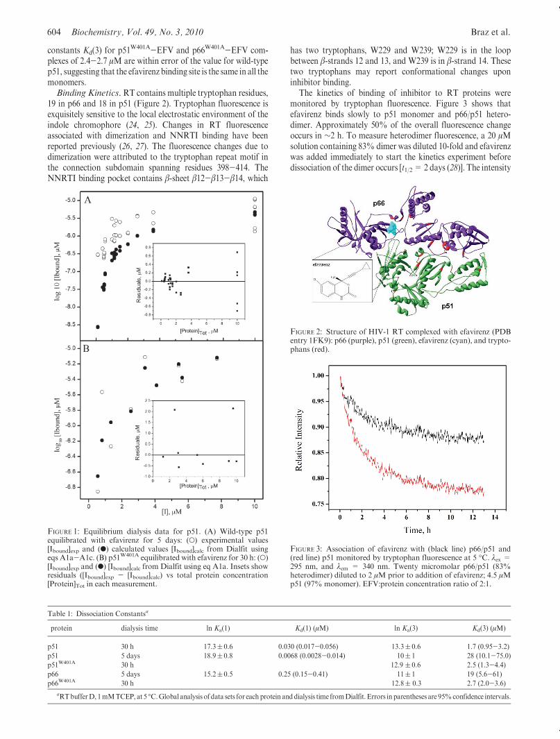
19 in p66 and 18 in p51 (Figure 2). Tryptophan fluorescence isexquisitely sensitive to the local electrostatic environment of theindole chromophore (24, 25). Changes in RT fluorescenceassociated with dimerization and NNRTI binding have beenreported previously (26, 27). The fluorescence changes due todimerization were attributed to the tryptophan repeat motif inthe connection subdomain spanning residues 398-414. TheNNRTI binding pocket contains β-sheet β12-β13-β14, which
has two tryptophans, W229 and W239; W229 is in the loopbetween β-strands 12 and 13, and W239 is in β-strand 14. Thesetwo tryptophans may report conformational changes uponinhibitor binding.
The kinetics of binding of inhibitor to RT proteins weremonitored by tryptophan fluorescence. Figure 3 shows thatefavirenz binds slowly to p51 monomer and p66/p51 hetero-dimer. Approximately 50% of the overall fluorescence changeoccurs in ∼2 h. To measure heterodimer fluorescence, a 20 μMsolution containing 83% dimer was diluted 10-fold and efavirenzwas added immediately to start the kinetics experiment beforedissociation of the dimer occurs [t1/2= 2 days (28)]. The intensity
FIGURE 1: Equilibrium dialysis data for p51. (A) Wild-type p51equilibrated with efavirenz for 5 days: (O) experimental values[Ibound]exp and (b) calculated values [Ibound]calc from Dialfit usingeqs A1a-A1c. (B) p51W401A equilibrated with efavirenz for 30 h: (O)[Ibound]exp and (b) [Ibound]calc from Dialfit using eq A1a. Insets showresiduals ([Ibound]exp - [Ibound]calc) vs total protein concentration[Protein]Tot in each measurement.
Table 1: Dissociation Constantsa
protein dialysis time ln Ka(1) Kd(1) (μM) ln Ka(3) Kd(3) (μM)
p51 30 h 17.3( 0.6 0.030 (0.017-0.056) 13.3( 0.6 1.7 (0.95-3.2)
p51 5 days 18.9( 0.8 0.0068 (0.0028-0.014) 10( 1 28 (10.1-75.0)
p51W401A 30 h 12.9 ( 0.6 2.5 (1.3-4.4)
p66 5 days 15.2( 0.5 0.25 (0.15-0.41) 11( 1 19 (5.6-61)
p66W401A 30 h 12.8( 0.3 2.7 (2.0-3.6)
aRTbufferD, 1mMTCEP, at 5 �C.Global analysis of data sets for each protein anddialysis time fromDialfit. Errors in parentheses are 95%confidence intervals.
FIGURE 3: Association of efavirenz with (black line) p66/p51 and(red line) p51 monitored by tryptophan fluorescence at 5 �C. λex =295 nm, and λem = 340 nm. Twenty micromolar p66/p51 (83%heterodimer) diluted to 2 μM prior to addition of efavirenz; 4.5 μMp51 (97% monomer). EFV:protein concentration ratio of 2:1.
FIGURE 2: Structure of HIV-1 RT complexed with efavirenz (PDBentry 1FK9): p66 (purple), p51 (green), efavirenz (cyan), and trypto-phans (red).

Article Biochemistry, Vol. 49, No. 3, 2010 605
change for the heterodimer is approximately half that of themonomer, consistent with an effect on tryptophan residues inonly one subunit.
Two kinetic mechanisms have been used to account for theslow binding of inhibitors to enzymes [Scheme 2 (29, 30)].Mechanism A depicts direct binding of inhibitor I to enzymeE, where the association and dissociation rate constants k1 andk-1, respectively, are inherently slow. Mechanism B depicts aninduced-fit model with fast equilibration of inhibitor and enzymeto form an intermediate complex EI, followed by slow isomer-ization of the EI complex to form the final complex EI*. Todiscriminate between the two mechanisms, the observed rateconstant kobs from the progress curve of the enzyme reaction isdetermined as a function of inhibitor concentration. A plot ofkobs versus inhibitor concentration is linear for mechanismA andhyperbolic for mechanism B.
Progress curves for efavirenz binding to p66W401A, p51W401A,and p66/p51 were measured at multiple inhibitor concentrations.Figure 4 shows the set of curves for p51W401A. The solid lines arethe fits to eq 2a to yield values of kobs. Similar curves wereobtained for p66W401A and p66/p51. Figure 5 shows the plots ofkobs versus inhibitor concentration. The linear fits are consistentwith mechanism A, where
kobs ¼ k-1 þ k1½I� ð3Þ
Values of the rate constants k1 and k-1 calculated from the slopesand intercepts of Figure 5 are given in Table 2. All three proteinshave similar association rate constants k1 of ∼13.5 M-1 s-1.Additionally, the dissociation rate constants k-1 were 5.9-8.1� 10-5 s-1, corresponding to a t1/2 of∼2.7 h. Having defined thebinding modality of efavirenz, we calculated values of Kd(3)
app
from the ratio of k-1/k1.The kinetics of dissociation of p66W401A- and p51W401A-EFV
complexes were measured under essentially irreversible condi-tions, so that at equilibrium, <3% of the monomer-EFVcomplex is present (Figure 6).Dissociation rate constants k-1(diss)of∼9.0� 10-5 s-1 (t1/2∼ 2.1 h) were obtained for bothmonomersfrom fitting the data to eq 2b. The k-1(diss) values fromkinetics measurements are close to the k-1 values determinedfrom the plots of kobs versus [I] (Table 2). The equilibriumdissociation constants Kd(3) of 6.6 μM calculated from theratio of the rate constants k-1(diss)/k1 for binding to themonomerare 2.5-fold higher than the values determined by equilibriumdialysis.Fluorescence Quantum Yields. Fluorescence quantum
yields of dimerization-defective monomers and wild-type hetero-dimer weremeasured in the absence andpresence of efavirenz. Tomeasure the extent of quenching, most of the protein must bebound to efavirenz. The quantum yields of monomer-EFVcomplexes were measured on solutions containing 1 μM mono-mer and 30 μMefavirenz equilibrated for 30 h at 5 �C, giving 94%monomer-EFV complex. The quantum yields of p66W401A andp51W401A monomers are the same within error (Table 3).Efavirenz binding decreases the quantum yield of bothmonomers
by a factor of 3. To measure the quantum yield of the hetero-dimer, we diluted a 20 μMsolution containing 83%dimer 50-foldand scanned it immediately as described above. The quantumyield of the heterodimer is approximately 25% lower than that ofthe monomers. The quantum yield of the p66/p51-EFV complex
Scheme 2: Mechanisms of Slow Binding Inhibition
FIGURE 4: Progress curves for binding of p51W401A to efavirenzmonitored by tryptophan fluorescence at 5 �C. λex = 295 nm, andλem=340 nm: 2.5 μMp51W401Awith (green line) 5, (blue line) 8, (redline) 11, and (black line) 14 μM efavirenz. Data acquired at 30 sintervals for the first 4-5 h and then at 5 min intervals. Data were fitto eq 2a to yield kobs.
FIGURE 5: Progress curve analysis of binding of efavirenz to (2) p66/p51, (red circles) p51W401A, and (blue squares) p66W401A. Lines arefits to eq 3. Error bars give the range of average values from twoexperiments.
Table 2: Kinetic Parametersa
protein
k1b
(M-1 s-1)
k-1b
(�10-5 s-1)
Kd(3)appc
(μM)
k-1(diss)d
(�10-5 s-1)
p51W401A 13.7( 0.7 5.9( 0.7 4.2( 0.5 9.1( 0.2
p66W401A 13.4( 0.6 8.1( 0.8 6.0( 0.3 8.9( 0.2
p66/p51 13.3( 0.9 6.7( 0.9 5.0( 0.3 nd
aRT buffer D, 1 mM TCEP, at 5 �C. bCalculated from eq 3 by linearregression of data in Figure 5. cKd(3)
app= k-1/k1.dCalculated from eq 2b.
Errors give the range of average values from two experiments.

606 Biochemistry, Vol. 49, No. 3, 2010 Braz et al.
was measured in solutions containing 20 μMp66/p51 and 40 μMefavirenz equilibrated for 1week at 5 �Cprior to dilution. Becauseefavirenz enhances dimerization 25-fold and bindsmore tightly tothe dimer than to the monomer, this solution contains 98% p66/p51-EFV complex. Efavirenz binding to heterodimer onlyquenches the fluorescence by a factor of 1.6.Native Gel Electrophoresis. BN-PAGE has been used to
monitor dimerization of RT proteins in the absence and presenceof efavirenz (21). The slow dissociation rate of efavirenz (t1/2 ∼ 2h) makes it possible to visualize binding of [14C]efavirenz tomonomer on gels. p66W401A and wild-type p66 were incubatedwith a 0.7:1.0 ratio of [14C]efavirenz to protein. Thewild-type p66concentration was 5 μM or approximately 53% homodimer.Lane 1 shows binding of [14C]efavirenz to the p66W401A mono-mer. Lane 2 shows binding of [14C]efavirenz to a mixture ofwild-type p66 monomer and p66/p66 homodimer. Lastly, lane 3shows enhancement of dimerization by efavirenz after equilibra-tion of wild-type p66 with [14C]efavirenz and excess cold efavir-enz for 1 week, giving 91% p66/p66-EFV complex. Thus, BN-PAGE supports the conclusions from equilibrium dialysis thatefavirenz binds to RT monomers as well as homodimers.
Nevirapine has been reported to have disparate effects onRT dimerization. Yeast two-hybrid experiments indicate asmall enhancement of dimerization, whereas urea denaturationstudies find no effect (16, 23). BN-PAGE was performed usingp66 incubated with excess efavirenz or nevirapine for 1 week.Figure 7B shows that both NNRTIs enhance dimerization, with
efavirenz having the stronger effect. These results are consistentwith the findings of the yeast two-hybrid experiments.
DISCUSSION
Although RT has been extensively studied for almost twodecades, new functions continue to be discovered for thisenigmatic enzyme. This paper reports two novel functions: (1)efavirenz, and presumably also other NNRTIs, binds to mono-meric forms ofRT, and (2) efavirenz is a slowbinding inhibitor ofmonomers and heterodimer. The biological significance ofmonomer binding is unknown at present. NNRTIs have beenfound to affect both early and late stages of theHIV-1 replicationcycle by multiple mechanisms (31, 32). Efavirenz interacts at thelevel of reverse transcription by inhibiting DNA polymeraseactivity, enhancing polymerase-dependent RNase H activity (30-DNA-directed), and partially inhibiting polymerase-independentRNase H activity (50-RNA-directed). It also inhibits plus-strandinitiation by affecting the ability of RT to bind the RNApolypurine tract. During late stages of HIV-1 replication, efavir-enz enhances processing and homodimerization of a 90 kDa Polpolyprotein in a yeast two-hybrid system and enhances intracel-lular processing of Gag and Gag-Pol precursor polyproteins inHIV-1-transfected cells (11). By increasing the processing of thesepolyproteins, efavirenz lowers viral production due to decreasedlevels of full constructs for incorporation into a budding particle.Essential to the above processes is defining the binding propertiesof the species, whether monomer or dimer, to which efavirenzbinds. Drug design requires an immense understanding of thetarget. This study suggests that monomeric forms of RT may bepotential targets for HIV-1 therapeutics. It also sparks develop-ment of high-throughput screening assays based on p66 and p51monomers in the evaluation of binding of new drugs to wild-typeand drug resistance mutant RTs.
The two crystal structures of RT-EFV complexes show 1:1binding stoichiometry (9, 33). Currently, no crystal structures
FIGURE 6: Dissociation of (red line) p51W401A-EFV and (blue line)p66W401A-EFV complexes monitored by tryptophan fluorescence at5 �C. λex= 295 nm, and λem=340 nm. Black curves are fits to eq 2b.Data acquired at 30 s intervals for the first 4-5 h and then at 5 minintervals.
Table 3: Quantum Yieldsa
protein Φ
p51W401A 0.14( 0.01
p51W401A-EFV complex 0.05( 0.02
p66W401A 0.15 ( 0.01
p66W401A-EFV complex 0.05( 0.02
p66/p51 0.11( 0.01
p66/p51-EFV complex 0.07( 0.01
NATA 0.23
aRT buffer D, 1 mM TCEP, at 5 �C. λex = 295 nm. Errors are standarddeviations of four experiments.
FIGURE 7: Blue Native polyacrylamide gel electrophoresis of p66 inthe absence and presence ofNNRTIs. (A)Monomer and homodimerbinding to [14C]EFV: (lane 1) 2 μM p66W401A incubated for 2 h with[14C]EFV, (lane 2) 5 μM p66 incubated for 2 h with [14C]EFV, and(lane 3) 5 μMp66 incubated for 1 weekwith [14C]EFV. (B)Wild-typep66 incubated for 1 week in the absence and presence of NNRTI:(lane 1) nativemarkers, (lane 2) 0.8 μMp66, (lane 3) 3 μMp66 and 25μM EFV, and (lane 4) 3 μM p66 and 25 μMNVP.

Article Biochemistry, Vol. 49, No. 3, 2010 607
are available for homodimers or monomers of RT. Equili-brium dialysis indicated a 1:1 stoichiometry for p66/p66- andp51/p51-EFV complexes. A 1:1 binding stoichiometry formonomer-EFV complexes was also obtained by equilibriumdialysis for wild-type p51 and dimerization-deficient p66W401A
and p51W401A, albeit with an affinity lower than that of thehomodimers (Table 1). The apparent free energies of binding ofefavirenz to homodimers at 5 �C (ΔG278=-RT lnKa) are-35.1kJ/mol for the p66/p66-EFV complex and-43.6 kJ/mol for thep51/p51-EFV complex. The more favorable binding energy ofthe p51/p51 homodimer may be attributed to better contactsbetween efavirenz and the protein or more facile formation of thebinding pocket in a dimer lacking two RNase H domains. Allmonomers have similar efavirenz binding energies ΔG278 of ∼-30 kJ/mol, which is 5-12 kJ/mol less favorable than that forbinding to homodimers.
The dissociation constants Kd(1) and Kd(3) from equilibriumdialysis (Table 1) and Kd(2) and Kd(4) from previous sedimenta-tion equilibrium experiments allow us to complete the thermo-dynamic linkage of NNRTI binding and subunit dimerizationproposed for RT (14). In the closed cycle of Scheme 1, ΔG = 0.Substituting ΔG278 gives
-RT ln Kað2Þ-RT ln Kað3Þ ¼ -RT ln Kað1Þ-RT ln Kað4Þð4Þ
In the case of p66, the left side of eq 4 sums to-68( 6 kJ/mol andthe right side to-64( 4 kJ/mol. In the case of p51, the left side ofeq 4 totals -64 ( 4 kJ/mol and the right side -63 ( 6 kJ/mol.These results for homodimers confirm the hypothesis thatNNRTI binding is coupled to subunit dimerization. Thus, wecan calculate the dissociation constant of the p66/p51-EFVcomplexKd(1) from the cycle in Scheme 1, whereKd(2) andKd(4)are the dissociation constants of the heterodimer in the presenceand absence of efavirenz (14), respectively, and Kd(3) is thedissociation constant of the p66- or p51-EFV complex. Re-arranging eq 4 gives RT lnKa(1) = 38( 6 kJ/mol orKd(1) = 92( 5 nM. Most studies of binding of efavirenz to RT haveemployed polymerase activity assays, conducted in the presenceof template/primer and dNTP (12, 34, 35). Maga et al. (35)reported a dissociation constant of 150 nM for the free RT-EFVcomplex extracted from enzymatic data. Geitmann et al. (36)measured binding of several NNRTIs to immobilized wild-typeand drug resistance mutant RTs by SPR at 25 �C in buffercontaining 0.005% surfactant and 3% (v/v) DMSO. An overalldissociation constant of 45 nM was obtained for binding ofefavirenz to wild-type RT.
The binding kinetics monitored by tryptophan fluorescenceestablish that efavirenz is a slow, tight binding inhibitorof RT. Dissociation rate constants k-1(diss) of 9.0 � 10-5 s-1
(t1/2 = 2.1 h) were obtained for monomer-EFV complexes.Association rate constants k1 of 13.5 M-1 s-1 were obtainedfor the reversible, direct binding of efavirenz to both monomersand heterodimer (Scheme 2, mechanism A). This suggests thatNNRTI binding occurs by an analogous process for monomericand dimeric species of RT, despite the difference in efavirenzbinding affinity. Slow binding inhibitors described in the litera-ture follow either direct binding or conformational changeinhibition models (Scheme 2). For example, small aza sugarinhibitors ofβ-glucosidase and yeast isomaltase bind by the directmechanism A with association rate constants ranging from 23 to7.3 � 104 M-1 s-1 (37). These inhibitors also have slow
dissociation rate constants of 0.16-6.7 � 10-2 s-1. By contrast,peptide R-keto acid analogues are slow binding inhibitors ofhepatitis C virus NS3 protease that bind by induced-fit mechan-ism B (38). These inhibitors undergo rapid equilibration with theenzyme in the first step of EI complex formation with a k1 of6.5 � 107 M-1 s-1 and a k-1 of 0.2 s
-1. The subsequent step is aslow isomerization to EI* with a k2 of 1.7-7.5 � 10-3 s-1 and ak-2 of 0.57-1.8 � 10-5 s-1 or a t1/2 of 11-48 h.
A few previous reports noted the slow onset of inhibition byNNRTIs. For example, 5-20 min preincubation periods of RTwith NNRTIs were required to witness inhibition of polymeraseand RNase H activity (31, 35, 39, 40). The slow association rateconstants reported here (Table 2) could be caused by a con-formational selection step involving exclusive binding of aweaklypopulated conformer of either protein or inhibitor. The chemicalstructure of efavirenz is provided in Figure 2. The benzoxazinonering system is rigid with free rotation of the cyclopropyl ethynylgroup (41). Efavirenz is in the same position in the binding pocketin both crystal structures of the wild-type RT-EFV complex.However, the cyclopropyl ethynyl group is rotated ∼100� in thedrug resistance mutant K103NRT-EFV complex relative to theposition in the wild-type structures (9, 33). The rigidity of theefavirenz core together with the ability of the binding pocket toaccommodate different orientations of the cyclopropyl ethynylgroup excludes conformational selection of the inhibitor as theculprit.
A selected-fit model has been proposed in which a conforma-tional pre-equilibration of the protein precedes inhibitor bind-ing (36, 42). In this model for slow binding, efavirenz would bindpreferentially to a less populated conformer of RT proteins.Productive collisions occurring between inhibitor and this con-former would induce a slow shift in the conformational equilib-rium favoring formation of the binding pocket and subsequentlythe EI complex. A less likely alternative would be severeorientation effects resulting in unproductive collisions of E andI that slow formation of the EI complex in the direct bindingmechanism A. A selected-fit model gave the best fit to the SPRdata for wild-type RT and efavirenz with an overall associationrate constant kon of 5.5� 104 M-1 s-1. This kon value is 3 ordersof magnitude faster then our association rate constant k1 forbinding of efavirenz to p66/p51. The dissociation rate constantkoff of ∼2.3 � 10-3 s-1 is ∼20-fold faster then the k-1 valueobtained from progress curve analysis. A probable explanationfor the faster binding kinetics is the different solution conditionsused in the SPR assay. Osmolytes such as detergents, organicsolvents, and salts affect protein solution structure and interac-tions (43, 44).
The NNRTI binding pocket is absent from structures of RTandRT-substrate complexes (45, 46). The polymerase domain iscomposed of four subdomains: fingers, palm, thumb, and con-nection. The structure of RT is asymmetric, and the subdomainsare in different orientations in the p66 and p51 subunits [Figure 8,top (45)]. In RT-NNRTI complexes, the binding pocket is in thepalmof the p66 subunit of the heterodimer [Figure 8, bottom (9)].LPC software identifies 14 residues that contact efavirenz in theNNRTI binding pocket of the p66 subunit: L100, K101, K103,V106, V179, Y181, Y188, G190, F227,W229, L234, H235, P236,and Y318 (47). The efavirenz contact residues are highlighted inFigure 8 to illustrate the location of these residues in the twosubunits. Although the contact residues are clustered in bothsubunits, these residues do not form a binding pocket in the p51subunit. The p66/p66 and p51/p51 homodimers may also have

608 Biochemistry, Vol. 49, No. 3, 2010 Braz et al.
asymmetric structures, because they both have polymeraseactivity (3). Additionally, the homodimers probably have similarNNRTI binding pockets in the subunit that binds the inhibitor,because p66 and p51 have identical amino acid sequences andsimilar folding patterns of the polymerase subdomains. Giventhat p66 and p51 monomers are capable of forming a competentNNRTI binding pocket, presumably the polymerase domain ofbothmonomers must adopt a conformation analogous to that ofthe p66 subunit in the heterodimer.
ACKNOWLEDGMENT
We are grateful to Ms. Kathryn J. Howard for suggesting thegel experiment with [14C]efavirenz and for advice about site-directedmutagenesis and protein purification.We also thankDr.ClareWoodward for suggesting thatNNRTIs bind tomonomersand Drs. Vernon Anderson and Jonathan Karn for helpfuldiscussions.
APPENDIX
Dialfit: MathematicalModels andDataAnalysis (by Leslie A.Holladay).
There are two different ways in which the model equationsmay be written: case A and case B.Case A. For case A, the following equilibrium constants are
defined as
Pþ I ¼ PI Kað3Þ ¼ ½PI�=ð½P�½I�Þ ðA1aÞPþP ¼ PP Kað4Þ ¼ ½PP�=½P�2 ðA1bÞ
PPþ I ¼ PPI Kað1Þ ¼ ½PPI�=ð½PP�½I�Þ ðA1cÞThe equilibrium dialysis experiment involves computing the totalmolar concentration of inhibitor bound to both monomer anddimer [Ibound], by subtracting the concentration of free inhibitoroutside the dialysis bag [Iout]= [I] from the total concentration ofinhibitor inside the bag [Iin] as in eq 1. For the data analysis, theobservable variable we wish to model is [Ibound]. The totalconcentration of protein [P]tot inside the bag is presumed to beknown to higher precision than the free and bound inhibitorconcentrations. [Ibound] is the observable to be predicted knowing[I] and [P]tot along with the current estimates forKa(1),Ka(3), andKa(4). Dialfit is applicable to any experiment that provides datafor [Ibound] and [I], knowing [P]tot.
To compute [Ibound], the concentration of free protein mono-mer [P] must first be computed. The conservation of massequation is
½P�tot ¼ ½P� þ ½PI� þ 2½PP� þ 2½PPI� ðA2ÞRearranging and substituting terms with the equilibrium con-stants from eqs A1 result in a quadratic in [P]
a½P�2 þ b½P� þ c ¼ 0 ðA3aÞwhere
a ¼ 2Kað4Þþ 2Kað1ÞKað4Þ½I� ðA3bÞ
b ¼ 1þKað3Þ½I� ðA3cÞ
c ¼ -½P�tot ðA3dÞThe only physical meaningful root of eq A3a is the positive one.The fitting function may be written as
½Ibound� ¼ Kað3Þ½P�½I� þKað1ÞKað4Þ½P�2½I� ðA4ÞThe values of [Ibound] determined over a wide range of totalinhibitor and total protein concentrations are globally fitted to eqA4.Note thatKa(1) andKa(4) cannot be separated, and thus, onevalue must be a fixed parameter.Case B. For case B, two equilibrium constants are defined in
eqs A1a and A1b; the third equilibrium constant is defined as
PIþP ¼ PPI Kað2Þ ¼ ½PPI�=ð½PI�½P�Þ ðA5ÞHere too, [Ibound] is the observable to be predicted knowing [I] and[P]tot, butwith the current estimates forKa(2),Ka(3), andKa(4). Theconservation of mass equation A2 and quadratic equations A3a,A3c, and A3d are the same as in case A; eq A3b becomes
a ¼ 2Kað4Þþ 2Kað2ÞKað3Þ½I� ðA6ÞAgain, the physically meaningful root of eq A3a is the positive one.The fitting function is
½Ibound� ¼ Kað3Þ½P�½I� þKað2ÞKað3Þ½P�2½I� ðA7ÞNote that cases A and B are mathematically equivalent from the
FIGURE 8: Structures of HIV-1 (top) RT (PDB entry 1DLO) and(bottom) the RT-EFV complex (PDB entry 1FK9). Polymerasedomains: fingers (blue), palm (red), thumb (green), and connection(orange) subdomains; RNase H domain (magenta). Efavirenz(yellow) and contact residues (gray) are shown using the van derWaals radii.

Article Biochemistry, Vol. 49, No. 3, 2010 609
relationship Ka(1)Ka(4) = Ka(2)Ka(3). The two parameters Ka(2)and Ka(3) are not separable, and thus, the value of Ka(2) must befixed.Weighted Least-Squares and Parameter Standard Er-
rors. The global data sets have a very wide range of values for[Ibound] and [I]. For radioactive counts, the relative standarddeviation is equal to
√N/N, whereN is the number of counts (48).
The relative variance is equal to 1/N. [Ibound] is computed fromthe difference in counts inside and outside the bag. Define thenumber of counts inside the bag as Ni and the number of countsoutside the bag asNo. Then the relative variance of the differenceNi-No= (NiþNo)/(NiNo). In the situation in which the valuesfor [Ibound] and [I] vary over several orders of magnitude, it isessential to use weighted least squares because the variance willalso vary over a wide range (49). The weightWj of any (Ni - No)jvalue is the reciprocal of the variance, soWj= [(NiNo)/(NiþNo)]j.The actual weights used are normalized so that the ΣjWj = 1 tocause the returned residual error in the fit to be correct. It is clearfrom Figure 1 (insets) that the errors in [Ibound] are veryheteroscedastic. The errors in the fitted variable vary greatlywith respect to the independent variable. Only if the errors arehomoscedastic is unweighted least squares appropriate.
Standard errors for the parameter values were computed usingthe “balanced bootstrap” with 100 trials (50, 51). If any of theindividual bootstrap trials was more than three standard devia-tions away from the parameter estimate, that trial was deleted asan outlier and the standard deviation recomputed. The 95%confidence intervals are computed using the standard deviationfrom the estimated parameter value.
REFERENCES
1. di Marzo Veronese, F., Copeland, T. D., DeVico, A. L., Rahman, R.,Oroszlan, S., Gallo, R. C., and Sarngadharan, M. G. (1986)Characterization of highly immunogenic p66/p51 as the reversetranscriptase of HTLV-III/LAV. Science 231, 1289–1291.
2. Hizi, A., McGill, C., and Hughes, S. H. (1988) Expression of soluble,enzymatically active, human immunodeficiency virus reverse tran-scriptase inEscherichia coli and analysis of mutants.Proc. Natl. Acad.Sci. U.S.A. 85, 1218–1222.
3. Restle, T., M€uller, B., and Goody, R. S. (1990) Dimerization ofhuman immunodeficiency virus type 1 reverse transcriptase. J. Biol.Chem. 265, 8986–8988.
4. Restle, T., M€uller, B., and Goody, R. S. (1992) RNase H activity ofHIV reverse transcriptase is confined exclusively to the dimeric forms.FEBS Lett. 300, 97–100.
5. Le Grice, S. F. J., Naas, T., Wohlgensinger, B., and Schatz, O. (1991)Subunit-selective mutagenesis indicates minimal polymerase activityin heterodimer-associated p51 HIV-1 reverse transcriptase. EMBO J.10, 3905–3911.
6. Tsai, C. H., Lee, P. Y., Stollar, V., and Li, M. L. (2006) Antiviraltherapy targeting viral polymerase. Curr. Pharm. Des. 12, 1339–1355.
7. De Clercq, E. (1998) The role of non-nucleoside reverse transcriptaseinhibitors (NNRTIs) in the therapy of HIV-1 infection. Antiviral Res.38, 153–179.
8. Kohlstaedt, L. A., Wang, J., Friedman, J. M., Rice, P. A., and Steitz,T. A. (1992) Crystal structure at 3.5 A resolution of HIV-1 reversetranscriptase complexed with an inhibitor. Science 256, 1783–1790.
9. Ren, J., Milton, J., Weaver, K. L., Short, S. A., Stuart, D. I., andStammers, D. K. (2000) Structural basis for the resilience of efavirenz(DMP-266) to drug resistance mutations in HIV-1 reverse transcrip-tase. Structure 8, 1089–1094.
10. Merluzzi, V. J., Hargrave, K. D., Labadia, M., Grozinger, K., Skoog,M., Wu, J. C., Shih, C.-K., Eckner, K., Hattox, S., Adams, J.,Rosenthal, A. S., Faanes, R., Eckner, R. J., Koup, R. A., and Sutton,J. L. (1990) Inhibition ofHIV-1 replication by a nonnucleoside reversetranscriptase inhibitor. Science 250, 1411–1413.
11. Figueiredo, A., Moore, K. L., Mak, J., Sluis-Cremer, N., de Bethune,M.-P., and Tachedjian, G. (2006) Potent nonnucleoside reversetranscriptase inhibitors target HIV-1 Gag-Pol. PLoS Pathog. 2,1051–1059.
12. Young, S. D., Britcher, S. F., Tran, L. O., Payne, L. S., Lumma, W.C., Lyle, T. A., Huff, J. R., Anderson, P. S., Olsen, D. B., Carroll, S.S., Pettibone,D. J., O’Brien, J. A., Ball, R.G., Balani, S.K., Lin, J. H.,Chen, I.-W., Schleif, S. A., Sardana, V. V., Long,W. J., Byrnes, V.W.,and Emini, E. A. (1995) L-743,726 (DMP-266): A novel, highly potentnonnucleoside inhibitor of the human immunodeficiency virus type 1reverse transcriptase. Antimicrob. Agents Chemother. 39, 2602–2605.
13. Tachedjian, G., Orlova, M., Sarafianos, S. G., Arnold, E., and Goff,S. P. (2001) Nonnucleoside reverse transcriptase inhibitors are che-mical enhancers of dimerization of the HIV type 1 reverse transcrip-tase. Proc. Natl. Acad. Sci. U.S.A. 98, 7188–7193.
14. Venezia, C. F., Howard, K. J., Ignatov, M. E., Holladay, L. A., andBarkley, M. D. (2006) Effects of efavirenz binding on the subunitequilibria ofHIV-1 reverse transcriptase.Biochemistry 45, 2779–2789.
15. Sluis-Cremer, N., Arion, D., and Parniak, M. A. (2002) Destabiliza-tion of the HIV-1 reverse transcriptase dimer upon interaction withN-acyl hydrazone inhibitors. Mol. Pharmacol. 62, 398–405.
16. Sluis-Cremer, N., Dmitrienko, G. I., Balzarini, J., Camarasa, M.-J.,and Parniak, M. A. (2000) Human immunodeficiency virus type 1reverse transcriptase dimer destablilization by 1-{spiro[40 0-amino-20 0,20 0-dioxo-10 0,20 0-oxathiole-50 0,30-[20,50-bis-O-(tert-butyldimethylsi-lyl)-β-D-ribofuranosyl]]}-3-ethylthymine. Biochemistry 39, 1427–1433.
17. Le Grice, S. F. J., Cameron, C. E., and Benkovic, S. J. (1995)Purification and characterization of human immunodeficiency virustype 1 reverse transcriptase. Methods Enzymol. 262, 130–144.
18. Ignatov, M. E., Berdis, A. J., Le Grice, S. F. J., and Barkley, M. D.(2005) Attenuation of DNA replication by HIV-1 reverse transcrip-tase near the central termination sequence. Biochemistry 44, 5346–5356.
19. Tachedjian, G., Aronson, H.-E., de los Santos, M., Seehra, J.,McCoy, J. M., and Goff, S. P. (2003) Role of residues in thetryptophan repeatmotif forHIV-1 reverse transcriptase dimerization.J. Mol. Biol. 326, 381–396.
20. Chen, R. F. (1967) Fluorescence quantum yields of tryptophan andtyrosine. Anal. Lett. 1, 35–42.
21. Braz, V.A., andHoward, K. J. (2009) Separation of protein oligomersby blue native gel electrophoresis. Anal. Biochem. 388, 170–172.
22. Ghosh,M., Jacques, P. S., Rodgers, D.W., Ottman,M., Darlix, J. L.,and Le Grice, S. F. J. (1996) Alterations to the primer grip of p66HIV-1 reverse transcriptase and their consequences for template-primer utilization. Biochemistry 35, 8553–8562.
23. Tachedjian, G., Aronson, H.-E. G., and Goff, S. P. (2000) Analysis ofmutations and suppressors affecting interactions between subunits ofthe HIV type 1 reverse transcriptase. Proc. Natl. Acad. Sci. U.S.A. 97,6334–6339.
24. Vivian, J. T., and Callis, P. R. (2001) Mechanisms of tryptophanfluorescence shifts in proteins. Biophys. J. 80, 2093–2109.
25. Callis, P. R., Petrenko, A., Mui~no, P. L., and Tusell, J. R. (2007) Abinitio prediction of tryptophan fluorescence quenching by proteinelectric field enabled electron transfer. J. Phys. Chem. B 111, 10335–10339.
26. Divita, G., Restle, T., andGoody, R. S. (1993) Characterization of thedimerization process of HIV-1 reverse transcriptase heterodimerusing intrinsic protein fluorescence. FEBS Lett. 324, 153–158.
27. Barnard, J., Borkow, G., and Parniak, M. A. (1997) The thiocarbox-anilide nonnucleoside UC781 is a tight-binding inhibitor of HIV-1reverse transcriptase. Biochemistry 36, 7786–7792.
28. Venezia, C. F., Meany, B. J., Braz, V. A., and Barkley, M. D. (2009)Kinetics of association and dissociation of HIV-1 reverse transciptasesubunits. Biochemistry 48, 9084–9093.
29. Copeland, R. A. (2005) Evaluation of Enzyme Inhibitors in DrugDiscovery, John Wiley & Sons, New York.
30. Szedlacsek, S. E., and Duggleby, R. G. (1995) Kinetics of slow andtight-binding inhibitors. Methods Enzymol. 249, 144–180.
31. Sluis-Cremer, N., and Tachedjian, G. (2008) Mechanisms of inhibi-tion of HIV replication by non-nucleoside reverse transcriptaseinhibitors. Virus Res. 134, 147–156.
32. Grobler, J. A., Dornadula, G., Rice, M. R., Simcoe, A. L., Hazuda,D. J., and Miller, M. D. (2007) HIV-1 reverse transcriptase plus-strand initiation exhibits preferential sensitivity to non-nucleosidereverse transcriptase inhibitors in vitro. J. Biol. Chem. 282, 8005–8010.
33. Lindberg, J., Sigurdsson, S., L€owgren, S., Andersson, H. O., Sahlberg,C., Nor�een, R., Fridborg, K., Zhang, H., and Unge, T. (2002)Structural basis for the inhibitory efficacy of efavirenz (DMP-266),MSC194 and PNU142721 towards the HIV-1 RT K103N mutant.Eur. J. Biochem. 269, 1670–1677.
34. Xia, Q., Radzio, J., Anderson, K. S., and Sluis-Cremer, N. (2007)Probing nonnucleoside inhibitor-induced active site distortion in

610 Biochemistry, Vol. 49, No. 3, 2010 Braz et al.
HIV-1 reverse transcriptase by transient kinetics analysis.Protein Sci.16, 1728–1737.
35. Maga, G., Ubiali, D., Salvetti, R., Pregnolato, M., and Spadari, S.(2000) Selective interaction of the human immunodeficiency virustype 1 reverse transcriptase nonnucleoside inhibitor efavirenz and itsthio-substituted analog with different enzyme-substrate complexes.Antimicrob. Agents Chemother. 44, 1186–1194.
36. Geitmann,M., Unge, T., andDanielson, H. (2006) Interaction kineticcharacterization of HIV-1 reverse transcriptase non-nucleoside in-hibitor resistance. J. Med. Chem. 49, 2375–2387.
37. Lohse, A., Hardlei, T., Jensen, A., Plesner, I. W., and Bols, M. (2000)Investigation of the slow inhibition of almond β-glucosidase and yeastisomaltase by 1-azasugar inhibitors: Evidence for the ‘direct binding’model. Biochem. J. 349, 211–215.
38. Narjes, F., Brunetti,M., Colarusso, S., Gerlach, B., Koch,U., Biasiol,G., Fattori, D., De Francesco, R.,Matassa, V.G., and Steink€uhler, C.(2000) R-Ketoacids are potent slow binding inhibitors of the hepatitisC virus NS3 protease. Biochemistry 39, 1849–1861.
39. Borkow, G., Fletcher, R. S., Barnard, J., Arion, D., Motakis, D.,Dmitrienko, G. I., and Parniak, M. A. (1997) Inhibition of theribonuclease H and DNA polymerase activities of HIV-1 reversetranscriptase by N-(4-tert-butylbenzoyl)-2-hydroxy-1-napthaldehydehydrazone. Biochemistry 36, 3179–3185.
40. Spence, R. A., Kati, W. M., Anderson, K. S., and Johnson, K. A.(1995) Mechanism of inhibition of HIV-1 reverse transcriptase bynonnucleoside inhibitors. Science 267, 988–993.
41. Wang, D.-E., Rizzo, R. C., Tirado-Rives, J., and Jorgensen, W. L.(2001) Antiviral drug design: Computational analyses of the effects of
the L100I mutation for HIV-RT on the binding of NNRTIs. Bioorg.Med. Chem. Lett. 11, 2799–2802.
42. Weikl, T. R., and von Deuster, C. (2009) Selected-fit versus induced-fit protein binding: Kinetic differences and mutational analysis.Proteins 75, 104–110.
43. Pegram, L. M., and Record, M. T., Jr. (2008) Thermodynamic originof Hofmeister ion effects. J. Phys. Chem. B 112, 9428–9436.
44. R€osgen, J., Pettitt,B.M., andBolen,D.W. (2005)Protein folding, stability,and solvation structure in osmolyte solutions. Biophys. J. 89, 2988–2997.
45. Hsiou, Y., Ding, J., Das, K., Clark, A. D., Jr., Hughes, S. H., andArnold, E. (1996) Structure of unligandedHIV-1 reverse transcriptaseat 2.7 A resolution: Implications of conformational changes forpolymerization and inhibition mechanism. Structure 4, 853–860.
46. Huang, H., Chopra, R., Verdine, G. L., and Harrison, S. C. (1998)Structure of a covalently trapped catalytic complex of HIV-1 reversetranscriptase: Implications for drug resistance. Science 282, 1669–1675.
47. Sobolev, V., Sorokine, A., Prilusky, J., Abola, E. E., andEdelman,M.(1999) Automated analysis of interatomic contacts in proteins. Bioin-formatics 15, 327–332.
48. Willard, H.H.,Merritt, L. L., Jr., andDean, J. A. (1965) InstrumentalMethods of Analysis, Vol. 256, D. Van Nostrand Company, New York.
49. Draper, N. R., and Smith, H. (1966) Applied Regression Analysis, pp77-81, Wiley & Sons, New York.
50. Efron, B., and Tibshrirani, R. (1986) Bootstrap methods for stan-dards errors, confidence intervals, and other measures of statisticalaccuracy. Stat. Sci. 1, 54–77.
51. Hall, P. (1992) The Bootstrap and Edgeworth Expansion, p 293,Springer-Verlag, New York.