effect of an industrial chemical waste on the uptake file · web viewolive leaf extract modulates...
TRANSCRIPT

J. Serb. Chem. Soc. 76 (9) 1207–1218 (2011) UDC 633.852.73+581.45+66.094.3+JSCS–4196 591.4:611.33/.36
Original scientific paper
Olive leaf extract modulates cold restraint stress-induced oxidative changes in rat liver
DRAGANA DEKANSKI1*, SLAVICA RISTIĆ1, NEVENA V. RADONJIĆ2, NATAŠA D. PETRONIJEVIĆ2, ALEKSANDAR DEKANSKI3# and DUŠAN M. MITROVIĆ4
1Biomedical Research, R&D Institute, Galenika a.d., Belgrade, 2Institute for Medical and Clinical Biochemistry, School of Medicine, University of Belgrade, 3Institute for Chemistry, Technology and Metallurgy, University of Belgrade and 4Institute for Medical Physiology
“Richard Burian”, School of Medicine, University of Belgrade, Serbia
(Received 4 February, revised 18 March 2011)
Abstract: Recently, the beneficial effects of different single doses of standardized dry olive (Olea europaea L.) leaf extract (OLE) in cold restraint stress (CRS)-in-duced gastric lesions in rats and its influence on oxidative parameters in gastric mucosa were demonstrated. The present study was undertaken to investigate the long-term pretreatment efficacy of OLE and its potential in the modulation of CRS-induced oxidative changes at the liver level. The experimental animals were divided into four groups, i.e., control, OLE-treated, CRS non-treated and CRS treated with OLE (CRS+OLE) groups. CRS caused severe gastric lesions in all non-pretreated animals and two-week pretreatment with OLE (80 mg per kg of body weight) attenuated stress-induced gastric lesions significantly. The malondialdehyde (MDA) level as an index of lipid peroxidation, superoxide dismutase (SOD) and catalase (CAT) activities were measured spectrophotometrically in liver tissue homogenates. The MDA level was increased in the CRS group and significantly decreased in the CRS+OLE group. The SOD and CAT activities were significantly decreased in the CRS group. In the CRS+OLE group, the activities of these two enzymes were significantly increased in comparison with the CRS group. The results obtained indicate that long-term supplementation with OLE provides oxidant/antioxidant balance in liver during stress condition.
Keywords: olive leaf; cold restraint stress; oxidative stress; liver.
INTRODUCTION
Stress, a condition in an organism that results from the action of several stressors, has been reported to affect the progression and severity of different di-seases. Environmental stress has been shown to be associated with altered ho-meostasis that may lead to oxidant–antioxidant imbalance. Under normal condi-
*Corresponding author. E-mail: [email protected]#Serbian Chemical Society member.doi: 10.2298/JSC110204107D
1207
1208 DEKANSKI et al.
tions, antioxidant systems of the cell minimize the perturbations caused by free radicals. When free radicals generation is increased to an extent that overcomes the cellular antioxidants, the result is oxidative stress.
It is known that immobilization stress accelerated by cold (a combination of two potent stressors) can disrupt the balance in an oxidant/antioxidant system and cause oxidative damage to several tissues by altering the enzymatic and non-en-zymatic antioxidant status, protein oxidation and lipid peroxidation.1
As a new strategy for alleviating oxidative damage, interest has been grow-ing in the usage of natural antioxidants. It was suggested that many of the nega-tive effects of oxidative stress are diminished upon supplementation with certain dietary antioxidants, such as vitamins and other non-nutrient antioxidants, e.g., plant flavonoids.2,3 There is an increasing interest in total medicinal plant ex-tracts, the greatest value of which may be due to the constituents that contribute to the modulation of the oxidative balance in vivo. Additionally, the obvious ad-vantage of total plant extracts is that they are easily attainable products, without purification of any of the fractions needed in order to apply them in possible pre-vention/treatment of diseases.2 Reasonably, the application of large quantities of plant extracts as dietary supplements is not to be recommends before assessment of important health issues regarding use of plant phenolics in general, and plant flavonoids in particular.3
Olive tree (Olea europaea L.) leaf has been used in traditional, folk medi-cine, in Mediterranean countries, particularly as an antimicrobial and cardiopro-tective agent.4 Recently, experimental animal studies demonstrated its antihyper-tensive, anti-atherogenic, anti-inflammatory, hypoglycemic and hypocholestero-lemic effects; all of these positive effects were at least partly related to its anti-oxidative action.5 Moreover, its antihypertensive effect in patients with stage-1 hypertension was confirmed in a double-blind, randomized, parallel and active-controlled clinical study.6
The main constituent of olive leaf is oleuropein, one of the iridoide monoter-penes, which is thought to be responsible for its pharmacological effects. In addi-tion, olive leaf contains triterpenes (important amounts of oleanolic and maslinic acid followed by minor concentrations of ursolic acid, erythrodiol, and uvaol), flavonoids (luteolin, apigenine, rutin, etc.), chalcones (olivin, olivine diglucoside) and tannins.4,7–9 It is its chemical content that makes olive leaf one of the most potent natural antioxidants. Oleuropein has remarkable antioxidant activity in vi-tro, comparable to a hydrosoluble analog of tocopherol10, as do other consti-tuents of olive leaf.11 Literature data on olive phenolics is mainly concerned with purified compounds, while the antioxidant properties of total extract have been poorly investigated. Being a complex mixture of compounds, the study of the protective effect of the total extract could be more representative than those of single components. It was shown that a total olive leaf extract had an antioxidant

OLIVE LEAF MODULATES HEPATIC OXIDATIVE STRESS 1209
activity higher than those of vitamin C and vitamin E, due to the synergy be-tween flavonoids, oleuropeosides and substituted phenols.12
The beneficial properties of olive leaf are further enhanced by the good ab-sorption of its phenolic constituents and their significant levels in the circula-tion.13,14
Although several studies have investigated the effects of cold-restraint stress on the antioxidant system and induction of lipid peroxidation in several tissues, to date, no information is available regarding the antioxidant effect of total dry olive leaf extract (OLE) on cold restraint stress (CRS)-induced hepatic oxidative stress. The influence of stress on the liver is also of interest from the clinical point of view, because stress plays a potential role in aggravating liver diseases in general and hepatic inflammation in particular, probably through the generation of reactive oxygen species (ROS). Thus, in this preclinical investigation, the ef-fect of CRS on oxidative stress and antioxidant defense system and the possible protective effect of OLE in rat liver tissue were investigated.
EXPERIMENTALMaterials
Olive leaf extract EFLA® 943, standardized to 18–26 % of oleuropein, was purchased from Frutarom Switzerland Ltd. (Wadenswil, Switzerland). The extract was manufactured from the dried leaves of Olea europaea L., applying an ethanol (80 % m/m) extraction proce-dure. After a patented filtration process (EFLA® Hyperpure), the crude extract was dried. The stability and microbiological purity were confirmed by the manufacturer. Further compre-hensive phytochemical analysis of the extract was previously realized and it was found to contain oleuropein (19.8 %), total flavonoids (0.29 %), including luteolin-7-O-glucoside (0.04 %), apigenine-7-O-glucoside (0.07 %) and quercetin (0.04 %), as well as caffeic acid (0.02 %), and tannins (0.52 %).15 The same batch of EFLA® 943 was used in the present study. Hydro-gen peroxide and thiobarbituric acid (TBA) were purchased from Sigma-Aldrich (Schnelldorf, Germany). All other reagents used in biochemical analysis were obtained from Merck (Darm-stadt, Germany).Animals, stress induction and stomach evaluation
Twenty-four male Wistar rats from the Biomedical Research Center, R&D Institute, Ga-lenika a.d. (Belgrade, Serbia), weighing 250±20 g, were used. The rats were housed 3 per cage under constant environmental conditions (20–24 °C; 12 h light/dark cycle), and were given ad libitum access to standard pelleted food and water. This study was approved by the Ethical Committee of the Medical School, University of Belgrade, and run in accordance to the state-ments of the European Union regarding the handling of experimental animals (86/609/EEC).
The animals were randomly divided into 4 groups each consisting of 6 rats: control, OLE, CRS, and CRS+OLE.
The first, control group received 1 ml of distilled water intragastrically (i.g.) using a me-tal tube for gavage for 14 days. This was the group of normal, healthy animals without any drug pretreatment or stress induction.
The OLE group received olive leaf extract (80 mg kg-1 daily, i.g.) dissolved in distilled water for 14 days.
1210 DEKANSKI et al.
The CRS group received distilled water i.g. for 14 days, and it was the group exposed to cold restraint stress on the last day of the experiment.
The CRS+OLE group received OLE (80 mg kg-1, daily, i.g.) dissolved in distilled water for 14 days. The last dose was administrated 120 min prior to CRS induction.
Day before the stress induction all experimental animals were placed in individual meta-bolic cages and were fasted for 24 h, but had free access to water. The rats from CRS and CRS+OLE group were immobilized in individual restraint boxes without the possibility of visual contact16 and subjected to cold (4 ±1 °C) stress for 3.5 h. This regimen of cold-restraint stress was reported to produce gastric ulcers in food-deprived rats,17,18 as well as plasma and hepatic tissue lipid peroxidation.19
At the end of this period, the animals were sacrificed under ether anesthesia, the ab-domen was opened by midline incision and the liver and the stomach were removed. The sto-mach was opened along the greater curvature, rinsed gently with water and pinned open for macroscopic examination. The number and severity of gastric lesions were evaluated accord-ing to the following rating scale:20 0 – no lesion; 1 – mucosal edema and petechiae; 2 – from 1 to 5 small lesions (1–2 mm); 3 – more than 5 small lesions or 1 intermediate lesion (3–4 mm); 4 – 2 or more intermediate lesions or 1 large lesion (greater than 4 mm); 5 – perforated ulcers. The sum of the total scores divided by the number of animals in the group was expressed as the ulcer index (UI)±standard deviation (SD). The percent inhibition of UI in relation to the CRS group was estimated from formula:
% Inhibition = (1–(UIOLE+CRS/UICRS))×100
Biochemical examination of liverThe liver from each animal was weighed, transferred to the ice-cooled test tube and
homogenized by Ultra-Turrax T25 (Janke & Kunkel GmbH. & Co., IKA®-Labortechnik, Staufen, Germany) in 20 mmol l-1 Tris buffer, pH 7.4, containing 5 mmol butylated hydroxy-toluene to prevent new lipid peroxidation that could occur during the homogenization. The homogenate was then centrifuged at 12000 rpm at 4 °C (Megafuge 2.0.R, Heraeus, Germany) for 10 min. The supernatant was aliquoted and stored at –80 °C until determination of the total protein, malondialdehyde (MDA), superoxide dismutase (SOD) and catalase (CAT).
The biochemical parameters were determined spectrophotometrically (UV–Vis spectro-photometer HP 8453, Agilent Technologies, Santa Clara, CA).
The protein content of the liver tissue samples was estimated by the method of Lowry et al.21 using bovine serum albumin as the standard.
Lipid peroxidation was determined at 533 nm and the MDA level was measured by the thiobarbituric acid (TBA) test according to the method suggested by Buege and Aust.22
The SOD activity in the liver was determined by measuring the inhibition of auto-oxida-tion of adrenaline at pH 10.2 at 30 °C by the method of Misra and Fridovich. 23 One unit of SOD activity represented the amount of SOD which was necessary to cause a 50 % inhibition of adrenaline auto-oxidation.
Activity of catalase in liver was determined according to the procedure of Goth24 by fol-lowing the absorbance of hydrogen peroxide at 230 nm and pH 7.0.Statistical analysis
All results are expressed as means±SD. Statistical analysis was realized using one-way ANOVA and the post hoc Tukey test. Values of P less than 0.05 were considered as signi-ficant.

OLIVE LEAF MODULATES HEPATIC OXIDATIVE STRESS 1211
RESULTS
Effect of OLE on gastric lesions induced by cold restraint stressCold restraint stress produced visible gastric lesions in all animals in the
CRS group. They were located mostly in the corpus. No visible lesions deve-loped in the non-secretary part of the rat stomach, which is a well-known res-ponse to CRS. Moreover, after opening, hemorrhagic content was found in sto-mach lumens. Following 3.5 h of cold-restraint stress, the average ulcer score in the non-pretreated group was very high (4.33±0.85). OLE (80 mg kg–1) signi-ficantly prevented the gastric mucosal lesions induced by cold-restraint stress. Ulcer index (UI) was 1.33±0.52. The percent of inhibition in UI was 70 %. Only gastric mucosal edema and petechiae were seen in almost all (5 of 6) animals in this experimental group. No visible sign of ulceration was observed in the control animals or in OLE group of animals.
Effect of OLE pretreatment on lipid peroxidation and the activity of antioxidative enzymes in the liver
Cold restraint stress significantly increased level of lipid peroxidation in liver, evaluated as MDA mg–1 protein (174.32±11.16 nmol mg–1 protein vs. 134.75±10.02 nmol mg–1 protein in the control (P<0.05)). The liver tissue MDA was reduced significantly by pretreatment with 80 mg kg–1 of OLE (137.47±21.06 nmol mg–1 protein). The difference was not statistically significant between the control and the OLE group (Fig. 1).
Fig. 1. Effect of intragastric pretreatment with olive leaf extract (OLE), applied at a dose of80 mg kg-1 for two weeks, on the malondialdehyde concentration (nmol (mg protein)-1)
in the liver of rat exposed to cold restraint stress (CRS); * indicates statistical significance(P < 0.05) of the difference in the MDA concentrations in non-pretreated rats exposed to
CRS as compared to the control animals; ** indicates statistical significance (P < 0.05) ofthe difference in MDA concentrations in pretreated rats as compared to the
CRS-exposed rats without pretreatment.

1212 DEKANSKI et al.
As shown in Fig. 2, the SOD activity averaged 115.70±3.10 U mg–1 protein in healthy rat liver. Following exposure of the rats to CRS, a significant decrease in SOD activity to the value of 99.07±3.09 U mg–1 protein was observed. OLE administration significantly reduced the decrease in SOD activity in the CRS+OLE group (109.70±5.12 U mg–1 protein) but did not influence the enzyme activity in the group of non-stressed animals.
Fig. 2. Effect of intragastric pretreatment with olive leaf extract (OLE), applied at a dose of80 mg kg-1 for two weeks, on the superoxide dismutase (SOD) activity (U (mg protein)-1) in
the liver of rat exposed to cold restraint stress (CRS); *indicates statistical significance(P < 0.05) of the difference in SOD activity in non-pretreated rats exposed to CRS as com-
pared to the control animals; **indicates statistical significance (P<0.05) of the difference in SOD activity in pretreated rats as compared to the CRS-exposed rats without pretreatment.
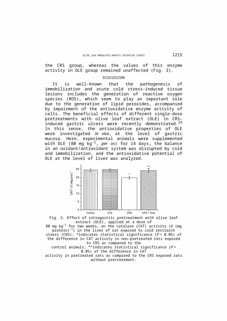
Catalase activity in the gastric mucosa was also significantly decreased after 3.5 h of CRS (24.53±1.00 U mg–1 protein in the control group vs. 19.77±0.8 U mg–1 protein in the CRS group). Pretreatment with OLE significantly reduced the decrease in CAT activity in the CRS group, whereas the values of this enzyme activity in OLE group remained unaffected (Fig. 3).
DISCUSSION
It is well-known that the pathogenesis of immobilization and acute cold stress-induced tissue lesions includes the generation of reactive oxygen species (ROS), which seem to play an important role due to the generation of lipid pero-xides, accompanied by impairment of the antioxidative enzyme activity of cells. The beneficial effects of different single-dose pretreatments with olive leaf ex-tract (OLE) in CRS-induced gastric ulcers were recently demonstrated.25 In this sense, the antioxidative properties of OLE were investigated in vivo, at the level of gastric mucosa. Here, experimental animals were supplemented with OLE (80 mg kg–1, per os) for 14 days, the balance in an oxidant/antioxidant system was

OLIVE LEAF MODULATES HEPATIC OXIDATIVE STRESS 1213
disrupted by cold and immobilization, and the antioxidative potential of OLE at the level of liver was analyzed.
Fig. 3. Effect of intragastric pretreatment with olive leaf extract (OLE), applied at a dose of80 mg kg-1 for two weeks, on the catalase (CAT) activity (U (mg protein)-1) in the liver of rat
exposed to cold restraint stress (CRS); *indicates statistical significance (P < 0.05) of the difference in CAT activity in non-pretreated rats exposed to CRS as compared to the
control animals; **indicates statistical significance (P < 0.05) of the difference in CATactivity in pretreated rats as compared to the CRS exposed rats without pretreatment.
In the present study, the protective activity of OLE was confirmed via CRS--induced gastric ulcers. CRS caused severe gastric lesions in animals pretreated with physiological saline solution. Seventy percent of inhibition of ulcer index, related to the non-pretreated group, was obtained in animals pretreated with 80 mg kg–1 of OLE for two weeks. In a previous trial, long-term pretreatment with the same dose was effective in absolute ethanol-induced gastric lesions, and a potent antioxidative activity of OLE in rat gastric mucosa was evidenced.26
The dose of OLE used in the present study was selected with respect to the nutraceutical/pharmaceutical level. It was calculated according to a clinical study in which OLE EFLA® 943 at 1000 mg daily effectively reduced blood pressure.6
For the extrapolation of the dosage from human to rat, food intake rather than bo-dy weight was taken as the criterion.27 Briefly, the estimated quantity of OLE ex-pressed per unit of human diet is 2 mg g–1 dry food, daily (1000 mg of OLE per 500 g dry food). For an adult rat (250 g b.w.) which consumes approximately 10 g of dry food daily, the consumption corresponded to an OLE dose of 80 mg kg–1.
Since lipid peroxidation is a well-established mechanism of cellular injury, changes in the malondialdehyde (MDA) concentrations were measured as an in-dicator of lipid peroxidation. MDA in liver tissue homogenates was found to be significantly increased in the rats exposed to CRS, when compared with the con-trol group. These results are in agreement with previous findings, which were re-lated to stress-induced lipid peroxidation in plasma and liver of experimental ani-mals.1,19 OLE pretreatment significantly decreased the MDA level in the liver of

1214 DEKANSKI et al.
CRS rats. Recent reports showed antioxidant properties of the main phenolics present in olive extracts, i.e., oleuropein and hydroxytyrosol (the main metabolite of oleuropein). Thus, both phenolics showed a substantial degree of inhibition of lipid peroxidation in vivo in rat liver microsomes28 and in oxidative stress in-duced by hydrogen peroxide or xanthine oxidase in vitro.29 The OLE used in the present study contained 19.8 % of oleuropein. Therefore, ≈ 16 mg kg–1 of oleu-ropein was administered daily per rat. It is interesting that this dose of oleuropein was the effective dose in attenuation of hepatic oxidative damage (thiobarbituric acid-reactive substances (TBARS) reduction) in alloxan-diabetic rats. The admi-nistration of oleuropein- and hydroxytyrosol-rich extracts for 4 weeks signifi-cantly decreased the serum glucose and cholesterols levels and restored the anti-oxidant perturbations in liver.30 The present results showed that the antioxidant system in liver was also affected by CRS. It was previously reported that CRS caused the inhibition of the activity of antioxidant enzymes in the liver and in the other tissues in rat.1,31 Six hours of immobilization stress caused a decrease in the liver levels of SOD, CAT and glutathione, while the level of MDA was in-creased, compared with non-stressed control rats.32 Cold stress (CS) alone also alters homeostasis, resulting in the creation of reactive oxygen species which lead to alterations in the antioxidant defense system. The MDA levels were increased, whereas the SOD, CAT and glutathione peroxidase activities and total gluta-thione level were significantly decreased in the CS group.33 In the present study, OLE administered to rats prior to stress induction attenuated the inhibition of SOD and CAT activity and, thus, additionally implicated its role in the modu-lation of the oxidative balance in liver. Jemai et al.34 reported that hydroxytyro-sol purified from olive tree leaves increased the SOD and CAT activities in the liver of Wistar rats fed a cholesterol-rich diet. In addition, in the same study, the content of TBARS in liver, heart, kidney, and aorta decreased significantly when hydroxytyrosol was orally administered. The antioxidative effect of the total OLE most probably resulted from the ability of its constituents to scavenge re-active oxygen species produced in CRS, which initiate lipid peroxidation. The performed phytochemical analysis of OLE EFLA® 943 showed a high oleuropein content together with other important constituents, i.e., apigenine-7-O-glucoside, luteolin-7-O-glucoside, quercetin and caffeic acid, as well as low concentration of tannins.15 The radical scavenging abilities of tannins, oleuropein and its metabolites, apigenine-7-O-glucoside, luteolin-7-O-glucoside, caffeic acid, and for total olive leaf extract were already reported.12 Furthermore, quercetin, luteolin-7-glucoside and caffeic acid showed protective potential against oxidative damages induced by tert-butyl hydroperoxide (t-BHP) in HepG2 cells. All the tested phenolic compounds were found to significantly decrease lipid peroxidation and prevent glutathione depletion induced by t-BHP; quercetin also significantly decreased DNA damage.35
OLIVE LEAF MODULATES HEPATIC OXIDATIVE STRESS 1215
Antioxidants are substances that delay or prevent the oxidation of cellular oxidizable substrates. The various antioxidants exert their effect by scavenging superoxide, or by activation of a battery of detoxifying/defensive proteins.36 The present finding that orally applied OLE had a significant protective effect in he-patic oxidative stress is very important. The phenolic compounds from OLE are food constituents, thus ingestion is the natural route for their intake. The potential of other antioxidant nutrients, such as vitamins A (retinol), E (tocopherol) and C (ascorbic acid) individually and in combination (vitamin E + C) to modulate res-traint stress-induced oxidative changes in liver was investigated, and the vitamin post-stress treatment was found to be effective in combating hepatic oxidative stress.32 In the present study, the additional intake of OLE influenced neither lipid peroxidation nor the activity of the investigated antioxidative enzyme in healthy animals. It was recently reported that supplementation of vitamin E under non-stress condition decreased liver SOD, however the hydrogen peroxide con-tent (as the subsequent product of SOD activity) and catalase activity remained unchanged.37 The results obtained in the present study are partly in agreement with these. Not only is the liver the main target for nutrient antioxidants once ab-sorbed from the gastrointestinal tract, but it is also the major place for their me-tabolism. Therefore, studies dealing with the metabolism of OLE constituents in liver should be given priority.
Several studies showed that phenolic substances increased the expression of SOD and CAT enzymes at the transcriptional level.38 Recently, some individual and combined olive leaf phenolics exhibited SOD-like activity in vitro.39 Fur-thermore, it was reported that oleuropein reduced the expression of a number of hepatic genes involved in oxidative stress responses and detoxification of lipid peroxidation products and pro-inflammatory cytokine genes.40 According to the biochemical parameters, the first step has been made in that it was shown that OLE synchronized antioxidant enzymes and inhibited lipid peroxidation in liver. Thus, this effect is worthy of further investigation of its potential in the regu-lation of cellular signaling, gene expression and protein synthesis; in one word, investigation at the molecular level.
CONCLUSIONS
Bearing in mind the significance of stress and its potential role in the aggra-vation of liver diseases, natural hepatoprotective antioxidants are of great impor-tance. A standardized olive leaf extract decreased lipid peroxidation in the liver of rats exposed to cold restraint stress. Superoxide dismutase and catalase en-zyme activity were increased in liver tissue homogenates. The obtained results indicate that olive leaf exhibits a potent antioxidative activity at the level of liver.

1216 DEKANSKI et al.
ABBREVIATIONSCAT – Catalase;CRS – Cold restraint stress;MDA – Malondialdehyde;OLE – Olive leaf extract;ROS – Reactive oxygen species;SOD – Superoxide dismutase;TBA – Thiobarbituric acid.
Acknowledgements. This study was supported by the Ministry of Science and Technological Development of the Republic of Serbia (Grant No.175096).
и з в о д
ЕКСТРАКТ ЛИСТА МАСЛИНЕ МОДУЛИШЕ ОКСИДАТИВНЕ ПРОМЕНЕ ИНДУКОВАНЕ ИМОБИЛИЗАЦИОНИМ СТРЕСОМ УБРЗАНИМ
ХЛАДНОЋОМ У ЈЕТРИ ПАЦОВА
ДРАГАНА ДЕКАНСКИ1, СЛАВИЦА РИСТИЋ1, НЕВЕНА В. РАДОЊИЋ2, НАТАША Д. ПЕТРОНИЈЕВИЋ2, АЛЕКСАНДАР ДЕКАНСКИ3 и ДУШАН М. МИТРОВИЋ4
1Biomedicinska ispitivawa, Institut za istra`ivawe i razvoj, Galenika a.d., Pasterova 2, 11000 Beograd, 2Institut za medicinsku i klini~ku biohemiju, Medicinski fakultet,
Univerzitet uBeogradu, Pasterova 2, 11000 Beograd, 3Institut za hemiju, tehnologiju i metalurgiju,
Wego{eva 12, 11000 Beograd i 4Institut za medicinsku fiziologiju “Rihard Burijan”, Medicinski fakultet, Univerzitet u Beogradu, Vi{egradska 26, 11 000 Beograd
Недавно су показани повољни ефекти различитих појединачних доза стандардизованог екстракта листа маслине (Olea europaea L.) на желудачне лезије пацова индуковане имо-билизационим стресом убрзаним хладноћом (CRS) и његов утицај на параметре оксида-тивног стреса у желудачној слузници. У овој студији испитиван је ефекат дуготрајног пре-третмана листом маслине и његов потенцијал у модулацији CRS-ом индукованих оксида-тивних промена на нивоу јетре. Експерименталне животиње су подељене у четири групе: контролна, третирана екстрактом листа маслине (OLE), CRS и група код које је CRS тре-тиран екстрактом (CRS+OLE). CRS је проузроковао озбиљна оштећења желуца код свих непретретираних животиња, а двонедељни претретман са OLE (80 mg kg -1 т.т.) значајно је смањио стресом индуковане желудачне лезије. Малондиалдехид (MDA), као показатељ липидне пероксидације, активности супероксид-дисмутазе (SOD) и каталазе (CAT) мерени су спектрофотометријски у хомогенатима ткива јетре. Ниво MDA се значајно повећао у CRS групи, а потом значајно смањио у CRS+OLE групи. Активности SOD и CAT биле су зна-чајно смањене у CRS групи, док је у CRS+OLE групи животиња активност ова два ензима знатно повећана у поређењу са CRS групом. Добијени резултати указују на то да дуготрајно прехрањивање екстрактом листа маслине помаже успостављање оксидативне–антиокси-дативне равнотеже у јетри током стреса.
(Примљено 4. фебруара, ревидирано 18. марта 2011)
REFERENCES1. E. Sahin, S. Gümüşlü, Clin. Exp. Pharmacol. Physiol. 34 (2007) 4252. B. Dimitrios, Trends Food Sci. Technol. 17 (2006) 505

OLIVE LEAF MODULATES HEPATIC OXIDATIVE STRESS 1217
3. B. Halliwell, Cardiovasc. Res. 73 (2007) 3414. Physician’s Desk References for Herbal Medicine, Medical Economics Company,
Montvale, NJ, 2000, p. 5565. S. N. El, S. Karakaya, Nutr. Rev. 67 (2009) 6326. E. Susalit, N. Agus, I. Effendi, R. R. Tjandrawinata, D. Nofiarny, T. Perrinjaquet-
Moc cetti, M. Verbruggen, Phytomedicine 18 (2011) 2517. J. Meirinhos, B. M. Silva, P. Valentao, R. M. Seabra, J. A. Pereira, A. Dias, P. B.
Andrade, F. Ferreres, Nat. Prod. Res. 68 (2005) 1898. A. P. Pereira, I. C. Ferreira, F. Marcelino, P. Valentao, P. B. Andrade, R. Seabra, L.
Estevinho, A. Bento, J. A. Pereira, Molecules 12 (2007) 11539. A. Guinda, M. Rada, T. Delgado, P. Gutiérrez-Adánez, J. M. Castellano, J. Agric.
Food. Chem. 58 (2010) 968510. E. Speroni, M. C. Guerra, A. Minghetti, N. Crespi-Perellino, P. Pasini, F. Piazza,
Phytother. Res. 12 (1998) S9811. R. Briante, M. Paturni, S. Terenziani, E. Bismuto, F. Febbraio, R. Nucci, J. Agric.
Food Chem. 50 (2002) 493412. O. Benavente-Garcia, J. Castillo, J. Lorente, A. Ortuno, J. A. Del Rio, Food Chem.
68 (2000) 45713. F. Visioli, C. Galli, F. Bornet, A. Mattei, R. Patelli, G. Galli, D. Caruso, FEBS Lett.
468 (2000) 15914. M. N. Vissers, P. L Zock, A. J. C. Roodenburg, R. Leenen, M. B. Katan, J. Nutr. 132
(2002) 40915. D. Dekanski, S. Janićijević-Hudomal, V. Tadić, G. Marković, I. Arsić, D. M.
Mitrović, J. Serb. Chem. Soc. 74 (2009) 36716. M. Popović, N. Popović, D. Bokonjić, S. Dobrić, Int. J. Neurosci. 91 (1997) 117. E. C. Senay, R. J. Levine, Proc. Soc. Exp. Biol. Med. 124 (1967) 122118. D. Das, R. K. Banerjee, Mol. Cell. Biochem. 125 (1993) 11519. C. Özer, S. Ercan, A. Babül, Z. S. Ercan, Turk. J. Biochem. 34 (2009) 3220. N. I. Büyükcoşkun, G. Güleç, B. C. Etöz, K. Ozlük, Turk. J. Gastroenterol. 18
(2007) 15021. O. H. Lowry, N. J. Rosenbrough, A. L. Farr, R. J. Randall, J. Biol. Chem. 193 (1951)
26522. J. A. Buege, S. D. Aust, Methods Enzymol. 52 (1978) 30223. H. P. Misra, I. Fridovich, J. Biol. Chem. 247 (1972) 317024. L. Goth, Clin. Chim. Acta 196 (1991) 14325. D. Dekanski, S. Janićijević-Hudomal, S. Ristić, N. V. Radonjić, N. D. Petronijević,
V. Piperski, D. M. Mitrović, Gen. Physiol. Biophys. 28 (2009) 13526. D. Dekanski, S. Ristić, D. M. Mitrović, Mediterr. J. Nutr. Metab. 2 (2009) 205 27. R. Rucker, D. Storms, J. Nutr. 132 (2002) 299928. V. R. Gutierrez, R. de la Puerta, A. Catalá, Mol Cell Biochem. 217 (2001) 3529. C. Manna, P. Galletti, V. Cucciolla, O. Moltedo, A. Leone, V. Zappia, J. Nutr. 127
(1997) 28630. H. Jemai, A. El Feki, S. Sayadi, J. Agric. Food Chem. 57 (2009) 879831. T. A. Shustanova, T. I. Bondarenko, N. P. Miliutina, Ross. Fiziol. Zh. Im. I M
Sechenova 90 (2004) 73 (in Russian)32. S. M. Zaidi, T. M. Al-Qirim, N. Banu, Drugs R D 6 (2005) 157

1218 DEKANSKI et al.
33. B. Ates, M. I. Dogru, M. Gul, A. Erdogan, A. K. Dogru, I. Yilmaz, M. Yurekli, M. Esrefoglu, Fundam. Clin. Pharmacol. 20 (2006) 283
34. H. Jemai, I. Fki, M. Bouaziz, Z. Bouallagui, A. El Feki, H. Isoda, S. Sayadi. J. Agric. Food Chem. 56 (2008) 2630
35. C. F. Lima, M. Fernandes-Ferreira, C. Pereira-Wilson, Life Sci. 79 (2006) 205636. J. M. Matés, Toxicology 153 (2000) 8337. S. F. Đurašević, J. Đorđević, N. Jasnić, I. Đorđević, P. Vujović, G. Cvijić, Arch.
Biol. Sci. Belgrade 62 (2010) 67938. J. Vina, C. Borras, M. C. Gomez-Cabrera, W. C. Orr, Free Radic. Res. 40 (2006)
11139. O. H. Lee, B. Y. Lee, Bioresour. Technol. 101 (2010) 375140. Y. Kim, Y. Choi, T. Park, Biotechnol. J. 5 (2010) 950.