effect of carbonate content and buffer type on calcium phosphate formation in sbf ... · ·...
TRANSCRIPT

J Mater Sci: Mater Med (2006) 17:697–707DOI 10.1007/s10856-006-9680-1
Effect of carbonate content and buffer type on calciumphosphate formation in SBF solutionsS. Jalota · S. B. Bhaduri · A. C. Tas
Received: 4 September 2004 / Accepted: 24 October 2005C© Springer Science + Business Media, LLC 2006
Coating of titanium-based biomedical devices with a layer ofcarbonated, apatitic calcium phosphate increases their bone-bonding ability. Simulated or synthetic body fluids (SBF)have the ability of forming apatitic calcium phosphates onthe immersed titanium alloys within a few days to 2 weeks.Apatite-inducing ability of 5 M NaOH-etched surfaces ofTi6A14V strips (10 × 10 × 1 mm) were tested by usingthree different SBF solutions all concentrated by a factor of1.5. SBF solutions used in this comparative study were: i)4.2 mM HCO3
− TRIS-HCl buffered SBF (conventional SBFor c-SBF), ii) 27 mM HCO3
− TRIS-HCl buffered SBF (Tas-SBF), and iii) 27 mM HCO3
− HEPES-NaOH buffered SBF(revised SBF or r-SBF). While the c-SBF of low carbonateion concentration (4.2 mM) was quite sluggish in forminga coat layer on Ti6A14V strips after 1 week of soaking at37◦C, Tas-SBF with a HCO3
− concentration exactly equal tothat of human plasma (27 mM) was able to fully coat theimmersed strips. Tas-SBF solution was able to coat even thevertically-suspended Ti6A14V strips in 1 week. The coatedstrips were characterized by using XRD, SEM, and FTIR.The differences in the coating morphology and surface cov-erage were assessed for each SBF solution of this study. Invitro characteristics of as-coated Ti6A14V strips was com-pared via the cell attachment, cell viability and total proteinamount tests by using rat osteoblasts (7 F2). In vitro tests pos-itively favored the calcium phosphate coatings of the TRIS-buffered, 27 mM HCO3
− SBF over those of the c- and r-SBFsolutions.
S. Jalota · S. B. Bhaduri · A. C. Tas (�)School of Materials Science and Engineering, ClemsonUniversity, Clemson, SC 29634, USAe-mail: c [email protected]
Introduction
SBF (simulated body fluid) solutions are able [1–3] to in-duce apatitic calcium phosphate formation on metals, ceram-ics or polymers (with proper surface treatments) immersedin them. SBF solutions, in close resemblance to the origi-nal Earle’s (EBSS) [4] and Hanks’ Balanced Salt Solution(HBSS) [5], were prepared to simulate the ion concentra-tions of human blood plasma. EBSS formulation of 1943,which has 26 mM of HCO3
− and a Ca/P molar ratio of 1.8,is relatively similar to today’s SBF [1–3] solutions. HBSSsolution has a Ca/P ratio of 1.62. EBSS and HBSS solutionsare derived from the physiological saline first developed byRinger in 1882 [6]. It was recently reported that HBSS so-lutions are also able to slowly induce apatite formation ontitanium [7], due to its low Ca/P ratio. For mimicking the ionconcentrations of human blood plasma, SBF solutions haverelatively low Ca2+ and HPO2−
4 concentrations of 2.5 mMand 1.0 mM, respectively [8]. pH values of SBF solutionswere fixed at the physiologic value of 7.4 by using buffers,such as TRIS (tris-hydroxymethyl-aminomethane)-HCl [3]or HEPES (2-(4-(2-hydroxyethyl)-1-piperazinyl)ethane sul-phonic acid)-NaOH [9, 10]. The buffering agent TRIS presentin conventional SBF (c-SBF) formulations, for instance, wasreported [11] to form soluble complexes with several cations,including Ca2+, which further reduces the concentration offree Ca2+ ions available for the real time calcium phosphatecoating. To the best of our knowledge, this behavior has notyet been reported for HEPES. HCO3
− concentration in SBFsolutions has been between 4.2 mM (equal to that of HBSS)[1] and 27 mM in revisited SBFs [12–14]. c-SBF, which wasfirst popularized by Kokubo in 1990 [1], can be regardedas a TRIS/HCl-buffered variant of HBSS, whose Ca/P mo-lar ratio was increased from 1.62 to 2.5. HBSS and c-SBFsolutions have the same low carbonate ion concentrations
Springer

698 J Mater Sci: Mater Med (2006) 17:697–707
Table 1 Ion concentrations of human plasma and synthetic solutions, mM
Blood Kokubo- Tas- Bigi- Kokubo-plasma Ringer6 EBSS4 HBSS5 c-SBF1 SBF12 SBF9 r-SBF10
Na+ 142.0 130 143.5 142.1 142.0 142.0 141.5 142.0K+ 5.0 4.0 5.37 5.33 5.0 5.0 5.0 5.0Ca2+ 2.5 1.4 1.8 1.26 2.5 2.5 2.5 2.5Mg2+ 1.5 0.8 0.9 1.5 1.5 1.5 1.5Cl− 103.0 109.0 123.5 146.8 147.8 125.0 124.5 103.0HCO3
− 27.0 26.2 4.2 4.2 27.0 27.0 27.0HPO2−
4 1.0 1.0 0.78 1.0 1.0 1.0 1.0SO2−
4 0.5 0.8 0.41 0.5 0.5 0.5 0.5Ca/P 2.5 1.8 1.62 2.5 2.5 2.5 2.5Buffer TRIS TRIS HEPES HEPESpH 7.4 6.5 7.2–7.6 6.7–6.9 7.25–7.4 7.4 7.4 7.4
(i.e., 4.2 mM). Tas et al. [12, 13] were the first in 1999 toraise the carbonate ion concentration in a TRIS-HCl bufferedSBF solution to 27 mM, while Bigi et al. [9] were the firstto do the same in a HEPES-NaOH buffered SBF solution.Table 1 summarizes these SBF compositions. Eagle’s min-imum essential medium (MEM) [15] and Dulbecco’s phos-phate buffer saline (PBS) [16], which are commonly used incell culture studies, may also be added to this table.
Dorozhkina et al. [17] underlined the influence of HCO3−
concentration in an SBF solution on the morphology andthickness of calcium phosphate coatings formed. They com-pared two types of TRIS-free SBF solutions in that study; (i)one with 4.2 mM HCO3
− and (ii) another SBF with 27 mMHCO3
− with the solution pH regulated by bubbling CO2.
The most important result drawn from this study was: “in-creasing the HCO3
− concentration in c-SBF from 4.2 to 27mM resulted in the formation of homogeneous and muchthicker carbonated hydroxyapatite layers” [17]. The samefact was also reported by Kim et al. [14] on PET substratesimmersed into r-SBF. Dorozhkina et al. [17] emphasized thatHEPES was rather unstable, in comparison to TRIS, and iteasily lost some of the initially present dissolved carbonates.Kokubo et al. [10], who developed the HEPES-buffered r-SBF recipe, also reported that r-SBF would release CO2 gasfrom the fluid, causing a decrease in HCO3
− concentration,and an increase in pH value, when the storage period waslong. Furthermore, they clearly stated that r-SBF would notbe suitable for long-term use in the biomimetic coating pro-cesses owing to its instability [10]. To accelerate the SBF-coating processes, solutions equal to 1.5 times the ionic con-centration of SBF were often used [18–20]. Habibovic etal. [21] prepared 5 × SBF-like solutions, without using anybuffering agents and also removing K+ and SO2−
4 ions fromtheir solutions. Recently, Tas et al. [22] demonstrated the suc-cessful use of 10 × SBF-like (i.e., 25 mM Ca2+ and 10 mMHPO2−
4 ) solutions in coating Ti6A14V surfaces at room tem-perature within 2 to 6 hours. Condensed (e.g., 5 or 10 times)
solutions similar to SBF are prone to form precipitates with ahigher carbonate content or with off-stoichiometric Ca/P mo-lar ratios [23–25]. The intention to coat otherwise bioinertmaterials (such as, PET [14] or PTFE [26]) should have beenthe formation of bonelike, carbonated (not greater than 6 to8% by weight) calcium phosphate layers with Ca/P molarratios in the range of 1.55 to 1.67.
r-SBF was previously tested in coating collagen fib-rils [27]. However, the in vitro apatite-inducing ability ofneither Tas-SBF [12] nor r-SBF [10] has yet been reportedon Ti6A14V substrates, in direct comparison to c-SBF. Themotivation for the present study stems from our interest infinding experimental evidence to the following questions:(a) do the use of different buffers (TRIS or HEPES) in SBFscause remarkable changes in the morphology or thickness ofthe calcium phosphate coat layers formed on Ti6A14V? (b)does the variation in HCO3
− concentration affect the apatite-inducing ability of SBFs? and (c) how would the in vitrotests of rat osteoblast response diffrentiate between coatingsformed by different SBFs?
Materials and methods
Ti6A14V strips (Grade 5, McMaster-Carr), with the dimen-sions of 10 × 10 × 1 mm, were used as substrates. The stripswere first abraded with a #1000 SiC paper (FEPA P#1000,Struers), and then washed three times, respectively with ace-tone, ethanol, and deionized water in an ultrasonic bath. Eachone of such strips was then immersed in 50 mL of a 5 M NaOHsolution at 60◦C for 24 hours in a glass bottle, followed bywashing with deionized water and drying at 40◦C.
Details of the SBF preparation routines are given inTable 2. Freshly prepared, 1.5× c-SBF [1], Tas-SBF [12],and r-SBF [10] solutions were used in coating experiments,unless otherwise noted. The order of addition of the reagentsto water is given in the first column of Table 2. The reagentamounts were reported in columns 3 through 5 of Table 2.
Springer

J Mater Sci: Mater Med (2006) 17:697–707 699
Table 2 1.5 × SBF preparation
Weight (g per L)
Order Reagent c-SBF1 Tas-SBF12 r-SBF1,10
1 NaCl 12.0540 9.8184 8.10452 NaHCO3 0.5280 3.4023 1.11003 Na2CO3 — — 3.06904 KCl 0.3375 0.5591 0.33755 K2HPO4·3H2O 0.3450 — 0.34506 Na2HPO4 — 0.2129 —7 MgCl2·6H2O 0.4665 0.4574 0.46658 1 M HCl 15 mL 15 mL —9 HEPES — — 17.892010 CaCl2·2H2O 0.5822 0.5513 0.582211 Na2SO4 0.1080 0.1065 0.108012 TRIS 9.0945 9.0855 —13 1 M HCl 50 mL 50 mL —14 1 M NaOH — — 0.8 mL
NaOH-treated Ti6A14V strips were soaked at 37◦C in50 mL of 1.5× c-SBF, Tas-SBF and r-SBF in tightly sealedPyrex® media bottles of 100 mL-capacity, for a period of 7, 14and 21 days. All the SBF solutions were replenished at every48 hours. Strips were removed from the SBF solutions at theend of respective soaking times, and washed with deionizedwater, followed by drying at 37◦C. The strips were placedeither “horizontally” on the base of the immersion bottles ordipped “vertically” into the solutions with a stainless steelwire [28]. Coated strips were examined by using an X-raydiffractometer (XRD; XDS 2000, Scintag Corp., Sunnyvale,CA), operated at 40 kV and 30 mA with monochromated CuK α radiation. X-ray data were collected at 2θ values from10◦ to 40◦ at a rate of 0.01◦ per minute. Fourier-transform in-frared (FTIR) analyses were performed directly on the coatedstrips (Nicolet 550, Thermo-Nicolet, Wobum, MA). Surfacemorphology of the sputter-coated (w/Pt) strips was evaluatedwith a scanning electron microscope (SEM; S-4700, HitachiCorp., Tokyo, Japan).
Calvarial rat osteoblast cells, designated 7 F2 (ATCC,Rockville, MD), were used for cell attachment studies onthe SBF-coated strips. Cells were first grown at 37◦C and5% CO2 in alpha MEM, augmented by 10% FBS. The cul-ture medium was changed every other day until the cellsreached a confluence of 90–95%. Osteoblasts were seeded ata density of 105 cells/cm2. Cell cytotoxicity measurementswere carried out after 24 hours, cell viability assessmentwas performed after 72 hours and total protein amount weremeasured after 7 days. Adhesion of the cells was quanti-fied 24 hours after seeding. Trypan blue was added andthe cells were counted using a light microscope (BX60,Olympus, Tokyo, Japan). Only cells that stain blue weredeemed necrotic because of plasma membrane damage. Forstatistics, all experiments were performed in triplicate where
n = 3. Analysis of variance was performed using the Tukey-Kramer multiple comparisons test. Osteoblast morphologyafter attachment was further examined using SEM. Prior toSEM investigations, samples were soaked in the primary fix-ative of 3.5% glutaraldehyde. Further, the cells were dehy-drated with increasing concentrations of ethanol (50%, 75%,90% and 100%) for 10 minutes each. Critical drying wasperformed according to the previously published procedures[29]. Samples were sputter-coated with Pt prior to the SEMimaging at 5 kV.
Results
Formation of a hydrated layer of sodium titanate on Ti6A14Vstrips was confirmed after 24 h of immersion in a 5 M NaOHsolution at 60◦C, as previously reported [30]. Alkali treat-ment also created a nano-scale roughness on the strip surfaceswith, as shown in Figure 1(a). XRD analysis of the alkali-treated strips affirmed the presence of that hydrous sodiumtitanate layer, Figure 1(b). During our preliminary studies,we also prepared 1×SBF solutions (i.e., c-, r-, and Tas-SBF)and tested the formation of calcium phosphates (CaP) onalkali-treated Ti6A14V strips for 1 week of soaking at 37◦C.There was almost no coating observed, regardless of the re-plenishment rate with these 1 × SBF solutions. For 1 ×SBFsolutions, more than 3 weeks of soaking is required to ob-serve only the onset of coating. To accelerate the coatingprocess, 1.5 ×SBF solutions were prepared (Table 2).
When the Ti6A14V strips were placed horizontally (i.e.,the strips were laying flat at the base of the bottles) in the1.5 × SBF-immersion bottles, precipitates forming in theSBF solutions coalesced on the surfaces of the strips toform a grape bunch-like morphology. SEM micrographs of
Springer

700 J Mater Sci: Mater Med (2006) 17:697–707
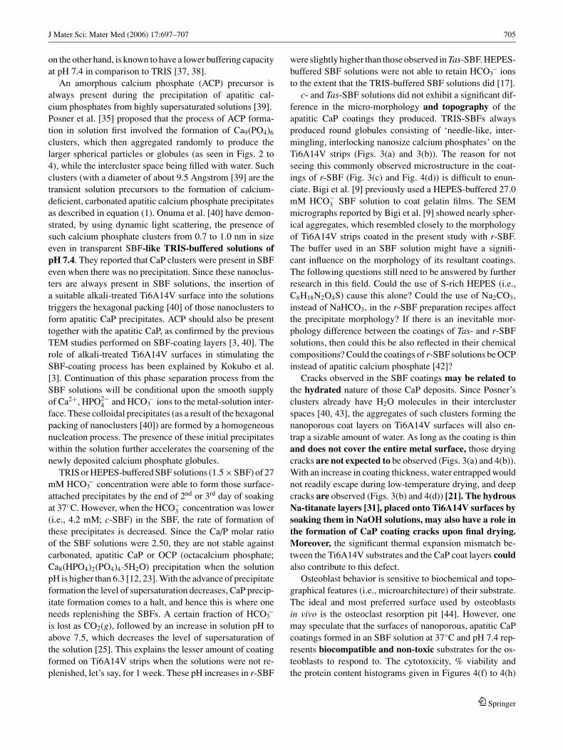
Fig. 1 5 M NaOH-treated Ti6A14V strips; (a) surface morphology, (b)XRD data, ∗Ti6A14V, ◦ Hydrous Na-titanate peaks.
Figures 2(a) through 2(c) show the surfaces of horizontally-placed strips soaked for 1 week, with replenishment on everyother day. c-SBF solutions were not able to form a uniformcoating (Fig. 2a), only occasional, agglomerated globules ofCaP were observed on the strip surfaces. Tas-SBF solutionswere found (Fig. 2b) to form an undercoat-like layer first,and then on top of it CaP globules were grown. r-SBF so-lutions were not able to form that undercoat layer (Fig. 2c).Figure 2(d), on the other hand, depicts the XRD data directlycollected from the strip surfaces after 3 weeks of horizontalimmersion in the respective SBF solutions. The sporadic andthin coating observed with the c-SBF solutions was not ableto suppress the X-ray intensities of the peaks of Ti6A14Vsubstrates. When the c-SBF solutions were not replenishedduring the immersion period (e.g., 1 week), there was al-most no CaP coating. Apatitic CaP coatings obtained byusing the Tas-SBF solutions were able to reduce the inten-sities of the specific X-ray diffraction peaks of Ti6A14Vsubstrates in comparison to those of r-SBF formulation,as shown in Fig. 2(d). As its main disadvantage, horizontal
placement did not allow the bottom faces of the strips to becoated.
The solution to the above problem was sought by verticallyimmersing the strips into the 1.5 × SBF solutions. Vertically-placed strips did not touch the bottom of the glass bottles,and they were placed at the halfway point along the entireheight of the solution level. Vertical placement of the stripseliminated the problem of irregular formation of globules ofapatitic CaP growing perpendicular to the coating surface, inall three SBF solutions tested [28]. Vertically-placed stripswere also coated on both sides. SEM micrographs given inFigures 3(a) through 3(c) show the drastic improvement in thecoating uniformity (in comparison to horizontal placement,Figs. 2(a)–2(c)), after 1 week in the SBFs. c-SBF solutionswith 4.2 mM HCO3
− again yielded a thin layer of calciumphosphate (CaP) coating, Fig. 3(a), in comparison to Tas-SBF solutions, Fig. 3(b). As seen in Fig. 3(a), CaP globuleswere formed in c-SBF solutions at the end of 1 week, butthey were not able to completely cover the available surface.The micro-morphology difference between the CaP coatingsof Tas- and r-SBF solutions was quite significant, Figs. 3(b)and 3(c).
XRD traces of the vertically-placed, 1 week-coatedTi6A14V strips are given in Figure 3(d). c-SBF solutionsstill formed a lesser quantity of apatitic CaP on the strips ascompared to those formed by the Tas- and r-SBF. There wasagain no coating observed on Ti6A14V strips in solutionswithout replenishment. These data clearly showed that (a)the carbonate ion concentration in 1.5 × SBF solutions ofpH 7.4 must be raised to the level of human blood plasma,i.e., 27 mM, to form a coating layer which fully covers theavailable strip surface in about 1 week, (b) the geometricalplacement of the samples in SBF solutions has a strong effecton the morphology of the coatings, and (c) nano-morphologyof the coatings obtained in r-SBF solutions were significantlydifferent than those obtained in c- and Tas-SBF solutions.
The initial progress of the SBF-coating, as a function ofsoaking time, on vertically-placed Ti6A14V strips was alsostudied. SEM micrographs of Figures 4(a) to 4(d) demon-strated the morphology differences between the CaP coat-ings of Tas- and r-SBF solutions, both having 27 mMHCO−
3 , after 2 and 4 days of soaking. In TRIS-HCl bufferedSBF solutions (c- and Tas- SBF), the increase in HCO3
−
concentration from 4.2 to 27 mM significantly enhancedthe coating kinetics. HEPES-NaOH buffered SBF solutions(i.e., r-SBF), on the other hand, were far from reproducingthat characteristic interlocking, needle- or plate-like nano-morphology of the coatings of TRIS-buffered SBFs. FTIRdata of the 3 weeks-soaked samples showed that all the coat-ings were consisting of carbonated (CO2−
3 ion absorptionbands seen at 1470-1420 and 875 cm−1) calcium phosphates,Figure 4(e). The absence of the stretching and the vibrationalmodes of the O-H group at 3571 and 639 cm−1 confirmed
Springer

Fig. 2 Horizontally-soaked Ti6A14V strips, 1 week; (a) c-SBF, (b) Tas-SBF, (c) r -SBF, (d) XRD traces: A: apatitic CaP, ∗Ti6A14V, ◦hydrousNa-titanate peaks.
Fig. 3 Vertically-soaked Ti6A14V strips, 1 week; (a) c-SBF, (b) Tas-SBF, (c) r -SBF, (d) XRD data; A: apatitic CaP, ∗Ti6A14V, ◦hydrous Na-titanatepeaks.

702 J Mater Sci: Mater Med (2006) 17:697–707
Fig. 4 (a) 2 days in Tas-SBF, (b) 4 days in Tas-SBF, (c) 2 days in r -SBF, (d) 4 days in r -SBF (e) FTIR data of CaP coatings; W: water, C:carbonate, P: phosphate bands, (f) number of osteoblasts attached on
the CaP coatings of different SBFs after 3 days, solid grey bars = deadcells, (g) cell viability for c-, Tas- and r -SBF coatings after 3 days, (h)protein concentration results for different SBFs after 7 days.
Springer

J Mater Sci: Mater Med (2006) 17:697–707 703
Fig. 5 SEM micrographs for the osteoblast attachment/adhesion on the CaP coatings (3 weeks of soaking time) of different SBF solutions,(a)−(b): c-SBF, (c)−(d): Tas-SBF, (e)−(f): r -SBF.
that these coating might not simply be named as “hydrox-yapatite.” From the FTIR data alone, it is rather difficult todistinguish between the coatings of different SBF solutions.
Rat osteoblasts showed significant differences in termsof the number of attached cells, cell viability, and proteinconcentration, as presented in Figures 4(f) through 4(h),between the apatitic calcium phosphate layers obtained by
using the SBF solutions of this study. The number of at-tached cells, %viability, and protein concentration were allfound to yield the highest values in the case of using a 27 mMHCO3
−-containing, TRIS-HCl buffered SBF solution (i.e.,Tas-SBF). Osteoblast attachment on the surfaces of the SBF-coatings (on 3 weeks-soaked samples) was monitored bySEM, and given in Figures 5(a) through 5(f). Osteoblasts
Springer

704 J Mater Sci: Mater Med (2006) 17:697–707
were attached to the surfaces of the CaP coatings of all theSBF solutions tested here, however, the high-magnificationpictures showed significant differences in osteoblast pro-liferation. The apatitic surfaces created by Tas-SBF solu-tions were found to be the most viable for osteoblasts toattach and grow in comparison to those of induced by c- andr-SBF.
Discussion
If CO2 gas is bubbled through an aqueous solution of 2.5 mMCaCl2·H2O, it would start precipitating sub-micron particlesof CaCO3 within few hours [31]. CO2 bubbles cause theformation of carbonic acid, H2CO3, which subsequently dis-sociate into HCO3
− and CO2−3 . The reversal of these disso-
ciation reactions will cause the release of CO2 gas out of thesolution, and a slight increase in solution pH. Marques etal. [25] have explained the buffering effect of HCO−
3 /CO2−3
pair. However, an addition of 27 mM NaHCO3 to the abovesolution can eliminate the necessity of bubbling CO2, and itwould again form CaCO3 precipitates [32]. Then, if one adds1 mM Na2HPO4 into this solution, it will precipitate calciumphosphates instead of CaCO3. This solution has a Ca/P molarratio of 2.50, with an ionic strength of 75 mM. It is saturatedwith respect to the formation of apatitic calcium phosphates,and it contains Ca2+, HPO2−
4 , HCO−3 , Na+, and Cl− ions.
When the pH of the above solution is brought down to therange of 5 to 6, for instance, by adding tiny droplets of diluteHCl, it will only precipitate CaHPO4·2 H2O [33]. If the pHvalue of that solution is kept at the physiological value of 7.4or higher, it would precipitate carbonated, apatitic calciumphosphates. Over the pH range of 6.2 to 10, HCO−
3 is themost stable carbonate species in aqueous solutions. The pre-cipitation of carbonated, apatitic calcium phosphates fromsuch a solution may be described by the following reaction[34]:
(10 − x − y)Ca2+ + (6−x − y)HPO2−4 + yHCO3
− + 2H+
+ 2OH− = Ca10−x−y[(HPO4)x(CO3)y(PO4)6−x−y](OH)2−x
+ (8 − x)H+. (1)
According to the above scheme, HPO2−4 and CO2−
3 ions com-pete for the PO3−
4 tetrahedra. The charge imbalance cre-ated by these substitutions will be compensated by the Ca-vacancies. Monovalent Na+ and K+ ions, as well as thedivalent Mg2+ ions, will also substitute over a certain frac-tion of the Ca-sites. In SBF-based CaP coating on Ti6A14Vsubstrates (or on ceramics and polymers), Na+, K+ andMg2+ ions will also participate in the complex chemistryof the formed apatitic CaP [13]. Bone mineral, having ex-tremely small sizes of biological apatite crystals (i.e., 20 to
30 nm), contains substantial amounts of CO2−3 (4 to 8 wt%),
0.5% Mg2+, 0.7% Na+, and is about 10% Ca-deficient withthe accompanying increase in reactivity related to this con-dition [35]. Precipitation of apatitic calcium phosphates mayalso be accompanied by a slight pH decrease (Eqn 1). All theprecipitated apatitic calcium phosphate powders, therefore,possess, some kind of Ca-deficiency, HPO2−
4 and CO2−3 ions
in their crystal structures. The addition of 5 mM K+ and/or0.5 mM SO4
2− ions into a solution as described above donot change the nature of the precipitates formed. Increas-ing the amount of Na+ and Cl− ions to the levels present inhuman blood plasma (i.e., 142 and 103 mM, respectively)would just increase the ionic strength of the new solution.c- and Tas-SBF have the same ionic strength values, i.e.,160.5 mM, whereas r-SBF has a significantly lower valueof 149.5 mM. Theoretically, if a solution has a low ionicstrength, this means that the ionic diffusion will be enhancedin such a solution. In a solution of low ionic strength andhigh ionic diffusion, more nucleation sites are present for theprecipitation reactions. CO2 is released from an aqueous so-lution at a faster rate if the solution has a low ionic strength[25]. The presence of NaCl in human blood plasma is forthe purpose of fixing the ionic strength at the value of 149.5mM. On the other hand, if one increases the ionic strengthof an SBF solution to above, e.g., 1100 mM, its rate of CO2
release would be slowed down significantly [22]. If one adds1.5 mM Mg2+ into the above solution, it is known that thismay slow down the growth rate of apatitic calcium phos-phates, favoring instead the formation of poorly-crystallizedcalcium phosphates [21].
Adjusting the pH of concentrated calcium phosphatesolutions to 7.4 can readily be achieved by using organicbuffers. For this purpose, the use of TRIS, (HOCH2)3CNH2,has been a good choice [1, 12]. TRIS is a base and its bufferingaction comes from the following reaction:
(HOCH2)3CNH2(aq) + H2O(1) = (HOCH2)3CNH3+
(aq) + OH−(aq). (2)
This is also known as the TRIS/TRISH+ buffer system, andthe slow addition of HCl to this system gives the user theability to maintain the pH value of solutions over the rangeof 7.2 to 7.4. Formulations of c- and Tas-SBF solutions usedTRIS-HCl as the buffering agent. TRIS/TRISH+ system hasa well-known proton buffering capacity [36]. This trait ofTRIS becomes especially important for incorporating aque-ous HCO3
− ions into the structures of apatitic calcium phos-phates in the form of CO2−
3 ions, as described in equation (1).Moreover, a working pH of 7.4 is near the lower end of thebuffering capacity of TRIS [36], and this facilitates the forma-tion of CaP clusters in SBF solutions. HEPES, C8H18N2O4S,
Springer
J Mater Sci: Mater Med (2006) 17:697–707 705
on the other hand, is known to have a lower buffering capacityat pH 7.4 in comparison to TRIS [37, 38].
An amorphous calcium phosphate (ACP) precursor isalways present during the precipitation of apatitic cal-cium phosphates from highly supersaturated solutions [39].Posner et al. [35] proposed that the process of ACP forma-tion in solution first involved the formation of Ca9(PO4)6
clusters, which then aggregated randomly to produce thelarger spherical particles or globules (as seen in Figs. 2 to4), while the intercluster space being filled with water. Suchclusters (with a diameter of about 9.5 Angstrom [39] are thetransient solution precursors to the formation of calcium-deficient, carbonated apatitic calcium phosphate precipitatesas described in equation (1). Onuma et al. [40] have demon-strated, by using dynamic light scattering, the presence ofsuch calcium phosphate clusters from 0.7 to 1.0 nm in sizeeven in transparent SBF-like TRIS-buffered solutions ofpH 7.4. They reported that CaP clusters were present in SBFeven when there was no precipitation. Since these nanoclus-ters are always present in SBF solutions, the insertion ofa suitable alkali-treated Ti6A14V surface into the solutionstriggers the hexagonal packing [40] of those nanoclusters toform apatitic CaP precipitates. ACP should also be presenttogether with the apatitic CaP, as confirmed by the previousTEM studies performed on SBF-coating layers [3, 40]. Therole of alkali-treated Ti6A14V surfaces in stimulating theSBF-coating process has been explained by Kokubo et al.[3]. Continuation of this phase separation process from theSBF solutions will be conditional upon the smooth supplyof Ca2+, HPO2−
4 and HCO3− ions to the metal-solution inter-
face. These colloidal precipitates (as a result of the hexagonalpacking of nanoclusters [40]) are formed by a homogeneousnucleation process. The presence of these initial precipitateswithin the solution further accelerates the coarsening of thenewly deposited calcium phosphate globules.
TRIS or HEPES-buffered SBF solutions (1.5 × SBF) of 27mM HCO3
− concentration were able to form those surface-attached precipitates by the end of 2nd or 3rd day of soakingat 37◦C. However, when the HCO−
3 concentration was lower(i.e., 4.2 mM; c-SBF) in the SBF, the rate of formation ofthese precipitates is decreased. Since the Ca/P molar ratioof the SBF solutions were 2.50, they are not stable againstcarbonated, apatitic CaP or OCP (octacalcium phosphate;Ca8(HPO4)2(PO4)4·5H2O) precipitation when the solutionpH is higher than 6.3 [12, 23]. With the advance of precipitateformation the level of supersaturation decreases, CaP precip-itate formation comes to a halt, and hence this is where oneneeds replenishing the SBFs. A certain fraction of HCO3
−
is lost as CO2(g), followed by an increase in solution pH toabove 7.5, which decreases the level of supersaturation ofthe solution [25]. This explains the lesser amount of coatingformed on Ti6A14V strips when the solutions were not re-plenished, let’s say, for 1 week. These pH increases in r-SBF
were slightly higher than those observed in Tas-SBF. HEPES-buffered SBF solutions were not able to retain HCO3
− ionsto the extent that the TRIS-buffered SBF solutions did [17].
c- and Tas-SBF solutions did not exhibit a significant dif-ference in the micro-morphology and topography of theapatitic CaP coatings they produced. TRIS-SBFs alwaysproduced round globules consisting of ‘needle-like, inter-mingling, interlocking nanosize calcium phosphates’ on theTi6A14V strips (Figs. 3(a) and 3(b)). The reason for notseeing this commonly observed microstructure in the coat-ings of r-SBF (Fig. 3(c) and Fig. 4(d)) is difficult to enun-ciate. Bigi et al. [9] previously used a HEPES-buffered 27.0mM HCO−
3 SBF solution to coat gelatin films. The SEMmicrographs reported by Bigi et al. [9] showed nearly spher-ical aggregates, which resembled closely to the morphologyof Ti6A14V strips coated in the present study with r-SBF.The buffer used in an SBF solution might have a signifi-cant influence on the morphology of its resultant coatings.The following questions still need to be answered by furtherresearch in this field. Could the use of S-rich HEPES (i.e.,C8H18N2O4S) cause this alone? Could the use of Na2CO3,instead of NaHCO3, in the r-SBF preparation recipes affectthe precipitate morphology? If there is an inevitable mor-phology difference between the coatings of Tas- and r-SBFsolutions, then could this be also reflected in their chemicalcompositions? Could the coatings of r-SBF solutions be OCPinstead of apatitic calcium phosphate [42]?
Cracks observed in the SBF coatings may be related tothe hydrated nature of those CaP deposits. Since Posner’sclusters already have H2O molecules in their interclusterspaces [40, 43], the aggregates of such clusters forming thenanoporous coat layers on Ti6A14V surfaces will also en-trap a sizable amount of water. As long as the coating is thinand does not cover the entire metal surface, those dryingcracks are not expected to be observed (Figs. 3(a) and 4(b)).With an increase in coating thickness, water entrapped wouldnot readily escape during low-temperature drying, and deepcracks are observed (Figs. 3(b) and 4(d)) [21]. The hydrousNa-titanate layers [31], placed onto Ti6A14V surfaces bysoaking them in NaOH solutions, may also have a role inthe formation of CaP coating cracks upon final drying.Moreover, the significant thermal expansion mismatch be-tween the Ti6A14V substrates and the CaP coat layers couldalso contribute to this defect.
Osteoblast behavior is sensitive to biochemical and topo-graphical features (i.e., microarchitecture) of their substrate.The ideal and most preferred surface used by osteoblastsin vivo is the osteoclast resorption pit [44]. However, onemay speculate that the surfaces of nanoporous, apatitic CaPcoatings formed in an SBF solution at 37◦C and pH 7.4 rep-resents biocompatible and non-toxic substrates for the os-teoblasts to respond to. The cytotoxicity, % viability andthe protein content histograms given in Figures 4(f) to 4(h)
Springer

706 J Mater Sci: Mater Med (2006) 17:697–707
clearly showed that the CaP-coated (in Tas-SBF) Ti6A14Vstrips always performed better than either bare Ti6A14Vor NaOH-treated TiA14V strips, as well as those coatedwith c-SBF and r-SBF solutions. Rat osteoblasts were ableto easily differentiate between CaP coatings coming fromdifferent SBF solutions. The main difference between theTRIS-buffered c- and Tas-SBF solutions was in their car-bonate content. In other words, osteoblasts favored a “TRIS-buffered SBF solution of 27 mM HCO3
−” over the other SBFsof this study. The improved osteoblast response to the CaPcoatings of Tas-SBF can therefore be considered to arisemainly from the differences in SBF solution chemistry,and as seen in the SEM micrographs of Figs. 5(a) to 5(f)the topographical features of both the coatings are essen-tially the same. It was quite interesting to note in Figure 4(h)that the adsorbed protein concentration measured in 7-dayssoaked samples of Tas-SBF was even higher than those of21-days soaked in c-SBF solution. It is a well-known factthat the surface chemistry of a material determines the initialin vitro interactions of proteins, such as fibronectin with in-tegrin cell-binding domains, which in turn regulate the celladhesion process. Osteoblast response to the CaP surfacesof this study can be regarded as the sum of their ability toattach, proliferate, and differentiate. In the attachment stage,osteoblast filopodia explore the substrate topography for ar-eas to which a greater surface area of the cell can adhere.These filopodia are used in sensing the substrate, and theyextend over significant distances to find areas appropriate forattachment [45]. SEM micrographs of Figures 5(a), 5(c) and5(e) revealed the extension of filopodia. Cracking of coat-ings became more pronounced in this case due to the criticalpoint-drying of the seeded cells.
On coating surfaces, cells were flattened and spread withclear actin fibers associated with vinculin adhesion plaques(Fig. 5). The SEM micrograph of Fig. 5(d), recorded on aTas-SBF-coated Ti6A14V surface, clearly showed the actincytoskeleton and the stress fibers. On the other hand, the mi-crographs of 5(b) and 5(f) displayed the vinculin adhesionplaque formation on samples produced by using c-SBF andr-SBF, respectively. Cells are seen to produce fewer adhe-sion plaques while still in the process of migration than oncepermanently settled in place [46]. Sun et al. [47] exposedcells to calcium phosphate particles and reported that HAparticles (100 nm) or β-TCP particles (100 nm) inhibitedthe growth of primary rat osteoblasts, while causing an in-crease in their expression of alkaline phosphatase [48]. Inaddition, Pioletti et al. [49] observed a decrease in growth,viability, and synthesis of extracellular matrix in primary ratosteoblasts that were exposed to β-TCP particles (1–10 μm)or CaHPO4·2H2O particles (1–10 μm). A 90% decrease inviability was reported by Pioletti et al. [49] when evaluatedusing a Live-Dead assay similar to what we have used inthe present study. The absence of such effects in this work
proved the biocompatible nature of all the SBF-coatingsstudied.
Conclusions
This study, for the first time, enabled the direct comparisonof HEPES- and TRIS-buffered solutions (r- and Tas-SBF)of 27 mM HCO3
− with one another, as well as with theTRIS-buffered, 4.2 mM HCO3
− SBF solution (c-SBF), interms of their apatite-inducing ability on Ti6A14V strips.The findings are: 1) A difference in coating morphology be-tween the HEPES- and TRIS-buffered SBF solutions, bothof 27 mM HCO3
−, was found. HEPES-buffered SBF solu-tions (r-SBF) formed more solution precipitates in compar-ison to TRIS-buffered SBF solutions (Tas-SBF); that wouldlimit their long term use in coating substrates. 2) The nom-inal carbonate ion concentration of an SBF solution influ-enced its CaP deposition rate; the apatitic CaP forming rateof c-SBF on NaOH-treated Ti6A14V was inferior to that ofTas-SBF. 3) SBF-coating process was considerably affectedby the placement geometry of the metallic substrates in theSBF solutions; horizontally-placed Ti6A14V strips exhib-ited a growth pattern of CaP deposits extending upwards inthe solution, while the vertically-soaked strips were coatedmore uniformly without such artifacts of sedimentation. 4)In vitro rat osteoblast culture tests performed on the apatiticCaP coatings were found to favor the TRIS-buffered, 27 mMSBF solutions (i.e., Tas-SBF) by yielding increased cell via-bility and protein concentrations.
Acknowledgments This work, a part of the MSc thesis of Sahil Jalota,was partially supported by an NSF grant no. 0409119. Authors are alsocordially grateful for the generous help of Prof. Thomas Boland of theDepartment of Bioengineering in performing the in vitro experiments.
References
1. T . K O K U B O, J. Non-Cryst. Solids 120 (1990) 138.2. T . K O K U B O, Acta Mater. 46 (1998) 2519.3. T . K O K U B O, H.-M. K I M, M. K A W A S H I T A and T.
N A K A M U R A, J. Mater. Sci. Mater. M. 15 (2004) 99.4. W. E A R L E, J. N. C. I. 4 (1943) 165.5. J . H . H A N K S and R. E . W A L L A C E, Proc. Soc. Exp. Biol.
Med. 71 (1949) 196.6. S . R I N G E R, J. Physiol. 4 (1880–1882) 29.7. L . F R A U C H I G E R, M. T A B O R E L L I , B . O. A R O N S S O N
and P . D E S C O U T S, Appl. Surf. Sci. 143 (1999) 67.8. H . -M. K I M, H. T A K A D A M A, F . M I Y A J I , T . K O K U B O,
S . N I S H I G U C H I and T. N A K A M U R A, J. Mater. Sci. Mater.M. 11 (2000) 555.
9. A . B I G I , E . B O A N I N I , S . P A N Z A V O L T A and N.R O V E R I , Biomacromolecules 1 (2000) 752.
10. A . O Y A N E, K. O N U M A, A. I T O, H. -M. K I M, T .K O K U B O and T. N A K A M U R A, J. Biomed. Mater. Res. 64 A(2003) 339.
Springer

J Mater Sci: Mater Med (2006) 17:697–707 707
11. A. P . S E R R O and B. S A R A M A G O, Biomaterials 24 (2003)4749.
12. D . B A Y R A K T A R and A. C. T A S, J. Eur. Ceram. Soc. 19 (1999)2573.
13. A . C . T A S, Biomaterials 21 (2000) 1429.14. H . -M. K I M, K. K I S H I M O T O, F . M I Y A J I , T . K O K U B O,
T . Y A O, Y. S U E T S U G U, J . T A N A K A and T.N A K A M U R A, J. Mater. Sci. Mater. M. 11 (2000) 421.
15. H . E A G L E, Science 122 (1955) 501.16. R . D U L B E C C O and M. V O G T, J. Exp. Med. 106 (1957) 167.17. E . I . D O R O Z H K I N A and S . V. D O R O Z H K I N, Coll. Surface.
A, 210 (2002) 41.18. Y . E . G R E I S H and P . W. B R O W N, J. Biomed. Mater. Res. 52
(2000) 687.19. H . T A K A D A M A, H.-M. K I M, T . K O K U B O and T.
N A K A M U R A, ibid. 57 (2001) 441.20. S . H . R H E E and J . T A N A K A, Biomaterials 20 (1999) 2155.21. P . H A B I B O V I C, F . B A R R E R E, C. A. V A N
B L I T T E R S W I J K, K. D E G R O O T and P . L A Y R O L L E, J.Am. Ceram. Soc. 85 (2002) 517.
22. A . C . T A S and S . B . B H A D U R I , J. Mater. Res. 19 (2004)2742.
23. E . I . D O R O Z H K I N A and S . V. D O R O Z H K I N, J. Biomed.Mater. Res. 67A (2003) 578.
24. S . V . D O R O Z H K I N, E . I . D O R O Z H K I N A and M. E P P L E,Cryst. Growth Des. 4 (2004) 389.
25. P . A . A. P . M A R Q U E S, M. C. F . M A G A L H A E S and R.N. C O R R E I A, Biomaterials 24 (2003) 1541.
26. L . G R O N D A H L, F . C A R D O N A, K. C H I E M, E .W E N T R U P-B Y R N E and T. B O S T R O M, J. Mater. Sci. Mater.M. 14 (2003) 503.
27. E . K. G I R I J A, Y. Y O K O G A W A and F. N A G A T A, Chem.Lett. 7 (2002) 702.
28. B . F E N G, J . Y . C H E N, S . Q. Q I , L . H E, J . Z . Z H A Oand X. D. Z H A N G, Biomaterials 23 (2002) 173.
29. T . J . W E B S T E R, R. W. S I E G E L and R. B I Z I O S, Biomate-rials 20 (1999) 1221.
30. P . L I , I . K A N G A S N I E M I , K. D E G R O O T and T.K O K U B O, J. Am. Ceram. Soc. 77 (1994) 1307.
31. D . R A U T A R A Y, K. S I N H A, S . S . S H A N K A R, S . D.A D Y A N T H A Y A and M. S A S T R Y, Chem. Mater. 16 (2004)1356.
32. O . G R A S S M A N N and P . L O E B M A N N, Biomaterials 25(2004) 277.
33. R . T A N G, C. A. O R M E and G. H. N A N C O L L A S, J. Phys.Chem. B 107 (2003) 10653.
34. T . L E V E N T O U R I , B . C . C H A K O U M A K O S, N.P A P A N E A R C H O U and V. P E R D I K A T S I S , J. Mater.Res. 16 (2001) 2600.
35. A . S . P O S N E R and F . B E T T S, Acc. Chem. Res. 8 (1975) 273.36. J . P R A T T, J . D . C O O L E Y, C. W. P U R D Y and D. C.
S T R A U S, Curr. Microbiol. 40 (2000) 306.37. B . R . V A N D Y K E, D. A. C L O P T O N and P . S A L T M A N,
Inorg. Chim. Acta, 242 (1996) 57.38. R . W. W I L S O N and M. G R O S E L L, Biochim. Biophys. Acta,
1618 (2003) 163.39. X . Y I N and M. J . S T O T T, J. Chem. Phys. 118 (2003) 3717.40. K . O N U M A and A. I T O, Chem. Mater. 10 (1998) 3346.41. Y . F . C H O U, W. A. C H I O U, Y. X U, J . C . Y. D U N N and
B. M. W U, Biomaterials 25 (2004) 5323.42. X . L U and Y. L E N G, Biomaterials 25 (2004) 1779.43. G . T R E B O U X, P . L A Y R O L L E, N. K A N Z A K I , K.
O N U M A and A. I T O, J. Phys. Chem. A 104 (2000) 5111.44. B . D. B O Y A N, Z . S C H W A R T Z, C. H. L O H M A N N, V.
L . S Y L V I A, D. L . C O C H R A N, D. D. D. D E A N and J . E .P U Z A S, J. Orthop. Res. 4 (2003) 638.
45. S . M. R E A, R. A. B R O O K S, A. S C H N E I D E R, S . M.B E S T and W. B O N F I E L D, J. Biomed. Mater. Res. 70B (2004)250.
46. M. J . D A L B Y, L . D I S I L V I O, G. W. D A V I E S and W.B O N F I E L D, J. Mater. Sci. Mater. M. 12 (2000) 805.
47. J . S . S U N, Y. H. T S U A N G, C. J . L I A O, H. C . L I U, Y.S . H A N G and F . H. L I N, J. Biomed. Mater. Res. 37 (1997) 324.
48. C . G. S I M O N, W. F . G U T H R I E and F . W. W A N G, ibid.68A (2004) 628.
49. D . P . P I O L E T T I , H . T A K E I , T . L I N, P . V . L A N D U Y T,Q. J . M A, S . Y. K W O N and K. L . P . S U N G, Biomaterials21 (2000) 1103.
Springer