effect of different etching time and concentration on microshear … · 2018-12-11 · effect of...
TRANSCRIPT

Effect of different etching time and
concentration on microshear bond strength
of CAD/CAM glass-ceramic blocks
to composite resin
Yookyung Kim
The Graduate School
Yonsei University
Department of Dental Science
Effect of different etching time and
concentration on microshear bond strength
of CAD/CAM glass-ceramic blocks
to composite resin
A Masters Thesis
Submitted to the Department of Dentistry
and the Graduate School of Yonsei University
in partial fulfillment of the
requirements for the degree of
Master of Dental Science
Yookyung Kim
June 2012

This certifies that the Masters Thesis of
Yookyung Kim is approved.
Byoungduck Roh
Sung-Ho Park
Jeong-Won Park
The Graduate School
Yonsei University
June 2010

감사의 글
2010년 설레임과 두려움을 안고 보존과에 첫 발을 내디뎠을 때가 엊그제 같은 데
어느새 이렇게 논문을 작성하는 시간까지 오게 되었습니다. 부족하지만 이렇게 하나의
결실을 맺게 되어 도움과 의지가 되어 주신 많은 분들께 감사의 말씀을 전하고자 합니다.
먼저 논문은 물론 수련 생활의 시작부터 끝까지 든든한 버팀목이 되어 주신 지도 교
수 노병덕 선생님께 진심으로 감사 드립니다. 항상 애정 어린 가르침과 격려를 주신 박
성호 선생님과 꼼꼼한 지적으로 완성도 높은 논문을 작성하도록 도와 주신 박정원 선생
님께도 깊은 감사의 말씀을 드립니다. 또한 오늘의 제가 있기까지 힘이 되고 방향을 잡
아주셨던 이찬영 선생님, 이승종 선생님, 김의성 선생님, 정일영 선생님, 신수정 선생님,
신유석 선생님께도 감사 드립니다. 가르침 받들어 자만하지 않고 항상 겸손한 자세로 꾸
준히 노력하는 좋은 모습을 보이도록 하겠습니다.
그 동안 같이 생활하면서 정들었던 2년차 선생님들과, 앞으로 든든하게 과를 꾸려나
갈 1년차 선생님들에게도 고마운 마음을 전합니다. 특히 3년 동안 같이 수련을 받으며
기쁘고 힘든 일들을 함께한 수련동기들이 있었기에 이 논문이 있을 수 있었다고 생각합
니다.
마지막으로 늘 한결 같은 마음으로 딸을 곁에서 지켜보고 지지하고 지원해주시는 부
모님과 힘든 와중에도 열렬히 응원해주는 든든한 동생 유현에게 그 동안 못다한 감사와
사랑을 전합니다.
2012년 6월
김 유 경
i
Table of Contents
List of Figure ................................................................................................................ ii
List of Table ................................................................................................................. iii
Abstract ........................................................................................................................ iv
I. Introduction ................................................................................................................ 1
II. Materials and methods ............................................................................................. 4
1. Materials
2. Ceramic specimen preparation
3. Experimental groups
4. Bonding procedure
5. Microshear bond test
6. Surface evaluation
7. Statistical analysis
III. Result ..................................................................................................................... 10
1. Microshear bond strength
2. Failure analysis
3. SEM evaluation
IV. Discussion ............................................................................................................. 20
V. Conclusion .............................................................................................................. 28
References .................................................................................................................. 29
국문 요약 ...................................................................................................................... 33
ii
List of Figure
Figure 1. Specimen preparation ................................................................................ 7
Figure 2. Microshear bond test ................................................................................. 8
Figure 3. Mean bond strength values of IPS Empress CAD .................................. 13
Figure 4. Mean bond strength values of IPS e.max CAD ....................................... 13
Figure 5. Distribution of failure modes among groups in IPS Empress CAD ....... 14
Figure 6. Distribution of failure modes among groups in IPS e.max CAD ............ 14
Figure 7. Representive micrograph of two failure modes ...................................... 15
Figure 8. SEM images of acid-etched surfaces (X1000) ..................................... 16
Figure 9. SEM images of acid-etched surfaces (X4000) ..................................... 18

iii
List of Table
Table 1. Ceramic block .............................................................................................. 4
Table 2. Hydrofluoric acid ......................................................................................... 4
Table 3. Experimental groups ................................................................................... 6
Table 4. Microshear bond strength of IPS Empress CAD ..................................... 11
Table 5. Microshear bond strength of IPS e.max CAD .......................................... 11
Table 6. Result of 3-way ANOVA analysis ........................................................... 12
Table 7. Result of 2-way ANOVA analysis in IPS Empress CAD ........................ 12
Table 8. Result of 2-way ANOVA analysis in IPS e.max CAD ............................ 12
Table 9. Standard composition of IPS Empress CAD and IPS e.max CAD ........... 20

iv
Abstract
Effect of different etching time and concentration on
microshear bond strength of CAD/CAM glass-ceramic
blocks to composite resin
Yookyung Kim, D.D.S.
Department of Dental Science, Graduate School, Yonsei University
(Directed by Prof. Byoung-duck Roh, D.D.S., M.S., Ph.D.)
1. Objective
Optimal surface preparation techniques for chemical and/or mechanical bonding to
ceramic substrates are crucial in order to ensure clinical success when placing indirect
ceramic restorations. The purpose of this article is to evaluate the effect of different
etching time and concentration on microshear bond strength of two different CAD/CAM
glass ceramic blocks to composite resin.
2. Materials and methods
140 ceramic plates were prepared, 70 from leucite based IPS Empress® CAD and another
70 from lithium disilicate based e.max® CAD. The ceramic surfaces were assigned into 7
groups of different surface treatments. The variables were the hydrofluoric acid etching
time (0, 20, 60, 120 seconds and 10 minutes) and the concentration of the gel (5% and
v
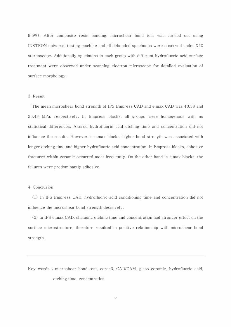
9.5%). After composite resin bonding, microshear bond test was carried out using
INSTRON universal testing machine and all debonded specimens were observed under X40
stereoscope. Additionally specimens in each group with different hydrofluoric acid surface
treatment were observed under scanning electron microscope for detailed evaluation of
surface morphology.
3. Result
The mean microshear bond strength of IPS Empress CAD and e.max CAD was 43.38 and
36.43 MPa, respectively. In Empress blocks, all groups were homogenous with no
statistical differences. Altered hydrofluoric acid etching time and concentration did not
influence the results. However in e.max blocks, higher bond strength was associated with
longer etching time and higher hydrofluoric acid concentration. In Empress blocks, cohesive
fractures within ceramic occurred most frequently. On the other hand in e.max blocks, the
failures were predominantly adhesive.
4. Conclusion
(1) In IPS Empress CAD, hydrofluoric acid conditioning time and concentration did not
influence the microshear bond strength decisively.
(2) In IPS e.max CAD, changing etching time and concentration had stronger effect on the
surface microstructure, therefore resulted in positive relationship with microshear bond
strength.
Key words : microshear bond test, cerec3, CAD/CAM, glass ceramic, hydrofluoric acid,
etching time, concentration

- 1 -
Effect of different etching time and concentration
on microshear bond strength of CAD/CAM glass-
ceramic blocks to composite resin
Directed by Prof. Byoung-duck Roh
The Graduate School, Yonsei University
Department of Dental Science
Yookyung Kim
I. Introduction
Advances in computer-aided design (CAD) and computer-aided
manufacturing (CAM) systems are providing new options for dentistry and
creating an alternative to the conventional impression and casting technique for
- 2 -
producing dental restorations. (Kamada et al., 1998) The CEREC (CEramic
REConstruction) system represents a unique CAD/CAM system that is used by
dentists for chairside fabrication and delivery of ceramic restorations. It offers a
considerable time savings over conventional, laboratory-generated restorations
that require multiple appointments.(Mehl and Hickel, 1999; Mormann, 1992)
A number of ceramic materials are available for use in CEREC restorations.
(Fasbinder, 2002) From the early feldspathic Vita Mark II (Vita, Bad Säckingen,
Germany) blocks, leucite reinforced glass-ceramic IPS Empress CAD (Ivoclar
vivadent AG, Schaan, Liechtenstein), and most recent lithium disilicate glass-
ceramic IPS e.max CAD (Ivoclar vivadent AG, Schaan, Liechtenstein) blocks are
provided and widely used by clinicians nowadays.
Optimal surface preparation techniques for chemical and/or mechanical
bonding to ceramic substrates are crucial in order to ensure clinical success
when placing indirect ceramic restorations and, when required, repairing them
intraorally (Alex, 2008). The modern generation of bonded porcelain
restorations was first described in 1980s, from Calamia’s works (Calamia, 1983,
1985; Calamia and Simonsen, 1984). He successfully utilized hydrofluoric acid
etching on porcelain surfaces in order to increase micromechanical retention,
and then applied silane to achieve stable chemical bonds between luting agent
and silicon dioxide on the ceramic surface. From then, hydrofluoric acid and
silane surface treatment was accepted as standard procedure in adhesive
cementation of ceramic restorations.
About usefulness of silane, common consensus has been reached (Hayakawa
et al., 1992; Matinlinna et al., 2004; Shimada et al., 2002). Not only providing
- 3 -
chemical interaction, which is attributed to its bifunctional characteristic, but it
also increases wettability of ceramic surfaces and creates better adhesive
environment.
But regarding hydrofluoric acid etching so far, the question is often raised if it
is generally required. Some studies clearly demonstrate that etching with HF has
the potential to significantly increase its bond strength to composite (Chen et al.,
1998; Nagayassu et al., 2006; Pisani-Proenca et al., 2006), while others lead to
the conclusion that acid etching can be eliminated, but not on the silanization,
relying more on the chemical adhesion (Aida et al., 1995; Hayakawa et al., 1992;
Shimada et al., 2002).
Moreover, when it comes to the specific time and concentration, the matter
becomes even more complex. In the initial stages, Calamia applied hydrofluoric
acid as long as 20 minutes, since he treated more acid-resistant feldspar
ceramics (Calamia, 1983). In recent years, numerous studies have tested
ceramic to resin bond, but the authors were inconsistent with their usage in
hydrofluoric acid, varying from 2.5 to 52% in concentration, 20 seconds to
several minutes with application time. In clinical situations, this inconsistency
may confuse dentists and laboratory technicians.
The purpose of this article is to evaluate the effect of different etching time
and concentration on microshear bond strength of two different CAD/CAM glass
ceramic blocks to composite resin. Leucite based IPS empress CAD and Lithium
disilicate based IPS e.max CAD were tested.

- 4 -
II. Materials and Methods
1. Materials
Two recent CAD/CAM glass-ceramic blocks and hydrofluoric acid with two
different concentrations were prepared. The materials employed in this study
are listed in Table 1 and 2.
Table 1. Ceramic block
Product Composition Shade Size Lot Manufacturer
IPS Empress®
CAD
Leucite-
reinforced
glass ceramic
LT
A3 C14 L39492
Ivoclar vivadent
AG, Schaan,
Liechtenstein
IPS e.max®
CAD
Lithium
disilicate
glass-ceramic
LT
A3 C14 N03151
Ivoclar vivadent
AG, Schaan,
Liechtenstein
Table 2. Hydrofluoric acid
Product Composition Lot Manufacturer
Porcelain
Etchant 9.5% Hydrofluoric acid 1100000095
Bisco, Inc.
Schaumburg, IL

- 5 -
IPS® Ceramic
etching gel <5% Hydrofluoric acid P02565
Ivoclar vivadent
AG, Schaan,
Liechtenstein
2. Ceramic specimen preparation
140 rectangular ceramic plates were made using low-speed diamond wheel
(Struers Minitom, DK-2610 Rodovre, Denmark), 70 from IPS Empress®CAD
and another 70 from IPS e.max®CAD blocks. The dimensions were 12mm in
width, 14mm in length and 2mm in thickness. E.max blocks were milled first in
bluish grey pre-crystallized metasilicate phase, and then followed by
subsequent crystallization process in Programat P300 (Ivoclar vivadent AG,
Schaan, Liechtenstein) furnace under crystallization temperature of 820~840℃
(Program no.81).
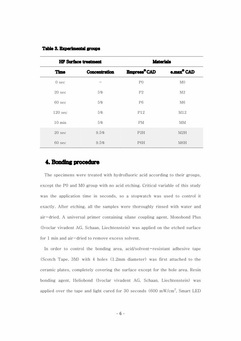
3. Experimental groups
Prepared specimens were randomly assigned to 7 groups of different surface
treatments. The variables were the hydrofluoric acid etching time (0, 20, 60,
120 seconds and 10 minutes) and the concentration of the gel (5% and 9.5%).
The descriptions of the tested groups are given in Table 3. In each groups, 10
samples were assigned.
- 6 -
Table 3. Experimental groups
HF Surface treatment Materials
Time Concentration Empress® CAD e.max® CAD
0 sec - P0 M0
20 sec 5% P2 M2
60 sec 5% P6 M6
120 sec 5% P12 M12
10 min 5% PM MM
20 sec 9.5% P2H M2H
60 sec 9.5% P6H M6H
4. Bonding procedure
The specimens were treated with hydrofluoric acid according to their groups,
except the P0 and M0 group with no acid etching. Critical variable of this study
was the application time in seconds, so a stopwatch was used to control it
exactly. After etching, all the samples were thoroughly rinsed with water and
air-dried. A universal primer containing silane coupling agent, Monobond Plus
(Ivoclar vivadent AG, Schaan, Liechtenstein) was applied on the etched surface
for 1 min and air-dried to remove excess solvent.
In order to control the bonding area, acid/solvent-resistant adhesive tape
(Scotch Tape, 3M) with 4 holes (1.2mm diameter) was first attached to the
ceramic plates, completely covering the surface except for the hole area. Resin
bonding agent, Heliobond (Ivoclar vivadent AG, Schaan, Liechtenstein) was
applied over the tape and light cured for 30 seconds (600 mW/cm2, Smart LED
plus, Sungbotech, Seoul, Korea).
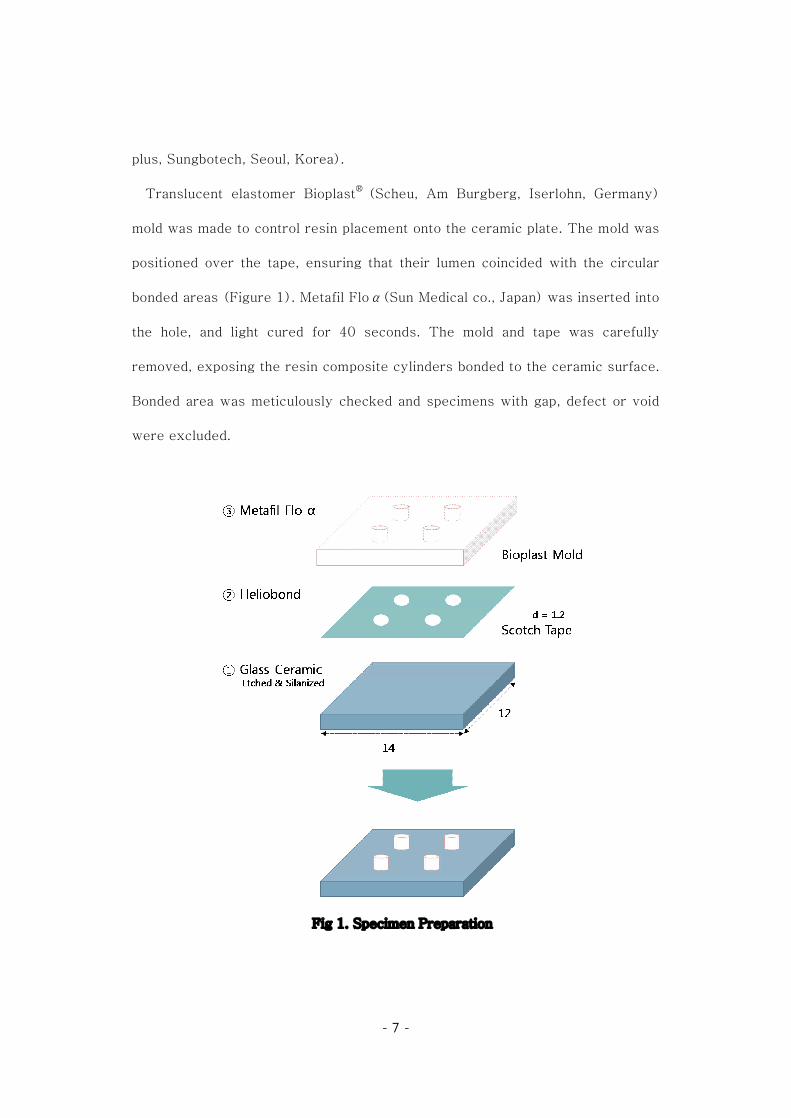
Translucent elastomer
mold was made to control
positioned over the tape, ensuring that their lumen coincide
bonded areas (Figure 1)
the hole, and light cured for 40 seconds. The mold and tape was carefully
removed, exposing the resin composite cylinders bonded to the ceramic surface.
Bonded area was meticulously checked and
were excluded.
- 7 -
plus, Sungbotech, Seoul, Korea).
Translucent elastomer Bioplast® (Scheu, Am Burgberg, Iserlohn, Germany)
control resin placement onto the ceramic plate. The mold was
positioned over the tape, ensuring that their lumen coincided with the circular
1). Metafil Floα(Sun Medical co., Japan) was in
light cured for 40 seconds. The mold and tape was carefully
removed, exposing the resin composite cylinders bonded to the ceramic surface.
was meticulously checked and specimens with gap, defect
Fig 1. Specimen Preparation
(Scheu, Am Burgberg, Iserlohn, Germany)
The mold was
with the circular
was inserted into
light cured for 40 seconds. The mold and tape was carefully
removed, exposing the resin composite cylinders bonded to the ceramic surface.
, defect or void

5. Microshear bond test
Following storage in distilled water at 37
was carried out using INSTRON
Co., Massachusetts, USA)
load was applied until bond failure of th
was recorded in Neuton
(τ=4P/πd2).
6. Surface evaluation
All debonded specimens after shear bond test were observed under
stereoscope (Leica, Microsystems Inc., Depew, New York,
the mode of failure. Failure modes were classified as follows:
- 8 -
5. Microshear bond test
Following storage in distilled water at 37℃ for 24 hours, microshear bond test
was carried out using INSTRON universal testing machine (Model 3366, Instron
Co., Massachusetts, USA) with cross-head speed of 1mm/min (Figure 2)
load was applied until bond failure of the specimen occurred. The load
Neutons(N) and converted to shear bond strength
Fig 2. Microshear bond test
evaluation
All debonded specimens after shear bond test were observed under
Microsystems Inc., Depew, New York, USA)
Failure modes were classified as follows:
for 24 hours, microshear bond test
Model 3366, Instron
(Figure 2). Shear
load at failure
ond strength in MPa
All debonded specimens after shear bond test were observed under X40
to determine

- 9 -
ž Adhesive failure at resin-ceramic interface
ž Cohesive failure within ceramic.
ž Mixed failure, involving bonding agent, resin and ceramic interfaces.
Furthermore, specimens in each group with different hydrofluoric acid surface
treatment were observed under scanning electron microscope (Hitachi, Tokyo,
Japan) for detailed evaluation of surface morphology.
7. Statistical analysis
Statistical analyses were performed using SPSS 11.5 software for Windows
(SPSS Inc., Chicago, IL, USA). 3-way analysis of variance (ANOVA) was
applied using bond strength (MPa) as the dependent variable and material,
etching time and concentration as factors. Tukey test was used in the post hoc
comparisons. When an interaction between the 3 factors was identified, the
differences were assessed statistically using two-way ANOVA, and Tukey tests.
In all analyses, the level of significance was set at α = 0.05.

- 10 -
III. Result
1. Microshear Bond Strength
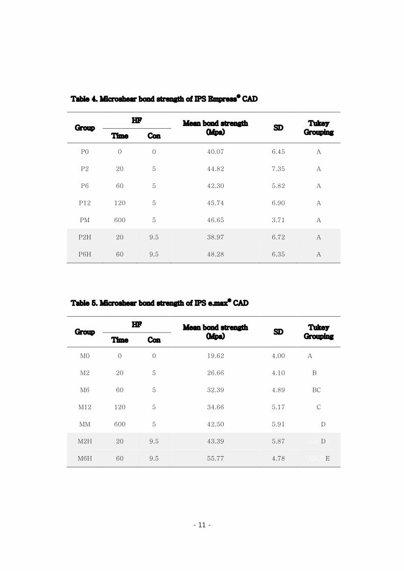
Results for the bond test are summarized in Table 4 and 5. The mean
microshear bond strength of IPS Empress CAD and e.max CAD was 43.38 and
36.43 MPa, respectively. 3-way ANOVA analysis (Table 6) showed interactions
between all three variables, therefore the effects of time and concentration were
separately analyzed by 2-way ANOVA in IPS Empress CAD and e.max CAD
blocks. In Empress blocks, all groups were homogenous with no statistical
differences. Altered hydrofluoric acid etching time and concentration did not
influence the results. However, e.max samples showed contrasting results. 2-
way ANOVA analysis showed significant effects of both time (p<0.0001) and
concentration (p<0.0001) on the bond strength of e.max blocks. Interaction
terms were also significant (p<0.05). In e.max blocks, higher bond strength was
associated with longer etching time and higher hydrofluoric acid concentration.
Figure 3 and 4 displays the clear differences between two blocks.
2. Failure Analysis
The distribution of failure modes is shown in Figure 5 and 6. In Empress
blocks, cohesive fractures within ceramic occurred most frequently. On the
other hand in e.max blocks, the failures were predominantly adhesive. The
representive micrographs are presented in figure 7.
- 11 -
Table 4. Microshear bond strength of IPS Empress® CAD
Group HF Mean bond strength
(Mpa) SD
Tukey Grouping Time Con
P0 0 0 40.07 6.45 A
P2 20 5 44.82 7.35 A
P6 60 5 42.30 5.82 A
P12 120 5 45.74 6.90 A
PM 600 5 46.65 3.71 A
P2H 20 9.5 38.97 6.72 A
P6H 60 9.5 48.28 6.35 A
Table 5. Microshear bond strength of IPS e.max® CAD
Group HF Mean bond strength
(Mpa) SD
Tukey Grouping Time Con
M0 0 0 19.62 4.00 ABCDE
M2 20 5 26.66 4.10 ABCDE
M6 60 5 32.39 4.89 ABCDE
M12 120 5 34.66 5.17 ABCDE
MM 600 5 42.50 5.91 ABCDE
M2H 20 9.5 43.39 5.87 ABCDE
M6H 60 9.5 55.77 4.78 ABCDE

- 12 -
Table 6. Result of 3-way ANOVA analysis
Source Sum of squares Mean square F Sig.
Material 1322.458 1322.458 36.243 .000
Time 1823.483 607.828 16.658 .000
Con 2052.534 2052.534 56.251 .000
Material*Time 405.851 135.284 3.708 .013
Material*Con 1970.211 1970.211 53.994 .000
Time*Con 398.687 398.687 10.926 .001
Material*Time*Con 26.006 26.006 .713 .400
Table 7. Result of 2-way ANOVA in IPS Empress® CAD
Source Sum of squares Mean square F Sig.
Time 281.251 93.750 1.958 .129
Concentration .421 .421 .009 .926
Time * Con 314.171 314.171 6.563 .013
Table 8. Result of 2-way ANOVA in IPS e.max® CAD
Source Sum of squares Mean square F Sig.
Time 1948.083 649.361 25.865 .000
Concentration 4022.324 4022.324 160.217 .000
Time * Con 110.522 110.522 4.402 .040

- 13 -
Fig 3. Mean bond strength values (MPa) of IPS Empress ® CAD.
Fig 4. Mean bond strength values (MPa) of IPS e.max® CAD
0
10
20
30
40
50
60
70
P0 P2 P6 P12 PM P2H P6H
MPa
0
10
20
30
40
50
60
70
M0 M2 M6 M12 MM M2H M6H
MPa

- 14 -
Fig 5. Distribution of failure modes (%) among groups in IPS Empress® CAD
(C) Cohesive (M) Mixed (A) Adhesive
Fig 6. Distribution of failure modes (%) among groups in IPS e.max® CAD
(C) Cohesive (M) Mixed (A) Adhesive
0%
20%
40%
60%
80%
100%
P0 P2 P6 P12 PM P2H P6H
A
M
C
0%
20%
40%
60%
80%
100%
M0 M2 M6 M12 MM M2H M6H
A
M
C
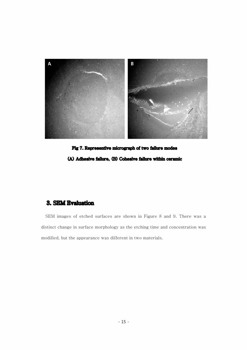
Fig 7. Representive micrograph of two failure modes
(A) Adhesive
3. SEM Evaluation
SEM images of etched surfaces are shown in Figure
distinct change in surface morphology
modified, but the appearance was different in two materials.
- 15 -
. Representive micrograph of two failure modes
(A) Adhesive failure, (B) Cohesive failure within ceramic
Evaluation
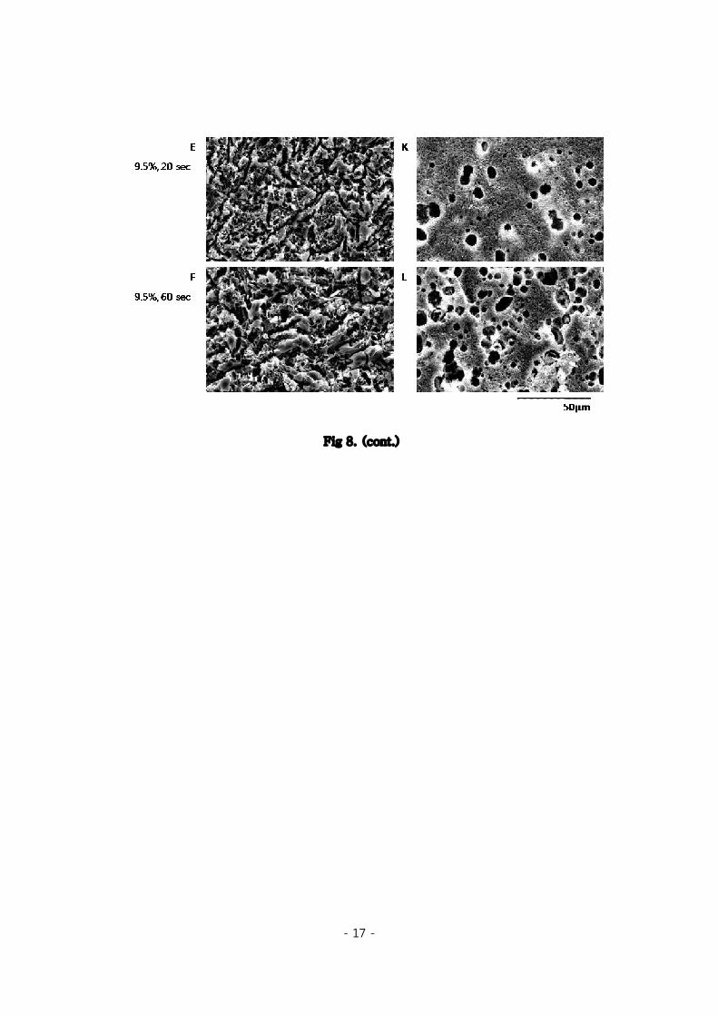
SEM images of etched surfaces are shown in Figure 8 and 9.
distinct change in surface morphology as the etching time and concentration
modified, but the appearance was different in two materials.
. Representive micrograph of two failure modes
failure, (B) Cohesive failure within ceramic
There was a
as the etching time and concentration was

Fig 8. SEM images
(A-F) IPS Empress CAD, (G
- 16 -
. SEM images of acid-etched surfaces (X1000)
F) IPS Empress CAD, (G-L) IPS e.max CAD.
- 17 -
Fig 8. (cont.)
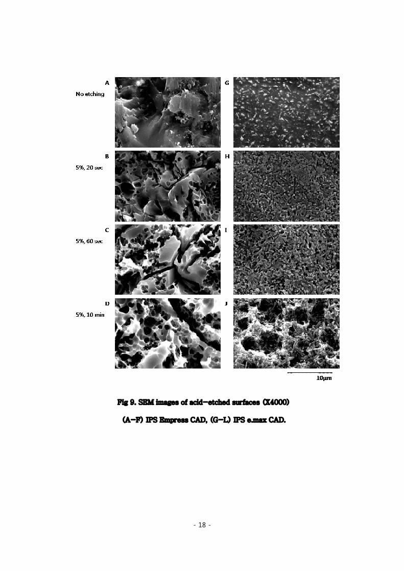
Fig 9. SEM images of acid
(A-F) IPS Empress
- 18 -
. SEM images of acid-etched surfaces (X4000)
F) IPS Empress CAD, (G-L) IPS e.max CAD.
etched surfaces (X4000)

- 19 -
Fig 9. (cont.)
- 20 -
IV. Discussion
IPS Empress CAD and e.max CAD are recommended to be conditioned for 60
seconds and 20 seconds with 5% hydrofluoric acid, respectively. Surprisingly,
the results of this study were quite inconsistent with the manufacturer’s
instructions.
First of all, there was a noticeable difference between the two CAD/CAM
blocks used in this study. From the beginning stages of adhesive cementation, it
was recommended to adjust etching time and concentration of hydrofluoric acid
depending on the specific porcelain being treated (Calamia, 1985). Since then,
several researchers have shown that the effects of different surface treatments
on bonding are strongly dependent on the type of ceramics (Aida et al., 1995;
Chen et al., 1998; Lacy et al., 1988).
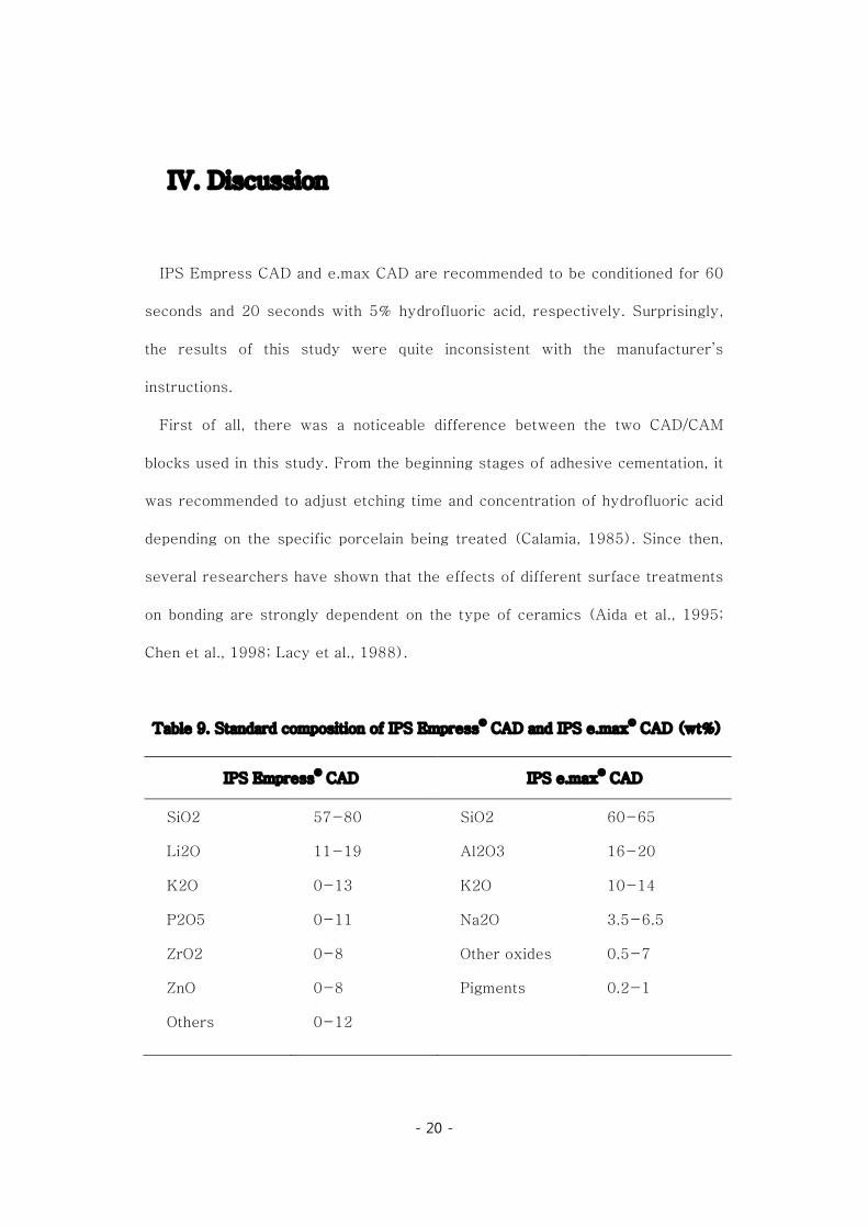
Table 9. Standard composition of IPS Empress® CAD and IPS e.max® CAD (wt%)
IPS Empress® CAD IPS e.max® CAD
SiO2
Li2O
K2O
P2O5
ZrO2
ZnO
Others
57-80
11-19
0-13
0-11
0-8
0-8
0-12
SiO2
Al2O3
K2O
Na2O
Other oxides
Pigments
60-65
16-20
10-14
3.5-6.5
0.5-7
0.2-1

- 21 -
The different responses to hydrofluoric acid appear to come from the
differences in ceramic microstructure and composition. IPS Empress CAD is a
leucite-based glass ceramic of the SiO2-Al2O3-K2O materials system, but
e.max CAD is a lithium disilicate glass-ceramic, and the chemical basis for the
material is the SiO2-Li2O system ("IPS e.max CAD: Scientific documentation;
IPS Empress CAD: Scientific documentation,"). From a materials perspective,
therefore, these materials are entirely different (Holand, 1998). Apart from the
chemical composition, there are also considerable differences in the
microstructures which is apparent in SEM images of non-etched samples
(Figure 8 and 9, A and G). The surface of e.max CAD shows dense
microstructure of lithium disilicate crystals measuring 0.5 to 5μm. The content
of these phyllosilicate crystals approximates 70% in volume of the glass-
ceramic, and is considerably higher than that of Empress CAD (35~45% vol%).
Stresses initiated by acid etching begin to form in the glass-crystal interface
and longer etching periods lead to the dissolution of crystals on the surface of
the glass-ceramic (Dorsch and Holand, 1994). Accordingly the microstructures
created by the glass and ceramic phases can greatly affect the surface
morphology created by acid etching. Therefore in this perspective, Empress and
e.max blocks will be discussed separately from now on.
In the Empress specimens, all groups showed no statistical differences.
Recently, Naves conducted a similar microshear bond test using 10%
hydrofluoric acid, changing etching periods from 10 to 120 seconds (Naves et al.,
2010). When unfilled resin was used, 60-second samples presented
significantly higher bond strength, but all the other groups showed similar
- 22 -
results without statistical differences. He suggested that combination of silane
and unfilled resin is responsible to this similarity, demonstrating complete
infiltration of resin into the irregularities created even by prolonged etching
periods. In an experiment run by the manufacturer itself (Dorsch and Holand,
1994) also revealed no differences in shear bond strength between etched and
non-etched samples.
These results can be somewhat confusing and difficult to interpret, because
etching with hydrofluoric acid clearly produced microporous and dendritic
retentive appearance on the ceramic surface in consistent with earlier studies
utilizing Leucite glass ceramics (Nagayassu et al., 2006; Naves et al., 2010). But
these mechanical interlocking appear to have little effect on the bond strength of
IPS Empress CAD. Advances in dental silane agent have brought us several
studies proposing possibilities to skip HF etching procedures (Aida et al., 1995;
Hayakawa et al., 1992; Shimada et al., 2002). Perhaps, relative high proportion
of glass matrix can explain this result. The dependence on chemical bonding by
silane coupling agent may override the influence of micro-mechanical retention
in Empress CAD.
Furthermore, there was no negative effect in bond strength from so-called
‘over-etching’ described in previous articles (Chen et al., 1998; Shimada et al.,
2002; Yen et al., 1993), despite the control group was applied as long as 10
minutes. The SEM images of current study could afford some explanation to this
result (Figure 8 and 9). After 60 seconds of 5% HF etching, the change over
time was relatively mild in Empress CAD samples. Samples treated with 9.5%
HF also showed similar surfaces with 5% samples. Maybe this is because the

- 23 -
crystalline content on the surface of Empress CAD is limited to certain degree
(35-45%). After exceeding this limit, perhaps which is after 60 seconds, there
may be no more crystals left to be dissolved by acid attacks. Therefore, no more
surface changes can be achieved, resulting in similar bond strengths.
Failure mode was predominantly cohesive within glass ceramics. This result is
in accordance with those of several studies that evaluated the bond strength at
ceramic-composite interface after silanization (Aida et al., 1995; Lu et al., 1992;
Nagayassu et al., 2006; Stangel et al., 1987). Some criticizes the cohesive
fractures associated with shear bond tests as a result of experimental design
and stress concentration (Armstrong et al., 2010; Braga et al., 2010; Della Bona
and van Noort, 1995), but in dealing with ceramic-composite interfaces this
mode to some extent may be inevitable because of brittleness of ceramic
materials. Thus, cautious interpretation is needed.
In e.max specimens, the results were totally different. 2-way ANOVA showed
positive correlation between shear bond strength and both of the variations,
etching time and concentration. Up to date, there are no published articles
dealing with bonding of IPS e.max, but there was one study with IPS Empress 2,
precursor of e.max, composed of same lithium disilicate crystals (Kim et al.,
2004). The result of their study showed similar results with this study
concluding that Empress 2 all-ceramic restorations require etching with 10%
hydrofluoric acid for 180~300 seconds to enhance the bond strength. This is
also lot more than the recommended of 5% and 20 seconds.
In the SEM images, in comparison with Empress samples, consequential
changes in the surface after increasing time and concentration were more
- 24 -
apparent on the e.max samples (Figure 6, G~L). These distinct increases in
microporosity can contribute to higher bond strength by providing more bond
area and creating undercuts for resin cements (al Edris et al., 1990). Calamia
evaluated the bond strengths of feldspatic porcelain etched for 2.5, 5, 10, and 20
minutes and found increased bond strength with increased etch times (Calamia,
1983). His SEM analysis of the porcelain surface was in accordance with ours,
indicating a definite potential for increased retention at each etch time. As
discussed above, the dense crystalline microstructures of e.max CAD surface
possibly have attributed to this high reactivity to hydrofluoric acid. In theory, the
reaction will continue until the available surface crystals are all used up.
Despite the apparent correlation with bond strength shown in this study, there
may be disadvantages utilizing higher HF concentrations and longer etching
periods. The manufacturer advice to etch for a very short period of 20 seconds
and they are unclear about what this recommendation is based on. Maybe the
answer to this question can be found in the material properties.
Lithium disilicate crystals are known be relatively susceptive to chemical
attacks (Holand, 1998). A recent study has been designed to evaluate the
durability of all-ceramic materials in oral environment. Substance lost following
wear testing with storage in artificial saliva was measured, and the result
showed significantly higher element release in lithium disilicate group than
leucite based groups (Dundar et al., 2003). According to the ISO standard
regarding dental ceramics ("ISO Dental Norm 6872,"), chemical resistance of a
material is tested by loss of mass with 4% acetic acid treatment for 16 hours at
80℃. Chemical solubility of IPS e.max CAD shows 40μg/cm2 and it is almost

- 25 -
twice compared with 25μg/cm2 of IPS Empress CAD ("IPS e.max CAD: Scientific
documentation; IPS Empress CAD: Scientific documentation,"). This value is
lower than maximum level permitted by the relevant standard (<100μg/cm2), but
it may affect the material in the acidic environments caused by etching
procedures. The biaxial strength of IPS e.max CAD after 20 seconds of
hydrofluoric acid etching was not reduced significantly (Holand, 1998), but no
data is provided with longer etching period or higher concentration.
All e.max specimens with only one exception etched with 5% hydrofluoric acid
showed adhesive failures till 120 seconds, but in the 10-minute etching group,
mixed and cohesive failure was increased. Particularly, specimens etched with
9.5% HF showed predominantly cohesive failures regardless of etching periods.
The SEM images uphold these findings (Figure 8, K and L). 9.5% HF created
highly destructive pitting on ceramic surfaces, even in the 20 second samples.
The involved area was not limited to the surface crystals measuring 0.5 to 1μm;
it contained much larger mass along with the glass matrix (~10μm).
Nevertheless, according to study of Yen, alteration of surface topography by
increased acid etching period from 30 seconds to 5 minutes did not have a
deleterious effect on the flexural strength of feldspathic porcelain and glass
ceramics (Yen et al., 1993). He concluded that flexural strength of ceramic
material is more dependent on internal bulk texture than on surface
characteristics. The clinical significance of these surface defects created by acid
etching is currently unknown.
The results of the current study have clinical implications. In Empress CAD,
prolonged etching periods or high HF concentration may not be necessary,

- 26 -
because it does not bring about higher bond strength. But in e.max CAD, the
bond strength increased significantly following longer etching time and higher
HF concentration. Therefore in this material, application of 9.5% HF in
laboratory use or 5% HF for a longer etching period in chairside may be
encouraged in clinical practice.
In this study, microshear test was performed to compare the bond strength.
Until the mid-nineties, bond strength tests were performed in specimens with
relatively large bonded areas, usually 3~6mm in diameter. However, problem in
stress distribution at the bonded interface was pointed out, and the need for new
methods to overcome these limitations led to the use of specimens with small
bonding areas, in the so-called micro-tensile and micro-shear tests(Braga et
al., 2010). Though, the general finding based upon finite element stress analysis
(FEA) shows that the shear force has its inherent limitations, which tends to
concentrate its force on to the base material rather than the strength of the
adhesive interface, resulting in cohesive fracture of the base material
(Armstrong et al., 2010; Della Bona and van Noort, 1995). But unfortunately, the
microtensile bond tests, although an effective method, also does not totally
eliminate the possibility of cohesive failures (Scherrer et al., 2010; Souza et al.,
2011), and above all, it is very technique sensitive to specimen preparation
procedures (Shimada et al., 2002). The high percentage of pre-test failures
reported and lack of standard interpreting these failures (Fabianelli et al., 2010;
Souza et al., 2011) can be one of the reasons not favoring this method especially
in the case of glass-ceramic samples. Compared to microtensile bond test,
trimming of the sample after the bonding procedure is not necessary for the
- 27 -
microshear test. Accordingly, the majority of studies on resin to ceramic bond
were designed and conducted as shear tests. (Hayakawa et al., 1992;
Kukiattrakoon and Thammasitboon, 2007; Lu et al., 1992; Nagayassu et al.,
2006; Naves et al., 2010; Shimada et al., 2002)
Lastly, it should be added that only the early bonding ability was investigated
in this study. It plays a fairly important role in clinical situations but of course,
the effects of aging on bonding should also be taken into consideration. There
are evidences that chemical adhesion of silane is unstable in aging situations.
(Meng et al., 2010; Pisani-Proenca et al., 2006) The oral environment
continuously stresses the bond interface by thermal changes, masticatory forces
and acidic challenges. Thus, further long-term investigations and clinical trials
are desired.
- 28 -
V. Conclusion
The aim of this article was to evaluate the effect of different etching time and
concentration on microshear bond strength of IPS Empress CAD and IPS e.max
CAD, two CAD/CAM blocks to composite resin.
Within the limitation of this study, the following conclusions were drawn:
(1) In IPS Empress CAD, hydrofluoric acid conditioning time and
concentration did not influence the microshear bond strength decisively.
(2) In IPS e.max CAD, changing etching time and concentration had stronger
effect on the surface microstructure, therefore resulted in positive relationship
with microshear bond strength.
- 29 -
References
Aida M, Hayakawa T, Mizukawa K: Adhesion of composite to porcelain with
various surface conditions. J Prosthet Dent 73: 464-470, 1995.
al Edris A, al Jabr A, Cooley RL, Barghi N: SEM evaluation of etch patterns by
three etchants on three porcelains. J Prosthet Dent 64: 734-739, 1990.
Alex G: Preparing porcelain surfaces for optimal bonding. Compend Contin
Educ Dent 29: 324-335; quiz 336, 2008.
Armstrong S, Geraldeli S, Maia R, Raposo LH, Soares CJ, Yamagawa J:
Adhesion to tooth structure: a critical review of "micro" bond strength
test methods. Dent Mater 26: e50-62, 2010.
Braga RR, Meira JB, Boaro LC, Xavier TA: Adhesion to tooth structure: a
critical review of "macro" test methods. Dent Mater 26: e38-49, 2010.
Calamia JR: Etched porcelain facial veneers: a new treatment modality based
on scientific and clinical evidence. N Y J Dent 53: 255-259, 1983.
Calamia JR: Etched porcelain veneers: the current state of the art.
Quintessence Int 16: 5-12, 1985.
Calamia JR, Simonsen RJ: Effect of coupling agents on bond strength of etched
porcelain. J Dent Res 63, 1984.
Chen JH, Matsumura H, Atsuta M: Effect of different etching periods on the
bond strength of a composite resin to a machinable porcelain. J Dent 26:
53-58, 1998.
Della Bona A, van Noort R: Shear vs. tensile bond strength of resin composite

- 30 -
bonded to ceramic. J Dent Res 74: 1591-1596, 1995.
Dorsch P, Holand W: Material Science of Empress Glass-Ceramics. In Ivoclar
Vivadent Report, 1994, pp. 3-8.
Dundar M, Artunc C, Toksavul S, Ozmen D, Turgan N: Determination of
elemental composition of substance lost following wear of all-ceramic
materials. Int J Prosthodont 16: 261-264, 2003.
Fabianelli A, Pollington S, Papacchini F, Goracci C, Cantoro A, Ferrari M, et al.:
The effect of different surface treatments on bond strength between
leucite reinforced feldspathic ceramic and composite resin. J Dent 38:
39-43, 2010.
Fasbinder DJ: Restorative material options for CAD/CAM restorations.
Compend Contin Educ Dent 23: 911-916, 918, 920 passim; quiz 924,
2002.
Hayakawa T, Horie K, Aida M, Kanaya H, Kobayashi T, Murata Y: The
influence of surface conditions and silane agents on the bond of resin to
dental porcelain. Dent Mater 8: 238-240, 1992.
Holand W: Materials Science Fundamentals of the IPS Empress 2 Glass-
Ceramic. In Ivoclar Vivadent Report, 1998, pp. 3-10.
IPS e.max CAD: Scientific documentation.
IPS Empress CAD: Scientific documentation.
ISO Dental Norm 6872.
Kamada K, Yoshida K, Atsuta M: Effect of ceramic surface treatments on the
bond of four resin luting agents to a ceramic material. J Prosthet Dent
79: 508-513, 1998.

- 31 -
Kim K, Choi K, Ahn S, Park C: Effect of etching time on shear bond strength of
resin cements to reinforced all-ceramic crowns. J Korean Acad Prost
42: 501-513, 2004.
Kukiattrakoon B, Thammasitboon K: The effect of different etching times of
acidulated phosphate fluoride gel on the shear bond strength of high-
leucite ceramics bonded to composite resin. J Prosthet Dent 98: 17-23,
2007.
Lacy AM, LaLuz J, Watanabe LG, Dellinges M: Effect of porcelain surface
treatment on the bond to composite. J Prosthet Dent 60: 288-291,
1988.
Lu R, Harcourt JK, Tyas MJ, Alexander B: An investigation of the composite
resin/porcelain interface. Aust Dent J 37: 12-19, 1992.
Matinlinna JP, Lassila LV, Ozcan M, Yli-Urpo A, Vallittu PK: An introduction to
silanes and their clinical applications in dentistry. Int J Prosthodont 17:
155-164, 2004.
Mehl A, Hickel R: Current state of development and perspectives of machine-
based production methods for dental restorations. Int J Comput Dent 2:
9-35, 1999.
Meng XF, Yoshida K, Gu N: Chemical adhesion rather than mechanical retention
enhances resin bond durability of a dental glass-ceramic with leucite
crystallites. Biomed Mater 5: 044101, 2010.
Mormann WH: Chairside computer-generated ceramic restorations: the Cerec
third generation improvements. Pract Periodontics Aesthet Dent 4: 9-
16, 1992.

- 32 -
Nagayassu MP, Shintome LK, Uemura ES, Araujo JE: Effect of surface
treatment on the shear bond strength of a resin-based cement to
porcelain. Braz Dent J 17: 290-295, 2006.
Naves LZ, Soares CJ, Moraes RR, Goncalves LS, Sinhoreti MA, Correr-
Sobrinho L: Surface/interface morphology and bond strength to glass
ceramic etched for different periods. Oper Dent 35: 420-427, 2010.
Pisani-Proenca J, Erhardt MC, Valandro LF, Gutierrez-Aceves G, Bolanos-
Carmona MV, Del Castillo-Salmeron R, et al.: Influence of ceramic
surface conditioning and resin cements on microtensile bond strength to
a glass ceramic. J Prosthet Dent 96: 412-417, 2006.
Scherrer SS, Cesar PF, Swain MV: Direct comparison of the bond strength
results of the different test methods: a critical literature review. Dent
Mater 26: e78-93, 2010.
Shimada Y, Yamaguchi S, Tagami J: Micro-shear bond strength of dual-cured
resin cement to glass ceramics. Dent Mater 18: 380-388, 2002.
Souza RO, Castilho AA, Fernandes VV, Bottino MA, Valandro LF: Durability of
microtensile bond to nonetched and etched feldspar ceramic: self-
adhesive resin cements vs conventional resin. J Adhes Dent 13: 155-
162, 2011.
Stangel I, Nathanson D, Hsu CS: Shear strength of the composite bond to
etched porcelain. J Dent Res 66: 1460-1465, 1987.
Yen TW, Blackman RB, Baez RJ: Effect of acid etching on the flexural strength
of a feldspathic porcelain and a castable glass ceramic. J Prosthet Dent
70: 224-233, 1993.

- 33 -
국문 요약
불산 농도 및 적용 시간에 따른 CAD/CAM 세라믹의
복합 레진에 대한 미세 전단 접착 강도 비교
연세대학교 대학원 치의학과
(지도 교수 노병덕)
김유경
1. 서론
기계적, 화학적 접착을 위한 표면 처리는 성공적인 세라믹 수복물의 합착을 위해서
필수적인 부분이다. 하지만 임상가들은 물론 연구자들 간에도 적절한 표면 처리에
대한 합의가 이루어지지 않아 혼동을 주고 있다.
이에 본 연구에서는 불산의 농도 및 적용 시간에 따른 CAD/CAM 세라믹의 복합
레진에 대한 전단 접착 강도를 비교하고자 하였다.
2. 본론
Leucite 기반의 IPS Empress®CAD와 lithium disilicate 기반의 e.max®CAD, 두
종류의 세라믹 블록과 5%, 9.5% 두 가지 농도의 불산을 사용하였다. 세라믹 시편

- 34 -
제작 후 불산을 0초, 20초, 60초, 120초 및 10분간 각각 적용하였고,
silane(Monobond Plus) 및 bonding agent(Heliobond)를 도포하였다. 복합 레진
접착 후 인스트론 만능 시험기를 사용하여 미세 전단 접착 강도(MPa)를 측정하였고,
stereoscope으로 파절면을 관찰하여 파절 양상을 기록하였다. 추가적으로 주사 전자
현미경으로 불산 처리 시간 및 농도에 따른 세라믹 표면 결정 구조의 변화를
관찰하였다. Two-way ANOVA test로 군간 접착 강도 사이의 유의성을 평가하였다.
실험 결과, IPS Empress CAD 는 모든 군에서 유의한 차이를 나타내지 않았으나,
IPS e.max CAD 의 경우, two-way ANOVA test 에서 불산 처리 시간 및 농도 모두
미세 전단 접착 강도와 유의한 관련성을 보였으며, 긴 처리 시간 및 높은 농도에서
보다 높은 미세 전단 접착 강도를 나타내었다.
파절 양상 관찰 결과 Empress CAD 는 세라믹 수복물의 cohesive failure 가 주를
이루었고, e.max CAD 의 경우 대부분 접착 계면에서 adhesive failure 를 나타내었다.
3. 결론
1) IPS Empress CAD 의 표면 처리시, 불산 적용 시간 및 농도를 변화시켜도 미세
전단 접착 강도에 유의한 변화가 나타나지 않았다.
2) IPS e.max CAD 의 경우 불산 적용 시간을 증가시키면 미세 전단 접착 강도도
유의하게 증가되는 양의 상관관계를 보였다.
3) IPS e.max CAD 의 경우 불산 농도를 5%에서 9.5%로 대체하여 사용 시
유의하게 높은 미세 전단 접착 강도를 보였으며, 세라믹에서의 cohesive failure
비중이 증가하였다.
Key words : microshear bond test, cerec3, CAD/CAM, glass ceramic,
hydrofluoric acid, etching time, concentration