effect of early implementation of electrical muscle
TRANSCRIPT
TitleEffect of early implementation of electrical muscle stimulationto prevent muscle atrophy and weakness in patients afteranterior cruciate ligament reconstruction
Author(s) Hasegawa, Satoshi; Kobayashi, Masahiko; Arai, Ryuzo;Tamaki, Akira; Nakamura, Takashi; Moritani, Toshio
Citation Journal of Electromyography and Kinesiology (2011), 21(4):622-630
Issue Date 2011-08
URL http://hdl.handle.net/2433/143566
Right © 2011 Elsevier Ltd.
Type Journal Article
Textversion author
Kyoto University
CORE Metadata, citation and similar papers at core.ac.uk
Provided by Kyoto University Research Information Repository
1
Title Page 1
2
EFFECT OF EARLY IMPLEMENTATION OF ELECTRICAL MUSCLE 3
STIMULATION TO PREVENT MUSCLE ATROPHY AND WEAKNESS IN 4
PATIENTS AFTER ANTERIOR CRUCIATE LIGAMENT RECONSTRUCTION 5
6
7
Author 8
Satoshi Hasegawa 9
Graduate School of Human and Environmental Studies, Kyoto University 10
11
Masahiko Kobayashi 12
Department of Orthopaedic Surgery, Kyoto University Hospital 13
14
Ryuzo Arai 15
Department of Orthopaedic Surgery, Kyoto University Hospital 16
17
Akira Tamaki 18
Department of Human Health Sciences, Kyoto University Graduate school of Medicine 19
20
Takashi Nakamura 21
Department of Orthopaedic Surgery, Kyoto University Hospital 22
23
Toshio Moritani (Corresponding Author) 24
Graduate School of Human and Environmental Studies, Kyoto University 25
E-mail: [email protected] 26
27
28
keywords: 29
Electrical muscle stimulation, Muscle atrophy, ACL reconstruction, Muscle training 30
2
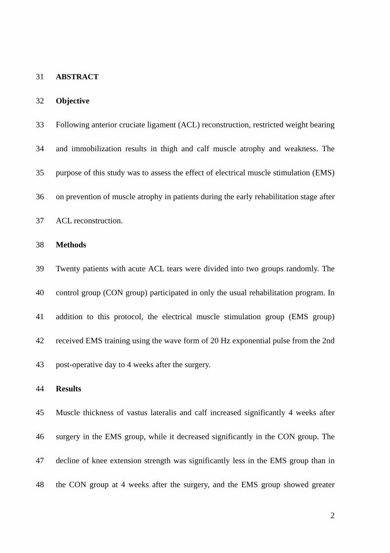
ABSTRACT 31
Objective 32
Following anterior cruciate ligament (ACL) reconstruction, restricted weight bearing 33
and immobilization results in thigh and calf muscle atrophy and weakness. The 34
purpose of this study was to assess the effect of electrical muscle stimulation (EMS) 35
on prevention of muscle atrophy in patients during the early rehabilitation stage after 36
ACL reconstruction. 37
Methods 38
Twenty patients with acute ACL tears were divided into two groups randomly. The 39
control group (CON group) participated in only the usual rehabilitation program. In 40
addition to this protocol, the electrical muscle stimulation group (EMS group) 41
received EMS training using the wave form of 20 Hz exponential pulse from the 2nd 42
post-operative day to 4 weeks after the surgery. 43
Results 44
Muscle thickness of vastus lateralis and calf increased significantly 4 weeks after 45
surgery in the EMS group, while it decreased significantly in the CON group. The 46
decline of knee extension strength was significantly less in the EMS group than in 47
the CON group at 4 weeks after the surgery, and the EMS group showed greater 48
3
recovery of knee extension strength at 3 months after surgery. 49
Conclusions 50
EMS implemented during the early rehabilitation stage is effective in maintaining 51
and increasing muscle thickness and strength in the operated limb. 52
53
54
55
56
57
58
59
60
61
62
63
64
65
66
4
INTRODUCTION 67
68
Following anterior cruciate ligament (ACL) reconstruction, immobilization and 69
restricted motion of the operated limb lead to unloading of the knee joint and 70
restricted weight bearing for 4 weeks after surgery, resulting in atrophy and 71
weakness of the quadriceps femoris and triceps surae muscles. Quadriceps atrophy 72
and strength loss often exceed 20% and 30%, respectively, during the first three 73
months following ACL reconstruction, and a 10% to 20% deficit in quadriceps size 74
and strength can persist for years after surgery, despite concentrated rehabilitation 75
efforts12. In addition, Nicholas et al. reported that ACL reconstruction resulted in a 76
significant decrease in thigh and calf girth at 3 weeks postoperation24. Therefore, a 77
primary focus of ACL rehabilitation protocols is the preservation and prompts 78
recovery of quadriceps femoris and triceps surae force production and function. We 79
believe it is important that patients start to exercise the quadriceps femoris and 80
triceps surae muscles during the early post-operative period in order to prevent 81
muscle atrophy and maintain muscle strength. One conventional choice for solving 82
this serious problem is electrical muscle stimulation (EMS). EMS elicits skeletal 83
muscle contractions through percutaneous electrodes that depolarize underlying 84
5
motor nerves. EMS using percutaneous electrodes is noninvasive and easy-to-use. 85
Several EMS studies have shown the potential advantages, both physiological and 86
clinical9, 20, 29. These previous studies have shown that EMS can be used to mimic 87
voluntary exercise and improve neuromuscular functions. There are other studies 88
showing better results of voluntary training versus electrical stimulation training and 89
that this varies depending on the type of individuals tested (healthy versus patients) 4, 90
5, 18, 30. 91
Previous studies had EMS protocols specific to each study’s purpose, making it 92
difficult to define the relationship between the EMS protocol and its effects. So it is 93
quite difficult to prescribe a flexible EMS protocol appropriate for the desired 94
purpose and participant’s condition. Our laboratory has focused on EMS protocols, 95
especially stimulus frequency characteristics. For example, our previous studies 96
demonstrated in human participants that 1) training with 20 Hz frequency 97
stimulation is more effective than 50 or 80 Hz frequency stimulations for inducing 98
muscle hypertrophy22, 2) EMS significantly increases glucose disposal rate (GDR) 99
during euglycemic clamp studies15, and a single bout of EMS to the lower 100
extremities can significantly enhance energy consumption, carbohydrate oxidation, 101
and whole body glucose uptake with low-intensity exercise13, and 3) EMS induces 102
6
selective fast-twitch MU activation of knee extensor muscles14. However, the 103
effects of long-term EMS training using our protocol are still unknown. Further 104
studies are necessary to test the therapeutic efficacy of our EMS device and 105
stimulation protocol. In most studies investigating the efficacy of EMS in patients 106
after knee surgery, the start time of the electric stimulation was often late (2-6 weeks 107
after surgery) and the muscles had already deteriorated and lost strength2, 6, 19, 25, 31, 32. 108
No one has reported the effects of EMS treatment implemented during the early 109
rehabilitation stage for prevention of muscle atrophy in patients with ACL 110
reconstruction. Moreover, there are no reports that evaluate changes in muscle 111
thickness of individual muscles during EMS training. 112
The purpose of this study was to determine the effects of electrical muscle 113
stimulation on the prevention of muscle atrophy in patients during the early 114
rehabilitation stage after ACL reconstruction using a modified EMS device and 115
stimulation protocol. 116
117
MATERIALS AND METHODS 118
Participants and Informed Consent 119

7
Twenty patients (16 male, 4 female), ranging in age from 13 to 54 years (26.3 ± 120
11.8 years) participated in this study. All patients had suffered an acute tear of the 121
ACL, and underwent an arthroscopically assisted semitendinosus autograft 122
reconstruction. The time from ACL tear until surgery were 3.1±1.4 months. They 123
had no history of neuromuscular disorders except for ACL injury. Each participant 124
provided informed consent prior to experimentation. The study protocol was 125
approved by the Medical Ethics Committee of our hospital. 126
127
Experimental Design 128
Twenty consecutive patients who underwent ACL reconstruction were 129
randomized and assigned to one of two groups: the control group (CON group) 130
included 10 patients (8 male, 2 female, age: 29.4±14.1 years, height: 165.9±5.9 cm, 131
weight: 60.1±10.1 kg, time from injury: 3.1±1.4 months) and the electrical muscle 132
stimulation group (EMS group) included 10 patients (8 male, 2 female, age: 133
23.5±9.3 years, height: 171.0±3.9 cm, weight: 68.1±6.3 kg, time from injury: 3.1±134
1.4 months). There were no significant differences between the groups in age, 135
physical characteristic, and the time from injury. The CON group received only the 136
usual rehabilitation program determined by our institute. In addition to this 137
8
standard rehabilitation protocol, the EMS group received EMS training for 4 weeks 138
beginning on post-operative day 2. Table 1 represents the rehabilitation program 139
determined by our institute, in which all patients in the study participated. To 140
determine the effects of EMS, we measured muscle thickness of the rectus femoris 141
(RF), vastus intermedius (VI), vastus lateralis (VL), and calf muscle (CA) before 142
surgery and at 4 weeks and 3 months after surgery. We also measured changes in 143
knee extensor muscle strength in isometric and isokinetic contractions before 144
surgery and at 4 weeks and 3 months after surgery. Moreover, we measured lower 145
extremity function using the Lysholm score before and at 6 months after the surgery. 146
147
EMS Training Protocol 148
The quadriceps femoris, hamstrings, tibialis anterior muscle, and triceps surae 149
were selected for EMS training in this study. The EMS training was performed on 150
the operated limb in patients of the EMS group, beginning the second day after 151
surgery and performed 5 days per week for a period of 4 weeks. Contractions of the 152
knee extensor, knee flexor, dorsi flexor, and plantar flexor muscles were elicited 153
simultaneously without involving movement of the joint by percutaneous muscle 154
stimulation for 20 minutes with the patient lying supine on a bed. 155
9
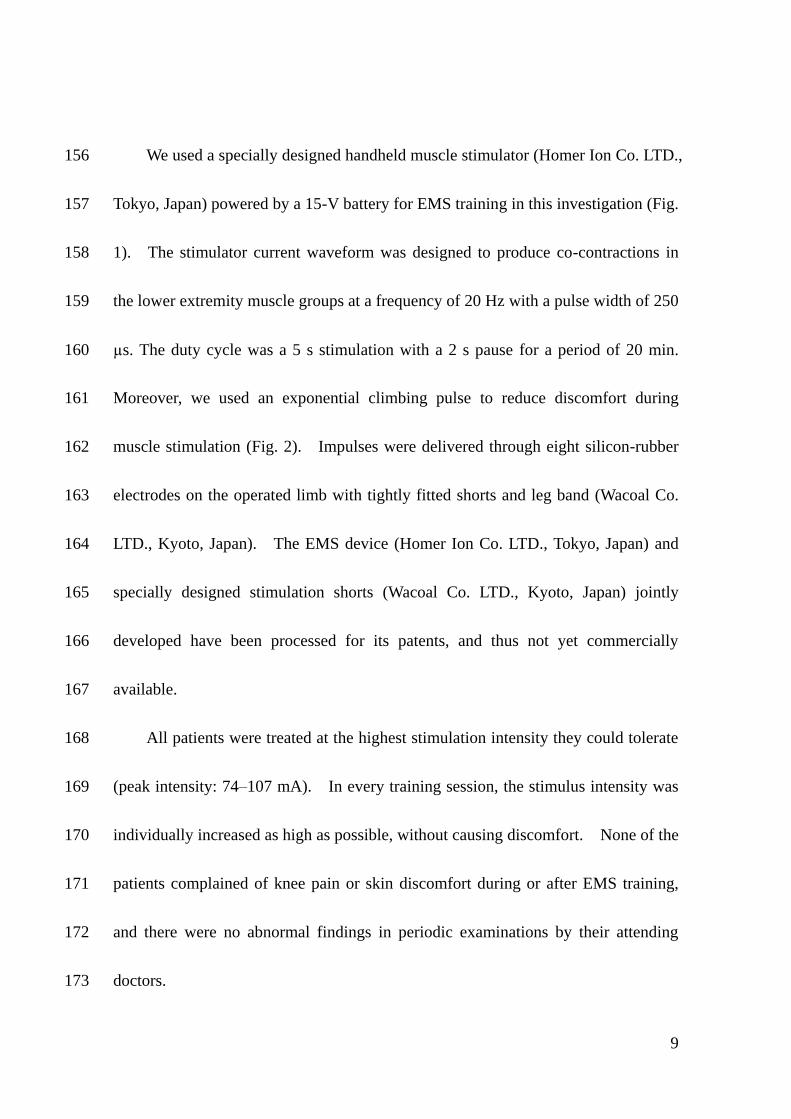
We used a specially designed handheld muscle stimulator (Homer Ion Co. LTD., 156
Tokyo, Japan) powered by a 15-V battery for EMS training in this investigation (Fig. 157
1). The stimulator current waveform was designed to produce co-contractions in 158
the lower extremity muscle groups at a frequency of 20 Hz with a pulse width of 250 159
µs. The duty cycle was a 5 s stimulation with a 2 s pause for a period of 20 min. 160
Moreover, we used an exponential climbing pulse to reduce discomfort during 161
muscle stimulation (Fig. 2). Impulses were delivered through eight silicon-rubber 162
electrodes on the operated limb with tightly fitted shorts and leg band (Wacoal Co. 163
LTD., Kyoto, Japan). The EMS device (Homer Ion Co. LTD., Tokyo, Japan) and 164
specially designed stimulation shorts (Wacoal Co. LTD., Kyoto, Japan) jointly 165
developed have been processed for its patents, and thus not yet commercially 166
available. 167
All patients were treated at the highest stimulation intensity they could tolerate 168
(peak intensity: 74–107 mA). In every training session, the stimulus intensity was 169
individually increased as high as possible, without causing discomfort. None of the 170
patients complained of knee pain or skin discomfort during or after EMS training, 171
and there were no abnormal findings in periodic examinations by their attending 172
doctors. 173

10
174
Muscle Thickness Analysis 175
Muscle thickness on the operated limb was measured using ultrasound still 176
images (GE Yokokawa Medical Co. LTD., Tokyo, Japan) obtained using an 8.0 MHz 177
probe with the patient lying supine or prone. Ultrasound is particularly useful 178
because it is safe, noninvasive, and portable. Strong correlations have been reported 179
between muscle thickness measured by B-mode ultrasound and site-matched 180
skeletal muscle mass measured by MRI7, 11, 21, 28, 34. Therefore, it is plausible to use 181
muscle thickness measurements to estimate muscle size and degree of muscle 182
atrophy. Previous studies have shown the reliability of the ultrasound technique for 183
measuring muscle thickness1, 17, 26, 33. Also, we measured the reliability of the 184
ultrasonographic measurement in this study. The intraclass correlation coefficients 185
in RF, VI, VL, and CA were 0.97 (0.88 – 0.99), 0.96 (0.85 – 0.99), 0.99 (0.97 – 1.0), 186
and 0.99 (0.96 – 1.0), respectively. Muscle thicknesses of the RF and VI were 187
measured at the level of the half distance between the anterior superior iliac spine 188
(ASIS) and the upper pole of the patella and on the line which linked the two points. 189
Muscle thickness of VL was measured at the level of lower one-thirds of the 190
distance between the ASIS and the upper pole of the patella, and 3 cm lateral from 191
11
the line which linked the patella to the ASIS in the supine position. Muscle thickness 192
of CA was measured at the level of the half distance between the head of fibula and 193
the lateral malleolus in the prone position. We measured muscle thickness with the 194
probe placed in the transverse plane. Measurements were performed before surgery 195
and at 4 weeks and 3 months after surgery. 196
197
Analysis of Knee Extensor Muscle Strength 198
We analyzed knee extensor muscle strength by measuring the maximal 199
voluntary isometric contraction of the quadriceps femoris using the CYBEX 200
HUMAC NORM® (Computer Sports Medicine, Inc., MA, USA.) dynamometer 201
before surgery and at 4 weeks and 3 months after surgery. The patients were 202
seated and stabilized in an electromechanical dynamometer with the knee flexed at 203
90 degrees where they attempted to maximally contract the quadriceps femoris 204
muscles for 5 seconds while verbal encouragement from the tester and visual 205
feedback from the dynamometer were provided. Similarly, we measured the 206
maximal isokinetic knee extension force with an angular velocity of 60 207
degrees/second before surgery and at 3 months after surgery. The peak torque 208
measured using the CYBEX HUMAC NORM®
was normalized with respect to 209
12
patient’s body weight, which was then expressed as the percent body weight (%BW). 210
This would allow a better understanding of the patient capacity (or muscle strength) 211
with respect to his/or her own body weight that needs to cope with in daily life. We 212
also calculated the ratio of changes at 4 weeks and 3 months after surgery in 213
comparison to the pre-operation. 214
215
Analysis of Lower Extremity Function 216
We measured lower extremity function using the Lysholm score before and at 6 217
months after the surgery. 218
219
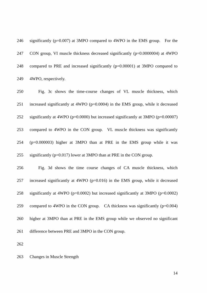
Statistics 220
We calculated the mean and standard error of the mean (SE) for all variables. 221
A two-way analysis of variance (ANOVA) followed by Fisher’s post-hoc test 222
procedure was used to test differences in the effects of EMS training on dependent 223
variables (muscle thickness and muscle strength in isometric and isokinetic 224
contraction) before surgery and after 4 weeks and 3 months. Also we calculated 225
the change ratio on operated side for muscle strength of knee extensor at 4 weeks 226
and 3 months after surgery in comparison to the pre-operation, and conducted a 227
13
two-way ANOVA followed by Fisher’s post-hoc test procedure to test differences in 228
effects of EMS training on dependent variables. The factors included in the two 229
way analysis of variance were time course (pre operation, 4 weeks after surgery, and 230
3 months after surgery) and training group (CON group and EMS group). 231
232
RESULTS 233
234
Changes in Muscle Thickness 235
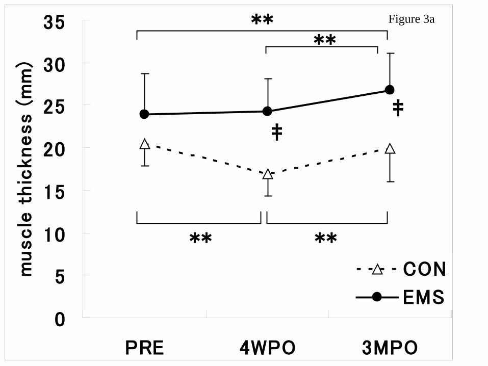
Fig. 3a shows RF muscle thickness of the operated side at pre-operation (PRE), 236
4 weeks post-operation (4WPO) and 3 months post-operation (3MPO) for both CON 237
and EMS groups. Two-way ANOVA with Fisher’s post-hoc test indicated that in 238
the EMS group there was no significant decline in RF muscle thickness between 239
PRE and 4WPO while the muscle thickness was significantly increased (p=0.003) at 240
3MPO. In contrast, RF muscle thickness decreased significantly (p=0.0001) at 241
4WPO compared to PRE and increased significantly (p=0.0006) at 3MPO compared 242
to 4WPO in the CON group. 243
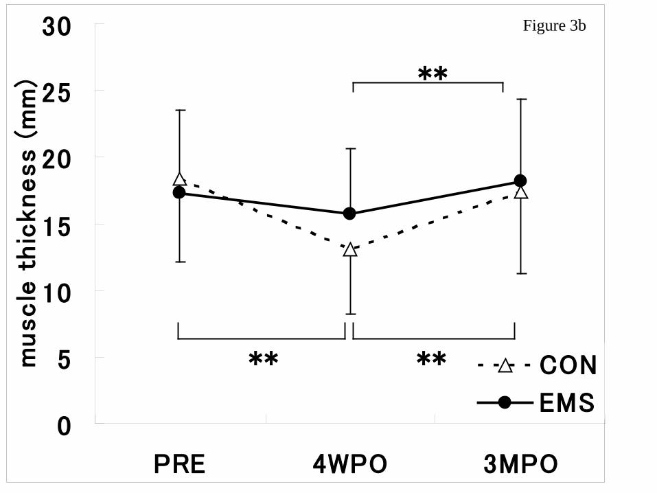
Fig. 3b shows the time-course changes of VI muscle thickness. There were no 244
significant changes between PRE and 4WPO and VI muscle thickness increased 245
14
significantly (p=0.007) at 3MPO compared to 4WPO in the EMS group. For the 246
CON group, VI muscle thickness decreased significantly (p=0.0000004) at 4WPO 247
compared to PRE and increased significantly (p=0.00001) at 3MPO compared to 248
4WPO, respectively. 249
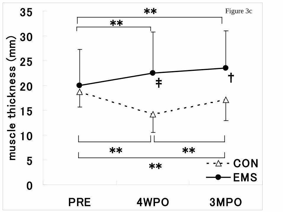
Fig. 3c shows the time-course changes of VL muscle thickness, which 250
increased significantly at 4WPO (p=0.0004) in the EMS group, while it decreased 251
significantly at 4WPO (p=0.0000) but increased significantly at 3MPO (p=0.00007) 252
compared to 4WPO in the CON group. VL muscle thickness was significantly 253
(p=0.000003) higher at 3MPO than at PRE in the EMS group while it was 254
significantly (p=0.017) lower at 3MPO than at PRE in the CON group. 255
Fig. 3d shows the time course changes of CA muscle thickness, which 256
increased significantly at 4WPO (p=0.016) in the EMS group, while it decreased 257
significantly at 4WPO (p=0.0002) but increased significantly at 3MPO (p=0.0002) 258
compared to 4WPO in the CON group. CA thickness was significantly (p=0.004) 259
higher at 3MPO than at PRE in the EMS group while we observed no significant 260
difference between PRE and 3MPO in the CON group. 261
262
Changes in Muscle Strength 263
15
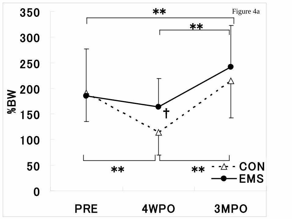
Fig. 4a shows the time-course changes of isometric knee extension strength 264
expressed as percentage of body weight (%BW) at PRE, 4WPO and 3MPO in both 265
groups. Isometric strength decreased significantly at 4WPO (p=0.001) and 266
increased significantly at 3MPO (p=0.00008) in the CON group, while there were no 267
significant changes between PRE and 4WPO and a significant increase at 3MPO 268
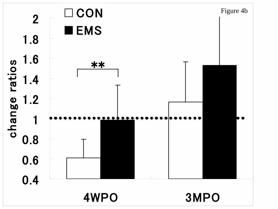
(p=0.001) in the EMS group. The changes in these values are shown in Fig. 4b. 269
Change ratios in the EMS group were significantly higher than the CON group at 4 270
weeks after surgery (-1.2% vs. 39.2%, p=0.008) and tended to be higher at 3 months 271
after surgery (52.7% vs. 16.3%, p=0.072), respectively. 272
Change ratios in isokinetic muscle strength measured at angular velocity of 60 273
degrees/sec at 3 months after surgery tended to be higher in the EMS group than in 274
the CON group (62.2% vs. 13.8%), but the difference did not reach the statistical 275
significance. 276
277
Changes in Lower Extremity Function 278
Lysholm scores for the CON and EMS groups were 59.2±7.8 vs. 63.6±4.9 at pre 279
operation, and 95.2±3.2 vs. 96.4±6.2 at 6 months after surgery, respectively. There 280
were no significant differences in Lysholm scores between the CON and the EMS 281
16
groups at 6months after the surgery. 282
DISCUSSION 283
284
The significant finding of this study was that 4 weeks of 20 Hz EMS training 285
beginning in the early rehabilitation stage following ACL reconstruction prevented 286
muscle atrophy and weakness. There have been some controversial findings 287
regarding the effects of EMS following ACL reconstruction. Sisk et al.31 288
demonstrated that there was no significant difference in strength between treatment 289
groups, but there was a significant difference in strength between competitive and 290
recreational athletes. Moreover, Lieber et al.19 demonstrated that 50 Hz 291
neuromuscular electrical stimulation and voluntary muscle contraction treatments, 292
when performed at the same intensity, are equally effective in strengthening skeletal 293
muscle that has been weakened by surgical repair of the ACL. On the other hand, 294
Delito et al.6 reported that patients in the EMS group finished a three-week training 295
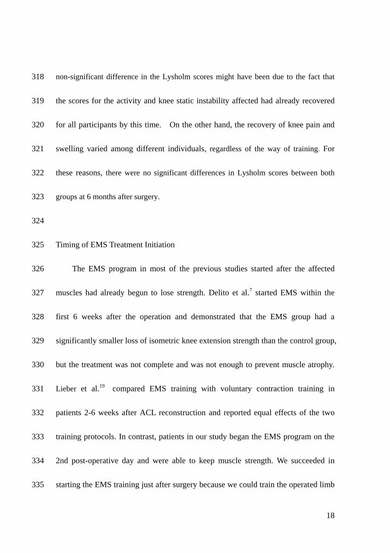
regimen with higher percentages of both extension and flexion torque when 296
compared to patients in the voluntary exercise group. Arvidsson et al.2 studied 297
different parts of the quadriceps in female patients and found less atrophy of the 298
vastus medialis after electrical stimulation. Snyder-Mackler et al.32 reported that 299
17
quadriceps strength averaged at least 70% of the strength on the uninvolved side in 300
patients treated with high-intensity electrical stimulation (either alone or combined 301
with low-intensity electrical stimulation), 57% in patients treated with high-level 302
active exercise, and 51% in patients treated only with low-intensity electrical 303
stimulation. Moreover, Fitzgerald et al.10 reported that use of the modified EMS 304
protocol as an adjunct to rehabilitation resulted in modest increases in quadriceps 305
torque output after 12 weeks of rehabilitation and in self-reported knee function at 306
12 and 16 weeks of rehabilitation, when compared to subjects who underwent 307
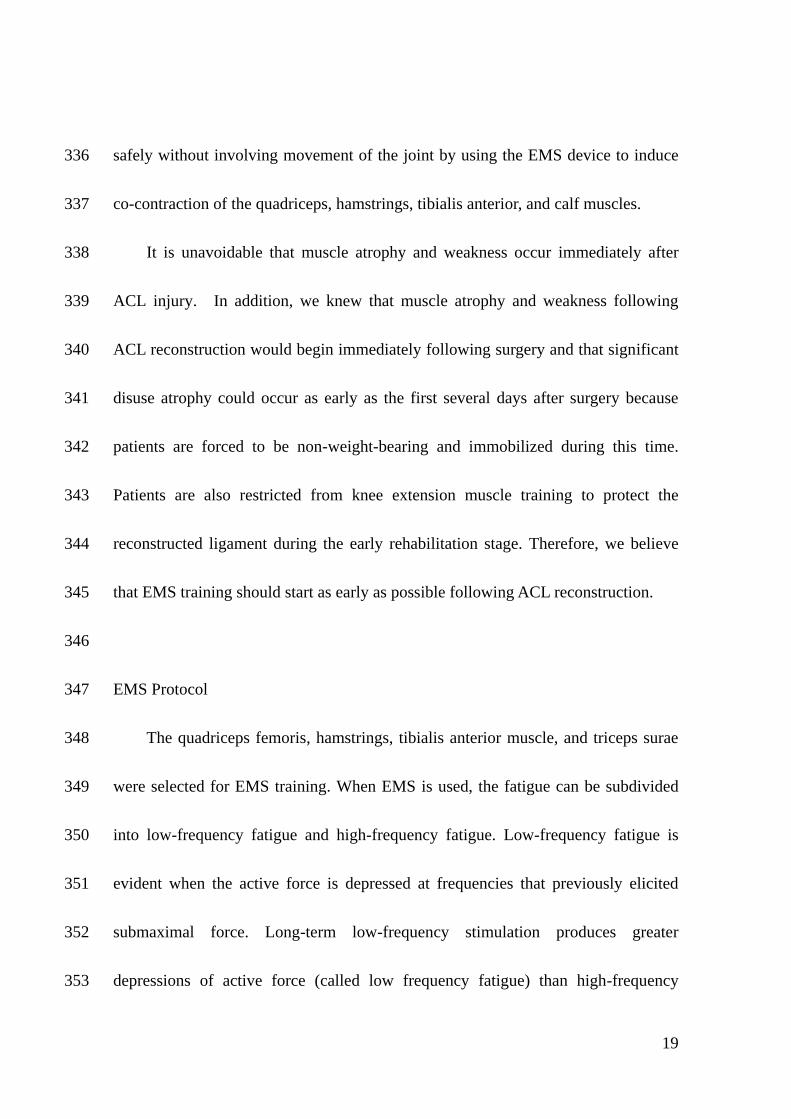
rehabilitation without EMS treatment. 308
Our present results confirmed significant efficacy of EMS training following 309
ACL surgery, but differ from previous studies on some points. Our current data 310
indicated that EMS training not only prevented muscle atrophy following ACL 311
reconstruction, but also resulted in VL and CA hypertrophy, which have not been 312
reported previously. We believe these different results are caused by differences in 313
the start timing of EMS, the EMS protocol, and the electrodes. 314
However, there were no significant differences in Lysholm scores between the 315
CON and the EMS groups. here were no significant differences in Lysholm scores 316
between the CON and the EMS groups at 6months after the surgery. The 317
18
non-significant difference in the Lysholm scores might have been due to the fact that 318
the scores for the activity and knee static instability affected had already recovered 319
for all participants by this time. On the other hand, the recovery of knee pain and 320
swelling varied among different individuals, regardless of the way of training. For 321
these reasons, there were no significant differences in Lysholm scores between both 322
groups at 6 months after surgery. 323
324
Timing of EMS Treatment Initiation 325
The EMS program in most of the previous studies started after the affected 326
muscles had already begun to lose strength. Delito et al.7 started EMS within the 327
first 6 weeks after the operation and demonstrated that the EMS group had a 328
significantly smaller loss of isometric knee extension strength than the control group, 329
but the treatment was not complete and was not enough to prevent muscle atrophy. 330
Lieber et al.19 compared EMS training with voluntary contraction training in 331
patients 2-6 weeks after ACL reconstruction and reported equal effects of the two 332
training protocols. In contrast, patients in our study began the EMS program on the 333
2nd post-operative day and were able to keep muscle strength. We succeeded in 334
starting the EMS training just after surgery because we could train the operated limb 335
19
safely without involving movement of the joint by using the EMS device to induce 336
co-contraction of the quadriceps, hamstrings, tibialis anterior, and calf muscles. 337
It is unavoidable that muscle atrophy and weakness occur immediately after 338
ACL injury. In addition, we knew that muscle atrophy and weakness following 339
ACL reconstruction would begin immediately following surgery and that significant 340
disuse atrophy could occur as early as the first several days after surgery because 341
patients are forced to be non-weight-bearing and immobilized during this time. 342
Patients are also restricted from knee extension muscle training to protect the 343
reconstructed ligament during the early rehabilitation stage. Therefore, we believe 344
that EMS training should start as early as possible following ACL reconstruction. 345
346
EMS Protocol 347
The quadriceps femoris, hamstrings, tibialis anterior muscle, and triceps surae 348
were selected for EMS training. When EMS is used, the fatigue can be subdivided 349
into low-frequency fatigue and high-frequency fatigue. Low-frequency fatigue is 350
evident when the active force is depressed at frequencies that previously elicited 351
submaximal force. Long-term low-frequency stimulation produces greater 352
depressions of active force (called low frequency fatigue) than high-frequency 353
20
stimulation in post-stimulation periods30. Impaired excitation-contraction coupling 354
is responsible for low-frequency fatigue, which is prolonged and preferentially 355
affects fast-twitch fibers8. High-frequency fatigue is evident when the active force is 356
depressed at frequencies that previously elicited maximal force. High-frequency 357
fatigue induces excessive loss of force, which can be due to electrical propagation 358
failure with a rapid decline in the evoked action potential amplitude. Jones et al.16 359
demonstrated that a reduction in extracellular [Na+] (or accumulation of [K+]) 360
accelerates the rate of force fatigue in an isolated preparation, as did an increase in 361
stimulus frequency. Moritani et al.22 have demonstrated that significantly less force 362
is generated after 30 seconds of high-frequency stimulation (50 Hz or 80 Hz) than 363
after a similar period of MVC. During this period of high-frequency force fatigue, 364
considerably greater force is generated at 20 Hz stimulation22. Thus, 365
high-frequency fatigue could be largely accounted for by a failure of electrical 366
transmission that may be due to reduced muscle membrane excitability leading to a 367
reduction in the evoked potential amplitude and conduction time3, 16, 22. 368
Most of the previous studies reported the efficacy of EMS using very 369
high-frequency (2500 Hz) or high-frequency stimulations (50 Hz or 80 Hz) 19, 31, 32. 370
Eriksson et al.9 showed that muscle enzyme activities, fiber size, and mitochondrial 371
21
properties in the quadriceps femoris did not change with 50 Hz EMS training 372
sessions over 4-5 weeks. Thus, patients in previous studies employing 373
high-frequency (50Hz or 80Hz) EMS training might have suffered from 374
high-frequency fatigue, so that the intended muscles were not effectively contracted. 375
This evidence indicates that 20 Hz EMS has the potential to elicit more effective 376
muscular improvement (a combined adaptation of neural factors and morphological 377
changes) than high-frequency (50 Hz or 80 Hz) EMS. Our present results are in 378
agreement with this previous evidence. Rebai et al.25 demonstrated that twelve 379
weeks after surgery, the quadriceps peak torque deficit in the operated limb with 380
respect to the non-operated limb at 180 degrees/s and 240 degrees/s was 381
significantly less in the 20 Hz group than in the 80 Hz group. Our data also 382
suggest that low-frequency (20 Hz) EMS training is effective in muscle training. 383
We specifically avoided the use of high frequency (50 Hz, 80 Hz, and more higher) 384
stimulations due to “high frequency fatigue”, i.e. a reduction of muscle membrane 385
excitability due to extracellular K+ accumulation which in turn results in force loss. In 386
other words, high frequency stimulations reduce the time necessary to fully perform 387
depolarization/repolarization to maintain the muscle membrane excitability. Use of 388
high frequency EMS would reduce the pain to a greater extent, but neurologically and 389

22
metabolically less effective when compared with low frequency stimulations. We have 390
shown this phenomenon with intramuscularly recorded M-wave and force 391
measurements22, 23
. We have also directly measured muscle energy metabolism during 392
low and high frequency stimulations and found that high frequency stimulations (50, 393
80Hz) resulted in significantly lower energy utilization due to “high frequency 394
fatigue”13
. Also, in our earlier preliminary studies, we have tried various stimulation 395
protocols (20, 50, 80Hz and different duty cycle) and measured directly the rate of 396
muscle fatigue, oxygen extraction level by near infrared spectroscopy, and 397
mechanomyogram (MMG). We found the presently used protocol is the best in terms 398
of avoiding fatigue accumulation without compromising muscular hypertrophy effects. 399
400
Wave Pattern and Electrodes 401
We used our original stimulus wave pattern and electrodes in the present study. 402
It is generally difficult to increase stimulus intensity to the level necessary for 403
effective muscle contraction using 20 Hz low-frequency stimulation because of skin 404
pain or discomfort. We were able to increase the stimulus intensity higher than in 405
previous studies without causing skin discomfort because we used an exponential 406
climbing pulse instead of a rectangular pulse (Fig 2). Moreover, our original 407
23
electrodes were large, wet-gel type electrodes that reduced source impedance so that 408
there were no complaints of skin discomfort during or after EMS training, and no 409
abnormal findings reported by the attending doctors. In our earlier studies13, 15, we 410
used square pulses without exponential climbing procedure. This stimulation 411
technique accompanied a quite pain on the skin surface, particularly when 412
stimulating at higher intensities. We therefore asked the EMS manufacture to invent 413
a new stimulation procedure to reduce such discomfort as much as possible by 414
avoiding initial sudden electrical discharge to the skin surface. A newly invented 415
this climbing pulse stimulation procedure has been successfully adopted in the 416
present study. This procedure includes initial phase of 10% of the final stimulus 417
voltage and gradually reaching the final intensity with in 100 msec. 418
419
Conclusion 420
We were able to prevent muscle weakness in patients with ACL reconstruction 421
by implementing our EMS protocol early in the rehabilitation stage following 422
surgery. The decrease in the quadriceps peak torque of the operated limb was 423
significantly less in the EMS group (1.2%) than in the CON group (39.2%) 4 weeks 424
after surgery. The recovery ratio in the EMS group was higher than in the CON 425
24
group at 3 months. We believe that the difference in muscle strength between the 426
EMS and CON groups at 3MPO was brought about by the prevention of muscle 427
atrophy by EMS training for 4 weeks. Consequently, we suggest that EMS training 428
with 20 Hz exponential climbing pulse beginning immediately after surgery can 429
prevent muscle atrophy and weakness in patients recovering from ACL 430
reconstruction using semitendinosus autograft. 431
432
433
434
REFERENCES 435
1. Abe T, Kondo M, Kawakami Y, Fukunaga T. Prediction equations for body 436
composition of Japanese adults by B-mode ultrasound. American Journal of 437
Human Biology 1994; 6: 161– 70. 438
439
2. Arvidsson I, Arvidsson H, Eriksson E, et al. Prevention of quadriceps wasting 440
after immobilization.: an evaluation of the effect of electrical stimulation. 441
Orthopedics 1986; 9: 1519-28. 442
443
25
3. Bigland-Ritchie B, Jones DA, and Woods JJ. Excitation frequency and muscle 444
fatigue. Electrical responses during human voluntary and stimulated contractions. 445
Experimental Neurology 1979; 64: 414-27. 446
447
4. Currier DP, Lehman J, Lightfoot P, Electrical stimulation in exercise of the 448
quadriceps femoris muscle. Physical Therapy 1979; 59: 1508-12. 449
450
5. Currier DP and Mann R. Muscular strength development by electrical stimulation 451
in healthy individuals. Physical Therapy 1983; 63: 915-21. 452
453
6. Delitto A, Rose SJ, McKowen JM, et al. Electrical stimulation versus voluntary 454
exercise in strengthening thigh musculature after anterior cruciate ligament 455
surgery. Physical Therapy 1988; 68: 660-63. 456
457
7. Dupont AC, Sauerbrei EE, Fenton, PV, Shragge, PC, Loeb GE, Richmond FJ. 458
Real-time sonography to estimate muscle thickness: comparison with MRI and 459
CT. Journal of Clinical Ultrasound 2001; 29: 230–36. 460
461
26
8. Edwards RH, Hill DK, Jones DA, and Merton PA. Fatigue on long duration in 462
human skeletal muscle after exercise. Journal of Physiology 1977; 272: 769-78. 463
464
9. Eriksson E, Haggmark T, Kiessling KH, and Karlsson J. Effects of electrical 465
stimulation on human skeletal muscle. International Journal of Sports Medicine 466
1981; 2: 18-22. 467
468
10. Fitzgerald G K, Piva SR, and Irrgang JJ. A modified neuromuscular electrical 469
stimulation protocol for quadriceps strength training following anterior cruciate 470
ligament reconstruction. Journal of Orthopedic and Sports Physical Therapy 2003; 471
33: 492-501. 472
473
11. Fukunaga T, Miyatani M, Tachi M, Kouzaki M, Kawakami Y, Kanehisa H. 474
Muscle volume is a major determinant of joint torque in humans. Acta 475
Physiological Scand 2001; 172: 249–55. 476
477
12. Gerber JP, Marcus RL, Dibble LE, Greis PE, Burks RT, LaStayo PC. Effects of 478
early progressive eccentric exercise on muscle structure after anterior cruciate 479
27
ligament reconstruction. J Bone Joint Surg Am 2007; 89: 559-70. 480
481
13. Hamada T, Hayashi T, Kimura T, Nakano K, and Moritani T. Electrical 482
stimulation of human lower extremities enhances energy consumption, 483
carbohydrare oxidation, and whole body glucose uptake. Journal of Applied 484
physiology 2004; 96: 911-16. 485
486
14. Hamada T, Kimura T, and Moritani T. Selective fatigue of motor units after 487
electrically elicited muscle contractions. Journal of Electromyography and 488
Kinesiology 2004; 14: 531-38. 489
490
15. Hamada T, Sasaki H, Hayashi T, Moritani T, and Nakano K. Enhancement of 491
whole body glucose uptake during and after human skeletal muscle 492
low-frequency electrical stimulation. Journal of Applied Physiology 2003; 94: 493
2107-12. 494
495
16. Jones DA, Bigland-Ritchie B, and Edwards RHT. Excitation frequency and 496
muscle fatigue: mechanical responses to voluntary and stimulated contractions. 497
28
Experimental Neurology 1979; 64: 401-13. 498
499
17. Kellis E, Galanis N, Natsis K, Kapetanos G.. Validity of architectural properties 500
of the hamstring muscles: correlation of ultrasound findings with cadaveric 501
dissection. Journal of Biomechanics 2009; 42: 2549–54. 502
503
18. Laughman RK, Youdas JW, Garrett TR, et al. Strength changes in normal 504
quadriceps femoris muscle as a result of electrical stimulation. Physical Therapy 505
1983; 63: 494-99. 506
507
19. Lieber RL, Silva PD, and Daniel DM. Equal effectiveness of electrical and 508
volitional strength training for quadriceps femoris muscles after anterior cruciate 509
ligament surgery. Journal of Orthopedic Research 1996; 14: 131-38. 510
511
20. Martin L, Cometti G, Pousson M, and Morlon B. Effect of electrical 512
stimulation training on the contractile characteristics of the triceps surae muscle. 513
European Journal of Applied Physiology and Occupational Physiology 1993; 67: 514
457-61. 515
29
516
21. Miyatani M, Kanehisa H, Ito M, Kawakami Y, Fukunaga T. The accuracy of 517
volume estimates using ultrasound muscle thickness measurements in different 518
muscle groups. European Journal of Physiology 2004; 91: 264–72. 519
520
22. Moritani T, Muro M, and Kijima A. Electromechanical changes during 521
electrically induced and maximal voluntary contractions: electrophysiolosic 522
responses of different muscle fiber types during stimulated contractions. 523
Experimental Neurology 1985; 88: 471-83. 524
525
23. Moritani, T., Muro, M., Kijima, A., Gaffney, F.A., and Persons, D. 526
Electromechanical changes during electrically induced and maximal voluntary 527
contractions: Surface and intramuscular EMG responses during sustained 528
maximal voluntary contraction. Experimental Neurology 1985; 88: 484-99. 529
530
24. Nicholas SJ, Tyler TF, McHugh MP, Gleim GW. The effect on leg strength of 531
tourniquet use during anterior cruciate ligament reconstruction: A prospective 532
randomized study. Arthroscopy 2001; 17 (6): 603-07. 533

30
534
25. Rebai H, Barra V, Laborde A, et al. Effect of two electrical stimulation 535
frequencies in yhigh muscle after knee surgery. International Journal of Sports 536
Medicine 2002; 23: 604-09. 537
538
26. Reeves ND, Maganaris CN, Narici MV. Ultrasonographic assessment of human 539
skeletal muscle size. European Journal of Physiology 2004; 91: 116–18. 540
541
27. Sale DG. Influence of exercise and training on motor unit activation. Exercise 542
and Sports Science Reviews 1987; 15 :95-151. 543
544
28. Sanada K, Kearns C, Midorikawa T, Abe T. Prediction and validation of total and 545
regional skeletal muscle mass by ultrasound in Japanese adults. European 546
Journal of Physiology 2006; 96: 24–31. 547
548
29. Scremin AME, Kurta L, Gentile A, Wiseman B, Perell K, Kunkel C, and 549
Scremin OU. Increasing muscle mass in spinal cord injured persons with a 550
functional electrical stimulation exercise program. Archives of Physical 551

31
Medicine and Rehabilitation 1999; 80: 1531-36. 552
553
30. Selkowitz DM. Improvement in isometric strength of the quadriceps femoris 554
muscle after training with electrical stimulation. Physical Therapy 1985; 65: 555
186-96. 556
557
31. Sisk TD, Stralka SW, Deering MB, Griffin JW. Effect of electrical stimulation on 558
quadriceps strength after reconstructive surgery of the anterior cruciate ligament. 559
American Journal of Sports Medicine 1987; 15: 215-20. 560
561
32. Snyder-Mackler L, Delitto A, Bailey SL, Stralka SW. Strength of the quadriceps 562
femoris muscle and functional recovery after reconstruction of the anterior 563
cruciate ligament. A prospective, randomized clinical trial of electrical 564
stimulation. Journal of Bone Joint Surgery 1995; 77: 1166-73. 565
566
33. Thoirs K, English C. Ultrasound measures of muscle thickness: intra- examiner 567
reliability and influence of body position. Clinical Physiology and Functional 568
Imaging 2009; 29: 440–46. 569
32
570
34. Walton JM, Roberts N, Whitehouse GH. Measurement of the quadriceps femoris 571
muscle using magnetic resonance and ultrasound imaging. British Journal of 572
Sports Medicine 1997; 31: 59–64. 573
574
575
576
577
Table 1. Rehabilitation Protocol in the Rehabilitation Unit of Kyoto University 578
Hospital 579
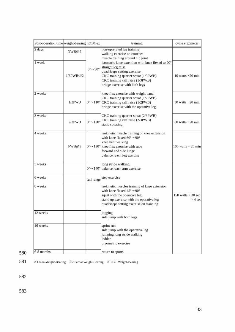
33
580
※1 Non-Weight-Bearing ※2 Partial Weight-Bearing ※3 Full Weight-Bearing 581
582
583
Post-operation time weight-bearing ROM ex training cycle ergometer
2 days non-oprerated leg training
walking exercise on crutches
muscle training around hip joint
1 week isometric knee extention with knee flexed to 90°
straight leg raise
quadriceps setting exercise
CKC training quarter squat (1/3PWB)
CKC training calf raise (1/3PWB)
bridge exercise with both legs
2 weeks knee flex exercise with weight band
CKC training quarter squat (1/2PWB)
CKC training calf raise (1/2PWB)
bridge exercise with the operative leg
3 weeks CKC training quarter squat (2/3PWB)
CKC training calf raise (2/3PWB)
static squating
4 weeks isokinetic muscle training of knee extension
with knee flexed 60°~90°
knee bent walking
knee flex exercise with tube
forward and side lunge
balance reach leg exercise
5 weeks long stride walking
balance reach arm exercise
6 weeks step exercise
8 weeks isokinetic muscles training of knee extension
with knee flexed 45°~90°
squat with the operative leg 150 watts × 30 sec
stand up exercise with the operative leg × 4 set
quadriceps setting exercise on standing
12 weeks jogging
side jump with both legs
16 weeks sprint run
side jump with the operative leg
jumping long stride walking
ladder
plyometric exercise
6-8 months return to sports
100 watts × 20 min
0°~140°
full range
0°~120° 60 watts ×20 min2/3PWB
FWB※3 0°~130°
NWB※1
0°~90°
1/3PWB※2 10 watts ×20 min
1/2PWB 0°~110° 30 watts ×20 min
34
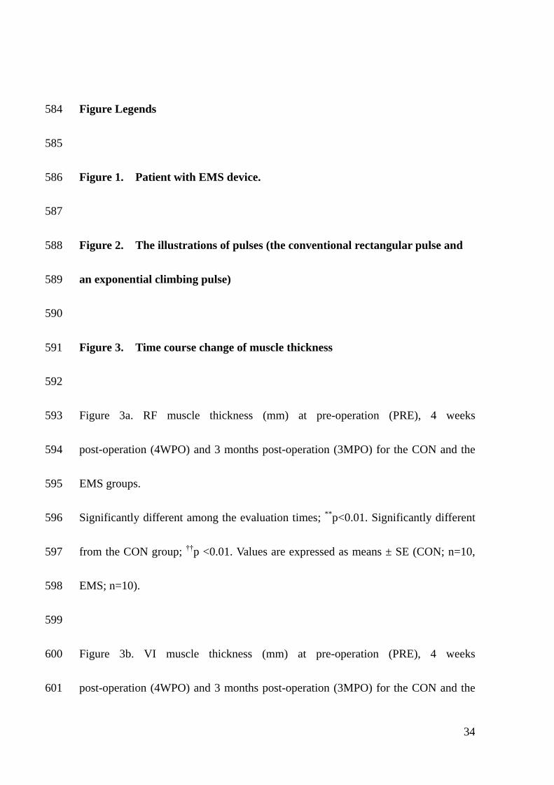
Figure Legends 584
585
Figure 1. Patient with EMS device. 586
587
Figure 2. The illustrations of pulses (the conventional rectangular pulse and 588
an exponential climbing pulse) 589
590
Figure 3. Time course change of muscle thickness 591
592
Figure 3a. RF muscle thickness (mm) at pre-operation (PRE), 4 weeks 593
post-operation (4WPO) and 3 months post-operation (3MPO) for the CON and the 594
EMS groups. 595
Significantly different among the evaluation times; **p<0.01. Significantly different 596
from the CON group; ††p <0.01. Values are expressed as means ± SE (CON; n=10, 597
EMS; n=10). 598
599
Figure 3b. VI muscle thickness (mm) at pre-operation (PRE), 4 weeks 600
post-operation (4WPO) and 3 months post-operation (3MPO) for the CON and the 601
35
EMS groups. 602
Significantly different among the evaluation times; **p<0.01. Values are expressed as 603
means ± SE (CON n=10, EMS n=10). 604
605
Figure 3c. VL muscle thickness (mm) at PRE, 4WPO and 3MPO for the CON group 606
and the EMS group. 607
Significantly different among the evaluation times; **p<0.01. Significantly different 608
from the CON group; ††p<0.01, †p<0.05. Values are expressed as means ± SE (CON; 609
n=10, EMS; n=10). 610
611
Figure 3d. CA muscle thickness (mm) at PRE, 4WPO and 3MPO for the CON and 612
the EMS groups. 613
Significantly different among the evaluation times; **p<0.01. Significantly different 614
from the CON group; ††p<0.01. Values are expressed as means ± SE (CON; n=10, 615
EMS n=10). 616
617
Figure 4. Time course change of muscle strength 618
619
36
Figure 4a. The isometric knee extension strength on an operated side at 620
pre-operation (PRE), 4 weeks post-operation (4WPO) and 3 months post-operation 621
(3MPO) for the CON and the EMS groups. 622
Significantly different among the evaluation times; **p<0.01. Significantly different 623
from the CON group; †p<0.05. Values are expressed as means ± SE (CON; n=10, 624
EMS; n=10). 625
626
Figure 4b. Changes ratios of isometric knee extension strength at 4WPO and 3MPO 627
compared to pre-operation in both the CON and EMS groups. 628
Significantly different; **p<0.01. Values are expressed as means ± SE (CON n=10, 629
EMS n=10). 630
631

EMS Device and Tight-fitting flexible electrodes
StimulatorPatient with EMS device
Figure 1

rectangular pulse exponential climbing pulse
illustrations of pulses
pulse pulse
SKIN SKIN
MUSCLEMUSCLE MUSCLEMUSCLE
electricity electricity
back pulse
pulse pulse
SKIN SKIN
MUSCLEMUSCLE MUSCLEMUSCLE
electricity electricity
back pulse
Figure 2
0
5
10
15
20
25
30
35
PRE 4WPO 3MPO
muscle
thic
kness (
mm
)
CON
EMS
****
** **
‡‡
Figure 3a
0
5
10
15
20
25
30
PRE 4WPO 3MPO
muscle
thic
kness (
mm
)
CON
EMS
** **
**
Figure 3b
0
5
10
15
20
25
30
35
PRE 4WPO 3MPO
muscle
thic
kness (
mm
)
CONEMS
** **
**
****
‡ †
Figure 3c

0
10
20
30
40
50
60
70
80
PRE 4WPO 3MPO
muscle
thic
kness (
mm
)
CON
EMS
** **
****
‡ ‡
Figure 3d
0
50
100
150
200
250
300
350
PRE 4WPO 3MPO
%B
W
CONEMS
** **
****
†
Figure 4a
0.4
0.6
0.8
1
1.2
1.4
1.6
1.8
2
4WPO 3MPO
change r
ati
os
CON
EMS
**
Figure 4b