effect of feeding micro-entrapped co-culture of ... · ruchi kushal, sanjeev kumar anand, harish...
TRANSCRIPT
HAL Id: hal-00895577https://hal.archives-ouvertes.fr/hal-00895577
Submitted on 1 Jan 2006
HAL is a multi-disciplinary open accessarchive for the deposit and dissemination of sci-entific research documents, whether they are pub-lished or not. The documents may come fromteaching and research institutions in France orabroad, or from public or private research centers.
L’archive ouverte pluridisciplinaire HAL, estdestinée au dépôt et à la diffusion de documentsscientifiques de niveau recherche, publiés ou non,émanant des établissements d’enseignement et derecherche français ou étrangers, des laboratoirespublics ou privés.
Effect of feeding micro-entrapped co-culture ofLactobacillus acidophilus and Bifidobacterium bifidum
on the immune response and protection of mice infectedwith Salmonella typhimurium
Ruchi Kushal, Sanjeev Kumar Anand, Harish Chander
To cite this version:Ruchi Kushal, Sanjeev Kumar Anand, Harish Chander. Effect of feeding micro-entrapped co-cultureof Lactobacillus acidophilus and Bifidobacterium bifidum on the immune response and protectionof mice infected with Salmonella typhimurium. Le Lait, INRA Editions, 2006, 86 (5), pp.387-399.�hal-00895577�
387Lait (2006) 387–399© INRA, EDP Sciences, 2006DOI: 10.1051/lait:2006013
Original article
Effect of feeding micro-entrapped co-culture of Lactobacillus acidophilus and Bifidobacterium
bifidum on the immune response and protection of mice infected with Salmonella typhimurium
Ruchi KUSHAL, Sanjeev Kumar ANAND*, Harish CHANDER
Division of Dairy Microbiology, National Dairy Research Institute, Karnal-132001, India
Received 18 July 2005 – Accepted 16 August 2006
Abstract – The present investigation evaluated the effectiveness of co-immobilized probioticcultures in terms of survival, carrying out a challenge test and protection assay against thetranslocation of a specific pathogen, Salmonella typhimurium, using antibiotic-decontaminatedmice models. The increase in the numbers of viable probiotic cultures in the intestinal tract, asachieved by feeding entrapped co-culture preparations, proved to be more effective at providingbetter protection to the host, as compared with the individual free cells. Our findings reinforced thatadministration of high numbers of probiotic cultures of lactobacilli and bifidobacteria, achieved byco-immobilization, reduced the translocation of enteropathogens such as S. typhimurium to extra-intestinal organs and thereby reduced the infection to the host. It was observed during the challengestudies that the survival rate of mice pre-treated with co-immobilized cultures was found to be ahundred percent as compared with 33 percent for the control group. A substantial increase in thecirculating antibodies was observed by the 10th day of feeding in the groups of mice fed on co-immobilized culture preparations. Similarly, lysosomal enzyme activities in peritonealmacrophages from the groups of mice administered with co-immobilized culture were generallyabout 2.07 and 1.18 times higher than those of the control group for β-glucuronidase andβ-galactosidase, respectively.
Lactobacillus / Bifidobacterium / micro-entrapped co-culture / immune response / protection
摘要 – 嗜酸乳杆菌和两歧双歧杆菌共培养物的微胶囊对小鼠的免疫应答和鼠伤寒沙门氏菌的抗感染作用。本文通过对无特殊病原菌小鼠的刺激试验和保护分析,研究和评价了嗜酸乳杆菌和两歧双歧杆菌 (Lactobacillus acidophilus, Bifidobacterium bifidum) 共固定化培养物对抑制感染后的鼠伤寒沙门氏菌 (Salmonella typhimurium) 向其他器官的迁移作用。灌喂益生菌共培养物微胶囊试验组与其他灌喂单一益生菌试验组比较,前者小鼠肠道中益生菌的数量明显增加,并对宿主有很好的保护作用。研究证实了高菌数的共固定化乳杆菌和双歧杆菌培养物可以有效地减少肠病原体如沙门氏菌向外肠器官的迁移,进而可以减少对宿主的感染。在刺激试验中还发现,经共固定化益生菌培养物预处理后的小鼠肠道中益生菌的存活率为 100%,对照组则为 33%,而且经过 10 天的灌喂后,在小鼠体内可以检测到大量的循环抗体。以 β- 葡糖苷酸酶和 β- 半乳糖苷酶表示巨噬细胞中溶酶体酶的活性,试验证明灌喂共固定化益生菌培养物试验组中,小鼠腹膜巨噬细胞中 β- 葡糖苷酸酶和 β- 半乳糖苷酶的活性分别是对照组的 2.07 和 1.18 倍。
乳杆菌 / 双歧杆菌 / 微胶囊化的培养物 / 免疫应答 / 机体保护
* Corresponding author (通讯作者): [email protected]
Article published by EDP Sciences and available at http://www.edpsciences.org/lait or http://dx.doi.org/10.1051/lait:2006013
388 R. Kushal et al.
Résumé – Effet de l’ingestion de cocultures micro-encapsulées de Lactobacillus acidophilus etBifidobacterium bifidum sur la réponse immune et la protection des souris infectées par Sal-monella typhimurium. La présente étude évalue l’efficacité de cultures probiotiques co-immobili-sées en terme de survie ; challenge test et protection contre la translocation d’un pathogène spécifi-que, Salmonella typhimurium, chez des souris modèles décontaminées par antibiotiques.L’augmentation du nombre de bactéries probiotiques viables dans le tractus intestinal montraitqu’une meilleure protection était obtenue chez l’hôte avec l’ingestion de co-cultures encapsulées,comparées aux cultures individuelles non encapsulées. Ces résultats réaffirment que l’administra-tion d’un nombre élevé de cultures co-immobilisées de probiotiques lactobacilles et bifidobactériesréduit la translocation vers les organes extra-intestinaux d’entéropathogènes tels que Salmonellatyphimurium et réduit par là même l’infection chez l’hôte. Au cours de l’étude du challenge test, ila été observé que le taux de survie des souris pré-traitées avec des co-cultures immobilisées était de100% contre 33% pour le témoin. Une augmentation substantielle des anticorps circulant étaitobservée après 10 jours d’ingestion de préparations de cultures co-immobilisées. De même, les acti-vités des enzymes lysosomales des macrophages péritonéaux était de façon générale plus élevéespour le groupe ayant reçu les cultures co-immobilisées par rapport au groupe témoin (2,07 à 1,18fois plus, respectivement pour β-glucuronidase et β-galactosidase).
Lactobacillus / Bifidobacterium / culture co-immobilisée / réponse immune / protection
1. INTRODUCTION
The microflora of the gut has a catalyticpotential and has been implicated in bothbeneficial and detrimental effects on thehealth and well-being of the host. It is pos-sible to prevent the deleterious effects andpromote the beneficial effects of the intes-tinal microflora by manipulating its com-position and metabolic activity. A goodexample is the introduction of live probioticlactic acid bacteria into the intestinal tract[3]. Species of lactobacilli and bifidobacte-ria have been reported to be prominentmembers of the normal flora of the gastroin-testinal tract throughout most of its lengthand for the life of the host [22]. Of these,bifidobacteria may comprise up to one-quarter of the gut flora of healthy adults.The two cultures have received greaterattention because they reportedly exhibit anumber of health benefits, including inhib-itory action towards different enteric path-ogens [5, 8, 10, 11, 15].
However, it has been frequentlyobserved that the probiotic bacterial strainswhen ingested either become established athigh or low population levels or be elimi-nated [6]. It has also been experienced thatseveral bacterial strains given by oral sup-plementation to conventional mice wereeliminated rapidly and only a few strainsremained in the subdominant flora. Several
authors have termed this phenomenon asthe barrier effect, which has also been rec-ognized as bacterial antagonism, bacterialinterference, colonization resistance [25] orcompetitive exclusion [14]. Thus, when thepopulation level of a given strain was below107 cfu·g–1 fecal material, it did not playany significant role in the gut ecosystemdue to the continuous renewal of intestinalcontents.
Therefore, it is apparent that in order toprovide any substantial health benefits, thecultures must reach the target site at a cer-tain minimum level of living probiotic bac-teria. Assuming a daily consumption offermented milk equal to 100 g, a minimumlevel for probiotic bacteria of 106 cfu·g–1
has been suggested in these products accord-ing to the daily efficient doses (108 cfu) asreported [27].
One way to improve probiotic cellcounts in carrier medium is by physicalentrapment of the organisms inside a poly-meric matrix prior to their incorporation inthe food product [26]. Previous work car-ried out in our laboratory has shown suc-cessful co-immobilization of probioticcultures of lactobacilli and bifidobacteria inthe most functional ratio of 2:1, respec-tively, for direct application to the host [12].
The studies relating to the mechanismunderlying intestinal micro-ecology haveso far relied extensively on in vitro models

Micro-entrapped co-culture and protection 389
that may not always reproduce the gutenvironment [9]. Many authors have sincefound small animals as in vivo systems toprovide more relevant models for the studyof microbial interactions. The two mostcommon methods followed are the use ofgerm-free (GF) mice and specific pathogen-free (SPF) or antibiotic-decontaminatedmice. These animal models were successfullyused to study colonization resistance; aquantitative indicator that was measured interms of the invading bacteria [18, 25]. TheSPF mice were also used to study the bac-terial translocation from the gastrointesti-nal tract in the immuno-compromised host[1, 16, 17, 28].
In view of the above, the present inves-tigation was undertaken to examine thein vivo probiotic effect of feeding highnumbers of cultures of L. acidophilus andB. bifidum as co-immobilized cells, andtheir protective effect.
2. MATERIALS AND METHODS
2.1. Handling of cultures
The strains of Lactobacillus acidophilus(NCDC 13) and Bifidobacterium bifidum(NCDC 255) were obtained from theNational Collection of Dairy Cultures of theNational Dairy Research Institute, Karnal,India. The cultures were maintained byweekly subculture in MRS and Yoshiokabroths [29], respectively. For the differen-tiation of Lactobacillus acidophilus cultureduring isolation from co-culture prepara-tions, Minimal Nutrient Agar with Salicin(MNAS) was used. On the other hand, Bifi-dobacterium bifidum was differentiatedfrom the mixed culture using ModifiedMRS (MMRS) with selective agents suchas nalidixic acid (0.15%), Cys HCl(5.0 g·L–1) and LiCl2 (30%). The culturesof Lactobacillus acidophilus and Bifido-bacterium bifidum were tested for probioticattributes such as resistance to low pH, biletolerance, resistance to lysozyme, surfacehydrophobicity and inhibition of enter-opathogens, and were co-immobilized incalcium alginate beads [12].
2.2. Carrier medium for direct delivery of culture preparations
Sterilized skim milk was used as a carrierfor oral administration of different culturepreparations in free and micro-entrappedstates to different groups of antibiotic-decontaminated mice (6 groups of 10 miceeach). It was given at a 20 percent reconsti-tution in drinking water. The level of thetwo cultures individually was between 1010
and 1011 cfu·100 mL–1 with an averagecount of about 8 × 10¹⁰ of the carriermedium, in the case of free cells. At thesame time, in the case of micro-entrappedcells, one gram of beads was suspended in100 mL of the carrier medium. This resultedin approximately 2 × 10¹¹ cells 100 mL–1
of Lactobacillus acidophilus and 1 × 10¹¹
cells 100 mL–1 of Bifidobacterium bifidumin the carrier medium. The control groupreceived 10 percent sterilized skim milkpowder at 20 percent in the drinking water.In addition to that, all groups of mice werefed ad libitum with a balanced sterilizeddiet.
2.3. In vivo evaluation in animal models
Swiss albino mice (4–5 weeks old)weighing 20 to 30 g were obtained from therandomly-bred colony maintained at theSmall Animal House of the National DairyResearch Institute, Karnal, India. Approvedguidelines were followed for animal han-dling.
Each experiment was conducted on anti-biotic-decontaminated mice (6 groups of 10mice each). These groups were fed for 8consecutive days before challenging:Group I (control), Group II (fed on free cellsof NCDC 13), Group III (fed on free cellsof NCDC 255), Group IV (fed on co-cultureof free cells of NCDC 13 and NCDC 255),Group V (fed on entrapped co-culture ‘A’beads; 2:1 ratio of NCDC 13 and NCDC255), and Group VI (fed on freeze-dried ‘A’beads). Each trial was conducted in dupli-cate and the data obtained were statisticallyanalyzed.
For the antibiotic-decontamination proc-ess, mice were given Cefaxone (Ceftriaxone,
390 R. Kushal et al.
Lupin Labs Ltd., Mumbai, India) as adecontaminating agent at a level of1 mg·mL–1 for two days ad libitum in theirdrinking water and the fecal counts weretaken as day 0 and after 2 days to confirmthe specific pathogen-free status, in thiscase Salmonella typhimurium. The antibi-otic-decontaminated mice thus establishedwere kept under barrier-sustained condi-tions in autoclaved polypropylene cages ina sterilized chamber. In addition to that, themice were fed a sterilized diet and weregiven acidified water (0.001 mol·L–1) afterwithdrawal of the carrier product.
The fecal samples were collected in ster-ile plastic containers and stored at 4 °C forno longer than two hours before analysis.One gram (wet weight) of sample takenfrom the container was diluted in 9 mL of0.1 percent sterile peptone and appropriateserial dilutions from each sample wereplated on selective media as indicatedabove (see Sect. 2.1).
2.4. Challenge test on decontaminated mice
After the 8 days of consecutive feeding,the above 6 groups of mice were challengedwith Salmonella typhimurium (MTCC-98,IMTECH, Chandigarh, India) cells in thesmooth phase. The cells were repeatedlywashed with saline solution before intuba-tions using an oral catheter. For the trans-location and antibody measurements, micereceived 20LD50 each. This dose allowedthe mice to survive for at least 15 days,while for the resistance assay (protectionassay) doses of 40LD50 each were used.
2.4.1. Bacterial translocation
Two mice of each group were sacrificedby cervical dislocation on days 2, 4, 6, 8 and10 after they were challenged with S. typh-imurium, and their abdomens were swabbedwith 70 percent ethyl alcohol. The skin andperitoneum were opened with sterile scis-sors and the spleen, liver and mesentericlymph nodes (MLN) were removed asepti-cally. The numbers of viable bacteria weredetermined in the control and experimentalgroups.
To determine translocation to themesenteric lymph nodes, the MLNs wereexcised and placed in 0.5 mL of brain heartinfusion broth and homogenized with aTeflon grinder, and aliquots of 0.2 mL wereplated on MacConkey’s agar. The plateswere incubated for 24 h at 37 °C. Three mil-liliters of brain heart infusion (BHI) wereadded to the remaining 0.1 mL of MLNhomogenate and incubated overnight at37 °C. Any growth was Gram-stained andsub-cultured on MacConkey’s agar [2].
The number of viable bacteria in the liverand spleen were determined both in the con-trol and experimental groups. At least twomice were sacrificed by cervical dislocationat different time intervals and the spleensand livers were removed aseptically.Organs were homogenized to a final vol-ume of 5 mL in 0.1 percent peptone waterwith a Teflon homogenizer. The cell sus-pension was serially diluted in peptonewater and plated in duplicate on MacConkey’sagar plates. Lactose-negative colonies werecounted after 24 to 48 h of incubation at37 °C. Confirmation of the identity of theisolates was done by biochemical tests [19,21].
2.4.2. Evaluation of host immune response
2.4.2.1. Circulating antibodies
Mice of different groups were bled fromthe retro-orbital venous plexus. The serawere diluted and the antibody titers weredetermined against lactobacilli, bifidobac-teria and salmonellae suspensions(109 cfu·mL–1) using a tube agglutinationtest [21].
2.4.2.2. Antibodies from the intestinal fluid
The procedure for the collection of intes-tinal fluid was a modification of the methodof Lin, Messhla and Watson [13] for the iso-lation of intestinal mucosal lymphoid cells.The small intestine was removed from eachmouse from the stomach-duodenum junc-tion and at the ileum-ascending colon junc-tion. The intestinal contents were washed
Micro-entrapped co-culture and protection 391
out with 1.0 mL of cold phosphate buffersaline (PBS) with a pH of 7.2, centrifugedat 2000× g for 30 min, and the supernatantwas collected for determination of antibod-ies. Antibody titers were determined bydiluting the intestinal fluid in PBS andagglutinating against lactobacilli, bifido-bacteria and salmonellae as describedabove. Circulating and intestinal fluid anti-bodies were measured on days 2, 6, 10 and14 post-challenge.
2.4.3. Determination of resistance/protection assay
Treated and control groups of mice(Groups I to VI with 10 mice each) that hadbeen fed for 8 consecutive days with co-immobilized probiotic cultures were chal-lenged with 40 LD50 of S. typhimurium andobserved for survival up to 21 d. The dailydeath count was recorded to determine per-cent survival in each group.
2.4.4. Evaluation of macrophage activity
During the above study samples werealso examined for the enzymatic activityand in vitro phagocytosis assay of perito-neal macrophages. Both tests were con-ducted on the 2nd, 5th and 8th days offeeding of culture preparations and a dayafter the challenge test on the groups ofdecontaminated mice.
2.4.4.1. Macrophage collection and culture
The mice were sacrificed by cervical dis-location and the peritoneal wash fluid wascollected using 5.0 mL of Hank’s medium(containing 100 units of penicillin andstreptomycin per mL and 0.1 percentbovine serum albumin without glucose andstain) after gentle massage of the abdomenof the animals.
Portions of the peritoneal exudates con-taining 106 cells per milliliter were used foran in vitro phagocytosis assay. The rest ofthe exudates was distributed into 35-mmpetri dishes and incubated in a humidifiedatmosphere of 5 percent CO2: 95 percent air
for 2 h at 37 °C to allow the cells to getattached. Non-adherent cells were removedby washing three times with phosphatebuffered saline. After being washed thecells were cultured in modified Hank’smedium for 18 h.
The macrophage activity was measuredby measuring the enzyme activity of β-glu-curonidase and β-galactosidase cells [21].
2.4.4.2. β-Glucuronidase assay
β-Glucuronidase activity was deter-mined on day 5 using the synthetic substratep-nitrophenyl-β-D-glucuronide (Sigma) [25].The assay was performed with 0.20 mL ofcell supernatant, 0.25 mL pnPG (31.5 mgreagent and 100 µL Triton X-100 in 100 mLof 0.05 mol·L–1 sodium acetate – acetic acidbuffer, pH 5.0) and 0.05 mL of buffer. Thereaction mixture was incubated for 5 h at37 °C and the reaction was stopped by add-ing 1.0 mL of 0.1 N NaOH. The absorbancewas read at 410 nm in a spectrophotometer.The standard curve was prepared using dif-ferent concentrations of p-nitrophenol (PNP).One unit of enzyme activity was defined asthe nanomoles of PNP liberated from thesubstrate per hour per 106 cells [21].
2.4.4.3. β- Galactosidase assay
β-Galactosidase was assayed on day 5using the synthetic substrate o-nitrophenyl-β-D-galactopyranoside (ONPG). A 0.2-mLsample was reacted with 1 mmol·L–1
ONPG in 0.2 mol·L–1 disodium phosphate0.1 mol·L–1 citric acid buffer, pH 3.8, in afinal volume of 0.8 mL. The mixture wasincubated for 5 h at 37 °C and the reactionwas stopped by the addition of 0.8 mL of0.5 mol·L–1 Na2CO3. The absorbance wasmeasured at 430 nm in a spectrophotome-ter. The standard curve was prepared usingdifferent concentrations of o-nitrophenol(ONP). One unit of enzyme activity wasdefined as the nanomoles of ONP liberatedfrom the substrate per hour per 106 cells[21].
2.4.5. In vitro phagocytosis assay
To measure the phagocytic activity, aliq-uots of peritoneal macrophages (106 cells
392 R. Kushal et al.
per milliliter) were incubated for 15 min at37 °C with the same volumes of bacterialsuspension (107 cells of salmonellae permilliliter). The incubation was stopped inan ice-cold bath. The mixture was centri-fuged for 5 min at 1500× g and the sedimentwas observed under an oil-immersion lensin a phase contrast microscope. The per-centage of macrophages with the ingestedbacteria was estimated by counting a totalof 200 cells.
2.5. Therapeutic effect
Two groups of SPF mice were used toseparately study the therapeutic effect offeeding co-immobilized probiotic cultures.In this experiment, 10 mice of each groupwere first challenged with a dose of20 LD50 of S. typhimurium. After 48 hGroup I was fed with free cells of a co-cul-ture preparation of L. acidophilus andB. bifidum and Group II was fed with co-immobilized culture preparation of the twoorganisms. This feeding was continued for7 consecutive days. During the feeding, thepattern of the growth of salmonellae in theMLN, liver and spleen were studied.
2.6. Statistical analysis
The data obtained in the above experi-ments were statistically analyzed [24] andmeans were compared for significantdifferences by Duncan’s Multiple RangeTest [7].
3. RESULTS AND DISCUSSION
Both the cultures Lactobacillus acido-philus (NCDC 13) and Bifidobacteriumbifidum (NCDC 255) were selected basedon a previous study conducted in our laband had probiotic characteristics, asrevealed by resistance to low pH levels (pH1.0 to 3.0), resistance to the highest bileconcentration of 2 percent for both the cul-tures even after 12 h of incubation, resist-ance to lysozyme (100 ppm), cell-surfacehydrophobicity (SAT values of 0.8 and 0.5,respectively) and antibacterial activity [12].
3.1. Studies in antibiotic-decontami-nated mice
To compare the effect of feeding variouspreparations of probiotic cultures on theresistance of mice to S. typhimurium and theimunomodulatory effects, antibiotic-decon-taminated mice were used. The first stepprior to the antibiotic treatment was thescreening of different groups of mice for thepresence of lactobacilli, bifidobacteria, sal-monella and coliforms in their feces. Theresults obtained in the present studyrevealed the complete absence of salmo-nella and bifidobacteria, with very lowlactobacilli in the feces of the mice. On theother hand, coliforms varied from log10 7.7to 9.0 per gram feces.
The oral antibiotic treatment with cefax-one (ceftriaxone) resulted in complete elim-ination of these organisms from theintestinal tract of the mice, as evident fromthe two days of treatment. These mice weresubjected to 8 days of consecutive feedingwith different probiotic culture prepara-tions to achieve their implantation prior tothe challenge test with S. typhimurium.
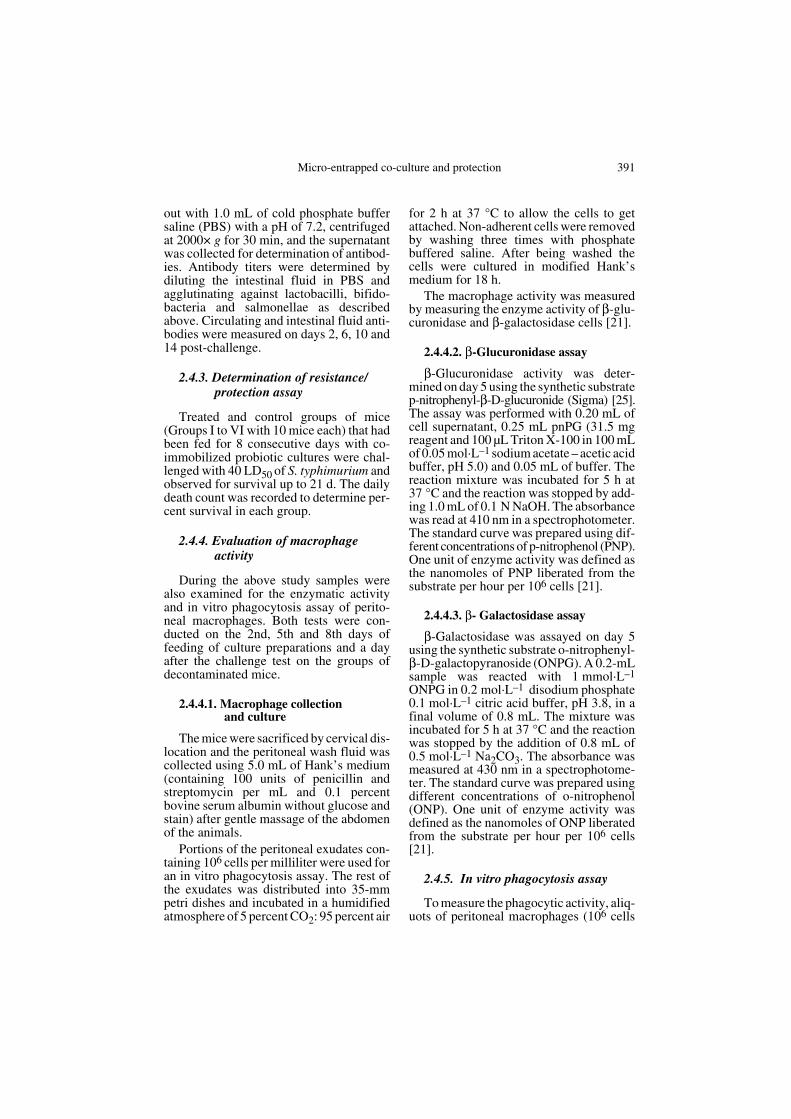
The fecal counts taken on the 8th day offeeding indicated high counts of lactoba-cilli and bifidobacteria in groups V to VI,with the highest average log counts of 8.8for lactobacilli (NCDC 13) and 8.3 for bifi-dobacteria (NCDC 255) in the group fed onmicro-entrapped co-culture (Fig. 1). Thestatistical analysis of the data also revealedsignificant differences in not only theentrapped co-culture and control groups,but also with the co-culture of free cells atboth the 5- and 1-percent levels. The oraladministration of single culture prepara-tions led to their survival in the host intes-tine as elaborated in Groups II and III. Allthe groups fed with probiotic preparations(Groups II to VI) showed the completeabsence of coliforms in their feces, whilethe control group registered the reappear-ance of coliforms in the feces after thedecontamination process.
3.2. Translocation assay using S. typhimurium
After feeding the different probiotic cul-ture preparations for 8 consecutive days, the
Micro-entrapped co-culture and protection 393
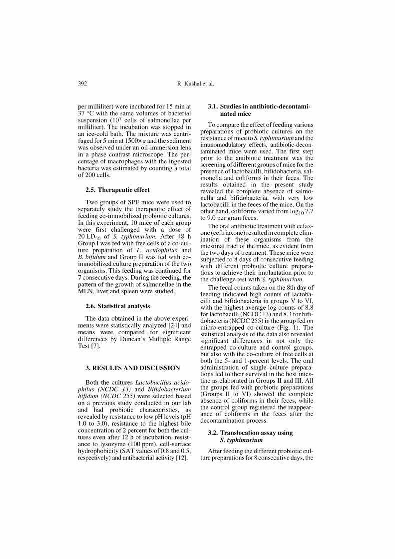
mice were orally challenged with 20 LD50of S. typhimurium. Viable bacteria in themesenteric lymph nodes (MLN) werepresent from the 2nd day onwards, reachingthe highest level on the 6th day (Fig. 2).
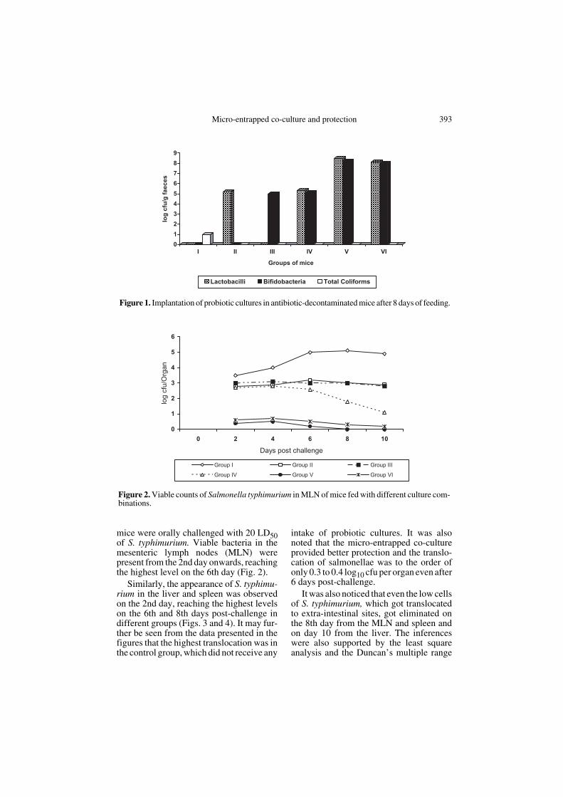
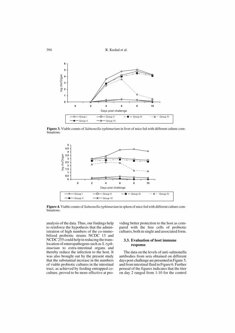
Similarly, the appearance of S. typhimu-rium in the liver and spleen was observedon the 2nd day, reaching the highest levelson the 6th and 8th days post-challenge indifferent groups (Figs. 3 and 4). It may fur-ther be seen from the data presented in thefigures that the highest translocation was inthe control group, which did not receive any
intake of probiotic cultures. It was alsonoted that the micro-entrapped co-cultureprovided better protection and the translo-cation of salmonellae was to the order ofonly 0.3 to 0.4 log10 cfu per organ even after6 days post-challenge.
It was also noticed that even the low cellsof S. typhimurium, which got translocatedto extra-intestinal sites, got eliminated onthe 8th day from the MLN and spleen andon day 10 from the liver. The inferenceswere also supported by the least squareanalysis and the Duncan’s multiple range
Figure 1. Implantation of probiotic cultures in antibiotic-decontaminated mice after 8 days of feeding.
Figure 2. Viable counts of Salmonella typhimurium in MLN of mice fed with different culture com-binations.
394 R. Kushal et al.
analysis of the data. Thus, our findings helpto reinforce the hypothesis that the admin-istration of high numbers of the co-immo-bilized probiotic strains NCDC 13 andNCDC 255 could help in reducing the trans-location of enteropathogens such as S. typh-imurium to extra-intestinal organs andthereby reduce the infection to the host. Itwas also brought out by the present studythat the substantial increase in the numbersof viable probiotic cultures in the intestinaltract, as achieved by feeding entrapped co-culture, proved to be more effective at pro-
viding better protection to the host as com-pared with the free cells of probioticcultures, both in single and associated form.
3.3. Evaluation of host immune response
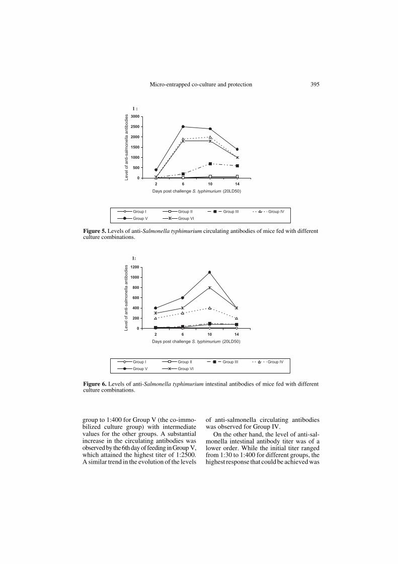
The data on the levels of anti-salmonellaantibodies from sera obtained on differentdays post-challenge are presented in Figure 5,and from intestinal fluid in Figure 6. Furtherperusal of the figures indicates that the titeron day 2 ranged from 1:10 for the control
Figure 3. Viable counts of Salmonella typhimurium in liver of mice fed with different culture com-binations.
Figure 4. Viable counts of Salmonella typhimurium in spleen of mice fed with different culture com-binations.
Micro-entrapped co-culture and protection 395
group to 1:400 for Group V (the co-immo-bilized culture group) with intermediatevalues for the other groups. A substantialincrease in the circulating antibodies wasobserved by the 6th day of feeding in Group V,which attained the highest titer of 1:2500.A similar trend in the evolution of the levels
of anti-salmonella circulating antibodieswas observed for Group IV.
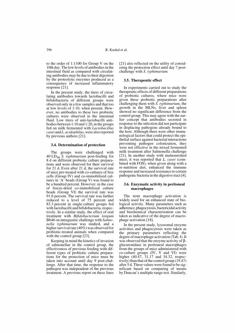
On the other hand, the level of anti-sal-monella intestinal antibody titer was of alower order. While the initial titer rangedfrom 1:30 to 1:400 for different groups, thehighest response that could be achieved was
Figure 5. Levels of anti-Salmonella typhimurium circulating antibodies of mice fed with differentculture combinations.
Figure 6. Levels of anti-Salmonella typhimurium intestinal antibodies of mice fed with differentculture combinations.
396 R. Kushal et al.
to the order of 1:1100 for Group V on the10th day. The low levels of antibodies in theintestinal fluid as compared with circulat-ing antibodies may be due to their digestionby the proteolytic enzymes produced as aconsequence of increased inflammatoryresponse [21].
In the present study, the titers of circu-lating antibodies towards lactobacilli andbifidobacteria of different groups wereobserved only in a few samples and that tooat low levels of 1:10, when present. How-ever, no antibodies to these two probioticcultures were observed in the intestinalfluid. Low titers of anti-lactobacilli anti-bodies between 1:10 and 1:20, in the groupsfed on milk fermented with Lactobacilluscasei and L. acidophilus, were also reportedby previous authors [21].
3.4. Determination of protection
The groups were challenged with40 LD50 S. typhimurium post-feeding for8 d on different probiotic culture prepara-tions and were observed for their survivalfor 21 d. Even after 21 d, the survival rateof mice pre-treated with co-cultures of freecells (Group IV) and co-immobilized cul-tures in ‘A’ beads (Group V) was found tobe a hundred percent. However, in the caseof freeze-dried co-immobilized culturebeads (Group VI) the survival rate was91.6 percent. The survival rate was furtherreduced to a level of 75 percent and83.3 percent in single-culture groups fedwith lactobacilli and bifidobacteria, respec-tively. In a similar study, the effect of oraltreatment with Bifidobacterium longumBb46 on intragastric challenge with Salmo-nella typhimurium was studied, and ahigher survival rate (40%) was observed forprobiotic-treated animals when comparedwith the control group [23].
Keeping in mind the kinetics of invasionof salmonellae in the control group, theeffectiveness of previous feeding with dif-ferent types of probiotic culture prepara-tions for the protection of mice must betaken into account until day 9 post-chal-lenge. After that time, the response to thepathogen was independent of the previoustreatment. A previous report on these lines
[21] also reflected on the utility of consid-ering the protection effect until day 7 post-challenge with S. typhimurium.
3.5. Therapeutic effect
In experiments carried out to study thetherapeutic effects of different preparationsof probiotic cultures, where mice weregiven these probiotic preparations afterchallenging them with S. typhimurium, thegrowth in the MLNs, liver and spleenshowed no significant difference from thecontrol group. This may agree with the ear-lier concept that antibodies secreted inresponse to the infection did not participatein displacing pathogens already bound tothe host. Although there were other immu-nological factors that could protect the epi-thelial surface against bacterial interactionspreventing pathogen colonization, theywere not effective in the mixed fermentedmilk treatment after Salmonella challenge[21]. In another study with malnourishedmice, it was reported that L. casei (com-bined with FOS), when given along with are-nutrition diet, enhanced the immuneresponse and increased resistance to certainpathogenic bacteria in the digestive tract [4].
3.6. Enzymatic activity in peritoneal macrophages
The term macrophage activation iswidely used for an enhanced state of bio-logical activity. Many parameters such asadherence, phagocytosis, bactericidal activityand biochemical characterization can betaken as indicative of the degree of macro-phage activation [18].
In the present study, lysosomal enzymeactivities and phagocytosis were taken asthe primary parameters reflecting thedegree of macrophage activation (Tab. I). Itwas observed that the enzyme activity of β-glucuronidase in peritoneal macrophagesfrom the groups of mice administered withco-culture groups (IV, V and VI) werehigher (40.47, 31.17 and 34.32, respec-tively) than that of the control group (19.47)after 5 d. These values were found to be sig-nificant based on comparing of meansby Duncan’s multiple range test. Similarly,
Micro-entrapped co-culture and protection 397
β-galactosidase activity of cultured perito-neal macrophages obtained from co-culturegroups was also higher (100.60, 93.59 and85.29, respectively) and was found to bestatistically significant as compared withthe control group (84.86). This indicates abetter response in the case of co-culturegroups; however, the reasons for the rela-tively higher values for the free co-culturegroup (Group IV) as compared with the co-immobilized groups (Group V and Group VI)could not be explained. In a previous studyalso, it was demonstrated that milk fer-mented with L. casei and L. acidophilus or
a mixture of both produced a remarkableeffect on immunomodulation in the host [18].
3.6.1. Enhancement of phagocytic activity
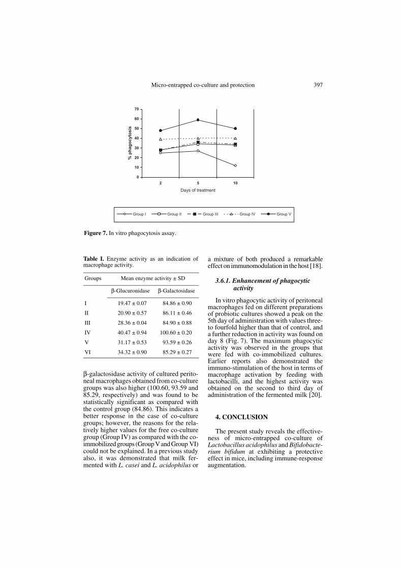
In vitro phagocytic activity of peritonealmacrophages fed on different preparationsof probiotic cultures showed a peak on the5th day of administration with values three-to fourfold higher than that of control, anda further reduction in activity was found onday 8 (Fig. 7). The maximum phagocyticactivity was observed in the groups thatwere fed with co-immobilized cultures.Earlier reports also demonstrated theimmuno-stimulation of the host in terms ofmacrophage activation by feeding withlactobacilli, and the highest activity wasobtained on the second to third day ofadministration of the fermented milk [20].
4. CONCLUSION
The present study reveals the effective-ness of micro-entrapped co-culture ofLactobacillus acidophilus and Bifidobacte-rium bifidum at exhibiting a protectiveeffect in mice, including immune-responseaugmentation.
Table I. Enzyme activity as an indication ofmacrophage activity.
Groups Mean enzyme activity ± SD
β-Glucuronidase β-Galactosidase
I 19.47 ± 0.07 84.86 ± 0.90
II 20.90 ± 0.57 86.11 ± 0.46
III 28.36 ± 0.04 84.90 ± 0.88
IV 40.47 ± 0.94 100.60 ± 0.20
V 31.17 ± 0.53 93.59 ± 0.26
VI 34.32 ± 0.90 85.29 ± 0.27
Figure 7. In vitro phagocytosis assay.
398 R. Kushal et al.
Acknowledgements: The research grant pro-vided in the form of a Sr. Fellowship by theNational Dairy Research Institute, Karnal, tothe first author is thankfully acknowledged.
REFERENCES
[1] Berg R.D., Bacterial translocation from gas-trointestinal tracts of mice receiving immu-nosuppressive chemotherapeutic agents,Current Microbiol. 8 (1983) 285–292.
[2] Berg R.D, Wommack E., Deiteh E.A.,Immunosuppression and intestinal bacterialovergrowth synergistically promote bacte-rial translocation, Arch. Surg. 123 (1988)1359–1364.
[3] Blaut M., The role of intestinal microflora inhealth and disease and novel methods tostudy it, IDF Nutr. Newsletter 5 (196) 16.
[4] Cano P.G., Perdigon G., Probiotics induceresistance to enteropathogens in a re-nour-ished mouse model, J. Dairy Res. 70 (2003)433–440.
[5] Coconnier-Polter M.H., Lievin-Le Moal V.,Servin A.L., Antibacterial effect of theadhering human Lactobacillus acidophilusstrain LB, Appl. Environ. Microbiol. 71(2005) 6115–6120.
[6] Contrepois M., Gouet P., Utilisation d’unetechnique microbiologique pour la mesurede la vitesse de transit des microparticulesdans le tractus digestif des ruminants, CRAcad. Sci. (Paris) 268 (1969) 1757–1759.
[7] Duncan D.B., Multiple range and F tests,Biometrics 11 (1955) 1–42.
[8] Fayol-Messaoudi D., Berger C.N., Coconnier-Polter M.H., Liévin-Le Moal V., ServinA.L., pH-, lactic acid-, and non-lactic acid-dependent activities of probiotic Lactoba-cilli against Salmonella enterica serovartyphimurium, Appl. Environ. Microbiol. 71(2005) 6008–6013.
[9] Fuller R., Probiotics: The Scientific Basis,1st edn., Chapman and Hall, London, 1992.
[10] Gilliland S.E., Acidophilus milk products: areview of potential benefits to consumers, J.Dairy Sci. 72 (1989) 2483–2494.
[11] Goldin B.R., Gorbach S.L., The effect ofmilk and lactobacilli feeding on humanintestinal bacterial enzyme activity, Am. J.Clin. Nutr. 39 (1984) 756–761.
[12] Kushal R., Anand S.K., Chander H., Devel-opment of a direct delivery system for co-culture of L. acidophilus and B. bifidum
based on micro-entrapment, Milchwissen-schaft 60 (2005) 130–134.
[13] Lin T.S., Messhla N., Watson R.R, Immunecomponents of the intestinal mucosa of age-ing and protein deficient mice, Immunology43 (1981) 401–407.
[14] Lloyd A.B., Cumming R.B., Kent R.D., Pre-vention of Salmonella typhimurium infec-tion in poultry by pre-treatment of chickensand poults with intestinal tract, Aust. Vet. J.53 (1977) 82–87.
[15] Mulet-Powell N., Lacoste-Armynot A.M.,Vinas M., de Buochberg M.S., Interactionsbetween pairs of bacteriocins from lacticacid bacteria, J. Food Prot. 61 (1988) 1210–1212.
[16] Owens W.E., Berg R.D., Bacterial transloca-tion from the gastrointestinal tract of ath-ymic (nu/nu) mice, Infect. Immun. 24 (1980)308–312.
[17] Owens W.E., Berg R.D., Bacterial transloca-tion from the gastrointestinal tract ofthymectomize mice, Current Microbiol. 7(1982) 169–174.
[18] Perdigon G., Alvarez S., Nader de MaciasM.E., Margni R.A., Oliver G., Hollando DeRuiz A.P., Lactobacilli administered orallyrelease enzymes from peritoneal macro-phages in mice, Milchwissenschaft 41(1986) 344–348.
[19] Perdigon G., Nader de Macias M.E., AlvarezS., Medici M., Oliver G., Pesce De RuizHolgado A.A., Effect of a mixture of Lacto-bacillus casei and Lactobacillus acidophilusadministered orally on the immune systemof mice, J. Food Prot. 49 (1986) 986–989.
[20] Perdigon G., Nader de Macias M.E., AlvarezS., Oliver G., Pesce De Ruiz Holgado A.A.,Systematic augmentation of the immuneresponse in mice by feeding fermented milkswith Lactobacillus casei and Lactobacillusacidophilus, Immunology 63 (1988) 17–23.
[21] Perdigon G., Alvarez S., Nader de MaciasM.E., The oral administration of lactic acidbacteria increases the mucosal intestinalimmunity in response to enteropathogens, J.Food Prot. 53 (1990) 404–410.
[22] Salminen S., Isolami E., Onnela T., Gutmicroflora in health and disease, Chemo-therapy 41 (Suppl.) (1995) 5–15.
[23] Silva A.M., Barbosa F.H.F., Duarte R.,Vieira L.Q., Arantes R.M.E., Nicoli J.R.,Effect of Bifidobacterium longum ingestionon experimental salmonellosis in mice, J.Appl. Microbiol. 97 (2004) 29–37.
Micro-entrapped co-culture and protection 399
[24] Snedecor G.W., Cochran W.G., StatisticalMethods, 6th edn., Oxford and IBH Pb. Co.India, 1968.
[25] van der Waaij D., Berghuis de Veries J.M.,Lekkerkert van der Wees J.E.C., Coloniza-tion resistance of the digestive tract in con-ventional and antibiotic treated mice, J.Hyg. (Camb.) 69 (1971) 405–411.
[26] van Pelt A.W., Weerkamp A.H., VyenM.H.W.J.C., Busscher H.J., deJong H.P.,Arends J., Adhesion of Streptococcus san-guis CH3 to polymers with different surfacefree energies, Appl. Environ. Microbiol. 49(1985) 1270–1275.
[27] Vanderhoof J.A., Young R.I., Use of probi-otics in childhood gastrointestinal disorders,J. Pediatr. Gastroenterol. Nutr. 27 (1988)323–332.
[28] Wells C.L., Jechorek R.P., Erlandsen S.L.,Evidence for the translocation of Enterococ-cus faecalis across the mouse intestinal tract,J. Infect. Dis. 162 (1990) 82–90.
[29] Yoshioka Y., Studies on the lactobacillusbifidum on the factor affecting the formationof bifidus flora of the intestinal tract ofinfants, Rep. Res. Lab. Snow Brand MilkProd. Co. Tokyo 72 (1971) 1–114.
To access this journal online:www.edpsciences.org