effect of heparin on the biological properties and
TRANSCRIPT
Gene 576 (2016) 292–303
Contents lists available at ScienceDirect
Gene
j ourna l homepage: www.e lsev ie r .com/ locate /gene
Research paper
Effect of heparin on the biological properties and molecular signature ofhuman mesenchymal stem cells
Ling Ling a,1, Emily T. Camilleri b,1, Torben Helledie a, Rebekah M. Samsonraj a,b, Drew M. Titmarsh a,Ren Jie Chua a, Oliver Dreesen a, Christian Dombrowski a, David A. Rider a, Mario Galindo c,d, Ian Lee e,Wanjin Hong e, James H. Hui f, Victor Nurcombe a, Andre J. van Wijnen b,⁎, Simon M. Cool a,f,⁎⁎a Institute of Medical Biology, Agency for Science, Technology and Research (A*STAR), 8A Biomedical Grove, #06-06 Immunos, Singapore 138648, Singaporeb Department of Orthopedic Surgery & Biochemistry and Molecular Biology, Mayo Clinic, Rochester, MN 55905, USAc Millennium Institute on Immunology and Immunotherapy, University of Chile, Chiled Program of Cellular and Molecular Biology, Institute of Biomedical Sciences, Faculty of Medicine, University of Chile, Casilla 70061, Correo 7, Santiago, Chilee Institute of Molecular and Cell Biology, Proteos, 61 Biopolis Drive, Singapore 138673, Singaporef Department of Orthopaedic Surgery, Yong Loo Lin School of Medicine, National University of Singapore, Singapore 119074, Singapore
Abbreviations: hMSCs, human bone marrow-derivedseq, high throughput next generation RNA sequencing;HBD, heparin-binding domain; VEGFs, vascular endothelheparin; LMWH, low-molecular-weight heparin; hESC, hheparan sulfate; ECM, extracellular matrix; FCS, fetal calfmononuclear cells; BrdU, 5-bromo-2-deoxyuridine; SVDALP, alkaline phosphatase; BSPII, bone sialoprotein II; Cprotein; ALBP, adipocyte lipid binding protein; CST3, Cyscyclin G2; INHB, inhibin; RPKM, reads per kilobasepair pe⁎ Correspondence to: A. vanWijnen, Department of Ort
and Molecular Biology, Mayo Clinic, 200 First Street SW55905, USA.⁎⁎ Correspondence to: S. Cool, Institute of Medical BiologImmunos, Singapore 138648, Singapore.
E-mail addresses: [email protected] (A.J. [email protected] (S.M. Cool).
1 These authors contributed equally.
http://dx.doi.org/10.1016/j.gene.2015.10.0390378-1119/© 2015 Elsevier B.V. All rights reserved.
a b s t r a c t
a r t i c l e i n f oArticle history:Received 8 October 2015Accepted 15 October 2015Available online 17 October 2015
Keywords:Mesenchymal stem cellsGlycosaminoglycansMultipotencyCell proliferationMicroarray
Chronic use of heparin as an anti-coagulant for the treatment of thrombosis or embolism invokes many adversesystemic events including thrombocytopenia, vascular reactions and osteoporosis. Here, we addressed whetheradverse effects might also be directed to mesenchymal stem cells that reside in the bonemarrow compartment.Harvested human bone marrow-derived mesenchymal stem cells (hMSCs) were exposed to varying doses ofheparin and their responses profiled. At low doses (b200 ng/ml), serial passagingwith heparin exerted a variableeffect on hMSC proliferation and multipotentiality across multiple donors, while at higher doses (≥100 μg/ml),heparin supplementation inhibited cell growth and increased both senescence and cell size. Gene expressionprofiling using cDNA arrays and RNA-seq analysis revealed pleiotropic effects of low-dose heparin on signalingpathways essential to hMSC growth and differentiation (including the TGFβ/BMP superfamily, FGFs, andWnts). Cells serially passaged in low-dose heparin possess a donor-dependent gene signature that reflectstheir altered phenotype. Our data indicate that heparin supplementation during the culturing of hMSCs canalter their biological properties, even at low doses. Thiswarrants caution in the application of heparin as a culturesupplement for the ex vivo expansion of hMSCs. It also highlights the need for careful evaluation of the bonemar-row compartment in patients receiving chronic heparin treatment.
© 2015 Elsevier B.V. All rights reserved.
mesenchymal stem cells; RNA-FGF, fibroblast growth factor;ial factors; UFH, unfractionateduman embryonic stem cell; HS,serum; BMMNCs, bone marrow, singular value decomposition;/EBP, CCAAT/enhancer bindingtatin-3; ITGA, integrin; CCNG2,r million mapped reads.hopedic Surgery & Biochemistry, MedSci 3-69, Rochester, MN
y, 8A Biomedical Grove, #06-06
n Wijnen),
1. Introduction
Heparin, a highly sulfated heparan glycosaminoglycan variantproduced and stored primarily by mast cells (Ronnberg and Pejler,2012), possesses the highest net negative charge density of all knownbiological molecules (Alter et al., 1987). Its negative charge binds topositively charged, heparin-binding domains (HBDs) present in a largenumber of extracellular proteins. This group of proteins includes fibro-blast growth factors (FGFs), vascular endothelial growth factors(VEGFs), bone morphogenetic proteins (BMPs) and large extracellularstructural molecules such as fibronectin and laminin, as well as itsmain clinical target antithrombin III (Naimy et al., 2008). Bothunfractionated (UFH) and low molecular weight heparin (LMWH)have been widely used as anticoagulants to enable surgery and dialysis,as well as to treat pathological conditions such as thrombosis andembolism.
The ability of heparin to interact with many proteins renders it apotential therapeutic agent beyond its use as an anti-thrombotic (Laneand Adams, 1993). Heparin's high affinity for protein has resulted in
293L. Ling et al. / Gene 576 (2016) 292–303
its application in cell culture to enhance the desirable activity of criticalextracellular biomolecules used as supplements for the expansion ofhuman stem cells. For example, heparin has been reported to promoteboth Wnt and FGF signaling in human embryonic stem cells (hESCs),thereby increasing their proliferation (Furue et al., 2008; Sasaki et al.,2008). Similarly, heparin has been shown to enhance Wnt-induceddifferentiation signals in osteogenic cells (Ling et al., 2010a), furtherhighlighting its diverse effects. Tissue culture surfaces coated withglycosaminoglycans such as heparin support greater proliferation ofMSCs (Uygun et al., 2009; Ratanavaraporn and Tabata, 2012).Heparin-functionalized hydrogels and heparinized nanoparticles havealso been developed to support the viability and differentiation ofhMSCs (Na et al., 2007; Benoit et al., 2007).
The widespread use of heparin in both clinical and research practicemakes a detailed study of both its mechanism of action and long-termeffects in cell culture highly advisable. Heparin non-selectively facili-tates the binding of a wide spectrum of proteins to high-affinity recep-tors on cells, particularly within the endothelium (Ferrara, 1999;Folkman and Shing, 1992; Gengrinovitch et al., 1995). Factors such asVEGF165 and FGF-2 normally associate with heparan sugars on cell sur-faces to form ligand:sugar:receptor complexes that induce proliferativesignals (Kim et al., 2011; Turnbull et al., 2001). Furthermore, VEGF165affinity-selected sugar has been shown to exert pro-angiogenic effectson endothelial cells (Wang et al., 2014). In contrast, short heparinfragments (~5.0-kDa) purified from porcine intestinal mucosa can sup-press VEGF165-mediated angiogenesis when delivered subcutaneously(Norrby and Ostergaard, 1997). Also, heparin derivatives as well asheparan sulfate (HS) isolated from bone marrow stromal cells andBMP-2 affinity-selected HS have been shown to increase the osteogenicpotential of BMP-2 (Ratanavaraporn and Tabata, 2012; Bramono et al.,2012; Murali et al., 2013). Genes encoding the synthesis of ECM proteinscan also be regulated by heparin (Hitraya et al., 1995). Heparin has beenshown to activatemetalloproteinase-2, leading to remodeling of the ECM(Tyagi et al., 1997). It can act as a heparanase inhibitor and is known toaffect the in vitro tubular morphogenesis of microvessels (Sweeneyet al., 1998). In certain fibroblasts, inflammatory cells, and tumor cells(most prominently), heparanase activity is enhanced, where the expres-sion of heparanasemRNA is known to correlatewith increasedmetastaticpotential (Hulett et al., 1999). Furthermore, type 1 diabetes has beenshown to be a heparanase-dependent disease (Dudakovic et al., 2014).These broad biological effects of heparin and heparin-degrading enzymesare consistent with the multiplicity of proteins that interact with itshyper-sulfated sugar chains and maintain tissue homeostasis.
In most tissues, heparin-binding proteins are usually controlled byphysiologically relevant and tissue-specific HS on the cell surface.There are notable differences in the structure between heparin andHS; most importantly heparin contains 3-O-sulfation and lacks discreteprotein-binding domains (Langeslay et al., 2011). Excess heparin withits greater negative-charge density can out-compete physiologically rel-evant HS-protein interactions and thus disrupt a number of biologicalprocesses associated with tissue development and repair that requireproper maintenance of stem cell pools. Also, safety concerns ascribedto heparin's binding promiscuity are evident from patients presentingwith heparin-induced thrombocytopenia (Bambrah et al., 2012),osteoporosis (Mazziotti et al., 2010; Sackler and Liu, 1973) and vascularreactions (Bounameaux et al., 1987; Gollub and Ulin, 1962). Indeed,heparin has been shown to enhance osteoclastic bone resorptionthrough an interaction with osteoprotegerin (OPG) (Irie et al., 2007),whilst other HS variants have been shown to exert anti-osteoclasticeffects (Ling et al., 2010b). Mastocytosis, a disorder characterized byincreased numbers ofmast cells that produce excessive heparin, is asso-ciated with osteoporosis, which again indicates the generally adverseeffect of heparin on skeletal tissue (Benucci et al., 2009). Even thoughchronic heparin use is associated with unwanted clinical events, it iswidely used as a stem cell culture supplement without a clear under-standing of its effects on stem cell phenotypes.
Adult stem cells are a key driver of natural tissue replenishment, andare among the small number of cells that can both undergo proliferationand differentiate into the various lineages needed to repair or regener-ate damaged tissue (Prockop and Oh, 2012; Samsonraj et al., 2015).Heparin supplementation in medium has been reported to promotehMSC proliferation (Mimura et al., 2011). Heparin-functionalizedhydrogels have been formulated in such a way that they are able toretain combinations of FGFs and ECM proteins and so support thegrowth, adhesion or differentiation of hMSCs (Na et al., 2007; Benoitet al., 2007; Benoit and Anseth, 2005; Bhakta et al., 2012). However,we lack precise knowledge of the biological effects of heparin on hMSCs.
This study set out to determine whether heparin, over a range ofdoses, could change the intrinsic properties of hMSCs in vitro. It provedcapable of altering the molecular profiles of hMSCs, even at low doses,and affected their potential for growth and differentiation in a donor-dependent manner. Our findings suggest that caution should beexercised whenever stem cells are serially passaged in heparin-supplemented media.
2. Materials and methods
Throughout the study, control data generated in hMSCs grown inmaintenance conditions (Helledie et al., 2012) was compared toheparin-treated hMSCs in Figs. 1A, B, 2 and 3. Experimentation withheparin was performed in parallel with studies using HS (Helledieet al., 2012). Data obtained with heparin and HS were analyzedseparately to address distinct findings on different biological propertiesof hMSCs that emerged from the two datasets.
2.1. Cell culture
Human MSCs were either directly purchased from Lonza or isolatedin our laboratory from human bone marrow mononuclear cells fromyoung healthy male donors aged 20–30 years provided by Lonza(Donors 1 to 3) as previously described (Samsonraj et al., 2015; Rideret al., 2008). Cells were expanded and maintained in completeDulbecco's Modified Eagle's Medium using our standard protocols(Helledie et al., 2012) and we reported the number of passages undervarious treatment conditions (experimental passage number). Unlessotherwise indicated, hMSCs were treated with 160 ng/ml of heparin(Sigma-Aldrich), a glycosaminoglycan dosage that is within the rangeused in other related stem cell studies (Mimura et al., 2011; Helledieet al., 2012).
2.2. Cell proliferation and cumulative growth analysis
HumanMSCs were plated in triplicate at 5000 cells/cm2 in the pres-ence or absence of heparin as indicated and proliferation determined bymonitoring viable cell number using the GUAVA PCA-96 benchtop flowcytometer as per manufacturer's instructions (Millipore). Briefly, cellswere dislodged by trypsinization and stained with GUAVA Flex dye.Cell suspensions were counted using the GUAVA Viacount program.
To monitor the cumulative growth, cells were plated at the samedensity (5000 cells/cm2) with or without heparin as indicated andsub-cultured upon reaching 70–80% confluency. The cells werere-plated under the same condition at each passage and the viablecells were counted using GUAVA system as described above.
2.3. Image cytometry
HumanMSCs were seeded into chamber slides at 3000 cells/cm2 andallowed to attach overnight. Cells were then treated with or without500 μg/ml heparin for 3 days. Cells were fixedwith 4% paraformaldehydeand stained with Rhodamine-conjugated phalloidin and DAPI (Life Tech-nologies). Samples were imaged with an Olympus IX83 inverted micro-scope with a Cool Pix HQ2 camera and MetaMorph software, using slide
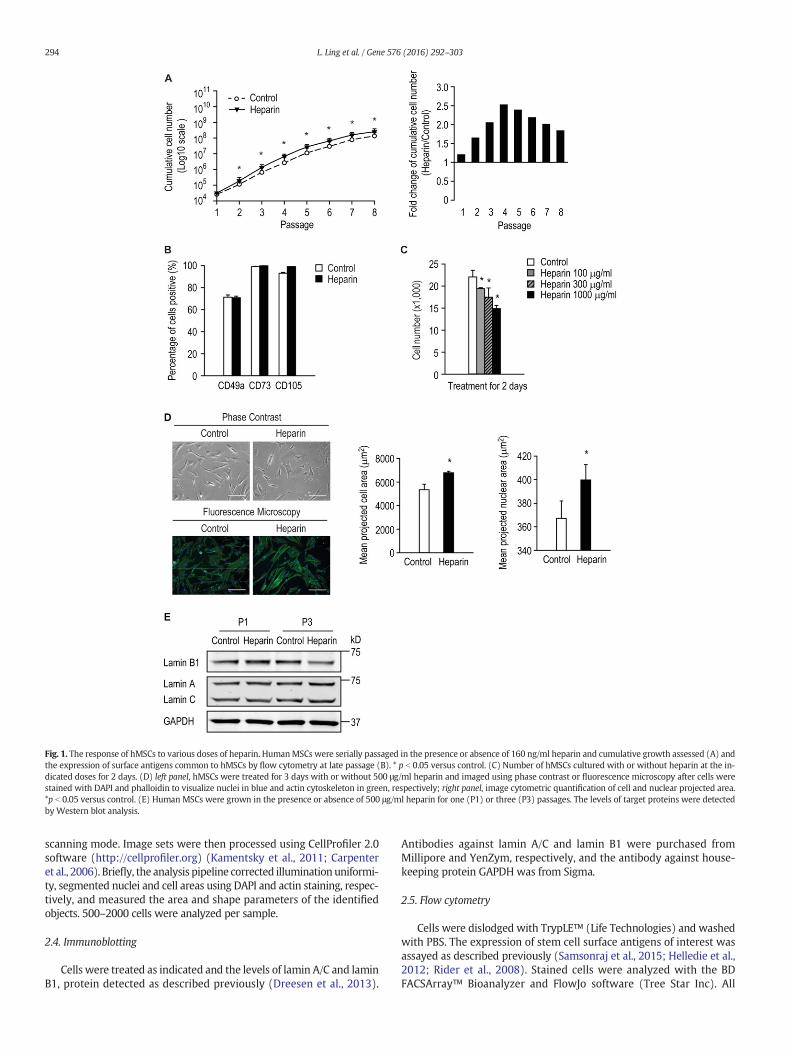
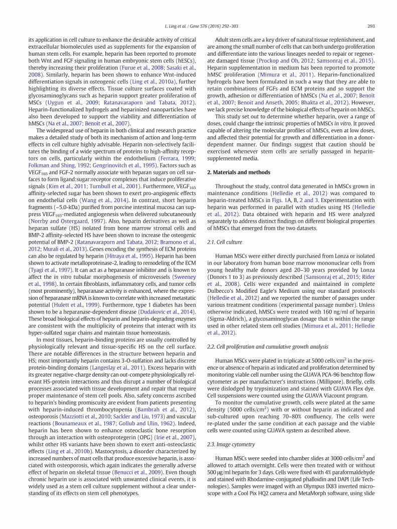
Fig. 1. The response of hMSCs to various doses of heparin. HumanMSCs were serially passaged in the presence or absence of 160 ng/ml heparin and cumulative growth assessed (A) andthe expression of surface antigens common to hMSCs by flow cytometry at late passage (B). * p b 0.05 versus control. (C) Number of hMSCs cultured with or without heparin at the in-dicated doses for 2 days. (D) left panel, hMSCs were treated for 3 days with or without 500 μg/ml heparin and imaged using phase contrast or fluorescence microscopy after cells werestained with DAPI and phalloidin to visualize nuclei in blue and actin cytoskeleton in green, respectively; right panel, image cytometric quantification of cell and nuclear projected area.*p b 0.05 versus control. (E) Human MSCs were grown in the presence or absence of 500 μg/ml heparin for one (P1) or three (P3) passages. The levels of target proteins were detectedby Western blot analysis.
294 L. Ling et al. / Gene 576 (2016) 292–303
scanning mode. Image sets were then processed using CellProfiler 2.0software (http://cellprofiler.org) (Kamentsky et al., 2011; Carpenteret al., 2006). Briefly, the analysis pipeline corrected illumination uniformi-ty, segmented nuclei and cell areas using DAPI and actin staining, respec-tively, and measured the area and shape parameters of the identifiedobjects. 500–2000 cells were analyzed per sample.
2.4. Immunoblotting
Cells were treated as indicated and the levels of lamin A/C and laminB1, protein detected as described previously (Dreesen et al., 2013).
Antibodies against lamin A/C and lamin B1 were purchased fromMillipore and YenZym, respectively, and the antibody against house-keeping protein GAPDH was from Sigma.
2.5. Flow cytometry
Cells were dislodged with TrypLE™ (Life Technologies) and washedwith PBS. The expression of stem cell surface antigens of interest wasassayed as described previously (Samsonraj et al., 2015; Helledie et al.,2012; Rider et al., 2008). Stained cells were analyzed with the BDFACSArray™ Bioanalyzer and FlowJo software (Tree Star Inc). All

Fig. 2.HumanMSCs grown in the presence of heparin exhibited an altered gene expression signature. HumanMSCs were serially passaged with or without 160 ng/ml heparin. Gene ex-pression signatures of hMSCs at P4, P6 and P8 were obtained using a stem cell-related chemiluminescent cDNA array. Expression profiles were analyzed by (A) singular value decompo-sition, or (B and C) hierarchical clustering and Pearson correlation distance/similarity approaches.
295L. Ling et al. / Gene 576 (2016) 292–303
antibodies were purchased from BD Biosciences. For Donor 1–3 sam-ples, STRO-1was kindly supplied by Prof Stan Gronthos, School of Med-ical Sciences, Faculty of Health Sciences, University of Adelaide,Australia, and the isotype control IgM (μ) was purchased from CaltagLaboratories.
2.6. Stem cell microarray
RNA was extracted from serially-passaged hMSCs (±heparinsupplementation), converted to cDNA and probed using the GEArray SSeries Human Stem Cell Gene Array (SABiosciences) as per
296 L. Ling et al. / Gene 576 (2016) 292–303
297L. Ling et al. / Gene 576 (2016) 292–303
manufacturer's instructions. The array was scanned using a Chemi-Smart 3000 image acquisition system (Vilber Lourmat). The stem cellsignature was created by using Singular Value Decomposition (SVD)(Holter et al., 2000; Alter et al., 2000), which reveals the relatedness ofdifferent biological samples. Graphical clustering of expression signa-tures indicates biological similarities.
The gene expression data were further analyzed using GeneSpringGC 11.0 software and DAVID Bioinformatics Resources 6.7 (http://david.abcc.ncifcrf.gov) (Huang da et al., 2009). The overlapping ofgene targets in different cells was acquired using VENNY software(http://bioinfogp.cnb.csic.es/tools/venny/index.html). Hierarchicalclustering was carried out using GENE-E (version 3.0.228; BroadInstitute, Cambridge, MA, USA).
2.7. Multilineage differentiation
Following serial passaging in the presence or absence of heparin,hMSCs were then induced for osteogenic, chondrogenic, or adipogenicdifferentiation and lineage commitment assessed by histochemicalstaining (Samsonraj et al., 2015; Helledie et al., 2012; Rider et al.,2008). For clarity, heparin was not included in any of the inductionmedia.
Osteogenic (Alizarin Red staining), adipogenic (Oil-Red-O staining)and chondrogenic (Alcian Blue) staining were performed as previouslydescribed (Samsonraj et al., 2015; Helledie et al., 2012). Intensities ofthe colorimetric dyes were quantified from digitally scanned imagesusing the “Unmix Colors” module in CellProfiler 2.1.1. Areas of interestwere isolated by masking (for culture wells) or manual tracing (pelletsections). Total dye intensity was then integrated in the area of interest,and normalized to the area (for pellet sections of varying size).
2.8. Next generation RNA sequencing and bioinformatic analysis
Human MSCs were harvested with TRIzol® Reagent (Invitrogen)and RNA was isolated according the manufacturer's protocol. RNAsequencing and bioinformatics was performed as detailed previously(Dudakovic et al., 2014). Briefly, oligo dT magnetic beads were used toselect polyadenylated mRNAs for the TruSeq RNA method (Illumina).Samples were indexed using TruSeq Kits (12-Set A and 12-Set B) formultiplexing on the flow cells of an Illumina HiSeq 2000 sequencer.Library preparation and concentration was test for quality controlusing an Agilent Bioanalyzer DNA 1000 chip and Qubit fluorometer(Invitrogen) respectively. Sequencing data was processed usingMAPRSeq (v.1.2.1), TopHat 2.0.6, HTSeq, and edgeR 2.6.2 work flows(Huang da et al., 2009; Robinson et al., 2010). Gene expression valueswere normalized to 1 million reads and corrected for gene length(reads per kilobasepair per million mapped reads, RPKM). Functionalgene annotation analysis and overlapping genes were performed asdescribed above.
2.9. Statistical analysis
Experimentswere performedwith at least three biological replicatesfor every condition and the data expressed asmean± S.D. unless statedotherwise. Differences among treatmentswere analyzed by a two-tailedunpaired t test. Significant differenceswere considered as thosewith a pvalue of b0.05 (*).
Fig. 3.Heparin controls a broad set of genes involved in signal transduction in hMSCs. HumanMexpression data were analyzed for changes during passaging or in response to heparin at each pevant changes during passage (left) or heparin treatment (right) for genes that are up- or down(B) Venn diagrams illustrate the number of genes that are consistently up- or down-regulated aright compares all genes that are up (59) or down (38) regulatedwith genes that are robustly eanalysis of selected genes to illustrate changes in expression during passage or heparin-treatmemembers of the BMP2/TGFβ family (left) or cell surface proteins (right). Gene expression was
3. Results
3.1. Heparin supplementation enhanced hMSC proliferation without affect-ing the expression of stem cell markers
Heparin binds and activates a large number of mitogenic factors andmorphogens that mediate proliferation and lineage commitment ofprogenitor cells. We evaluated its mitogenic properties on hMSCs bymonitoring cumulative cell growth. This was consistently enhancedwhen cells were serially passaged in the presence of 160 ng/ml heparincompared with the control (Fig. 1A). Interestingly, at earlier passages(≤passage 4) the proliferative effect of heparin was greatest afterwhich a decline was observed (Fig. 1A, right panel). Heparin has beenpreviously reported to accelerate proliferation of genetically modifiedhMSCs (Mimura et al., 2011), consistent with our observations at earlypassage.
Having established the effect of heparin on the culture-expansion ofhMSCs, we next examined the stem cell quality after serially-passagingby assessing the expression of particular stem cell surface antigens. Theexpression of surface antigens CD49a, CD73 and CD105, standardmarkers for the identification of MSCs, did not change appreciably onserially-passaged cells in the presence of heparin (Fig. 1B). These resultssuggest that routine expansion of hMSCs in the presence of heparinmaynot affect the selected cell surface antigens of a given donor.
3.2. High-dose heparin significantly inhibits hMSC growth
Having established that low-dose heparin (160 ng/ml) modestlyincreased hMSC proliferation, we next examined their response to arange of higher heparin concentrations. The results showed that growthof hMSCs were inhibited by heparin at 100–1000 μg/ml (Fig. 1C). Cellstreated with 500 μg/ml heparin for 3 days also exhibited an alteredmorphology; these cells were flatter and larger than control cells,which preserved their fibroblast-like spindle shape (Fig. 1D, left panel).Quantification by image cytometry of the mean projected cell andnuclear areas confirmed that high-dose heparin treatment resulted insignificantly larger cells and nuclei (Fig. 1D, right panel), morphologicalfeatures that are consistent with onset of a dormant (quiescent or se-nescent) cellular state. Also, hMSCs cultured with 500 μg/ml heparinfor three passages have reduced level of lamin B1 (Fig. 1E), a senescencemarker reported previously (Dreesen et al., 2013), whilst the levels oflamin A or C were not affected. Thus, although heparin has positiveeffects on proliferation at low-doses, it is a potent growth inhibitorand might cause premature cellular senescence at higher doses.
3.3. Heparin supplementation in culture supports expansion of hMSCs butmay compromise cell fate decisions
Because chronic heparin treatment appears to influence the naïveundifferentiated state of hMSCs, we next examinedmolecular pathwaysinvolved in this response using cDNA arrays. RNA sampleswere isolatedfrom control and heparin-treated hMSCs at passages (P4, 6 or 8) andmRNA levels were determined using a customized cDNA array thatmonitors expression of a large set of extracellular ligands (e.g., growthfactors, cytokines), cell surface proteins (e.g., growth factor receptors,integrins), cytoplasmic proteins and nuclear transcription factors(Figs. 2 and 3).
The expression profiles of hMSCs were analyzed and the resultinghMSC signatures were evaluated by Singular Value Decomposition
SCs were serially passagedwith or without 160 ng/ml heparin. (A) Stem cell related geneassage as a six componentmatrix. The pie chart depicts the number of genes showing rel--regulated (N1.4 fold change; “Up” or “Down”) or shows no appreciable change (“Other”).t the three passages (P4, P6 or P8) and/or in response to heparin. The Venn diagram to thexpressed (Signal N0.1). This analysis yields 20 genes that meet both criteria. (C) Expressionnt. (D) Bar graphs depict the fold change in expression in response to heparin for selectedanalyzed at three different passages (P4, P6 or P8) as indicated.

298 L. Ling et al. / Gene 576 (2016) 292–303
(SVD) (Holter et al., 2000; Alter et al., 2000) (Fig. 2A). These studieswere modeled on similar approaches previously used to distinguishtumor subtypes (Bild et al., 2006; Pomeroy et al., 2002). Themicroarraydata compared the expression of 262 genes across 6 samples. Eachexpression value was log-transformed and centered across samplesbefore application of Singular Value Decomposition analysis. Projectionwas onto the first 2 right singular vectors, which together capture about70% of the variation in the original data. Thereafter, each sample wasdefined by its score — a linear combination of the gene expressionvalues and the coefficients of the 2 singular vectors. Samples withsimilar phenotypes tend to cluster on a score plot. The analysis revealedthat the temporal expression of genes in cells cultured under controlconditions are different in short-term versus long-term culture(Fig. 2A). These differences indicate that heparin alters the molecularphenotype of hMSCs as a function of time in culture. Human MSCs atpassage 4 (the lowest passage analyzed), appear to have a signatureresembling that of naïve hMSCs. Parallel cultures of hMSCs exposed toheparin have their own unique signature when compared to controlcells, which reflect stem cell-related alterations in their molecularcharacteristics phenotype. These observations were corroboratedby hierearchical clustering (Fig. 2B) and Pearson correlation distance/similarity analysis (Fig. 2C) of representative genes that exhibit changesin gene expression (Fig. 3). Taken together, culture expansion in thepresence of heparin modifies the passage-related gene expressionphenotype of hMSCs.
3.4. Heparin controls a broad set of genes involved in signal transduction inhMSCs
Weanalyzed changes in gene expression over passage (independentof the effect of heparin) or upon heparin treatment (independent of theeffect of passaging) in hMSCs using arbitrary cut-offs (N1.4 fold or b0.7fold) to filter for biologically relevant changes (Fig. 3A). In addition, weapplied optional filters related to relative expression levels (signal N1 orN0.1 arbitrary units) as indicated to focus on genes with prominentlevels of expression. The arbitrary cut-offs for signal strength N1 or 0.1were selected to obtain a manageable number of genes that illustratewhat genes are robustly expressed in the basal state in the absence ofheparin (signal N1) and/or which set of genes shows biologically inter-esting changes in gene expression in response to heparin (signal N0.1and fold change N1.4 or b0.7). Of the 262 genes analyzed, there areonly 8 genes that are consistently expressed at high levels (signal N1)in hMSCs regardless of passage or heparin treatment in all sixconditions. These genes are the glycosylated cell surface protein CD24,the intracellular proteinase inhibitor Cystatin-3 (CST3), the extracellularligandWnt8A, integrinα5 (ITGA5), three different transcription factors(SOX1, EGR2/Krox20 and POU5F1/Oct4) and the cell cycle inhibitorCDKN2D. The abundant expression of these genes is consistent withthe multi-lineage potential of hMSCs. CD24, SOX1 and POU5F1/OCT4are markers of progenitor cells (Qiu et al., 2011; Elkouris et al., 2011;Boyer et al., 2005), while EGR2,Wnt8A and ITGA5 are linked to differen-tiation in distinct cell lineages (Lejard et al., 2011; Erter et al., 2001;Hamidouche et al., 2010).
During successive passaging from P4 to P8, 70 genes are down-regulated and 11 are up-regulated, reflecting modulations in prolifera-tive potential and lineage phenotype (Fig. 3A, left pie chart). Heparintreatment consistently up-regulates 59 genes and down-regulates 38genes by at least 1.4 fold, regardless of the passage number (Fig. 3A,right pie chart). The vast majority of genes regulated by heparin(~90%) are different from those regulated as a consequence of passage.Only 1 of the 11 genes thatwere up-regulated and only 8 of the 70 genesthat were down-regulated (~10%) during passage were alsomodulatedby heparin (Fig. 3B, left panel). Taken together, these findings indicatethat heparin selectively alters expression of genes that are distinctfrom those related to continuous culture of hMSCs.
Of the 97 (i.e., 38 plus 59) genes that are up- or down-regulated byheparin, at least 20 genes had robust basal expression (signal N0.1)(Fig. 3B, right panel). Three of these have known roles in cell cycle con-trol. The cell cycle regulators CDKN1B (p27) and CCNG2 (cyclin G2),both associated with cell growth inhibition, are each up-regulated,while the tumor suppressor protein RBL2 (p130) was down-regulated(Fig. 3C). In addition, heparin significantly increases expression of a se-ries of growth factors, including FGF14, FGF20, BDNF, IL6 and VEGF, butnot FGF2 (Fig. 3C and data not shown) as well as the expression of anumber of cell surface proteins, including ITGA6 (integrin α6) andNGFR, but not ITGA5 or ITGB3 (Fig. 3C and data not shown). In addition,heparin increases themRNA levels of a series of transcription factors, in-cluding FOXG1A, FOXH1, PAX6 and SOX13, but not GATA4 (Fig. 3C anddata not shown). Thus, heparin selectively stimulates expression of anumber of different cell surface proteins and nuclear effectors; suchpleiotropic effects are expected based on its biochemical activity as amultivalent co-ligand.
Heparin'smost striking gene expression effect is themajor inductionof multiple members of the TGFβ/BMP/GDF superfamily includingBMP3, BMP4, GDF3, GDF5 and GDF11 (Fig. 3D). In addition, heparinsuppresses the corresponding inhibitors NOG (noggin) and INHB1A (in-hibin 1A) (Fig. 3D). These changes are consistentwith auto- or paracrineenhancement of TGFβ/BMP/GDF signaling. Heparin also increases theexpression of selected Wnt and FGF members (Fig. 3C and data notshown), as well asmodulates (up or down) cell surfacemarkers includ-ing FZD4, NGFR, PDGFRA and PTPRC (Fig. 3D).We conclude that heparincompromises expression ofmRNA transcripts encoding proteins knownto play a role in cell fate decisions.
3.5. Heparin exhibits variable effects on the proliferation of hMSCs from dif-ferent donors
Having established that heparin at 160 ng/ml increased the numberof hMSCs in culture and negatively affects their molecular signature, wenext sought to determine whether this finding was similar for hMSCsfreshly isolated from human bone marrow mononuclear cells. HumanMSCs from different donors responded variably to long-term heparintreatment, as their growthwas either slightly increased at late passages(≥P5), not affected, or significantly inhibited (Fig. 4A).
Similar to the data in Fig. 1B, the effect of long-term passaging withheparin on the expression of CD49a, CD73 and CD105 in hMSCs fromdifferent donors was also minimal (Fig. 4B). Expression of three addi-tional stem cell markers STRO-1, CD146 and SSEA4 (Sacchetti et al.,2007; Gang et al., 2007; Simmons and Torok-Storb, 1991) was similar,except that heparin slightly increased SSEA4 expression in hMSCsfrom donor 2 (Fig. 4B). Multilineage staining for hMSCs from thesethree donors showed that long-term culturing with heparin supple-mentation also had little effect on the multipotency of hMSCs (Fig. 4C).
3.6. RNA-seq analysis reveals genome-wide differences that modify cellgrowth characteristics of MSCs upon chronic heparin treatment
Because hMSCs from three different donors exhibit different cellgrowth responses to heparin treatment, we investigated how heparinaffects themolecular properties of hMSCs using transcriptomic analysis.We focused our RNA-seq analysis on Donor 3 in which heparin exhibitsa clear negative effect on cell proliferation, and compared our datasetwith expression results obtained for Donor 2 which is refractory to thegrowth modulatory effects of heparin (see Fig. 4A). As such, Donor 2represents a negative control that permits elimination of gene expres-sion effects of heparin independent of biological effects on cell prolifer-ation. We compared gene expression patterns of late cultured cells inthe presence or absence of heparin (Fig. 5). We filtered the dataset forgenes with reads per kilobasepair per million mapped reads (RPKMs)greater than 0.3, which reduces false discovery of mRNA detection, aswell as for fold-changes in expression values greater than 2-fold (up

Fig. 4. The long-term effect of heparin on hMSCs frommultiple donors. (A) HumanMSCs serially passaged in the presence or absence of 160 ng/ml heparin. Cumulative cell numberswerecalculated. Significance is reported (*p b 0.05)when all 6 passages showed a difference. (B and C)HumanMSCswere serially passaged in the presence or absence of 160 ng/ml heparin andassessed at late passage for the expression of cell surface antigens by flow cytometry, or their ability to differentiate into the osteogenic, adipogenic and chondrogenic lineages as assessedby histochemical staining for Alizarin Red, Oil-Red-O or Alcian Blue and the intensity of stain quantified by densitometry.
299L. Ling et al. / Gene 576 (2016) 292–303
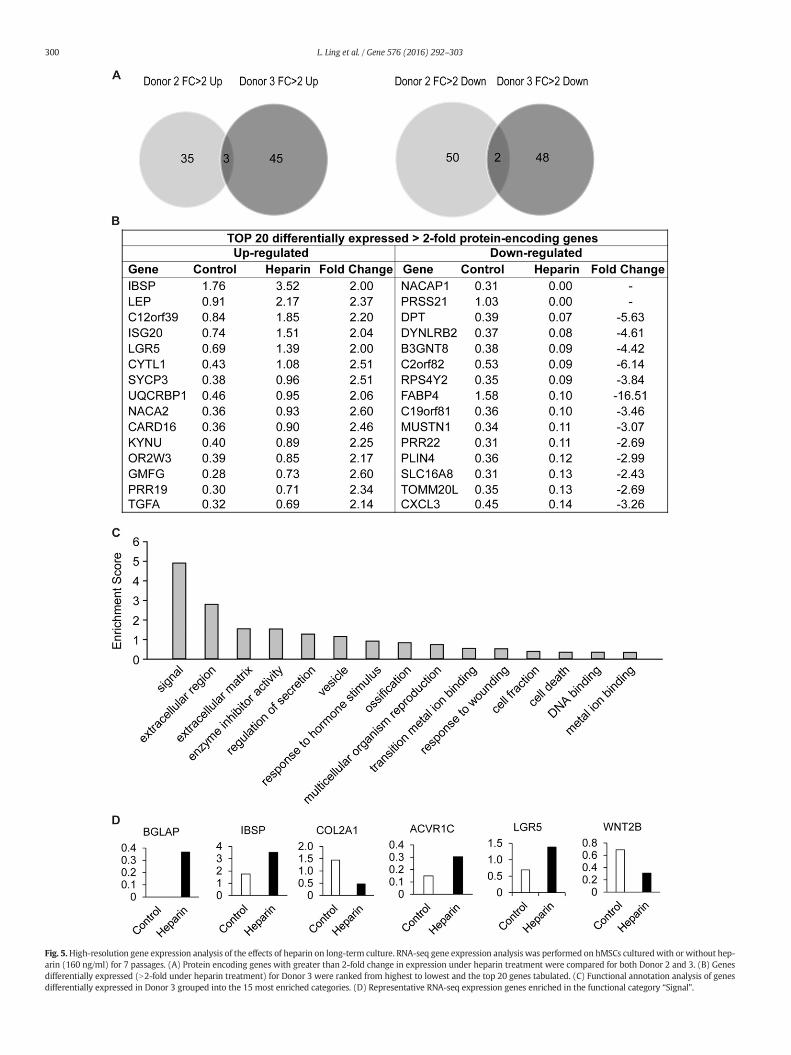
Fig. 5.High-resolution gene expression analysis of the effects of heparin on long-term culture. RNA-seq gene expression analysis was performed on hMSCs culturedwith or without hep-arin (160 ng/ml) for 7 passages. (A) Protein encoding genes with greater than 2-fold change in expression under heparin treatment were compared for both Donor 2 and 3. (B) Genesdifferentially expressed (N2-fold under heparin treatment) for Donor 3 were ranked from highest to lowest and the top 20 genes tabulated. (C) Functional annotation analysis of genesdifferentially expressed in Donor 3 grouped into the 15 most enriched categories. (D) Representative RNA-seq expression genes enriched in the functional category “Signal”.
300 L. Ling et al. / Gene 576 (2016) 292–303

301L. Ling et al. / Gene 576 (2016) 292–303
or down) upon heparin treatment for each donor. Strikingly, weobserved very limited overlaps in the groups of genes that are heparinresponsive between the two donors (Fig. 5A), consistent with theobservation that heparin has donor-dependent pleiotropic effects oncell growth (see Fig. 4A).
We observed that there are 45 genes that are uniquely upregulatedby heparin in Donor 3 only, while 48 genes are differentially down-regulated by heparin compared to Donor 2 (Fig. 5A). To identify themost prominently regulated genes within these groups, we sortedgenes for highest expression values (in RPKM) yielding a set of 15heparin-responsive up- or down-regulated genes with most robustexpression (Fig. 5B). None of this diverse set of genes appear to haveobvious functions that can directly account for the cell growth perturba-tions that are evident in biological assays (see Fig. 4). Hence, unique cellproliferative effects of heparin on donor 3 may affect primarily transla-tional or post-translational mechanisms that do not perturb geneexpression at the mRNA level (e.g., miRNAs, protein stability, proteinphosphorylation).
To understand the biological activities of the entire set of genesboth up- and down-regulated by heparin, we performed gene ontologyanalysis (Fig. 5C). The two most enriched categories are genes involvedin signaling and extracellular region (enrichment scores of ~5 and ~3,respectively). Interestingly, these two categories are directly related tothe main activities of heparin that control the interaction of signalingligands with extracellular proteoglycans to modulate intracellularkinase pathways. Among the category signaling, heparin modulatesthe expression of at least two receptors (i.e., ACVR1C and LGR5), aligand (Wnt2B) and three skeletal ECM proteins (i.e., BGLAP, ISBP andCOL2A1). Compared to our earlier qPCR profiling studies with one orig-inal donor (Figs. 1 to 3) that prompted later studieswith three addition-al donors (Figs. 4 and 5), we observed that different sets of genes aremodulated. This result is expected from the observation that differentdonors respond both biologically (Figs. 1A and 4A) and molecularly(Figs. 2, 3 and 5A) to heparin. Taken together, the RNA-seq results sug-gests that heparin may affect cell growth at least in part by alteringmRNA levels of distinct sets of cell growth-related receptor signalingpathways in a donor dependent manner.
4. Discussion
Although widely used as an anti-clotting agent for patients at risk ofthrombosis or embolism, heparin's broad ability to bind and potentiatethe activity of a plethora of factors unrelated to blood coagulation hasrendered it a versatile agent that is widely used as a culture supplementfor manymammalian cell types, including hMSCs and hESCs. For exam-ple, heparin has been used as an additive for stem cell expansion andhydrogel formulation to promote stem cell pluripotency (Furue et al.,2008).
Notwithstanding its widespread use as a stem cell culture additive,the results here show that heparin supplementation invites potentialrisk to ex vivo cultured stem cells from the bone marrow compartment.The adverse effect of long-term heparin therapy as an anti-coagulant onskeletal tissues is widely recognized, adding further caution to its use asa culture reagent. Heparin reduces bone density either through increas-ing bone resorption or decreasing bone formation (Rajgopal et al.,2008). The high affinity of heparin for BMPs can dysregulate the activityof those osteogenic growth factors, and thus osteoblast-induced boneformation. A number of studies have demonstrated that heparinrestricts the signaling and inhibits the activity of BMPs 2, 4, 6 and 7(Irie et al., 2003; Brkljacic et al., 2013; Ohkawara et al., 2002; Fisheret al., 2006).
Our data suggests that heparin supplementation might modestlyincrease hMSC proliferation at low doses, but strongly suppressesgrowth at high doses. High dose heparin also significantly alteredcell morphology, including cell and nuclear area and transformedthe hMSCs to a more senescent phenotype. During osteogenic
differentiation, MSCs gradually lose their original morphology, by flat-tening and spreading (Matsuoka et al., 2013), which correlates withloss of proliferative ability (Banfi et al., 2000), findings supported byour current study. Notably, heparin at high dosewas reported to be del-eterious to hESCs (Furue et al., 2008).
Continuous supplementation of hMSCs with a growth-permissivedose of heparin (as seen in Fig. 1), resulted in changes in mRNA expres-sion of multiple lineage-specific markers. These effects collectively repre-sents amajor drawback for the exploitation of hyper-sulfatedheparin as astemcell culture additive.Moreover, cultures supplementedwithheparinshow an altered molecular signature. Ultimately, hMSCs serially-passaged in heparin must be tested for their in vivo efficacy in well-accepted animalmodels. For example, rodentmodels of bone fracture re-pair have been used to assess hMSC quality (Samsonraj et al., 2015;Helledie et al., 2012; Rai et al., 2010) and provide a robust assessmentof the effect of ex vivo expansion strategies on their therapeutic utility.
The analysis here demonstrates that heparin exerts long-termeffects on cells that are reflected by changes in gene expression. GlobalmRNA expression profiles of control versus heparin-expanded cells asmeasured by cDNA arrays or RNA-seq are distinct in several donors atrepresentative passages. Importantly, heparin alters the gene expres-sion profiles of multiple ligands, receptors and downstream transcrip-tion factors in a donor-dependent manner. For example, heparinincreases expression of several FGFs and lineage commitment-relatedfactors such as BMP4, VEGF, GDF5 and Wnt2B, depending on thedonor and/or method of analysis (i.e., cDNA array versus RNA-seq).Because FGF, BMP and Wnt signaling are central to the proliferationand differentiation capacities of hMSCs, the observed modulation ofthese transcripts by heparin during long-term culture may account forat least some of the changes in cell proliferation and ability to controlcell fate decisions.
Another prominent finding of the gene expression profiling inresponse to heparin is the donor-dependent coordinate up-regulationof multiple members of the TGFβ/BMP/GDF superfamily and concomi-tant down-regulation of pathway inhibitors (i.e., noggin and inhibin1A). TGFβ signaling inhibits osteogenic differentiation and promotessenescence (Furue et al., 2008; Samsonraj et al., 2013; Galindo et al.,2005). In contrast, BMPs stimulate stem cell differentiation (Xu et al.,2005). Therefore, changes in the auto- or paracrine production ofTGFβ/BMP family members may directly contribute to loss ofmultipotentiality and ‘stemness’.
We note that donor-to-donor variability contributes significantly to-wards the observed effects of heparin at both biological and molecularlevels. Heparin treatment of hMSCs from four different donors revealedeither growth enhancement, inhibition or no difference. The pleiotropiceffects of heparinmay biologically amplify the inherent donor-to-donorvariability of hMSCs. Indeed, analyses by cDNA arrays or high resolutionRNA-seq revealed donor-dependent differences in gene expression inhMSCs from different donors following long term culture in heparin.
Although our study indicates that heparin may not be suitable as anadditive for long-term proliferative expansion of naïve multipotenthMSCs, there are HS variants that may be more suitable for routine cul-turing of hMSCs. Both heparin andHS consist of a repeatingdisaccharideunit with highly variable sulfation patterns. Sulfation most often occurson N-, 2-O-, and 6-O-groups and these sulfated groups are responsiblefor the interaction with growth factors or adhesion proteins. Thoughheparin is closely related to HS, it is primarily distinguished fromHS by its higher sulfation, lack of domain structure, higher frequencyof N-sulfation, presence of 3-O-sulfation, and more O-sulfation thanN-sulfation (Gallagher and Walker, 1985). Differences in the temporalexpression of HS variants as tissues grow andmaturemay permit selec-tive binding and co-activation of distinct growth factor-dependent sig-naling pathways that regulate cell growth and differentiation(Nurcombe et al., 2007).
Indeed, leveraging this biochemical diversity, we have isolated HSvariants that possess unique domains structures that target protein
302 L. Ling et al. / Gene 576 (2016) 292–303
ligands controlling the phenotypic properties of hMSCs (e.g., FGF2,BMP2, VEGF165) (Wang et al., 2014; Murali et al., 2013; Helledie et al.,2012; Nurcombe et al., 2000; Murali et al., 2011; Bramono et al.,2011).We anticipate that better preparations of HS, rather thanheparin,may be useful for routine culture of hMSCs without adversely affectingthe biological properties of the cells.
5. Conclusions
This study assessed the effects of low-dose heparin on hMSCs invitro, and highlights the potential risk to patients undergoing chronicheparin therapy— the possible impairment of naïve stem cell replenish-ment and subsequentmaintenance of tissue homeostasis from the bonemarrow. Our work demonstrates the hazard of using heparin and sug-gests caution in its use as an adjuvant during stem cell bioprocessing.
Acknowledgments
We thank all members of the Glycotherapeutics Group from the In-stitute of Medical Biology (IMB), Agency for Science, Technology andResearch (A*STAR) and the Orthopedic Research Laboratories at MayoClinic for stimulating discussions. In particular, we thank Ms. Phua ZerCheng, and A*STAR's Advanced Molecular Pathology Laboratory. Weacknowledge that datawas initially collected at the Institute ofMolecularand Cell Biology, A*STAR and continued at IMB. This work was alsosupported by the National Medical Research Council of Singapore(NMRC/1257/2010) and in part by the National Institutes of HealthGrants AR49069 (AvW). We also appreciate the generous philanthropicsupport of William and Karen Eby, as well as the charitable foundationin their names (AvW).
References
Alter, O., Brown, P.O., Botstein, D., 2000. Singular value decomposition for genome-wideexpression data processing and modeling. Proc. Natl. Acad. Sci. U. S. A. 97,10101–10106.
Alter, S.C., Metcalfe, D.D., Bradford, T.R., Schwartz, L.B., 1987. Regulation of human mastcell tryptase. Effects of enzyme concentration, ionic strength and the structure andnegative charge density of polysaccharides. Biochem. J. 248, 821–827.
Bambrah, R.K., Pham, D.C., Zaiden, R., Vu, H., Tai, S., 2012. Heparin-induced thrombocyto-penia. Clin. Adv. Hematol. Oncol. 9, 594–599.
Banfi, A., Muraglia, A., Dozin, B., Mastrogiacomo, M., Cancedda, R., Quarto, R., 2000.Proliferation kinetics and differentiation potential of ex vivo expanded human bonemarrow stromal cells: implications for their use in cell therapy. Exp. Hematol. 28,707–715.
Benoit, D.S., Anseth, K.S., 2005. Heparin functionalized PEG gels that modulate proteinadsorption for hMSC adhesion and differentiation. Acta Biomater. 1, 461–470.
Benoit, D.S., Durney, A.R., Anseth, K.S., 2007. The effect of heparin-functionalized PEGhydrogels on three-dimensional human mesenchymal stem cell osteogenic differen-tiation. Biomaterials 28, 66–77.
Benucci, M., Bettazzi, C., Bracci, S., Fabiani, P., Monsacchi, L., Cappelletti, C., Manfredi, M.,Ciolli, S., 2009. Systemic mastocytosis with skeletal involvement: a case report andreview of the literature. Clin. Cases Miner. Bone Metab. 6, 66–70.
Bhakta, G., Rai, B., Lim, Z.X., Hui, J.H., Stein, G.S., vanWijnen, A.J., Nurcombe, V., Prestwich,G.D., Cool, S.M., 2012. Hyaluronic acid-based hydrogels functionalized with heparinthat support controlled release of bioactive BMP-2. Biomaterials 33, 6113–6122.
Bild, A.H., Yao, G., Chang, J.T., Wang, Q., Potti, A., Chasse, D., Joshi, M.B., Harpole, D.,Lancaster, J.M., Berchuck, A., Olson Jr., J.A., Marks, J.R., Dressman, H.K., West, M.,Nevins, J.R., 2006. Oncogenic pathway signatures in human cancers as a guide totargeted therapies. Nature 439, 353–357.
Bounameaux, H., Schneider, P.A., Mossaz, A., Suter, P., Vasey, H., 1987. Severe vasospasticreactions (ergotism) during prophylactic administration of heparin-dihydroergotamine. Vasa 16, 370–372.
Boyer, L.A., Lee, T.I., Cole, M.F., Johnstone, S.E., Levine, S.S., Zucker, J.P., Guenther, M.G.,Kumar, R.M., Murray, H.L., Jenner, R.G., Gifford, D.K., Melton, D.A., Jaenisch, R.,Young, R.A., 2005. Core transcriptional regulatory circuitry in human embryonicstem cells. Cell 122, 947–956.
Bramono, D.S., Murali, S., Rai, B., Ling, L., Poh, W.T., Lim, Z.X., Stein, G.S., Nurcombe, V., vanWijnen, A.J., Cool, S.M., 2012. Bone marrow-derived heparan sulfate potentiates theosteogenic activity of bone morphogenetic protein-2 (BMP-2). Bone 50, 954–964.
Bramono, D.S., Rider, D.A., Murali, S., Nurcombe, V., Cool, S.M., 2011. The effect of humanbone marrow stroma-derived heparan sulfate on the ex vivo expansion of humancord blood hematopoietic stem cells. Pharm. Res. 28, 1385–1394.
Brkljacic, J., Pauk, M., Erjavec, I., Cipcic, A., Grgurevic, L., Zadro, R., Inman, G.J., Vukicevic, S.,2013. Exogenous heparin binds and inhibits bonemorphogenetic protein 6 biologicalactivity. Int. Orthop. 37, 529–541.
Carpenter, A.E., Jones, T.R., Lamprecht, M.R., Clarke, C., Kang, I.H., Friman, O., Guertin, D.A.,Chang, J.H., Lindquist, R.A., Moffat, J., Golland, P., Sabatini, D.M., 2006. CellProfiler:image analysis software for identifying and quantifying cell phenotypes. GenomeBiol. 7, R100.
Dreesen, O., Chojnowski, A., Ong, P.F., Zhao, T.Y., Common, J.E., Lunny, D., Lane, E.B., Lee,S.J., Vardy, L.A., Stewart, C.L., Colman, A., 2013. Lamin B1 fluctuations have differentialeffects on cellular proliferation and senescence. J. Cell Biol. 200, 605–617.
Dudakovic, A., Camilleri, E., Riester, S.M., Lewallen, E.A., Kvasha, S., Chen, X., Radel, D.J.,Anderson, J.M., Nair, A.A., Evans, J.M., Krych, A.J., Smith, J., Deyle, D.R., Stein, J.L.,Stein, G.S., Im, H.J., Cool, S.M., Westendorf, J.J., Kakar, S., Dietz, A.B., van Wijnen, A.J.,2014. High-resolution molecular validation of self-renewal and spontaneous differ-entiation in clinical-grade adipose-tissue derived human mesenchymal stem cells.J. Cell. Biochem. 115, 1816–1828.
Elkouris, M., Balaskas, N., Poulou, M., Politis, P.K., Panayiotou, E., Malas, S., Thomaidou, D.,Remboutsika, E., 2011. Sox1 maintains the undifferentiated state of cortical neuralprogenitor cells via the suppression of Prox1-mediated cell cycle exit andneurogenesis. Stem Cells 29, 89–98.
Erter, C.E., Wilm, T.P., Basler, N., Wright, C.V., Solnica-Krezel, L., 2001. Wnt8 is required inlateral mesendodermal precursors for neural posteriorization in vivo. Development128, 3571–3583.
Ferrara, N., 1999. Vascular endothelial growth factor: molecular and biological aspects.Curr. Top. Microbiol. Immunol. 237, 1–30.
Fisher, M.C., Li, Y., Seghatoleslami, M.R., Dealy, C.N., Kosher, R.A., 2006. Heparan sulfateproteoglycans including syndecan-3 modulate BMP activity during limb cartilage dif-ferentiation. Matrix Biol. 25, 27–39.
Folkman, J., Shing, Y., 1992. Control of angiogenesis by heparin and other sulfated polysac-charides. Adv. Exp. Med. Biol. 313, 355–364.
Furue, M.K., Na, J., Jackson, J.P., Okamoto, T., Jones, M., Baker, D., Hata, R., Moore, H.D., Sato,J.D., Andrews, P.W., 2008. Heparin promotes the growth of human embryonic stemcells in a defined serum-freemedium. Proc. Natl. Acad. Sci. U. S. A. 105, 13409–13414.
Galindo, M., Pratap, J., Young, D.W., Hovhannisyan, H., Im, H.J., Choi, J.Y., Lian, J.B., Stein,J.L., Stein, G.S., vanWijnen, A.J., 2005. The bone-specific expression of Runx2 oscillatesduring the cell cycle to support a G1-related antiproliferative function in osteoblasts.J. Biol. Chem. 280, 20274–20285.
Gallagher, J.T., Walker, A., 1985. Molecular distinctions between heparan sulphate andheparin. Analysis of sulphation patterns indicates that heparan sulphate and heparinare separate families of N-sulphated polysaccharides. Biochem. J. 230, 665–674.
Gang, E.J., Bosnakovski, D., Figueiredo, C.A., Visser, J.W., Perlingeiro, R.C., 2007. SSEA-4identifies mesenchymal stem cells from bone marrow. Blood 109, 1743–1751.
Gengrinovitch, S., Greenberg, S.M., Cohen, T., Gitay-Goren, H., Rockwell, P., Maione, T.E.,Levi, B.Z., Neufeld, G., 1995. Platelet factor-4 inhibits the mitogenic activity ofVEGF121 and VEGF165 using several concurrent mechanisms. J. Biol. Chem. 270,15059–15065.
Gollub, S., Ulin, A.W., 1962. Heparin-induced thrombocytopenia in man. J. Lab. Clin. Med.59, 430–435.
Hamidouche, Z., Fromigue, O., Ringe, J., Haupl, T., Marie, P.J., 2010. Crosstalks betweenintegrin alpha 5 and IGF2/IGFBP2 signalling trigger human bone marrow-derivedmesenchymal stromal osteogenic differentiation. BMC Cell Biol. 11, 44.
Helledie, T., Dombrowski, C., Rai, B., Lim, Z.X., Lee, I., Rider, D.A., Stein, G.S., Hong, W., vanWijnen, A.J., Hui, J.H., Nurcombe, V., Cool, S.M., 2012. Heparan sulfate enhances theself-renewal and therapeutic potential of mesenchymal stem cells from humanadult bone marrow. Stem Cells Dev. 21, 1897–1910.
Hitraya, E.G., Tan, E.M., Rudnicka, L., Jimenez, S.A., 1995. Expression of extracellular matrixgenes in adult human dermal microvascular endothelial cells and their regulation byheparin and endothelial cell mitogens. Lab. Investig. 73, 393–402.
Holter, N.S., Mitra, M., Maritan, A., Cieplak, M., Banavar, J.R., Fedoroff, N.V., 2000. Funda-mental patterns underlying gene expression profiles: simplicity from complexity.Proc. Natl. Acad. Sci. U. S. A. 97, 8409–8414.
Huang da, W., Sherman, B.T., Lempicki, R.A., 2009. Systematic and integrative analysis oflarge gene lists using DAVID bioinformatics resources. Nat. Protoc. 4, 44–57.
Hulett, M.D., Freeman, C., Hamdorf, B.J., Baker, R.T., Harris, M.J., Parish, C.R., 1999. Cloningof mammalian heparanase, an important enzyme in tumor invasion and metastasis.Nat. Med. 5, 803–809.
Irie, A., Habuchi, H., Kimata, K., Sanai, Y., 2003. Heparan sulfate is required for bone mor-phogenetic protein-7 signaling. Biochem. Biophys. Res. Commun. 308, 858–865.
Irie, A., Takami, M., Kubo, H., Sekino-Suzuki, N., Kasahara, K., Sanai, Y., 2007. Heparin en-hances osteoclastic bone resorption by inhibiting osteoprotegerin activity. Bone 41,165–174.
Kamentsky, L., Jones, T.R., Fraser, A., Bray, M.A., Logan, D.J., Madden, K.L., Ljosa, V., Rueden,C., Eliceiri, K.W., Carpenter, A.E., 2011. Improved structure, function and compatibilityfor CellProfiler:modular high-throughput image analysis software. Bioinformatics 27,1179–1180.
Kim, S.H., Turnbull, J., Guimond, S., 2011. Extracellular matrix and cell signalling: the dy-namic cooperation of integrin, proteoglycan and growth factor receptor. J. Endocrinol.209, 139–151.
Lane, D.A., Adams, L., 1993. Non-anticoagulant uses of heparin. N. Engl. J. Med. 329,129–130.
Langeslay, D.J., Beni, S., Larive, C.K., 2011. Detection of the 1 H and 15 N NMR resonancesof sulfamate groups in aqueous solution: a new tool for heparin and heparan sulfatecharacterization. Anal. Chem. 83, 8006–8010.
Lejard, V., Blais, F., Guerquin, M.J., Bonnet, A., Bonnin, M.A., Havis, E., Malbouyres, M.,Bidaud, C.B., Maro, G., Gilardi-Hebenstreit, P., Rossert, J., Ruggiero, F., Duprez, D.,2011. EGR1 and EGR2 involvement in vertebrate tendon differentiation. J. Biol.Chem. 286, 5855–5867.
Ling, L., Dombrowski, C., Foong, K.M., Haupt, L.M., Stein, G.S., Nurcombe, V., van Wijnen,A.J., Cool, S.M., 2010a. Synergism betweenWnt3a and heparin enhances osteogenesis
303L. Ling et al. / Gene 576 (2016) 292–303
via a phosphoinositide 3-kinase/Akt/RUNX2 pathway. J. Biol. Chem. 285,26233–26244.
Ling, L., Murali, S., Stein, G.S., van Wijnen, A.J., Cool, S.M., 2010b. Glycosaminoglycansmodulate RANKL-induced osteoclastogenesis. J. Cell. Biochem. 109, 1222–1231.
Matsuoka, F., Takeuchi, I., Agata, H., Kagami, H., Shiono, H., Kiyota, Y., Honda, H., Kato, R.,2013. Morphology-based prediction of osteogenic differentiation potential of humanmesenchymal stem cells. PLoS One 8, e55082.
Mazziotti, G., Canalis, E., Giustina, A., 2010. Drug-induced osteoporosis: mechanisms andclinical implications. Am. J. Med. 123, 877–884.
Mimura, S., Kimura, N., Hirata, M., Tateyama, D., Hayashida, M., Umezawa, A., Kohara, A.,Nikawa, H., Okamoto, T., Furue, M.K., 2011. Growth factor-defined culture mediumfor human mesenchymal stem cells. Int. J. Dev. Biol. 55, 181–187.
Murali, S., Leong, D.F., Lee, J.J., Cool, S.M., Nurcombe, V., 2011. Comparative assessment ofthe effects of gender-specific heparan sulfates on mesenchymal stem cells. J. Biol.Chem. 286, 17755–17765.
Murali, S., Rai, B., Dombrowski, C., Lee, J.L., Lim, Z.X., Bramono, D.S., Ling, L., Bell, T.,Hinkley, S., Nathan, S.S., Hui, J.H., Wong, H.K., Nurcombe, V., Cool, S.M., 2013.Affinity-selected heparan sulfate for bone repair. Biomaterials 34, 5594–5605.
Na, K., Kim, S., Park, K., Kim, K., Woo, D.G., Kwon, I.C., Chung, H.M., Park, K.H., 2007. Hep-arin/poly(L-lysine) nanoparticle-coated polymeric microspheres for stem-cell thera-py. J. Am. Chem. Soc. 129, 5788–5789.
Naimy, H., Leymarie, N., Bowman, M.J., Zaia, J., 2008. Characterization of heparin oligosac-charides binding specifically to antithrombin III using mass spectrometry. Biochemis-try 47, 3155–3161.
Norrby, K., Ostergaard, P., 1997. A 5.0-kD heparin fraction systemically suppressesVEGF165-mediated angiogenesis. Int. J. Microcirc. Clin. Exp. 17, 314–321.
Nurcombe, V., Goh, F.J., Haupt, L.M., Murali, S., Cool, S.M., 2007. Temporal and functionalchanges in glycosaminoglycan expression during osteogenesis. J. Mol. Histol. 38,469–481.
Nurcombe, V., Smart, C.E., Chipperfield, H., Cool, S.M., Boilly, B., Hondermarck, H., 2000.The proliferative and migratory activities of breast cancer cells can be differentiallyregulated by heparan sulfates. J. Biol. Chem. 275, 30009–30018.
Ohkawara, B., Iemura, S., ten Dijke, P., Ueno, N., 2002. Action range of BMP is defined by itsN-terminal basic amino acid core. Curr. Biol. 12, 205–209.
Pomeroy, S.L., Tamayo, P., Gaasenbeek, M., Sturla, L.M., Angelo, M., McLaughlin, M.E., Kim,J.Y., Goumnerova, L.C., Black, P.M., Lau, C., Allen, J.C., Zagzag, D., Olson, J.M., Curran, T.,Wetmore, C., Biegel, J.A., Poggio, T., Mukherjee, S., Rifkin, R., Califano, A., Stolovitzky,G., Louis, D.N., Mesirov, J.P., Lander, E.S., Golub, T.R., 2002. Prediction of centralnervous system embryonal tumour outcome based on gene expression. Nature 415,436–442.
Prockop, D.J., Oh, J.Y., 2012. Medical therapies with adult stem/progenitor cells (MSCs): abackward journey from dramatic results in vivo to the cellular and molecularexplanations. J. Cell. Biochem. 113, 1460–1469.
Qiu, Q., Hernandez, J.C., Dean, A.M., Rao, P.H., Darlington, G.J., 2011. CD24-positive cellsfrom normal adult mouse liver are hepatocyte progenitor cells. Stem Cells Dev. 20,2177–2188.
Rai, B., Lin, J.L., Lim, Z.X., Guldberg, R.E., Hutmacher, D.W., Cool, S.M., 2010. Differences be-tween in vitro viability and differentiation and in vivo bone-forming efficacy ofhuman mesenchymal stem cells cultured on PCL-TCP scaffolds. Biomaterials 31,7960–7970.
Rajgopal, R., Bear, M., Butcher, M.K., Shaughnessy, S.G., 2008. The effects of heparin andlow molecular weight heparins on bone. Thromb. Res. 122, 293–298.
Ratanavaraporn, J., Tabata, Y., 2012. Enhanced osteogenic activity of bone morphogeneticprotein-2 by 2-O-desulfated heparin. Acta Biomater. 8, 173–182.
Rider, D.A., Dombrowski, C., Sawyer, A.A., Ng, G.H., Leong, D., Hutmacher, D.W.,Nurcombe, V., Cool, S.M., 2008. Autocrine fibroblast growth factor 2 increases themultipotentiality of human adipose-derived mesenchymal stem cells. Stem Cells26, 1598–1608.
Robinson, M.D., McCarthy, D.J., Smyth, G.K., 2010. edgeR: a bioconductor package fordifferential expression analysis of digital gene expression data. Bioinformatics 26,139–140.
Ronnberg, E., Pejler, G., 2012. Serglycin: the master of the mast cell. Methods Mol. Biol.836, 201–217.
Sacchetti, B., Funari, A., Michienzi, S., Di Cesare, S., Piersanti, S., Saggio, I., Tagliafico, E.,Ferrari, S., Robey, P.G., Riminucci, M., Bianco, P., 2007. Self-renewing osteoprogenitorsin bone marrow sinusoids can organize a hematopoietic microenvironment. Cell 131,324–336.
Sackler, J.P., Liu, L., 1973. Heparin-induced osteoporosis. Br. J. Radiol. 46, 548–550.Samsonraj, R.M., Raghunath, M., Hui, J.H., Ling, L., Nurcombe, V., Cool, S.M., 2013.
Telomere length analysis of human mesenchymal stem cells by quantitative PCR.Gene 519, 348–355.
Samsonraj, R.M., Rai, B., Sathiyanathan, P., Puan, K.J., Rotzschke, O., Hui, J.H., Raghunath,M., Stanton, L.W., Nurcombe, V., Cool, S.M., 2015. Establishing Criteria for HumanMesenchymal Stem Cell Potency. Stem Cells.
Sasaki, N., Okishio, K., Ui-Tei, K., Saigo, K., Kinoshita-Toyoda, A., Toyoda, H., Nishimura, T.,Suda, Y., Hayasaka, M., Hanaoka, K., Hitoshi, S., Ikenaka, K., Nishihara, S., 2008.Heparan sulfate regulates self-renewal and pluripotency of embryonic stem cells.J. Biol. Chem. 283, 3594–3606.
Simmons, P.J., Torok-Storb, B., 1991. Identification of stromal cell precursors in humanbone marrow by a novel monoclonal antibody, STRO-1. Blood 78, 55–62.
Sweeney, S.M., Guy, C.A., Fields, G.B., San Antonio, J.D., 1998. Defining the domains of typeI collagen involved in heparin- binding and endothelial tube formation. Proc. Natl.Acad. Sci. U. S. A. 95, 7275–7280.
Turnbull, J., Powell, A., Guimond, S., 2001. Heparan sulfate: decoding a dynamic multi-functional cell regulator. Trends Cell Biol. 11, 75–82.
Tyagi, S.C., Kumar, S., Katwa, L., 1997. Differential regulation of extracellular matrixmetal-loproteinase and tissue inhibitor by heparin and cholesterol in fibroblast cells. J. Mol.Cell. Cardiol. 29, 391–404.
Uygun, B.E., Stojsih, S.E., Matthew, H.W., 2009. Effects of immobilized glycosaminoglycanson the proliferation and differentiation of mesenchymal stem cells. Tissue Eng. A 15,3499–3512.
Wang, C., Poon, S., Murali, S., Koo, C.Y., Bell, T.J., Hinkley, S.F., Yeong, H., Bhakoo, K.,Nurcombe, V., Cool, S.M., 2014. Engineering a vascular endothelial growth factor165-binding heparan sulfate for vascular therapy. Biomaterials 35, 6776–6786.
Xu, R.H., Peck, R.M., Li, D.S., Feng, X., Ludwig, T., Thomson, J.A., 2005. Basic FGF andsuppression of BMP signaling sustain undifferentiated proliferation of human EScells. Nat. Methods 2, 185–190.