effect of jcv on expression of -catenin in colorectal ... f.aldrubi, et al.pdf · (enam et al.,...
TRANSCRIPT

Int.J.Curr.Microbiol.App.Sci (2015) 4(12): 215-228
215
Original Research Article
Effect of JCV on Expression of -Catenin in Colorectal Carcinoma
Ibrahim F.Aldrubi1*, Ban A. Abdul-Majeed2 and Faiza A. Mukhlis3
1Department of Virology/Al-yarmok Teaching Hospital, Iraq 2Department of Pathology/ College of Medicine /Al-Nahrin University, Iraq
3Department of Microbiology/ College of Medicine / Baghdad University, Iraq *Corresponding author
A B S T R A C T
Introduction
Colorectal carcinoma is the third most common cancer in the United States after prostate and lung/bronchus cancers in men and after breast and lung/bronchus cancers in women (Siegel et al., 2011). More than 90% of colorectal carcinomas are
adenocarcinomas originating from epithelial cells of the colorectal mucosa (Hamilton SR et al., 2010).It has been estimated that one fifth of all cancer is caused by some infectious agent(s).The role of viruses in human cancer, especially small DNA viruses
ISSN: 2319-7706 Volume 4 Number 12 (2015) pp. 215-228 http://www.ijcmas.com
John Cunningham virus (JCV) is a type of human polyomavirus. It s widespread virus detected in different population throughout the word. JCV encodes for T-Ag which have oncogenic capability through bind and inactivate tumor suppressor protein p53 and pRband can interfere with cell cycle control and genomic instability mechanisms. The association between JCV and colorectal carcinoma still awaits the final conformation. Determine the role of JCV in malignant transformation of colorectal carcinoma and to study the interaction of JC viral T-Ag and estimation the possible interaction of T-Ag with B-catenin. involved the use of 28 colonic tissue biopsies taken through colonoscopy from patients with colorectal cancer and 31 tissue biopsies from patients without CRC attended to GIT Endoscopic Unite of Oncology Hospital, Baghdad Teaching Hospital, and Al-Yarmouk Teaching Hospital during the period from June 2013 to march 2014. JCV T Ag was detected by real time PCR and by in situe hybridization, Viral DNA load of positive samples were determined by quantitative real-time PCR.B-catenin was detected by out of 28 CRC cases, 16(57.1%) were positive for T-Ag compared to 9/33(27.3%) in non CRC patient (p=0.018), 12/28(42.9%) of CRC cases were positive for agnoprotein gene compared to 5/33(15.2%) in non CRC patient (p=0.016). Nuclear localization of B-catenin was detected in 12/28(42.86%) of CRC groups, while none of the control group revealed positive nuclear staining (p<0.05). No significant associations found neither to grades nor to the site of tumor. Colorectal carcinomas contain more viral copie and express JCV T-Ag and agnoprotein genes compared to normal colonic tissues.
K e y w o r d s
Colorectal cancer, JCV, T-Ag.
Int.J.Curr.Microbiol.App.Sci (2015) 4(12): 215-228
216
(e.g., polyomaviruses, papillomaviruses and Epstein-Barr virus) including Merkel cell carcinoma, cervical cancer, Burkitt's lymphoma, Hodgkin s lymphoma, nasopharyngeal carcinoma is well recognized (Antonic et al., 2013).John Cunningham virus can play a role in human CRC and first reports suggesting this since 1999 (Laghi et al., 1999). Some reportsshowed the expression of the viral oncogenic early protein, T-antigen, and the late auxiliary protein, agnoprotein, in different percentage of the CRC samples(Hana et al., 2015).T Ag may control cellular proliferation bydysregulatingWnt signaling pathway through stabilization of B-catenin. T Ag also interacts and activates the IGF-1 receptors signaling system which implicated in cell survival and generation of proliferation signals that contribute to cell transformation (Enam et al., 2002). Chromosomal instability has been reported to be caused by T-antigen in vitro and its DNA sequences were found in 77% of CRCs studied (Goel et al., 2006).
Patients and Methods
Sixty one patients attending the GIT endoscopic units of Oncology Hospital, Baghdad Teaching Hospital and Al-Yarmouk Teaching Hospital were selected as subjects in the period extended from June 2013 to March 2014. All study groups underwent diagnostic colonoscopic biopsies. Patient were further divided into 2 groups namely carcinoma group including 28 patients proved to have colorectal carcinoma their age ranged from 19-70 years and control group including 33 patients who did not have colorectal carcinoma, their age ranged from 20-70 years. Ethical approval for the study was obtained from the ethical committee of College of Medicine University of Baghdad. Two colonic biopsy
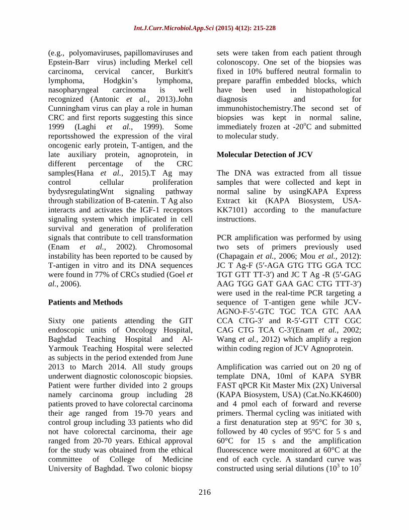
sets were taken from each patient through colonoscopy. One set of the biopsies was fixed in 10% buffered neutral formalin to prepare paraffin embedded blocks, which have been used in histopathological diagnosis and for immunohistochemistry.The second set of biopsies was kept in normal saline, immediately frozen at -20oC and submitted to molecular study.
Molecular Detection of JCV
The DNA was extracted from all tissue samples that were collected and kept in normal saline by usingKAPA Express Extract kit (KAPA Biosystem, USA- KK7101) according to the manufacture instructions.
PCR amplification was performed by using two sets of primers previously used (Chapagain et al., 2006; Mou et al., 2012): JC T Ag-F (5 -AGA GTG TTG GGA TCC TGT GTT TT-3 ) and JC T Ag -R (5 -GAG AAG TGG GAT GAA GAC CTG TTT-3 ) were used in the real-time PCR targeting a sequence of T-antigen gene while JCV-AGNO-F-5 -GTC TGC TCA GTC AAA CCA CTG-3 and R-5 -GTT CTT CGC CAG CTG TCA C-3 (Enam et al., 2002; Wang et al., 2012) which amplify a region within coding region of JCV Agnoprotein.
Amplification was carried out on 20 ng of template DNA, 10ml of KAPA SYBR FAST qPCR Kit Master Mix (2X) Universal (KAPA Biosystem, USA) (Cat.No.KK4600) and 4 pmol each of forward and reverse primers. Thermal cycling was initiated with a first denaturation step at 95°C for 30 s, followed by 40 cycles of 95°C for 5 s and 60°C for 15 s and the amplification fluorescence were monitored at 60°C at the end of each cycle. A standard curve was constructed using serial dilutions (103 to 107

Int.J.Curr.Microbiol.App.Sci (2015) 4(12): 215-228
217
copies of JCV DNA) JCV Bio probe (ENZO life sciences, Ny. USA) were performed for every plate run starting with 1.81X10⁷copies / g with10X dilution factor. Three replicates were performed for each sample and real-time PCR data were analyzed using Mx3005P Real-Time PCR system Stratagen (Agilent Technologies, USA).
Histopathological and Immunohistochemistry
Two sections of 4 micrometers thickness were taken from each paraffin embedded tissue block. First sections were put on ordinary slide for Haematoxylin and Eosin (H&E) staining to confirm diagnosis and to detect the histological types and grades of tumor. The second sections were put on the charged slide for immunohistochemistry (IHC) using Dako EnVision+ System-HRP (DAB), (DAKO, Denmark) Code K4006 and Monoclonal Mouse Anti-Human Beta-Catenin Clone -Catenin-1 Code M3539 (DAKO, Denmark). IHC was given intensity and percentage scores, based on intensity of positive (brownish discoloration at nuclear localization) staining and number of stained cell per 100 cells. The intensity score was divided into negative, low, and high. Positive cells were counted in 10 different fields for each sample and the average of positive cells was determined assigning to one of the 3 following categories of percentage score.Score 1: 1 30% cells positivity, score 2: 31 60% cells positivity and score 3: 61% and more cells positivity(Enam et al., 2002).
Statistical Analysis
Statistical analysis tests were applied to compare and correct results of different methodology applied in the present work. Frequencies of positive and negative results were recorded as percentages of the total.
Chie square test with Fisher extract correction was applied to compare between frequencies. T-test was applied to compare between means. In each test, a p-value <0.05 was considered significant.
Cohen Kappa was used as an index of inter-rater reliability to measure agreement between 2 sets of dichotomous rating. A value of K=0.4-0.59 indicate moderate inter-rater reliability, 0.60-0.79 indicate substantial and 0.8 indicating outstanding.
Results and Discussion
Age
The age of carcinoma group ranged from 19-70 years with mean (52.85 ± 11.74) years while the age of control group ranged from 22-73 years with mean age (46.75 ± 13.22) years. The results revealed no significant statistical difference in mean age between study groups. The highest frequency (35.71%) of patients with colorectal carcinoma was within 50-59 years. In the control group the highest frequency (33.33%) was within 50-59 years.
Gender
Out of 28 carcinoma group, 18 (64.28%) patients were males and 10 (35.72%) were females with male to female ratio (1.8:1). For the control group 26 (78.78%) were males and 7(21.22%) were females with male to female ratio (3.7:1). Individuals in carcinomas and control groups did not differ significantly according to sex (P= 0.208).
Histopathological Types
Microscopic examination of H&E stained slides from carcinoma group showed that 24 out of 28 (85.72%) cases were of non-mucinous adenocarcinoma while only 4

Int.J.Curr.Microbiol.App.Sci (2015) 4(12): 215-228
218
(14.28%) cases were of mucinous adenocarcinoma.
Histological grading of adenocarcinoma in carcinoma group showed that 21(75%) of cases was of moderately differentiated adenocarcinoma while 3 (10.7%) cases were of well differentiated and only 4 (14.3%) cases were of poorly differentiated adenocarcinoma as shown in Figure (3)
Tumor Site
Thirteen out of 28 (46.42%) cases of the carcinoma group were from left colon, 7(25%) cases from right colon and 8 (28.58%) were from rectum. For control group 14 out of 33 (42.4%) were from left colon, 8 (24.2%) from right colon and 11 (33.3%) from rectum. Statistical analysis showed no significant difference (P=0.919) between study groups.
Detection and Quantification of JCV by Real Time PCR.
Detection and Quantification of T-Ag Gene by Real Time PCR.
The T-Ag gene was detected in 16 out of 28 (57.1%) in carcinoma group, while 9 out of 33 (27.3%) cases of the control group were positive for JCV T-Ag gene (Table 1). The results revealed significant difference between study groups (P=0.018). The viral load (VL) (figure 4) for carcinoma group ranged from 1.00e+02copies/ µg to 8.39e+02 copies/µg with a mean of (416.937 ± 217.779 SE 56.230). For control group the VL was ranged from to 9.88E+01 to4.11e+02 copies/µgwith a mean of (229.866 ±111.492 SE 39.418). The difference between the two groups was statistically significant regarding the mean viral load; p<0.05.T Ag gene detection by real time PCR was 57.14% sensitive and
72.73% specific. The positive predictive value (PPV) was 64% and the negative predictive value (NPV) was 66.67%.
Ten out of 21 (47.6%) cases of moderately differentiated adenocarcinoma were positive for JCV T-Ag gene. Positive T-Ag gene was also detected in 2 out of 3 (66.7%) cases and 4 cases out of 4 (100%) in well and poorly differentiated adenocarcinomas respectively. However, the difference failed to reach the level of statistical significant (p>0.05).
The standard dilutions were (1.81X10⁷, 1.81X10⁶, 1.81X10⁵, 1.81X10⁴,and 1.81X10³). The photography was taken directly from the real time PCR machine. Agilent technology
Regarding Tumor Site
T-Ag gene was detected in 7(100%), 7 cases out of 13(53.85%) and only 2 out of 8 (25%) cases of right, left colon and rectum respectively in carcinomas group. While in control group T-Ag gene was detected in 1 out of 8 (12.5%) cases of Rt. colon, 5 out of 14 (35.7%) cases of LT colon and 3 out of 11 (27.27%) cases of rectum. A significant statistical difference was noticed in the carcinomas group, p<0.05.
Detection and Quantification of Agnoprotein Gene by Real Time PCR
The agnoprotein gene of JCV was detected in 12 out of 28(42.85%) cases of the carcinomas group while only 5 out of 33 (15.15%) cases of control group were positive (table 3). Statistical analysis of the results showed significant difference between study groups as (p = 0.016). The viral loads of carcinomas group (table-3) were ranged from (1.22e+02 - 4.55e+02) copies/µg with a mean of (317 ± 129.121), while that of the control group were ranged

Int.J.Curr.Microbiol.App.Sci (2015) 4(12): 215-228
219
from (3.32e+02 - 6.45e+01) copies/µg with a mean of (152.94 ± 105.149). The difference between carcinomas group and control group was significant regarding the viral load (P<0.05). The test showed a sensitivity of 42.86%, specificity of 84.85%, PPV of 70.59% and NPV of 63.64%.
Regarding grades of tumor, JCV agnoprotein gene was detected in 2 out of 3 (66.67%) cases, 7 out of 21(33.33%) and 3 out of 4 (75%) cases of well, moderately, and poorly differentiate adenocarcinoma respectively. No significant difference was found between the presence of JCV agnoprotein gene in the different grades of tumor among carcinomas group, p = 0.206.
Regarding site of tissue biopsies, Agnoprotein gene was detected in 6 out of 7(85.7%) cases, 5 cases out of 13(35.5%) cases and only 1 out of 8 (12.5%) cases of right, left colon and rectum respectively. A significant statistical difference was detected among the carcinomas groups (P= 0.015). While for control group only 1case out of 8 (12.5%) from RT colon and 4 cases out of 14(28.6%) from LT colon were positive for agnoprotein DNA. No significant difference was detected in the control group. Statistical analysis reached the level of significant difference on comparing variables between study groups (p<0.05).
Detection of -Catenin by IHC among Study Groups
Expression of B- catenin was detected as brownish discoloration at nuclear localization (figure 5). Nuclear localization of - catenin was detected by IHC in 12/28 (42.86%) cases of carcinoma group, and none in the control group. Cytoplasmic -catenin was detected in 26/28 (92.86%) cases of carcinoma group and 24/33 (72.73%) of the control group. (Table5). A
significant difference was found among study groups (P<0.05) regarding nuclear -catenin while the level of statistical significant difference was not reached in cytoplasmic B-catenin (p>0.05).
Eight cases (28.6%) revealed percentage score 1, two cases (7.14%) revealed score 2 and 2 (7.14%) cases revealed score3 of nuclear -catenin by IHC in the carcinoma group (figure 6). Statistical analysis was invalid. The detection of -catenin by IHC in colonic biopsies of colorectal cancer showed 42.86% sensitivity and 100%specificity. The PPV was 100% and NPV was 67.35%.
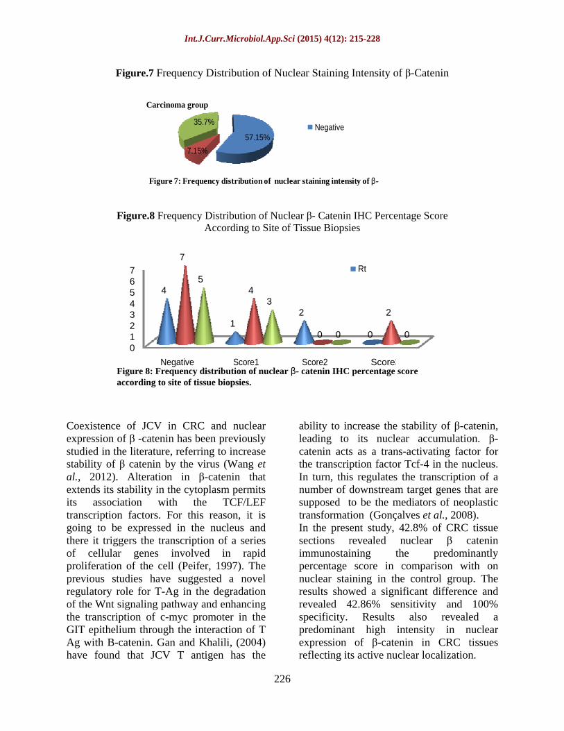
Figure (7) shows the frequency distribution of nuclear staining intensity of -catenin of study groups. High staining intensity was detected in 10 (35.7%) of the CRC specimen. Statistical analysis was invalid.
The results of association for -catenin nuclear score and the grade of tumor revealedthe highest frequency was in the moderately differentiated adenocarcinoma, in which 7/28 cases were score 1 and 2/28 cases were score 2. Statistical analyses were invalid.
The frequency of percentage score distribution of IHC detection of -catenin according to the site is shown in figure 8. Score 1 was shown in 4 (50%) cases of Lt, 1(12.5%) of right and 3 (37.5%) cases of rectum. Score 2 was only detected in 2 (100%) cases of the right side, and score 3 was detected in 2 (100%) cases of the Lt side. Statistical analysis is invalid.
Higher frequency of nuclear high staining intensity was found among moderately differentiated adenocarcinoma 8 out of 12(80%) cases, while 2 (20%) cases were poor differentiated adenocarcinoma. On the

Int.J.Curr.Microbiol.App.Sci (2015) 4(12): 215-228
220
other hand 2 out of 12 cases were of low staining intensity, one was well differentiated and the other was moderately differentiated adenocarcinoma (Table 7). No significant association was found between the grade of tumor and staining intensity (p=0.313).
Table (8) shows the distribution of -catenin nuclear staining intensity according to site of tissue biopsies. The highest frequency was of high intensity detected in 5 (50%), cases 3(30%) cases and 2 (20%) cases in LT, rectum and RT colon respectively. Statistical analysis failed to reach significant level (p>0.05) of difference between the site of tumor according to intensity of staining.
Agreement and Inter-Rater Reliability between JCV T-Ag Detected by Real Time PCR and B-Catenin among Study Groups
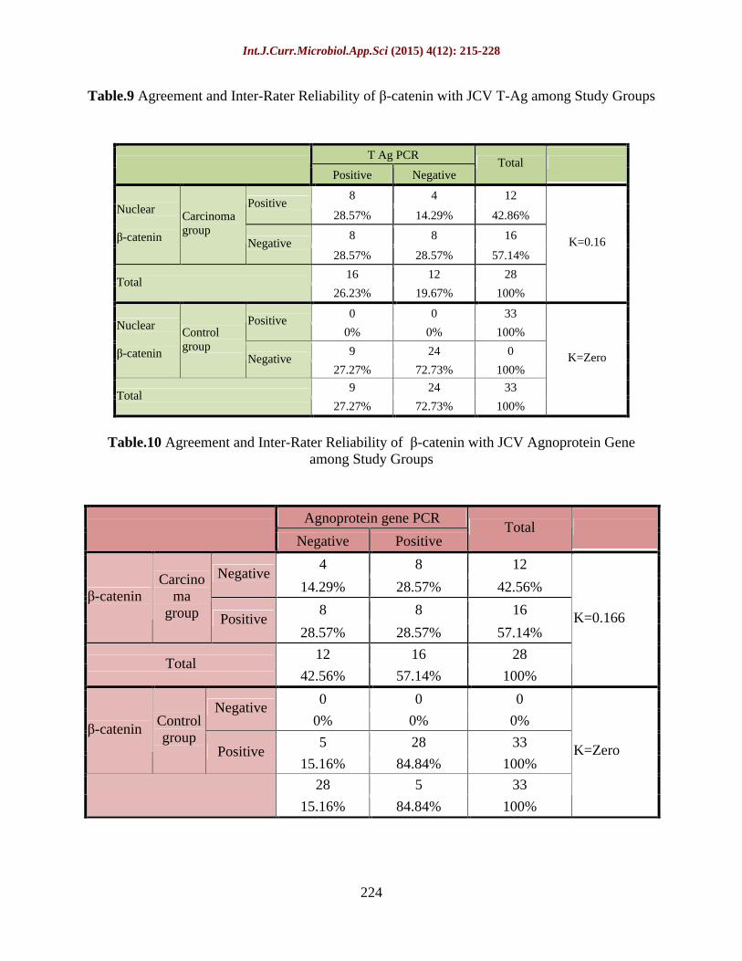
Table 9 shows the association between JCV T-Ag with -catenin in study groups. The result revealed that 8/28 (28.57%) cases showed positive results for both JCV T-Ag and -catenin, in carcinoma group while 8/28(28.57%) cases showed positive result for JCV T-Ag and negative for -catenin. There was no agreement between the 2 tests. The calculated K for the carcinoma group was 0.16 and for the control group was zero
Agreement and Inter-Rater Reliability of JCV Agnoprotein Gene Detected by Real Time PCR with -Catenin in Study Groups
The association of -catenin with JCV agnoprotein gene is illustrated in table (10). In carcinomas group 4/28 (14.29%) cases showed positive results for both -catenin and JCV agnoprotein gene, while 8/28 (28.57%) cases were negative for both. In the control group none of the cases showed positive results of both -catenin and JCV
agnoprotein. There was no agreement between the 2 tests. The calculated K for the carcinoma group was 0.116 and for the control group was zero.
Bowel cancer is strongly related to age. In this study the mean age of patients with colorectal carcinoma was (52.85 ± 11.74) years and the frequency of cases were decreases with young age. Other studies in Iraq found that the mean age of patients with colorectal carcinoma was 56.03±2.37 and 56.88 ±1.99 years respectively (Qasim., 2009; Qasim., 2010). The highest frequency of cases was observed within age group of 50-59 years. This was a little bit different from another study in Iraq which found that the highest frequency was among 60-69 years of age (A.Majid et al., 2009; Iraqi-Cancer-Registry, 2012).
The sex specific incidence in colorectal carcinoma showed a slight preponderance in males with ratio of male to female (1.8:1) and this was consistent with other Iraqi study reported male to female ratio of (1.4:1) in colonic carcinoma(Al-Humadi and Buffalo, 2008).
The most frequent histological grade was moderately differentiated adenocarcinoma (75%). Although in different frequencies, moderately differentiated adenocarcinoma was also the most common grade and represented (53%), (66. 7) % and (90%) in different other studies (A.Majid et al., 2009; Abdulhussain and Othman, 2012; Hana et al., 2015).
Several studies have attempted to confirm the presence of JC virus DNA in colonic tissue using paraffin embedded specimen, (Enam et al., 2002) other study failed to detect the presence of JC virus DNA (Newcomb et al., 2004). Since the virus is a small circular DNA virus and may be existent in relatively small amounts in

Int.J.Curr.Microbiol.App.Sci (2015) 4(12): 215-228
221
colorectal neoplasms, the formalin fixation may break the DNA and make it more difficult to recover adequate quantities of integral JCV DNA for amplification. The decision was made in this study to use fresh
frozen specimen which is never been fixed with formalin since they were tiny biopsies and there was a fear of losing large amount of the DNA (Boland, 2004).
Table.1 Frequency Distribution of T-Ag Gene by Real Time PCR Mong Study Groups
T Ag gene by PCR
Positive Negative Total
16 12 28 Carcinoma
57.1% 42.9% 100.0%
9 24 33
Study groups
Control 27.3% 72.7% 100.0%
25 36 61 Total
41.0% 59.0% 100.0%
P=0.018
Table.3 Frequency Distribution of Agnoprotein Gene by Real Time PCR among Study Groups.
Agnoprotein gene Agnoprotein gene
Positive Negative Total
12 16 28 Carcinoma
42.9% 57.1% 100.0%
5 28 33
Study groups
Control 15.2% 84.8% 100.0%
17 44 61 Total
27.9% 72.1% 100.0% P= 0.016
Table.4 Comparison of Viral Load Between Study Groups According to JCV Agnoprotein Gene Amplification by Real Time PCR
JCV Agnoprotein gene Carcinomas group Control group
Mean viral load 317 152.94 Standard Deviation 129.121 105.149
Standard Error of the Mean
38.931 52.574
Total Numbers : 12 5 The P= 0.024

Int.J.Curr.Microbiol.App.Sci (2015) 4(12): 215-228
222
Table.5 Detection of -Catenin Nuclear and Cytoplasmic Localization in Study Groups by IHC
Nuclear -catenin Cytoplasmic -catenin
Positive Negative
Total Positive Negative
Total
12 16 28 26 2 28 Carcinoma
42.86% 57.14% 100% 92.86% 7.14% 100% 0 33 33 24 9 33
Control 0% 100% 100% 72.73% 27.27% 100% 12 49 61 50 11 61
19.67% 80.33% 100% 81.97% 18.03% 100%
P < 0.05 P>0.05
Table.6 Frequency Distribution of IHC for -Catenin Staining Percentage Score According to Tumor Grade
Grade of tumor
Well Moderate Poor Total
2 12 2 16 Negative
12.5% 75.0% 12.5% 100%
1 7 0 8 Score 1
12.5% 87.5% 0% 100%
0 0 2 2 Score 2
0% 0% 100% 100%
0 2 0 2
Nuclear
-catenin
Score 3 0% 100% 0% 100%
3 21 4 28 Total
10.7% 75.0% 14.3% 100%
P=0.287
Moderatly differentiated
75%Poorly
differentiated14.3 %
Well differentiated
10.7 %
F

Int.J.Curr.Microbiol.App.Sci (2015) 4(12): 215-228
223
Table.7 Distribution of -Catenin Nuclear Staining Intensity Score According to Grades of Tumor
Grade - catenin signals intensity
Well Moderate Poor Total
2 12 2 16 Negative
12.5% 75% 12.5% 100%
1 1 0 2 Low
50% 50% 0% 100%
0 8 2 10
Carcinoma group
High 0% 80% 20% 100%
3 21 4 28 Total
10.7% 75.0% 14.3% 100%
P=0.310
Table.8 Frequency Distribution of -catenin IHC Nuclear Staining Intensity According to Site of Tissue Biopsies
Site - catenin signals intensity Rt. Lt. Rectum
Total
4 7 5 16 Negative
25% 43.75% 31.25% 100%
1 1 0 2 Low
50% 50% 0% 100%
2 5 3 10
Carcinoma group
High 20% 50% 30% 100%
7 13 8 28 Total
25.0% 46.4% 28.6% 100.0%
p=0.988
Figure.4
Int.J.Curr.Microbiol.App.Sci (2015) 4(12): 215-228
224
Table.9 Agreement and Inter-Rater Reliability of -catenin with JCV T-Ag among Study Groups
T Ag PCR
Positive Negative
Total
8 4 12
Positive 28.57% 14.29% 42.86%
8 8 16
Nuclear
-catenin
Carcinoma group
Negative 28.57% 28.57% 57.14%
16 12 28 Total
26.23% 19.67% 100%
K=0.16
0 0 33 Positive
0% 0% 100%
9 24 0
Nuclear
-catenin
Control group
Negative 27.27%
72.73%
100%
9 24 33 Total
27.27% 72.73% 100%
K=Zero
Table.10 Agreement and Inter-Rater Reliability of -catenin with JCV Agnoprotein Gene among Study Groups
Agnoprotein gene PCR
Negative Positive Total
4 8 12 Negative
14.29% 28.57% 42.56%
8 8 16 -catenin
Carcinoma
group Positive 28.57% 28.57% 57.14%
12 16 28 Total
42.56% 57.14% 100%
K=0.166
0 0 0 Negative
0% 0% 0%
5 28 33 -catenin
Control group
Positive 15.16% 84.84% 100%
28 5 33
15.16% 84.84% 100%
K=Zero

Int.J.Curr.Microbiol.App.Sci (2015) 4(12): 215-228
225
Figure.5 Illustrate Different Colonic Biopsies of Carcinoma Group and Control Group with Different Percentage and Intensity Score
A. Membranous and cytoplasmic staining X40 B. Nuclear staining of -catenin( arrow) X40 C. Nuclear staining of -catenin (arrow) X100 D. Nuclear staining of -catenin high score X100
Figure.6 Frequency Distribution of Nuclear -Catenin by IHC According to Percentage Score
0
5
10
15
20
25
30
35
Negative Score1 Score 2 Score 3
Patient group
A
B
C
D
Int.J.Curr.Microbiol.App.Sci (2015) 4(12): 215-228
226
Figure.7 Frequency Distribution of Nuclear Staining Intensity of -Catenin
57.15%
7.15%
35.7%
Carcinoma group
Negative
Figure 7: Frequency distribution of nuclear staining intensity of -
Figure.8 Frequency Distribution of Nuclear - Catenin IHC Percentage Score According to Site of Tissue Biopsies
01234567
Negative Score1 Score2 Score3
4
12
0
7
4
0
2
5
3
0 0
Rt
Figure 8: Frequency distribution of nuclear - catenin IHC percentage score according to site of tissue biopsies.
Coexistence of JCV in CRC and nuclear expression of -catenin has been previously studied in the literature, referring to increase stability of catenin by the virus (Wang et al., 2012). Alteration in -catenin that extends its stability in the cytoplasm permits its association with the TCF/LEF transcription factors. For this reason, it is going to be expressed in the nucleus and there it triggers the transcription of a series of cellular genes involved in rapid proliferation of the cell (Peifer, 1997). The previous studies have suggested a novel regulatory role for T-Ag in the degradation of the Wnt signaling pathway and enhancing the transcription of c-myc promoter in the GIT epithelium through the interaction of T Ag with B-catenin. Gan and Khalili, (2004) have found that JCV T antigen has the
ability to increase the stability of -catenin, leading to its nuclear accumulation. -catenin acts as a trans-activating factor for the transcription factor Tcf-4 in the nucleus. In turn, this regulates the transcription of a number of downstream target genes that are supposed to be the mediators of neoplastic transformation (Gonçalves et al., 2008). In the present study, 42.8% of CRC tissue sections revealed nuclear catenin immunostaining the predominantly percentage score in comparison with on nuclear staining in the control group. The results showed a significant difference and revealed 42.86% sensitivity and 100% specificity. Results also revealed a predominant high intensity in nuclear expression of -catenin in CRC tissues reflecting its active nuclear localization.
Int.J.Curr.Microbiol.App.Sci (2015) 4(12): 215-228
227
There was no significant association with biopsy sites nor with tumor grade. This could implies that when there is participation of - catenin abnormalities in CRC pathways it take place probably early in the process. It does not affect tumor grade nor take a role in increasing the grade. One could relate this to the presence of JCV infection which may affect -catenin. However, no agreement was found in the present study between - catenin immunostaining from one side and detection of JCV T Ag gene, agnoprotein gene, or CISH. In other word the present study failed to relate the -catenin expression to the presence of JCV.
This study highlighted the possible role of JCV which might participate in different ways in the pathogenesis of colorectal carcinoma. Despite these evidences the role of JCV in colorectal malignancies and its oncoproteins in promoting transformation is still far from clear.
Reference
A.Majid, T, Shakir, W M and Mahmmod, a S (2009) Colorectal Carcinoma Presentation and Management. COLORECTAL CARCINOMA THE IRAQI POSTGRADUATE MEDICAL JOURNAL 8.
Abdulhussain, S S and Othman, O H (2012) epidemiological study of colorectal and anal cancer in kirkuk. Iraqi society of Gastroenterology and Hepatology 6 33.
Al-Humadi and Buffalo, S (2008) Epidemiology of Colon & Rectal Cancer In Iraq. Word Journal of Colorectal Surgery 1.
Antonic, V, Stojadinovic, A, Kester, K E, et al. (2013) Significance of infectious agents in colorectal cancer development. J Cancer 4 227-240.
Boland, C R (2004) Evidence for an Association between JC Virus and Colorectal Neoplasia. Cancer Epidemiology, Biomarkers & Prevention 13.
Chapagain, M L, Nguyen, T, Bui, T, Verma, S and Nerurkar, V R (2006) Comparison of real-time PCR and hemagglutination assay for quantitation of human polyomavirus JC. Virol J 3 3.
Enam, S, Del Valle, L, Lara, C, et al. (2002) Association of human polyomavirus JCV with colon cancer: evidence for interaction of viral T-antigen and beta-catenin. Cancer Res 62 7093-7101.
Goel, A, Li, M S, Nagasaka, T, et al. (2006) Association of JC virus T-antigen expression with the methylator phenotype in sporadic colorectal cancers. Gastroenterology 130 1950-1961.
Gonçalves, P, M and P, J (2008) The betacatenin/TCF4 pathway modifies alternative splicing through modulation of SRp20 expression. RNA 2008;14:2538-2549. 14 22538-22549.
Hamilton Sr, Bosman Ft and Boffetta P, E A (2010) Carcinoma of the colon and rectum. WHO Classification of Tumours of the Digestive System Bosman FT, Carneiro F, Hruban RH, Theise ND, eds. Lyon: IARC Press, 134-146.
Hana, D B, Azhar A.F.Al-Attraqhchi and Khattab, Y I (2015) Molecular and Immunohistochemical Detection of JC Polyomavirus in Human Colorectal Cancer in Sample of Iraqi Patients. International Journal of Scientific & Engineering Research 6 934.
Iraqi-Cancer-Registry (2012) IRAQI CANCER REGISTRY CENTER.

Int.J.Curr.Microbiol.App.Sci (2015) 4(12): 215-228
228
Ministry of health/ Iraq P.O.BOX 707 /12112. FAX (+) 9641- 4150292 BAGHDAD, IRAQ.
Laghi, L, Randolph, a E, Chauhan, D P, et al. (1999) JC virus DNA is present in the mucosa of the human colon and in colorectal cancers. Proc Natl Acad Sci U S A 96 7484-7489.
Mou, X, Chen, L, Liu, F, et al. (2012) Prevalence of JC virus in Chinese patients with colorectal cancer. PLoS One 7 e35900.
Newcomb, P A, Bush, a C, Stoner, G L, Lampe, J W, Potter, J D and Bigler, J (2004) No evidence of an association of JC virus and colon neoplasia. Cancer Epidemiol Biomarkers Prev 13 662-666.
Peifer, M (1997) B-Catenin as oncogene: the smoking gun. Science (Wash. DC) 275 1752 1753,.
Qasim., B (2009) Immunohistochemical expression of molecular markers: matrix metalloproteinase-7(MMP-7), CD34, P53, Bcl2, proliferating cell nuclear antigen, estrogen and progesterone receptors in human colorectal carcinogenesis using specialized automated cellular image analysis system. A clinicopathological study. A Thesis Submitted to College of Medicine -Al-Nahrain University
Qasim., Z (2010) CO-expression of (VEGF A, VEGF C, COX2 and EGFR) biomarkers in human colorectal cancer and their association eith lymph node metastasis and angiogenesis. A Thesis Submitted to College of Medicine-Al-Nahrain University.
Siegel, R, Ward, E, Brawley, O and Jemal, A (2011) the impact of eliminating socioeconomic and racial disparities on premature cancer deaths. CA Cancer J Clin 61 212-236.
Wang, J P, Wang, Z Z, Zheng, Y S, et al. (2012) JC virus existence in Chinese gastrointestinal carcinomas. Oncol Lett 3 1073-1078.