effect of salt on ros homeostasis, lipid peroxidation and
TRANSCRIPT

Ann. For. Sci. 66 (2009) 211 Available online at:c© INRA, EDP Sciences, 2009 www.afs-journal.orgDOI: 10.1051/forest/2008093
Original article
Effect of salt on ROS homeostasis, lipid peroxidation and antioxidantmechanisms in Pinus pinaster suspension cells
Herlânder Azevedo*, Vítor Amorim-Silva, Rui M. Tavares
Departamento de Biologia, Universidade do Minho, Campus de Gualtar, 4710-057 Braga, Portugal
(Received 20 February 2008; accepted 19 November 2008)
Keywords:H2O2 /
maritime pine /NaCl /superoxide
Abstract• In the Pinus genus, information on the effectiveness of oxidative defence mechanisms during expo-sure to salt is lacking. The effect of salt stress imposition on ROS homeostasis was investigated usingmaritime pine (Pinus pinaster Ait.) suspension cells as a model system.• Cells were maintained in MS-based medium, exposed to salt (50, 100 and 150 mM NaCl) andanalysed for biomass production, evidencing a decreasing growth capacity. Use of 100 mM NaClimposed severe salt stress without affecting cell viability, being chosen for subsequent studies on theROS homeostasis of salt shock-treated suspension cells.• Increased total ROS levels were evident on the second day of salt exposure, but a superoxideion transient burst was immediately noticeable. Additionally, lipid peroxide formation seemed tocorrelate with superoxide ion breakdown. In-gel superoxide dismutase activity evidenced a FeSODhomodimer with strongly increasing activity between hours 12–48 of salt stress imposition. Subse-quently, P. pinaster Fe-Sod1 and csApx1 genes were isolated from a cDNA library and expressionwas shown to increase within 12–24 h.• Results show that severe salt treatment generates oxidative stress in P. pinaster cells despite theinduction of antioxidant systems, and suggest a putative involvement of ROS in salt stress signalling.
Mots-clés :H2O2 /
pin maritime /NaCl /superoxide
Résumé – Effet du stress salin sur l’homéostasie des formes réactives d’oxygène, la peroxydationdes lipides et les mécanismes antioxydants dans des suspensions cellulaires de Pinus pinaster.• Les informations sur les mécanismes de défense oxydative du pin en réponse à un stress salin sontrares. L’effet d’une exposition au sel sur l’homéostasie des formes réactives d’oxygène (FRO) a étéétudié en utilisant une suspension cellulaire de pin maritime (Pinus pinaster Ait.) comme modèle.• Les cellules cultivées dans un milieu MS modifié ont été exposées au sel (50, 100 et 150 mM NaCl)et l’analyse de la production de biomasse a révélé une réduction de leur croissance. Une concentra-tion de 100 mM NaCl, stress sévère qui n’affecte cependant pas la viabilité cellulaire, a été choisiepour les études suivantes.• L’augmentation des teneurs en FRO est évidente le jour suivant l’enrichissement du milieu en selsmais une production transitoire d’ions superoxyde est immédiatement constatée. De plus, l’appari-tion de produits issus de la peroxydation des lipides semble concomitante à la disparition des ionssuperoxyde. La mesure par tests in-gel de l’activité de la superoxyde dismutase supporte l’implica-tion d’un homodimère de FeSOD dont l’activité augmente fortement au bout de 12 et jusqu’à 48 hd’exposition au sel. Les gènes Fe-Sod1 et csApx1, isolés d’une banque d’ADNc de P. pinaster, voientleur expression augmenter au bout de 12 h et jusqu’à 24 h de traitement.• Les résultats montrent que de fortes concentrations de sels provoquent un stress oxydatif dans lescellules de P. pinaster malgré l’induction de réponses antioxydantes et suggèrent l’implication desERO dans les voies de transduction du stress salin.
* Corresponding author: [email protected]
Article published by EDP Sciences

Ann. For. Sci. 66 (2009) 211 H. Azevedo et al.
1. INTRODUCTION
In plants, abiotic stress has been shown to disrupt the cel-lular homeostasis of cells and consequently generate oxida-tive stress (Mittler, 2002; Zhu, 2001). Particularly, salt stressseverely affects plant productivity, being responsible for thegeneration of ion imbalances and hyperosmotic stress. Asa consequence, secondary effects occur that include oxida-tive damage to cellular constituents (Zhu, 2001; Bor et al.,2003). Partially reduced and excited species of oxygen, usu-ally designated reactive oxygen species (ROS), can react withmany cellular substances leading to the oxidative destructionof cells (Mittler, 2002). In plants, the major ROS include hy-drogen peroxide (H2O2), superoxide ion (O−2 ), singlet oxy-gen (1O2) and hydroxyl radical (HO•) (Mittler et al., 2004).The maintenance of homeostatic levels of ROS in cells isachieved by the pools of antioxidants, namely of the ascorbate-glutathione cycle, and by the activity of ROS scavenging en-zymes, the most important being superoxide dismutase (SOD,EC 1.15.1.1), catalase (CAT, EC 1.11.1.6), ascorbate perox-idase (APX, EC 1.11.1.1) and glutathione peroxidase (GPX,EC 1.11.1.9) (Mittler et al., 2004). These enzymes have beenfound in most cellular organelles, demonstrating the impor-tance of ROS scavenging to cellular viability. SODs, by dis-mutating O−2 into H2O2 and O2, constitute the only enzymaticmechanism of scavenging superoxide. Hydrogen peroxide canbe scavenged by CAT and by peroxidases like APX and GPX,that reduce H2O2 via ascorbate and glutathione, respectively(reviewed by Mittler et al., 2004). The role of antioxidant en-zymes in the tolerance to diverse environmental stresses, in-cluding salt stress, has been reported (Bor et al., 2003).
Maritime pine (Pinus pinaster Ait.) is one of the three mainpine species in Europe, being native to the western Mediter-ranean basin. As a major reforestation species, P. pinasterhas been grown extensively in setting dunes of coastal areas,where plants are highly exposed to salt spray. As a conse-quence of different salt tolerance between species, vegetationzonation can be observed in proportion to seashore distance.The high suitability, particularly amongst pines, of P. pinasterto the setting dune environment is a strong indicator of thepresence of salt-adaptative mechanisms. We have addressedthis issue at plant level, observing that tolerance to high NaClconcentrations is accompanied by changes in both the photo-synthetic apparatus and the homeostasis of ROS (unpublisheddata). The association between salt and oxidative stresses hasbeen previously confirmed: salt decreases water availabilityand disrupts the homeostasis of water potential and ion dis-tribution, a process which is the basis for oxidative stressgeneration (Zhu, 2001). At plant level, over-reduction of thephotosynthetic apparatus constitutes a major source of ROS(Osmond and Grace, 1995), but salt-associated oxidative stresscan also be mediated by superoxide and hydrogen peroxideradicals at the levels of mitochondria and peroxisomes, respec-tively (Corpas et al., 1993; Hernandez et al., 1993). Further-more, a novel role has been established for ROS as signallingintermediates during abiotic stress resistance (reviewed byMittler et al., 2004), and the importance of non-photosynthetic
ROS generation during abiotic stress responses has been re-cently substantiated (reviewed by Van Breusegem et al., 2008).
The present study addresses the cell-level role of non-photosynthetic ROS during the maritime pine salt stress re-sponse, by use of a heterotrophic suspension cell system. Saltstress impact on growth was measured and the homeostasisof various ROS subsequently analysed. Activation of ROS-scavenging mechanisms was monitored at both protein andgene expression levels. For the purpose, a candidate gene ap-proach was used to identify ROS-scavenging proteins puta-tively involved in the P. pinaster salt stress response.
2. MATERIALS AND METHODS
2.1. P. pinaster suspension culture maintenanceand characterization
A heterotrophic Pinus pinaster suspension cell culture waspreviously established in our laboratory from root segments ofmaritime pine seedlings (Azevedo et al., 2008a). Suspensioncells were maintained in Murashige and Skoog (MS) medium(Murashige and Skoog, 1962) supplemented with 3% glucose,5 mg L−1 dithiothreitol, 0.1 g L−1 myo-inositol, 1 mg L−1 BAand 2 mg L−1 2,4-D, at pH 6. Suspensions were maintainedat 25 ◦C and 100 rpm in the dark. Subcultures were performedduring the late exponential phase (every 12–14 days), by trans-ferring 10 mL of the culture into 70 mL of fresh medium. Forgrowth analysis under salt stress, flasks were supplementedwith 50, 100 or 150 mM NaCl. Suspension cells from three in-dependent cultures were gathered into a single inoculum thatwas used to inoculate five separate cultures to be character-ized. Biomass was determined by removing aliquots contain-ing 3 mL of suspended cells every 3 days under sterile con-ditions. Samples were filtered using pre-weighed GF/C filters(Whatman, Clifton, NJ, USA), and oven dried at 60 ◦C for24 h. For salt stress experiments, P. pinaster suspended cellsin mid-exponential growth phase were gathered from three in-dependent cultures, centrifuged at 5000 g for 5 min and resus-pended in MS medium, at a final density of 0.1 g FW mL−1.Medium was supplemented with 100 mM NaCl when re-quired, and experiments were carried out using five indepen-dent replicas.
2.2. Protein extraction
Suspension cells were ground to a fine powder in liquidnitrogen. Approximately 1 g of fresh weight was thawed in2–3 mL of protein extraction buffer (50 mM sodium phos-phate at pH 7.0, 1 mM benzamidine, 0.1% 2-mercaptoethanoland 1% PVPP) and incubated on ice for 5 min. After centrifu-gation at 15 000 g for 15 min at 4 ◦C, the supernatant wasrecovered and immediately used for enzyme assays or storedat −80 ◦C. Protein was quantified using the Coomassie Bluemethod (Sedmak and Grossberg, 1977).
211p2

ROS in salt-stressed pine suspensions Ann. For. Sci. 66 (2009) 211
2.3. ROS homeostasis analysis
The superoxide radical (O−2 ) was quantified by the re-duction of XTT (Invitrogen-Molecular Probes, OR, USA)to a soluble formazan (Able et al., 1998). Immediately be-fore salt stress imposition, 0.5 mM XTT was added to thecell suspension, followed by incubation in the dark, at roomtemperature with agitation. Aliquots were removed period-ically, and the reduced XTT form was quantified by mea-suring the absorbance of the supernatant at 470 nm. Theoverall oxidative stress state of the cell was quantified us-ing the cell-permeant 2’,7’-dichlorodihydrofluorescein diac-etate (H2DCFDA; Invitrogen-Molecular Probes, OR, USA) aspreviously described (Allan et al., 2001). H2DCFDA is con-verted by non-specific cellular esterases to H2DCF, which ox-idizes in the presence of H2O2 and other reactive oxygen inter-mediates. The end product 2’,7’-dichlorofluorescein is highlyfluorescent and able to diffuse out of the cell. This propertywas used to quantify the intracellular production of 2’,7’-dichlorofluorescein, by performing a spectrofluorimetric anal-ysis of the supernatant. During the time course of salt stressimposition to pine suspension cells, 1 mL aliquots were re-moved and added 10 µL of 20 µM H2DCFDA. Cells wereincubated in the dark, at room temperature, for 30 min withagitation. Samples were centrifuged at 8000 g for 5 min andthe supernatant recovered. Relative fluorescence was quan-tified using a LS 50 Luminescence Spectrometer (PerkinElmer) at an excitation wavelength of 488 nm and an emis-sion wavelength of 525 nm. Lipid peroxidation was quanti-fied spectrophotometrically by the MDA-TBA method, whichquantifies the end product of lipid peroxidation malondialde-hyde (MDA) by reaction at low pH and high temperaturewith 2-thiobarbituric acid (TBA) (Loreto and Velikova, 2001).The reaction was initiated by adding 75 µL of protein ex-tract from salt stressed pine suspension cells, to 250 µL ofchilled reaction mixture, composed of 0.5% (w/v) TBA in20% (w/v) TCA. The mixture was incubated at 95 ◦C for30 min and placed immediately on ice. Samples were cen-trifuged at 10 000 g for 5 min at 4 ◦C, and the supernatantrecovered. Quantification of the MDA-TBA complex was per-formed by determining the absorbance of the supernatant at532 nm and deducting non-specific absorbance at 600 nm.The molar extinction coefficient of MDA-TBA complex, at532 nm, is 155 mM−1 cm−1.
2.4. Enzymatic assays
SOD activity was determined after Native-PAGE elec-trophoresis on a 10% acrylamide gel (Beauchamp andFridovich, 1971). Following the PAGE separation of 30 µg ofprotein, the gel was incubated in 50 mM Tris-HCl (pH 8.0),0.106 mM riboflavin, 53.7 µM EDTA, and 0.245 mM NBT,for 30 min, in the dark, with agitation. SOD isoforms weredifferentiated according to their sensitivity to KCN and H2O2(Fe-SODs are resistant to KCN and inhibited by H2O2). Inhi-bition assays were carried out using gel replicas, pre-incubatedfor 30 min in 50 mM Tris-HCl (pH 8.0) containing 2 mM KCN
or 5 mM H2O2, followed by incubation in SOD reaction solu-tion for 30 min, in the dark, with agitation. After incubation,the gels were transferred to a light box and exposed to whitelight. SOD activity was revealed as an achromatic band againsta dark purple background.
2.5. cDNA isolation and Northern blot analysis
A P. pinaster cDNA library was constructed using the ZAPExpressTM Synthesis Kit (Stratagene) and the ZAP ExpressTM
Gigapack� III Gold Cloning Kit (Stratagene) (Azevedo et al.,2003). Screening of the cDNA library was carried out ac-cording to the supplier’s instructions. Heterologous cDNAprobes Fe-Sod1 (AF094831) and Apx1 (AF053474) fromZantedeschia aethiopica were used to isolate maritime pineFe-Sod1 (AY536055) and csApx1 (AY485994), respectively.Cell suspensions were ground to a fine powder in a mortarusing liquid nitrogen. Total RNA extraction was performedusing a CTAB-based method (Azevedo et al., 2003). Sam-ple normalization and integrity assessment was carried outby formaldehyde gel electrophoresis with ethidium bromidestaining. For Northern blot analysis, total RNA was isolatedfrom pine suspension cells during the time course of salt stressimposition. RNA (20 µg) was resolved by 1.2% formaldehydeagarose gel electrophoresis and transferred to Hybond-N+ ny-lon membranes (Amersham Biosciences). Membranes werehybridized with 100 ng of 32P-labelled Fe-Sod1 and csApx1cDNAs. Overnight hybridization was carried out at 42 ◦C in50% formamide, 5 mM EDTA (pH 8.0), 50 mM sodium phos-phate, 0.9 M NaCl, 10× Denhardts reagent, 0.1% SDS and250 µg/mL denatured salmon sperm DNA, followed by suc-cessively stringent washes, until a final wash using 1× SSCand 0.1% SDS, at 65 ◦C, for 30 min. Membranes were thenexposed to BioMax MS film (Kodak) for three days.
2.6. Phylogenetic analysis
Unrooted phylogenetic trees were constructed using thePHYLIP software suit (http://evolution.genetics.washington.edu/phylip.html) and maximum likelihood as the methodof inference. The phylogenetic analysis of Fe-Sod1 wasperformed considering Fe-Sod sequences from other plantspecies, bacteria and Entamoeba histolytica as outroot. Phylo-genetic analysis of Apx1 was performed considering Apx fromother higher plants and yeast cytochrome c peroxidase (CCP)as outroot.
3. RESULTS
3.1. P. pinaster suspension cell growth under salt stress
A previously established Pinus pinaster suspension cellculture was analysed for its growth response to different con-centrations of NaCl (Fig. 1). In the absence of NaCl, a 15 day
211p3

Ann. For. Sci. 66 (2009) 211 H. Azevedo et al.
Figure 1. Dry weight estimation in P. pinaster suspension culturesgrown for 18 days in MS medium supplemented at day 0 with 0 mM(open square), 50 mM (closed triangle), 100 mM (closed square) and150 mM (open triangle) NaCl. Bars represent standard errors of themeans of five independent cultures, unless too small to visualize.
exponential growth phase was observed, immediately fol-lowed by a stationary phase. Increasing NaCl resulted in aconcentration-dependant reduction of growth: 50 mM delayedmaximum biomass levels by three days; 100 mM imposeda severe decline in biomass production; 150 mM resulted ingrowth arrest and ultimately in biomass loss, suggesting a lossin cell viability. In light of these results, subsequent experi-ments were carried out using 100 mM NaCl, as it imposedsevere salt stress without affecting cell viability.
3.2. Analysis of ROS homeostasis
To analyse the homeostasis of ROS during salt stress, P.pinaster suspension cells in mid-exponential growth (day 8)were transferred to fresh medium containing 100 mM NaCl,and analysed for the production of ROS and lipid peroxides.Results from the quantification of intracellular ROS levels(Fig. 2a) show that in the initial 24 h, ROS were maintainedat a basal level, increasing in the subsequent 24 h period.The tetrazolium dye XTT was used to measure the intracel-lular production of superoxide radical (O−2 ) in the presence of100 mM NaCl. A burst of O−2 production was observed im-mediately after stress imposition, reaching a maximum peakwithin 12 h (Fig. 2b). Results suggest that NaCl immediatelygenerates superoxide production, but the diminishing of su-peroxide levels within 12 h of incubation suggests the activa-tion of anti-oxidant systems, namely the induction of super-oxide dismutase. Lipid peroxidation is generally considereda marker for extensive oxidative stress and occurs as a con-sequence of ROS production (Petersen et al., 1999). Resultsfrom the quantification of lipid peroxide levels in salt-stressedsuspension cells are depicted (Fig. 2c). According to this fig-ure, no lipid peroxidation was observed in the initial 6 h, afterwhich levels suffered a steady increase.
Suspension cells subjected to sugar starvation (day 13) andtherefore evidencing increased endogenous ROS levels, wereresuspended in sugar-containing medium, resulting in a recov-ery to basal ROS levels (Fig. 2d). In the presence of 100 mM
Figure 2. Analysis of various oxidative stress parameters inP. pinaster suspension cells subjected to salt stress imposition. Eight-day old cultures supplemented with 0 mM (open square) and 100 mM(closed square) NaCl were analysed during a 48 h time course, for(a) the fluorescence produced by H2DCFDA as a consequence of in-tracellular ROS, (b) the absorbance at 470 nm induced by the reactionof XTT with the superoxide radical, (c) the production of MDA-TBAcomplexes as a consequence of lipid peroxide presence. Thirteen-dayold cultures supplemented with 0 mM (open square) and 100 mM(closed square) NaCl were analysed during a 24 h time course, for(d) the fluorescence produced by H2DCFDA as a consequence of in-tracellular ROS. Bars represent standard errors of the means of fiveindependent cultures, unless too small to visualize.
211p4

ROS in salt-stressed pine suspensions Ann. For. Sci. 66 (2009) 211
Figure 3. In-gel activity assay for superoxide dismutase (SOD) af-ter Native PAGE electrophoretic separation of protein extracts, from100 mM NaCl-challenged P. pinaster suspension cells. Superoxidedismutase activity is depicted as achromatic bands in a dark back-ground. Depiction of gel replicas (t = 0) incubated with KCN andH2O2 prior to SOD activity assay.
NaCl, sugar-starved cells showed an increase in the recoveryrate. These results are strongly supportive of an induction ofanti-oxidant systems following challenging with NaCl, sug-gesting that basal ROS levels observed in the early stagesof salt stress imposition may result from increased ROS-scavenging capacity rather than the absence of oxidative stress.
3.3. Evaluation of the superoxide dismutase activity
ROS homeostasis is balanced between the rates of ROSgeneration, reaction with cellular components and substances,and degradation by enzymatic/non-enzymatic antioxidants(reviewed by Mittler et al., 2004). In light of the previous re-sults, superoxide dismutase activity was evaluated in proteinextracts of pine suspended cells, during the time course ofsalt stress imposition. P. pinaster suspension cells have evi-denced the presence of six isoforms of SOD: four MnSOD,one FeSOD and one Cu, ZnSOD (data not shown). The in-gelactivity assay revealed that one homodimer isoform, identi-fied by KCN/H2O2 specific inhibition to be a FeSOD, was theonly isoform suffering changes in protein levels, with activitysteadily increasing after 12 h of stress imposition (Fig. 3).
3.4. Phylogenetic analysis of P. pinaster Fe-SODand csAPX
The antioxidant capacity of P. pinaster cells during saltstress was analysed at the transcript level using a candidategene approach to isolate genes involved in ROS-scavengingmetabolism. Full-coding cDNA sequences were obtained byscreening a maritime pine cDNA library (Azevedo et al.,2003), resulting in the identification of the genes Fe-Sod1 (acc.No. AY536055) and Apx1 (acc. no. AY485994).
The Fe-Sod1 cDNA (929 bp) contained a putative 750 bpopen reading frame. The predicted protein of 249 a.a. residueshas a molecular mass of 28.6 kDa and a 7.63 isoelectricpoint. Comparison with full-sequence Fe-Sod genes indicatedthe presence of a putative N-terminal signalling peptide (datanot shown). However, FeSOD1 protein targeting predictionusing TargetP and PSORT softwares was inconclusive be-tween a chloroplastic or peroxisomal targeting: despite theN-terminus transit peptide, the a.a. sequence also presenteda C-terminus peroxisomal matrix targeting sequence, PST1
[(S/A/C)(K/R/H)L], (Volokita, 1991). P. pinaster Fe-Sod1 wasthe first described nucleotide sequence encoding a chloroplas-tic Fe-SOD in gymnosperms. Unrooted phylogenetic tree anal-ysis clearly showed that plant Fe-SOD proteins cluster in adistinct subclade (Fig. 4). Results also suggest the existence ofparalogue differentiation within Fe-SODs, which is in accor-dance with previous observations (Lino-Neto, 2001).
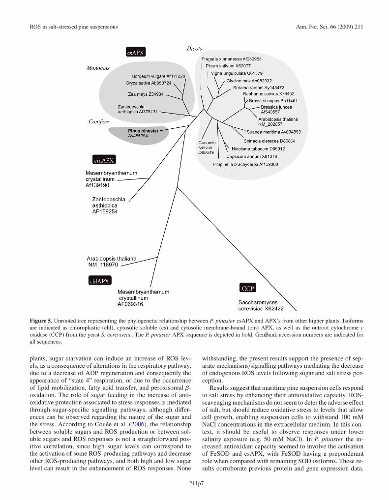
The Apx1 cDNA (1084 bp) contained a putative750 bp open reading frame. The deduced a.a. sequenceof 249 residues presented a predicted 27.3 kDa mass anda 5.44 isoelectric point. APX1 sequence analysis indicatedthe absence of protein signalling peptides or of membrane-spanning regions, suggesting a cytosolic localization. Un-rooted phylogenetic tree analysis suggests that P. pinasterApx1 codes for a cytosol-soluble (csAPX) isoform (Fig. 5).This is corroborated by the identity percentages observed be-tween P. pinaster APX1 and csApx (73.3–76.9%) in contrast tocmApx (58.7–58.8%). P. pinaster Apx1 was the first database-available nucleotide sequence encoding a cytosolic APX ingymnosperms.
3.5. Expression analysis of P. pinaster Fe-Sod1and csApx1
Transcript levels of Fe-Sod1 and csApx1 were analysed toascertain the involvement of these enzymes in P. pinaster re-sistance to salt stress, as suggested by previous results. Resultsdepicted in Figure 6 show that transcript levels of both genesincreased slightly 12 h after salt-challenging, and increasedsubstantially within 24 h of stress imposition, suggesting anactivation of antioxidant enzymatic mechanisms during severesalt stress in maritime pine cell suspensions.
4. DISCUSSION
Pinus pinaster heterotrophic suspension cells have beensuccessfully used to study plant-pathogen interactions andchanges in nutrient uptake profiles (Azevedo et al., 2008a;2008b). In the present study, this model was used to anal-yse the homeostasis of ROS during salt stress under non-photosynthetic conditions. With regards to cell culture growth,it was observed that under normal conditions, P. pinaster sus-pension cells evidenced exponential growth and an absent lagphase, a behaviour already observed in other suspension cellmodels (Shimon-Kerner et al., 2000). Increasing concentra-tions of salt resulted in growth inhibition and eventual arrest,as expected from previous reports (Elkahouia et al., 2005). Aconcentration of 100 mM NaCl was subsequently chosen toimpose severe salt stress without preventing cell growth.
Maritime pine suspension cells were able to maintain a lowoverall oxidative state in the initial 24 h of exposure to salt,but results also evidenced a transient O−2 burst. The correlationshown to exist between salt challenging and the production ofsuperoxide radical (O−2 ) in mitochondria (Corpas et al., 1993),might suggest that the observed O−2 burst may result from an
211p5

Ann. For. Sci. 66 (2009) 211 H. Azevedo et al.
Figure 4. Unrooted tree representing the phylogenetic relationship between P. pinaster Fe-SOD and Fe-SOD’s from other plants, Bacteria andProtozoa. The corresponding coding nucleotide sequences were analyzed using ClustalW and maximum-likelihood algorithms. The P. pinasterFe-SOD sequence is depicted in bold. GenBank accession numbers are indicated for all sequences.
affection of the respiratory electron chain. Meanwhile, stud-ies have shown that not only biotic but also abiotic stress canbe accompanied by an oxidative burst, which is mediated bythe O−2 -generating enzyme NADPH oxidase, and is correlatedwith stress signalling (reviewed by Mittler et al., 2004).
Lipid peroxidation can be initiated by the hydroxyl radi-cal (HO•) and requires active O2 uptake (Fridovich, 1985).Since the reduction of transition metal by O−2 is required forthe generation of HO• (together with H2O2 presence), the dis-mutation of O−2 by SODs should suppress the generation ofHO•. However, results seem to indicate that steady levels ofHO•/lipid peroxides are being generated in maritime pine sus-pension cells as a consequence of salt stress imposition. In thecell, lipid peroxidation leads to membrane permeability andloss of integrity, and ultimately to solute leakage and cellu-lar damage (Bor et al., 2003). During salt stress, low levels oflipid peroxides have been related to the increased antioxidantcapacity of salt tolerant/resistant species or cultivars (Radicet al., 2006; Ruiz et al., 2005), whereas high lipid peroxidelevels were associated with salt-sensitivity (Bor et al., 2003;Koca et al., 2007; Masood et al., 2006).
As previously stated, it has been hypothesised that mostforms of stress, by generating ROS, led to the developmentof novel roles for ROS as versatile signalling molecules. Theyact not only in abiotic but also in biotic stress sensing, with
a predominant role being led by superoxide ion (reviewedby Mittler et al., 2004). Even though a transient burst of O−2was observed in maritime pine cells, the specific thresholdsfor each ROS that determine their roles as signal modulatorsor toxic molecules are still unknown. The subsequent accu-mulation of total ROS and lipid peroxides, which are tightlylinked with the incapacity to scavenge excess ROS, suggestthat 100 mM NaCl causes severe oxidative stress in P. pinastersuspension cells.
When suspension cells subjected to sugar starvation andtherefore evidencing increased endogenous ROS levels weretransferred to sugar-containing medium, they were able to re-cover to basal ROS levels, showing an increase in the recov-ery rate in the presence of salt. These results are stronglysupportive of an induction of anti-oxidant systems followingchallenging with NaCl, suggesting that basal ROS levels ob-served in the early stages of salt stress imposition may re-sult from increased ROS-scavenging capacity rather than theabsence of oxidative stress. The involvement of soluble sug-ars (e.g. sucrose, glucose, fructose) in the dynamics of ROSand the response of plants to oxidative stress has been re-ported (revised by Couée et al., 2006). Soluble sugars seemto assume a dual role with respect to ROS, being involved inboth ROS-producing pathways and the feeding of NADPH-producing pathways, which contribute to ROS scavenging. In
211p6
ROS in salt-stressed pine suspensions Ann. For. Sci. 66 (2009) 211
Figure 5. Unrooted tree representing the phylogenetic relationship between P. pinaster csAPX and APX’s from other higher plants. Isoformsare indicated as chloroplastic (chl), cytosolic soluble (cs) and cytosolic membrane-bound (cm) APX, as well as the outroot cytochrome coxidase (CCP) from the yeast S. cereviseae. The P. pinaster APX sequence is depicted in bold. GenBank accession numbers are indicated forall sequences.
plants, sugar starvation can induce an increase of ROS lev-els, as a consequence of alterations in the respiratory pathway,due to a decrease of ADP regeneration and consequently theappearance of “state 4” respiration, or due to the occurrenceof lipid mobilization, fatty acid transfer, and peroxisomal β-oxidation. The role of sugar feeding in the increase of anti-oxidative protection associated to stress responses is mediatedthrough sugar-specific signalling pathways, although differ-ences can be observed regarding the nature of the sugar andthe stress. According to Couée et al. (2006), the relationshipbetween soluble sugars and ROS production or between sol-uble sugars and ROS responses is not a straightforward pos-itive correlation, since high sugar levels can correspond tothe activation of some ROS-producing pathways and decreaseother ROS-producing pathways, and both high and low sugarlevel can result in the enhancement of ROS responses. None
withstanding, the present results support the presence of sep-arate mechanisms/signalling pathways mediating the decreaseof endogenous ROS levels following sugar and salt stress per-ception.
Results suggest that maritime pine suspension cells respondto salt stress by enhancing their antioxidative capacity. ROS-scavenging mechanisms do not seem to deter the adverse effectof salt, but should reduce oxidative stress to levels that allowcell growth, enabling suspension cells to withstand 100 mMNaCl concentrations in the extracellular medium. In this con-text, it should be useful to observe responses under lowersalinity exposure (e.g. 50 mM NaCl). In P. pinaster the in-creased antioxidant capacity seemed to involve the activationof FeSOD and csAPX, with FeSOD having a preponderantrole when compared with remaining SOD isoforms. These re-sults corroborate previous protein and gene expression data.
211p7

Ann. For. Sci. 66 (2009) 211 H. Azevedo et al.
Figure 6. Time course expression analysis of P. pinaster Fe-Sod1and csAPX1 during 100 mM NaCl challenging of maritime pine sus-pension cells. Total RNA samples were separated on a denaturingformaldehyde gel, blotted and hybridized with homologous cDNA32P-labeled probes. Total RNA was used as loading control.
SOD activity was long predicted to help in the scavenging ofexcessive amounts of superoxide ions generated, for instance,in mitochondria (Boveris and Chance, 1973). An induction inthe activity of SOD following salt stress has been frequentlyobserved, either with total activity (Koca et al., 2007; Masoodet al., 2006) or isoform analysis (Elkahouia et al., 2005). Pre-vious evidence has shown that, in tobacco suspension cells,FeSOD is the main isoform involved in the control of cellularO−2 levels (Kurepa et al., 1997).
SOD enzymatic activity is suggested to protect cells byscavenging O−2 , and also by decreasing the risk of HO• gener-ation by the transition metal-catalyzed Haber-Weiss reaction.On the other hand, ascorbate is the most important reducingsubstrate for H2O2 detoxification (Noctor and Foyer, 1998).Ascorbate peroxidase (APX) is a multigenic family with var-ious isoforms (Asada, 1994), in which cytosolic APX playsa fundamental role in non-photosynthetic tissues, by prevent-ing H2O2 dependent inhibition of cytosolic enzymes (Verni-quet et al., 1991). Overexpression of various isoforms of SODhas been shown to confer improved tolerance to oxidativestress (Bowler et al., 1991; Perl et al., 1993). Moreover, over-expression of SOD and APX isozymes has been previouslyco-related with salt tolerance (Kaminaka et al., 1999; Wanget al., 1999), and the involvement of APX in the mechanismsof salt tolerance has also been substantiated at protein level(Elkahouia et al., 2005; Koca et al., 2007; Masood et al., 2006).Finally, present results are corroborated by microarray tran-script profiling of salt-challenged Arabidopsis plants, in whichall three Fe-SOD, but none other SOD isoform, presented in-duced expression. A similar induction was observed for thecytosolic APX1 (reviewed by Mittler et al., 2004).
With the onset of worldwide soil salinization, a better un-derstanding of plant-salinity relations is determinant not onlyfor cultivars but also for forest species. Present data confirmsthe activation of ROS-scavenging mechanisms as part of theP. pinaster cellular response to salt stress, and suggests a
possible role of transient superoxide production in stress sig-nalling. The use of in vitro heterotrophic suspension culturesmay allow further insight onto the cellular/molecular aspectsof resistance, particularly with regards to the role of ROS ofnon-photosynthetic origin. Nonetheless, and in order to clar-ify the extent of maritime pine tolerance/resistance to salt, fur-ther studies are being carried out at plant level, correlating saltstress with ROS homeostasis and the effect of salt impositionon the photosynthetic apparatus.
Acknowledgements: H. Azevedo is supported by FCT, grant refer-ence SFRH/BPD/17198/2004. V. Amorim-Silva is supported by FCT,grant reference SFRH/BD/38583/2007.
REFERENCES
Able A.J., Guest D.I., and Sutherland M.W., 1998. Use of a newtetrazolium-based assay to study the production of superoxide rad-icals by tobacco cell cultures challenged with avirulent zoosporesof Phytophthora parasitica var nicotianae. Plant. Physiol. 117: 491–499.
Allan A.C., Lapidot M., Culver J.N., and Fluhr R., 2001. An early tobaccomosaic virus-induced oxidative burst in tobacco indicates extracellu-lar perception of the virus coat protein. Plant Physiol. 126: 97–108.
Asada K., 1994. Production and action of active oxygen species inphotosynthetic tissues. In: Foyer C.H. and Mullineaux P.M. (Eds.),Production and action of active oxygen species in photosynthetic tis-sues, CRC Press, Boca Raton, pp. 77–104.
Azevedo H., Lino-Neto T., and Tavares R.M., 2003. An improved methodfor high-quality RNA isolation from needles of adult maritime pinetrees. Plant Mol. Biol. Rep. 21: 333–338.
Azevedo H., Dias A.C.P., and Tavares R.M., 2008a. Establishment andcharacterization of Pinus pinaster suspension cell cultures. Plant CellTiss. Organ Cult. 93: 115–121.
Azevedo H., Lino-Neto T., and Tavares R.M. 2008b. The necrotrophBotrytis cinerea induces a non-host Type II resistance mechanismin Pinus pinaster suspension-cultured cells. Plant Cell Physiol. 49:386–395.
Beauchamp C. and Fridovich I., 1971. Superoxide dismutase: improvedassay and an assay applicable to acrylamide gels. Anal. Biochem. 44:276–286.
Bor M., Ozdemir F., and Turkan I., 2003. The effect of salt stress on lipidperoxidation and antioxidants in leaves of sugar beet Beta vulgarisL. and wild beet Beta maritima L. Plant Sci. 164: 77–84.
Boveris A. and Chance B., 1973. The mitochondrial generation of hydro-gen peroxide. Biochem. J. 134: 707–716.
Bowler C., Slooten L., Vandenbraden S., Rycke R.D., Botterman J.,Sybesma C., Montagu M.V., and Inze D., 1991. Manganese superox-ide dismutase can reduce cellular damage mediated by oxygen radi-cals in transgenic plants. EMBO J. 10: 1723–1732.
Corpas F.J., Gomez M., Hernandez J.A., and Del Rio L.A., 1993.Metabolism of activated oxygen in peroxisomes from two Pisumsativum L. cultivars with different sensitivity to sodium chloride. J.Plant Physiol. 141: 160–165.
Couée I., Sulmon C., Gouesbet G., and El Amrani A., 2006. Involvementof soluble sugars in reactive oxygen species balance and responses tooxidative stress in plants. J. Exp. Bot. 57: 449–459.
Elkahouia S., Hernández J.A., Abdellyc C., Ghrira R., and Limama F.,2005. Effects of salt on lipid peroxidation and antioxidant enzymeactivities of Catharanthus roseus suspension cells. Plant Sci. 168:607–613.
211p8

ROS in salt-stressed pine suspensions Ann. For. Sci. 66 (2009) 211
Fridovich I., 1985. Biological effects of the superoxide radical. Arch.Biochem. Biophys. 247: 1–11.
Hernandez J.A., Corpas F.J., Gomez M., del Rio L.A., and Sevilla F.,1993. Salt-induced oxidative stress mediated by activated oxygenspecies in pea leaf mitochondria. Physiol. Plant. 89: 103–110.
Kaminaka H., Morita S., Tokumoto M., Masumura T., and Tanaka K.,1999. Differential gene expressions of rice superoxide dismutase iso-forms to oxidative and environmental stresses. Free Rad. Res. 31:219–225.
Koca H., Bor M., Ozdemir F., and Turkan I., 2007. The effect of salt stresson lipid peroxidation, antioxidative enzymes and proline content ofsesame cultivars. Environ. Exp. Bot. 60: 344–351.
Kurepa J., Hérouart D., Van Montagu M., and Inzé D., 1997. Differentialexpression of CuZn- and Fe-superoxide dismutase genes of tobaccoduring development, oxidative stress, and hormonal treatments. PlantCell Physiol. 38: 463–470.
Lino-Neto T., 2001. Role of oxidative stress enzymes duringZantedeschia aethiopica spathe whitening and regreening, MinhoUniversity, Braga, 284 p.
Loreto F. and Velikova V., 2001. Isoprene produced by leaves protectsthe photosynthetic apparatus against ozone damage, quenches ozoneproducts and reduces lipid peroxidation of cellular membranes. PlantPhysiol. 127: 1781–1787.
Masood A., Shah N.A., Zeeshan M., and Abraham G., 2006. Differentialresponse of antioxidant enzymes to salinity stress in two varieties ofAzolla (Azolla pinnata and Azolla filiculoides). Environ. Exp. Bot.58: 216–222.
Mittler R., 2002. Oxidative stress, antioxidants and stress tolerance.Trends Plant Sci. 7: 405–410.
Mittler R., Vanderauwera S., Gollery M., and Van Breusegem F., 2004.Reactive oxygen gene network of plants. Trends Plant Sci. 9: 490–498.
Murashige T. and Skoog F., 1962. A revised medium for rapid growth andbio assays with tobacco tissue cultures. Physiol. Plant. 15: 473–497.
Noctor G. and Foyer C.H., 1998. Ascorbate and glutathione: keeping ac-tive oxygen under control. Annu. Rev. Plant Physiol. Plant Mol. Biol.49: 249–279.
Osmond C.B. and Grace S.C., 1995. Perspectives on photoinhibition andphotorespiration in the field: quintessential inefficiencies of the lightand dark reactions of photosynthesis. J. Exp. Bot. 46: 1351–1362.
Perl A., Perl-treves R., Galili G., Aviv D., Shalgi E., Malkin S., and GalunE., 1993. Enhanced oxidative-stress defence in transgenic potatoplants expressing tomato Cu, Zn superoxide dismutase. Theor. Appl.Genet. 85: 568–576.
Petersen D.R., Reichard J., Kolaja K.L., and Hartley D.P., 1999. 4-Hydroxynonenal and malondialdehyde heatic protein adducts in ratstreated with carbon tetrachloride: immuno-chemical dection and lob-ular localization. Toxicol. Appl. Pharm. 161: 23–33.
Radic S., Radic-Stojkovic M., and Pevalek-Kozlina B., 2006. Influence ofNaCl and mannitol on peroxidase activity and lipid peroxidation inCentaurea ragusina L. roots and shoots. J. Plant Physiol. 163: 1284–1292.
Ruiz J.M., Blasco B., Rivero R.M., and Romero L., 2005. Nicotine-free and salt-tolerant tobacco plants obtained by grafting to salinity-resistant rootstocks of tomato. Physiol. Plant. 124: 465–475.
Sedmak J.J. and Grossberg S.E., 1977. A rapid, sensitive, and versatile as-say for protein using Coomassie Brilliant Blue G250. Anal. Biochem.79: 544–552.
Shimon-Kerner N., Mills D., and Merchuk J.C., 2000. Sugar utilizationand invertase activity in hairy-root and cell-suspension cultures ofSymphytum officinale. Plant Cell Tissue Organ Cult. 62: 89–94.
Van Breusegem F., Bailey-Serres J., and Mittler R., 2008. Unravelingthe tapestry of networks involving reactive oxygen species in plants.Plant Physiol. 147: 978–984.
Verniquet F., Gaillard J., Neuberger M., and Douce R., 1991. Rapid in-activation of plant aconitase by hydrogen peroxide. Biochem. J. 276:643–648.
Volokita M., 1991. The carboxy-terminal end of glycolate oxidase directsa foreign protein into tobacco leaf peroxisomes. Plant J. 1: 361–366.
Wang J., Zhang H., and Allen R.D., 1999. Overexpression of anArabidopsis peroxisomal ascorbate peroxidase gene in tobacco in-creases protection against oxidative stress. Plant Cell Physiol. 40:725–732.
Zhu J.-K., 2001. Plant salt tolerance. Trends Plant Sci. 6: 66–71.
211p9