effect of various stressors on the blood acth and corticosterone concentration in normotensive...
TRANSCRIPT
www.elsevier.com/locate/ygcen
General and Comparative Endocrinology 153 (2007) 217–220
Effect of various stressors on the blood ACTHand corticosterone concentration in normotensive Wistar
and spontaneously hypertensive Wistar-Kyoto rats
Jelena Djordjevic *, Tamara Vuckovic, Nebojsa Jasnic, Gordana Cvijic
Institute of Physiology and Biochemistry, Faculty of Biology, University of Belgrade, Studentski Trg 16,
P.O. Box 52, 11000 Belgrade, Serbia and Montenegro
Received 13 September 2006; revised 30 January 2007; accepted 2 February 2007Available online 12 February 2007
Abstract
The role of the sympathetic nervous system (SNS) and the hypothalamo–pituitary–adrenal (HPA) axis in the stress response is welldocumented. The imbalance in a central and peripheral SNS activity accompanied by the HPA hyperresponsivity has been observed inessential and experimental hypertension. The spontaneously hypertensive rats (SHR) are extensively used in studying mechanisms of theessential hypertension. The blood ACTH and corticosterone concentration was examined in spontaneously hypertensive (Wistar-Kyoto)and normotensive (Wistar) adult male rats exposed to acute cold (2 h) or immobilization (2 h) stress as well as chronic (21 days) isolationstress or their combination. The present results show that SHR in basal conditions have higher blood ACTH and corticosterone level ascompared to the normotensive rats. Both the acute exposure to cold and immobilization stress induced a higher increment in SHRplasma ACTH in respect to Wistar rats. The similar pattern of ACTH response occurred when SHR were previously chronically isolatedand acutely exposed to both applied stressors. Surprisingly, corticosterone concentration did not differ between control rats with or with-out 21 days isolation or those exposed to a cold or immobile acute stressor.� 2007 Elsevier Inc. All rights reserved.
Keywords: ACTH; Corticosterone; Hypertension; Stress
1. Introduction
The spontaneously hypertensive rats (SHR) provide awell-accepted animal model for the study of human essen-tial hypertension. While the etiology of hypertension inSHR is not well understood, it is known that SHR exhibitincreased sympathetic activity during the hypertensiondevelopment (Iriuchijima, 1973). During aging the sympa-thetic activity rises and so does the hyperreactivity tostressors (Sato, 1987). Krukoff et al. (1999) showed thatchronic elevation in sympathetic activity in SHR is associ-ated with hyperreactiveness of the hypothalamo–pituitary–
0016-6480/$ - see front matter � 2007 Elsevier Inc. All rights reserved.
doi:10.1016/j.ygcen.2007.02.004
* Corresponding author. Fax: +381 11 639064.E-mail address: [email protected] (J. Djordjevic).
adrenocortical (HPA) system and may have importantimplications for response of HPA axis during stress.
The purpose of this study was to investigate the reactionof HPA system of spontaneously hypertensive and normo-tensive rats acutely and/or chronically exposed to varioustypes of stressors (cold as environmental, immobilizationas psychophysical and isolation as psychosocial) by mea-suring the blood ACTH and corticosterone concentration.
2. Materials and methods
Male spontaneously hypertensive rats (SHR) of Wistar-Kyoto strainand normotensive rats of Wistar strain, 15-weeks-old, were used for theexperiments. The rats were acclimated to 22 ± 1 �C, kept at a 12:12 hlight–dark cycle, with the dark cycle onset from 6 p.m. The animals weregiven commercial rat food and tap water ad libitum and housed two percage for 15 days before starting the experiment. The blood pressure was
0
50
100
150
200
250
300
pg/m
l pla
sma
immobilization (2h)
†
**WistarSHR
controls
*
controls: acute stress
*
†*
chronic stress: i s o l a t i o n (21 days)cold (2h)
† Wistar : SHR
*
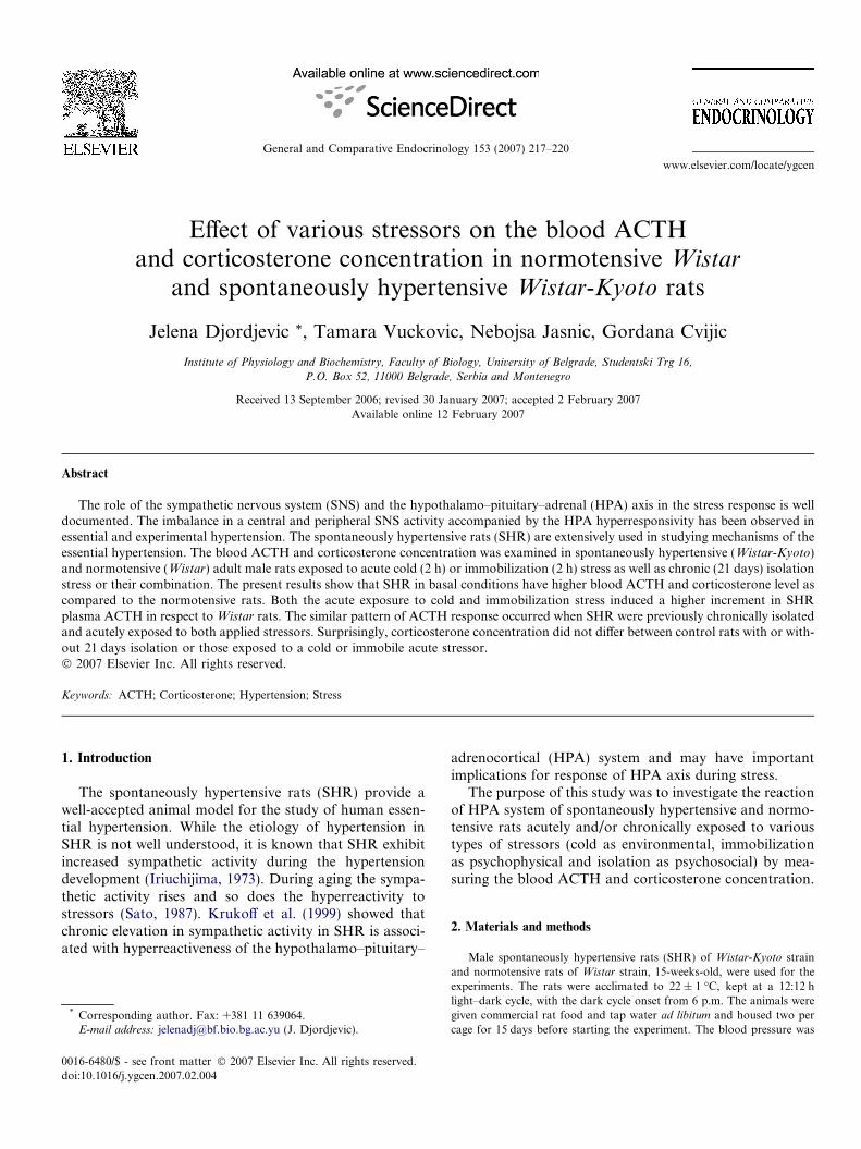
Fig. 2. Plasma ACTH concentration. The effect of acute cold (6 �C for2 h) and immobilization stress (2 h) on the plasma ACTH concentration inpreviously chronically isolated (21 days) spontaneously hypertensive andnormotensive rats. The data are presented as means ± SE of six animals(n = 6) in pg/ml. Differences between the groups: *p < 0.05, **p < 0.01unstressed animals vs. animals exposed to acute stressors; �p < 0.05 SHRvs. Wistar strain.
Wistar
218 J. Djordjevic et al. / General and Comparative Endocrinology 153 (2007) 217–220
monitored one week prior to the experiment, using tail cuff plethysmogra-phy in restrained, conscious animals. The mean arterial pressure forWistar rats was 100 ± 5 mm Hg and for SHR 180 ± 5 mm Hg. The exper-iments were performed according to the rules of animal care proposed bythe Serbian Laboratory Animal Science Association (SLASA).
The rats were divided into 12 groups, each consisting of six animals.The first group contained intact SHR controls. The SHR from the secondand third groups were exposed to the acute stressors, cold (6 �C for 2 h)and immobilization (2 h), respectively, and killed after the stress termina-tion. The immobilization stress was performed according to Kvetnanskyand Mikulaj (1970) by fixing all four limbs to a board with adhesive tape.The heads were also fixed by a metal loop over the neck area, thus limitingtheir motion. Acute stress exposures were exerted between 8:00 and 11:00a.m. to avoid the effects of circadian rhythms. The fourth group of SHRwas subjected to the social isolation for 21-day-period, remaining withoutany visual connection with other animals, being killed on the 22nd day.The fifth and sixth groups were chronically stressed at the same way asthe fourth group (21 days of the isolation) and on the 22nd day acutelyexposed to the novel stressors: cold (6 �C for 2 h) and immobilization(2 h). These animals were killed at the onset of acute stressors. The sameexperimental procedure was performed with the six groups of Wistar rats.
The rats were always killed by decapitation without anaesthesia withguillotine (Harvard-Apparatus, Holliston, MA, USA). Blood was col-lected from the trunk and divided into two sets of tubes. For plasmaobtaining EDTA was added in one of the sets. Serum and plasma werefrozen for corticosterone and ACTH determination. Plasma ACTH wasdetermined by chemiluminescent method using IMMULITE automaticanalyzer (DPC, Los Angeles, CA). The values are expressed as pgACTH/ml plasma. Serum corticosterone was determined by RIA kit(ICN Biochemicals, Costa Mesa, CA) and the values expressed as lgCORT/100 ml serum. Two Factor ANOVA Model 1 followed by Holm-Sidak posterior comparation test has been employed for the comparisonof the experimental groups. The level of significance was set at p < 0.05.
30
40
erum
SHR †
† Wistar : SHR
3. Results
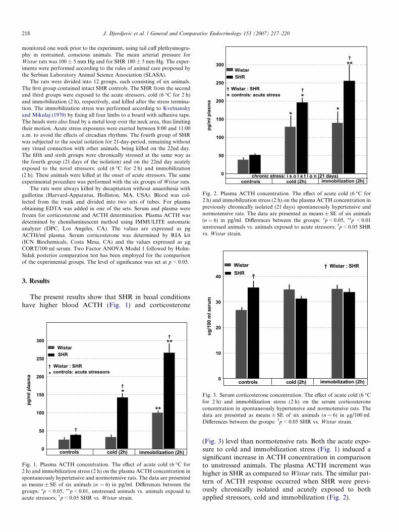
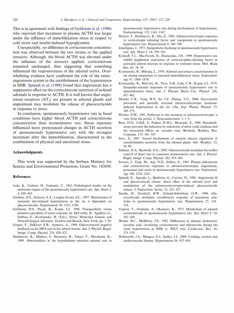
The present results show that SHR in basal conditionshave higher blood ACTH (Fig. 1) and corticosterone
0
50
100
150
200
250
300
pg/m
l pla
sma
cold (2h) immobilization (2h)
**WistarSHR
*
**
†
†
†
† Wistar : SHR
* controls: acute stressors
controls
Fig. 1. Plasma ACTH concentration. The effect of acute cold (6 �C for2 h) and immobilization stress (2 h) on the plasma ACTH concentration inspontaneously hypertensive and normotensive rats. The data are presentedas means ± SE of six animals (n = 6) in pg/ml. Differences between thegroups: *p < 0.05, **p < 0.01, unstressed animals vs. animals exposed toacute stressors; �p < 0.05 SHR vs. Wistar strain.
0
10
20
ug/1
00 m
l s
cold (2h) immobilization (2h)controls
Fig. 3. Serum corticosterone concentration. The effect of acute cold (6 �Cfor 2 h) and immobilization stress (2 h) on the serum corticosteroneconcentration in spontaneously hypertensive and normotensive rats. Thedata are presented as means ± SE of six animals (n = 6) in lg/100 ml.Differences between the groups: �p < 0.05 SHR vs. Wistar strain.
(Fig. 3) level than normotensive rats. Both the acute expo-sure to cold and immobilization stress (Fig. 1) induced asignificant increase in ACTH concentration in comparisonto unstressed animals. The plasma ACTH increment washigher in SHR as compared to Wistar rats. The similar pat-tern of ACTH response occurred when SHR were previ-ously chronically isolated and acutely exposed to bothapplied stressors, cold and immobilization (Fig. 2).

0
10
20
30
40
50
ug/1
00 m
l ser
um
chronic stress: i s o l a t i o n (21 days)cold (2h) immobilization (2h)
WistarSHR
† Wistar : SHR
†
controls
Fig. 4. Serum corticosterone concentration. The effect of acute cold (6 �Cfor 2 h) and immobilization stress (2 h) on the serum corticosteroneconcentration in previously chronically isolated (21 days) spontaneouslyhypertensive and normotensive rats. The data are presented as means± SE of six animals (n = 6) in lg/100 ml. Differences between the groups:�p < 0.05 SHR vs. Wistar strain.
J. Djordjevic et al. / General and Comparative Endocrinology 153 (2007) 217–220 219
However, the results concerning corticosterone concen-tration are different. Hence, the acute cold and immobiliza-tion stress with or without previously 21 days isolation didnot significantly changed serum corticosterone level in bothexamined strains (Figs. 3 and 4).
4. Discussion
Morphological abnormalities have been reported in thepituitary and adrenals of SHR (Aoki et al., 1963), indi-cating that the hypertension may be related to a dis-turbed HPA function. The results concerning the bloodACTH and corticosterone level in basal condition inSHR are controversial. Hattori et al. (1986) did not finddifferences in blood ACTH and vasopressin level between6- and 11-week-old SHR and normotensive Wistar-Kyoto
rats (WKY). However, basal corticosterone level washigher only in 6-week-old SHR. Six weeks represents anearly stage of hypertension, whereas hypertension isnearly established in 11-week-old SHR. Sowers et al.(1981) reported an increased corticosterone level in twomonths old SHR, Yamori et al. (1973) did not find differ-ences in plasma corticosterone level between six monthold SHR and WKY while Wexler and McMurty (1982)found a decreased corticosterone level in SHR as com-pared to WKY. Thus, the inconsistent results regardingthe basal ACTH and corticosterone level in SHR andWKY could be age-dependent (Gomez et al., 1998).Our results show that 15-week-old SHR in basal condi-tions have higher blood ACTH and corticosterone con-centration as compared to normotensive age-matchedWistar rats.
There is a possibility that glucocorticoids and ACTHper se may be the factors in hypertension development,since adrenalectomy or hypophysectomy prevents thedevelopment of hypertension in SHR while corticosteronesupplementation restores hypertension after adrenalectomy(Hashimoto et al., 1989; Gardner et al., 1997). Besides, aprogression of the SHR syndrome is associated with ahypertrophy of the adrenals cortical zone (Aoki et al.,1963). There is also a growing evidence that the elevatedglucocorticoids contribute to the pathogenesis of essentialhypertension in humans (Withworth et al., 2000).
The mechanisms of glucocorticoid-mediated control ofthe arterial pressure is still poorly understood. Glucocorti-coids can increase vascular reactivity to noradrenaline andangiotensin and can blunt the induction of nitric oxide(NO) synthesis in smooth muscle (Rees et al., 1990). Suzukiet al. (1996) provide additional evidence for a link betweenglucocorticoids and altered production of endothelium-derived agents since they showed that the impaired dilatorresponse to histamine in SHR is related to an enhancedadrenal glucocorticoid secretion. Mondo et al. (2006)induced a rise of blood pressure in Sprague–Dawley ratsby ACTH treatment and showed the hypertension is asso-ciated with reduced NO. On the other hand, there is someevidence that glucocorticoids attenuate arterial barorecep-tor reflex function (Scheuer and Bechtold, 2002).
The involvement of stress in the pathophysiology ofhypertension has always been suspected although there isa lack of clear evidence documenting a direct link betweenstress and hypertension. Inconsistent results exist in theliterature regarding the HPA response to stress in SHRand WKY rats. Hashimoto et al. (1989) reported a smallerplasma ACTH and greater corticosterone response tohemorrhage and ether stress in 7-weeks-old SHR in com-parison to age-matched WKY. Gomez et al. (1998) failedto find differences in the HPA response to the acute tailshock between SHR and WKY. However, Kvetnanskyet al. (1979) reported a higher circulating level of catechol-amines and corticosterone in SHR during the acute immo-bilization stress. Our results show that naive or previouslychronically isolated SHR exhibit an enhance ACTHresponse to acutely applied stressors, cold and immobiliza-tion as compared to normotensive rats. It is well knownthat the ACTH response in acute stress is mostly dependenton the interaction of corticotropin-releasing-hormone(CRH) and vasopressin. Because the contribution of eachsecretagogue to ACTH release depends on the intensity,duration and type of stressor (Plotsky, 1991), the differentresponse of these strains could explain why no consistentdifferences in their HPA response to stress have beenfound. However, Krukoff et al. (1999) reported that acutestress is capable of upregulating CRH gene expression inSHR through an activation of the neurons that do notsynthesize CRH under normotensive condition.
The present study also shows that the highest elevationof plasma ACTH was observed after the immobilization,as compared to the acute cold and the long term isolation.
220 J. Djordjevic et al. / General and Comparative Endocrinology 153 (2007) 217–220
This is in agreement with findings of Goldstein et al. (1996)who reported that increment in plasma ACTH was largerunder the influence of immobilization stress in respect tocold stress and insulin-induced hypoglycemia.
Unexpectedly, no difference in corticosterone concentra-tion was observed between the two strains or the appliedstressors. Although, the blood ACTH was elevated underthe influence of the stressors applied, corticosteroneremained unchanged, thus suggesting that somethinginfluenced the responsiveness of the adrenal cortex. Over-whelming evidence have confirmed the role of the renin–angiotensin system in the establishment of the hypertensionin SHR. Spinedi et al. (1989) found that angiotensin has asuppressive effect on the corticosterone secretion of isolatedadrenals in response to ACTH. It is well known that angio-tensin receptors (AT1) are present in adrenal glands andangiotensin may modulate the release of glucocorticoidsin response to stress.
In conclusion, spontaneously hypertensive rats in basalconditions have higher blood ACTH and corticosteroneconcentration than normotensive rats. Stress exposureinfluenced more pronounced changes in ACTH secretionof spontaneously hypertensive rats with the strongestincrement after the immobilization, characterized as thecombination of physical and emotional stress.
Acknowledgments
This work was supported by the Serbian Ministry forScience and Environmental Protection, Grant No. 143050.
References
Aoki, K., Tankaw, H., Fujinami, Y., 1963. Pathological studies on theendocrine organs of the spontaneously hypertensive rats. Jpn. Heart J.4, 426–442.
Gardner, D.S., Jackson, A.A., Langley-Evans, S.C., 1997. Maintenance ofmaternal diet-induced hypertension in the rat is dependent onglucocorticoids. Hypertension 30, 1525–1530.
Goldstein, D.S., Pacak, K., Kopin, I.J., 1996. Nonspecificity versusprimitive specificity of stress response. In: McCarthy, R., Aguilera, G.,Sabban, E., Kvetnansky, R. (Eds.), Stress: Molecular Genetic andNeurobiological Advances. Gordon and Breach, New York, pp. 3–20.
Gomez, F., DeKloet, E.R., Armario, A., 1998. Glucocorticoid negativefeedback on the HPA axis in five inbred strains. Am. J. Physiol. Regul.Integr. Comp. Physiol. 274, 420–427.
Hashimoto, K., Makino, S., Hirasawa, R., Takao, T., Murakami, K.,1989. Abnormalities in the hypothalamo–pituitary–adrenal axis in
spontaneously hypertensive rats during development of hypertension.Endocrinology 125, 1161–1167.
Hattori, T., Hashimoto, K., Ota, Z., 1986. Adrenocorticotropin responsesto corticotropin releasing factor and vasopressin in spontaneouslyhypertensive rats. Hypertension 8, 386–390.
Iriuchijima, J., 1973. Sympathetic discharge in spontaneously hypertensiverats. Jpn. Heart J. 14, 350–356.
Krukoff, T.L., MacTavish, D., Jhamandas, J.H., 1999. Hypertensive ratsexhibit heightened expression of corticotrophin-releasing factor inactivated central neurons in response to restraint stress. Mol. BrainRes. 65, 70–79.
Kvetnansky, R., Mikulaj, L., 1970. Adrenal and urinary catecholamines inrat during adaptation to repeated immobilization stress. Endocrinol-ogy 87, 1868–1874.
Kvetnansky, R., McCarty, R., Thoa, N.B., Lake, C.R., Kopin, I.J., 1979.Sympatho-adrenal responses of spontaneously hypertensive rats toimmobilization stress. Am. J. Physiol. Heart Circ. Physiol. 236,457–462.
Mondo, C.K., Yang, W.S., Su, J.Z., Huang, T.G., 2006. Atorvastatinprevented and partially reversed adrenocorticotropic hormone-induced hypertension in the rat. Clin. Exp. Pharm. Physiol. 33,369–373.
Plotsky, P.M., 1991. Pathways to the secretion of adrenocorticotropin: aview from the portal. J. Neuroendocrinol. 3, 1–9.
Rees, D.D., Cellek, S., Palmer, R.M.J., Moncada, S., 1990. Dexameth-asone prevents the induction by endotoxin of nitric oxide synthase andthe associated effects on vascular tone. Biochem. Biophys. Res.Commun. 173, 541–547.
Sato, A., 1987. Neural mechanisms of somatic sensory regulation ofcatecholamines secretion from the adrenal gland. Adv. Biophys. 23,39–80.
Scheuer, D.A., Bechtold, A.G., 2002. Glucocorticoids modulate baroreflexcontrol of heart rate in conscious normotensive rats. Am. J. Physiol.Regul. Integr. Comp. Physiol. 282, 475–483.
Sowers, J., Tuck, M., Asp, N.D., Sollars, E., 1981. Plasma aldosteroneand corticosterone responses to adrenocorticotropin, angiotenzin,potassium and stress in spontaneously hypertensive rats. Endocrinol-ogy 108, 1216–1221.
Spinedi, E., Aguado, L., Basilotta, G., Carrizo, D., 1989. Angiotenzin IIand glucocorticoid release: direct effect al the adrenal level andmodulation of the adrenocorticotropin-induced glucocorticoidsrelease. J. Endocrinol. Invest. 12, 321–327.
Suzuki, H., Zweifach, B.W., Schmid-Schonbein, G.W., 1996. Glu-cocorticoids modulate vasodilatator response of mesenteric arte-rioles in spontaneously hypertensive rats. Hypertension 27, 114–118.
Yamori, Y., Ooshima, A., Okamoto, K., 1973. Metabolism of adrenalcorticosteroids in spontaneously hypertensive rats. Jpn. Heart J. 14,162–164.
Wexler, B.C., McMurty, J.P., 1982. Differences in adrenal cholesterol,ascorbic acid, circulating corticosterone and aldosterone during theonset hypertension in SHR vs. WKY rats. Cardiovasc. Res. 16,573–579.
Withworth, J.A., Mangos, G.J., Kelley, J.J., 2000. Cushing, cortisol andcardiovascular disease. Hypertension 36, 912–916.