effective risk stratification using exercise myocardial ... · effective risk stratification using...
TRANSCRIPT
34 JACC Vol. 28, No. 1 July 19969-44
Effective Risk Stratification Using Exercise Myocardial Perfusion SPECT in Women: Gender-Related Differences in Prognostic Nuclear Testing RORY HACHAMOVITCH, MD, DANIEL S. BERMAN, MD, FACC, HOSEN KIAT, MD, FRACP, FACC, C NOEL BAIREY MERZ, MD, FACC, ISHAC COHEN, PHD, J. ARTHUR CABICO, JOHN FRIEDMAN, MD, FACC, GUIDO GERMANO, PHD, KENNETH F. VW TRAIN, GEORGE A. DIAMOND, MD, FACC
Los Angeles, Colifomia .-
fIll&fives. ‘Ibis study was designed to evaluate the incremen- tal pmgmdic value over dhdcal and exercise variables of rest thaRllila-2OVexerdse tedmetium-991~ sestamibi einglf-photon emlsslon computed tonmgmphy (SPECT) in mlmen comparefl with men and to detemh wðer this test caa be wed ta elktidyrlskstra~pa~tsofhotkgende~
gender- -m
tbewbeneedtoide&ifyauaninvasivetestingstrategytbatis ~toaccwtelyaudeffecWyriskstrati@wamen.
Mefbods. We Identified 4,136 omsecntive patients (2,742 men, 1,394 wamen) wba underwent daal-isotope SPECT. The iacremea- btvataD~nn~rtestiogwssde~~osisgbottrastepwise Cox pmportlonal hazards model and RapIan-Meter survival analysis. Receiver opersting cbaraderistie curve analysis was performed to deter&e test discriminatioa for high risk patients iumeuandwwmen.
electrocardiim. Cox proportional haxards analysis revealed that nuclear testing added incremental pragnastic value in both men aud women aRer indnsion of the most prediive dhdcal and exerdse variables (overall cbiiqnare 89 in men vs. 120 in wamen, p e 0.005). Raplan-Meier survival analysis demanstrated that oudear testing further sb-atllkd men and warna with both intermediate to bigb and low prescan lRwliboads of coronary artery disease (p < 0.005 far all). Receiver aperating ebaracter- istic curve analysis demonstrated superior discrhnination for the nuclear scan results in ideatitjiq high risk wumea than mea (area under the curve: 0.84 * 0.03 vs. 0.71 f 0.03 in Illea, p c 0.0005). Tbe odds ratio wmparhg event rates in patients with abnormal versus those with narmal scan results vms greater ia wamen than ia men, suggesting superior stratificatian using nuclear testing in wnnen.
ResuRs. Tlte patient popalation was fallawed up far 20 f 5 umtks for events (cardiac death or nonfatal mywarditi infarc- tion). During this time, 63 myacardii infarctiaas and 32 cardiac deaths ocamed in the men, and 31 q yacardial iefarctions and 14 cardiac deaths oceorred in tbe women. Nuclear testing signi& amtly stratil&d bath men and women irrespective of their rest
Condusions. Dnal-isotope myacarxliai perfasion imaging yie8d.v incremental pragnostic value in botb ma and women. This madallty ideatilies low risk women and men equally well but relatively bii risk women more accurately than titiely high risk men and, thus, is able to stratify women anwe e&ctively than men.
(J Am coil cardid 1996;28:34-44)
Ischemlc heart disex is the leading cause of death i? both men and women in the United States (1). Although the incidence of nonfatal coronary artery disease has doubled among women in the past decade (2), and the rate of referral
of women to interventional testing and revascularization has also increased (2), a number of studies have found that coronary artery disease in women is identified less often (3), at a later stage (4) and treated less aggressively than in men (s-10).
Some investigators suggest that these differences are appro- priate when axrected for baseline group differences (g-11), but others consider them illustrative of a gender-related bii in the diagnosis and management of coronary artery dii (6-9). To minim& this bii in the evahtation of women, there is a need for a noninvasive test strategy that is able to aeauately and economically risk stratify women, thus identi- fying a subset of patients in need of further, invasive testing and possible revtition. In this conte& akhough previ- ous studies have contrasted tbe d&no&c efficq of electro-

JACC Vol. 28, No 1 July 19%3l-44
cardiographic (ECG) and nuclear s&es testing in women and men, prognostic efficacy has not been similarly compared.
We undertook the present study to determine whether nuclear exercise terting adds similar incremental progmdc information over that provided by clinical and exercise data in women compared with men and whether this modality, incor- porated in a clinical strategy, can be used to effectively risk stratify both men and women.
Methods Study pnpuIatIon. This study was a retrospective analysis of
data from 4,620 consecutke patients (3,1(x) men, 1520 women) who undenvent rest thallium-Ml/exercise technetium-99m sesta- miii separate-acquisition dual-isotope single-photon emission computed tomography (SPW between January I,1991 and December 1,1993 at Cedar&ii Medical Center. Patients who were known to have valvular heart disease or primary cardiimy- opathy were exduded. Of the initial population, I39 men and 59 women were lost to fdlow-up, and 12 men and 4 women were exduded because of missing data, resulting in a study population of 4,406 (2$49 men, 1,457 women).
Patients who underwent revascularization within 60 days of the index dual-isotope SPECT exercise test were censored from all analysis and were not considered in the analysis of patient outcomes after 60 days. Of the 4,406 patients (2,949 men, 1,457 women [95% of the initial population]), 207 men and 63 women were thus censored. This temporal threshold was utilii to provide discrimination between patients with and without clinical deterioration that may have resulted in late referral to revascularization. We have previously reported (12,13) that patients who are referred to revascularization within the first 60 days after nuclear testing do so, i,~ large part, on the basis of their scan results, whereas patients who are referred to revescularization >6O days after nuclear testing tend to be referred because of worsening of diniil status. Thus, the data presented here are based on a subset of 4,136 patients (2,742 men, 1,394 women).
Exe&e myvnaniIal perksIon proto&. All patients un- derwent exercise dual-isotope myocardial perfusion imaging as previously descrbed (13). Whenever possible, beta-adrenergic
bIockirg agents and calcium channel antagonists were discon- tinued 48 h before testing, and nitrate compounds were discontinued for 6 h before testing. Before exercise, thallium- 231 (2.5 to 3.0 mCi) was injected intravenously at rest, with dose variation based on patient weight. Rest thallium-201 SPECT imaging was initiated 10 min after injection of the isotope. In a minority of parients, thallium redistribution images (24-h images) were obtained, and the results were &red in Our atmlysii in pIace of the rest thallium scores. After thallium imaging ali padents performed a symptom- Iimited exercise treadtniI1 test (E’IT) using standard protocols with 12-lead ECXi tecordmg of each minute of exercise. Blood presauewasrecordedatre&attheendofeachexercisestage ad at peak exercise. MaximaI degree of ST segment change at
snofwAxw VERTtCAL LOWGNUS
Figure 1. A&nment of mp.xiiil regions for scoring of SPEU imap. t&ally. scam with multiple segments scored as having stress’ rest scores of 1 to P or a si@e w-1 with a stress score of 2 were classified as equivocal. Scans with two segments assigned stress scores of 2 were daxsitied as probably abnormal. and scans nltaining two or more strw segments assigned uzores of Z or ram with one or more xgmenn assigned rftnfi of 3 were classdied as definitety abnormal. .Assignment of segmental wares took into aunt kncmledge of normal segment variation. Reversibility of .wgmental scores influenced the interpretation torvard ahnormal. When fixed defects on the strw study were an&wed to be secondary la attenuation. their score was decreased to 1. If apparent apical defects were aani&red likely I(I represent normal apical thinnk or if defects were considered to be secondary to breast attenuation. lhey were assigned a score of 1. On the basis of these scoring guidelines the ohsewers judged each patient‘s study results as normal, p&ably normal, equivocal. probably ahnormal or definitely abnwnnal in a reading blinded to the patients clinical historical and exercise treadmill information. The oktven were then made aware of the patient’s other relevant. nonnudear information and formed a final interpretation of the study (scan result), which by agreement among ohservers could noi \a~ by more than one grade from the initial (blinded) interpre’ ‘ion.
SO ms after the J point of the ECG was measured and assessed as horizontal. upsloping or downsloping.
The ECG mponse to testing was categorized as either nonischemic (no significant ECG changes), ischemic (>l-mm horizontal or down&ping ST segment elevation or depression except in leads without significant Q waves or in lead aVR), t-qiwd (brderlioe ECG changes) or m (exe&z- ilKiUCdwsGChiUlgeSUIliilterpetaMebecaurof~~uSe,
pacedmythmbundlebmnchbkck).Theciinicn~to exercisewasalsoazses&a5eithernfmk&m& is&emic(angiiii ymptonls during exeti), tyimal or crlnamal (exertiona hypote~or iqpropkte slnnmes of breath).
At near-maximal exercise, a 2% to 30-mCi k of Tc-99m sestamibi was injected (actual patient dose varied with patient weight). and exercise continued for one additional minute after injection. TII SPECT imaging was begun 30 miu after isotope injeciion and was performed as previously described (14). All images were subject to quality control measures (15).
Image iattrpM&k Semiquantitative visual interpreta- tion was performed using short-axis. vertical long-axis and horizontal long-axis tomograms (15). The short-axis and verti- cal long-axis myocardial tomograms ‘were divi& into 20 segments and were scored by consensus of two experienced obrmers(Fig. 1).
Scintig&dci&exes.Asummedstressscorewasob- tainedbyaddingthe~~ofthe;TOsegmenboftfKmess images. A summed rest thaIhum defect score was obtained by simiIarlyaddingthe2OsegmentsoftberestthaIliumimages. Tbesumofthedifferencesbetweeneachofthe20~ntson

36 HACHAMOVITCH ET AL PROGNOSTIC F4UCLEAR TESTING IN WOMEN
the stress and rest images was defined as the summed difference score and represents the amount of isshemia present. All three of these derived nuclear variables measure both the extent and severity of perfusion abnormalities (13,14). Whenever avail- able, scores of late redistribution rather than rest thallium images were used. Fewer women than men had late imaging (43 [3.1%] of 1,394 women, 155 15.681 of 2,742 men. p < 0.091).
P&at foflnw-np. Patient follow-up was performed by scripted telephone interview by interviewers who had knowl- edge of the patient’s test results. Everus were defined as either cardiac death (confirmed by review of death certificate and hospital chart or physician’s records) or nonfatal myocardial infarction (documented by appropriate ECG and cardiac enzyme level changes). If a patient was found to have had both more than one event after nuclear testing, the more serious event (e.g., cardiac death) was considered; if two myocardial infarctions occurred, the temporally proximal event to the index test was considered. When interventions (cardiac cathe- terizatior., coronary artery bypass surgery or percutaneous transluminal coronary angioplasty) were identified, these out- comes were contirmed by hospital records or physician’s office records All patients included in this report were followed up for at least 1 year (mean 20 2 5 months).
LikelW of coronary artery disease. For purposes of analyzing patients in digerent risk subsets, we used analysis of the pteexercise and prescan likelihood of coronary artery disease as aggregate descriptors of proven prognostic impor- tance based on Bayesian analysii of age, gender, symptom classiition, rest ECG, cardiac risk factors and (for prescan likelihood) the results of ECG stress testing but not the nuclear scan information (exercise heart rate, blood pressure, dura- tion, magnitude and slope of ST segment changes, exertional hypotension md rest ECG characteristics) and calculated using CADENZA (16). In patients with a prior history of myocardi II infarction, the likelihood oi coronary artery disease was assumed to be 1, and in patients with previously docu- mented coronary artery disease without prior myocardial in- farction, the value represents the likelihood of exercise- induced ischemia rather than likelihood of anatomic disease (17).
Statistical analpir Comparisons between patient groups were performed using a one-way analysis of variance for contin- uous variables and a &i-square test for categoric variables. All continuous variablea are descded as mean value t SD. Receiver operatingCh8aUe&ticcurveaaredescriiasmeanvalueLt SEld A p value < 0.05 was considered staustically sign&ant.
incremental v&e. To determine the additive prognostic value of a test it is necessary io include all other information known regarding the patient before that time. With this in mind, incremental prognostic value was determined in three different ways:
krt.rtrtv~?u ANALYSTS. The Cox proportional hazards model (BMDP version 7) (l&19) was developed in a stepwise fasbmn to determine four diiinet statistical models each determining tbe best predictor of events on the basis of 1)
clinical information, 2) exercise information, 3) nuclear infor- mation, and 4) the increase in prognostic information after adding the most predictive clinical and exercise variables (model 2) to a model that “forces in” the best clinical variables (model I), and 5) a model to determine the increase in prognostic information after adding the most predictive nu- clear variables (model 3) to a model that “forces in” the best clinical and exercise variables (model 4). Entry significance threshold into the model was p < 0.05. A statistically signiti- cant increase in the global chi-square of the model after the addition of the nuclear variables would indicate incremental prognostic value. Details of this analysis are described in the Appendix.
su~vJv.41. ANALY~s. Cumulative survivat rates as a function of time after the index nuclear exercise test were calculated using the Kaplan-Meier method and compared using the Mantel-Cox test (19,20). Patients were first stratified in this analysis by prescan likelihood of coronary artery disease (thus incorporating information derived from patient historical data and risk factors as well as the results of exercise ECG testing) into low and high clinical risk subgroups. These subgroups were then further stratified by the results of the nuclear scan. Increme~rul value was defined as a statistically significant difference in the survival rates of the subgroups after inclusion of nuclear information (p < 0.05 by Mantel-Cox test).
Test disctiminahon. The areas under the receiver operating characteristic (ROC) cuscs (expressed as the area t SEM) were compared to assess the relative ability of nuclear testing to discriminate between low and high risk pat&us in both men and women. Differences between ROC curve areas were exprccsd relative to a baseline area of 0.5, a value reflecting absence of discrimination (20-25).
Eficrriwness of srratificarion. Effectiveness of stratification, that is, the assignment of patients to high and low risk groups, was expressed as an odds ratio of an event given an abnormal versus a normal scan. These odds ratios in men and women were compared using the Mantel-Haenszel statistic and test of homogeneity of odds ratios (19).
Results initial patient population. The 2,742 men and 1,394
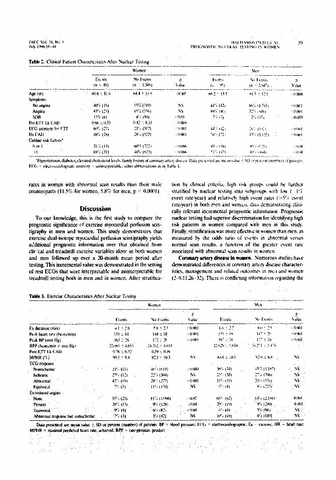
women included in the prognosis analysis are characterized in Table 1. The women were significantly older and presented with nonanginal symptoms or atypical angina and multiple cardiac risk factors more frequently than did the men, who more commonly presented without symptoms at the time of nuclear testing. The men had a more frequent history of previous myocardial infarction or cardiovascular intervention (cardiac catheterization, bypass surgery or percutaneous trans- luminal coronary angioplasty) and had a higher pre-ElT and prescan likelihood of coronary artery disease.
Onteome events. Among the 2,742 men and 1,394 women, a total of 95 events occurred during the follow-up period in men and 45 events in women. These include.d 63 myocardii infarctions and 32 cardii deaths in the men and 31 myocardial
JACC Vd ZX, No. I July I‘M&-4t
TaMe I. Patient Characlcristia --
Men Wcma’n P 01 r 2.142) (n 1.394) :&.
4s CY) 61.7 c I?.? 64.5 z I1.S 4Mcti Cardiac rbk factctrs
0 D’c ((135) :w (259) <O.UO4 I X’i (I.Oi6) 34’; (477) NS 2 267 (719) 31’2 (431) <Il.mlI
>2 14c: (372) 16p~ (227) NS Syaptoms
AS)mp:0lTK4C 43% (1.193) 2wi (N17) 4m1 NWallgiIt~l 217 (SY?) 27r, (3741 4lSY)I Alypic.4 angina 2lFi (554) ?!I? l.36) dl.IIt! Typical angina 1211 (WI) 14!‘1 (IYI) NS SOB 25 (62) 4’; (56) vs
lix MI 24’: (666J l4ci (IW) WUI lix cath 3Y4 (1 ,U70) Wi (271) -‘ll.Wl Hx PTCA 147 (3Yn) 6G (91) CWJOI Ha CABG 17°C (466) 6”; (lb) CO.WI Hx CAD 46% ( 1.267) zs’, (353) c OSXJI Prc-El-f L.k CAD 0.49 5 0.36 0.42 T 034 4001 Postscan lk CAD Lt.49 + U.42 0.41 t u.3li <MJOI
-.- Dara presented arc mean value f SD or peroeot (numh:r) of partents.
CABG = coronary artery bypass surgery; CAD = cormury artery disease, cath = cardiac catheterization: FIT = ex&(e test: Hx = histq of; U = likelihood or; Ml = myorardial infarction; PTCA = percutaac~, translluminal amnary angioplaq; SOB = shortnc?6 of hrcath.
infarctions and 14 cardiac deaths in the women. The overall event rate was 3.5% in the men and 3.2% in the women (p = NS). The censored revascularization rate (revascularization within the first 60 days after nuclear testing) was 7.5% in men (207 patients) and 4.5% in women (63 patients, p 1 O.P4).
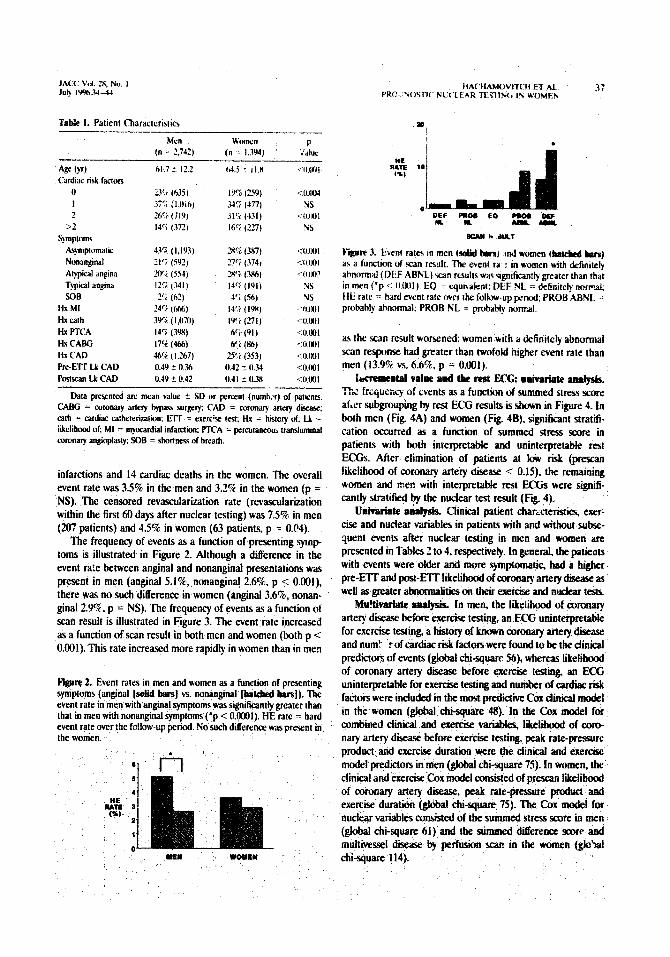
The frequency of events as a function of presenting symp tams is illustrated in Figure 2. Although a difference in the event ra:e between angina1 and nonanginal presentations was present in men (anginal 5.1%. nonanginal 2.6%, p i 0.001). there was no such difference in women (angina1 3.66, nonan- ginal 29%. p = NS). The frequency of events as a function of scan result is illustrated in Figure 3. The event rate increased as a function of scan result in both men and women (both p < 0.001). This rate increased more rapidly in women &an in men
Flgtwe 2. Event rates in men and women as a function of presentiag symptoms (anginal [solid biusl vs. nonanginal [bate&d bars]). The event rate in men with angina1 symptoms was significantly greater than that in men with nonanginal symptoms (‘p < O.ooOl). HE rate = hard event rate over the fc!!ow-up period. No such difference was present in the women.
IWkduLT
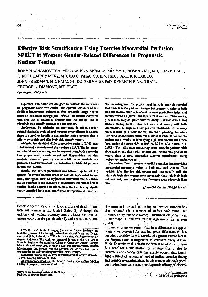
F@ue 3. Event rare5 in men (s&M bnra) WI women #Wekd &a) as a function of rcan result. The event ra : in women tith definitely abnormal (DEF ABNL) +can resul~c was signilicanrly greater lhan that in men (‘p c: ll.fMl). EQ = equnaknt: DEF NL = dcfinitefy ourmal; HE rate =: hard event rate ovct rk follow-up periud; PRO6 ABNL -= probably abnormal: PROB NL = probably normal.
as the SC~ZI result worsened: women with a definilely abnormal scan response had greater than twofoid higher event rate than men (139% vs. 6.6%. p = 0.001).
-~V~lPcolldtBorestECG:R~~~ The frqxrq of events as a function of summed stress score after subgrouping by rest ECG results is shown in Figure 4. in both men (Fig. 4A) and women (Fig. 48). significant stratifi- cation occuned as a function of summed stress score in patients with both interpretable and uninterpretable rest ECGs. After elimination of patients at low risk (presfan likelihood of coronary artery disease < 0.15). the remaining women and rzen with interpretable rest ECGs were signifi- cantly stratiksl by the nuclear test result (Fig. 4).
U&aria& anal@x. Clinical patient charzcteristii exer- cise and nuclear variables in patients with and without subse- quent events after nuclear testing in men and women are presented in Tables 2 to 4, respectively. In general, the patients with events were older and more symptomatic, had a higher pre-IZIT and post-ETT likelihood of coronary artery disease as wellasgreaterabmm&i&ontheirexer&e andnudcartesh
Mm!tivariate ramly&. In mea the likelihood of coromuy artery dii before exe&e test& an ECG uninterpreMe for exercise test& a history of known coronary artery disease and num! rof cardiac risk factors were found to be the diiical predictors of events (global &square 56), whereas likelihood of coronary artery disease before exercise testing, an ECG uninterpretable for exercise tedng and number of cardiac risk factors were included in tbe most ptedi&e Cox dinical model in the women (global chi-square 48). lo the Cox model for combined cliiical and exercise variab&, likeiibood of core- nary artery d&ase before exercise testing, peak rate-me prod~a and exe&e duration were the clinical and exercise model predictors in men (giobal &i-square 75). IO women, the clinical and exercise Cox model amsisted of prescan likelihond of coronary artery disease, peak rate-pressure product and exercise duration (global chi-square 75). The Cnx tnaiel for nuclearvari&escom&edofthesummedstressscoreinmen (global chi-square 61) and the summed difference mrc and multivessel dii by perfusii scan in the women (glo‘pi &-square 114).

I,\(‘( Vol. 28. tie. 1 July Iwm-44
FigoR 4. Event rates in men (A) ar,d v/u&n (8) subgrouped hy rest electrocardiogram interpretable for t&+,jil\ rating (IN’rERp ECG), uninterpretabie for treadmill testin& \&lh$‘PPP E@) and inter- pretable for treadmill testing but t&@& patiehts with a pr,+can likelii of coronary artery disek, 6u.l~ (&QEAp ECG: INT- HIGH LK CAD). Within each of tt\y:$ three shbgroups, ti bth men and women. there was a sign&ant d&rt@& k&J in the hard event rate over the follow-up period as a &Q&fl of the summed stress score. Solid bars = summed stress s$& tA (fiohllai); Ut&pd bfs = summed stress score 4 to 8 (mildly @t&t): @y bars s sumged stress ssore >8 (severely abnormal), g ‘: O,u5.
InCremW(al value. Car pro@&Pl /IQ&&. &i shown in Fi&e 5, when added to the cli&~ afitj exercise nlodel in the Cox proportional hazards analy@, P@leat data provided 17% additional prognostic informati& in the her aQd 37% in the women compared with clinical fold e%ercie va&!es alone (both p < 0.001). The gain in tot+’ &.~@te was IS in the mm (global chiqoarc 90) md 45 $ Q flflen (global chi-square 120).
&ph-Meier a~&. In t$ he4, Stfatificatioh of the stue cohort into low clinical bS\ \pfe&n lilt&h&d of coronary artery disease <O.lS, $ ti 97)) and high clihical risk (prescan likelihood of coronarJl $t& disease > 0.15, n = 1,765) resulted in populations I&, S@icaPtly di@ererlt event rates (low risk 1.0% vs. high risg !,A$, p * O.OO@, &-square 14) (Fig. 6A). The use of nuc& testing stratified @e high clinical risk group into a subgroup & horeal wtt re&s aud a low event rate (1.9% [ l.l%j)‘~# of fr&w-up]) afld sub- groups with significantly higher &fit rates With abnowal scan
results (svznt rate 6.2%. p <: O.OUJl, chi-square I9 vs. normal scan results) (Fig. KY). The low clinical risk group was also further stratified into a low event rate subgroup with normal scan rchults (0.2%’ evpnt rate) and a higher risk group with ahnormal scan results (event rate 4.6%, p = 0.00001. chi- square 20 vs. normal scan results and low clinical risk) (Fig. hB). In both low and high risk subgroups, tue use of scan findings resulted in a fivefold difference in event rates between normal and abnormal scan results.
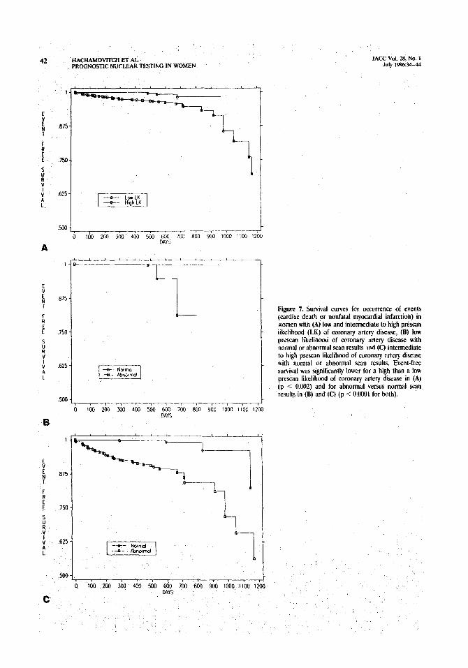
In rhe women, a qualitatively similar but quantitatively more impressive stratification was present. Yrescan likelihood of coronary artery disease separated the cohort into low clinical risk (n = 5S6, event rate 0.7%) and high clinical risk (n = 838, event rate 4.4%) subgroups, p < 0.01, chi-square 11) (Fig. 7A). The high clinical risk group was stratified into a subgroup with rI(jrmal scan rcsaltc and a low (0.8%) event rate and a high risk group with abnormai scan results (event rate i2.7%, p < 0.0001, chi-square 58 vs. normal scan results) (Fig. “C). Significant stAfication was noted in the low clinical risk women; a normal sc,m result identified a low risk subgroup (cvcnt rate 0.2%). whzreas an abnormal scan result identified 3 subgroup with a bigher event rate (4.3% [2 events in 46 patients], p < 0.0002. chi-square 14 vs. normal scan results) (Fig. 78). These analyses demonstrate a more clinically rele- vant incremental prognostic value of nuclear testing in men and women.
Comparis0n of discrimiaatioa Of nuclear testing in men aad women. To directly compare the relative discrimination of nuclear testing in men versus women with respect to identify- ing high risk subjects, we compared the areas under the ROC curves to compare the discrimination for predicting events using the summed stress score. The area under the curve in women (0.84 i: 0.03) was significantly greater than that for men (0.71 it 0.03, p < 0.0005 vs. women). This finding demonstrates that nuclear testing is better able to identify women at high risk of future events than men independently of baseline event rates, diagnostic thresholds or selection bias.
Test effectiveness. Nuclear testing also risk stratified women more effectively than men (odds ratio [OR] for an event with abnormal vs. normal Scan results: men 4.4, women 22.8, Mantel-Haenszel OR 6.8,95% confidence interval ([Cl] 4.7 to 9.7, chi-square 109, p < O.OOOl). This significant difference in stratification effectiveness was present between men and women in all prescan likelihood categories, demon- strating that this effectiveness was independent of underlying patient characteristics and ECG exercise test results (Mantel- Naenszel OR 5.1,95% Cl 2.2 to Il.9 for low [<0.15] prescan likelihood of coronaty artery disease; OR 8.0, (95% ‘CI 4.2 to IS.4 for intermediate [0.15 to 0.851 prescan likelihood of coronary artery disease; OR 3.6, (95% Cl 1.9 to 6.9 for high [ >0.85] prescan ‘likelihood of coronary artery disease). Thus, although the results of nuclear testing risk stratified both meti and women, the resultant stratification was more effective in women-similar low event rates in both male and female patients with normal 5can results but significantly higher event
JACC Vol. 2x, No I July l’i%24-44
rates in women with abnormal scan results than their male counterparts (1 IX, for women. 5.86 for men, p c WtX~l ).
Discussion To our knowledge, this is the first study to compare the
prognostic significance of exercise myocardial perfusion xin- tigraphy in men and women. This study demonstrates that exercise dual-isotope myocardial perfusion scintigraphy yields additional prognostic information over that obtained from clir ‘Cal and treadmill exercise variables alonr in h>th women and men followed up over a 20-month mean period after testing. This incremental value was demonstrated in the setting of rest EC& that were interpretable and uninterpretable for treadmill testing both in men and in women. After stratitica-
Tabk 3. Exercise Char&&tics After Nuclear Testing
tion by clinical criteria. high ri4 group could bc funhcr stratified by nuclear te~lrng into &group\ nith Itn\ ( -’ l ’ i event r;lleIycar) and relatitrly high event rates IWi cvcnt rate/year) in both pen and womcu, ihtis dcmonstra:ing &i- tally relevant incremental prognostic informalton. Prognostic nuclear testing had superior discrimination for identi$inp high risk patients in women compared with men in thn srudy. Finally, stratification was mDre effective in women than men. as measured b the odds ratio of events in abnormal versus normal scan results. a function of the greater event rate associated with ahnorma! ran results in uomen.
Corcmary &ery diise in women. Numsrou5 studifs hare demonstrated differences in coronary anery dise;LI(: character- istics. management and related outcome< in melI and women (2-9.11.26-32). There i+ conflicting informauon iegarding the

40 HAcHAMovITcH ET AL PROGNOSTIC NUCLEAR WSTING IN WOMEN
TOWC 4. Nuclear Variables After Nudear Testing’ WOIXII
Events No Even& ss 112 + 8.4 2.1 5 5.5 SDS 8.9 z 8.1 1.9 i: 3.4 SRS 22 f 4.0 0.8 + 3.1 -defects 0.9 + 0.7 0.3 ?r 12 RevenrMedefeus 3.5 ir 3.0 0.6 + 1.6 r!mubeddaease 82% (37) 5% w NOItdsfaoILWlS 13% (6) 7% (1.0%)
14.1 2 11.1 6.5 c 8.9 7.9 lr 7.9 4.2 t 6.0 6.0 + 8.3 2.?%57 22 2 2.9 03 c 2.0 3.2 + 3.2 I.6 + 2s 39% (37) 14% (38d) 21% (20) 54% (1.4?7)
*p < 0.001 for all compariums Data presented are mean ~+Iue 2 SD or percent (number of patients). SDS = summed diierence SCI -c: SRS = summed rest score; SSS = summed stress scare.
accuracy of noninvasive methods in identifying coronary artery disease in women. Although we previously showed (33) that the diagnostic accuracy of perfusion scintigraphy is similar in women and men, technical dificulties in the interpretation of scans in women has led to a perception of reduced diagnostic accmmy in this group.
Risk stra~bisn in women by nnainvasive testing. Studies assessing the prognostic value of noninvasive testing in women have been limited. Data from the Coronary Artery Surgery Study indicate that exercise ECG testing was able to risk strati@ both men and women (34). Recently, Panchoiy et al. (35) found that nuclear testing was prognostically predictive in women and added incremental prognostic information over cl&al and exercise variables in a catheterized population but did not compare their prognosis with a male group. The present study also demonstrates the incremental prognostic value of m&ear testing in women and extends these findings by demon8trating that nuclear testing has superior effectiveness and greater discrimination for the identification of high risk patients in women than in men.
Greater eatdine risk in wemen. In our current study, although men and women had similar low event rates after a normal scan, women had a higher event rate than men after an abnormal nuclear scan, explaining, in part the statistically superior prognostic performance of nuclear testing in women. Previous reports have also shown that women with known coronary artery dii, myocardial infarction or coronary artery bypass surgery are at higher risk of an adverse outcome than their male counterparts (B-32). Shaw et al. (6) also found that women referred for noninvasive testing had a markedly greater rate of myocardial infarction or death than men (69% vs. 2.4%, p < 0.002). Several hypotheses have been advanced to account for these gender-related mortality differ- ences. Surgical mortality increases with decreased patient heiit and lower coronary artery lumen diameter, both char- aeteristics associated with women (36), possibly predisposing women to lowed thresh&s for coronary occh&n by acute tbrombw Potentially, delayed referral or underreferral of women to intervention may also play a role in mortality and morbidity Merences. However, we previously demonstrated
150
I
YEW
JACC Vol. 28. No. I July iY%34-44
r
WOMEN
Fire 5. Resulrs of determination of incremental prognostic value using the Cox proportional hazards model in men and women for the three mud&i tested (clinical variables [s&l aarS], clinical *us exercise variables [btcbd bars], clinii plus exercise plus nuclear variables [open bars]). Tbe chbsquare of the model including all variables was significantly greater than that for clinical plus exercise variables in both men ani women. *p < O.ooO1.
(11) that after consideration of the extent and severity of stress perfusion abnormalities, no referral bias to catheterization or revascularization was present between men and women after nuclear testing. In fact, a greater rate of referral to catheter- izztion was present in women with severe ischemia than in their male counterparts (11). The greater risk of adverse outcome in women with coronary artery disease and the difficulties associated with identification of high risk women on clinical grounds emphasize the need to identify a noninvasiv-, testing modality to identify women at high risk of future events. The results of the current study suggest that this need car? be met by nuclear testing in appropriate patient subsets.
clinical implications for risk stratificatino. Although the Cox proportional hazards analysis documented the increlran- tal value of nuclear testing in the overall patient population, the Kaplan-Meier analysis extended these findings by demon- strating the ability of nuclear testing to further stratify the patients in both low and high clinical risk groups. The us of nuclear testing in patients with a low prescan likelihood of coronary artery drsease is questionable because of their low overall risk (37). However, the re&s of this analysis in the population of patients w:th an intermediate to high pre;can likelihood of coronary artery disease is of clear importance because of their overall intermediate risk, indicating the need for further risk stratification.
When the two genders were compared directly, women were stratified significantly more effectively than men in the current study. Because proportionally fewer women than men had abnormal scan results and were thus categorized as high risk after nuclear testing, fewer women would require referral to further testing (e.g., catheterization). Thus, a clinical strat- egy incorporating nuclear testing may be less costly in women than men. However, this possibility requires further investiga- tion and should be evaluated in future trials.
IncresaewtoI v&w as a hwtion of rest ECG. Two recent studii have demonstrated that the incremental diagnostic value of nuclrar resting for identifying severe coronary artery disease in patients with normal rest ECG findings is too small

JAW ‘,‘.,I 3. No I July l%:.M-44
Fll 6. Survival curves for occurrence of events 5 icardiac death or nonfatal myocardiil infarction) in pen with (A) low and intermediate to high prcscan
F
.ikelihod (WI) of coronary artery disease, (B) k~ :
prescan likelihocd of coronary artery with normal or E
abnormal scan results and (C) intermediate to high prescan likelii of coronary artery disease with
:
normal or abnormal scan resutts Event-free survival t
was si@ican$ lower for a high than a low prescap likelihood of mronary artery disease in (A) (p c 0 KJI )
i i
and for abnormal versus normal scan results ;B) and (C) (p < 0.005 for both).
1
075
7%
.625 i
.625
HACHAMOVITCH ET AL PROGNOSTIC NUCLEAR TESTING IN WOMEN
JACC Vol. 28, No. 1 July 19963444
0 100 2011 MO 400 500 6M) 700 803 900 1000 1100 1200 DAYS
Fiim 7. Survival curves for occurrence of events (cardiac death or nonfatal myocardial infarction) in +vomen with (A) low and intermediate to high prescan iikelihwd (1.K) of coronary artery disease, (8) low prexan likelihood of coronary artery disease with normal or abnormal scan results ?nd (C) intermediate to high prescan likelihood of coronary ztery disease with normal or abnormal scan results. Event-free survival was significantly lower for a high than a low prescan likelihood of coronary artery disease in (A) (p < 0.002) and for abnormal versus normal scan results in (B) and (C) (p <: 0.0001 for both).
0 100 200 m 4M MO 600 700 800 900 moo 1100 1200 DAYS

JACC Vol. 28. No. 1 July I‘M%-44
to justify its use (38,39). We previously demonstrated (37) that when an optimized noninvasive strategy is used in sele&ng patients for testing, sestamibi imaging sign&candy enhances both risk stratification and reduces the cost of the testing strategy in patients with normal rest ECG findings. The current study demonstrates that significant risk stratification is achieved by nuclear testing in both men and women with rest ECGs that are interpretable fol exercise testing. This finding was present even after exclusion of patients with a low prescan likelihood of coronary artery disease who may not have required nuclear testing (Fig. 4).
Study limitations. Txhnical. Scintigraphic studies in the present study were assessed by experienced observers using a standardized, semiquantitative approach to visual interpreta- tion that we previously developed (14) and documented to be highly reproducible (15). Nonetheless, the subjective nature of this analysis and its dependence on the expertise of the observer present a limitation with respect to the extrapolation of our results to those of other centers that would have been avoided by the use of quantitative methods for analysis of technetium%m myocardial perfusion SPECT studies (40). These programs correlate highly with both visual Scan assess- ment and coronary angiography (41). At the time of collection of the SPECT studies in this patient population, we did not have a quantitative analysis technique in operation on all of our camera/computer systems. Prognostic studies using quan- titz’ive ana$sis would be of interest.
The results of the present study may not be generalizab!e to myocardial perfusion imaging performed in women with stress-rest thallium protocols because the ability to assess stress-induced lung uptake of thallium, a powerful prognostic variable, is not possible with stress sestamibi protocols. How- ever, the use of sestamibi in women may be advantageous because of its improved image quality and potentially reduced attenuation artifacts frequently found in women (42).
Patient cohort. Our patient population is taken from a group referred to exercise myocardial perfusion imaging for both prognostic and diagnostic testing, and we cannot exclude the possibility of bias introduced by way of this referral. The patients are nevertheless typical of those referred to a com- munity hospital (university affiliated) in a major urban area, and the results of the present study should be applicable to this setting.
StatisticaL The use of the Cox proportional hazards model is limited by the number of events accumulated during the follow-up period. The iow loss to follow-up rate, the large patient group used and the adequate number of events favor the accuracy of our multivariate results (20).
Conelusions. The results of the present study demonstrate that nuclear stress perfusion imaging is an effective noninvasive means to risk stratify women into patient subgroups who are at low or relatively high #risk of future cardiac events irrespective of rest ECG findings. In light of previous work demonstrating a gender-related referral bias in the diagnosis and treatment of cardiac disease, as well as gender-related diierences in cardiac risk, this modality can play an important role in the assessment,
HACHAMOVITCH ET AL. 43 PROGNOSllC NLTLEAR TESTING IN WOMEN
and perhaps guide clinical management. of coronary artery of ischemic heart disease in women.
Appendix
Cm Proportional Hazards Model In performing Cox proportional hazards analysis we limited the
number of variables entered into any model to I per 10 events of interest to avoid overfitting (IO). The variables initially considered for an@& inch&d clinical (all those listed in Table 1 as well as the rest ECG and the presence of individual cardiac risk factors); exercise (all those listed in Table 3 as well as Mw! pressure response to exercise and piescar. likc%od of awnary disease); and nuclear (all those listed in Table 4 as well a< the presence of transient ischemic dilation of the left ventricle) vafables.
The selection of the variables for entry into the muitiwiate models detailed here was based on t’le results of univariate anal+ Variables were examined for colinearity, and the proportional hazards assump- tion was tested For men the particular variables entered into the models included 1) c!M&pre-exercise treadmill testing (EIT) likelihood of coronary artery disease, age, number of cardiac risk factors and presenting symptoms; 2) ewcise-prescan likelihood of coronary artery disease, clinical response to exercise, exercise duration. rate-pressure product and ECG reywtse to exercise; 3) dinical p/w ewcire-pre-EIT likelihood of coronary artery disease, uninterpret- able rest ECG, coronary artery disease, number of cardiac risk factors, prescan likelihood of coronary artery disease, rate-pressure product and exercise duration; 4) rurckar-multivesse! disease by scan. summed strffs score and summed difference score: and 5) cliniclrl phs txercise v.urablps forced in, with nuclear variables added- uninterpretable root EC%. coronary artery disease and number of cardiac risk factors. exercise duration, rate-pressure product forced in and the summed stress score added Those for w01nen inch&d 1) clinira-pre-ETT likelihood of coronary artery disease. history of coronary artery disease. number of cardiac risk factors. age, uninter- pretabie rest ECG for exercise testing 2) aerciw-prescan likelihood of coronary artcry disease, rate-pressure product. exercise dwation, clinical response to exercise and ECG response to exercise: 3) clincul pita erercise-pre-ElT likelihood of coronary artery disease, titer- pretabie rest ECG. number ot cardiac risk factors exercise duration and rate-p- produd; 4) nucZear--multives?el disease by ran summed stress score and summed ditference score; 5) &I&/ $us aercise variablm forced in Gth nuclear variables added-exercise duration. prescan iiielihood of cr;onq artery diiase. rate-pressure product foroed in and the summed difference score and muhiwsel disease by scan added.
Refepms 1. Faker ED. C&sebnm JH, Sacks FM. Wenger NK. Whimant JP, Wii M.
American Heart Asaciah Mediitific Statemmr on Cutiascu- tar lhxse in women. CiicuktiIm 1993388:1999-m.
2. Destafano F, Metitr RK. AMa RF, Casper ML E&r ED. Trends in nonfatalwmnaryheartd&seintheUnitedStacesA3chlotemMed I!ml53289-94.
3. T& JN. Waswtbsil-Smoltcr S. Wexler JP. Steia& RM, Budner N, ler6eLSeXbiBincon5ideringuxowybypagsurgeiy.Annbue!nMcd 1o8f;107:19-25.

44 HACHAMOVITCH ET AL PROGNOSTlC NUCLEAR TESTING IN WOMEN
mortality of women in corooaty artev bypass surgery: evidence for referral bias Ann intern Med 1990;112:561-7.
5. Krmnholz HM, Douglas PS, Laoer MS, Pastemak RC. Selection of patients for coronary angiography and coronary revaau!arization early after myocar. diil infrmiion: is there evidence for a gender bias? Ann Intern Med 1992;116:785-90.
6. Shaw LJ, Miller DD, Romeis JC, Xxgl D. Younis LT, Chaitman BR. Gender di5erences in the noninvasive evaluation and management ol patients with suspected coronary artery disease. Ann intern Med 1994;120: 559-66.
7. Ayaaian J& Epstein AM. Differences in the use of procedures between men andwomen hospitalized for coronary heart disease. N Engl J Med I991:3?Z: 221-5.
8. Steingart RM, Packer M, Ham” P. et al. Sex dikrencxs in the management of coronary artery disease. N Engl J Med lY91;325:226-u).
9. Bickell NA, Piepcr KS, Lee K1 et al. Referral pa!tcms for coronary artery disease treatment: gender bias or good clinical judgment? Ann Intern Med 1992;116:791-7.
10. Mark DB, Shaw LK. DcLong DR. Califf RM, Pryor DB. Abscncc of sex bias in the referral of patients for cardiac catheterization. N Engl J Med 1YY4;33lk1101-6.
11. Hachamovitch R, Berman DS, Kiat H, et al. Gender-related dilferences in clinical management after exercise nuclear testing. J Am Coil Cardiol lYY5;2fc1457-64.
12. Staniloff HIM, Forrcster IS, Berman DS, Swan HJC. Prediction of death, myocardial infarction, and worsening chest pain using thallium scintigraphy and exercise electmcardiography. J Nucl Med 198h:27:1842-8.
13. Ladeoheim ML Pollock BH, Rozanski A, et al. Extent and severity of myocardial mrfusion as predictors of prognosis in patients with sus- pected coronary artery discax 3 Am Coil Cardiol lY86;7:464-71.
14. Berman DS, Kiat H, Friedman JD. et 41. Separate acquisition rest thallium- 2Olktress technetium Y?hn sestamibi d&isotope myocardial perfusion single-photon emission computed towphy: a clinical validation study. J Am Coil Cardiol lY9%2?:1455-64.
IS. Berman DS, Kiat H, Van Train K. Tc-9Ym sestamihi imaging in the assessment of chronic coronary artery disease. Semin Nucl Med 1991:21: 190-212.
16. Diamond GA, Staniloff HM, Forrester JS. Pollock BH, Swan HJC. Com- puter assisted diagnosis in the noninvasive evaluation of patients with suspeaed coronary artery disease. J Am Coil Cardiol 1983;1:444-55.
17. Staniloff HM, Diamond GA, Pollock BH. Probabilistic diagnosis and prog- nosis of coronary artery disease. J Cardiac Rehabil 1984;4:518-29.
18. Cox DR. Regression models and life-tables. J R Stat Sot (B) lY7?;34:187- 202.
19. Diion WI, editor. BMDP Statistical Software Manual. Berkeley (CA): Univrrsity of California Press. 1992.
20. Kaplal EL, M&r P. Nonparametric estimation from incomplete obseerva- tions. J Am Stat Assoc lY58;53:457-81.
21. Diamond GA. ROC steady: a receiver operator characteristic curve that is invariate relative to selection bias. bled Decis Making 1987;7:238-43.
22. McNeil BJ, Hanley JA. Statistical approaches to the analysis of receiver operator characteristic (RQC) curves. Med Dccis Making 19f$4:137-50
23. Hanley JA, McNeil BJ. The meaning and use of the area under a receibcr oper..tor characteristic (ROC) curve. Radiology 1982;143:29-36.
24. Diamond GA. Future imperfed: the limitations of clinical prediction mod& and the limits of clinical prediction. J Am Coil Cardiol 1989;14:I?A-??A.
25. Kotler T, Diamond GA. Exercise thallium-201 scintigraphy in the diagnosis
JACC Vol. 28, No. I July I’%%-44
and prognosis of coronary artery disease. Aon l&m Med 1990;113:684- 702.
26. Kennedy JW, Killip R, Fisher LD. The clinical spectrum of coronary artery d&case and its surgical and medical management, 1974-1979. The Coronary Artery Surgery Study. Circulation 198266 Suppl III:lH-16-23.
27. The principle investigators of CASS and their associates: the National Heart, Lung. and Blood Institute Coronary Artery Surgery Study (CA%). Ciila- tion lY81;63 Suppl 1:1-l-81.
28. Lemer DJ. Kannel WB. Patterns of coronary heart disease morbidity and mortality in the sexes: a ‘%-year follow-up of the Framingham population. Am Heart J 19R6:11l:3&90.
29. Tofler GH, Stone PH. Muller JE. Forman S, Solomon DE, Braunwald E. Effects of gender and race on prognosis after myocxdial infarctiotr adverse prognosis for women, particularly black women. J Am Co11 Cardiol 148x9: 473-82.
30. Dittrich H, Gilpin E. Nicod P, Cali G. Henning H, Ross J Jr. Acote myocardial infarction in women: intlucrlces uf gender on mortality and prognostic variables. Am J Cardiol 1988$2:1-7.
31. Greenland P. Reicher-Ross H, Goldbourt U. LI al. In-hospital and I year mortalitv in 1,524 women after myocardial infarction: comparison with4315 men. Ci&dation 1991;83:484-Yl.
32. Becker RC, Terrin M, Ross R, Knatterod GL, Gore JM, Braunwaid E. Comparison of clinical outcomes for women and men after acute Ml. Ano Intern Mcd 1993:120:638-45.
33. Van Train K. Maddahi J. Berman DS, et al. Quantitative analysis of tomographic stress thallium-201 myocardial scintigrams: a multicenter trial. J Nucl Med 1990;31:1168-79.
3J. Wiener DA, Ryan TJ, Parsons L, et al. Long term prognostic value of exerciw testing in men and women from the coronary artery surgery study registry. Am J Cardiol 199%75:865-70.
35 Pancholy SB. Fattah AA, Kamal AM, Ghods M, Heo I, Mcandrian AS. Independent and incremental prognostic value of exercise thallium single- photon emission computed tomographic imaging in women. J Nod Cardid 1995:?:110-6.
36. Fisher LD, Kennedy JW. Davis KB. et al. kuociation of sex, physical size and operative mortality after corona? artery bypass in the corvnaly artery surgery study (CASS). J Thorac Cardlovasc Surg 1%2;84:334-41.
37. Brrman DS, Hachrmovitch R, Kiat H, et al. Incremental prognostic value and cost implications of normal and equivocal exercise Tc-YYm se&m&i myocardial perfusion SPECT. J Am Coil Cardiil lY95:2663Y-47.
38. Gibbons RI, Zinsmeister AR, Miller TD. Clements 1P. Supine exercise electrocardiography compared with exercise radionoclide angiography in noninvasive identification of severe coronary artery disease. Ann Intern Med 1990;112:74~Y,
39. Christian TF, Miller TD, Bailey KR. Gibbons RJ. Exercise tomographic n-201 imaging in patients with coronary artely disease and normal EC% Ann Intern Med 1994;121:825-32.
40. Garcia EV, Cooke D, Van Train KF, et al. Technical aspects of myccardkd SPECT imaging wrth technetium-YYm sestainibi. Am J Cardiol lY90:66:23E- 3lE.
41. Van Train K, Garcia EV, Maddahi J, et al. Multicenter trial valiitioa for quantitative analysis of same-day rest-stress technetium Wm-sestamibi myo- cardial tomograms. 3 Nucl Med lY94;35:609-18.
42. Taillefer R. DePuey GE, Udelsen J. et al. Comparison of thallium2Ol and technetium 99” sestamihi (perfusion and gated SPECT) myocardial perfu- sion imaging in detection of coronary artery disease in women [abstract]. Circulation lY9592 Suppl f&129.