effectiveness of antimicrobial peptide …aac.asm.org/content/58/9/5229.full.pdfeffectiveness of...
TRANSCRIPT

Effectiveness of Antimicrobial Peptide Immobilization for PreventingPerioperative Cornea Implant-Associated Bacterial Infection
Xiao Wei Tan,a Tze Wei Goh,a P. Saraswathi,b Chan Lwin Nyein,a Melina Setiawan,a Andri Riau,a R. Lakshminarayanan,b,e
Shouping Liu,b,e Donald Tan,c,d,e Roger W. Beuerman,b,c,f Jodhbir S. Mehtaa,b,d,e
Tissue Engineering and Stem Cell Group, Singapore Eye Research Institute, Singaporea; Antimicrobials Group, Singapore Eye Research Institute, Singaporeb; Yong Loo LinSchool of Medicine, National University of Singapore, Singaporec; Singapore National Eye Centre, Singapored; Duke-NUS Graduate Medical School, Singaporee; Duke-NUSSRP Neuroscience and Behavioral Disorders, Singaporef
Titanium (Ti) is a promising candidate biomaterial for an artificial corneal skirt. Antimicrobial peptide (AMP) immobili-zation may improve the bactericidal effect of the Ti substrate. In this study, we tested the bactericidal efficacy of a func-tionalized Ti surface in a rabbit keratitis model. A corneal stromal pocket was created by a femtosecond laser. The Ti filmswere then inserted into the pocket, and Staphylococcus aureus or Pseudomonas aeruginosa was inoculated into the pocketabove the implant films. The corneas with Ti-AMP implants were compared with the corneas implanted with unprotected Ti byslit lamp observation and anterior segment optical coherence tomography (AS-OCT). Inflammatory responses were evaluated bybacterium counting, hematoxylin-eosin staining, and immunostaining. There was a lower incidence and a lesser extent of infec-tion on rabbit corneas with Ti-AMP implants than on those with unprotected Ti implants. The bactericidal effect of AMP againstS. aureus was comparable to that of postoperative prophylactic antibiotic treatment; hence, SESB2V AMP bound to the Ti im-plant provided functional activity in vivo, but its efficacy was greater against S. aureus than against P. aeruginosa. This worksuggests that SESB2V AMP can be successfully functionalized in a rabbit keratitis model to prevent perioperative cornealinfection.
Infection or biofilm formation on the surface of implants follow-ing the implantation of surgical devices is a clinically devastating
complication that accounts for about 60% of all hospital-associ-ated infections (1). Devices that penetrate the skin or those thatare within tissues, e.g., the cornea, present the highest risks forinfection (2, 3). Endophthalmitis, a severe panocular infection,remains one of the most devastating complications after the im-plantation of any artificial corneal device, e.g., intracorneal stro-mal rings to correct refractive errors (4, 5), Boston keratoprosthe-sis (6–8), and osteo-odonto keratoprosthesis (OOKP) (9, 10) usedto restore vision in patients with end-stage corneal blindness. Pa-tients with endophthalmitis present with discharge, lid swelling,pain, reduced vision acuity, implant erosion, or exposure (11).Infected patients normally receive systemic or local antibiotic/antifungal treatment once their infection has been diagnosed, andimplant removal is sometimes required. Implant-related infec-tions are generally difficult to manage, result in an adverse effecton patient quality of life, require prolonged hospital admission,and culminate in a higher final cost to the patient and the healthcare provider (12, 13). Currently, there are limited strategies forpreventing infections associated with surgical implants. Hence,clinically applicable and effective strategies are urgently needed toaddress this issue.
Both systemic and locally applied antibiotic agents have beenused to prevent infections associated with surgical implants. An-timicrobial agents applied locally usually provide higher drug lev-els on the surface of devices and the immediate vicinity than doessystemic administration (1). Various approaches to local prophy-laxis have been tried to prevent bacterial colonization, includingantimicrobial irrigation of the surgical field (such as sterilizing theocular surface with povidone-iodine before ophthalmic surgery[14]), placement of antimicrobial carriers (e.g., intraoperative in-sertion of antibiotic-treated prosthetic devices in orthopedics
[15]), dipping implants into antimicrobial solutions (e.g., immer-sion of a heart valve prosthesis into antibiotic solution before im-plantation [16]), and coating surgical implants with antimicrobi-als (17). Among these methods, coating implants withantimicrobials provides the unique advantage of potentially deliv-ering the antimicrobial drug in a controllable manner via condi-tioned biodegradation of chemical bonds that cross-link the im-plant surface with the antimicrobial drugs. Clinical trials with theuse of Silzone-coated St. Jude medical valves (18) or the use ofsilver-coated pins in orthopedics have proven that antimicrobialcoatings are effective (19).
In previous studies, we showed that titanium (Ti) is a prospec-tive biomaterial for synthetic osteo-odonto keratoprosthesis(OOKP) skirts, as it shows excellent biocompatibility (20) and Tiis stable in inhospitable environments often seen in ocular micro-bial infections (21, 22). We (23) and others (24, 25) previouslyshowed the effectiveness of functionalized Ti with immobilizedantimicrobial coatings in vitro. Compared with other antimicro-bial agents, such as antibiotics, antimicrobial peptides (AMPs)have a broad spectrum of activity and possess a low propensity fordeveloping pathogen resistance (26). Antimicrobial peptides can
Received 24 March 2014 Returned for modification 2 May 2014Accepted 15 June 2014
Published ahead of print 23 June 2014
Address correspondence to Jodhbir S. Mehta, [email protected].
Supplemental material for this article may be found at http://dx.doi.org/10.1128/AAC.02859-14.
Copyright © 2014, American Society for Microbiology. All Rights Reserved.
doi:10.1128/AAC.02859-14
The authors have paid a fee to allow immediate free access to this article.
September 2014 Volume 58 Number 9 Antimicrobial Agents and Chemotherapy p. 5229 –5238 aac.asm.org 5229
on June 25, 2018 by guesthttp://aac.asm
.org/D
ownloaded from

be effectively immobilized on Ti substrates by the use of a phos-phate lipid spray or cross-linking agent, e.g., sinalization or poly-ethylene glycol (PEG) (27, 28). Despite several publications on thein vitro efficacy of AMPs (23–25), there are few studies demon-strating their efficacy in vivo (29). In the current study, we exam-ined the effectiveness of a functionalized implant in a rabbit ker-atitis model for its potential application as an artificial cornea skirtto prevent perioperative corneal infection. The coating of a syn-thetic Ti OOKP with AMPs may improve the biocompatibilityand bactericidal effect of an artificial corneal device for this groupof patients with end-stage corneal diseases.
MATERIALS AND METHODSMIC of soluble SESB2V. MICs were obtained in Mueller-Hinton broth(MHB) using the broth macrodilution method following CLSI guide-lines (30). SESB2V peptide was dissolved in water to make 1,000-�g/ml stock solutions. Serial 2-fold dilutions of the peptide were pre-pared in cation-adjusted MHB in test tubes. The bacterial suspensionwas added in each tube to maintain a final concentration of approxi-mately 5 � 105 CFU/ml. The tubes were then incubated at 35°C for 20to 22 h. The bacterial growth was checked in all the test tubes andtabulated for each concentration as “growth” or “no growth.” TheMIC of the peptide was defined as the lowest concentration that pre-vented the visible growth of bacteria.
Preparation of substrates and SESB2V peptide coating. The SESB2Vand truncated peptides were purchased from EZBiolab Inc. (Carmel,IN). The Ti substrate and AMP coating were prepared as previouslydescribed (23). Briefly, Ti substrates were cleaned ultrasonically for 10min each in dichloromethane, acetone, and water and placed in HNO3
(40%) for 40 min for surface passivation. Acid-treated titanium sub-strates were washed with ultrapure water before further treatment. Forsurface polydopamine (PDOP) coating, the titanium substrates wereimmersed in a dopamine hydrochloride solution (2 mg/ml) that wasdissolved in Tris buffer (10 mmol, pH 8.5) for 12 h in darkness. ThePDOP-coated substrates were then coated with the SESB2V solution(500 �g/ml) on both surfaces. After being briefly washed with phos-phate-buffered saline (PBS), modified substrates were stored at 4°Cprior to further analysis. Thin (0.005-mm-thickness) Ti films (Good-fellow, Cambridge, United Kingdom) were used for in vitro peptidestability testing and in vivo studies. For in vitro peptide stability testing,fluorescein isothiocyanate (FITC)-conjugated SESB2V peptide wasimmobilized onto the Ti surface. The functionalized substrate filmswere then soaked in PBS and kept in the dark. Fluorescence imageswere captured by a camera attached to a microscope (Axioplan 4.4;Carl Zeiss, Gottingen, Germany).
Bactericidal activities of SESB2V peptide. The bactericidal functionof SESB2V was analyzed with a Live/Dead BacLight bacterial viability kit(Molecular Probes, Carlsbad, CA). Staphylococcus aureus (ATCC 29213)and Pseudomonas aeruginosa (ATCC 9027) were used for the bactericidalassays. Yeast extract-dextrose broth containing peptone (10 g/liter), beefextract (8 g/liter), sodium chloride (5 g/liter), glucose (5 g/liter), and yeastextract (3 g/liter) was used as the growth medium. The bacteria wereincubated overnight at 37°C with agitation and then washed and resus-pended in phosphate-buffered saline (PBS) at an optical density at 600 nm(OD600) of 1.5. A bacterial suspension (500 �l) was then added to eachsubstrate in a 24-well plate and incubated for 30 min at 37°C. The sub-strates (n � 3 for each) were then rinsed with PBS to remove nonadherentbacteria before analyzing with the protocol provided by the manufacturer.Images of live/dead bacteria were taken with a fluorescence microscope(Carl Zeiss), and images of six areas were randomly captured. The num-bers of live/dead bacteria in each image were counted in three replicates.
Animal surgery. The animal protocol of the study adhered to theStatement for Use of Animals in Ophthalmic Vision and Research by theAssociation for Research in Vision and Ophthalmology. The protocol was
approved by the Institutional Animal Care and Use Committee and Insti-tutional Biosafety Committee at the Singapore Eye Research Institute.
Thirty-six New Zealand White rabbits, aged 1 to 2 months and weigh-ing 2 to 2.5 kg, were used in the study. The animals were anesthetizedusing an intramuscular injection of ketamine hydrochloride (35 mg/kg ofbody weight; Parnell Laboratories, Alexandria, Australia) and xylazinehydrochloride (5 mg/kg; Troy Laboratories, Smithfield, Australia). Theright eye of each rabbit was chosen for surgery. The contralateral eye of theimplanted rabbit served as the control. Following anesthesia, a 7-mm-diameter and 75% deep corneal stromal pocket was created using aVisuMax femtosecond laser machine (Carl Zeiss Meditec, Jena, Ger-many). A 5-mm-wide circumferential incision was made by a guardeddiamond knife (Storz; Bausch and Lomb). While forceps gently held theanterior flap in place, a Siebel spatula was inserted to separate the corneaalong the laser-created pocket. A 4-mm-diameter implant film was theninserted into the pocket. Following insertion, the incision was then closedwith interrupted 10/0 nylon sutures (see Fig. S1A to C in the supplementalmaterial).
All the rabbits received topical medications immediately after implan-tation, including 0.3% tobramycin (Tobrex; Alcon Laboratory, Inc., FortWorth, TX) and 1% prednisolone acetate (Pred Forte; Allergan, Irvine,CA). At 1 week postoperation, a 50-�l bacterial solution (2 � 104 CFU/ml) was inoculated into the corneal pocket above the titanium discs witha syringe attached to a 29-gauge needle (Fig. 1D). For rabbits with corneaswith pristine Ti implants (n � 21), one group was sacrificed at day 2 afterinoculation of S. aureus (n � 9) or P. aeruginosa (n � 6), and the othergroup (n � 6) received antibiotic eye drops immediately after S. aureus(n � 3) or P. aeruginosa (n � 3) inoculation. The antibiotics used were0.3% gatifloxacin eye drops (Zymar) (one drop every 2 h) and an antibi-otic solution (100 �l of 330 mg/ml cefazolin and 100 �l of 40 mg/mlgentamicin) injected subconjunctivally once a day. The group of rabbitsthat received the antibiotic solution (n � 6) were sacrificed after treatmentwith antibiotics continuously for 2 days. The rabbits with corneas withTi-AMP implants (n � 15) were also divided into 2 groups. One group ofrabbits was followed until day 2 after inoculation of the bacteria (n � 6 forS. aureus and n � 3 for P. aeruginosa), and another group of rabbits (n �3 for S. aureus and n � 3 for P. aeruginosa) was followed until day 7postinoculation. The study design is pictured in Fig. 1.
Slit lamp imaging and anterior segment optical coherence tomogra-phy. Slit lamp photographs and anterior segment optical coherence to-mograms (AS-OCT) were taken on the days before and after Ti implan-tation, the day before bacterial inoculation, and days 1, 2, and 7 afterbacterial inoculation. Slit lamp photographs were taken with a Zoom slitlamp NS-2D (Righton, Tokyo, Japan). Postinfection corneal slit lampimages were graded by a scoring method reported by Johnson et al. (31).Briefly, the infection grade (0 to 4) is based on the evaluation of thefollowing infection symptoms: none to severe conjunctival infection,none to severe chemosis, none to severe iritis (cell and flare), none tosevere fibrin formation, the area of hypopyon formation, the area of stro-mal infiltration (percentage of stroma with white cell infiltrate), and thearea of stromal edema (percentage of stroma which is swollen). Cornealcross-sectional visualization was performed with a Visante AS-OCT (CarlZeiss). Corneal thicknesses were measured using the software provided bythe manufacturer.
Quantification of viable bacteria from infected rabbit corneas. Atthe determined time points, the infected eyes (both controls and treated)were enucleated from the rabbits and used for bacterial quantification.The corneas were dissected from the rest of the eye ball and individuallyhomogenized in sterile phosphate-buffered saline (PBS) using plastic pes-tles followed by fine homogenization with bead beating using sterile glassbeads (2 mm). The homogenate then underwent serial dilution platingusing Trypticase soy agar (TSA) plates (Beckman, USA). The plates wereincubated at 35°C for 48 h. The numbers of colonies were counted, andthe results are expressed as the log10 number of CFU/cornea.
Tan et al.
5230 aac.asm.org Antimicrobial Agents and Chemotherapy
on June 25, 2018 by guesthttp://aac.asm
.org/D
ownloaded from

Tissue fixation and sectioning. After euthanization, the rabbit cor-neas were removed and postfixed in 4% paraformaldehyde, followed bydehydration with a serial concentration of ethanol. After dehydration,tissue blocks were embedded into paraffin for hematoxylin-eosin (H&E)staining. For immunostaining, following fixation, tissues blocks were em-bedded in optimum cutting temperature (OCT) cryocompound (LeicaMicrosystems, Nussloch, Germany). Frozen tissue blocks were stored at�80°C until sectioning. Serial sagittal corneal 10-�m sections were cutusing a cryostat (Microm HM550; Microm, Walldorf, Germany). Sec-tions were placed on polylysine-coated glass slides and air dried for 15min.
H&E and immunofluorescent staining. For H&E staining, tissue sec-tions were immersed in hematoxylin (Sigma-Aldrich, Oakville, Canada)and eosin (Sigma) solutions for 10 to 20 s before cleaning with purexylene. For immunostaining, tissue sections were postfixed with 4% para-formaldehyde for 15 min, washed with 1� PBS, and blocked with 10%normal goat serum in 1� PBS and 0.15% Triton X-100 for 1 h. Thesections were incubated with either rat monoclonal antibody againstCD11b (Abcam, San Francisco, CA) diluted 1:100, rat monoclonal anti-body against F4/80 (Abcam) diluted 1:50, or mouse monoclonal antibodyagainst MMP9 (Sigma) diluted 1:200 at 4°C overnight. After washing with1� PBS, the sections were incubated with goat anti-mouse or goat anti-ratAlexa Fluor 488-conjugated secondary antibody (Invitrogen, Carlsbad,CA) at room temperature for 1 h. Slides were then mounted with Ultra-Cruz mounting medium containing 4=,6-diamidino-2-phenylindole(DAPI) (Santa Cruz Biotechnology, Santa Cruz, CA). For negative con-trols, nonimmune serum was used in place of the specific primary anti-body. Sections were observed and imaged with a fluorescence microscope(Carl Zeiss).
Statistical analysis. Data are presented as means � the standard errors(SE). The P value was determined using one-way analysis of variance(ANOVA) with Microsoft Excel 2007 software (Microsoft, Redmond,WA). The Student t test was used for counting the live/dead bacterialratios. Data were considered to be statistically significant when the P valuewas �0.05.
RESULTS
Previous studies have indicated that incubation of the full-lengthpeptide with trypsin generates a number of proteolytically pro-cessed fragments and a partial loss of antimicrobial activities in thepresence of tear fluid (32). Since the microenvironment in the
stroma encompasses a number of proteases, we sought to investi-gate the efficacy of the full-length SESB2V peptide and its varioussequentially truncated variants. First, we determined the MICs offull-length and truncated peptides against S. aureus and P. aerugi-nosa strains. As shown in Fig. 2A, the MICs of truncated peptidesto S. aureus, except the shortest peptide, [(R)2K]2KK, varied from3.125 �g/ml to 25 �g/ml. The MIC of the full-length peptide,[(RGRKVVRR)2K]2KK, was 6.125 �g/ml. For P. aeruginosa theMICs of the truncated peptides, except [(VVRR)2K]2KK and[(R)2K]2KK, varied from 6.25 �g/ml to 25 �g/ml. The MIC of thefull-length peptide was 12.5 �g/ml. These results indicate thatSESB2V peptide retained a high bactericidal effect on the S. aureusand P. aeruginosa strains even after truncation.
We then studied the in vitro bactericidal effect of the immobi-lized AMP with a Live/Dead bacterial imaging assay. Clusters oflive bacteria (stained green) were seen on pristine Ti substrates(Fig. 2B), while on SESB2V-functionalized Ti substrates, most ofthe bacteria were dead (stained red). The quantitative live/deadratio of bacteria was calculated to compare the antimicrobialproperties of the pristine Ti and Ti-AMP substrates (Fig. 2B). Themean live/dead ratios of S. aureus and P. aeruginosa cells on pris-tine Ti substrates (15.8:1 for S. aureus and 1.66:1 for P. aeruginosa)were significantly higher than those on Ti-AMP substrates (2.5:1for S. aureus [P � 0.01 compared to the Ti group] and 0.48:1 for P.aeruginosa [P � 0.03 compared to the Ti group]).
We also studied the stability of the AMP coating on Ti metalsubstrates in vitro. The fluorescence signal of the FITC-conjugatedSESB2V peptide (SESB2V-FITC) lasted at least 60 days after cova-lent binding onto Ti substrates (Fig. 2C). Pristine Ti showed nofluorescence after laser excitation by the same microscope.
The bactericidal effects of AMP were tested in a rabbit keratitismodel. No clinical signs of toxicity were observed in any rabbiteyes after Ti or Ti-AMP implantation. After the S. aureus bacterialinjection, signs of inflammation and infection were visible clini-cally in the pristine Ti-implanted rabbits by day 1 postinoculation(n � 9/9), and there was worsening in seven of the nine eyes by day2. Clinical symptoms included conjunctival chemosis, discharge,
FIG 1 Design of the rabbit study.
AMP Prevents Rabbit Cornea Implant-Associated Infection
September 2014 Volume 58 Number 9 aac.asm.org 5231
on June 25, 2018 by guesthttp://aac.asm
.org/D
ownloaded from

hypopyon in the anterior chamber, severe corneal edema, andiritis. Most of the corneas (n � 5/9) melted due to severe edema,inflammatory cellular infiltration, and cell sloughing. However,for the corneas with Ti-AMP implants, most of the corneas (n �7/9) developed no obvious signs of inflammation and remainedclear after bacterial inoculation. Three eyes were followed up to 7days. Two of the three eyes still had minimal signs of infection/inflammation up to day 7 postinoculation (Fig. 3A). For rabbitswith the P. aeruginosa inoculation, signs of inflammation and in-fection occurred in corneas with both pristine Ti (n � 6/6) andAMP-coated Ti (n � 4/6) implants on day 1 postinoculation andworsened on day 2 postinoculation (n � 6/6 for pristine Ti groupand n � 4/6 for Ti-AMP group). However, for corneas with Ti-AMP implants, the infection was localized to the central area of thecornea and was not as extensive compared with that in the pristineTi implants. On day 7 postinoculation, there were still signs oflocalized inflammation and infection in the Ti-AMP group (n �2/3) (see Fig. S2A in the supplemental material). Due to the severemelting of the corneas, the rabbits in the pristine Ti implantationgroup were sacrificed at day 2 postinoculation; hence, there wereno data collected at day 7 from this group.
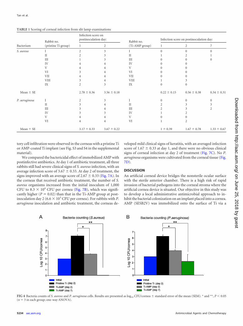
Table 1 summarizes the mean ocular infection score for eachbacterial inoculum group obtained by slit lamp examination. Inthe pristine Ti implant group, all of the eyes (n � 9) developedsigns of bacterial S. aureus infection within 48 h postinoculation,while in the Ti-AMP implant group, two out of nine eyes devel-oped signs of infection. The mean (�SE) ocular inflammationscore for the Ti group was 2.78 � 0.36 on day 1 postinoculationand 3.56 � 0.18 on day 2 postinoculation, while the mean ocular
inflammation score for the Ti-AMP group was 0.22 � 0.15 on day1 postinoculation, 0.56 � 0.38 on day 2 postinoculation (P � 0.01between the Ti and Ti-AMP groups at both day 1 and day 2 timepoints), and 0.34 � 0.31 on day 7 postinoculation. As for the P.aeruginosa inoculation group, all six eyes with Ti implants devel-oped severe signs of infection, and four of the six eyes with Ti-AMP implants developed mild to moderate signs of infection. Themean ocular inflammation score for the Ti group was 3.17 � 0.33on day 1 postinoculation and 3.67 � 0.22 on day 2 postinocula-tion, while the mean ocular inflammation score for the Ti-AMPgroup was 1 � 0.39 on day 1 postinoculation, 1.67 � 0.78 on day2 postinoculation (P � 0.01 between the Ti and Ti-AMP groups atboth day 1 and day 2 time points), and 1.33 � 0.67 on day 7postinoculation.
On AS-OCT examination, the Ti film appeared as a hyperre-flective line on the scanned image, confirming the position of theimplant (Fig. 3B). Within 48 h of inoculation with S. aureus, themean corneal thickness anterior to pristine Ti implants increasedto 757 � 76.7 �m, while the mean corneal thickness anterior toTi-AMP implants increased to 408 � 37 �m (P � 0.02 betweenthe Ti and Ti-AMP groups). At day 7 postinoculation, the meancorneal thickness was 360 � 12.3 �m in the Ti-AMP group (Fig.3C). Infection of the AMP-coated Ti corneas was worse after P.aeruginosa inoculation than that after S. aureus inoculation.Within 48 h of P. aeruginosa inoculation, the mean corneal thick-ness anterior to pristine Ti implants increased to 1,010.7 � 225.4�m, while the mean corneal thickness anterior to Ti-AMP im-plants increased to 460.3 � 100.1 �m (P � 0.01 between the Tiand Ti-AMP groups). At day 7 postinoculation, the mean corneal
FIG 2 In vitro bactericidal effect and stability of SESB2V AMP. (A) MICs of the full-length and truncated SESB2V peptides. (B) Representative images from theLive/Dead bactericidal assay. Green, live bacteria; red, dead bacteria. The live/dead ratios were determined and graphed. *, P � 0.05 (n � 6, Student’s t test). (C)Fluorescence images of Ti surface after immobilization with SESB2V-FITC peptide in phosphate-buffered saline.
Tan et al.
5232 aac.asm.org Antimicrobial Agents and Chemotherapy
on June 25, 2018 by guesthttp://aac.asm
.org/D
ownloaded from

thickness of the Ti-AMP group was 409.2 � 31.4 �m (see Fig. S2Cin the supplemental material).
We counted bacterial clones from the rabbits’ corneal tissue toanalyze the bacterial loads. By 48 h postinoculation, the number ofS. aureus organisms increased from the initial inoculum of 1,000CFU per cornea to approximately 1.3 � 107 CFU per cornea in theTi group and increased to 6.6 � 103 CFU per cornea in the Ti-AMP group (P � 0.01 between the Ti and Ti-AMP groups). At day7 postinoculation, there was no bacterial growth in the Ti-AMPgroup culture plates. Similarly, within 48 h of bacterial inocula-tion, the number of P. aeruginosa organisms increased from theinitial inoculum of 1,000 CFU per cornea to approximately 1.9 �106 CFU per cornea in the Ti group and 9.4 � 103 CFU per corneain the Ti-AMP group (P � 0.01 between the Ti and Ti-AMPgroups). At day 7 postinoculation, there was 1.74 � 102 CFU percornea in the Ti-AMP group (Fig. 4).
Tissue sections were analyzed to further evaluate inflammatoryresponses following bacterial inoculation. At 48 h postinocula-tion, H&E staining of S. aureus-infected corneas with a pristine Tiimplant revealed loss of corneal integrity as a result of melting ofthe epithelial layer in the central cornea (Fig. 5B). Blood vesselsgrew into the corneal stroma (Fig. 5C), and inflammatory cellsinfiltrated the central cornea surrounding the area of the implantpocket (Fig. 5C). However, S. aureus-infected corneas with a Ti-AMP implant revealed very mild cellular infiltration (Fig. 5E andF), and the keratocyte nuclei were aligned as they would be in anormal cornea (Fig. 5G).
In corneas with a pristine Ti implant, the inflammatory mark-ers CD11b, F4/80, and MMP9 were all highly expressed in theanterior corneal stroma area, while there was reduced expressionin the corneas of the Ti-AMP implant group (Fig. 6). For the P.aeruginosa-infected group, similar different patterns of inflamma-
FIG 3 In vivo bactericidal effect of SESB2V AMP against S. aureus. (A) Slit lamp examination of the rabbit eyes before and after implantation and bacterialinoculation. (B) AS-OCT scanning of the rabbit eyes before and after implantation and bacterial inoculation. (C) Measurement of corneal thickness. Only cornealthickness anterior to the implant was measured at pre- and postinoculation day 1 and day 2. *, P � 0.05 (one-way ANOVA) between the pristine Ti and Ti-AMPgroups at postinoculation day 1; **, P � 0.05 (one-way ANOVA) between the pristine Ti and Ti-AMP groups at postinoculation day 2.
AMP Prevents Rabbit Cornea Implant-Associated Infection
September 2014 Volume 58 Number 9 aac.asm.org 5233
on June 25, 2018 by guesthttp://aac.asm
.org/D
ownloaded from
tory cell infiltration were observed in the corneas with a pristine Tior AMP-coated Ti implant (see Fig. S3 and S4 in the supplementalmaterial).
We compared the bactericidal effect of immobilized AMP withpostinfective antibiotics. At day 1 of antibiotic treatment, all threerabbits still had severe clinical signs of S. aureus infection, with anaverage infection score of 3.67 � 0.33. At day 2 of treatment, thesigns improved with an average score of 2.67 � 0.33 (Fig. 7A). Inthe corneas that received antibiotic treatment, the number of S.aureus organisms increased from the initial inoculum of 1,000CFU to 8.3 � 104 CFU per cornea (Fig. 7B), which was signifi-cantly higher (P � 0.02) than that in the Ti-AMP group at post-inoculation day 2 (6.6 � 103 CFU per cornea). For rabbits with P.aeruginosa inoculation and antibiotic treatment, the corneas de-
veloped mild clinical signs of keratitis, with an averaged infectionscore of 1.67 � 0.33 at day 1, and there were no obvious clinicalsigns of corneal infection at day 2 of treatment (Fig. 7C). No P.aeruginosa organisms were cultivated from the corneal tissue (Fig.7D).
DISCUSSION
An artificial corneal device bridges the nonsterile ocular surfacewith the sterile anterior chamber. There is a high risk of rapidinvasion of bacterial pathogens into the corneal stroma where theartificial cornea device is situated. Our objective in this study wasto develop a local administrative antimicrobial approach to in-hibit the bacterial colonization on an implant placed into a cornea.AMP (SESB2V) was immobilized onto the surface of Ti via a
TABLE 1 Scoring of corneal infection from slit lamp examinations
BacteriumRabbit no.(pristine Ti group)
Infection score onpostinoculation day:
Rabbit no.(Ti-AMP group)
Infection score on postinoculation day:
1 2 1 2 7
S. aureus I 2 3 I 0 0 0II 2 3 II 1 2 1III 1 3 III 0 0 0IV 4 4 IV 0 0V 4 4 V 0 0VI 3 4 VI 0 0VII 4 4 VII 0 0VIII 3 4 VIII 1 3IX 2 3 IX 0 0
Mean � SE 2.78 � 0.36 3.56 � 0.18 0.22 � 0.15 0.56 � 0.38 0.34 � 0.31
P. aeruginosa I 2 3 I 0 0 0II 3 4 II 2 3 3III 3 4 III 2 3 2IV 3 3 IV 1 2V 4 4 V 0 0VI 4 4 VI 1 2
Mean � SE 3.17 � 0.33 3.67 � 0.22 1 � 0.39 1.67 � 0.78 1.33 � 0.67
FIG 4 Bacteria counts of S. aureus and P. aeruginosa cells. Results are presented as log10 CFU/cornea � standard error of the mean (SEM). * and **, P � 0.05(n � 3 in each group; one-way ANOVA).
Tan et al.
5234 aac.asm.org Antimicrobial Agents and Chemotherapy
on June 25, 2018 by guesthttp://aac.asm
.org/D
ownloaded from

cross-linker, polydopamine (33, 34). To mimic keratoprosthesissurgery, Ti films were then inserted into rabbit corneal stromalpockets. Ti implants whose surfaces were functionalized withAMP efficiently inhibited corneal bacterial infections with mini-mal adjacent tissue damage. Immobilized AMP proved to be moreefficient in vivo against S. aureus than against P. aeruginosa. Wealso demonstrated that the Ti-AMP implants showed superiorbactericidal activity against S. aureus compared to the use of post-inoculation topical antibiotics.
Current strategies for combating implant-associated ocular in-fections include the continuous use of topical antibiotics (1).However, a serious concern regarding the continuous use of con-ventional antibiotics is the potential development of antibiotic-resistant pathogens such as methicillin-resistant Staphylococcusaureus (MRSA) (35–37) and fungi (7). Antibiotic-resistant infec-tions can lead to devastating effects and uncontrolled bacterialinfection. Therefore, AMPs that have a wide spectrum of bacteri-cidal activity and that are less likely to develop bacterial resistance(26, 37) are an ideal substitute. There have been few studies on thein vivo efficacy of covalently bound AMPs. Gao et al. demon-strated the efficacy of AMP-coated Ti wire used in orthopedicsurgery in a dorsal skin pocket model of wound infection (29).
FIG 5 Hematoxylin-eosin (H&E) pictures of rabbit corneas after inoculationwith S. aureus. (A) Normal rabbit cornea. (B to D) Rabbit cornea with pristineTi implantation and bacterial infection: corneal epithelium (B), central cor-neal stroma (C), and corneal stroma surrounding the implant pocket (D). (Eto G) Rabbit cornea with Ti-AMP implantation and bacterial infection: cor-neal epithelium (E), central corneal stroma (F), and corneal stroma surround-ing the implant pocket (G).
FIG 6 Immunostaining pictures of rabbit corneas after infection with S. aureus. Green, inflammatory cell markers; blue, DAPI cell nucleus counterstaining. (Ato C) CD11b staining: normal rabbit cornea (A), cornea with pristine Ti implant (B), and cornea with Ti-AMP implant (C). (D to F) F4/80 staining: normal rabbitcornea (D), cornea with pristine Ti implant (E), and cornea with Ti-AMP implant (F). (G to I) MMP9 staining: normal rabbit cornea (G), cornea with pristineTi implant (H), and cornea with Ti-AMP implant (I).
AMP Prevents Rabbit Cornea Implant-Associated Infection
September 2014 Volume 58 Number 9 aac.asm.org 5235
on June 25, 2018 by guesthttp://aac.asm
.org/D
ownloaded from

Here, we report the successful surface functionalization of a me-tallic implant with immobilized AMP in an infectious keratitismodel. In contrast to other studies, our model mimics preciselythe clinical in situ placement of synthetic artificial cornea devices(29). We previously proved that SESB2V has a wide spectrum ofbactericidal activity against Gram-positive and Gram-negativebacteria (23). We also determined the MIC of SESB2V againstMRSA (DM21455) to be 6.25 �g/ml. In this study, we showed alow MIC (3.125 to 25 �g/ml) of soluble SESB2V and its truncatedpeptides against Gram-positive S. aureus and Gram-negative P.aeruginosa bacterial strains. These results indicate that fragmentedSESB2V peptide still maintained a bactericidal effect, which is im-portant clinically following protease digestion in vivo. These prop-erties make SESB2V peptide a promising candidate for oculartherapeutic use. After immobilization onto Ti substrates, SESB2Vpeptide retained its bactericidal activities against S. aureus and P.aeruginosa, with a coating concentration of 500 �g/ml, which isabout 80 times higher than the MICs of soluble SESB2V. Weused this concentration, since the covalent binding of the AMPto the substrate surface is likely to alter the MIC. Previousstudies have shown higher MICs of immobilized AMPs com-pared to those of soluble peptides (39–41). The difference inthe live/dead ratio between S. aureus and P. aeruginosa cells on apristine Ti surface is possibly because P. aeruginosa contains muchhigher levels of dead/weakened bacteria. Another possibility isthat pristine Ti itself has a different bactericidal effect on differentbacterial strains. However, these results are still valid, since wecompared the live/dead ratio of bacteria before and after AMPimmobilization.
Titanium films with immobilized SESB2V peptide were placedinto the rabbits’ corneal stroma, and there were no clinical signs of
corneal inflammation within 1 week after implantation comparedto those with a pristine Ti implant. AMPs have been shown to haveimmunomodulatory activities that include angiogenesis, modula-tion of cytokine activity, and reduction of lipopolysaccharide(LPS)-mediated proinflammatory responses (42, 43). The cyto-toxicity associated with AMPs is usually related to the high con-centrations used to compensate for their relatively short half-livesdue to rapid tissue protease digestion (44). Covalent immobiliza-tion of SESB2V may decrease the long-term cytotoxicity to cor-neal tissue, since covalent binding would prolong the life span andfunction of the peptides and reduce the necessity for using highconcentrations of AMP (44, 45). We observed that SESB2V wasstable in a humidified environment for at least 2 months afterimmobilization onto a Ti surface. Additionally, the bactericidaleffect of functionalized Ti discs may be achieved even after 2 weeksof implantation (1 week after bacterial inoculation). The resultsindicate that AMP activity was still present, even though thesepeptides may face the risk of degradation by tissue protease. Thedegraded product of SESB2V may still possess the bactericidaleffect in vivo, as evidenced by the low MIC of the truncatedSESB2V peptides. However, we observed a difference in the bac-tericidal efficacy of the AMP-coated implants against both S. au-reus and P. aeruginosa at day 7 postinoculation (2 weeks afterimplantation). This was possibly due to the biodegradation ordysfunction of SESB2V peptide in the corneal stroma. Our resultsare encouraging for the early postoperative period, but bacterialendophthalmitis may occur months or years after the placementof a device (4–6). Hence, future work should examine the role ofAMPs combined with a controlled drug delivery system, e.g.,nanoparticles or biodegradable films.
The clinical score of corneal inflammation on slit lamp imagesand the quantitative analysis of corneal thickness on AS-OCT im-ages, as well as the histological studies, demonstrated that immo-bilized SESB2V significantly reduced corneal inflammation andstromal swelling caused by either S. aureus or P. aeruginosa patho-gens. Moreover, immobilized SESB2V was more effective in pre-venting postimplantation infection caused by S. aureus than thatcaused by P. aeruginosa. After the inoculation of S. aureus in theAMP-coated implants, the clinical analysis and histological im-ages revealed less obvious signs of infection/inflammation than inthose inoculated with P. aeruginosa. This was possibly because P.aeruginosa was less sensitive to the bactericidal activities ofSESB2V peptide than the same inoculum of S. aureus. Addition-ally, we observed that the inoculation of S. aureus or P. aeruginosacaused the infiltration of different types of inflammatory cells inthe absence of AMP. For example, an inoculation of S. aureuscaused the infiltration of more F4/80-positive cells near the im-plant site, while an inoculation of P. aeruginosa caused the infil-tration of more CD11b-positive cells. Hence, we cannot excludethe possibility that the difference in the bactericidal effects was dueto the difference in the virulence of the pathogens (46, 47). Therabbits developed a more severe keratitis after P. aeruginosa inoc-ulation than after S. aureus inoculation, even without the presenceof AMP. However, the superior efficacy against S. aureus is clini-cally advantageous, since S. aureus is the leading cause of endoph-thalmitis and bacterial keratitis after ocular implantation (7).
Following the clinical diagnosis of an ocular implant infection,the initial treatment will involve the use of intensive topical anti-biotics. Hence, we compared the effect of intensive topical antibi-otic therapy in a clinical situation similar to that of the immobi-
FIG 7 Antibiotic treatment of rabbit corneas after inoculation with S. aureusand P. aeruginosa. (A and C) Scoring of slit lamp examinations after inocula-tion of S. aureus (A) and P. aeruginosa (C). (B and D) Bacterium counting ofcorneal tissue after inoculation of S. aureus (B) and P. aeruginosa (D). *, P �0.05 (one-way ANOVA).
Tan et al.
5236 aac.asm.org Antimicrobial Agents and Chemotherapy
on June 25, 2018 by guesthttp://aac.asm
.org/D
ownloaded from
lized SESB2V treatment used in this study. Compared with topicalantibiotics, there was a significantly lower clinical inflammationscore and fewer bacterial clones with the AMP coating following S.aureus inoculation. However, the topical antibiotics were superiorto SESB2V peptide in treating infections caused by P. aeruginosa,as the inflammatory symptoms resolved and there was no growthof bacterial clones after treatment by the current choice of antibi-otics (Fig. 7C and D). In clinical reality, the topical therapy wouldbe used as an adjunct to the AMP coating; hence, localized controlof the infection by the AMP coating, as indicated by our results,may prevent further spread of the infection into fulminant endo-phthalmitis.
Infections associated with the implantation of biomaterials re-main one of the major barriers to the use of medical devices inpatients. We have successfully immobilized a novel AMP to de-liver a bactericidal effect in vivo. Hence, our study demonstratesthe efficacy of a local antimicrobial prophylactic measure in pre-venting implant-associated corneal infections in an infectious ker-atitis model.
ACKNOWLEDGMENTS
We thank Mageswari D/O Muthusamy for assistance with animal tech-niques.
This work was supported by Singhealth Research Foundation (grantSHF/FG488S/2010).
REFERENCES1. Darouiche RO. 2004. Treatment of infections associated with surgical
implants. N. Engl. J. Med. 350:1422–1429. http://dx.doi.org/10.1056/NEJMra035415.
2. Baker AS, Schein OD. 1989. Ocular infections, p 75–92. In Bisno L,Waldvogel FA (ed), Infections associated with indwelling medical devices.ASM Press, Washington, DC.
3. Behlau I, Gilmore MS. 2008. Microbial biofilms in ophthalmology andinfectious diseases. Arch. Ophthalmol. 126:1572–1581. http://dx.doi.org/10.1001/archopht.126.11.1572.
4. Bourcier T, Borderie V, Laroche L. 2003. Late bacterial keratitis afterimplantation of intrastromal corneal ring segments. J. Cataract Refract.Surg. 29:407– 409. http://dx.doi.org/10.1016/S0886-3350(02)01484-0.
5. Shehadeh-Masha’our R, Modi N, Barbara A, Garzozi HJ. 2004. Keratitisafter implantation of intrastromal corneal ring segments. J. Cataract Re-fract. Surg. 30:1802–1804. http://dx.doi.org/10.1016/j.jcrs.2004.01.034.
6. Chan CC, Holland EJ. 2012. Infectious keratitis after Boston type 1keratoprosthesis implantation. Cornea 31:1128 –1134. http://dx.doi.org/10.1097/ICO.0b013e318245c02a.
7. Robert MC, Moussally K, Harissi-Dagher M. 2012. Review of endoph-thalmitis following Boston keratoprosthesis type 1. Br. J. Ophthalmol.96:776 –780. http://dx.doi.org/10.1136/bjophthalmol-2011-301263.
8. Nouri M, Terada H, Alfonso EC, Foster CS, Durand ML, Dohlman CH.2001. Endophthalmitis after keratoprosthesis: incidence, bacterial causes,and risk factors. Arch. Ophthalmol. 119:484 – 489. http://dx.doi.org/10.1001/archopht.119.4.484.
9. Hughes EH, Mokete B, Ainsworth G, Casswell AG, Eckstein MB,Zambarakji HJ, Gregor Z, Rosen PH, Herold J, Okera S, Liu CS. 2008.Vitreoretinal complications of osteoodontokeratoprosthesis surgery. Ret-ina 28:1138 –1145. http://dx.doi.org/10.1097/IAE.0b013e318174e10e.
10. Tan A, Tan DT, Tan XW, Mehta JS. 2012. Osteo-odonto keratopros-thesis: systematic review of surgical outcomes and complication rates.Ocul. Surf. 10:15–25. http://dx.doi.org/10.1016/j.jtos.2012.01.003.
11. Endophthalmitis Vitrectomy Study Group. 1995. Results of the Endo-phthalmitis Vitrectomy Study, a randomized trial of immediate vitrec-tomy and of intravenous antibiotics for the treatment of postoperativebacterial endophthalmitis. Arch. Ophthalmol. 113:1479 –1496. http://dx.doi.org/10.1001/archopht.1995.01100120009001.
12. Boxma H, Broekhuizen T, Patka P, Oosting H. 1996. Randomisedcontrolled trial of single dose antibiotic prophylaxis in surgical treatmentof closed fractures: the Dutch Trauma Trial. Lancet 347:1133–1137. http://dx.doi.org/10.1016/S0140-6736(96)90606-6.
13. Whitehouse JD, Friedman ND, Kirkland KB, Richardson WJ, SextonDJ. 2002. The impact of surgical-site infections following orthopedic sur-gery at a community hospital and a university hospital: adverse quality oflife, excess length of stay, and extra cost. Infect. Control Hosp. Epidemiol.23:183–189. http://dx.doi.org/10.1086/502033.
14. Apt L, Isenberg S, Yoshimori R, Paez HJ. 1984. Chemical preparation ofthe eye in ophthalmic surgery: III. Effect of povidone-iodine on the con-junctiva. Arch. Ophthalmol. 102:728 –729. http://dx.doi.org/10.1001/archopht.1984.01040030584025.
15. Musella M, Guido A, Musella S. 2001. Collagen tampons as aminogly-coside carriers to reduce postoperative infection rate in prosthetic repairof groin hernias. Eur. J. Surg. 167:130 –132. http://dx.doi.org/10.1080/110241501750070592.
16. Actis Dato A, Jr, Chiusolo C, Cicchitti GC, Actis Dato GM, Porro MC,Bello A. 1992. Antibiotic pretreatment of heart valve prostheses. MinervaCardioangiol. 40:225–229.
17. Darouiche RO. 2003. Antimicrobial approaches for preventing infectionsassociated with surgical implants. Clin. Infect. Dis. 36:1284 –1289. http://dx.doi.org/10.1086/374842.
18. Schaff H, Carrel T, Steckelberg JM, Grunkemeier GL. 1999. ArtificialValve Endocarditis Reduction Trial (AVERT): protocol of a multicenterrandomized trial. J. Heart Valve Dis. 8:131–139.
19. Masse A, Bruno A, Bosetti M, Biasibetti A, Cannas M, Gallinaro P.2000. Prevention of pin track infection in external fixation with silvercoated pins: clinical and microbiological results. J. Biomed. Mater. Res. 53:600 – 604. http://dx.doi.org/10.1002/1097-4636(200009)53:5�600::AID-JBM21�3.0.CO;2-D.
20. Tan XW, Perera AP, Tan A, Tan D, Khor KA, Beuerman RW, Mehta JS.2011. Comparison of candidate materials for a synthetic osteo-odontokeratoprosthesis device. Invest. Ophthalmol. Vis. Sci. 52:21–29. http://dx.doi.org/10.1167/iovs.10-6186.
21. Pullamsetti SS, Savai R, Janssen W, Dahal BK, Seeger W, GrimmingerF, Ghofrani HA, Weissmann N, Schermuly RT. 2011. Inflammation,immunological reaction and role of infection in pulmonary hypertension.Clin. Microbiol. Infect. 17:7–14. http://dx.doi.org/10.1111/j.1469-0691.2010.03285.x.
22. Punnia-Moorthy A. 1987. Evaluation of pH changes in inflammation ofthe subcutaneous air pouch lining in the rat, induced by carrageenan,dextran and Staphylococcus aureus. J. Oral Pathol. 16:36 – 44. http://dx.doi.org/10.1111/j.1600-0714.1987.tb00674.x.
23. Tan XW, Lakshminarayanan R, Liu SP, Goh E, Tan D, Beuerman RW,Mehta JS. 2012. Dual functionalization of titanium with vascular endo-thelial growth factor and beta-defensin analog for potential application inkeratoprosthesis. J. Biomed. Mater. Res. B Appl. Biomater. 100:2090 –2100. http://dx.doi.org/10.1002/jbm.b.32774.
24. Behlau I, Mukherjee K, Todani A, Tisdale AS, Cade F, Wang L,Leonard EM, Zakka FR, Gilmore MS, Jakobiec FA, Dohlman CH,Klibanov AM. 2011. Biocompatibility and biofilm inhibition of N,N-hexyl,methyl-polyethylenimine bonded to Boston keratoprosthesismaterials. Biomaterials 32:8783– 8796. http://dx.doi.org/10.1016/j.biomaterials.2011.08.010.
25. Kazemzadeh-Narbat M, Kindrachuk J, Duan K, Jenssen H, HancockRE, Wang R. 2010. Antimicrobial peptides on calcium phosphate-coatedtitanium for the prevention of implant-associated infections. Biomaterials31:9519 –9526. http://dx.doi.org/10.1016/j.biomaterials.2010.08.035.
26. Seo MD, Won HS, Kim JH, Mishig-Ochir T, Lee BJ. 2012. Antimicrobialpeptides for therapeutic applications: a review. Molecules 17:12276 –12286. http://dx.doi.org/10.3390/molecules171012276.
27. Kazemzadeh-Narbat M, Lai BF, Ding C, Kizhakkedathu JN, Hancock RE,Wang R. 2013. Multilayered coating on titanium for controlled release ofantimicrobial peptides for the prevention of implant-associated infections.Biomaterials 34:5969–5977. http://dx.doi.org/10.1016/j.biomaterials.2013.04.036.
28. Gabriel M, Nazmi K, Veerman EC, Nieuw Amerongen AV, Zentner A.2006. Preparation of LL-37-grafted titanium surfaces with bactericidal activ-ity. Bioconjug. Chem. 17:548–550. http://dx.doi.org/10.1021/bc050091v.
29. Gao G, Lange D, Hilpert K, Kindrachuk J, Zou Y, Cheng JT, Kazemza-deh-Narbat M, Yu K, Wang R, Straus SK, Brooks DE, Chew BH,Hancock RE, Kizhakkedathu JN. 2011. The biocompatibility and biofilmresistance of implant coatings based on hydrophilic polymer brushes con-jugated with antimicrobial peptides. Biomaterials 32:3899 –3909. http://dx.doi.org/10.1016/j.biomaterials.2011.02.013.
AMP Prevents Rabbit Cornea Implant-Associated Infection
September 2014 Volume 58 Number 9 aac.asm.org 5237
on June 25, 2018 by guesthttp://aac.asm
.org/D
ownloaded from

30. Clinical and Laboratory Standards Institute. 2009. Methods for dilutionantimicrobial susceptibility tests for bacteria that grow aerobically; ap-proved standard, 8th ed, M07-A8. Clinical and Laboratory Standards In-stitute, Wayne, PA.
31. Johnson MK, Hobden JA, Hagenah M, O’Callaghan RJ, Hill JM, ChenS. 1990. The role of pneumolysin in ocular infections with Streptococcuspneumoniae. Curr. Eye Res. 9:1107–1114. http://dx.doi.org/10.3109/02713689008997584.
32. Lakshminarayanan R, Liu S, Li J, Nandhakumar M, Aung TT, Goh E,Chang JY, Saraswathi P, Tang C, Safie SR, Lin LY, Riezman H, Lei Z,Verma CS, Beuerman RW. 2014. Synthetic multivalent antifungal pep-tides effective against fungi. PLoS One 9:e87730. http://dx.doi.org/10.1371/journal.pone.0087730.
33. Lee H, Dellatore SM, Miller WM, Messersmith PB. 2007. Mussel-inspired surface chemistry for multifunctional coatings. Science 318:426 –430. http://dx.doi.org/10.1126/science.1147241.
34. Xie J, Michael PL, Zhong S, Ma B, MacEwan MR, Lim CT. 2012. Musselinspired protein-mediated surface modification to electrospun fibers andtheir potential biomedical applications. J. Biomed. Mater. Res. A 100:929 –938. http://dx.doi.org/10.1002/jbm.a.34030.
35. Campoccia D, Montanaro L, Arciola CR. 2006. The significance of infectionrelated to orthopedic devices and issues of antibiotic resistance. Biomaterials27:2331–2339. http://dx.doi.org/10.1016/j.biomaterials.2005.11.044.
36. Campoccia D, Montanaro L, Speziale P, Arciola CR. 2010. Antibiotic-loaded biomaterials and the risks for the spread of antibiotic resistancefollowing their prophylactic and therapeutic clinical use. Biomaterials 31:6363– 6377. http://dx.doi.org/10.1016/j.biomaterials.2010.05.005.
37. Trampuz A, Zimmerli W. 2006. Antimicrobial agents in orthopaedicsurgery: prophylaxis and treatment. Drugs 66:1089 –1105. http://dx.doi.org/10.2165/00003495-200666080-00005.
38. Park SC, Park YK, Hahm KS. 2011. The role of antimicrobial peptides inpreventing multidrug-resistant bacterial infections and biofilm formation.Int. J. Mol. Sci. 12:5971–5992. http://dx.doi.org/10.3390/ijms12095971.
39. Bagheri M, Beyermann M, Dathe M. 2009. Immobilization reduces theactivity of surface-bound cationic antimicrobial peptides with no influ-ence upon the activity spectrum. Antimicrob. Agents Chemother. 53:1132–1141. http://dx.doi.org/10.1128/AAC.01254-08.
40. Cho WM, Joshi BP, Cho H, Lee KH. 2007. Design and synthesis of novelantibacterial peptide-resin conjugates. Bioorg. Med. Chem. Lett. 17:5772–5776. http://dx.doi.org/10.1016/j.bmcl.2007.08.056.
41. Haynie SL, Crum GA, Doele BA. 1995. Antimicrobial activities of am-phiphilic peptides covalently bonded to a water-insoluble resin. Antimi-crob. Agents Chemother. 39:301–307. http://dx.doi.org/10.1128/AAC.39.2.301.
42. Hale JD, Hancock RE. 2007. Alternative mechanisms of action of cationicantimicrobial peptides on bacteria. Expert Rev. Anti Infect. Ther. 5:951–959. http://dx.doi.org/10.1586/14787210.5.6.951.
43. Jenssen H, Hancock RE. 2010. Therapeutic potential of HDPs as immu-nomodulatory agents. Methods Mol. Biol. 618:329 –347. http://dx.doi.org/10.1007/978-1-60761-594-1_20.
44. Chen R, Cole N, Willcox MD, Park J, Rasul R, Carter E, Kumar N.2009. Synthesis, characterization and in vitro activity of a surface-attachedantimicrobial cationic peptide. Biofouling 25:517–524. http://dx.doi.org/10.1080/08927010902954207.
45. Costa F, Carvalho IF, Montelaro RC, Gomes P, Martins MC. 2011.Covalent immobilization of antimicrobial peptides (AMPs) onto bioma-terial surfaces. Acta Biomater. 7:1431–1440. http://dx.doi.org/10.1016/j.actbio.2010.11.005.
46. Karthikeyan RS, Priya JL, Leal SM, Jr, Toska J, Rietsch A, Prajna V,Pearlman E, Lalitha P. 2013. Host response and bacterial virulence factorexpression in Pseudomonas aeruginosa and Streptococcus pneumoniae cor-neal ulcers. PLoS One 8:e64867. http://dx.doi.org/10.1371/journal.pone.0064867.
47. Dajcs JJ, Thibodeaux BA, Girgis DO, O’Callaghan RJ. 2002. Corneal virulenceof Staphylococcus aureus in an experimental model of keratitis. DNA CellBiol. 21:375–382. http://dx.doi.org/10.1089/10445490260099656.
Tan et al.
5238 aac.asm.org Antimicrobial Agents and Chemotherapy
on June 25, 2018 by guesthttp://aac.asm
.org/D
ownloaded from