effects of atp and actin-filament binding on the dynamics of the myosin ii s1 domain
TRANSCRIPT

1624 Biophysical Journal Volume 105 October 2013 1624–1634
Effects of ATP and Actin-Filament Binding on the Dynamics of the Myosin IIS1 Domain
Joseph L. Baker and Gregory A. Voth*Department of Chemistry, James Franck Institute, Institute for Biophysical Dynamics, and Computation Institute, University of Chicago,Chicago, Illinois
ABSTRACT Actin and myosin interact with one another to perform a variety of cellular functions. Central to understandingthe processive motion of myosin on actin is the characterization of the individual states along the mechanochemical cycle.We present an all-atom molecular dynamics simulation of the myosin II S1 domain in the rigor state interacting with an actinfilament. We also study actin-free myosin in both rigor and post-rigor conformations. Using all-atom level and coarse-grainedanalysis methods, we investigate the effects of myosin binding on actin, and of actin binding on myosin. In particular, wedetermine the domains of actin and myosin that interact strongly with one another at the actomyosin interface using a highlycoarse-grained level of resolution, and we identify a number of salt bridges and hydrogen bonds at the interface of myosinand actin. Applying coarse-grained analysis, we identify differences in myosin states dependent on actin-binding, or ATPbinding. Our simulations also indicate that the actin propeller twist-angle and nucleotide cleft-angles are influenced by myosinat the actomyosin interface. The torsional rigidity of the myosin-bound filament is also calculated, and is found to be increasedcompared to previous simulations of the free filament.
INTRODUCTION
Myosin is a motor protein that is involved in a variety ofcellular functions, including contraction of muscle cellsand the transport of cargo along actin filaments (1–3). Agreat deal of understanding about myosin has been obtainedvia both experimental and computational methods, and thereare extensive reviews on myosin and its interactions withactin in the literature (1–8). There are many different typesof myosin, because the protein belongs to a diverse familyof proteins (3). In particular, myosin II, also known asconventional or muscle myosin, has been very well charac-terized, including a structure of the actomyosin system inthe rigor state (ATP-free state of myosin) which was deter-mined using cryo-electron microscopy (cryo-EM) (9). Morerecently, a structure of myosin and tropomyosin on actinwas solved using cryo-EM in combination with docking/flexible fitting of crystal structures into the map (10). Toperform its variety of functions, myosin interacts with actinfilaments in a mechanochemical cycle (1,4,5) (referred to asthe Lymn-Taylor cycle (11)).
Computer simulation has offered valuable insight into thefunctioning of isolated myosin protein (not including actin)in a variety of its nucleotide states (using both all-atom andsimplified representations of myosin) (12–18). For example,the process of ATP hydrolysis in myosin has been investi-gated using QM/MM methods (19–22), and elastic networkmodels at an a-carbon level of resolution of myosin II andmyosin V bound to actin have been studied to elucidatehow actin binding can lead to phenomena such as phosphaterelease in myosin, and other protein conformational changes
Submitted April 19, 2013, and accepted for publication August 22, 2013.
*Correspondence: [email protected]
Editor: Anne Houdusse.
� 2013 by the Biophysical Society
0006-3495/13/10/1624/11 $2.00
(23,24). However, all-atom simulation of myosin on a fullyperiodic actin filament, including both the myosin leverarm and light chains, has not yet been performed. Previoussimulation work on actin interacting with myosin at the all-atom level has been limited to small systems (25,26). Forexample, the presence of an important electrostatic inter-action between actin and myosin was established over thecourse of 15-ns simulations in a study of myosin interactingwith an actin trimer (25), and simulation of myosin interact-ing with an actin dimer in an implicit solvent was used tostudy interactions at the actin/myosin interface (26). Ourall-atom molecular dynamics (MD) simulations are, to ourknowledge, the first of their kind for the myosin S1 domain,including the lever-arm binding light chains, interactingwith a fully periodic actin filament. By including a fullyperiodic actin filament we are able to improve upon theseprevious simulations by ensuring that each actin monomerhas appropriate contacts within the filament environment,and also gain the new capability to calculate filament-levelproperties such as the torsional rigidity of the actin filamentfrom all-atom and coarse-grained (CG) analysis, whichcannot be calculated from actin dimer and trimer systems.
In this study we simulate rigor actomyosin, consistingof a periodic actin filament (built from an actin 13-mer)and the myosin II S1 domain from squid muscle (27). TheS1 domain of myosin consists of the motor domain (whereactin-binding and ATP hydrolysis occur), and the lever arm(7). In addition to our simulation of rigor actomyosin, wesimulate myosin in the rigor state in the absence of actin,as well as myosin in the post-rigor state (the state withATP present in the myosin nucleotide cleft). All-atom sim-ulations of the actomyosin filament can shed light on thepresence of stable interactions between actin and myosin
http://dx.doi.org/10.1016/j.bpj.2013.08.023

CG Analysis of Actomyosin 1625
at the interface of the two proteins. All-atom MD in explicitsolvent has an advantage in that it can be used to gain infor-mation about the dynamical interactions between actin andmyosin—information that, necessarily, cannot be providedby static structures. By making comparisons of the simula-tions of the myosin rigor state bound to actin to the rigorstate free of actin and the post-rigor state, we can also under-stand the influence of actin binding (and ATP binding) onmyosin dynamics. Furthermore, because extensive simula-tions of the free actin filament have been previously carriedout (28–32), comparisons can be made to our simulation ofactomyosin to assist understanding of how myosin modu-lates actin filament dynamics. For example, it has beendemonstrated experimentally that myosin increases thetorsional rigidity of actin filaments (33–35). This can haveimportant functional consequences for actomyosin systems.It has been suggested that actin’s ability to act as a torsionalspring could help to strain the flexible connection of themyosin motor to the thick filament (36), and that a largertorsional rigidity might aid in establishing cooperativitybetween actin monomers in the filament (34).
In addition to all-atom level analysis of interactions be-tween actin and myosin, we also make use of coarse-grained(CG) methods to analyze the dynamics and interactions inthe actomyosin system and the myosin rigor and post-rigorstates. For large proteins and protein complexes, CG repre-sentation and analysis allows one to reduce an all-atom sys-tem to a smaller number of domains and interactions (37). Inthis study, we used the essential dynamics coarse-graining(ED-CG) (38) and the heterogeneous elastic network model(hENM) (39) methods to perform a CG analysis. Thesemethods have previously been applied by our group to assistunderstanding of the nature of heterogeneity in myosin-freeactin filaments (29), interactions in the large ribosomal sub-unit (40), and NBAR domain proteins (41). ED-CG allowsus to investigate how the definitions of dynamic domainsin myosin and actin are influenced by their interactionswith one another in the actomyosin rigor state. In contrastto other approaches for coarse-graining proteins, whichmay use an a-carbon level of resolution as in the G�o model(42) or a few residues per CG site as in the MARTINI model(43,44), methods like ED-CG can be used to group aminoacids into CG sites at a highly coarse-grained level of reso-lution where tens of amino acids are assigned to each CGdomain (29,40,41). Specifically, the ED-CGmethod demon-strates that the N-terminal region of the myosin-bound actinmonomer is influenced by myosin binding, and that thevarious squid muscle myosin states have relatively similarregions of collective motion along the myosin primarysequence. Furthermore, the hENM method allows thedetermination of the regions of myosin that most stronglyinteract with actin, and reveals differences between domaincouplings in myosin dependent on whether it is actin-boundor has ATP in its nucleotide cleft. For example, we find thatthe two domains comprising the actin-binding cleft of
myosin have a larger effective coupling in the bound-rigorcompared to the free-rigor and post-rigor states, and thatswitch I and the P-loop are much more strongly coupledin the post-rigor state than in either the bound-rigor orfree-rigor states. This provides further evidence for thereciprocal coupling of the actin binding cleft and the nucle-otide clefts of myosin. We also observe, by calculating theactin propeller twist and nucleotide cleft angles, that myosinhas an influence on both of these properties at the monomerwith the largest myosin interface. From our simulations, wealso create hENM-EDCG models of the actomyosin systemthat can reproduce the torsional rigidity, which we calculatefrom the all-atom myosin-bound actin filament. The valuesthat we obtain are consistent with the observation that thetorsional rigidity of actin filaments bound by myosin S1domain in the rigor state is increased in comparison to thefree filament (33–35).
METHODS
All-atom MD simulations
All-atomMD simulations were run using the software NAMD (45) with the
CHARMM27 force field (46), including CMAP corrections. For myosin,
we used the myosin II S1 domain (including the lever-arm and lever-arm
binding light chains) rigor (PDB:3I5G) and post-rigor (PDB:3I5F) states
from squid muscle (27). Missing loop regions were modeled using the
software MODELER (47) and the Falc-Loop server (48,49). The structure
for the actin 13-mer filament was built the same way as has been previously
discussed in the literature (30–32), with the monomer structure based on
the Oda structure (PDB:2ZWH) (50). The starting structure for the filament
includes Mg2þ and ADP in the nucleotide cleft, as well as active site waters
coordinating the Mg2þ ion (30). Periodic boundary conditions were en-
forced so that the actin filament is continuous across the cell boundaries,
leading to a filament of infinite length, as in previous simulations of the
periodic actin filament (30–32).
To build the actomyosin complex, the molecular-dynamics flexible-
fitting (MDFF) refined actomyosin structure (51) was used as a template
for positioning of the squid muscle myosin II rigor state (27) onto the actin
13-mer. The middle five monomers of the 13-mer filament were aligned
with the five monomers from the MDFF model by root mean-square devi-
ation (RMSD) fitting the backbone a-carbons (51). The squid rigor state
was then aligned to the myosin II protein from the aligned MDFF structure
using the built-in MULTISEQ tool (52) in the program VMD (53) by using
the STAMP structural alignment algorithm (54) on the residues in the
myosin II motor domain. The final system is shown in Fig. 1. Systems
were solvated with sufficient water to provide padding between the protein
and its periodic image (15 A of padding for isolated myosin states along
each box direction, and 8 A of padding for the filament system perpendic-
ular to the filament axis, with no water caps along the filament axis, as in
previous work). All residues were modeled in their standard protonation
states at a pH of 7. In addition, the N-terminus of the actin monomers
was acetylated. Neutralization with 0.180 M KCl was performed (with
number of K/Cl ions equal to 238/220, 297/277, and 1378/1178 for
free-rigor, post-rigor, and bound-rigor, respectively). Full solvation and
neutralization of the system were accomplished with the VMD SOLVATE
and AUTOIONIZE plug-ins (53), respectively.
Systems were energy-minimized with constraints released in a stepwise
fashion. For the isolated myosin II proteins a long, restrained equilibration
phase was performed, with harmonic restraints gradually released on the
protein backbone. To not disturb the initial configuration of the magnesium
and crystal waters used in building the filament, the actomyosin system was
Biophysical Journal 105(7) 1624–1634
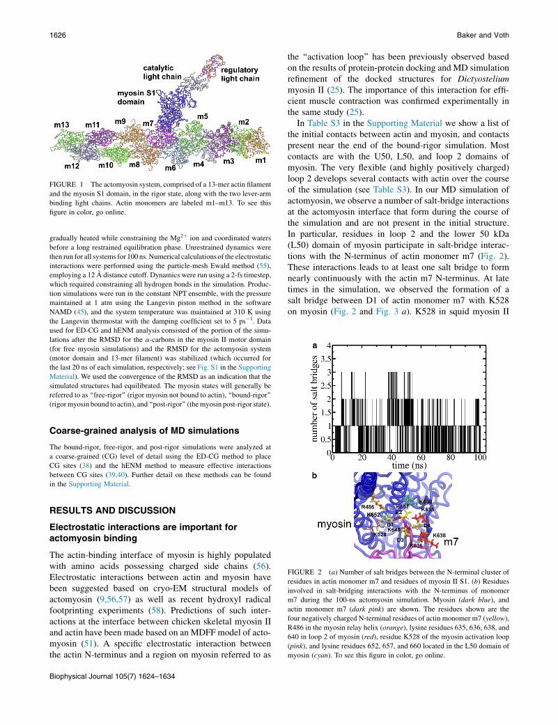
FIGURE 1 The actomyosin system, comprised of a 13-mer actin filament
and the myosin S1 domain, in the rigor state, along with the two lever-arm
binding light chains. Actin monomers are labeled m1–m13. To see this
figure in color, go online.
1626 Baker and Voth
gradually heated while constraining the Mg2þ ion and coordinated waters
before a long restrained equilibration phase. Unrestrained dynamics were
then run for all systems for 100 ns.Numerical calculations of the electrostatic
interactions were performed using the particle-mesh Ewald method (55),
employing a 12 A distance cutoff. Dynamics were run using a 2-fs timestep,
which required constraining all hydrogen bonds in the simulation. Produc-
tion simulations were run in the constant NPT ensemble, with the pressure
maintained at 1 atm using the Langevin piston method in the software
NAMD (45), and the system temperature was maintained at 310 K using
the Langevin thermostat with the damping coefficient set to 5 ps�1. Data
used for ED-CG and hENM analysis consisted of the portion of the simu-
lations after the RMSD for the a-carbons in the myosin II motor domain
(for free myosin simulations) and the RMSD for the actomyosin system
(motor domain and 13-mer filament) was stabilized (which occurred for
the last 20 ns of each simulation, respectively; see Fig. S1 in the Supporting
Material). We used the convergence of the RMSD as an indication that the
simulated structures had equilibrated. The myosin states will generally be
referred to as ‘‘free-rigor’’ (rigor myosin not bound to actin), ‘‘bound-rigor’’
(rigormyosin bound to actin), and ‘‘post-rigor’’ (themyosin post-rigor state).
Coarse-grained analysis of MD simulations
The bound-rigor, free-rigor, and post-rigor simulations were analyzed at
a coarse-grained (CG) level of detail using the ED-CG method to place
CG sites (38) and the hENM method to measure effective interactions
between CG sites (39,40). Further detail on these methods can be found
in the Supporting Material.
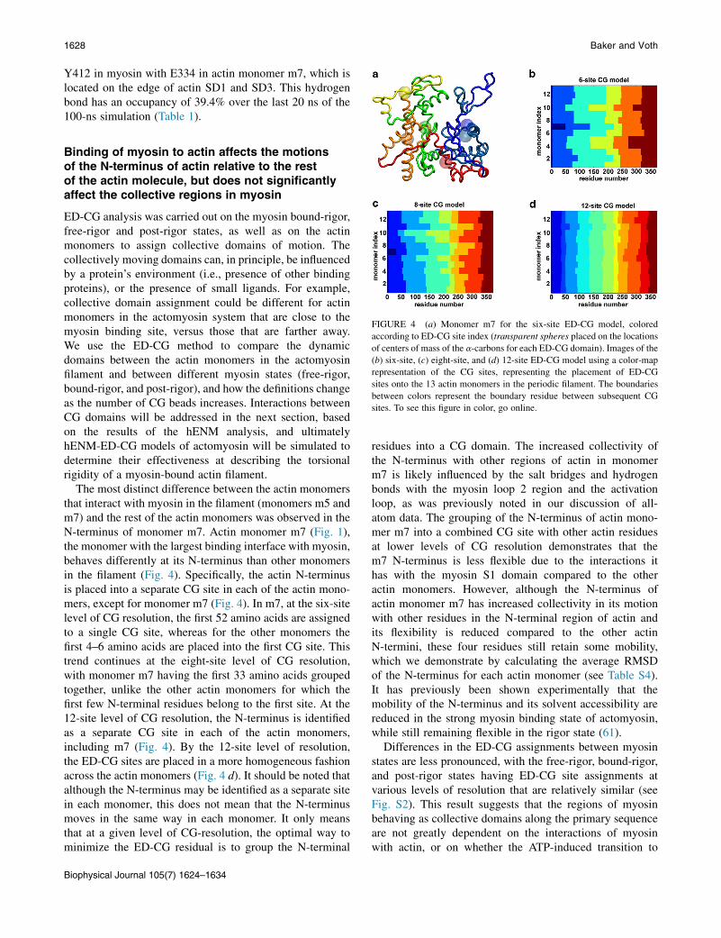
FIGURE 2 (a) Number of salt bridges between the N-terminal cluster of
residues in actin monomer m7 and residues of myosin II S1. (b) Residues
involved in salt-bridging interactions with the N-terminus of monomer
m7 during the 100-ns actomyosin simulation. Myosin (dark blue), and
actin monomer m7 (dark pink) are shown. The residues shown are the
four negatively charged N-terminal residues of actin monomer m7 (yellow),
R486 in the myosin relay helix (orange), lysine residues 635, 636, 638, and
640 in loop 2 of myosin (red), residue K528 of the myosin activation loop
(pink), and lysine residues 652, 657, and 660 located in the L50 domain of
myosin (cyan). To see this figure in color, go online.
RESULTS AND DISCUSSION
Electrostatic interactions are important foractomyosin binding
The actin-binding interface of myosin is highly populatedwith amino acids possessing charged side chains (56).Electrostatic interactions between actin and myosin havebeen suggested based on cryo-EM structural models ofactomyosin (9,56,57) as well as recent hydroxyl radicalfootprinting experiments (58). Predictions of such inter-actions at the interface between chicken skeletal myosin IIand actin have been made based on an MDFF model of acto-myosin (51). A specific electrostatic interaction betweenthe actin N-terminus and a region on myosin referred to as
Biophysical Journal 105(7) 1624–1634
the ‘‘activation loop’’ has been previously observed basedon the results of protein-protein docking and MD simulationrefinement of the docked structures for Dictyosteliummyosin II (25). The importance of this interaction for effi-cient muscle contraction was confirmed experimentally inthe same study (25).
In Table S3 in the Supporting Material we show a list ofthe initial contacts between actin and myosin, and contactspresent near the end of the bound-rigor simulation. Mostcontacts are with the U50, L50, and loop 2 domains ofmyosin. The very flexible (and highly positively charged)loop 2 develops several contacts with actin over the courseof the simulation (see Table S3). In our MD simulation ofactomyosin, we observe a number of salt-bridge interactionsat the actomyosin interface that form during the course ofthe simulation and are not present in the initial structure.In particular, residues in loop 2 and the lower 50 kDa(L50) domain of myosin participate in salt-bridge interac-tions with the N-terminus of actin monomer m7 (Fig. 2).These interactions leads to at least one salt bridge to formnearly continuously with the actin m7 N-terminus. At latetimes in the simulation, we observed the formation of asalt bridge between D1 of actin monomer m7 with K528on myosin (Fig. 2 and Fig. 3 a). K528 in squid myosin II

FIGURE 3 (a) Distance between side chains of three N-terminal actin
residues that form a salt bridge with the activation-loop residue K528 in
myosin II. (Lines) Actin residues D1 (black), E2 (red cross), and D3 (green
square). (b) Distance between the side chains of loop-3 residues in myosin
and residues in SD1 of actin monomer m5. (Lines) Distances between
E100-K569 (black), E100-K572 (red cross), E99-K569 (green square),
and E99-K572 (blue circle). (c) Distances between salt-bridging residues
in the myosin U50 domain with residues in the SD3 domain of actin mono-
mer m7. Distance between actin residue E167 (in the W-loop of actin) and
myosin residue K544 (black) and the distance between K328 in actin
and E373 in myosin (red cross). (d) Shown are myosin (dark blue), actin
monomer m5 (green), m6 (light blue), and m7 (dark pink). Also shown
are salt-bridging residues K569 and K572 in myosin (red), in the loop-3
region, and residues E99 and E100 in SD1 of actin (orange). Residues
K544 and E373 (cyan) in myosin (both in the U50 domain), which interact
with residues E167 (W-loop of actin) and K328, are also shown. To see this
figure in color, go online.
TABLE 1 Percentage of time during last 20 ns for hydrogen-
bond formation
Myosin residue Actin residue
Occupancy
percentage
E538 (side chain, L50) T351 (side chain, C-terminus) 75.4
E538 (side chain, L50) T351 (main chain, C-terminus) 50.4
K569 (side chain, L50) E99 (side chain, SD1) 43.4
Y412 (side chain, CM-loop) E334 (side chain, SD1) 39.4
K572 (side chain, L50) E100 (side chain, SD1) 38.3
K635 (main chain, Loop 2) E4 (side chain, N-terminus) 37.9
E373 (side chain, U50) K328 (side chain, SD3) 36.4
K636 (side chain, Loop 2) E100 (side chain, SD1) 30.9
K569 (side chain, L50) E100 (side chain, SD1) 30.7
K544 (side chain, L50) E167 (side chain, SD3) 28.0
K636 (main chain, Loop 2) E4 (side chain, N-terminus) 26.6
CG Analysis of Actomyosin 1627
is homologous to R520 in Dictyostelium myosin (the acti-vation-loop residue).
In another study, the salt bridge between R520 and theN-terminal cluster was formed from the beginning of theprotein-protein docking phase (25). Our observation thatthis salt bridge forms over the course of 100-ns MD simula-tions suggests this interaction is indeed important for thedefinition of the actomyosin interface, and not specific toa single isoform of myosin II. The initial distance betweenany of the N-terminal actin residues and the activation-loop residue K528 is 20–30 A, falling quickly below 20 Awithin the next 20 ns, and during the last 20 ns maintainingdistances of<15 A (including the close approach within oursalt bridge cutoff of 3.2 A by D1 in actin near the end of thesimulation; Fig. 3 a). Similar to a recent study in which theeffect observed upon mutation of R520 in Dictyosteliumdiscoideum myosin II (25), we would expect, based on oursimulations, that mutation of K528 to a neutral or negativelycharged amino acid in squid muscle myosin could influencethe efficiency of myosin.
We also observe particularly stable salt-bridge inter-actions between actin monomer m5 (Fig. 3, b and d) and a
region of myosin referred to as ‘‘loop 3’’ (squid myosin IIresidues 567–580). This loop has been previously hypo-thesized to interact with actin (51,56). It has also been sug-gested that myosin II has interactions with the actin W-loop(residues 165–172) via a salt-bridge interaction betweenactin residue E167 and myosin residue K544 (51). Inter-action between these residues was observed experimentallyfor rabbit skeletal muscle myosin (58). We observe asalt-bridge interaction (and hydrogen-bonding interaction,Table 1) between myosin and the W-loop in our simulation,as well as between another upper 50 kDa (U50) domainresidue and another SD3 residue in actin (Fig. 3, c and d).Because the myosin salt bridge to the W-loop was predictedfor the chicken skeletal myosin II (51) and observedexperimentally for rabbit skeletal muscle (58), that wealso observe this for squid muscle myosin suggests thatthis interaction between myosin and the W-loop is impor-tant for the various myosin isoforms in defining theactomyosin interface. To confirm the presence of thisinteraction between squid muscle myosin and actin (whichwould help to demonstrate that the actin/myosin interfacehas many common features across different myosin iso-forms), cysteine-mutant-plus-fluorescence-labeling experi-ments similar to those performed for the rabbit skeletalmyosin (58) could be carried out.
Several regions of myosin participate in hydrogenbonding with actin monomers m5 and m7 (Table 1). Thehydrogen bonds listed in Table 1 were formed over thecourse of the simulation. A long-lived hydrogen bond isformed between the side chain of E538 in the L50 domainof myosin and the side chain of T351 in the C-terminus ofactin (Table 1). This is the most stable hydrogen bondobserved, having an occupancy of 75.4% over the last20 ns of simulation. Likewise, in the cardiomyopathy(CM) loop—a region of myosin in which mutation hasbeen related to the condition familial hypertrophic CM(59,60)—it has been proposed that the actin residuesP332–E334 could interact with myosin CM loop residues(56). Supporting the idea that these regions interact in theactomyosin complex, we observe a hydrogen bond between
Biophysical Journal 105(7) 1624–1634
1628 Baker and Voth
Y412 in myosin with E334 in actin monomer m7, which islocated on the edge of actin SD1 and SD3. This hydrogenbond has an occupancy of 39.4% over the last 20 ns of the100-ns simulation (Table 1).
FIGURE 4 (a) Monomer m7 for the six-site ED-CG model, colored
according to ED-CG site index (transparent spheres placed on the locations
of centers of mass of the a-carbons for each ED-CG domain). Images of the
(b) six-site, (c) eight-site, and (d) 12-site ED-CG model using a color-map
representation of the CG sites, representing the placement of ED-CG
sites onto the 13 actin monomers in the periodic filament. The boundaries
between colors represent the boundary residue between subsequent CG
sites. To see this figure in color, go online.
Binding of myosin to actin affects the motionsof the N-terminus of actin relative to the restof the actin molecule, but does not significantlyaffect the collective regions in myosin
ED-CG analysis was carried out on the myosin bound-rigor,free-rigor and post-rigor states, as well as on the actinmonomers to assign collective domains of motion. Thecollectively moving domains can, in principle, be influencedby a protein’s environment (i.e., presence of other bindingproteins), or the presence of small ligands. For example,collective domain assignment could be different for actinmonomers in the actomyosin system that are close to themyosin binding site, versus those that are farther away.We use the ED-CG method to compare the dynamicdomains between the actin monomers in the actomyosinfilament and between different myosin states (free-rigor,bound-rigor, and post-rigor), and how the definitions changeas the number of CG beads increases. Interactions betweenCG domains will be addressed in the next section, basedon the results of the hENM analysis, and ultimatelyhENM-ED-CG models of actomyosin will be simulated todetermine their effectiveness at describing the torsionalrigidity of a myosin-bound actin filament.
The most distinct difference between the actin monomersthat interact with myosin in the filament (monomers m5 andm7) and the rest of the actin monomers was observed in theN-terminus of monomer m7. Actin monomer m7 (Fig. 1),the monomer with the largest binding interface with myosin,behaves differently at its N-terminus than other monomersin the filament (Fig. 4). Specifically, the actin N-terminusis placed into a separate CG site in each of the actin mono-mers, except for monomer m7 (Fig. 4). In m7, at the six-sitelevel of CG resolution, the first 52 amino acids are assignedto a single CG site, whereas for the other monomers thefirst 4–6 amino acids are placed into the first CG site. Thistrend continues at the eight-site level of CG resolution,with monomer m7 having the first 33 amino acids groupedtogether, unlike the other actin monomers for which thefirst few N-terminal residues belong to the first site. At the12-site level of CG resolution, the N-terminus is identifiedas a separate CG site in each of the actin monomers,including m7 (Fig. 4). By the 12-site level of resolution,the ED-CG sites are placed in a more homogeneous fashionacross the actin monomers (Fig. 4 d). It should be noted thatalthough the N-terminus may be identified as a separate sitein each monomer, this does not mean that the N-terminusmoves in the same way in each monomer. It only meansthat at a given level of CG-resolution, the optimal way tominimize the ED-CG residual is to group the N-terminal
Biophysical Journal 105(7) 1624–1634
residues into a CG domain. The increased collectivity ofthe N-terminus with other regions of actin in monomerm7 is likely influenced by the salt bridges and hydrogenbonds with the myosin loop 2 region and the activationloop, as was previously noted in our discussion of all-atom data. The grouping of the N-terminus of actin mono-mer m7 into a combined CG site with other actin residuesat lower levels of CG resolution demonstrates that them7 N-terminus is less flexible due to the interactions ithas with the myosin S1 domain compared to the otheractin monomers. However, although the N-terminus ofactin monomer m7 has increased collectivity in its motionwith other residues in the N-terminal region of actin andits flexibility is reduced compared to the other actinN-termini, these four residues still retain some mobility,which we demonstrate by calculating the average RMSDof the N-terminus for each actin monomer (see Table S4).It has previously been shown experimentally that themobility of the N-terminus and its solvent accessibility arereduced in the strong myosin binding state of actomyosin,while still remaining flexible in the rigor state (61).
Differences in the ED-CG assignments between myosinstates are less pronounced, with the free-rigor, bound-rigor,and post-rigor states having ED-CG site assignments atvarious levels of resolution that are relatively similar (seeFig. S2). This result suggests that the regions of myosinbehaving as collective domains along the primary sequenceare not greatly dependent on the interactions of myosinwith actin, or on whether the ATP-induced transition to

CG Analysis of Actomyosin 1629
the post-rigor state has occurred. Because the ED-CGmethod places sites along the primary sequence to preservechemical connectivity within the CG site, a direct compari-son to the structural domains, for example in Houdusse et al.(62), cannot be made because the structural domains are notsequence-continuous (see Table S1). However, hENM anal-ysis can be used to measure the effective couplings betweenthe myosin structural domains, allowing us to determinehow strongly various structural elements of myosin interactwith one another, and the differences between states. Anal-ysis of myosin with the hENM method is discussed in thenext section.
FIGURE 5 (a) Plot of data from the last 20 ns for the myosin states in
post-rigor (red squares), free-rigor (black circles), and bound-rigor (green
triangles). Each point represents a trajectory frame from the simulations,
with the horizontal axis showing the width of the outer cleft (near to actin)
and the vertical axis showing the width of the inner cleft (near to nucleo-
tide). (b) Image of myosin S1 domain (from post-rigor simulation) showing
the location of the residues used to calculate the width of the outer cleft
(brown spheres) and the inner cleft (purple spheres). Also shown is the loca-
tion of the ATP molecule, which is near the inner-cleft residues. Switch I
(orange) and switch II (green) are shown, as well as the U50 and L50
domains (blue and red, respectively). (c) Plots of simulation frames during
the last 20 ns of the free-rigor (black circles), bound-rigor (green triangles),
and post-rigor (red squares) states. The horizontal axis shows the distance
between the center of mass of the a-carbons of the P-loop (residues 175–
183) and switch I (residues 237–247), and the vertical axis shows the dis-
tance between the center of mass of the a-carbons of switch I and switch
II (residues 463–473). (d) Image of myosin with the P-loop (blue/purple),
switch I (red/yellow), and switch II (green/orange) from a representative
snapshot taken from the last 20 ns of the MD simulations for the free-
rigor/post-rigor states. To see this figure in color, go online.
Analysis of interactions between CG sites revealsthat both filament binding and ATP binding affectdynamics in myosin
The hENM method can be used to describe the effectiveinteractions between the CG sites within a protein or proteincomplex (29,39–41). To evaluate the differences betweenmyosin states, and to probe interactions between myosinand actin, we have utilized the hENM method as a CG-levelanalysis tool. Two CG sites that are coupled by a large springconstant in an hENMmodel interact more strongly with oneanother than those with smaller spring constants. BecauseED-CG does not assign the same dynamic domains in everymyosin state or between every actin monomer (nor shouldit, because ED-CG is designed to capture the differencesin dynamic motions between states), we do not use theED-CG site assignments for this analysis. Instead, a singleset of CG sites based on the functional domains of myosin(or actin) is chosen, so that there is a consistent set of CGsite definitions for which interactions can be comparedeffectively (see Table S1 and Table S2). The site definitionsare chosen to represent specific structural elements inmyosin and actin, as has been done previously to compareinteractions between actin sites in the free filament (29).
Several differences are observed in the hENM profilebetween myosin states at the CG level. For example, it hasbeen proposed that the actin-binding cleft of myosin,between the U50 and L50 domains of the protein, is tightlyclosed in the rigor state, and upon binding of ATP intothe myosin nucleotide cleft the myosin actin-binding cleftopens as myosin transitions to the post-rigor state andreleases the actin filament (27). The interaction strengthbetween the U50 CG site and the L50 CG site was measuredusing the hENM analysis method. We observed for thethree myosin states that the U50-L50 coupling has strengthin the order of
kU50-L50-BOUND-RIGOR>kU50-L50-FREE-RIGOR>kU50-L50-POST-RIGOR:
The values of the effective spring constants between the U50and L50 domains for these three states are 5.8 kcal/mol/A2,
4.1 kcal/mol/A2, and 1.0 kcal/mol/A2, respectively. There-fore, in the post-rigor state the U50-L50 coupling is muchweaker than in either the free-rigor or bound-rigor state.
The spring constant for the post-rigor state being thesmallest is consistent with weaker interactions betweenthe U50 and L50 domains being associated with the pres-ence of ATP in the nucleotide cleft (27). It also demonstratesthat myosin being bound to the actin filament increases thestrength of the coupling between the two domains comparedto the free-rigor state. Therefore, our hENM analysis sug-gests that binding of myosin to actin acts to stabilize thebound rigor-state, actin-binding cleft relative to the free-rigor state as evidenced by the increased strength of thecoupling between the U50 and L50 domains. This is furthersupported by the small outer-cleft width in the bound-rigorstate of myosin (Fig. 5, a and b). In the squid rigor crystalstructure, the far outer cleft has a width of ~13.2 A, whereas
Biophysical Journal 105(7) 1624–1634

1630 Baker and Voth
the Dictyostelium myosin II rigor state has a width of~20.3 A (27). Relative to the crystal structure values, weobserve widening of this cleft in both the free-rigor andbound-rigor states, with less widening in the bound-rigorstate. Specifically, in our simulations, the free-rigor stateand post-rigor states sample the largest outer-cleft widthsin the last 20 ns of the simulation, whereas the bound-rigorstate samples a smaller outer-cleft width than either of thetwo simulations (Fig. 5). Therefore, although actin bindingis not required for the closed myosin actin-binding cleft, ashas been demonstrated by the ability to produce closed-cleftstructures in the crystal environment (27) and by spin-label-ing and electron-paramagnetic resonance experiments (63),actin binding does help to stabilize the more closed outer-cleft state of the myosin protein (63).
Two of the myosin sites that sense the nucleotide state inmyosin also have greatly differing interactions as probed bythe hENM analysis method. The P-loop and switch I bothinteract with nucleotide, and the sensing of ATP in thissite has been suggested to be related to the widening ofthe actin-binding cleft of myosin (5) (see Fig. S3). In thepost-rigor state the coupling between these two elementsis measured to be
kpLOOP-SWI-POST-RIGOR ¼ 18:3 kcal=mol=�A2;
which is the largest spring constant observed between anydomains in myosin. The ATP molecule present in thepost-rigor cleft leads to the very strong coupling betweenthese regions of myosin. Comparatively, in the free-rigorstate the coupling between these elements is only
kpLOOP-SWI-FREE-RIGOR ¼ 0:05 kcal=mol=�A2;
a factor of ~360-smaller than in the post-rigor state. In thebound-rigor state, the value of the coupling is
kpLOOP-SWI-BOUND-RIGOR ¼ 1:6 kcal=mol=�A2;
which is a factor-of-30 larger than in the free-rigor state(and a factor of ~10-smaller than the post-rigor state). Thishierarchy of scales for the P-loop/switch I coupling,
kpLOOP-SWI-POST-RIGOR>kpLOOP-SWI-BOUND-RIGOR
>kpLOOP-SWI-FREE-RIGOR;
suggests that there is an influence of both actin binding andATP binding on the dynamics of the nucleotide-sensingcleft. The large difference between the couplings in thepost-rigor state compared to the rigor state is also consistentwith electron-paramagnetic resonance measurements usingspin-labeled nucleotides, which demonstrated a more opennucleotide pocket in the strong actin-binding state of myosincompared to the ATP state (64).
Biophysical Journal 105(7) 1624–1634
In Fig. 5, c and d, we show that the P-loop/switch I dis-tance distribution (and absolute distances) are smallest forthe post-rigor state, and largest for the free-rigor state. Itis possible that the ability of the P-loop/switch I distanceto probe larger average separations in the bound-rigor statecompared to the post-rigor state could be important forthe nucleotide cleft to be able to bind ATP, initiating thetransition to the post-rigor conformation. Furthermore, thesmaller average separation in bound-rigor compared tofree-rigor suggests that actin-binding could aid in the pro-cess of the recruitment of switch I to the P-loop to bindATP in the myosin nucleotide cleft. Fig. S4 shows that theP-loop/switch I distance and the outer-cleft width are bothstabilized toward smaller average separations by the pres-ence of actin (comparing bound-rigor to free-rigor). In theabsence of actin (free-rigor), the P-loop/switch I andouter-cleft widths can assume larger average values. Thepresence of ATP in the myosin cleft more tightly couplesthe P-loop and switch I elements. In the bound-rigorsimulation, a hydrogen-bonding interaction between S178(P-loop) and N244 (switch I) appears to be preferentiallystabilized compared to the same hydrogen bond in thefree-rigor state (43 and 1% occupancy respectively, overthe last 20 ns of simulation). This difference in hydrogenbonding likely contributes to the difference in effectivecouplings between the P-loop and switch I in these twosimulations. Taken together, our result for the hierarchy ofP-loop/switch I couplings and the difference in the effectivecouplings between the U50/L50 domains of myosin in therigor versus post-rigor state, provide additional support forthe reciprocal coupling between the actin-binding cleftand the nucleotide cleft in myosin.
We have also analyzed effective couplings among switchII, the relay helix/loop, the SH3 motif in the N-terminaldomain of myosin, and the converter/lever domains. TableS5 shows the values of the hENM springs between theseelements for the three myosin states. Both the switch II/relay helix and relay loop/converter effective couplingsare smaller in post-rigor compared to the bound-rigor andfree-rigor states (see Table S5). However, the couplingsare still quite large in each instance (none are close tozero), indicating that there is a continuous mechanicalpathway from the nucleotide cleft of myosin (switch II) tothe converter, as expected for communication between thesesites. We also observed a stronger coupling between the SH3domain and the converter in bound-rigor compared to post-rigor and free-rigor. The SH3 domain has been implicated inhelping to establish an interaction between the N-terminalextension of the essential light chain (ELC) and an actinN-terminus (65). Although our simulations did not includethe ELC N-terminal extension, the enhanced coupling ofthe interaction between the SH3 domain and the converter(comparing our bound-rigor to free-rigor result) may beimportant for SH3 to communicate the ELC/actin contactback to the converter domain in the strongly bound state.

CG Analysis of Actomyosin 1631
We also find that there is an influence of actin binding(comparing bound-rigor to free-rigor) on the strength ofthe interactions between the converter domain and themyosin lever arm (see Table S5). The converter is coupledto the lever more strongly by ~20% in bound-rigor versusfree-rigor. Additionally, the presence of ATP in the nucleo-tide cleft increases the converter/lever coupling by ~30%compared to free-rigor. This is consistent with the increasedcoupling of the converter into the neck domain of myosin Vas observed in Cecchini et al. (66). An increased effectivecoupling between these domains in post-rigor is likelyimportant for coupling the converter rotation to the leverarm during the recovery stroke. It is also interesting tonote that none of the states have a converter/lever coupling~0, indicating that the converter is not disengaged from thelever in either rigor or post-rigor. In Cecchini et al. (66), itwas also observed that for myosin V the converter wasmore coupled to the motor domain in rigor than in post-rigor. The increased coupling of the converter to the relayloop in the rigor state compared to post-rigor is also con-sistent with the results for myosin V, which indicated thatthe converter is more coupled into the motor domain inrigor than in post-rigor (66).
U50/L50 domains and loop 2 interactmost strongly with actin monomers m5and m7 in the periodic filament
The hENM analysis was also used to probe interactionsbetween myosin and actin domains. Large spring constantsindicate that two domains in the protein complex are stronglyinteracting. As shown in Table S3, most contacts betweenmyosin and actin are with the U50/L50 domains and loop 2of myosin (in addition to the activation-loop contact betweenactin and the myosin relay helix). Therefore, we examine thecouplings among myosin and actin for the U50 domain, theL50 domain, and the loop-2 regions of myosin, which areall near the actomyosin interface. The regions of the U50and L50 domains that are near to the surface of actin remainlargely unchanged during the simulation, and residues onloop 2 orient to form favorable electrostatic contacts withthe actin filament (see the Supporting Material for a discus-sion of structural changes at the actomyosin interface due tothe MD relaxation, as well as several structural figures,Fig. S5, Fig. S6, Fig. S7, and Fig. S8). The U50 domain ofmyosin has strong interactions with SD2 of actin monomerm5 (kU50-SD2(m5) ¼ 0.61 kcal/mol/A2), with SD3 of actinmonomerm7 (kU50-SD3(m7)¼ 0.18 kcal/mol/A2), andwith theN-terminal residues of actin monomer m7 (kU50-NTERM(m7)¼0.36 kcal/mol/A2). On the other hand, the L50 domain ofmyosin has strong interactions with SD2 of actin monomerm5 (kL50-SD2(m5)¼ 0.52 kcal/mol/A2), the C-terminal domainof actin monomer m7 (kL50-CTERM(m7) ¼ 0.64 kcal/mol/A2),and its two strongest interactions with the SD1 and SD3domains of actin monomer m7, which have k values of 1.8
and 1.3 kcal/mol/A2, respectively. The myosin loop 2 regionis observed to have interactions with the N-terminus of actinmonomer m7 (kLOOP2-NTERM(m7) ¼ 0.26 kcal/mol/A2), aswell as with the C-terminus, SD1, SD2, and SD3 of actinmonomer m7 (with k values of 0.57, 0.80, 0.13, and1.23 kcal/mol/A2, respectively, for these domains). Hence,hENM suggests that the strongest interactions betweenmyosin and actin occur between myosin loop 2 and SD3 ofactin monomer m7, and between the L50 domain and SD1and SD3 of actin monomer m7.
Also, both the U50 and loop 2 domains of myosin interactwith the N-terminus of actin monomer m7, which, based onthe all-atom analysis, is reduced in its flexibility comparedto the N-terminus of the other actin monomers (see TableS4). Interactions between myosin and actin monomer m5are approximately a factor of 2 smaller than the interactionswith monomer m7. Because a majority of the interfacebetween myosin and actin is the overlap of myosin withmonomer m7 and not with m5, stronger interactions withmonomer m7 are expected. However, interactions betweenthe myosin U50 and L50 domains with both actin monomersm5 and m7 are important for defining the strong actin-bind-ing rigor state (51). The interactions of myosin with actinalso affect the actin monomers near the myosin bindingsite. In particular, the dihedral angle related to the actin pro-peller twist (SD2-SD1-SD3-SD4 dihedral) is flatter formonomers m7 and m6 (see Fig. S9 and Table S6), and theactin SD2-SD1-SD3 angle, which is related to the actinnucleotide cleft (28), is smaller for monomer m7 (see TableS6). These results suggest that myosin S1 has the ability toregulate the extent of the propeller twist motion of indi-vidual actin monomers, and access to the nucleotide cleftof actin. Experimental studies have previously suggestedthat myosin S1 has an effect on the dynamics of the actinnucleotide cleft (58,67,68). Our result suggests that theeffect on the nucleotide cleft may result from myosin S1’sability to influence both the propeller twist- and cleft-angles.Furthermore, the interactions of loop 2 with SD3, and L50with SD1 and SD3 of m7 found by hENM, suggest thatthose interactions might be leading to the propeller-twistand cleft-angle effects.
Torsional rigidity of the myosin-boundactin filament increases based on the resultsof all-atom and hENM-ED-CG simulationsof actomyosin
The torsional rigidity of an actin filament is an importantproperty for muscle contraction, as it has been demonstratedthat the myosin power stroke can produce axial rotations inthe thin filament (69), and that myosin can increase thetorsional rigidity of the actin filament (33–35). With regardto the functional role of actin torsional rigidity in relation tointeractions of actin with myosin, it has been suggested thatthe flexible joint connecting myosin to the thick filament
Biophysical Journal 105(7) 1624–1634

1632 Baker and Voth
should become taut during the powerstroke and that theability for actin to behave as a torsional-spring could assistin straining these joints because myosin pulls on the actinfilament (36). A more torsionally stiff filament may alsobe able to assist in promoting cooperativity between actinmonomers by not allowing the influence of myosin bindingat one location to dissipate quickly (34). Other actin-bindingproteins also influence the torsional rigidity of the filament.For example, the protein cofilin is an actin-severing pro-tein that binds along the actin filament, greatly reducingthe stiffness compared to the free filament (70). SeveralCG level simulations of the actomyosin system were carriedout using the software GROMACS (71). The CG sites wereobtained using the ED-CG method (38), and the interactionsbetween sites were determined using the hENM method(39). Data from the hENM-ED-CG simulations were usedin the analysis of the torsional rigidity of the CG actinfilaments. Our calculations of the torsional rigidity of themyosin-bound actin filament from the hENM-EDCG simu-lations agree well with those based upon the all-atom data,and are described in the Supporting Material. The all-atomfilament gave a result of 0.87 � 10�26 N m2/rad 50.12 � 10�26 N m2/rad. For the torsional rigidity of theCG filaments, we obtained values consistent with the resultof the all-atom calculation for each level of CG resolutionused (see Table S7). These results are a factor of ~2 largerthan the ADP-bound bare actin filament that was describedin Fan et al. (29). We find that the value of the torsionalrigidity is practically independent of the number of CG sitesthat are used in the model (see Table S7). This is consistentwith the result found for the free actin filament in Fan et al.(29). In that study it was found that heterogeneous interac-tions between CG sites were important for the constructionof a model that can describe filament properties such as thetorsional rigidity (29). This study demonstrates that hetero-geneous interactions between CG sites can also correctlyreproduce the all-atom simulations measurement of thisfilament property in the presence of actin-binding proteins(in this case, myosin) at various levels of CG resolution.Our results for the torsional rigidity of the myosin-boundactin filament are also in qualitative agreement with exper-imental observations that the binding of myosin S1 in therigor state to actin filaments leads to an increase in filamenttorsional stiffness (33–35).
CONCLUSIONS
In this study we have presented, to our knowledge, the firstall-atom simulations of myosin II S1 interacting with anactin filament. At the all-atom level of resolution we identi-fied a number of salt-bridge and hydrogen-bond interactionsbetween actin and myosin. Regions of the filament thatwere found to be involved in such interactions includedthe N-terminal region of actin monomer m7, which salt-bridges with residues in the myosin loop-2 region as well
Biophysical Journal 105(7) 1624–1634
as in other regions, and also with the loop-3 region of myo-sin (which is involved in the previously predicted Milligancontact (56)). Hydrogen bonding was observed betweena residue in the cardiomyopathy loop of myosin II, and aresidue on the border of the SD1/SD3 domains of the actinfilament monomer m7.
The ED-CG analysis performed on the actin filamentdemonstrated that actin monomer m7 behaves differentlyin its N-terminal region compared to the other actin mono-mers in the filament, because its N-terminus is not identifiedas a single CG site until the 12-site level of resolution. TheN-terminus was identified as its own CG site at the lowestlevel of resolution (six-site EDCG model) for each of theother monomers in the filament. ED-CG analysis assignedsimilar CG sites for the myosin states. Analysis with thehENM method distinguishes between the various myosinstates in many of the key interactions. For example, theU50 and L50 domains are most strongly coupled in thebound-rigor state, and most weakly coupled in the post-rigorstate. There is also a large difference in hENM couplings forCG sites in the myosin nucleotide cleft, especially betweenthe P-loop and switch I, which are very tightly coupled inthe post-rigor myosin state. Taken together with the resultfor the U50/L50 coupling, these two results provide furthersupport for the existence of a reciprocal coupling betweenthe actin-binding cleft and nucleotide-binding cleft of myo-sin. The hENM analysis also demonstrates that the mecha-nical channel from switch II to the converter is intact inboth rigor and post-rigor states, and that the SH3 domainis coupled to the converter of myosin most strongly inbound-rigor. Communication between the SH3 and con-verter domains may be important when the ELC N-terminalextension is guided by the SH3 domain toward the actinfilament. This method also identified regions of strong inter-action between myosin and the actin filament, and theseregions agree with those that interact through various elec-trostatic interactions based on the all-atom data.
At the actin/myosin interface, we find that actin monomerm7 has both its propeller twist and its actin nucleotide cleftangle influenced by the presence of myosin. To our knowl-edge, this is the first observation of such a modification ofthese properties of the actin monomer by myosin, and theability of myosin to regulate these angles can explain howmyosin can influence the actin nucleotide cleft withoutdirectly binding into the cleft region itself. Based on thehENM analysis, it appears that loop 2/SD3, and L50/SD1plus L50/SD3 interactions, may be responsible for the regu-lation of those angles. Finally, we were able to calculate alarge-scale property of the actin filament with myosinbound, namely the torsional stiffness of the filament. Wecalculated this property both from the all-atom data, andfrom CG simulations based upon an hENM-ED-CG model,as has been previously carried out in our group for freeactin filaments (29). These calculations demonstrate thatthe actin filament with myosin bound has an increase in

CG Analysis of Actomyosin 1633
its torsional rigidity by approximately a factor of two overthe value previously calculated for the free actin filament.This increase is qualitatively consistent with experimentalresults that indicate a stiffening of actin filaments uponmyosin II binding (33–35).
The creation of a CG model of actomyosin that is able tocapture the complete mechanochemical cycle is an ambi-tious task, especially at the highly CG level. Here we havetaken a first step toward understanding how a CG modelof just a single state along the Lymn-Taylor cycle can becharacterized, and whether available CG methods are ableto lead to models that can correctly reproduce propertiesconsistent with all-atom data and experimental measure-ments of the actomyosin system. Much future work remainsto be done, including understanding how to coarse-grain theprocess of ATP hydrolysis, and to build multistate modelsthat can correctly capture the main features of the iterative,force-producing cycle of this system.
SUPPORTING MATERIAL
Nine figures, seven tables, supplemental information and further analysis
are available at http://www.biophysj.org/biophysj/supplemental/S0006-
3495(13)00970-3.
The authors thank Drs. Marissa Saunders, Jun Fan, Daniel Parton, and
Martin McCullagh for helpful discussions related to this work. We also
acknowledge the use of resources at the National Institute for Computa-
tional Sciences and the Texas Advanced Computing Center.
This work was supported by the National Science Foundation Materials
Research Science and Engineering Center at the University of Chicago
(grant No. DMR-0820054). Simulations were performed using the Extreme
Science and Engineering Discovery Environment (XSEDE), which is sup-
ported by National Science Foundation grant No. OCI-1053575, and in part
by the National Institutes of Health resources provided by the Computation
Institute and the Biological Sciences Division of the University of Chicago
and Argonne National Laboratory, under grant No. S10 RR029030-01.
REFERENCES
1. Llinas, P., O. Pylypenko, ., A. M. Houdusse. 2012. How myosinmotors power cellular functions: an exciting journey from structureto function. Based on a lecture delivered at the 34th FEBS Congressin Prague, Czech Republic, July 2009. FEBS J. 279:551–562.
2. Hammer, 3rd, J. A., and J. R. Sellers. 2012. Walking to work: rolesfor class V myosins as cargo transporters. Nat. Rev. Mol. Cell Biol.13:13–26.
3. Sellers, J. R. 2000. Myosins: a diverse superfamily. Biochim. Biophys.Acta. 1496:3–22.
4. Sweeney, H. L., and A. Houdusse. 2010. Structural and functionalinsights into the myosin motor mechanism. Annu. Rev. Biophys. 39:539–557.
5. Geeves, M. A., and K. C. Holmes. 2005. The molecular mechanism ofmuscle contraction. Adv. Protein Chem. 71:161–193.
6. De La Cruz, E. M., and E. M. Ostap. 2004. Relating biochemistry andfunction in the myosin superfamily. Curr. Opin. Cell Biol. 16:61–67.
7. Root, D. D. 2002. The dance of actin and myosin: a structural and spec-troscopic perspective. Cell Biochem. Biophys. 37:111–139.
8. Geeves, M. A., and K. C. Holmes. 1999. Structural mechanism ofmuscle contraction. Annu. Rev. Biochem. 68:687–728.
9. Holmes, K. C., I. Angert, ., R. R. Schroder. 2003. Electron cryo-microscopy shows how strong binding of myosin to actin releasesnucleotide. Nature. 425:423–427.
10. Behrmann, E., M. Muller, ., S. Raunser. 2012. Structure of the rigoractin-tropomyosin-myosin complex. Cell. 150:327–338.
11. Lymn, R. W., and E. W. Taylor. 1971. Mechanism of adenosine triphos-phate hydrolysis by actomyosin. Biochemistry. 10:4617–4624.
12. Baumketner, A., and Y. Nesmelov. 2011. Early stages of the recoverystroke in myosin II studied by molecular dynamics simulations. ProteinSci. 20:2013–2022.
13. Tehver, R., and D. Thirumalai. 2010. Rigor to post-rigor transition inmyosin V: link between the dynamics and the supporting architecture.Structure. 18:471–481.
14. Ovchinnikov, V., B. L. Trout, and M. Karplus. 2010. Mechanicalcoupling in myosin V: a simulation study. J. Mol. Biol. 395:815–833.
15. Elber, R., and A. West. 2010. Atomically detailed simulation of therecovery stroke in myosin by Milestoning. Proc. Natl. Acad. Sci.USA. 107:5001–5005.
16. Cecchini,M.,Y.Alexeev, andM.Karplus. 2010. Pi release frommyosin:a simulation analysis of possible pathways. Structure. 18:458–470.
17. Takagi, F., and M. Kikuchi. 2007. Structural change and nucleotidedissociation of myosin motor domain: dual G�o model simulation.Biophys. J. 93:3820–3827.
18. Mesentean, S., S. Koppole,., S. Fischer. 2007. The principal motionsinvolved in the coupling mechanism of the recovery stroke of themyosin motor. J. Mol. Biol. 367:591–602.
19. Yang, Y., and Q. Cui. 2009. The hydrolysis activity of adenosinetriphosphate in myosin: a theoretical analysis of anomeric effects andthe nature of the transition state. J. Phys. Chem. A. 113:12439–12446.
20. Yang, Y., H. Yu, and Q. Cui. 2008. Extensive conformational transi-tions are required to turn on ATP hydrolysis in myosin. J. Mol. Biol.381:1407–1420.
21. Schwarzl, S. M., J. C. Smith, and S. Fischer. 2006. Insights into thechemomechanical coupling of the myosin motor from simulation ofits ATP hydrolysis mechanism. Biochemistry. 45:5830–5847.
22. Li, G. H., and Q. Cui. 2004. Mechanochemical coupling in myosin: atheoretical analysis with molecular dynamics and combined QM/MMreaction path calculations. J. Phys. Chem. B. 108:3342–3357.
23. Zheng, W. J. 2011. Coarse-grained modeling of conformational transi-tions underlying the processive stepping of myosin V dimer along fila-mentous actin. Proteins. 79:2291–2305.
24. Zheng, W. 2010. Multiscale modeling of structural dynamics underly-ing force generation and product release in actomyosin complex.Proteins. 78:638–660.
25. Varkuti, B. H., Z. Yang,., A. Malnasi-Csizmadia. 2012. A novel actinbinding site of myosin required for effective muscle contraction. Nat.Struct. Mol. Biol. 19:299–306.
26. Liu, Y. M., M. Scolari,., H. J. Woo. 2006. Protein-protein interactionsin actin-myosin binding and structural effects of R405Q mutation: amolecular dynamics study. Proteins. 64:156–166.
27. Yang, Y. T., S. Gourinath, ., C. Cohen. 2007. Rigor-like structuresfrom muscle myosins reveal key mechanical elements in the transduc-tion pathways of this allosteric motor. Structure. 15:553–564.
28. Saunders, M. G., and G. A. Voth. 2012. Comparison between actin fila-ment models: coarse-graining reveals essential differences. Structure.20:641–653.
29. Fan, J., M. G. Saunders, and G. A. Voth. 2012. Coarse-grainingprovides insights on the essential nature of heterogeneity in actin fila-ments. Biophys. J. 103:1334–1342.
30. Saunders, M. G., and G. A. Voth. 2011. Water molecules in the nucle-otide binding cleft of actin: effects on subunit conformation andimplications for ATP hydrolysis. J. Mol. Biol. 413:279–291.
31. Pfaendtner, J., E. Lyman,., G. A. Voth. 2010. Structure and dynamicsof the actin filament. J. Mol. Biol. 396:252–263.
Biophysical Journal 105(7) 1624–1634

1634 Baker and Voth
32. Chu, J. W., and G. A. Voth. 2005. Allostery of actin filaments:molecular dynamics simulations and coarse-grained analysis. Proc.Natl. Acad. Sci. USA. 102:13111–13116.
33. Prochniewicz, E., and D. D. Thomas. 1997. Perturbations of functionalinteractions with myosin induce long-range allosteric and cooperativestructural changes in actin. Biochemistry. 36:12845–12853.
34. Rebello, C. A., and R. D. Ludescher. 1999. Differential dynamicbehavior of actin filaments containing tightly-bound Ca2þ or Mg2þ
in the presence of myosin heads actively hydrolyzing ATP. Biochem-istry. 38:13288–13295.
35. Ng, C. M., and R. D. Ludescher. 1994. Microsecond rotationaldynamics of F-actin in ActoS1 filaments during ATP hydrolysis.Biochemistry. 33:9098–9104.
36. Yasuda, R., H. Miyata, and K. Kinosita, Jr. 1996. Direct measurementof the torsional rigidity of single actin filaments. J. Mol. Biol. 263:227–236.
37. Saunders, M. G., and G. A. Voth. 2012. Coarse-graining of multiproteinassemblies. Curr. Opin. Struct. Biol. 22:144–150.
38. Zhang, Z. Y., L. Y. Lu,., G. A. Voth. 2008. A systematic methodologyfor defining coarse-grained sites in large biomolecules. Biophys. J.95:5073–5083.
39. Lyman, E., J. Pfaendtner, and G. A. Voth. 2008. Systematic multiscaleparameterization of heterogeneous elastic network models of proteins.Biophys. J. 95:4183–4192.
40. Zhang, Z. Y., K. Y. Sanbonmatsu, and G. A. Voth. 2011. Key intermo-lecular interactions in the E. coli 70S ribosome revealed by coarse-grained analysis. J. Am. Chem. Soc. 133:16828–16838.
41. Ayton, G. S., E. Lyman, and G. A. Voth. 2010. Hierarchical coarse-graining strategy for protein-membrane systems to access mesoscopicscales. Faraday Discuss. 144:347–357, discussion 445–481.
42. Ueda, Y., H. Taketomi, and N. Go. 1978. Studies on protein folding,unfolding, and fluctuations by computer-simulation. 2. 3-Dimensionallattice model of lysozyme. Biopolymers. 17:1531–1548.
43. Monticelli, L., S. K. Kandasamy, ., S. J. Marrink. 2008. TheMARTINI coarse-grained force field: extension to proteins. J. Chem.Theory Comput. 4:819–834.
44. Marrink, S. J., H. J. Risselada,., A. H. de Vries. 2007. The MARTINIforce field: coarse grained model for biomolecular simulations. J. Phys.Chem. B. 111:7812–7824.
45. Phillips, J. C., R. Braun, ., K. Schulten. 2005. Scalable moleculardynamics with NAMD. J. Comput. Chem. 26:1781–1802.
46. MacKerell, Jr., A. D., M. Feig, and C. L. Brooks, 3rd. 2004. Extendingthe treatment of backbone energetics in protein force fields: limitationsof gas-phase quantum mechanics in reproducing protein confor-mational distributions in molecular dynamics simulations. J. Comput.Chem. 25:1400–1415.
47. Sali, A., and T. L. Blundell. 1993. Comparative protein modeling bysatisfaction of spatial restraints. J. Mol. Biol. 234:779–815.
48. Ko, J., D. Lee,., C. Seok. 2011. TheFALC-Loopweb server for proteinloop modeling. Nucleic Acids Res. 39(Web Server issue):W210–W214.
49. Lee, J., D. Lee,., C. Seok. 2010. Protein loop modeling by using frag-ment assembly and analytical loop closure. Proteins. 78:3428–3436.
50. Oda, T., M. Iwasa, ., A. Narita. 2009. The nature of the globular- tofibrous-actin transition. Nature. 457:441–445.
51. Lorenz, M., and K. C. Holmes. 2010. The actin-myosin interface. Proc.Natl. Acad. Sci. USA. 107:12529–12534.
Biophysical Journal 105(7) 1624–1634
52. Roberts, E., J. Eargle, ., Z. Luthey-Schulten. 2006. MULTISEQ:unifying sequence and structure data for evolutionary analysis. BMCBioinformatics. 7:382.
53. Humphrey, W., A. Dalke, and K. Schulten. 1996. VMD: visual molec-ular dynamics. J. Mol. Graph. 14:33–38, 27–28.
54. Russell, R. B., and G. J. Barton. 1992. Multiple protein sequencealignment from tertiary structure comparison: assignment of globaland residue confidence levels. Proteins. 14:309–323.
55. Darden, T., D. York, and L. Pedersen. 1993. Particle mesh Ewald—anN.log(N) method for Ewald sums in large systems. J. Chem. Phys.98:10089–10092.
56. Milligan, R. A. 1996. Protein-protein interactions in the rigor actomy-osin complex. Proc. Natl. Acad. Sci. USA. 93:21–26.
57. Rayment, I., H. M. Holden, ., R. A. Milligan. 1993. Structure ofthe actin-myosin complex and its implications for muscle contraction.Science. 261:58–65.
58. Oztug Durer, Z. A., J. K. Kamal, ., E. Reisler. 2011. Myosin bindingsurface on actin probed by hydroxyl radical footprinting and site-directed labels. J. Mol. Biol. 414:204–216.
59. Dausse, E., M. Komajda, ., S. al-Mahdawi. 1993. Familial hypertro-phic cardiomyopathy. Microsatellite haplotyping and identification of ahot spot for mutations in the b-myosin heavy chain gene. J. Clin. Invest.92:2807–2813.
60. Geisterfer-Lowrance, A. A., S. Kass, ., J. G. Seidman. 1990. Amolecular basis for familial hypertrophic cardiomyopathy: a b cardiacmyosin heavy chain gene missense mutation. Cell. 62:999–1006.
61. Korman, V. L., S. E. B. Anderson, ., D. D. Thomas. 2006. Structuraldynamics of the actin-myosin interface by site-directed spectroscopy.J. Mol. Biol. 356:1107–1117.
62. Houdusse, A., A. G. Szent-Gyorgyi, and C. Cohen. 2000. Three confor-mational states of scallop myosin S1. Proc. Natl. Acad. Sci. USA.97:11238–11243.
63. Klein, J. C., A. R. Burr, ., D. D. Thomas. 2008. Actin-binding cleftclosure in myosin II probed by site-directed spin labeling and pulsedEPR. Proc. Natl. Acad. Sci. USA. 105:12867–12872.
64. Naber, N., T. J. Purcell, ., R. Cooke. 2007. Dynamics of thenucleotide pocket of myosin measured by spin-labeled nucleotides.Biophys. J. 92:172–184.
65. Lowey, S., L. D. Saraswat, ., D. Hanein. 2007. Evidence for an inter-action between the SH3 domain and the N-terminal extension of theessential light chain in class II myosins. J. Mol. Biol. 371:902–913.
66. Cecchini, M., A. Houdusse, and M. Karplus. 2008. Allosteric commu-nication in myosin V: from small conformational changes to largedirected movements. PLOS Comput. Biol. 4:e1000129.
67. Root, D. D., and E. Reisler. 1992. The accessibility of etheno-nucleo-tides to collisional quenchers and the nucleotide cleft in G- and F-actin.Protein Sci. 1:1014–1022.
68. Adams, S. B., and E. Reisler. 1994. Sequence 18–29 on actin: antibodyand spectroscopic probing of conformational changes. Biochemistry.33:14426–14433.
69. Nishizaka, T., T. Yagi, ., S. Ishiwata. 1993. Right-handed rotation ofan actin filament in an in vitro motile system. Nature. 361:269–271.
70. Prochniewicz, E., N. Janson, ., E. M. De la Cruz. 2005. Cofilinincreases the torsional flexibility and dynamics of actin filaments.J. Mol. Biol. 353:990–1000.
71. Hess, B., C. Kutzner, ., E. Lindahl. 2008. GROMACS 4: algorithmsfor highly efficient, load-balanced, and scalable molecular simulation.J. Chem. Theory Comput. 4:435–447.