effects of dietary supplementation of sonneratia...
TRANSCRIPT

274
Tropical Biomedicine 36(1): 274–288 (2019)
Effects of dietary supplementation of Sonneratia alba
extract on immune protection and disease resistance in
goldfish against Aphanomyces invadans
Afzali, S.-F.1 and Wong, W.-L.1*
1Department of Biological Science, Faculty of Science, Universiti Tunku Abdul Rahman, Jalan Universiti,Bandar Barat, 31900 Kampar, Perak, Malaysia*Corresponding author e-mail: [email protected] 27 June 2018; received in revised form 21 December 2018; accepted 24 December 2018
Abstract. A 30-day study was conducted on the effects of diets supplementation with 0,1.0%, 3.0%, and 5.0% Sonneratia alba leaf extracts on healthy goldfish, Carassius auratus
against Aphanomyces invadans. Results showed that the numbers of white blood cellsignificantly increased in the infected fish fed with 3.0% and 5.0% supplementation diets afterthe second week of experiments. Whilst the numbers of red blood cell significantly decreasedin the infected fish fed with 0 and 1.0% supplementation diets. After the third week of feedingtrials, the total protein, albumin level and lysozyme activity were significantly increased inthe infected fish fed with 3.0% and 5.0% supplementation diets. However, the myeloperoxidaseactivity significantly increased after two weeks in the infected fish were fed with 3.0% and5.0% supplementation diets. The cumulative mortality rate of goldfish decreased up to 17%when the infected fish were fed with 3.0% supplementation diets. This study indicates thatenriched fish feed with 3.0% and 5.0% S. alba leaf extracts enhanced the non-specific immunityand survival rate of the goldfish, suggesting that the extract may serve as a potentialprophylactic treatment against A. invadans.
INTRODUCTION
The epizootic ulcerative syndrome (EUS)caused by the oomycete fungus,Aphanomyces invadans (David & Kirk, 1997)can be characterized histologically by itsmycotic granulomas in infected fish. It isknown to be one of the infectious fish diseasesfor farmed and wild fishes in fresh andbrackish-water from the Asia-Pacific region,America and Africa (OIE, 2017; Kar, 2015).Fish such as Channa spp., barbs, major carps,gourami, catfish, goldfish and mullets areknown as highly vulnerable to EUS (Afzaliet al., 2014, 2015; Bruno & Wood, 1999).Since EUS progressively spread throughoutthe world and caused huge economic losses(US$ 9 billion per year) (Harikrishnan et
al., 2011a), it had been documented by theOffice International des Epizooties (OIE) in
the infectious diseases list since 1995.Hitherto, there is neither standard chemicalagents, which can be applied to treat thisdestructive infection in the time of outbreak,nor available vaccine for prevention (OIE,2017).
Some toxic chemicals such as malachitegreen and formalin (Campbell et al., 2001),and antibiotics including streptomycin,oxolinic acid (Lilley and Inglis, 1997) andoxytetracyclin (Saha and Pal, 2002) hadbeen applied in EUS-infected fish ponds tominimize fish losses. These had causedseveral undesirable effects such as anti-biotic resistance, environmental pollution,bioaccumulation and food toxicity issues(Maqsood et al., 2011). Therefore, applyingplant extracts as immunostimulants forEUS treatments are recently considered asan alternative strategy for fish disease

275
managements and controls (Pandey et al.,
2012). Plant-based immunostimulants are notonly safe for the environment, but also easilybiodegradable, and locally available in lowcost, which make them a suitable choice forproduction (Valladao et al., 2015). Plant-based extracts usually possess effectiveactive compounds including phenols,polyphenols, alkaloids, quinones, terpenoids,lectines, and polypeptides that have shownto improve fish immune system againstthe pathogen and decrease fish mortality(Harikrishnan et al., 2009a, b, 2011a; 2018;Citarasu, 2010).
Several plants with biomedicineproperties have been applied as treatmentsagainst EUS. For examples, Mikania cordata
(Burm) (see Kumar et al., 2015), Rauvolfia
tetraphylla (L.) (see Yogeshwari et al., 2015),paddy husk extract (see Jahan et al., 2014),Lawsonia inermis (L.), (see Uthayakumaret al., 2014), neem (Azadirachta indica A.Juss) (see Alam et al., 2014; Harikrishnan et
al., 2009; Harikrishnan et al., 2005; Campbellet al., 2001; Fairweather, 1999), turmeric(Curcuma longa L.) (see Chowdhury andRahman, 2013; Campbell et al., 2001),Curcuma zedoaria (Christm.), Indian sorrel(Oxalis corniculata L.), akand (Calotropis
gigantea L.) (see Chowdhury and Rahman,2013), and Kalojira (Nigella sativa L.) (seeAlam et al., 2014; Chowdhury and Rahman,2013) have shown to be effective in inhibitingmycelial growth of A. invadans in vitro andin vivo. Among these plants, A. indica isknown to be a more suitable candidate and apotential source of feedstuffs due to its broadspectrum activity and lower doserequirements (Immaraju, 1998).
Sonneratia alba J.E. Smith (also knownas mangrove apple or “perepat” in Malaysia)belongs to the sonneratiaceae family (Backerand Van Steenis, 1951). Morphologically,S. alba can be identified with its roundedand leathery leaf, falcate shape seeds,smooth and shiny calyx and corolla widthfruits (Tomlinson, 1994). It can be found fromsoutheastern Africa to India and southernChina to the western islands of the PacificOcean (Duke and Jackes, 1987) where theseregions are prone to have EUS outbreak (OIE,2017). Traditionally, the fruits and leaves of
S. alba are applied to treat intestinalparasites, coughs, swellings and sprains(Peter and Sivasothi, 1999; Bandaranayake,1998). The leaves of mangrove plants are richin cyclitol, polyol, sucrose, glucose, fructose,condensed and hydrolysable tannins,minerals, nucleotides (Bandaranayake,2002; Popp et al., 1984). These compounds,particularly, tannins were reported to havean antimicrobial activity and may be act as astrong barrier against microbial attack byincreasing resistance against pathogenicorganisms (Balasooriya et al., 1982). Saadet al. (2012) investigated the antimicrobialactivity of S. alba against some infectiousdiseases caused by human fungal andbacterial pathogens. However, the authorsconcluded that S. alba leaf extracts were noteffective against the tested fungal strain,Candida albicans (Robin) and yeast,Cryptococcus neoformans (Sanfelice) eventhough the extracts were used at a highconcentration (10,000 ppm).
In a preliminary study, our scientific teamproved that the organic extract of S. alba
effectively inhibited the mycelial growthof A. invadans in vitro at a minimumconcentration of 1000 ppm (Afzali and Wong,2017). In the present study, we aimed todetermine the effects of S. alba leaf extracton the immune response and survival ofthe EUS-infected goldfish in vivo. Thegoldfish, Carassius auratus L. was chosento be used in the present study because ithas been widely employed in EUS-relatedscientific studies for detection of A. invadans
and for artificially EUS-infected fish (Afzaliet al., 2015; Phadee et al., 2004).
MATERIALS AND METHODS
The present study was approved by UTARScientific and Ethical Review Committee(SERC) meeting on 19th October 2015 andUTAR Institutional Biosafety Committee(IBC).
Preparation herbal extract
Fresh leaves of S. alba were collected fromPrai river, Penang, Malaysia that are shownin Figure 1. The plants were morphologically

276
Figure 1. Sonneratia alba mangrove plant characterised with (a) rounded tips leaf (arrow), youngfruit (circle), and (b) flower with small petals (circle).
identified to the species level according toDuke and Jackes (1987). The leaves werewashed thoroughly with distilled water toremove any dirt and dust on the leaves. Then,the leaves were placed in the dark and air-dried for 48 hrs. Afterward, the leaves wereplaced at -20°C in a refrigerator overnightbefore transferring them to a freeze dryerdevice (Christ, Germany) for seven days. Atotal amount of 359.44 g dried leaves wasobtained from 1 kg of fresh leaves. Driedleaves were ground to coarse powder witha mechanical grinder, weighed and thentransferred into a thimble for organicextraction procedure using the Soxhletextraction apparatus. In each extraction,30 g of ground powder was soaked with600 ml methanol solvent (GENE Chemicals)for 5-6 hr. The solvent was then distillatedusing a rotary vacuum evaporator (Buchi,Switzerland) to dryness under reducedpressure at 60 mbar and 40°C, and theresidues were stored in airtight containersat -20°C until required. The final yield ofextraction recorded was 8 g herbal powderper 30 g dried leaves.
Culturing of Aphanomyces invadans
Aphanomyces invadans isolate (NJM9701)was obtained from the Centre for Environ-ment, Fisheries and Aquaculture Science(Cefas), U.K., (courtesy of Dr. Oidtmann B).The isolate was originally isolated from
naturally infected Ayu in Japan. The cultureswere stored on slopes of ‘glucose-peptone’(GP) agar media (3 gL-1 glucose, 1 gL-1
peptone, 0.128 gL-1MgSO4.7H2O, 0.014 gL-1
KH2PO4, 0.029 gL-1 CaCl2.2H2O, 2.4 mgL-1
FeCl3.6H2O, 1.8 mgL-1 MnCl2.4H2O, 3.9 mgL-1
CuSO4.5H2O, 0.4 mgL-1 ZnSO4.7H2O, 12 gL-1
technical agar, 10 mlL-1 Penicillin) inuniversal tubes, filled with sterile lightparaffin oil and kept at room temperatureaccording to the standard procedures byLilley et al. (1998). The suspension of motilesecondary zoospores was prepared andquantified using a Neubauer countingchamber and adjusted to the requiredconcentration (100 spores/ml) according toJohnson et al. (2004).
Preparation supplementation feed
The experimental diets were prepared usingfish meal, soya bean powder, coconut oil cake,corn flour, wheat flour, fish oil, vitamin mixtureand different percentages of S. alba leafextract, and the proximate compositions ofthe formulated diets were determined asshown in Table 1. Four experimental pelletdiets, 0 (basal diet without S. alba extract),1.0%, 3.0%, and 5.0% of S. alba extractswere prepared for the experiments. Theingredients were mixed well in hot water andsteamed for 25 min to make them into a softpaste. After cooling at room temperature,vitamin mixture and plant extracts were

277
Table 1. Formulations and proximate compositions of the experimental fish feeds
Ingredients (g/kg-1) Sonneratia alba extract in diets (%)
0.0 1.0 3.0 5.0
Fish meal 200 200 200 200Soya bean powder 180 180 180 180Coconut oil cake 180 180 180 180Wheat flour 180 170 150 130Corn flavor 180 180 180 180Fish oil 50 50 50 50Vitamin-mineral mix1 30 30 30 30S. alba extract powder 0.0 10 30 50
Proximate Composition
Crude protein 39.0 39.2 40.5 40.8Crude lipid 8.6 8.6 8.5 8.4Crude ash 7.5 7.5 7.6 7.7Fiber 5.0 5.0 4.5 4.0Moisture 8.5 8.3 8.1 5.0
1 Vitamin and mineral mixture (mg/kg-1): Vitamin A 5 000 I.U., Vitamin D 600 I.U., Vitamin B1 10, Vitamin B2 20, PantothenicAcid 30, Niacin 50, Vitamin C 200, KI 0.1, CaHPO4. CuSO4 5H2O Cu 10, FeSO4 7H2O 100, MnSO4 H2O 50, ZnO 50, CaCI2 6H2O 0.05.
added to the paste and extruded through amanual noodle extruder with an openingdiameter of 0.5 mm. The pellets were initiallyair-dried, placed in an oven at 40ºC for18 hrs, packed in airtight container andthen stored in a freezer at -20ºC until used.
Fish
Healthy goldfish (mean weight: 22 ± 3 g)obtained from a local fish farm in Ipoh,Perak, Malaysia were transferred to thelaboratory in plastic bags filled with aeratedclean water. The fish were acclimatized in50 L plastic aquariums equipped withaeration and sponge filters for two weeksbefore initiating the experiments. Fishwere fed twice per day (at 0900 and 1600)with the formulated basal diet at a rate of2.0% of their body weight. The waterquality parameters including temperature(23 ± 2 °C), concentration of dissolvedoxygen (6.3 ± 2 mg l-1) and pH (5.7–6.92)were maintained during the experiments.
Experimental design and challenge study
Fish were divided into five groups of 20 fisheach in three replicates. The five groupswere: i) uninfected fish (control) fed withbasal diet (without S. alba leaf extract), (ii)infected fish fed with basal diet (without
S. alba leaf extract), (iii) infected fish fedwith enriched diet supplemented with 1.0%S. alba leaf extract, (iv) infected fish fed withenriched diet supplemented with 3.0% S. alba
leaf extract, and (v) infected fish fed withenriched diet supplemented with 5.0% S. alba
leaf extract. After the first week of feedingtrial, the fish from all the groups except thecontrol fish were injected intramuscularlyat the left side of the body below the dorsalfin with 0.1 ml of A. invadans sporesuspension (100 spores/ml) and the controlwas injected with 0.1 ml of autoclaved pondwater (APW). In addition, a group of 20 fishin each group (not subjected to any bloodsampling) were kept in a different tankfor each experimental group in order toobserve for daily mortality until the end ofexperiment. Fish in all the groups were fedwith the experimental diets at a rate of2.0% body weight twice a day within theexperiment period. All the fish weremonitored daily for EUS characteristicclinical signs. When ulcers were observedon moribund fish, tissues underlying theulcers were isolated and screened ifthe isolated fungus fulfilled the Koch’spostulates, thus confirming the infection ofA. invadans.
Sonneratia alba extract in diets

278
Blood sampling
Six fish from each tank were sampledrandomly in weeks 1, 2, 3, and 4 aftertreatments and were put under anaesthesiain 150 ppm buffered MS-222 (Sigma-Aldrich, St. Louis, MO, USA) solution. Theanaesthetized fish were then bled throughtheir caudal vasculature using a 24-gaugesyringe needle and the collected blood wastransferred into heparinized tubes. An aliquotof the blood, which was kept in serumcollection tubes, were placed at roomtemperature for 1 hr. The sera were separatedby centrifugation at 2700 rpm for 10 minand stored at -20ºC for determining thebiochemical parameters of blood.
Total blood cells counts
The blood samples were diluted by addingTurk’s dilution fluid for white blood cell(WBC) and Hayem’s solution fluid for redblood cell (RBC). After incubating the bloodfor 5 min at room temperature, the WBC andRBC cells were counted using a Neubauerhaemocytometer chamber and expressedas cells ml-1.
Total protein content
The total protein content of the serum wasdetermined using a micro protein assay kit(Sigma-Aldrich, U.S.A) according to Bradford(1976). The procedure was based onemploying the brilliant blue G dye and theamount of absorption at 595 nm wasrecorded using a spectrophotometer(Thermo Spectronic GENESYS 20 VisibleSpectrophotometer, U.S.A). The concen-trations of total proteins in the serum wereexpressed as mg/ml.
Albumin assay
The albumin level in the serum was measuredusing the bromocresol green albumin assaykit (Sigma-Aldrich, U.S.A) (Doumas et al.,
1971). The intensity of the color wasmeasured at 620 nm using a spectro-photometric multi-well plate reader(Infinite® 200 PRO multimode reader,Switzerland). A standard curve was obtainedfor each set of the assays to determine thesample albumin concentration (g/dL). The
globulin was calculated by subtracting thealbumin value from the total plasma protein.
Serum lysozyme activity
The lysozyme detection kit (Sigma-Aldrich,U.S.A) was used to determine the presenceof lysozyme activity in the serum accordingto Shugar (1952). A 0.01% (w/v) suspensionof Micrococcus lysodeikticus cells wasemployed as the lysozyme substrate in a2.6 ml reaction mixture (66 mM potassiumphosphate, pH 6.24). The mixture was thenincubated at 25ºC. The decrease in absor-bance at 450 nm was monitored andrecorded for 5 min using a thermostatedspectrophotometer (Thermo SpectronicGENESYS 20 Visible Spectrophotometer,U.S.A) to obtain the maximum linear rate(ΔA450 /minute). The lysozyme concentrationwas expressed in unit/ml enzyme.
Myeloperoxidase (MPO) assay
The MPO activity in the fish serum wasmeasured using the MPO fluorometricactivity assay kit (Sigma-Aldrich, U.S.A)according to the manufacturer’s protocol(Nauseef, 2007). The procedure is based onthe production of NaClO from H2O2 and NaClwhich react with Aminophenyl fluoresceinto generate fluorescein that can be detectedat 525 nm using a fluorescence multi-wellplate reader device (FLUOstar OmegaMicroplate Reader – BMG Labtech, MARSData analysis software). Ten µl of serumwas diluted with 40 µl of MPO assay bufferto reach to a final volume of 50 µl. Themixture was then added with the reactionbuffer and incubated for 2 min. The initialfluorescence intensity (FLU) was measuredat 525 nm, which required being in thelinear range of the standard curve. Themeasurements of FLU were recordedevery 5 min and the penultimate readingwas considered as the final measurementfor calculating the enzyme activity. Thefluorescein standard curve was obtained foreach assay. The FLU of each sample wascompared to the standard curve to determinethe amount of fluorescein generated by theMPO assay between the initial and final time.
279
Statistical analysis
To compare the significant differences inthe hematological and immunologicalparameters data obtained from theexperiments, the analysis of variance testwas performed using the SPSS version 22.Prior to data analysis, normality test andhomogeneity test of variances were applied.The Welch test and mean comparison wasdone using Tamhane test at a confidencelevel of 0.05.
RESULTS
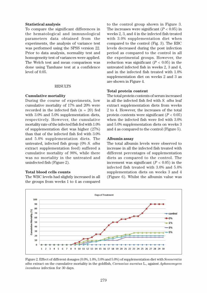
Cumulative mortality
During the course of experiments, lowcumulative mortality of 17% and 20% wererecorded in the infected fish (n = 20) fedwith 3.0% and 5.0% supplementation diets,respectively. However, the cumulativemortality rate of the infected fish fed with 1.0%of supplementation diet was higher (27%)than that of the infected fish fed with 3.0%and 5.0% supplementation diets. Theuntreated, infected fish group (0% S. alba
extract supplementation feed) suffered acumulative mortality of 90%, while therewas no mortality in the untreated anduninfected fish (Figure 2).
Total blood cells counts
The WBC levels had slightly increased in allthe groups from weeks 1 to 4 as compared
to the control group shown in Figure 3.The increases were significant (P < 0.05) inweeks 2, 3, and 4 in the infected fish treatedwith 3.0% supplementation diet whencompared to the control (Fig. 3). The RBClevels decreased during the post infectionperiod as compared to the control in allthe experimental groups. However, thereduction was significant (P < 0.05) in theuntreated infected fish in weeks 2, 3 and 4,and in the infected fish treated with 1.0%supplementation diet on weeks 2 and 3 asare shown in Figure 4.
Total protein content
The total protein contents of serum increasedin all the infected fish fed with S. alba leafextract supplementation diets from weeks2 to 4. However, the increases of the totalprotein contents were significant (P < 0.05)when the infected fish were fed with 3.0%and 5.0% supplementation diets on weeks 3and 4 as compared to the control (Figure 5).
Albumin assay
The total albumin levels were observed toincrease in all the infected fish treated withdifferent percentages of supplementationdiets as compared to the control. Theincrement was significant (P < 0.05) in theinfected fish treated with 3.0% and 5.0%supplementation diets on weeks 3 and 4(Figure 6). Whilst the albumin value was
Figure 2. Effect of different dosages (0.0%, 1.0%, 3.0% and 5.0%) of supplementation diet with Sonneratia
alba extract on the cumulative mortality in the goldfish, Carassius auratus L., against Aphanomyces
invadans infection for 30 days.

280
Figure 4. Effects of different dosages (0.0%, 1.0%, 3.0% and 5.0%) of supplementation diet with Sonneratia
alba extract on the total red blood cell (RBC) in the goldfish, Carassius auratus L., against Aphanomyces
invadans infection for four weeks. Data are presented as mean ± S.E. and significant differences (*p< 0.05) are indicated by asterisk over the bars.
Figure 3. Effects of different dosages (0.0%, 1.0%, 3.0% and 5.0%) of supplementation diet with Sonneratia
alba extract on total white blood cell (WBC) in the goldfish, Carassius auratus L., against Aphanomyces
invadans infection for four weeks. Data are presented as mean ± S.E. and significant differences (*p< 0.05) are indicated by asterisk over the bars.
slightly decreased in the untreated, infectedgroup on weeks 2 and 3.
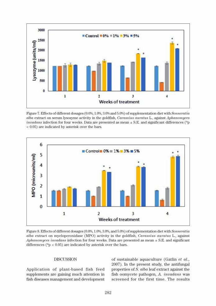
Serum lysozyme activity
The lysozyme activity of serum did notshow any significant changes during weeks1 and 2 in all the infected fish treated with
different percentages of supplementationdiets. Whilst in the infected fish treated with3.0% and 5.0% supplementation diets, thelysozyme levels were significantly increased(P < 0.05) as compared to the control onweeks 3 and 4 (Figure 7).

281
Figure 5. Effects of different dosages (0.0%, 1.0%, 3.0% and 5.0%) of supplementation diet with Sonneratia
alba extract on total serum protein in the goldfish, Carassius auratus L., against Aphanomyces
invadans infection for four weeks. Data are presented as mean ± S.E. and significant differences (*p< 0.05) are indicated by asterisk over the bars.
Figure 6. Effects of different dosages (0.0%, 1.0%, 3.0% and 5.0%) of supplementation diet with Sonneratia
alba extract on total serum albumin in the goldfish, Carassius auratus L., against Aphanomyces
invadans infection for four weeks. Data are presented as mean ± S.E. and significant differences (*p< 0.05) are indicated by asterisk over the bars.
Myeloperoxidase (MPO) assay
Myeloperoxidase activities were signifi-cantly increased when the fish group treatedwith supplementation diets containing 3.0%and 5.0% of S. alba extract on weeks 2, 3
and 4 as compared to the control. Thesevalues were not statistically changed inthe infected fish treated with 1.0% supple-mentation diet (Figure 8).
282
Figure 7. Effects of different dosages (0.0%, 1.0%, 3.0% and 5.0%) of supplementation diet with Sonneratia
alba extract on serum lysozyme activity in the goldfish, Carassius auratus L., against Aphanomyces
invadans infection for four weeks. Data are presented as mean ± S.E. and significant differences (*p< 0.05) are indicated by asterisk over the bars.
Figure 8. Effects of different dosages (0.0%, 1.0%, 3.0%, and 5.0%) of supplementation diet with Sonneratia
alba extract on myeloperoxidase (MPO) activity in the goldfish, Carassius auratus L., againstAphanomyces invadans infection for four weeks. Data are presented as mean ± S.E. and significantdifferences (*p < 0.05) are indicated by asterisk over the bars.
DISCUSSION
Application of plant-based fish feedsupplements are gaining much attention infish diseases management and development
of sustainable aquaculture (Gatlin et al.,
2007). In the present study, the antifungalproperties of S. alba leaf extract against thefish oomycete pathogen, A. invadans wasscreened for the first time. The results

283
showed that goldfish fed diet enriched withS. alba leaf extract at concentrations of 3.0%and 5.0% may improve the immunity anddisease resistance against A. invadans. Thecumulative mortality was recorded lower(17% and 20%) in the infected fish fed with3.0% and 5.0% supplementation diets,respectively as compared to the othertreated groups. However, 90% of the fishdied from EUS infection in the untreatedgroups. This has shown that S. alba leafextract has probably improved the resistanceof fish against EUS. Similarly, Yogeshwariet al. (2015) showed that the cumulativemortalities of the A. invadans-infectedIndian major carp, Labeo rohita (Hamilton)administered with 5.0% and 10% supple-mentation diets of the herbal extract,Rauvolfia tetraphylla had lower to 15% and25%, respectively. In addition, Harikrishnanet al. (2011b) found that Japanese snowbell,Styrax japonicas (Siebold and Zucc)enriched diets significantly increased thesurvival rate of Kelp grouper, Epinephelus
bruneus (Bloch) fed for 30 days after beingchallenged with a marine bacterium, Vibrio
harveyi (Johnson and Shunk) and a ciliateprotozoan, Uronema marinum (Womersley).Furthermore, the lysozyme activity, totalprotein and myeloperoxidase levels of theblood in E. bruneus significantly increasedwhen the fish were fed with 1.0% and 2.0%enriched diets, respectively, indicating anenhancement in the immune responses ofthe Kelp groupers against the targetpathogens (Harikrishnan et al., 2011b).
The first defense mechanism againstpathogens in fish is the innate (non-specific)immune system (Ahilan et al., 2010). Thecommon components of the innate immunesystem of fish are physical parameters,cellular and humoral factors, which mayinfluence the activity of innate immuneparameters (Magnadottir, 2006; Secombesand Fletcher, 1992). White blood cells(leukocytes: neutrophils, lymphocytes,monocytes, eosinophils, and basophils) arean important part of immune systems,which protect fish body against infectionspathogens (Abbas et al., 2014). Red bloodcells (erythrocytes) are responsible for blood
oxygen transportation and alterations in thisvalue reflect the health status of fish (Abbaset al., 2014). In this study, the WBC level wassignificantly higher (P < 0.05) in the infectedfish treated with 3.0% supplementation feedafter the second week of treatment. Thisindicated a significant improvement in thegeneral immune system status in the infectedfish within a short period of time after theadministration of S. alba leaf extract inthe feed. On the other hand, the RBC levelsdecreased in all the infected fish after theEUS-infection, and the reduction of RBC wassignificant in the untreated infected fishand the infected fish treated with 1.0%supplementation feed. These indicate thatthe severity of EUS-infection may have beensuppressed following the feeding of 3.0% and5.0% S. alba leaf extract supplementationfeeds. Similar observation was reported in astudy of goldfish infected with the bacterialpathogen, Aeromonas hydrophila (Chester)(Harikrishnan et al., 2010a). In their study,the WBC levels significantly increased in thefish group fed with 100 and 200 mg kg-1 ofmixed herbal extract supplementationfeeds while the RBC level decreased afterthe treatments. Harikrishnan et al. (2003) hadalso indicated that comparable changes inthe WBC and RBC counts in the ‘Commoncarp Cyprinus carpio L.’ which had beenartificially infected with A. hydrophila anddip-treated with neem (A. indica) leafextract for 10 days. They reported that 30days of oral administration of A. indica
ethanol extract supplementation diet mayprotect the hematological and biochemicalparameters in EUS-infected carp.
The total proteins in blood normallydecreased when an animal is infected bypathogenic organisms or affected by anyenvironmental stressors. This may due to afall in the absorbed amino acids that areessential for protein synthesis. In presentstudy, the total serum protein and albuminincreased significantly in the infected fishtreated with 3.0% and 5.0% supplementationfeeds after the third week of infection. Therewere no remarkable differences in thesevalues when the infected fish supplementedwith 1.0% S. alba leaf extract during the

284
experimental period. The decreases in thetotal protein and albumin in the blood ofthe untreated infected group highlightedthe positive effects of the administrated S.
alba leaf extract. The results of the presentstudy are in conformity with the findingby Harikrishnan et al. (2010a) who alsoobserved an increase in the total serumprotein in A. hydrophila-infected goldfishfollowed by the application of 400 and 800mg kg-1 of mixed herbal enriched diets onweek 4 of the treatments. In addition, Mariet al. (2014) suggested that the increases inthe total protein and albumin, lysozymeactivity in the infected Indian major carp,Cirrhinus mrigala (Hamilton) fed with1.0% chitosan enriched diet may haveenhanced the immunity of fish against A.
invadans infection. In another Indian majorcarp, Catla catla (Hamilton), the level of totalserum protein also increased when the fishwas fed with enriched diet containing ‘Chaff-flower Achyranthes aspera L.’ seed on day14 and 21 of post-injection (Vasudeva andChakrabarti, 2005). However, the authorsnoted a significant increase in the globulinbut not in the total protein or albumin.
Lysozyme activity has an importantrole in the innate immunity of fish and givesan indication of protection against invasivemicrobes (Saurabh and Sahoo, 2008). Inthe present study, the serum lysozymeconcentration was significantly higher (P <0.05) in the infected goldfish fed with 3%and 5% supplementation feeds on weeks 3and 4, suggesting that the immune systemof the infected goldfish may have beenimproved. There was no alteration in thetreated group with 1.0% S. alba leaf extract.Whilst a sharp decrease was noted in theinfected fish that did not have any S. alba
extract in their diets. Dhayanithi et al. (2015)recorded similar lysozyme activity whenthe authors applied the Gray mangrove,Avicennia marina (Forsk.) extractsenriched diets in the Clownfish, Amphiprion
sebae (Bleeker) against Vibrio alginolyticus
(Miyamoto) infection on weeks 6 and 8. Theyconcluded that administration of 4.0% and8.0% A. marina extracts may strengthen theimmune system of the infected clownfish.
Also the tilapia, Oreochromis niloticus L.which was fed with 0.1% and 0.5% oftraditional Chinese medicine (derived fromAstragalus) for a week, had also showed anincrement in the lysozyme activity (Yin et
al., 2006). Moreover, it was reported thatintramuscular administration of the tri-herbalcompounds (azadirachtin, camphor andcurcumin) at a concentration of 100 ppmcould significantly enhanced (P < 0.05) theserum lysozyme activity in C. mrigala
against A. invadans (Harikrishnan et al.,
2009, 2010b). Similarly, the effects of Chineseherb (Astragalus) on the non-specificimmunity of Jian carp, Cyprinus carpio
(var. Jian) and large yellow croaker,Pseudosciaena crocea (Richardson) werereported (Jian and Wu 2003; 2004).
Myeloperoxidase is the most abundantneutrophil granule protein, which issynthesized during myeloid differentiation,that plays a key role in the various functionsof neutrophils in innate and adaptiveimmunity (Odobasic et al., 2016). TheMPO values of the goldfish serum weresignificantly increased (P < 0.05) in the fishgroup treated with 3.0% and 5.0% of S. alba
extract supplementation feeds on weeks3 and 4 (present data). However, suchchanges in the MPO activity were notsignificant in the fish group treated with1% supplementation feed. Moreover, anobvious decline in the MPO level wasobserved in the untreated infected fish,which may be attributed to the invasivespread of A. invadans in the goldfish. InC. catla, Kumar et al. (2015) showed thatthere is a significant increase in the MPOactivity when the EUS-infected catla werefed with the leaf extract of heartleafhempvine, Mikania cordata (Burm. f.).
Many plant-based products have beenfound to have non-specific immuno-modulatory effects in animals (Pandey et al.,
2012), to improve the innate system, and mayserve as general immune prophylacticcontrol in organic aquaculture (Magnadottir,2010). Nowadays, crude plant extracts withantibacterial activity are potentially to bebeneficial alternatives in aquaculturebecause they may provide cheaper treat-

285
ment sources, which are less toxic thanchemotherapeutic agents (Maqsood et al.,
2011). The present study demonstrated thatthe immune system and survival rate of theA. invadans-infected goldfish may havebeen elevated when the fish were fed with3.0% to 5.0% of S. alba leaf extract supple-mentation diets. This study may also providean insight for upcoming research on theapplications of different dosages of S. alba
leaf extract in other EUS-susceptible fish,and the identification of specific bioactivecompounds present in its leaf extract forpotential development of antifungalcompounds.
Acknowledgements. This research wassupported by a research grant from theUniversiti Tunku Abdul Rahman, Malaysia[UTARRF vote no. 6200/W48] awarded toWong WL and Afzali SF, and by the UniversitiTunku Abdul Rahman post-doctoral researchscholarship awarded to Afzali SF.
Conflict of Interest
The authors declare no conflict of interests.
REFERENCES
Abbas, A.K., Lichtman, A.H. & Pillai, S. (2014).Cellular and Molecular Immunology. 8th
ed. Philadelphia: Elsevier.Afzali, S.F., Daud, H.H.M., Sharifpour, I.,
Afsharnasab, M. & Shankar, S. (2015).Experimental infection of Aphanomyces
invadans and susceptibility in sevenspecies of tropical fish. Veterinary World
8(9): 1038-1044.Afzali, S.F., Hassan, M.D. & Mutalib, A.R.
(2014). Induction of skin ulcers in moonlight gourami (Trichogaster microlepis)with Aphanomyces invadans zoospores.Pertanika Journal of Tropical Agri-
cultural Science 37(1): 133-140.Afzali, S.F. & Wong, W.L. (2017). In vitro
screening of Sonneratia alba extractagainst the oomycete fish pathogen,Aphanomyces invadans. Iranian
Journal of Fisheries Science 16(4): 1333-1340.
Ahilan, B., Nithiyapriyatharshini, A. &Ravaneshwaran, K. (2010). Influence ofcertain herbal additives on the growth,survival and disease resistance ofgoldfish, Carassius auratus (Linnaeus).Tamil Nadu Journal of Veterinary and
Animal Sciences 6(1): 5-11.Alam, M.N., Ahmed, G.U. & Chowdhury,
M.B.R. (2014). Performance of herbalextracts on diseased fish. Bangladesh
Journal of Veterinary Medicine 12(2):225-230.
Backer, C.A. & van Steenis, C.G.G.J. (1951).Sonneratiaceae. Flora Malesiana-Series1, Spermatophyta 4(1): 280-289.
Balasooriya, S.J., Sotheeswaran, S. &Balasubramanium, S. (1982). Econo-mically useful plants of Sri Lanka. PartIV. Screening of Sri Lanka plants fortannins. Journal of the National Science
Council of Sri Lanka 10: 213-219.Bandaranayake, W.M. (1998). Traditional
and medicinal uses of mangroves.Mangroves and Salt Marshes 2(3): 133-148.
Bandaranayake, W.M. (2002). Bioactivities,bioactive compounds and chemicalconstituents of mangrove plants.Wetlands Ecology and Management
10(6): 421-452.Bradford, M.M. (1976). A rapid and sensitive
method for the quantitation of microgramquantities of protein utilizing the principleof protein-dye binding. Analytical Bio-
chemistry 72: 248-254.Bruno, D. & Wood, B., 1999. Saprolegnia and
other oomycetes. Mycotaxon 95: 335-340.Campbell, R.E., Lilley, J.H., Panyawachira,
V. & Kanchanakhan, S. (2001). In vitro
screening of novel treatments forAphanomyces invadans. Aquaculture
Research 32(3): 223-233.Chakrabarti, R. (2005). Stimulation of
immunity in Indian major carp Catla catla
with herbal feed ingredients. Fish &
Shellfish Immunology 18(4): 327-334.Chowdhury, M.B.R. & Rahman, T. (2013). Use
of low-cost chemotherapeutic andmedicinal plants against Thai silver barb(Barbonymus gonionotus). Journal of
the Bangladesh Agricultural University
10(2): 385-390.

286
Citarasu, T. (2010). Herbal biomedicines: anew opportunity for aquaculture industry.Aquaculture International 18: 403-414.
David, J.C. & Kirk, P.M. (1997). Index of Fungi
6(13): 706-752.Dhayanithi, N.B., Kumar, T.T.A., Arockiaraj,
J., Balasundaram, C. & Harikrishnan, R.(2015). Dietary supplementation ofAvicennia marina extract on immuneprotection and disease resistance inAmphiprion sebae against Vibrio
alginolyticus. Fish & Shellfish Immu-
nology 45(1): 52-58.Doumas, B.T., Watson, W.A. & Biggs, H.G.
(1971). Albumin standards and themeasurement of serum albumin withbromcresol green. Clinica Chimica Acta
31(1): 87-96.Duke, N.C. & Jackes, B.R. (1987). A
systematic revision of the mangrovegenus Sonneratia (Sonneratiaceae) inAustralasia. Blumea-Biodiversity,
Evolution and Biogeography of Plants
32(2): 277-302.Fairweather, D.J. (1999). Development of
a bath challenge system to studycomponent causes, and preventativetreatments, of epizootic ulcerativesyndrome (EUS) in snakehead fish(Channa striata). MSc thesis, Univer-sity of Plymouth, Plymouth.
Gatlin, D.M., Barrows, F.T., Brown, P.,Dabrowski, K., Gaylord, T.G., Hardy, R.W.& Overturf, K. (2007). Expanding theutilization of sustainable plant productsin aquafeeds: a review. Aquaculture
Research 38(6): 551-579.Harikrishnan, R., Rani, M.N. & Balasundaram,
C. (2003). Hematological and bio-chemical parameters in common carp,Cyprinus carpio, following herbaltreatment for Aeromonas hydrophila
infection. Aquaculture 221(1): 41-50.Harikrishnan, R., Balasundaram, C. &
Bhuvaneswari, R. (2005). Restorativeeffect of Azadirachta indica aqueousleaf extract dip treatment onhaematological parameter changes inCyprinus carpio (L.) experimentally
infected with Aphanomyces invadans
fungus. Journal of Applied Ichthyology
21: 410-413. doi: 10.1111/j.1439-0426.2005.00614.
Harikrishnan, R., Balasundaram, C.,Dharaneedharan, S., Moon, Y.G., Kim,M.C., Kim, J.S. & Heo, M.S. (2009a). Effectof plant active compounds on immuneresponse and disease resistance inCirrhina mrigala infected with fungalfish pathogen, Aphanomyces invadans.Aquaculture Research 40(10): 1170-1181.
Harikrishnan, R., Balasundaram, C., Kim,M.C., Kim, J.S. & Heo, M.S. (2009b).Effective administration route ofazadirachtin and its impact onhaematological and biochemicalparameters in goldfish (Carassius
auratus) infected with Aeromonas
hydrophila. Bulletin of the Veterinary
Institute in Pulawy 53(1): 613-619.
Harikrishnan, R., Balasundaram, C. & Heo,M.S. (2010a). Herbal supplementationdiets on hematology and innate immunityin goldfish against Aeromonas hydro-
phila. Fish & Shellfish Immunology
28(2): 354-361.Harikrishnan, R., Balasundaram, C. &
Heo, M. (2010b). Supplementation dietcontaining probiotics, herbal andazadirachtin on hematological andbiochemical changes in Cirrhina
mrigala against Aphanomyces
invadans. Journal of Fisheries and
Aquaculture 4: 1-11.Harikrishnan, R., Balasundaram, C. & Heo,
M.S. (2011a). Impact of plant productson innate and adaptive immune systemof cultured finfish and shellfish.Aquaculture 317(1): 1-15.
Harikrishnan, R., Kim, J.S., Kim, M.C.,Balasundaram, C. & Heo, M.S. (2011b).Styrax japonica supplementation dietenhances the innate immune responsein Epinephelus bruneus againstbacterial and protozoan infections.Experimental Parasitology 129(3): 260-265.

287
Harikrishnan, R., Jawahar, S., Thamizharasan,S., Paray, B.A., Al-Sadoon, M.K. &Balasundaram, C. (2018). Immunedefense of emodin enriched diet inClarias batrachus against Aeromonas
hydrophila. Fish & Shellfish Immuno-
logy 76: 13-20.Immaraju, J.A. (1998). The commercial use
of azadirachtin and its integrationinto viable pest control programmes.Pesticide Science 54(3): 285-289.
Jahan, R., Rahman, S., Rehana, F., Anwar,M.M., Kalpana, M.A., Anwarul Bashar,A.B.M. & Rahmatullah, M. (2014).Treatment of epizootic ulcerativesyndrome (EUS) in Anabas testudineus
with an alkali-soluble fraction frompaddy husk. Advances in Natural &
Applied Sciences 8(9): 7-12.Jian, J. & Wu, Z. (2003). Effects of traditional
Chinese medicine on nonspecificimmunity and disease resistance of largeyellow croaker, Pseudosciaena crocea
(Richardson). Aquaculture 218(1): 1-9.Jian, J. & Wu, Z. (2004). Influence of traditional
Chinese medicine on non-specificimmunity of Jian Carp (Cyprinus carpio
var. Jian). Fish & Shellfish Immunology
16(2): 185-191.Johnson, R.A., Zabrecky, J., Kiryu, Y. & Shields,
J.D. (2004). Infection experiments withAphanomyces invadans in four speciesof estuarine fish. Journal of Fish
Diseases 27(5): 287-295.Kar, D. (2015). Epizootic Ulcerative Fish
Disease Syndrome. U.K.: AcademicPress.
Kumar, V., Suvra, R. & Debtanu, B. (2015).Effect of Mikania cordata (Burm) BLRobins on non-specific immune responseof Catla catla (Hamilton, 1822) againstAphanomyces invadans. Fishery
Technology 52: 20-25.Lilley, J.H. & Inglis, V. (1997). Comparative
effects of various antibiotics, fungicidesand disinfectants on Aphanomyces
invaderis and other saprolegniaceousfungi. Aquaculture Research 28(6): 461-469.
Lilley, J., Callinan, R.B., Chinabut, S. &Kanchanakhan, S. (1998). EpizooticUlcerative Syndrome (EUS) TechnicalHandbook. pp. 88. Bangkok: The AquaticAnimal Health Research Institute.
Magnadottir, B. (2010). Immunologicalcontrol of fish diseases. Journal of
Marine Biotechnology 12: 361-379.Magnadottir, B. (2006). Innate immunity
of fish (overview). Fish & Shellfish
Immunology 20(2): 137-151.Maqsood, S., Prabjeet, S., Munir, H.S. &
Khusheeba, M. (2011). Emerging roleof immunostimulants in combatingthe disease outbreak in aquaculture.International Aquatic Research 3: 147-163.
Mari, L.S.S., Jagruthi, C., Anbazahan, S.M.,Yogeshwari, G., Thirumurugan, R.,Arockiaraj, J. & Harikrishnan, R.(2014). Protective effect of chitin andchitosan enriched diets on immunity anddisease resistance in Cirrhina mrigala
against Aphanomyces invadans. Fish &
Shellfish Immunology 39(2): 378-385.Nauseef, W.M. (2007). Isolation of human
neutrophils from venous blood. In: Quinn,M.T., DeLeo, F.R. & Bokoch, G.M. eds.Neutrophil Methods and Protocols.Methods in Molecular BiologyTM 412
Totowa: Human Press. p 15-20.Odobasic, D., Kitching, A.R. & Holdsworth,
S.R. (2016). Neutrophil-mediatedregulation of innate and adaptiveimmunity: the role of myeloperoxidase.Journal of Immunology Research 2016.p 11. doi:10.1155/2016/2349817
OIE (2017). Chapter 2.3.2 Infection withAphanomyces invadans (Epizooticulcerative syndrome). In: DiagnosticManual for Aquatic Animal Diseases.Second edition. http://www.oie.int/fileadmin/Home/eng/Health_standards/aahm/current/chapitre_aphanomyces_invadans.pdf (Cited 22 Oct 2017).
Pandey, G., Madhuri, S. & Mandloi, A.K. (2012)Medicinal plants useful in fish diseases.Plant Archives 12(1): 1-4.

288
Peter, K.L.N. & Sivasothi, N. (1999). A Guideto the Mangroves of Singapore I: theEcosystem and Plant Diversity.Singapore: Singapore Science Centre. p136-137.
Phadee, P., Kurata, O., Hatai, K., Hirono, I. &Aoki, T. (2004). Detection and identifi-cation of fish-pathogenic Aphanomyces
piscicida using polymerase chainreaction (PCR) with species-specificprimers. Journal of Aquatic Animal
Health 16(4): 220-230.Popp, M., Larher, F. & Weigel, P. (1984).
Chemical composition of Australianmangroves III. Free amino acids, totalmethylated onium compounds andtotal nitrogen. Zeitschrift für
Pflanzenphysiologie 114(1): 15-25.Saad, S., Taher, M., Susanti, D., Qaralleh, H.
& Awang, A.F.I.B. (2012). In vitro anti-microbial activity of mangrove plantSonneratia alba. Asian Pacific Journal
of Tropical Biomedicine 2(6): 427-429.Saha, D. & Pal, J. (2002). In vitro antibiotic
susceptibility of bacteria isolated fromEUSaffected fishes in India. Letters in
Applied Microbiology 34(5): 311-316.Saurabh, S. & Sahoo, P.K. (2008). Lysozyme:
an important defence molecule of fishinnate immune system. Aquaculture
Research 39(3): 223-239.Secombes, C.J. & Fletcher, T.C. (1992). The
role of phagocytes in the protectivemechanisms of fish. Annual Review of
Fish Diseases 2: 53-71.Shugar, D. (1952). The measurement of
lysozyme activity and the ultra-violetinactivation of lysozyme. Biochimica et
Biophysica Acta 8: 302-309.
Tomlinson, P.B. (1994). The Botany ofMangroves. Cambridge: CambridgeUniversity Press.
Uthayakumar, V., Chandirasekar, R., Sreedevi,P.R., Senthilkumar, D., Jayakumar, R. &Ramasubramanian, V. (2014). Immuno-stimulatory effect and disease resistanceinduced by Lawsonia inermis againstAphanomyces invadans in stripedmurrels (Channa striatus). Malaya
Journal of Biosciences 1(4): 231-241.Valladao, G.M.R., Gallani, S.U. & Pilarski, F.
(2015). Phytotherapy as an alternativefor treating fish disease. Journal of
Veterinary Pharmacology and Thera-
peutics 38(5): 417-428.Vasudeva, R.Y. & Chakrabarti, R. (2005).
Stimulation of immunity in Indianmajor carp Catla catla with herbalfeed ingredients. Fish & Shellfish
Immunology 18: 327-334.Yin, G., Jeney, G., Racz, T., Xu, P., Jun, X. &
Jeney, Z. (2006). Effect of two Chineseherbs (Astragalus radix and Scutellaria
radix) on non-specific immune responseof tilapia, Oreochromis niloticus.Aquaculture 253(1): 39-47.
Yogeshwari, G., Jagruthi, C., Anbazahan,S.M., Mari, L.S.S., Selvanathan, J.,Arockiaraj, J. & Ramasamy, H. (2015).Herbal supplementation diet on immuneresponse in Labeo rohita againstAphanomyces invadans. Aquaculture
437: 351-359.