effects of different phenolic compounds on antioxidant...
TRANSCRIPT
Chapter 3
EFFECTS OF DIFFERENT PHENOLIC COMPOUNDS ON ANTIOXIDANT ENZYMES
AND LIPID PEROXIDATION IN OREOCHROMIS MOSSAMBICUS
3.1 Introduction
3.1.1 The Oxygen Radical Cascade 3.1.2 Lipid Peroxidation
3.2 Materials and methods 3.2.1 Parameters investigated
3.3 Statistical analysis 3.4 Results
3.4.1 Superoxide Dismutase (SOD) 3.4.2 Catalase (CAT) 3.4.3 Glutathione peroxidase (GPx) 3.4.4 Glutathione-S-transferase (GST) 3.4.5 Total reduced Glutathione (GSH) 3.4.6 Conjugated dienes (CD) 3.4.7 Hydroperoxides (HP) 3.4.8 Malondialdehyde (MDA)
3.5 Discussion
Co
nt
en
ts
Effects of different phenolic compounds on antioxidant enzymes and ………
BIOCHEMICAL EFFECTS OF DIFFERENT PHENOLIC COMPOUNDS ON OREOCHROMIS MOSSAMBICUS (PETERS) 60
3.1 Introduction
Oxidative stress and oxidative damage to fundamental biomolecules and to
antioxidant defenses of organisms is an established field in environmental
toxicology and ecotoxicology (Kelly et al., 1998; Regoli et al., 2002a, b).The
biochemistry of ‘reactive oxygen species’ (ROS) is an important field with
practical implications. This is because oxygen is an essential component of living
organisms and the formation of reactive oxygen intermediates seems to be
common in aerobically metabolizing cells. In addition to aerobic metabolism-
encompassing electron-transfer chains and certain enzyme activities,
environmental sources such as air pollutants, photochemical smog, industrial
chemicals, ionizing radiations, as well as metabolism of xenobiotics contribute to
the cellular steady-state concentration of ROS. Further, reactive species are
formed as a response to diverse stimuli by specialized physiological reactions: the
formation of oxyradicals during respiratory burst and the release of the
endothelium-derived releasing factor, identified as nitric oxide are such examples.
Regulated production of free radicals in higher organisms and maintenance of
‘‘redox homeostasis’’ are essential for the physiological health of organisms
(Ames et al., 1993). But during these metabolic processes, a small proportion (2–
3%) of free radicals may escape from the protective shield of antioxidant
mechanisms, causing oxidative damage to cellular components.
Other endogenous sources of ROS within cells are several oxidizing
enzymes, such as tryptophan dioxygenase, xanthine oxidase and cytochrome P450
reductase which can produce O2.-, while enzymes such as guanyl cyclase and
glucose oxidase generate H2O2 (Vigo-Pelfrey, 1990). Cytochrome P450
involvement in the production of ROS is of additional interest in toxicology
because it is involved in the metabolism of xenobiotics (Fridovich, 1978).
3.1.1 The Oxygen Radical Cascade
The oxygen activation follows a series of electron transfer reactions and the
pertinent ones are as follows.

Effects of different phenolic compounds on antioxidant enzymes and ………
BIOCHEMICAL EFFECTS OF DIFFERENT PHENOLIC COMPOUNDS ON OREOCHROMIS MOSSAMBICUS (PETERS) 61
O2+e- → O2.-
O2.- +H+ → HO2
HO2+ H+ e- → H2O2
H2O2+ e- → OH+OH-
The endogenous sources for production of O2.- ranges from small to large
autooxidizable molecules such as catecholamines, ubihydroquinone and
oxidoreductases such as haemoproteins and flavin enzymes. Thus O2.- is generated
in virtually all sub cellular compartments including cytosol, mitochondria,
endoplasmic reticulum, nuclei etc.
General harmful effects of reactive oxygen species in cell are: oxidations of
polyunsaturated fatty acids in lipids (lipid peroxidation), damage of DNA,
oxidations of amino acids in proteins, oxidative inactivation of specific enzymes
by oxidation of co-factors.
3.1.2 Lipid Peroxidation
The term lipid peroxidation is broadly defined as the formation of lipid
radical, which would then react with molecular oxygen in the form of a chain of
reactions resulting in the breakdown of polyunsaturated fatty acids (PUFA). This
reaction sequence is known alternatively as lipid peroxidation or “oxidative
deterioration of polyunsaturated lipids”. Lipid peroxidation is a free radical
mediated chain reaction that results in the oxidative degradation of unsaturated
lipids especially PUFA, such as C20:4 and C22:6 which occurs in the
phospholipids of most biological membrane assemblies. Biological membranes
rich in unsaturated fatty acids are in close contact with oxygen rich metal ion
containing fluid. Therefore membrane lipids are highly susceptible to
peroxidative damage (Ray and Akhtar, 2002).
Initiation of lipid peroxidation in a membrane or free fatty acid is due to the
attack of any species that has sufficient reactivity to abstract a methylene hydrogen
atom from the diene portion of an unsaturated fatty acid (Fig. 3.1) and forms a lipid

Effects of different phenolic compounds on antioxidant enzymes and ………
BIOCHEMICAL EFFECTS OF DIFFERENT PHENOLIC COMPOUNDS ON OREOCHROMIS MOSSAMBICUS (PETERS) 62
radical. Reaction of the lipid free radical with molecular oxygen leads to the formation
of a lipid hydroperoxy radical (L00.). The lipid hydroperoxy radical could then react
along either of the two pathways. It could: - abstract a methylene hydrogen from a
neighbouring unsaturated fatty acid forming a lipid hydroperoxide and a second lipid
radical or undergo an intermolecular–cyclisation to form a five membered lipid
endoperoxide radical. These lipid radicals formed would then react with molecular
oxygen and with other unsaturated lipids resulting in the extraction of methylene
hydrogen and continuation of the radical chain reactions (Ray and Akhtar, 2002).
Breakdown of lipid hydroperoxides and endoperoxides leads to formation of more than
twenty known products of lipid peroxidation (Fridovich, 1983).
Fig.3.1 Formation of Conjugated diene (CD) and lipid hydroperoxide (HP)
following hydrogen abstraction from a polyunsaturated fatty acid.

Effects of different phenolic compounds on antioxidant enzymes and ………
BIOCHEMICAL EFFECTS OF DIFFERENT PHENOLIC COMPOUNDS ON OREOCHROMIS MOSSAMBICUS (PETERS) 63
The resulting lipid hydroperoxides can easily decompose into several
reactive species including lipid alkoxyl radicals, aldehydes (e.g. malondialdehyde,
OHC–CH2– CHO), alkanes, lipid epoxides, and alcohols. Most of these products
are toxic and active mutagens (Esterbauer et al., 1990., Porter et al., 1995;
D’Ischia et al., 1996). The only mechanism, which produces malondialdehyde
(MDA) in biological systems, is lipid peroxidation. MDA is not the major product
of lipid peroxidation, but a typical degradation product. Lipid peroxidation
products may form DNA adducts giving rise to mutations and altered patterns of
gene expression (Marnett, 1999). MDA reacts with nitrogenous base of DNA to
form DNA adducts (Fujimoto et al., 1984). Uncontrolled lipid peroxidation leads
to the disruption of phospholipid membranes and to cell lysis. Peroxides also
interact with macromolecules such as sulphydryl groups of proteins and bases of
nucleic acids abrupting normal function and causing mutation (Slater and
Benedetton, 1981). Additionally peroxides and free radial are produced by the
immune system to mediate response to a foreign organism. Peroxidized
membranes become rigid and lose permeability and integrity.
It is evident then that ‘oxidative stress is a chain event, and a single
initiating event caused by a prooxidant may cascade into a widespread chain
reaction that produces many deleterious products in concentrations many
magnitudes greater than the initiator (Ahmad, 1992). This is exemplified by the
fact that thousands of PUFA molecules may be destroyed by a lipid peroxidation
chain reaction initiated by a single initiator free radical (McCord, 1985). It is
imperative that in order to prevent this vicious chain reaction, the O2 radical
cascade to. O2.- and H2O2 must be attenuated, and the peroxides converted to
innocuous metabolites. The prevention of lipid peroxidation is an essential
process in all the aerobic organisms, as lipid peroxidation products can cause
DNA damage. On the other hand, hydrogen peroxide has been implicated as an
intracellular messenger that affects cellular processes including protein
phosphorylation, transcription and apoptosis (Choi et al., 1998).
Effects of different phenolic compounds on antioxidant enzymes and ………
BIOCHEMICAL EFFECTS OF DIFFERENT PHENOLIC COMPOUNDS ON OREOCHROMIS MOSSAMBICUS (PETERS) 64
DNA in cellular nuclei is another key cellular component that is particularly
susceptible to oxidative damage by ROS (Cerutti, 1985). The polyanionic nature
of DNA provides a useful substrate for infiltration through membranes and
adherence of metal cations, thus facilitating the formation of HO. adjacent to these
critical biological targets (Halliwell and Aruoma, 1991). Additionally, the
heterogeneity of DNA molecules allows for HO. attacks, including the
nucleobases and the sugar–phosphate backbone (Buxton et al., 1988). Hydroxyl
radicals react with nucleobases approximately five times faster than with the
nucleic acid backbone (Cadet et al., 1997). Other hydroxyl radical attacks can be
directed towards the sugar–phosphate backbone of DNA, causing different
lesions, including apurinic sites where the base has been removed, fragmentation
of deoxyribose with single-strand breaks, and oxidation of the sugar moiety
(Dizdaroglu et al., 1975; Breen and Murphy, 1995).
Protein oxidation reactions involve various propagating radicals and ROS
and the results are oxidative modifications of amino acid side chains, reactive
oxygen species mediated peptide cleavage, reactions of peptides with lipids and
carbohydrate oxidation products and formation of carbonyl derivatives of
proteins. Oxidatively modified proteins accumulate during ageing. Of the various
indices of protein oxidation, protein carbonyl formation is the best studied
(Stadtman and Berlett, 1999). There are a variety of pathways through which
protein carbonyls are formed (Levine et al., 2000). Experimental studies showed
that oxidative damage to proteins by ROS leads to the accumulation of oxidatively
modified forms of enzymes which are implicated in ageing (Wolff and Dean,
1986). This accumulation during ageing reflects a loss in the capacity of the
organisms to degrade oxidised proteins, with subsequent effects on the
transcriptional and translational fidelity mechanisms (Duikan et al., 2000).
The aquatic environment receives daily substantial amounts of
environmental pollutants that have the potential to cause oxidative stress in
aquatic organisms through free radical and ROS mechanisms. The uptake of these

Effects of different phenolic compounds on antioxidant enzymes and ………
BIOCHEMICAL EFFECTS OF DIFFERENT PHENOLIC COMPOUNDS ON OREOCHROMIS MOSSAMBICUS (PETERS) 65
pollutants by aquatic organisms can occur from sediments, suspended particulate
matter with toxic properties and food sources. Exposure to these contaminants
will depend on the particular dietary and ecological lifestyles of the aquatic
organisms. Current knowledge and recent advances of oxidative toxicity by
xenobiotics in aquatic organisms provide a fertile field for aquatic toxicology
studies (Livingstone, 1998). Aquatic organisms were chosen as test species
because of their filtration capacity and sensitivity to oxidative damage from
concerning chronic exposure or sub-lethal concentrations. Aquatic organisms
can provide model systems for investigation of how ROS damage cellular
components, how cells respond, how repair mechanisms ameliorate this
damage and how oxidative stress can lead to disease (Di Giulio et al., 1989;
Livingstone et al., 1994). Aquatic organisms are more sensitive to exposure
and toxicity compared to terrestrial organisms including mammals and in this
respect they may provide experimental data for evaluation of subtle effects of
oxidative stress, mutagenicity, and other adverse effects of pollutants
(Lackner, 1998). Some aquatic organisms can provide better models for
linking malignant neoplasms with carcinogenic pollutants (Malins et al.,
1988). Environmental toxicity studies on aquatic organisms have focused
primarily on redox cyclic compounds (quinones, aromatic hydrocarbon-
quinones, nitropyrene, lindane, paraquat, nitrobenzoic acid, etc.) and their
effects on subcellular fractions (microsomes) of the major organs of
biotransformation using the adult or larval stages (catfish, rainbow trout,
flounder, mussels, etc.) (Garcia Martinez and Livingstone, 1995; Lemaire and
Livingstone, 1997; Sjo lin and Livingstone, 1997).
Biological systems have developed during their evolution adequate
enzymatic and non- enzymatic antioxidant mechanisms to protect their cellular
components from oxidative damage. Consequently, all aerobic organisms possess
elaborate defense mechanisms to prevent the formation of toxic forms of oxygen
and to remove peroxides formed.

Effects of different phenolic compounds on antioxidant enzymes and ………
BIOCHEMICAL EFFECTS OF DIFFERENT PHENOLIC COMPOUNDS ON OREOCHROMIS MOSSAMBICUS (PETERS) 66
Aquatic organisms metabolize organic xenobiotics by phase I metabolism
which produces reactive oxygen species (ROS) as by-products (Livingstone, 1991).
Oxidative stress occurs when reactive oxygen species (ROS), such as superoxide ion
(O2.-), hydrogen peroxide (H2O2), hydroxyl radical (.OH) and singlet oxygen (1O2)
react with lipids, proteins or nucleic acids resulting in several biochemical injuries
(Yu and Anderson, 1997; Pinchuk and Lichtenberg, 2002; Valvanidis et al., 2006).
Detoxification of ROS is one of the prerequisites of aerobic life (McCord, 2000), and
many defenses have evolved providing an antioxidant system which is able to
prevent, intercept and repair damages. It consists of non-enzymatic ROS scavengers
such as: ascorbic acid, reduced glutathione, α- tocopherol, flavonoids, β-carotene and
urate, and also of an enzymatic system that includes superoxide dismutase,
glutathione peroxidase, catalase, NADPH quinine oxidoreductase, DT-diaphorase,
epoxide hydrolase, glucose-6-phosphate dehydrogenase and a few conjugation
enzymes (Sies, 1991; Valvanidis et al., 2006).
Oxidative stress develops when the levels of antioxidants are lowered or
when production of reactive oxygen species (ROS) exceeds the capacity of the
cell to dispose of them. ROS are produced by the univalent reduction of dioxygen
to superoxide anion, which in turn disproportionates to H2O2 and O2
spontaneously or through a reaction catalyzed by superoxide dismutase (SOD).
2 .O2.- + 2 H+ H2O2 + O2
SOD (EC 1.15.1.1) was first discovered by McCord and Fridovich in 1969.
The SOD family consists of four metalloforms; two forms containing copper and
zinc, one form containing manganese and another form containing iron.
Cu,ZnSOD is found in the cytosol of most eukaryotic cells (Fridovich,1975). A
different form of Cu,ZnSOD is found in extracellular fluids, where it is called
ECSOD (Marklund,1984; Marklund et al.,1985). MnSOD is located in the
mitochondrial matrix as well as in bacteria, while FeSOD is present in many
aerobic bacteria (Fridovich, 1974). In eukaryotic cells, three forms of SOD are
known to exist: Cu,ZnSOD, EC-SOD, and MnSOD.
Effects of different phenolic compounds on antioxidant enzymes and ………
BIOCHEMICAL EFFECTS OF DIFFERENT PHENOLIC COMPOUNDS ON OREOCHROMIS MOSSAMBICUS (PETERS) 67
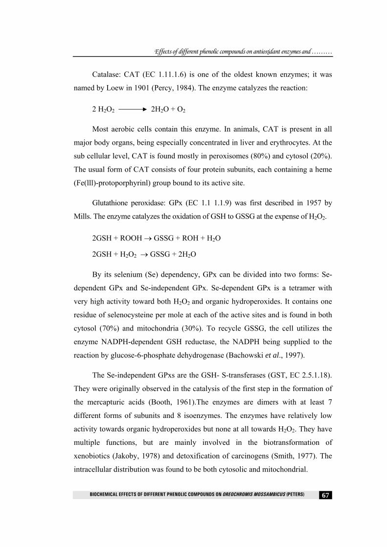
Catalase: CAT (EC 1.11.1.6) is one of the oldest known enzymes; it was
named by Loew in 1901 (Percy, 1984). The enzyme catalyzes the reaction:
2 H2O2 2H2O + O2
Most aerobic cells contain this enzyme. In animals, CAT is present in all
major body organs, being especially concentrated in liver and erythrocytes. At the
sub cellular level, CAT is found mostly in peroxisomes (80%) and cytosol (20%).
The usual form of CAT consists of four protein subunits, each containing a heme
(Fe(lll)-protoporphyrinl) group bound to its active site.
Glutathione peroxidase: GPx (EC 1.1 1.1.9) was first described in 1957 by
Mills. The enzyme catalyzes the oxidation of GSH to GSSG at the expense of H2O2.
2GSH + ROOH → GSSG + ROH + H2O
2GSH + H2O2 → GSSG + 2H2O
By its selenium (Se) dependency, GPx can be divided into two forms: Se-
dependent GPx and Se-independent GPx. Se-dependent GPx is a tetramer with
very high activity toward both H2O2 and organic hydroperoxides. It contains one
residue of selenocysteine per mole at each of the active sites and is found in both
cytosol (70%) and mitochondria (30%). To recycle GSSG, the cell utilizes the
enzyme NADPH-dependent GSH reductase, the NADPH being supplied to the
reaction by glucose-6-phosphate dehydrogenase (Bachowski et al., 1997).
The Se-independent GPxs are the GSH- S-transferases (GST, EC 2.5.1.18).
They were originally observed in the catalysis of the first step in the formation of
the mercapturic acids (Booth, 1961).The enzymes are dimers with at least 7
different forms of subunits and 8 isoenzymes. The enzymes have relatively low
activity towards organic hydroperoxides but none at all towards H2O2. They have
multiple functions, but are mainly involved in the biotransformation of
xenobiotics (Jakoby, 1978) and detoxification of carcinogens (Smith, 1977). The
intracellular distribution was found to be both cytosolic and mitochondrial.

Effects of different phenolic compounds on antioxidant enzymes and ………
BIOCHEMICAL EFFECTS OF DIFFERENT PHENOLIC COMPOUNDS ON OREOCHROMIS MOSSAMBICUS (PETERS) 68
Glutathione transferases are a family of detoxifying enzymes with broad and
overlapping substrate specificities towards carcinogenic, mutagenic, toxic and
pharmacologically active compounds (Boyland and Chasseaud, 1969). Among the
substrates are many reactive intermediates formed via the P450 system
(Chasseaud, 1978), as well as compounds formed during lipid peroxidation
(Ahlin, 1985). Glutathione S-transferases (GST) play an important role in the
detoxification and excretion of xenobiotics by catalyzing the conjugation of the
tripeptide glutathione (GSH) with the xenobiotic in the phase II of the
biotransformation process promoting its elimination from the organism (Leaver
et al., 1992). This enzyme has been used in laboratory and field studies as a
biomarker for several contaminants (Almar et al., 1998; Fenet, 1998).
Glutathione (γ-_-glutamyl-cysteinyl-glycine) is a tripeptide that is mainly
present in cells in its reduced form (GSH), which basically acts as an intracellular
reductant and nucleophile. It functions in the synthesis of proteins and DNA, amino
acid transport, maintenance of the thiol-disulphide status, free radical scavenging,
signal transduction, as an essential cofactor of several enzymes, as a non-toxic storage
form of cysteine, and as a defence against oxidizing molecules and potentially
harmful xenobiotics such as metals (Pena-Llopis et al., 2001; Elia et al., 2003).
The depletion of dissolved oxygen concentration of waters polluted with
phenolic wastes (Phipps et al., 1981) leads to formation of free radicals, especially
superoxide (O2.-) which acts by oxidizing various cellular substrates, especially
unsaturated fatty acids, which are very susceptible to free radical damage (Rady,
1993). In living organisms, it is necessary to limit the DNA damage caused by
reactive species with antioxidants in order for them to survive. Most components
of cellular structure and function are likely to be the potential targets of oxidative
damage and the most susceptible substrates for auto oxidation are polyunsaturated
fatty acids of the cell membrane, which undergo peroxidation rapidly. This may
lead to muscle degradation, impairment of the nervous system, haemolysis,
general deterioration of the cellular metabolism and eventual cell death.

Effects of different phenolic compounds on antioxidant enzymes and ………
BIOCHEMICAL EFFECTS OF DIFFERENT PHENOLIC COMPOUNDS ON OREOCHROMIS MOSSAMBICUS (PETERS) 69
Many environmental pollutants, such as phenol, may cause oxidative stress in
aquatic organisms by inducing ROS production (Sayeed et al., 2003; Oruc et al.,
2004). In biological systems, the balance between both endogenous and exogenous
pro-oxidant factors versus antioxidant defenses can be used to assess oxidative
damage induced by different classes of chemical pollutants (Valvanidis et al., 2006).
Changes of activity of antioxidant enzymes may depict a change in the ROS within
the cells. Therefore, these enzymes can be used as biomarkers for oxidative stress
(Roche and Boge, 2000; Valvanidis et al., 2006). Aquatic organisms are usually more
sensitive than terrestrials and may be better experimental subjects to evaluate subtle
effects of oxidative stress (Ahmad et al., 2000; Valvanidis et al., 2006).
In the present study biomarkers of oxidative stress in the gills, liver, kidney
and muscle of O. mossambicus exposed to different phenolic compounds for 21
days were experimented. The oxidative stress biomarkers studied included the
non-enzymatic antioxidant glutathione and antioxidant enzymes such as catalase,
superoxide dismutase, glutathione peroxidase and glutathione-S-transferase. Also
indicators of lipid peroxidation such as malondialdehyde, hydroperoxide and
conjugated dienes were estimated.
3.2 Materials and methods
Collection, maintenance, acclimatization and experimental design and
preparation of tissue samples were the same as explained in detail in chapter 2,
section 2.2.
3.2.1 Parameters investigated 3.2.1.1 Assay of superoxide dismutase (SOD) (E.C.1.15.1.1)
Superoxide dismutase in different tissues was determined using the method
of Kakkar et al. (1984).
Reagents
0.33 M sucrose, n-butanol , 0.052 M sodium pyro-phosphate buffer (pH 8.3),
0.0025 M Tris-HCl buffer (pH 7.4), 186 μM phenazine methosulphate (PMS), 300
μM Nitro blue tetrazolium (NBT), 780 μM NADH and glacial acetic acid.

Effects of different phenolic compounds on antioxidant enzymes and ………
BIOCHEMICAL EFFECTS OF DIFFERENT PHENOLIC COMPOUNDS ON OREOCHROMIS MOSSAMBICUS (PETERS) 70
Procedure
Weighed samples of tissues were homogenised in 0.33 M sucrose and
subjected to differential centrifugation under cold conditions to obtain the cytosol
fraction. Before estimating the activity, an initial purification was done by
precipitating the protein from the supernatant with 90% ammonium sulphate and
this fraction was then dialysed against 0.0025 M Tris- HCl buffer (pH 7.4). The
supernatant was used as the enzyme source. Assay mixture contained 1.2 ml of
sodium pyrophosphate buffer, 0.1 ml of PMS, 0.3 ml of NBT, 1.3 ml of distilled
water and 0.1 ml of the enzyme source. The tubes were kept at 30°C for one
minute and then 0.2 ml of NADH were added and incubated at 30°C for 90
seconds and the reaction was stopped by the addition of 1 ml of glacial acetic
acid. Reaction mixture was shaken vigorously with 4.0 ml of n-butanol. The
mixture was allowed to stand for 10 minutes and centrifuged. The upper butanol
layer was removed. Absorbance of the chromogen in butanol was measured at
560 nm against n-butanol blank. A system devoid of enzyme served as control.
One unit of enzyme activity is defined as the enzyme concentration required to
inhibit chromogen production by 50% in one minute under the assay conditions
and specific activity is expressed as units / mg protein.
3.2.1.2 Assay of Catalase (CAT) (E.C.1.11.1.6)
Catalase level in different tissues was determined using the method of
Maehly and Chance (1955).
Reagents 0.01M phosphate buffer (pH 7.0), 30mM H2O2.
Procedure
The estimation was done spectrophotometrically following the decrease in
absorbance at 230 nm. The reaction mixture contained 0.01 M phosphate buffer,
30 mM hydrogen peroxide and the enzyme extract prepared by homogenizing the
tissue in phosphate buffer and centrifuging at 5000 rpm. Specific activity was

Effects of different phenolic compounds on antioxidant enzymes and ………
BIOCHEMICAL EFFECTS OF DIFFERENT PHENOLIC COMPOUNDS ON OREOCHROMIS MOSSAMBICUS (PETERS) 71
expressed as International Units / mg protein. 1 1U = change in absorbance / min/
extinction coefficient (0.021).
3.2.1.3 Assay of Glutathione peroxidase (GPx) (E.C. 1.11.1.9)
Glutathione peroxidase in different tissues was estimated by the method of
Rotruck (1973).
Reagents
0.4 M Tris buffer (pH 7.0) 10mM sodium azide solution, 10% Trichloro
acetic acid (TCA) , 0.4 mM Ethylene diamine tetra acetic acid (EDTA), 0.2 mM
Hydrogen peroxide (H2O2) , 2 mM glutathione solution (GSH).
Procedure
Weighed samples of different tissues were homogenized in a known volume
of Tris buffer. To 0.2 ml of Tris buffer, 0.2 ml EDTA, 0.1 ml sodium azide and
0.5 ml tissue homogenate were added and mixed well. To this mixture 0.2ml of
GSH followed by 0.1 ml H2O2 solution were added. The contents were mixed and
incubated at 37°C for 10 minutes along with a control containing all reagents
except tissue homogenate. After 10 minutes the reaction was arrested by the
addition of 0.5 ml of 10% TCA. Tubes were centrifuged and the supernatant was
assayed for GSH. The values are expressed as μg of GSH / min/ mg protein.
3.2.1.3 Assay of Glutathione–S-transferase (GST) (E.C.2.5.1.18)
Glutathione-S-transferase in different tissue was determined using the
method of Beutler et al. (1986).
Reagents
0.5 M phosphate buffer (pH 6.5), 25 mM of 1-chloro-2, 4-dinitro benzene
(CDNB) in 95% ethanol, 20 mM glutathione (GSH)
Procedure
All the tissues were homogenized in 0.5 M phosphate buffer. The reaction
mixture contained 200μl phosphate buffer, 20 μl CDNB and 680 μl distilled
Effects of different phenolic compounds on antioxidant enzymes and ………
BIOCHEMICAL EFFECTS OF DIFFERENT PHENOLIC COMPOUNDS ON OREOCHROMIS MOSSAMBICUS (PETERS) 72
water. Then the tubes were incubated at 37°C for 10 minutes and added 50 μl of
GSH. After mixing well, added 50μl of tissue extract to the tube. Increase in
absorbance was noted at 340nm for 5 minutes in a UV–visible spectrophotometer.
Values are expressed in µmoles of CDNB complexed / min/ mg protein. The
extinction coefficient between CDNB –GSH conjugate is 9.6 mM-1 cm-1.
3.2.1.4 Estimation of total reduced glutathione (GSH)
Total reduced glutathione was estimated by the method of Ellman (1958).
Reagents
DTNB (0.6 Mm) in 0.2 M phosphate buffer (pH-8.0), TCA 5%, standard
glutathione.
Procedure
Precipitated protein in the homogenates of gills, liver, kidney and muscle
with 0.1 ml 5% TCA and 0.4 ml distilled water. Mixed the contents well for
complete precipitation of proteins and centrifuged. To 0.5 ml clear supernatant,
added 2.5 ml of 0.2 M phosphate buffer and 50 µl of DTNB. Read the absorbance
at 412 nm against a blank containing all the reagents. A series of standards were
run along with blank treated in a similar manner to determine the glutathione
content. Values were expressed as nmoles/100 g wet tissue.
3.2.1.5 Estimation of Conjugated dienes (CD)
The concentration of conjugated dienes was estimated according to the
method of Retnagal and Ghoshal (1966).
Procedure
Membrane lipids were extracted and evaporated to dryness as described for
the iodometric assay for hydroperoxides. The lipid residue was dissolved in 1.5 ml
of cyclohexane and the absorbance at 233 nm was determined against a
cyclohexane blank. Molar extinction coefficient of conjugated dienes is 2.52 x
104 M-1 cm -1.

Effects of different phenolic compounds on antioxidant enzymes and ………
BIOCHEMICAL EFFECTS OF DIFFERENT PHENOLIC COMPOUNDS ON OREOCHROMIS MOSSAMBICUS (PETERS) 73
3.2.1.6 Estimation of Hydroperoxides (HP)
Hydroperoxides was estimated by method of Mair and Hall, 1977.
Reagents
Potassium iodide, 0.5% cadmium acetate
Procedure
1ml of the tissue homogenate of the different tissues was mixed thoroughly
with 5 ml of chloroform: methanol (2:1) followed by centrifugation at 1000×g for
5 minutes to separate the phases. 3 ml of the lower chloroform layer was
recovered using a syringe and placed in a test tube and dried in a 45°C water bath.
1 ml of acetic acid: chloroform (3:2) mixture followed by 0.05 ml of potassium
iodide was quickly added and the test tubes were stoppered and mixed. The tubes
were placed in the dark at room temperature for exactly 5 minutes followed by the
addition of 3 ml of cadmium acetate. The solution was mixed and centrifuged at
1000 × g for 10 minutes. The absorbance of the upper phase was read at 353 nm
against a blank containing the complete assay mixture except the tissue
homogenate. Molar extinction coefficient of hydroperoxide is 1.73×10⁴ M-1 cm-1.
3.2.1.7 Estimation of Malondialdehyde
Malondialdehyde was estimated by the method of Niehaus and Samuelson, 1958.
Reagents
TCA-TBA-HCl reagent: 15% (w/v) Trichloro acetic acid, 0.375% (w/v)
Thiobarbituric acid (TBA) in 0.25 N HCl. 0.1 M Tris-HCl buffer (pH7.5).
Procedure
The tissue homogenate of different tissues were prepared in Tris-HCI buffer
and was combined with thiobarbituric acid reagent and mixed thoroughly and
heated for 15 minutes in a boiling water bath. It was then cooled and centrifuged for
10 minutes at 600×g. The absorbance of the sample was read spectrophotometrically
at 535 nm against a reagent blank that contained no tissue extract. The extinction

Effects of different phenolic compounds on antioxidant enzymes and ………
BIOCHEMICAL EFFECTS OF DIFFERENT PHENOLIC COMPOUNDS ON OREOCHROMIS MOSSAMBICUS (PETERS) 74
coefficient for malondialdehyde is 1.56 ×10⁵ M-1 cm-1. The values are expressed as
millimoles / 100g wet wt of tissue.
3.2.1.8 Estimation of Protein
Protein was estimated by the method of Lowry et al. (1951), (Chapter 2,
Section 2.2.6.11).
3.3 Statistical analysis
The statistical analysis was carried out using the software SPSS 13.0
package. Two-way analysis of variance (ANOVA) was carried out to compare
between different phenolic compounds treated groups and also between tissues. If
significant difference were revealed by the ANOVA test, Tukey’s test was used to
further elucidate which tissues and treatments were significantly different. One-way
ANOVA followed by Tukey’s test was also carried out for the comparison between
different treatments in each tissue. Significance level (P value) was set at 0.05 in all
tests.
3.4 Results 3.4.1 Superoxide Dismutase (SOD)
Two-factor ANOVA followed by Tukey’s test showed that there was
significant (P<0.05), (Fig 3.1 and Table 3.1) variation in SOD activity between
treatments and also between tissues. Between the treated groups, both phenol and m-
cresol treated groups showed significant variation in SOD activity and also with the
control. SOD activity was found to be significantly (P<0.05) elevated in gills, liver
and kidney of O. mossambicus treated with phenol compared to control and among
these tissues liver showed the maximum activity, whereas the fishes treated with m-
cresol showed significantly elevated activity in liver, kidney and muscle compared to
control. A significantly (P<0.05) decreased activity compared to control was shown
by gills treated with m-cresol and muscle treated with phenol.

Effects of different phenolic compounds on antioxidant enzymes and ………
BIOCHEMICAL EFFECTS OF DIFFERENT PHENOLIC COMPOUNDS ON OREOCHROMIS MOSSAMBICUS (PETERS) 75
Fig. 3.1 Effect of different phenolic compounds on SOD activity in
O. mossambicus. Each bar diagram represents mean ± S.D. On each set of bars, values with different lower case letters vary significantly (P<0.05) in each tissue on different treatments.
Table 3.1 Effect of different phenolic compounds on SOD activity in O. mossambicus. Values in the same column with different upper case letters vary significantly (P<0.05) between tissues and values in the same row with different lower case letters vary significantly (P<0.05) between treatment groups.
SOD activity
Groups Tissues
Control Phenol m-cresol
Gills b12.57±2.73B c23.11±2.76B
a5.24±2.86B
Liver b12.70±1.10D c27.60±2.47D
a15.90±1.54D
Kidney b13.0±0.25C c17.10±0.45C
a15.21±1.17C
Muscle b3.73±0.13A c2.11±0.45A
a4.60±0.77A
Values are expressed as units/mg protein. One unit is defined as the amount of enzyme which gives 50% inhibition of formazon formation / minute.
Each value represents the mean ± S.D of six separate experiments

Effects of different phenolic compounds on antioxidant enzymes and ………
BIOCHEMICAL EFFECTS OF DIFFERENT PHENOLIC COMPOUNDS ON OREOCHROMIS MOSSAMBICUS (PETERS) 76
3.4.2 Catalase (CAT)
In the present study catalase activity in different tissues of O. mossambicus
treated with different phenolic compounds showed significant variations (P
<0.05), (Fig 3.2 and Table 3.3) compared to control group. Tukey’s test showed
significant difference between phenolic compounds treated groups and also with
the control. Highest CAT activity was found in the liver of fishes treated with m-
cresol. On treatment with both phenol and m-cresol gills, liver and kidney showed
significantly elevated CAT activity compared to control. Comparison between
groups treated with different phenolic compounds revealed that there was
significant increase (P<0.05) in CAT activity in all tissues compared to control
except in muscle. Muscle showed a statistically significant decreased activity
compared to control.
Fig 3.2. Effect of different phenolic compounds on CAT activity in O. mossambicus. Each bar diagram represents mean ± S.D. On each set of bars, values with different lower case letters vary significantly (P <0.05) in each tissue on different treatments.

Effects of different phenolic compounds on antioxidant enzymes and ………
BIOCHEMICAL EFFECTS OF DIFFERENT PHENOLIC COMPOUNDS ON OREOCHROMIS MOSSAMBICUS (PETERS) 77
Table 3.2. Effect of different phenolic compounds on CAT activity in O. mossambicus. Values in the same column with different upper case letters vary significantly (P<0.05) between tissues and values in the same row with different lower case letters vary significantly (P<0.05) between treatment groups.
CAT activity
Groups Tissues
Control Phenol m-cresol
Gills a11.07±0.10C b15.06±3.22C
c14.44±1.32C
Liver a11.50±2.76D b34.34±2.74D c38.87±6.49D
Kidney a4.81±0.48B b8.62±0.98B c9.86±2.31B
Muscle a3.24±0.11A b2.97±0.99A c2.53±0.98A
One IU = Change in absorbance at 230 nm / min, Extinction Coefficient = 0.021
Each value represents the mean ± S.D of six separate experiments
3.4.3 Glutathione peroxidase (GPx)
Glutathione peroxidase activity showed an overall significant change
(P<0.05) in experimental groups of animal (Fig 3.3and Table 3.4) compared to
control. Tukey’s test showed significant difference between phenolic
compounds treated groups and also with the control. Statistical analysis
between tissues showed that GPx activity was found to show a statistically
significant (P<0.05) decreased activity in liver and kidney of the treated groups
compared to control. Whereas gills treated with phenol showed a decreased
GPx activity compared to control. On treatment with both phenol and m-cresol
muscle showed a significantly (P<0.05) elevated activity compared to control.

Effects of different phenolic compounds on antioxidant enzymes and ………
BIOCHEMICAL EFFECTS OF DIFFERENT PHENOLIC COMPOUNDS ON OREOCHROMIS MOSSAMBICUS (PETERS) 78
Fig.3.3 Effect of different phenolic compounds on GPx activity in O. mossambicus. Each bar diagram represents mean ± S.D. On each set of bars, values with different lower case letters vary significantly (P<0.05) in each tissue on different treatments.
Table 3.3 Effect of different phenolic compounds on GPx activity in O. mossambicus. Values in the same column with different upper case letters vary significantly (P<0.05) between tissues and values in the same row with different lower case letters vary significantly (P<0.05) between treatment groups.
GPx activity Groups Tissues
Control Phenol m-cresol Gills c11.98±1.10D
a6.45±2.22D b15.25±1.32D
Liver c10.36±2.76A a3.89±1.74A
b3.2±1.49A Kidney c12.42±0.48C
a4.95±0.98C b8.11±2.31C Muscle 5.72±1.11B
a7.07±0.99B b8.82±0.98B
Values are expressed as μg of GSH/min/mg protein. Each value represents the mean ± S.D of six separate experiments.
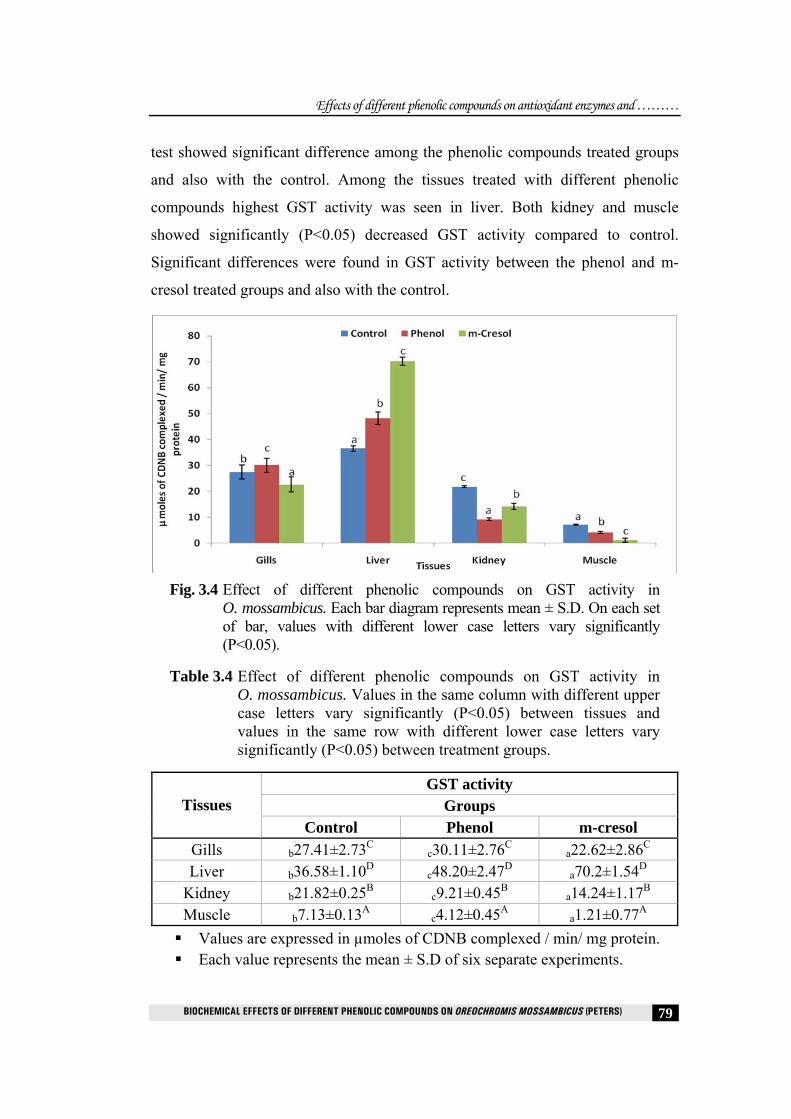
3.4.4 Glutathione-S-transferase (GST)
In the present study, glutathione-S-transferase activity in different tissues of
O. mossambicus treated with different phenolic compounds showed significant
variations (P<0.05), (Fig 3.2 and Table 3.5) compared to control group. Tukey’s
Effects of different phenolic compounds on antioxidant enzymes and ………
BIOCHEMICAL EFFECTS OF DIFFERENT PHENOLIC COMPOUNDS ON OREOCHROMIS MOSSAMBICUS (PETERS) 79
test showed significant difference among the phenolic compounds treated groups
and also with the control. Among the tissues treated with different phenolic
compounds highest GST activity was seen in liver. Both kidney and muscle
showed significantly (P<0.05) decreased GST activity compared to control.
Significant differences were found in GST activity between the phenol and m-
cresol treated groups and also with the control.
Fig. 3.4 Effect of different phenolic compounds on GST activity in
O. mossambicus. Each bar diagram represents mean ± S.D. On each set of bar, values with different lower case letters vary significantly (P<0.05).
Table 3.4 Effect of different phenolic compounds on GST activity in O. mossambicus. Values in the same column with different upper case letters vary significantly (P<0.05) between tissues and values in the same row with different lower case letters vary significantly (P<0.05) between treatment groups.
GST activity Groups Tissues
Control Phenol m-cresol Gills b27.41±2.73C
c30.11±2.76C a22.62±2.86C Liver b36.58±1.10D
c48.20±2.47D a70.2±1.54D Kidney b21.82±0.25B
c9.21±0.45B a14.24±1.17B Muscle b7.13±0.13A
c4.12±0.45A a1.21±0.77A Values are expressed in µmoles of CDNB complexed / min/ mg protein. Each value represents the mean ± S.D of six separate experiments.
Effects of different phenolic compounds on antioxidant enzymes and ………
BIOCHEMICAL EFFECTS OF DIFFERENT PHENOLIC COMPOUNDS ON OREOCHROMIS MOSSAMBICUS (PETERS) 80
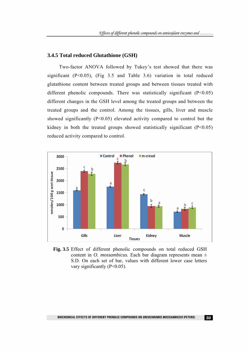
3.4.5 Total reduced Glutathione (GSH)
Two-factor ANOVA followed by Tukey’s test showed that there was
significant (P<0.05), (Fig 3.5 and Table 3.6) variation in total reduced
glutathione content between treated groups and between tissues treated with
different phenolic compounds. There was statistically significant (P<0.05)
different changes in the GSH level among the treated groups and between the
treated groups and the control. Among the tissues, gills, liver and muscle
showed significantly (P<0.05) elevated activity compared to control but the
kidney in both the treated groups showed statistically significant (P<0.05)
reduced activity compared to control.
Fig. 3.5 Effect of different phenolic compounds on total reduced GSH content in O. mossambicus. Each bar diagram represents mean ± S.D. On each set of bar, values with different lower case letters vary significantly (P<0.05).

Effects of different phenolic compounds on antioxidant enzymes and ………
BIOCHEMICAL EFFECTS OF DIFFERENT PHENOLIC COMPOUNDS ON OREOCHROMIS MOSSAMBICUS (PETERS) 81
Table 3.5 Effect of different phenolic compounds on total reduced GSH content in O. mossambicus. Values in the same column with different upper case letters vary significantly (P<0.05) between tissues and values in the same row with different lower case letters vary significantly (P<0.05) between treatment groups.
GSH content Groups Tissues
Control Phenol m-cresol Gills a1601.5±20.1C
c2401±40.2C b2290.3±69.3C
Liver a1757.8±25.7D c2745.8±52.7D
b2681.3±61.4D
Kidney a1436.8±30.4B c952.6±64.9B
b937.4±42.3B
Muscle a716.1±27.1A c830.2±60.9A
b892.2±56.9A
Values were expressed as nmoles/100 g wet tissue. Each value represents the mean ± S.D of six separate experiments.
3.4.6 Conjugated dienes (CD)
Conjugated diene level in all the phenolic compounds treated groups was
significantly (Fig 3.6 and Table 3.7) (P<0.05) different when compared to control.
Among the tissues gills, liver and muscle showed a statistically significant
elevated CD level in both the treated groups compared to control whereas the
kidney in both the treated groups showed a statistically significant (P<0.05)
reduced level compared to control.
Fig. 3.6 Effect of different phenolic compounds on level of CD in
O. mossambicus. Each bar diagram represents mean ± S.D. On each set of bar, values with different lower case letters vary significantly (P<0.05).

Effects of different phenolic compounds on antioxidant enzymes and ………
BIOCHEMICAL EFFECTS OF DIFFERENT PHENOLIC COMPOUNDS ON OREOCHROMIS MOSSAMBICUS (PETERS) 82
Table 3.6 Effect of different phenolic compounds on level of CD in O. mossambicus. Values in the same column with different upper case letters vary significantly (P<0.05) between tissues and values in the same row with different lower case letters vary significantly (P<0.05) between treatment groups.
CD level Groups Tissues
Control Phenol m-cresol Gills a30.26±1.1C
c45.78±2.2C b45.08±1.3C
Liver a32.71±2.7D c50.51±1.7D
b40.37±1.4D
Kidney a10.72±0.4A c9.91±0.9A
b8.84±2.3A
Muscle a20.11±1.1B c23.11±0.9B
b24.44±0.9B
Values are expressed as mmoles/100g wet tissue. Each value represents the mean ± S.D of six separate experiments.
3.4.7 Hydroperoxides (HP)
The level of hydroperoxides in the groups treated with both the phenol and m-
cresol showed statistically significant (P<0.05) (Fig 3.6 and Table 3.8) difference
between them and also with the control group. Tissues such as gills, liver, kidney and
muscle showed statistically significant (P<0.05) elevated levels compared to control.
Among the tissues the highest level of hydroperoxide was seen in liver.
Fig.3.7 Effect of different phenolic compounds on level of HP in
O. mossambicus. Each bar diagram represents mean ± S.D. On each set of bar, values with different lower case letters vary significantly (P<0.05).

Effects of different phenolic compounds on antioxidant enzymes and ………
BIOCHEMICAL EFFECTS OF DIFFERENT PHENOLIC COMPOUNDS ON OREOCHROMIS MOSSAMBICUS (PETERS) 83
Table 3.7 Effect of different phenolic compounds on level of HP in O. mossambicus. Values in the same column with different upper case letters vary significantly (P<0.05) between tissues and values in the same row with different lower case letters vary significantly (P<0.05) between treatment groups.
HP level Groups Tissues
Control Phenol m-cresol Gills a13.5±0.10B
c15.33±3.2B b7.91±1.32B
Liver a27.21±2.76D c38.07±2.7D
b33.90±6.49D
Kidney a14.21±0.48C c17.22±0.98C
b16.08±2.31C
Muscle a8.31±0.11A c11.24±0.99A
b12.73±0.98A
Values are expressed as mmoles/100g wet tissue. Each value represents the mean ± S.D of six separate experiments.
3.4.8 Malondialdehyde (MDA)
No significant difference (Fig 3.8 and Table 3.9) in MDA level was found in
gills, kidney and muscle among the treated groups. Among the tissues, statistically
significant (P<0.05) elevated MDA level was found in gills and liver compared to
control.
Fig.3.8 Effect of different phenolic compounds on MDA level in
O. mossambicus. Each bar diagram represents mean ± S.D. On each set of bar, values with different lower case letters vary significantly (P<0.05).

Effects of different phenolic compounds on antioxidant enzymes and ………
BIOCHEMICAL EFFECTS OF DIFFERENT PHENOLIC COMPOUNDS ON OREOCHROMIS MOSSAMBICUS (PETERS) 84
Table 3.8 Effect of different phenolic compounds on MDA level in O. mossambicus. Values in the same column with different upper case letters vary significantly (P<0.05) between tissues and values in the same row with different lower case letters vary significantly (P<0.05) between treatment groups.
MDA level Groups Tissues
Control Phenol m-cresol Gills a0.081±0.032C
b0.095±0.021C b0.091±0.022C
Liver a0.142±0.031B b0.158±0.023B
b0.161±0.025B
Kidney a0.032±0.015A b0.030±0.018A
b0.033±0.015A
Muscle a0.052±0.014D b0.050±0.011D
b0.050±0.013D
Values are expressed as mmoles/100g wet tissue. Each value represents the mean ± S.D. of six separate experiments.
3.5 Discussion
The antioxidant defense mechanism of O. mossambicus was responsive to
the exposure of different phenolics. Xenobiotics such as phenol, are metabolized
by the multienzymatic system cytochrome P450 (CYP) (Andersson and Förlin,
1992). Sometimes biotransformation processes lead to increase of toxicity of
individual compounds by the formation of electrophilic metabolites that may bind
and damage DNA or enzymes. The enzymatic bioactivation of phenolics
catalyzed by cytochrome P450 leads to the formation of products such as
hydroquinones, catechols and benzoquinones. The metabolites formed can cause
increased generation of reactive oxygen species (ROS) or oxidative stress.
Aerobic organisms have developed through evolutionary processes antioxidant
defense mechanisms designed to prevent cellular damage from ROS.
In the present study, almost all the tissues treated with phenol and m-cresol
for 21 days in O. mossambicus showed significantly elevated SOD and CAT
activity compared to control. SOD is the first enzyme to respond against oxygen
radicals (McCord and Fridovich, 1969) and is the one that offers the greatest

Effects of different phenolic compounds on antioxidant enzymes and ………
BIOCHEMICAL EFFECTS OF DIFFERENT PHENOLIC COMPOUNDS ON OREOCHROMIS MOSSAMBICUS (PETERS) 85
response to oxidative stress (Winston and Di Giulio, 1991). The tissue specific
increase in SOD activity showed the following trend for fishes treated with
phenol: kidney > gills > liver whereas the muscle showed a significantly
decreased SOD activity compared to control. On treatment with m-cresol, tissues
such as liver, kidney and muscle showed a significantly elevated activity whereas
gills showed a significantly decreased activity compared to control. Changes in
the levels of superoxide dismutase have been detected in fishes exposed to various
degrees of oxygen tension (Lushchak et al., 2001) and environmental
perturbations (Achuba, 2002). Superoxide dismutase is inducible in mammals and
microorganisms and the level of the enzyme increases with an increased need of
protection against toxic oxygen radicals (Fridovich 1974; Trostler et al., 1979).
Mn-containing superoxide dismutase and Cu/Zn dependent superoxide dismutase
are involved in the general defense system against natural or chemically induced
production of reactive oxygen species (Fridovich, 1986). Catechol increases the
reduction of O2 and this may have resulted in an increased SOD activity. Also
catechol reduces the dismutation of O2, and thus leads to the production of larger
amounts of H2O2. Thus for the detoxification of increased H2O2 generated a
significantly elevated CAT activity was observed in gills, liver and kidney of
fishes treated with both the phenolics whereas muscle showed a significantly
decreased CAT activity compared to control in both phenol and m-cresol treated
groups. An increased generation of H2O2 may have occurred due to several
reasons such as oxygen depletion, (Penning et al., 1996), dismutation reaction of
O2.- catalyzed by increased SOD activity.
The elevated CAT activity observed may be for the detoxification of
increased H2O2 formed from different reactions. Therefore, the SOD-CAT system
provides the first defense against oxygen toxicity. Perhaps a peroxisomal
proliferation may have also occurred as they are cell organelles that play key roles
in multiple cell functions (Mannaerts and Van Veldhoven, 1993) especially in the
metabolism of ROS (Singh, 1996). The most abundant peroxisomal enzyme is
CAT and the proliferation may have resulted in elevated CAT activity. Increase of

Effects of different phenolic compounds on antioxidant enzymes and ………
BIOCHEMICAL EFFECTS OF DIFFERENT PHENOLIC COMPOUNDS ON OREOCHROMIS MOSSAMBICUS (PETERS) 86
SOD and CAT in liver is reported in some fish species under oxidative stress
(Bainy et al., 1996; Sayeed et al., 2003; Güll et al., 2004; Zhang et al., 2004; Nam
et al., 2005; Wilhelm-Filho et al., 2005). Considering the results for each tissue in
both treated groups, it was found that liver showed the highest SOD and CAT
antioxidant activity, both enzymes appearing to have an important role in
combating the sequential generation of superoxide radical (O2.- ) and hydrogen
peroxide (H2O2) from the intense metabolic activity characteristic of this tissue.
The significant increase in catalase and superoxide dismutase activities in gills,
liver and kidney examined may represent an adaptive response to protect the fish
from free radical toxicity induced by phenolic compounds.
GPx glutathione peroxidase activity, a seleno-enzyme that neutralizes ROS
such as organic and hydrogen peroxides (Matés, 2000) activity in gills, liver and
kidney of fishes treated with phenol and m-cresol showed a significantly
decreased activity compared to control. Whereas muscle in both treated groups
showed a significantly enhanced activity compared to control. CAT and GPx
activities are fundamental to remove hydrogen peroxide from cytoplasm,
however, only the GPx activity was decreased in O. mossambicus exposed to both
the phenolics. In theory, reduced enzymatic activity implies that some ROS are
not being quenched, thus predisposing cells to oxidative stress. The low GPx
activity might be due to a direct phenol inhibition of enzyme synthesis or due to
increased generation of hydroperoxide which may have inhibited the enzyme
activity. Also catechol toxicity is mainly associated with damage to the protein
and generation of hydrogen peroxide, which is capable of causing further damage
(Barreto et al., 2009). Significantly elevated GPx activity in muscle shows that an
induction in glutathione peroxidase activity has occurred in this tissue.
GST is a multicomponent enzyme involved in the detoxification of many
xenobiotics, which plays an important role in protecting tissues from oxidative
stress (Fournier et al., 1992). GST was found to be strongly inhibited in kidney
and muscle on exposure to different phenolic compounds. GST activity was found

Effects of different phenolic compounds on antioxidant enzymes and ………
BIOCHEMICAL EFFECTS OF DIFFERENT PHENOLIC COMPOUNDS ON OREOCHROMIS MOSSAMBICUS (PETERS) 87
to be highly elevated in liver on exposure to phenolics, since liver plays an
important role in the detoxification of xenobiotics and in elimination by
conjugating them with glutathione. GST-mediated conjugation may be an
important mechanism for detoxifying peroxidised lipid breakdown products,
which have a number of adverse biological effects when present in high amounts.
Induced GST activity indicates the role of this enzyme in protection against the
toxicity of xenobiotic-induced lipid peroxidation (Leaver and George, 1998).
Many studies analyzing GST in liver of fish exposed to different insecticides
showed an enzymatic induction (Andersson et al., 1985; Rodriguez et al., 1991;
Leaver et al., 1992; Scott et al., 1992). However, inhibition of GST activity has
also been reported in gills of mosquito fish exposed to carbofuran (Rondon et al.,
2005). Thus, it is possible that the enzyme is regulated in vivo by, for instance,
thiol-disulphide interchange and proteolysis or by some other mechanism.
Since reactive metabolites of foreign compounds are substrates for glutathione
transferase, an attractive idea would be that these metabolites modify the
microsomal glutathione transferase covalently, thereby increasing the enzyme
activity by which these reactive metabolites are eliminated through
conjugation. This would allow the cell to adjust rapidly to exposure to reactive
compounds. The microsomal metabolism of phenol to species which will bind
to proteins is most likely catalyzed by P450 monooxygenases (; Sawahata
et al., 1983; Wallin et al., 1985). These enzymes are probably the major targets
for the covalent binding of phenol. It is likely that the electrophilic metabolites
benzoquinone and 2- hydroxybenzoquinone conjugate with the sulphydryl
group of the enzyme, thereby activating the enzyme (Irons, 1981). In
summary, microsomal glutathione transferase can be activated by reactive
metabolites of phenol and m-cresol, and is caused by covalent binding of the
metabolites to the enzyme.
GSH is the major cytosolic low molecular weight sulphydryl compound that
acts as a cellular reducing and a protective reagent against numerous toxic substances
including most inorganic pollutants, through the –SH group (Stryer, 1988). Gills,

Effects of different phenolic compounds on antioxidant enzymes and ………
BIOCHEMICAL EFFECTS OF DIFFERENT PHENOLIC COMPOUNDS ON OREOCHROMIS MOSSAMBICUS (PETERS) 88
liver and muscle showed elevated GSH level when treated with phenolics. Among
the tissues, GSH level was found to be highest in liver compared to other tissues
which may be due to an adaptive mechanism to slight oxidative stress through an
increase in its synthesis which can be provided for the increased GST activity.
However, a depletion of GSH was observed in kidney which shows that severe
oxidative stress may suppress GSH levels due to loss of adaptive mechanisms and
the oxidation of GSH to GSSG. During scavenging the ROS, GSH is oxidized and
forms glutathione-protein mixed disulphides; hence, the cell’s ability to reduce or
synthesize GSH is the key to how effectively the cell can manage the oxidative
stress. Total glutathione will be a prospective biological index to indicate
exposure to contaminants (Stein et al., 1992). Due to its function in resisting the
reactive oxygen toxicity, the changing degree for total glutathione can serve as
markers of exposure to pollutants which disturb the piscine oxyradicals.
The conjugated diene level was found to be elevated in liver, kidney and
muscle of both the treated groups and also in gills treated with phenol. CD is
the initial peroxidative product and is an accurate indicator of lipid
peroxidation and its elevated level indicated that lipid peroxidation has been
initiated. An increased hydroperoxide level was observed in liver, kidney and
muscle of both the treated groups which may be due to decreased GPx activity
observed in these tissues. This maybe because GPx catalyzes the reduction of
H2O2 derived from oxidative metabolism as well as peroxides from oxidation
of lipids and is considered the most effective enzyme against lipid peroxidation
(Winston and Di Giulio, 1991). Being more polar than parent lipids,
hydroperoxides perturb membrane structure/function and can be deleterious to
cells (Girotti, 1998). An increased MDA level was observed in both gills and
liver on exposure to different phenolics indicating that elevated antioxidant
enzyme activities were not efficient enough to prevent lipid peroxidation in
these tissues. Significant oxidative damage and lipid peroxidation should
theoretically occur if antioxidant defenses were overwhelmed by ROS
production (Kappus, 1987; Halliwell and Gutteridge, 1989; Winston and

Effects of different phenolic compounds on antioxidant enzymes and ………
BIOCHEMICAL EFFECTS OF DIFFERENT PHENOLIC COMPOUNDS ON OREOCHROMIS MOSSAMBICUS (PETERS) 89
Di Giulio, 1991). In addition to changes in the antioxidant defense system, one
of the hallmarks of oxidative stress is damage to biological macromolecules
such as the phospholipids of cell membranes (Shi et al., 2004). MDA is a
major oxidation product of peroxidized polyunsaturated fatty acids and
increased MDA content is an important indicator of lipid peroxidation. Taken
as a whole, our data seems to implicate phenolic compounds as a potent
mediator of free radical generation in fish.
…… ……