effects of early and intensive neuro-rehabilitative treatment on muscle synergies in acute...
TRANSCRIPT
RESEARCH Open Access
Effects of early and intensive neuro-rehabilitativetreatment on muscle synergies in acutepost-stroke patients: a pilot studyPeppino Tropea1†, Vito Monaco1†, Martina Coscia1, Federico Posteraro2,3 and Silvestro Micera1,4*
Abstract
Background: After a stroke, patients show significant modifications of neural control of movement, such asabnormal muscle co-activation, and reduced selectivity and modulation of muscle activity. Nonetheless, resultsreported in literature do not allow to unequivocally explain whether and, in case, how a cerebrovascular accidentaffects muscle synergies underlying the control of the upper limb. These discrepancies suggest that a completeunderstanding of the modular re-organization of muscle activity due to a stroke is still lacking. This pilot studyaimed at investigating the effects of the conjunction between the natural ongoing of the pathology and theintense robot-mediated treatment on muscle synergies of the paretic upper limb of subacute post-stroke patients.
Methods: Six subacute patients, homogenous with respect to the age and the time elapsed from the trauma, andten healthy age-matched subjects were enrolled. The protocol consisted in achieving planar movement of theupper limb while handling the end-effector of a robotic platform. Patients underwent 6 weeks long treatmentwhile clinical scores, kinematics of the end-effector and muscle activity were recorded. Then we verified whethermuscle coordination underlying the motor task was significantly affected by the cerebrovascular accident and howmuscle synergies were modified along the treatment.
Results: Results show that although muscle synergies in subacute stroke patients were qualitatively comparable tothose of healthy subjects, those underlying the movement of the shoulder can reflect the functional deficit inducedby the pathology. Moreover, the improvement of motor performance due to the treatment was achieved inconjunction with slight modifications of muscle synergies. In this regard, modifications of muscle synergiesappeared to be influenced by the different recovering mechanisms across patients presumably due to theheterogeneity of lesions, sides and location of the accident.
Conclusions: The results support the hypothesis that muscle synergies reflect the injury of the cerebrovascularaccident and could document the effects of the functional recovery due to a suitable and customized treatment.Therefore, they open up new possibilities for the development of more effective neuro-rehabilitation protocols.
Keywords: Stroke, Neuro-rehabilitation, Upper limb, Muscle synergies
* Correspondence: [email protected]†Equal contributors1The BioRobotics Institute, Scuola Superiore Sant’Anna, Piazza Martiri dellaLibertà 33, Pisa 56127, Italy4Translational Neural Engineering Lab, Center for Neuroprosthetics andInstitute of Bioengineering, School of Engineering, Ecole PolytechniqueFederale de Lausanne (EPFL), Lausanne, SwitzerlandFull list of author information is available at the end of the article
J N E R JOURNAL OF NEUROENGINEERINGAND REHABILITATION
© 2013 Tropea et al.; licensee BioMed Central Ltd. This is an open access article distributed under the terms of the CreativeCommons Attribution License (http://creativecommons.org/licenses/by/2.0), which permits unrestricted use, distribution, andreproduction in any medium, provided the original work is properly cited.
Tropea et al. Journal of NeuroEngineering and Rehabilitation 2013, 10:103http://www.jneuroengrehab.com/content/10/1/103

BackgroundAs widely described in literature, a cerebrovascular acci-dent affects the ability of patients to effectively controltheir arms during complex motor tasks. Specifically, sub-jects who experienced a stroke usually show weaknessand slowness while moving their arm, difficulty whilegenerating and sustaining force, and delayed musclecontraction [1-3]. Moreover, a cerebrovascular accidentalters the ability to selectively recruit muscle groups dur-ing upper limb related motor tasks [4-7], and involvesstrong torque coupling among muscles crossing elbowand shoulder joints [8-10]. Therefore, a brain damage isexpected to significantly influence muscle enrolmentand activity, involving an abnormal control of the armwhile executing a motor task.In order to investigate the relationship between the
signals descending from damaged cortical areas andthose leading the activation of muscles, some researchgroups have analyzed the effects of a cerebrovascular ac-cident on “muscle synergies”. Briefly, the muscle syner-gies are considered as a potential strategy adopted bythe Central Nervous System (CNS) to reduce the com-putational workload underlying the estimation of muscleactivity (see Material and methods, for further details),reflect the modular organization of complex motortasks, and seem to document the behavior of neuronalnetworks downstream of the neocortex [11,12]. Severalexperimental evidences on mammals [13,14] have cor-roborated this hypothesis showing that the control ofthe limb can be generated by combining modular roles,i.e., muscle synergies, encrypted in the neuronal net-works of the spinal cord.Due to the abnormal muscle activity of post-stroke pa-
tients and its consequent altered biomechanics, a cere-brovascular accident is supposed to significantly affectthe modular organization underlying the motor task. Itis therefore expected that the coordination of musclesynergies may be somehow influenced by the cerebro-vascular trauma and may also reflect the severity of theimpairment. Despite of this, literature provides contrast-ing results that do not allow to unequivocally clarify theeffects of the trauma on the coordinated activity ofmuscle groups.As matter of the fact, Cheung and colleagues [11] ob-
served that the modular organization underlying muscleactivity recorded during upper limb related tasks inpost-stroke survivors, almost all characterized by mildseverity of the impairment (i.e., Fugl-Meyer > 30/66),was very similar between affected and unaffected sidesand despite differences in motor performance betweenarms, size and location of the cerebral lesion. Moreover,muscle synergies of patients appeared striking similar tothose of a healthy control group [11]. In spite of this, theauthors noticed that a cerebrovascular accident may affect
the activation coefficients of muscle synergies, leading tothe hypothesis that the trauma alters the cortical activa-tion patterns for downstream muscle synergies involvingmotor dysfunctions in the affected arm.Similar results were also observed by other authors
[15] who observed that a cerebrovascular accident doesnot significantly alter the modular structure of themuscle synergies underlying stretch reflex coordinationof the upper arm in post-stroke patients, but it can ba-sically affect the recruitment patterns.More recently, the previous research group [16]
reported that motor modules underlying muscle activityof the upper limb in a larger group of patients reflectboth the severity of functional impairment and the tem-poral distance from stroke onset. Specifically, in case ofsevere impairment, synergies related to the affected armappeared as the merging of those of the unaffected one,which are assumed to be not changed by either the cere-bral lesion or the elapse of time after that. On the otherhand, in chronic post-stroke patients, the synergies ofthe affected arm appeared to be fractions of those ob-served in the contralateral arm. On the whole, accordingto these authors [11,16], the preservation, the mergingand the fractioning of muscle synergies are three distinctre-adaptation strategies following a stroke which may re-flect the multiple neural responses that occur after a cor-tical damage.Roh and colleagues [17] similarly investigated the modu-
lar organization of muscle activity in severe chronic post-stroke patients while carrying out a 3D isometric contrac-tion with the upper arm. Their results show that a cere-brovascular accident induces abnormal coordination ofmuscle activation by altering the structure of muscle syn-ergies. Compared to earlier studies [11,15,16], the authorsnoticed that these systematic alterations did not reflectmerging or fractioning of normal muscle synergies, butthey involved further re-adaptation strategies followingthe trauma [17].Other authors [18-21] also found that the fundamental
modular organization of leg muscle co-excitation isqualitatively comparable between healthy and eitherpost-stroke or spinal cord injured patients while walking.Nevertheless, they also observed that muscle synergiescan be differently merged due to the severity of thestroke [18], or can reflect a variable muscle coordinationacross spinal cord injured patients [20,21].According to reported results, the effect of a cerebrovas-
cular accident on the coordinated activity of musclegroups still remains an open issue. Moreover, since almostall studies mainly aimed at investigating stroke related al-terations of muscle synergies in a single experimental ses-sion, it is not possible to clarify whether the differentresults can be ascribed to the methodological differencesand/or to the inherent inter-patients variability.
Tropea et al. Journal of NeuroEngineering and Rehabilitation 2013, 10:103 Page 2 of 15http://www.jneuroengrehab.com/content/10/1/103

For these reasons, we designed a new preliminary studyaimed at investigating whether the modifications ofmuscle synergies can be observed during the subacutephase (i.e., from about 1 week to 3/4 months after thetrauma; see [22]) of a group of post stroke patients under-going intense neuro-rehabilitative treatments. This periodlikely reflects rapid recovery of the motor performancedue to the spontaneous ongoing of the pathology as wellas the effects of therapeutic interventions [23]. Moreover,intense neuro-rehabilitative treatments have been demon-strated to be effective in reduce motor impairments ofacute and subacute post-stroke patients [24].According to the proposed experimental design, we
wanted to test the hypothesis that the modifications ofthe functional performance of post-stroke patients due tocombination of the natural ongoing of the pathology andthe intense neuro-rehabilitative treatment are reflected inthe re-organization of the modular activity underlying thecontrol of the affected upper limb. If confirmed, our studywill provide a significant evidence that muscle synergiesare able to flexibly re-adapt under the influence of bothintrinsic (i.e., the cerebral infarct) and extrinsic (i.e., thetreatment) constraints.
Materials and methodsParticipantsSix patients (4 males and 2 females, age 71.8 ± 5.4 years,range 66–82) were recruited during the subacute phase.All patients experienced a single unilateral stroke. Exclu-sion criteria were: bilateral impairment, severe sensorydeficits in the limb, cognitive impairment or affectivedysfunction that would have influenced the ability tocomprehend task instructions or to perform experi-ments, physical impairments that would have impededmotor tasks, and inability to provide an informed con-sent. Table 1 reports a summary of features related to allpatients at the begin of the experimental sessions.Ten neurologically intact age matched subjects (5 men
and 5 women, age 71.2 ± 5.8 years, range 64–80) wereinvolved in the study as control group. Healthy partici-pants exhibited normal range of motion and muscle
strength, and they did not show any apparent functionaldisability.All participants signed an informed consent before
starting experimental sessions.
Procedures and technical apparatusThe neuro-rehabilitative treatment, already described inliterature [25], consisted in controlling the position ofthe end-effector of a planar robot by means of the par-etic limb, while taking it forward and backward from acentral target to eight ones placed around a circumfer-ence with a radius of 0.14 m (Figure 1A). When the sub-jects carried out all the 16 subsequent sub-movements,they completed one full turn.The robot adopted for this study was the InMotion2
(Interactive Motion Technologies, Inc. Cambridge,Massachusetts), a platform designed to enable subjects toaccomplish reaching tasks in the horizontal plane by com-bining elbow and shoulder angular movements [26]. Inorder to allow comfortable positioning, before starting thetreatment, participants were asked to check if they wereable to move the arm through their own full range ofmovement. The robot was also provided with a forearmsupport to compensate for the action of the gravity. Dur-ing the trials, the trajectory of end-effector was recordedby the robot (sample rate at 200 Hz), and a visual feedbackof the ongoing exercise was provided to each subject. Eachpatient received 45 minutes of robot-mediated therapy,five days per week, for six weeks, completing, at least, 65turns per sessions. The physical therapist instructed theparticipants to move the handle from one target to an-other one while keeping the trajectory as straight as pos-sible. Assisting force was provided by the robot whensubjects were not able to reach specified targets.Starting from the first day and every two weeks, pa-
tients attended an additional session where they carriedout one further full turn, without providing any assist-ance. During this session, EMG signals were recordedfrom ten upper arm and shoulder muscles: Biceps, BIC;brachial, BRAC; brachioradialis, BRAD; anterior deltoid,DELA; medial deltoid, DELM; posterior deltoid, DELP;
Table 1 Summary of stroke patients recruited in this study
Subjects ID Gender Age yrs Days elapsed from the accident Dominance Paretic side Stroke type Lesion location
Sub 01 M 82 37 R L I Right cortical-subcortical frontal
Sub 02 F 66 29 R L I Right Frontal-temporal-parietal
Sub 03 M 70 27 R L I Right cortical-subcortical precentral
Sub 04 M 70 24 R R H Left internal capsule
Sub 05 M 72 14 R L I Right cortical-subcortical parietal
Sub 06 F 71 19 R L I Right paramedian Pontis
Labels in the 2nd, 5th, 6th, and 7th columns refer to: F, Female; M, Male; L, Left; R, Right; H, Hemorrhagic; I, Ischemic.
Tropea et al. Journal of NeuroEngineering and Rehabilitation 2013, 10:103 Page 3 of 15http://www.jneuroengrehab.com/content/10/1/103

latissimus dorsi, LAT; pectoralis major, PECM; upper tra-pezius, TRAP; and triceps, TRI. Dual Ag–AgCl snapelectrodes with an inter-electrode spacing of 2 cm wereused during the experiments. A standard procedure, inaccordance with surface electromyography for non-invasive assessment of muscles (SENIAM) guidelines [27],was used for skin preparation and electrode placement.The reference electrode was placed over the electricallyneutral lateral epicondyle where it interfered least with themovement and other electrode sites. EMG electrodes wereconnected to a hub and wirelessly transmitted to theNoraxon data acquisition system (NORAXON, Telemyo2400T, V2), to enable unimpeded movements. Sample ratewas set at 1500 Hz.Healthy subjects underwent a protocol similar to the
one used for post-stroke patients consisting of five ex-perimental sessions, at different cadences, composed ofa 10 minute long warm-up period and 5 full turns. Onlydata related to the first turn were used for further ana-lysis. Trials were carried out without using robotic as-sistance or resistance but constrained by the beat of ametronome at the following frequencies: 24, 30, 40, 60,and 80 beats per minute (bpm). Then, only data moreclosely related to the cadence of patients at dischargewere used for further analysis.
The protocol was approved by the Local EthicalCommittee.
Data analysisClinical assessmentThe severity of the impairment of patients was evaluatedusing clinical scales provided by an experienced physiat-rist. In particular, muscle spasticity was quantified withthe Modified Ashworth Scale (MAS), by rating resist-ance to passive stretch [28]. The sensorimotor status ofeach patient was evaluated using the upper limb sectionof the Fugl-Meyer Assessment (FMA) scale includingitems assessing upper extremity motion, balance, sensa-tion and range of movement [29,30]. The upper limbcomponent of the Motricity Index (Motricity) was thenused to grade motor activity in muscles of the upperlimb [31]. For each subject, the clinical assessment wasperformed at the admission, in the middle, and at thedischarge of the rehabilitative treatment.
Analysis of the end-effector trajectoryThe end-effector trajectory was low-pass filtered (zero-lagButterworth, 4th order, cut off at 10 Hz) and analyzed tomonitor the effectiveness of the ongoing therapy. For this
Figure 1 Experimental setup, cadence of movement and robotic parameters during therapy. (A) A subject during rehabilitative treatment.The patients moved the handle from a central target to eight ones placed around a circumference. During this task EMG signals were recordedfrom ten upper arm and shoulder muscles (B) Mean and standard deviation of cadence (bpm) in patients (four experimental sessions), andhealthy participants (single session). The label * highlights when the difference between data of healthy and post-stroke patients are significantlydifferent (p < 0.05). (C) Modifications of robotic parameters pre vs. post therapy. The significance of pre vs. post comparison, carried out by theWilcoxon test (p-value), is reported above each subplot. The right column represents data related to the healthy control group. The significance(p-value) of the comparison between patients and healthy subjects is reported above each subplot. In particular: labels “pre” and “post” refer totrials before and after the treatment. P-values highlighted in bold are those statistically significant (p < 0.05).
Tropea et al. Journal of NeuroEngineering and Rehabilitation 2013, 10:103 Page 4 of 15http://www.jneuroengrehab.com/content/10/1/103
purpose, the following metrics, related to the whole turn,were computed:
– number of Peaks (#Peaks) of the speed profile; if apoint-to-point reaching movement has a lownumber of peaks, it means that few acceleration anddeceleration periods are present [32];
– smoothness described by the Teulings’s index (TI) isthe rate of change of the acceleration in amovement [33]; a lower value of TI indicates asmoother movement;
– movement accuracy was evaluated using theNormalized Path Length parameter (nPL), asdescribed by Colombo and colleagues [34]; itvirtually is the line obtained by normalizing theeffective length path with the ideal one; when thisparameter approximates one, movement accuracy isvery high;
– the absolute hand path error (e), as computed byFranklin and colleagues [35], is the area between theactual movement path and the straight line; this wasconsidered an index of learning and a reduction of eindicates a better adaptation to the required task.
Extraction of muscle synergiesBefore extracting muscle synergies, raw EMG signalswere pre-processed in accordance with previous litera-ture [11]. Specifically, signals firstly underwent the cas-cade of high-pass-filtering (FIR filter, 50th order, cutoffat 50 Hz), rectification, low-pass-filtering (FIR filter, 50thorder, cutoff at 20 Hz) and were finally integrated over25 ms intervals. Then, filtered EMG data related to thecomplete turn were normalized to have zero mean andunit variance, and post-processed for synergy extraction.The extraction of muscle synergies basically consists
in decomposing a set of pre-processed EMG signals as alinear combination of basic temporal components [12],in accordance with the following equation:
EM�t
¼ WM�N
⋅ CN�tþ RES ð1Þ
where:
– EM�t
is the matrix of pre-processed EMGsignals related to M muscles recorded along a t-longtime window;
– CN�t
is the matrix of the N (with N<M) basictemporal components, also named activationcoefficients;
– WM�N
is the matrix of the weight coefficientsrepresenting the algebraic transformation betweenthe temporal components and the EMG signals; it isreferred as the matrix of muscle synergies since ithighlights which are the muscles working together
and functionally activated by a specific temporalcomponent;
– RES is a residual term that is supposed to accountfor noise related information.
As well known, several algorithms, based on different hy-pothesis, can be used to extract muscle synergies (see [36]for an exhaustive review). In this study we adopted theFactor Analysis (FA) with “varimax” rotation, which hasbeen shown to be one among those best performing [36].According to the equation 1), N should be chosen as the
minimum number of muscle synergies able to capture thestructural variation of the dataset, that is, each furthersynergy will only add noise. In order to only retain signifi-cant synergies, we adopted two alternative criteria:
– the eigenvalue > 1 criterion; this criterion is based onthe assumption that a factor can be consideredsignificant if its explained variance is at least asmuch as that of one original variable [37-39];
– the number of synergies at which the slope of thecumulative variance drops below the 75% of theslope related to the shuffled dataset [11]; inparticular, after shuffling, the dataset loses itsintermuscular relationship and the cumulativevariance of synergies extracted form it increaseswith almost constant slope, that is, all synergiesaccount for the same amount of data information;this slope therefore refers to a structureless matrixand has been adopted as a reasonable threshold toidentify synergies which informativeness can beconsidered negligible [11]; for each subject and eachrecord, pre-processed EMG matrices have beenshuffled 100 times and the 75% of the average slopeacross all runs was used as a threshold.
Statistical analysisSince data were not distributed in a Gaussian fashion, onlynon-parametric statistic tests were used. More in detail, inorder to verify whether clinical scores were characterizedby a significant trend throughout the rehabilitative treat-ment, the Spearman correlation coefficient (ρ) was calcu-lated with respect to the three clinical assessment sessions(i.e., Admission, Middle, and Discharge). As regard roboticparameters, for each subject, the median values over thefirst and the last four days were considered as representa-tive of his/her admission and discharge status. Then, theWilcoxon test was used to verify whether the patient’s per-formance was significantly affected by the treatment.Moreover, robotic parameters were also compared be-tween healthy and impaired subjects, at the most similarcadence, by using the Wilcoxon test, to verify the degreeof improvement of the kinematic patterns of the end-effector with respect to those of healthy subjects.
Tropea et al. Journal of NeuroEngineering and Rehabilitation 2013, 10:103 Page 5 of 15http://www.jneuroengrehab.com/content/10/1/103

Before comparing muscle synergies and activation coeffi-cients, they were firstly grouped across subjects using thebest-matching scalar product of weight coefficients nor-malized to the Euclidean norm, according to previousauthors [40,41]. Herein we will only refer to ordered syner-gies. Then, the scalar product of the weight coefficient vec-tors normalized to their Euclidean norm (dot) of twohomologous muscle synergies was adopted to define a syn-thetic measure of their degree of similarity, More in detail:
– the median value of all dots calculated betweenweighting coefficient related synergies of coupledsubjects within each group provided the intra-groupsimilarity (dotintra);
– the median value of all dots obtained comparinghomologous synergies of a patient and all healthysubjects were adopted to characterized the degree ofsimilarity between that patient and the healthy controlgroup; then, the median value across all patients wasconsidered representative of the degree of similaritybetween patients and healthy subjects (dotinter).
Before comparing temporal activation components,they were firstly interpolated over 16000 points (i.e., datarelated to each of the 16 submovements were interpo-lated over 1000 points) in order to have datasets withthe same length. Then, the activation coefficients foreach of the 8 directions (i.e., N, NE, E, SE, S, SW, W,and NW) related to left hemiparetics were mirrored inorder to correctly achieve the comparisons betweengroups. After that, the Pearson correlation coefficient (r)was used to compare the temporal components relatedto two homologous muscle synergies for each of the 8directions of the motor task. In accordance with the ap-proach adopted for the weight coefficients, rintra refers tothe comparison within each group and rinter describesthe degree of similarity of the activation between pa-tients and healthy subjects.Then, the trend of dotintra, dotinter throughout the re-
habilitative treatment (i.e., 0, 2, 4, and 6 weeks) was ana-lyzed by using the Spearman’s correlation coefficient.The comparison between dotintra/dotinter related to pa-tients and dotintra related to the healthy control grouphas been achieved by using the Wilcoxon test.Data were processed by using custom routines devel-
oped under Matlab environment (Mathworks Inc.,Natick, MA, USA). For all statistical tests, the signifi-cance was set at α = 0.05.
ResultsClinical assessmentFive patients were characterized by left hemiparesis; theremaining one was affected by right hemiparesis. For all
patients the onset of the trauma occurred on average25 ± 8 days prior to the experimental session (Table 1).The robotic-aided therapy led to a reduction of the
impairment of the hemiparetic limb in all patients, asshown by the trend of clinical scores throughout thetherapy (see ρ in Table 2). In particular, the degree ofimprovement in accuracy and voluntary isolated move-ment was significantly documented by an averaged in-crement of the FMA score of 72.8% (range 33.3-147.4 %)between admission and discharge across all patients(Table 2). Moreover, patients were also characterized bya positive variation of the Motricity index (Table 2).Analysis of the MAS score concerning both districts
(i.e., shoulder and elbow) did not reveal significanttrends, even though ρ suggested that spasticity tendedto decrease with the treatment.
Analysis of the end-effector trajectoryThe patients appreciably increased the cadence of theirmovements (Figure 1) throughout the therapy, such thatat discharge they were, on average, able to carry outexercises with 23 bpm. Accordingly, data concerningpost-stroke patients were compared to those of healthysubjects related to the constraining frequency of 24bpm. The Wilcoxon test showed that only during thebaseline (i.e., see “0 week” in Figure 1B) the cadence ofthe patients was significantly lower (p < 0.05) than thatof the healthy control group.The trend of all robotic related metrics reflected an
improvement of motor performance, generally consistingof the reduced length of the path and its increasedsmoothness (Figure 1C). Nonetheless, the only metricsshowing a significant difference between pre- and post-treatment were TI and #Peaks, which reached valuescomparable to those of the healthy control group duringthe last experimental session (Figure 1C).
EMG signals and muscle synergiesEMG signals in healthy subjects were characterized bya significant modulation along the whole exercise. Inparticular, the visual inspection of the signals allowedto identify the anti-phase behavior of muscle groupsleading elbow and shoulder flex/extension and the co-ordinated activity of deltoids/trapezious groups. Inpost-stroke patients, EMG signals were generally char-acterized by a higher background activity. In addition,the amplitude modulation related to some musclegroups appeared altered when compared to the healthycontrol group (e.g., see deltoids in Figure 2A).Four synergies were typically considered significant to
reconstruct individual muscle activation across patientsand healthy subjects according to the adopted criteria(Figure 2B and 2C) even though a small subset ofdatasets required more modules. Despite of this, four
Tropea et al. Journal of NeuroEngineering and Rehabilitation 2013, 10:103 Page 6 of 15http://www.jneuroengrehab.com/content/10/1/103

Table 2 Comparison in clinical outcomes during rehabilitation
MAS shoulder MAS elbow FMA Motricity
A M D ρ A M D ρ A M D ρ A M D ρ
(p-Value) (p-Value) (p-Value) (p-Value)
Sub 01 0 0 0 0 1 1 21 33 41 40 64 67
Sub 02 2 0 0 3 3 2 8 12 14 50 55 60
Sub 03 0 0 0 −0.344 0 0 0 −0.131 21 29 28 0.319 51 55 55 0.371
Sub 04 1 0 1 (0.113) 1 0 0 (0.419) 19 41 47 (0.049) 40 84 84 (0.023)
Sub 05 1 0 0 1 0 0 36 47 49 66 79 92
Sub 06 0 0 0 0 1 0 18 25 27 29 40 45
Labels refer to: A, Admission; M, Middle; D, Discharge; ρ – Spearman correlation coefficient.Text in bold highlights the statistically significant correlation coefficients.
Figure 2 EMG signals, cumulative variance and number of retained muscle synergies according to the adopted criteria. (A) EMG signalsrelated to ten upper limb muscles were collected during robot-mediated treatment. Representative EMGs for a healthy and subacute patient(at 0 and 6 weeks) are shown. Only for graphical purpose data were normalized in amplitude for their maximum value. Labels on the vertical axisare: BRAD, brachioradialis; BRAC, brachialis; BIC, biceps brachii; TRI, triceps brachii; DELA, anterior part of deltoid; DELM, medial part of deltoid; DELP,posterior part of deltoid; LAT, latissimus dorsi; TRAP, trapezius superior; PECM, pectoralis major. Labels on the horizontal axis are: N, North; NE,Northeast; E, East; SE, Southeast; S, South; SW, Southwest; W, West; NW, Northwest. (B) Average values of the cumulative variance related tomuscle synergies extracted form the original datasets (solid line) and those extracted from shuffled dataset (dot line) as a functions of thenumber of extracted synergies for healthy subjects and subacute patients. (C) Histograms of the number of muscle synergies to be retained inaccordance with the eigenvalue > 1 criterion (left column) and the slope of the cumulative variance-based criterion (right column).
Tropea et al. Journal of NeuroEngineering and Rehabilitation 2013, 10:103 Page 7 of 15http://www.jneuroengrehab.com/content/10/1/103
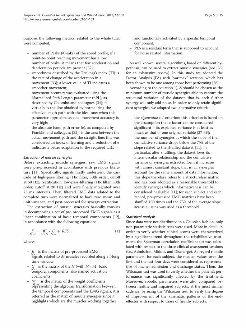
synergies were retained from all datasets to allow an easyintra- and inter- group comparison, as already done byprevious authors [17]. The cumulative variance explainedby the retained synergies, in both groups, was about the70% (Figure 2B).Figure 3 shows both muscle synergies and temporal
components underlying the coordination of muscle activ-ity in both healthy subjects and patients. With respect tohealthy control group, the first (S1) and the second (S2)synergies consisted of the activity of muscles controllingthe upper arm during the abduction and the flexion/ex-tension of the shoulder: S1 was loaded by DELM andDELP, and a lower contribution of TRI and DELA; S2 wasloaded by DELA and PECM, and the lower contribution ofBIC and DELM. The third synergy (S3) reflected the activ-ity of BRAD, TRI and the lower contribution of BRAC,while controlling elbow flex/extension whereas the fourthsynergy (S4) revealed the coupled coordination of elbowflexors (i.e., BRAD, BRAC and BIC) and the pectoralis
major, even though it was characterized by a wide datadispersion across subjects.Muscle synergies related to post-stroke patients (Figure 3)
were qualitatively similar to those of the healthy controlgroup even though some specific features characterizedthem. Specifically, S1 basically accounted for the contri-bution of TRI, DELA, DELM, and DELP. Furthermore,with the ongoing of the therapy, the contribution of theTRI decreased while DELA became more consistent. S2was mainly characterized by the activity of BRAD andBRAC. S3 also showed slightly modifications through-out the rehabilitative treatment: it initially reflected thespread activity of many muscle groups while, at theend, similar to healthy subjects, it was mainly loaded byBIC and TRI. S4 was characterized by great variabilityacross patients which increased with the ongoing of thetreatment.The intra-group similarity of muscle synergies related
to both healthy control group and post-stroke patients
Figure 3 Muscle synergies and temporal components in healthy and stroke patients. Weight coefficients (A) and activation coefficientprofiles (B) for each of extracted synergies in healthy and stroke patients before and after the treatment. Concerning the subplots A, gray barsshow the weight coefficient for each subjects involved in the study and black bar profiles indicate group means and standard deviations. Labelson the horizontal axis are: BRAD, brachioradialis; BRAC, brachialis; BIC, biceps brachii; TRI, triceps brachii; DELA, anterior part of deltoid; DELM,medial part of deltoid; DELP, posterior part of deltoid; LAT, latissimus dorsi; TRAP, trapezius superior; PECM, pectoralis major. Labels on thehorizontal axis are: 0 w, 0 weeks (pre treatment); 6 w, 6 weeks (post treatment). Concerning the subplots B, gray and thin lines represent theactivation profiles for each individual subjects while the thick lines represent the group mean. Labels on the horizontal axis are: N, North; NE,Northeast; E, East; SE, Southeast; S, South; SW, Southwest; W, West; NW, Northwest.
Tropea et al. Journal of NeuroEngineering and Rehabilitation 2013, 10:103 Page 8 of 15http://www.jneuroengrehab.com/content/10/1/103
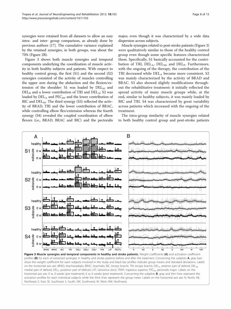
was characterized by a decreasing trend with respect tothe number of retained synergies (Figure 4). For in-stance, in healthy subjects mean values of dotintra rangedfrom about 0.84 in S1 to about 0.18 in S4 (Figure 4)while in post-stroke patients values were generally lower.Moreover, due to the ongoing of the treatment (i.e., at 0,2, 4 and 6 weeks), the dotintra related to patients signifi-cantly decreased for synergies S1, S2 and S4 (Table 3).The dotinter between healthy subjects and subacute pa-
tients related to S1 was significantly high (on average,about 0.76; Figure 4) whereas that related to S3, and S4was generally characterized by averaged values lowerthan 0.40 (Figure 4). Differently than the other synergies,the dotinter related to S2 was characterized by an in-creasing trend with the exception of data referring tothe “0 week”, suggesting that this synergy became moresimilar to that of healthy subjects with the ongoing of thetreatment (Figure 4). For all synergies, the Spearman coef-ficient highlighted that the rehabilitative treatment did notmodify dotinter (p-values > 0.05 in Table 3).The temporal component related to S1 (Figure 3) in
healthy subjects was characterized by well shaped peaksoccurring when subjects inverted the direction of thehandle (i.e., from forward to backward) which amplitudewas greater during the ipsilateral movements (i.e., fromthe N to the SE directions) than during the contralateralones (i.e., from the S to the NW directions). Patients didnot show a consistent behavior of S1 at the beginning of
the treatment. However, during the following experi-mental sessions, a more uniform modulation of this acti-vation coefficient across patients appeared characterizedby a greater amplitude during the ipsilateral movementsof the handle.Concerning the temporal component related to S2, in
healthy subjects it was characterized by wide peaks ba-sically during the movements toward the north-relateddirections. In post-stroke patients, before the treatment,the peaks of activation were consistently present duringthe ipsilateral movements of the handle. After the treat-ment, they disappeared or were characterized by a scarceconsistence across patients.The temporal component of S3, leading elbow flex-
extension, showed the expected peaks along all directionsin healthy subjects. Conversely, it was characterized bynot uniform behavior across post-stroke patients.
Figure 4 Metrics to compare weight coefficients. Mean and standard deviation of the metric adopted to describe intra-group (i.e., dotintra)and inter-groups (i.e., dotinter) similarity between homologous synergies. Labels on the horizontal axis refer to: 0 w, pre treatment; 2 w, 2 weeks; 4w, 4 weeks; 6 w, post treatment. The label * highlights when values for post-stroke subjects are significantly different from healthy controlgroup (p < 0.05).
Table 3 Analysis of the trend of dotintra and the dotinteralong the weeks related to patients
S1 S2 S3 S4
dotintra ρ −0.70 0.26 −0.46 −0.42
p-value <0.01 0.22 0.03 0.04
dotinter ρ −0.09 0.22 0.06 −0.31
p-value 0.67 0.29 0.78 0.14
ρ and p-value refer respectively to the Sperman’s correlation coefficient and itssignificance. Values in bold highlights the statistically significantcorrelation coefficients.
Tropea et al. Journal of NeuroEngineering and Rehabilitation 2013, 10:103 Page 9 of 15http://www.jneuroengrehab.com/content/10/1/103

The temporal component of S4 in healthy subjectsshowed consistent peaks across the subjects in almostall directions, even though those contralateral (i.e., S,SW and W) were characterized by the greatest ampli-tude. In post-stroke patients, it did not show a regularshape along the whole therapy cycle, even though afterthe 6 weeks of treatment it showed a better modulation.According to the visual inspection of temporal compo-
nents, the intra-group similarity of activation patternsrelated to healthy subjects showed that each synergy wasbasically elicited by a set of specific directions. Specific-ally (Figure 5): the temporal component related to S1was characterized by a consistent peak across subjectswhen the movement was directed toward NE-E-SE di-rections; that related to S2 was elicited by movementsdirected toward W-NW-N directions; those concerningS3 and S4 were more consistent when referring to all di-rections from N to SW, clockwise, even though, withvariable value of rintra.
The comparison of the temporal coefficients withinand between groups (Figure 5) related to post-stroke pa-tients showed that at the beginning of the treatment theywere not consistent either among patients or with thehealthy subjects, and generally assumed lower values ofrintra, and rinter than the control group. With the ongoingof the treatment, both rintra, and rinter increased suggestingthat the related temporal components were elicited andbecame more consistent depending on the direction of themovement. Moreover, the anisotropic behavior of rinterwas in slight accordance with data related to the healthysubjects.
DiscussionThis study aimed at verifying whether the expected im-provements in motor performance of subacute-patientsdue to conjunction between the spontaneous recoveryand the intense neuro-rehabilitative treatment werereflected in the modular coordination of muscular
Figure 5 Metrics to compare temporal components. Mean value of the Pearson’s correlation coefficient adopted to compare intra-group(rintra) and inter-groups (rinter) similarity between homologous temporal components for each of the 8 directions. Labels on the plots are: N,North; NE, North-East; E, East; SE, South-East; S, South; SW, South-West; W, West; NW, North-West. Labels on the horizontal axis are: 0 w, pretreatment; 2 w, 2 weeks; 4 w, 4 weeks; 6 w, post treatment.
Tropea et al. Journal of NeuroEngineering and Rehabilitation 2013, 10:103 Page 10 of 15http://www.jneuroengrehab.com/content/10/1/103

activity. This pilot study was focused on a narrow sam-ple of patients (see Materials and methods) due to theneed of having a homogeneous group with respect tothe time elapsed from the onset of the stroke.As expected, muscle recruitment both in healthy and
hemiparetic subjects is characterized by a certain degreeof coordination, such that the activation of 10 musclescan be described by a smaller number of muscle syner-gies (Figure 2B), in accordance with previous studies[11,40]. Results therefore corroborate the general finding[42] that movement planning is mainly based on theorganization of sub-modules which, in the framework ofthe adopted protocol, can be functionally related to sup-port the arm against gravity (see S1 in Figure 3), to flex/extend the shoulder (see S2 in Figure 3), and to flex/ex-tend the elbow (see S3 in Figure 3). Although S4 re-vealed the coupled coordination of elbow flexors (i.e.,BRAD, BRAC and BIC) and pectoralis major, it wascharacterized by a wider data dispersion across subjectsthan previous modules.The modular organization of roles underlying muscle
activity appeared to be significantly influenced by boththe cerebrovascular accident and the functional recovery.Specifically, due to the trauma, the intra-group consist-ence of muscle synergies (Figures 3 and 4) and temporalcoefficients (Figures 3 and 5) was generally lower than inthe control group. Moreover, the ongoing of the treat-ment involved more consistent activation patterns whichappeared related to specific directions of the movement(Figure 5).
Consistence of muscle synergiesAs well known, one of the key issues of the extraction ofmuscle synergies consists in identifying the number ofmodules capturing only the systematic behavior of theoriginal dataset. In this respect, the number of synergiesat which the curve of the cumulative variance shows anabrupt change of slope is generally considered suitablefor this purpose [11].Indeed, several authors have shown that the cumula-
tive variance grows gradually making difficult the identi-fication of the correct number of synergies by visualinspection [11,39,41,43]. For these reasons several add-itional criteria have been adopted to disentangle signalfrom noise.With respect to our study, we adopted 2 different cri-
teria to identify the correct number of synergies. Thesemethods suggested that 4 modules can be consideredsuitable to reconstruct the original datasets andaccounted for about the 70% of the total data variance.These values are in good accordance with those reportedin previous works where the authors noticed that 5-6 syn-ergies were sufficient for explaining about the 75-80% ofdata variance related to 12 muscles recorded from
mild-to-severe post-stroke patients while carrying outseveral upper limb related motor tasks [11,16].On the whole, since the aim of our study consisted in
comparing muscle synergies describing both a group ofsubacute post-stroke patients and a group of healthysubjects, we focused our attention on modules commonto all subjects. Therefore, the number of retained syner-gies appeared to be well suited to describe the greatestamount of data information coded in recorded EMGsignals.Concerning the consistence of weight coefficients, re-
sults showed that only S1 in both healthy subjects andpost-stroke patients was characterized by a significant de-gree of intra-group consistency (on average, dotintra > 0.75;see Figure 4) while dotintra of S2, S3, and S4 was usuallylower (on average, dotintra ranged between about 0.15 and0.6). Indeed, these values do not appear in agreement withthose reported in previous studies [11,40,44,45], eventhough this discrepancy can be reasonably explained bytwo main reasons concerning the experimental designand, the data processing.As matter of the fact, there is no unanimous consen-
sus concerning how to pre-process raw data, such thatprevious authors have already highlighted that differentapproaches can lead to different conclusions [18,39].Moreover, synergy extraction is itself a sort of data filter-ing because it allows the algebraic complexity of the dataset to be reduced by selecting only those factors, whichare supposed to account for the greatest amount of in-formation [41]. In this regard, by adopting different cri-teria to retain synergies [18,43] or to pre-process data[46], it is possible to obtain different results. Finally,other authors [47] noticed that the attitude of somefactorization algorithms to accurately capture the mainfeatures of a dataset can strongly depend both on thesparseness and the diversity of the actual modulesunderlying the structure of the dataset being factorized,and on how uniformly the space accounting for suchactual modules is parsed.Concluding, different experimental paradigms (e.g.,
motor task, recorded muscle groups, age, and path-ology), pre-processing and factorization (i.e., time variantversus time invariant factorizing algorithms) algorithmscan have led to different degrees of similarity withinhomologous groups of synergies. Therefore, further ef-forts are needed to achieve wider agreement among theauthors regarding both methods for data pre-processingand synergy comparison.
Effects of the stroke on muscle synergiesResults (Figure 4) showed that the intra-group consistencyat the baseline (i.e., “0 weeks”) of S1 and S2 of the post-stroke patients was generally lower than that of healthysubjects. This was accompanied by a very low intra-group
Tropea et al. Journal of NeuroEngineering and Rehabilitation 2013, 10:103 Page 11 of 15http://www.jneuroengrehab.com/content/10/1/103

consistency of the temporal components of all modulesthat was also inhomogeneous with respect to the directionof the movement (see rintra in Figure 5), and by a lowinter-group degree of similarity of temporal components(see rinter in Figure 5). Despite of this, muscle synergies ofpost-stroke patients appeared similar to that of healthysubjects (see dotinter in Figure 4) suggesting that they werecharacterized by the same modular organization.Noticeably, these results cannot be completely as-
cribed to the different cadence between healthy andpost-stroke patients because they were similarly con-firmed when both groups carried out the motor taskswith comparable speed (Figures 4 and 5).These evidences support previous findings concerning
the robustness of muscular organization within each syn-ergy and suggest that although the cerebrovascular acci-dent may increase the variability between patients, thebasic structure of each module is not significantly alteredwhen compared to that of healthy subjects [11,15]. Con-versely, the temporal components enabling each synergyappeared significantly altered by the trauma, indicatingthat they presumably result by the abnormal motor com-mands descending from the damaged hemisphere [11,15].In this respect, our results corroborated the hypothesisthat muscle synergies may be encrypted in neural circuitslocated in the spinal cord and/or in the brainstem and areinconsistently recruited due to trauma [11,17].With respect to the specific effect of the trauma on
weight coefficients related to S1 and S2, results (Figure 3)showed that patients at the baseline were mainly char-acterized by the abnormal contribution of deltoid headson synergies underlying the control of the shoulder.Specifically, in post-stroke patients, the DELA signifi-cantly loaded S1 whereas, according to data referring tothe healthy control group (Figure 3), it was expected tocontribute to the flexion of the shoulder described byS2. Moreover, the contribution of the anterior andmedial heads of the deltoid to S2 was appreciablyattenuated.Actually, previous authors [17] already noticed that,
during an isometric task, the alteration of the structureof muscle synergies was mainly confined at the proximaldistrict and affected in a negative fashion the motor per-formance of the patients. They hence hypothesized thatthis abnormal muscle recruitment in their chronic pa-tients could result as an adaptive response to the weak-ness following the trauma.Our study confirms these previous findings and reveals
that the re-modulation of the contribution of the deltoidheads to the synergy leading the proximal joint may dir-ectly reflect the altered muscle recruitment early afterthe trauma. In this regard, it is possible to speculate thatthis abnormal muscle-recruitment can be consideredpredictive of the degree of impairment.
Actually, from the best of our knowledge, our study isthe first one aimed at investigating muscle synergies ofonly subacute post-stroke patients. Therefore some ofthe results of previous studies which have been con-firmed by our analysis (e.g., robustness of modules andalteration of temporal coefficients, re-modulation of thebehavior of muscles crossing the proximal district) canbe more directly ascribed to the effect of the trauma ra-ther than to compensative strategies induced by thechronic stage of the pathology. However, further studiesare required to stronger support to this hypothesis.Muscle synergies related to S3 and S4 did not show
significant differences between the two groups of sub-jects, both in term of inter- and intra-group consistence.This result is in agreement with that reported by Rohand colleagues [17] who did not notice significant alter-ation of muscle synergies leading the control of the dis-tal joint. However, we cannot reject the hypothesis thatit could be due to the evidence that these two synergies,together, accounted for about the 20-25% of the varianceof the datasets and were therefore characterized by anintrinsically greater variability than S1 and S2. In this re-gard, we believe that some of the methodological issuesunderlying all factorization approaches should be stillclarified in order to allow a confident interpretation ofthe analysis of muscle synergies.
Effects of the ongoing of the treatment on musclesynergiesThe conjunction between the spontaneous recovery andthe intensive treatment improved motor performance,both in terms of kinematics of the end-effector (Figure 1Band 1C) and functional scores (Table 2). In particular,the therapy generally favored the reorganization of theupper limb related motor tasks and modified patient’smotor capabilities. This evidence is in accordance withprevious studies, which have already highlighted thatrobot-assisted therapy can improve motor performance,allowing patients to learn how to coordinate their jointsin adaptable patterns in order to increase the functionaloutcome [24,48].Although the treatment involved functional improve-
ments, the degree of similarity between muscle synergiesof post-stroke patients and healthy participants did notsignificantly change with the ongoing therapy (seedotinter in Figure 4 and Table 3). Conversely, the intra-group consistence of muscle synergies related to S1, S3and S4 in patients was characterized by a slight and signifi-cantly (p < 0.05) declining trend along the treatment dur-ation (see dotintra in Figure 4 and Table 3). On the otherhand, the temporal components reflected a certain degreeof adaptability of the Central Nervous System (CNS) andsuggested that a treatment provided early after the traumainvolves the reorganization of descending signals (Figure 5).
Tropea et al. Journal of NeuroEngineering and Rehabilitation 2013, 10:103 Page 12 of 15http://www.jneuroengrehab.com/content/10/1/103
Despite of this, the activation patterns were not character-ized by a stereotyped behaviour both across subjects andwith the ongoing of the treatment (Figure 5).Results therefore contrasted the hypothesis that a bet-
ter motor outcome of post-stroke patients, as assessedby clinical scores and kinematics of the end-effector, wasreflected in a muscle coordination more similar to thatof the healthy control group and more consistent acrosssubjects. In this regard, we believe that the reasonsunderlying these results reside in the complex relation-ship between the modifications of the motor outcomeand the re-organization of muscle activity due to thetreatment, and involves both methodological and neuro-physiological aspects.From the methodological viewpoint, the improvements
of motor performance due to neuro-rehabilitative therap-ies mainly consist of changes in amplitude modulation be-tween agonist and antagonist muscle groups [7,10,49,50].The FA instead captures the communality of a set of zero-scored EMG signals by means of their correlation, whichmainly reflects the concomitance of signal bursts, i.e., thetiming of the activity. Hence, although the FA is able tohighlight the inter-muscular coordination, it may be notenough sensitive to characterize variations of the signals’amplitude. This result is corroborated by previous authors[39,43,51] who investigated the principal roles underlyingthe coordination of muscle activity during walking in awide range of speeds and reported that, despite theEMG amplitude is significantly affected by the speed,muscle synergies do not seem to be influenced by thepace of the subjects. Further methodological improve-ments are hence required to capture all available infor-mation encrypted in the coordinated activity of manymuscle groups and, in case, highlight the trend of thedotinter due to the treatment.From the neuro-physiological side, the decreasing con-
sistence of similarity across patients during the ongoingof the treatment (see dotintra in Figure 4) could be as-cribed to the different recovering mechanisms due tothe heterogeneity of lesions, sides and location of the ac-cident. In other words, the improvements of motor per-formance across patients occur in accordance with theirown clinical picture, that is, they are characterized bygreat inter-subjects variability, which does not facilitateto capture univocal and common features of the wholegroup. In this regard, a greater number of participantscan provide further evidence for this result. Moreover,from the rehabilitative viewpoint, the analysis of the cor-relation between motor outcome and muscle synergiesshould be carried out for each single patient in order toavoid bias due to the inherent inter-patients variability.Concluding, as also observed by other authors [11,17],
the analysis of muscle synergies seems to be effective inproviding a theoretical support for the design of therapeutic
interventions for post-stroke patients because it can high-light the neural re-organization of motor control resultingafter a cerebrovascular accident and leaded by a neuro-rehabilitative treatment.
Limits of the studyThe first limit of this study was the small group of par-ticipants which involves a limited strength of the statis-tical findings. However, our intention was to enroll ahomogenous group of patients, in term of age (range:66-82 ys), onset from the trauma (range: 14-37 days)and absence of bilateral impairments, in order to reduce,as much as possible, the potential bias of the intrinsiclack of homogeneity across patients which may be oneof the main reasons underlying the discrepant resultsamong previous literature. Accordingly, this work hasbeen designed as a pilot study and further investigations,according to the reported results, are guaranteed.The second potential limit concerns the slow speed
adopted by the patients before the treatment. This maybias the interpretation of the results because the value ofall metrics describing kinematic parameters and musclesynergies are conjunctly affected by both the functionalcapabilities of all patients before starting the therapy andthe slow speed achieved during the exercises. However,from one hand, the increasing speed and smoothnessthroughout the treatment supported the hypothesis thatthe therapy is effective from the clinical viewpoint. On theother hand, since during the following experimental ses-sions (i.e., 2, 4 and 6 weeks) patients achieved a cadencecomparable to that of healthy participants (Figure 1B), ourexperimental design allows to extrapolate that the degreeof similarity of muscle synergies between healthy subjectsand post-stroke patients is characterized by a monotonicbehavior.The last limit of this study was that in only one case, a
patient was affected by paresis of the dominant arm (seeSub 04 in Table 1). Actually, the effect of the interactionbetween dominance and affected side is widely discussedin literature even though there is not an exhaustive un-derstanding [52,53]. With respect to our study, we didnot observed any apparent difference between this pa-tient and the others which might justify his exclusionfrom this study. However, we acknowledge that furtherstudies are required to explore this issue.
ConclusionsThis pilot study supports the hypothesis that the coordi-nated activity of muscle groups, i.e., muscle synergies, aresignificantly affected by a cerebrovascular accident. More-over, results suggest that due to the treatment, patientsmodify the coordinated activity of muscle groups eventhough the reorganization of rules underlying motorcontrol are characterized by a significant inter-patients
Tropea et al. Journal of NeuroEngineering and Rehabilitation 2013, 10:103 Page 13 of 15http://www.jneuroengrehab.com/content/10/1/103

variability. If confirmed with a significant robustness,these insights would support the hypothesis that suitableand customized treatments can be designed to favouritethe functional recovery of post-stroke patients.
Competing interestThe authors declare that they have no competing interest.
Authors’ contributionsPT designed and carried out experiments, analyzed data and wrote thepaper; VM designed experiments, analyzed data and wrote the paper; MCcarried out experiments; FP designed the study; SM designed the study,analyzed data, and wrote the paper. All authors read and approved the finalmanuscript.
AcknowledgmentsWe would like to thank all participants enrolled for the study and expressour special thanks to Ms. Rossella Crecchi, PT, for her precious support.
GrantsThis work was partially supported by the MIRROR Project (Novel Approachesfor Robot-Mediated Neuro-Rehabilitation) funded by a local Bank Foundation(Fondazione MPS).
Author details1The BioRobotics Institute, Scuola Superiore Sant’Anna, Piazza Martiri dellaLibertà 33, Pisa 56127, Italy. 2Rehabilitation Department Versilia Hospital,AUSL 12, Viareggio, Italy. 3Bioengineering Rehabilitation Laboratory, AuxiliumVitae Rehabilitation Centre, Volterra, Italy. 4Translational Neural EngineeringLab, Center for Neuroprosthetics and Institute of Bioengineering, School ofEngineering, Ecole Polytechnique Federale de Lausanne (EPFL), Lausanne,Switzerland.
Received: 14 October 2012 Accepted: 26 September 2013Published: 5 October 2013
References1. Canning CG, Ada L, O'Dwyer N: Slowness to develop force contributes to
weakness after stroke. Arch Phys Med Rehabil 1999, 80:66–70.2. Hammond MC, Kraft GH, Fitts SS: Recruitment and termination of
electromyographic activity in the hemiparetic forearm. Arch Phys MedRehabil 1988, 69:106–110.
3. Bourbonnais D, Vanden Noven S: Weakness in patients with hemiparesis.Am J Occup Ther 1989, 43:313–319.
4. Cruz EG, Waldinger HC, Kamper DG: Kinetic and kinematic workspaces ofthe index finger following stroke. Brain 2005, 128:1112–1121.
5. Dewald JP, Pope PS, Given JD, Buchanan TS, Rymer WZ: Abnormal musclecoactivation patterns during isometric torque generation at the elbowand shoulder in hemiparetic subjects. Brain 1995, 118(Pt 2):495–510.
6. Musampa NK, Mathieu PA, Levin MF: Relationship between stretch reflexthresholds and voluntary arm muscle activation in patients withspasticity. Exp Brain Res 2007, 181:579–593.
7. Lum PS, Burgar CG, Shor PC: Evidence for strength imbalances as asignificant contributor to abnormal synergies in hemiparetic subjects.Muscle Nerve 2003, 27:211–221.
8. Beer RF, Given JD, Dewald JP: Task-dependent weakness at the elbow inpatients with hemiparesis. Arch Phys Med Rehabil 1999, 80:766–772.
9. Ellis MD, Acosta AM, Yao J, Dewald JP: Position-dependent torquecoupling and associated muscle activation in the hemiparetic upperextremity. Exp Brain Res 2007, 176:594–602.
10. Dewald JP, Beer RF: Abnormal joint torque patterns in the paretic upperlimb of subjects with hemiparesis. Muscle Nerve 2001, 24:273–283.
11. Cheung VC, Piron L, Agostini M, Silvoni S, Turolla A, Bizzi E: Stability ofmuscle synergies for voluntary actions after cortical stroke in humans.Proc Natl Acad Sci U S A 2009, 106:19563–19568.
12. Bizzi E, Cheung VC, d'Avella A, Saltiel P, Tresch M: Combining modules formovement. Brain Res Rev 2008, 57:125–133.
13. d’Avella A, Saltiel P, Bizzi E: Combinations of muscle synergies in theconstruction of a natural motor behavior. Nat Neurosci 2003, 6:300–308.
14. Ting LH, Macpherson JM: A limited set of muscle synergies for forcecontrol during a postural task. J Neurophysiol 2005, 93:609–613.
15. Trumbower RD, Ravichandran VJ, Krutky MA, Perreault EJ: Contributions ofaltered stretch reflex coordination to arm impairments following stroke.J Neurophysiol 2010, 104:3612–3624.
16. Cheung VC, Turolla A, Agostini M, Silvoni S, Bennis C, Kasi P, Paganoni S,Bonato P, Bizzi E: Muscle synergy patterns as physiological markers ofmotor cortical damage. Proc Natl Acad Sci U S A 2012, 109:14652–14656.
17. Roh J, Rymer WZ, Perreault EJ, Yoo SB, Beer RF: Alterations in upper limbmuscle synergy structure in chronic stroke survivors. J Neurophysiol 2013,109:768–781.
18. Clark DJ, Ting LH, Zajac FE, Neptune RR, Kautz SA: Merging of healthymotor modules predicts reduced locomotor performance and musclecoordination complexity post-stroke. J Neurophysiol 2010, 103:844–857.
19. Gizzi L, Nielsen JF, Felici F, Ivanenko YP, Farina D: Impulses of activationbut not motor modules are preserved in the locomotion of subacutestroke patients. J Neurophysiol 2011, 106:202–210.
20. Grasso R, Ivanenko YP, Zago M, Molinari M, Scivoletto G, Castellano V,Macellari V, Lacquaniti F: Distributed plasticity of locomotor patterngenerators in spinal cord injured patients. Brain 2004, 127:1019–1034.
21. Ivanenko YP, Grasso R, Zago M, Molinari M, Scivoletto G, Castellano V,Macellari V, Lacquaniti F: Temporal components of the motor patternsexpressed by the human spinal cord reflect foot kinematics.J Neurophysiol 2003, 90:3555–3565.
22. Kalra L, Langhorne P: Facilitating recovery: evidence for organized strokecare. J Rehabil Med 2007, 39:97–102.
23. DeJong G, Horn SD, Conroy B, Nichols D, Healton EB: Opening the blackbox of post-stroke rehabilitation: stroke rehabilitation patients,processes, and outcomes. Arch Phys Med Rehabil 2005, 86:S1–S7.
24. Masiero S, Armani M, Rosati G: Upper-limb robot-assisted therapy inrehabilitation of acute stroke patients: focused review and results ofnew randomized controlled trial. J Rehabil Res Dev 2011, 48:355–366.
25. Posteraro F, Mazzoleni S, Aliboni S, Cesqui B, Battaglia A, Dario P, Micera S:Robot-mediated therapy for paretic upper limb of chronic patientsfollowing neurological injury. J Rehabil Med 2009, 41:976–980.
26. Krebs HI, Hogan N, Aisen ML, Volpe BT: Robot-aided neurorehabilitation.IEEE Trans Rehabil Eng 1998, 6:75–87.
27. Hermens HJ, Freriks B, Disselhorst-Klug C, Rau G: Development ofrecommendations for SEMG sensors and sensor placement procedures.J Electromyogr Kinesiol 2000, 10:361–374.
28. Bohannon RW, Smith MB: Interrater reliability of a modified Ashworthscale of muscle spasticity. Phys Ther 1987, 67:206–207.
29. Fugl-Meyer AR, Jaasko L, Leyman I, Olsson S, Steglind S: The post-strokehemiplegic patient. 1. a method for evaluation of physical performance.Scand J Rehabil Med 1975, 7:13–31.
30. Fugl-Meyer AR: Post-stroke hemiplegia assessment of physical properties.Scand J Rehabil Med 1980, 7:85–93.
31. Collin C, Wade D: Assessing motor impairment after stroke: a pilotreliability study. J Neurol Neurosurg Psychiatry 1990, 53:576–579.
32. Rohrer B, Fasoli S, Krebs HI, Hughes R, Volpe B, Frontera WR, Stein J, HoganN: Movement smoothness changes during stroke recovery. J Neurosci2002, 22:8297–8304.
33. Teulings HL, Contreras-Vidal JL, Stelmach GE, Adler CH: Parkinsonismreduces coordination of fingers, wrist, and arm in fine motor control. ExpNeurol 1997, 146:159–170.
34. Colombo R, Pisano F, Micera S, Mazzone A, Delconte C, Carrozza MC, DarioP, Minuco G: Assessing mechanisms of recovery during robot-aidedneurorehabilitation of the upper limb. Neurorehabil Neural Repair 2008,22:50–63.
35. Franklin DW, Osu R, Burdet E, Kawato M, Milner TE: Adaptation to stableand unstable dynamics achieved by combined impedance control andinverse dynamics model. J Neurophysiol 2003, 90:3270–3282.
36. Tresch MC, Cheung VC, d'Avella A: Matrix factorization algorithms for theidentification of muscle synergies: evaluation on simulated andexperimental data sets. J Neurophysiol 2006, 95:2199–2212.
37. Davis BL, Vaughan CL: Phasic Behavior of Emg Signals during Gait - Useof Multivariate-Statistics. J Electromyogr Kines 1993, 3:51–60.
38. Sabatini AM: Identification of neuromuscular synergies in natural upper-arm movements. Biol Cybern 2002, 86:253–262.
39. Monaco V, Ghionzoli A, Micera S: Age-related modifications of musclesynergies and spinal cord activity during locomotion. J Neurophysiol 2010,
Tropea et al. Journal of NeuroEngineering and Rehabilitation 2013, 10:103 Page 14 of 15http://www.jneuroengrehab.com/content/10/1/103

104:2092–2102.40. d’Avella A, Portone A, Fernandez L, Lacquaniti F: Control of fast-reaching
movements by muscle synergy combinations. J Neurosci 2006, 26:7791–7810.41. Cheung VC, d'Avella A, Tresch MC, Bizzi E: Central and sensory
contributions to the activation and organization of muscle synergiesduring natural motor behaviors. J Neurosci 2005, 25:6419–6434.
42. Kisiel-Sajewicz K, Fang Y, Hrovat K, Yue GH, Siemionow V, Sun CK, JaskolskaA, Jaskolski A, Sahgal V, Daly JJ: Weakening of synergist muscle couplingduring reaching movement in stroke patients. Neurorehabil Neural Repair2011, 25:359–368.
43. Ivanenko YP, Poppele RE, Lacquaniti F: Five basic muscle activationpatterns account for muscle activity during human locomotion. J Physiol2004, 556:267–282.
44. d’Avella A, Fernandez L, Portone A, Lacquaniti F: Modulation of phasic andtonic muscle synergies with reaching direction and speed. J Neurophysiol2008, 100:1433–1454.
45. d’Avella A, Portone A, Lacquaniti F: Superposition and modulation ofmuscle synergies for reaching in response to a change in targetlocation. J Neurophysiol 2011, 106(6):2796–2812.
46. Hug F, Turpin NA, Couturier A, Dorel S: Consistency of muscle synergiesduring pedaling across different mechanical constraints. J Neurophysiol2011, 106:91–103.
47. Burkholder TJ, van Antwerp KW: Practical limits on muscle synergyidentification by non-negative matrix factorization in systems withmechanical constraints. Med Biol Eng Comput 2012, 51:187–196.
48. Dipietro L, Krebs HI, Fasoli SE, Volpe BT, Stein J, Bever C, Hogan N:Changing motor synergies in chronic stroke. J Neurophysiol 2007, 98:757–768.
49. Barker RN, Brauer S, Carson R: Training-induced changes in the pattern oftriceps to biceps activation during reaching tasks after chronic andsevere stroke. Exp Brain Res 2009, 196:483–496.
50. Hughes AM, Freeman CT, Burridge JH, Chappell PH, Lewin PL, Rogers E:Shoulder and elbow muscle activity during fully supported trajectorytracking in people who have had a stroke. J Electromyogr Kinesiol 2010,20:465–476.
51. Cappellini G, Ivanenko YP, Poppele RE, Lacquaniti F: Motor patterns inhuman walking and running. J Neurophysiol 2006, 95:3426–3437.
52. Haaland KY, Elsinger CL, Mayer AR, Durgerian S, Rao SM: Motor sequencecomplexity and performing hand produce differential patterns ofhemispheric lateralization. J Cogn Neurosci 2004, 16:621–636.
53. Harris JE, Eng JJ: Individuals with the dominant hand affected followingstroke demonstrate less impairment than those with the nondominanthand affected. Neurorehabil Neural Repair 2006, 20:380–389.
doi:10.1186/1743-0003-10-103Cite this article as: Tropea et al.: Effects of early and intensive neuro-rehabilitative treatment on muscle synergies in acute post-strokepatients: a pilot study. Journal of NeuroEngineering and Rehabilitation2013 10:103.
Submit your next manuscript to BioMed Centraland take full advantage of:
• Convenient online submission
• Thorough peer review
• No space constraints or color figure charges
• Immediate publication on acceptance
• Inclusion in PubMed, CAS, Scopus and Google Scholar
• Research which is freely available for redistribution
Submit your manuscript at www.biomedcentral.com/submit
Tropea et al. Journal of NeuroEngineering and Rehabilitation 2013, 10:103 Page 15 of 15http://www.jneuroengrehab.com/content/10/1/103