effects of familial alzheimer's disease-linked
TRANSCRIPT

Sede Amministrativa: Università degli Studi di Padova
Dipartimento di Scienze Biomediche
SCUOLA DI DOTTORATO DI RICERCA IN : BIOSCIENZE E
BIOTECNOLOGIE
INDIRIZZO: NEUROBIOLOGIA
CICLO XXV
EFFECTS OF FAMILIAL ALZHEIMER’S DISEASE-LINKED
PRESENILIN 2 MUTANTS ON Ca2+ HOMEOSTASIS
OF GOLGI APPARATUS SUB-COMPARTMENTS
Direttore della Scuola : Ch.mo Prof. Giuseppe Zanotti
Coordinatore d’indirizzo: Ch.ma Prof.ssa Daniela Pietrobon
Supervisore : Dott.ssa Paola Pizzo
Dottoranda : PAOLA CAPITANIO

Index
Riassunto 1
Summary 3
Introduction 5
1. CALCIUM SIGNALLING 5
1.1 Intracellular Ca2+ stores and Ca2+ dynamics 6
1.1.1 The Endoplasmic Reticulum as an intracellular Ca2+ store 6
1.1.2 The Golgi Apparatus (GA) as an intracellular Ca2+ store 14
1.1.3 Peroxisomes, mithocondria and endolysosomal
compartments in intracellular Ca2+ dynamics 21
1.1.3.1 Mithocondria 21
1.1.3.2 Peroxisomes 22
1.1.3.3 Endolysosomal compartments 22
1.2 Ca2+ entry from the extracellular space 23
1.2.1 Voltage operated Ca2+ entry 23
1.2.2 Store operated Ca2+ entry 27
2. GENETICALLY ENCODED Ca2+ INDICATORS 31
2.1 FRET-based cameleon probes 32
3.THE ALZHEIMER’S DISEASE 35
3.1 Clinical and pathological features of AD 35
3.2 The molecular pathogenesis of AD 38
3.2.1 The amyloid hypothesis 39
3.2.1.1 -secretase and -cleavage 40
3.2.1.2 -Secretase and -cleavage 41
3.2.1.3 -Secretase and -cleavage 41
3.2.2 The hyperphosphorylated tau hypothesis 44
3.2.3 The Ca2+ hypothesis 46
Materials and Methods 49
Results 53

1. CHARACTERIZATION OF Ca2+ HANDLING BY THE
CIS/MEDIAL-GOLGI 53
1.1 A new Cameleon Ca2+ probe specifically targeted to
cis/medial-Golgi 53
1.1.1 Creation of the new Cameleon Ca2+ probe 53
1.1.2 Localization of the new probe 54
1.1.3 Calibration of the new mGo-D1cpv 57
1.2 Ca2+ handling in the medial-Golgi 57
2. EFFECTS OF THE FAMILIAL ALZHEIMER’S DISEASE
LINKED-PRESENILIN 2-T122R MUTANT ON Ca2+
HOMEOSTASIS IN THE GOLGI APPARATUS
SUBCOMPARTMENTS 61
2.1 Intracellular localization of PS2 in SH-SY5Y cells 61
2.2 Both FAD-linked PS2 and PS1 mutants strongly affect the
Ca2+ level of the medial-Golgi apparatus but do not affect the Ca2+
level of the trans-Golgi apparatus 62
2.3 FAD-linked PS2 reduced medial-Golgi Ca2+ uptake by
inhibiting SERCA pump activity 64
2.4 FAD-linked PS2 increases medial-Golgi Ca2+ leak 67
2.5 FAD-linked PS2 increase IP3 sensitivity of medial-Golgi
Apparatus 68
Discussion and conclusions 71
Abbreviations 73
References 77

1
Riassunto
La malattia di Alzheimer’s (AD) è un disordine neurodegenerativo e la
forma più comune di demenza senile.
La caratteristica istopatologica di AD è la presenza di depositi
neurofibrillari intracellulari e di placche amiloidi, costituite da aggregati
di peptide amiloide (A ), che si depositano nella matrice extracellulare del
cervello. I peptidi A sono il risultato di due tagli sequenziali della
Proteina Precursore dell'Amiloide (APP); A viene poi rilasciato dall’enzima
-secretasi. Le più abbondanti specie peptidiche di A , prodotte anche
fisiologicamente per tutta la vita, sono A 40 e A 42, quest’ultimo più
insolubile e più incline all’aggregazione.
Sebbene la maggior parte dei casi di AD siano sporadici, una piccola
percentuale di pazienti è affetta dalla forma ereditaria di Alzheimer
(malattia familiare di Alzheimer, FAD), causata da mutazioni dominanti in
uno dei geni codificanti per APP, presenilina-1 (PS1) e presenilina-2 (PS2);
le PSs sono le subunità catalitiche del complesso enzimatico della -
secretasi ma funzionano anche in maniera indipendente da tale attività
enzimatica.
Le mutazioni in PSs legate a FAD portano ad un aumento nel rapporto
A 42/A 40, che promuove la deposizione di placche amiloidi. Oltre a
questo effetto, è stato ampiamente dimostrato che molte mutazioni in
PS1 e PS2 provocano alterazioni della omeostasi del Ca2+ intracellulare,
rendendo così i neuroni più sensibili agli stimoli eccitotossici e apoptotici.
L'apparato di Golgi (GA) rappresenta, insieme al reticolo endoplasmatico
(ER), il principale deposito intracellulare di Ca2+, IP3 sensibile, e la sua
funzionalità è fondamentale per il controllo delle risposte citosoliche di
Ca2+.
Sempre maggiori evidenze suggeriscono che il GA sia un organello
eterogeneo in termini di Ca2+ handling, essendo dotato di un diverso
toolkit molecolare per il Ca2+ rispetto a quello espresso nell’ ER. Ad
esempio, come meccanismi di uptake per il Ca2+, il GA esprime la classica
pompa SERCA (Sarco-Endoplasmic Reticulum Ca2+ ATPase) ma anche un
ulteriore pompa, detta SPCA1 (Secretory Pathway Ca2+ ATPase1).
L'utilizzo di uno specifico sensore per il Ca2+ specificatamente indirizzato
al trans-Golgi, ci ha precedentemente permesso di dimostrare

2
direttamente la eterogeneità funzionale del GA, mostrando il
comportamento distinto di questo sub-compartimento: i meccanismi di
uptake di Ca2+ sono mediati esclusivamente dalla SPCA1 (e non dalla
SERCA); non rilascia Ca2+ in risposta alla generazione IP3, ma piuttosto si
accumula il catione come conseguenza dell’aumento di Ca2+ citoplasmatico.
Per quanto riguarda gli altri sub-compartimenti del GA, abbiamo generato
un nuovo indicatore per il Ca2+ fuso alla sequenza di indirizzamento
dell'enzima 1,6 N-acetylglucosaminyltransferasi (C2gnT) residente del
cis/medial-Golgi. La nuova sonda co-localizza con il marcatore di
cis/medial-Golgi Giantina e quindi è stata utilizzata per studiare le
dinamiche di Ca2+ in questo sub-compartimento a livello di singola cellula.
Complessivamente i dati ottenuti suggeriscono che il GA sia unico in
termini di omeostasi del Ca2+, con tali sub-compartimenti separati da pochi
micron, e in equilibrio molto rapido tra loro, ma comunque in grado di
mantenere differenze consistenti in termini di concentrazione dello ione e
risposta a stimoli esterni .
Le differenze tra i due sub-compartimenti del GA sono confermate dall’
effetto specifico sulla omeostasi del Ca2+ dell'espressione della forma
mutata di PS2T122R legata alla malattia familiare di Alzheimer. Le cellule
che esprimono tale proteina mostrano una diminuzione del contenuto di
Ca2+ nel cis/medial-Golgi ma nessun effetto sull’ omeostasi del Ca2+ nel
trans-Golgi. PS2T122R sembra inibire l'assorbimento di Ca2+ nel
cis/medial-Golgi, inibendo l'attività della pompa SERCA, mentre non
influenza l'assorbimento di Ca2+, mediato dalla SPCA1, nel trans-Golgi.
Il GA sembra quindi giocare un ruolo importante nella patogenesi di AD e
comprendere il contributo di tale organello nella patogenesi di AD e la sua
base fisiopatologica potrà avere un forte impatto sulla possibilità di
sviluppare terapie più efficaci per AD.
3
Summary
Alzheimer’s Disease (AD) is a progressive neurodegenerative disorder and
the most common form of senile dementia. The characteristic
histopathological hallmarks of AD are the intracellular neurofibrillary
tangles and the amyloid plaques, made of aggregated amyloid peptides
(A ), that deposit in the extracellular matrix of the brain. A peptides
are the result of two sequential cleavages of the amyloid precursor
protein (APP); A is eventually released by the -secretase enzyme. The
most abundant A peptide species, both physiologically produced
throughout life, are A 40 and A 42, which is more insoluble and
aggregation-prone.
Although most AD cases are sporadic, a small percentage of patients is
affected by the hereditary form of AD (Familial Alzheimer’s Disease,
FAD), caused by dominant mutations in one of three genes. These genes
code for the APP, presenilin-1 (PS1) and presenilin-2 (PS2); PSs are the
catalytic subunits of the -secretase enzyme complex but they also
function in a -secretase indipendent manner.
FAD-linked mutations in PSs lead to an increased A 42/A 40 ratio, that
promotes A plaques deposition. Beside this effect on A production,
many mutations in PS1 and PS2 have been extensively demonstrated to
cause alterations in the intracellular Ca2+ homeostasis, thus making
neurons more sensitive to excitotoxic stimuli and apoptosis.
The Golgi apparatus (GA) represents, together with the endoplasmic
reticulum (ER), the major IP3-sensitive, rapidly mobilizable, intracellular
Ca2+ store and its functionality is thus important for shaping cytosolic
Ca2+ responses. Increasing evidence suggests that the GA is an
heterogeneous Ca2+ handling organelle, equipped with a diverse molecular
Ca2+ toolkit compared to the one expressed in the ER. For example, as
Ca2+ uptake mechanisms, the GA expresses the classical sarco-
endoplasmic reticulum Ca2+ ATPase (SERCA) but also an additional Ca2+
pump, the secretory pathway Ca2+ ATPase1, SPCA1.
The use of a specific Cameleon Ca2+ sensor targeted to the trans-Golgi,
allowed us to directly demonstrate the functional GA heterogeneity by
showing the distinct behavior of this sub-compartment: it takes up Ca2+
almost exclusively via SPCA1 (and not by SERCA); it does not release Ca2+
4
in response to IP3 generation, but rather accumulates the cation as a
consequence of the cytoplasmic Ca2+ rise.
As regard to the other GA compartments, we generated a new FRET-
based Ca2+ indicator fused to the cis/medial-Golgi targeting sequence of
the enzyme 1,6 N-acetylglucosaminyltransferase (C2gnT). The new probe
very nicely co-localizes with the cis/medial-Golgi marker Giantin and thus
was used to study Ca2+ dynamics in this compartment at single cell level.
The data collected suggest that the GA is unique in terms of Ca2+
homeostasis, with compartments that are separated by a few microns,
and in very rapid equilibrium with each other, that still maintain quite
substantial differences in terms of ion concentration and response to
external stimuli.
The differences between the two GA sub-compartments, the medial and
the trans-one, are confirmed by the specific effect on Ca2+ homeostasis
of the expression of the FAD-linked PS2 T122R mutation. Cells
expressing the mutated form of the protein show a decreased Ca2+
content in the cis/medial-Golgi but no effects on trans-Golgi Ca2+
homeostasis. PS2-T122R seems to inhibiting Ca2+ uptake in the cis/medial-
Golgi by inhibiting SERCA pump activity while does not affect Ca2+ uptake,
mediated by SPCA1, in the trans-Golgi.
As a major Ca2+ store, the GA could play an important role in AD and
understanding the contribution of GA Ca2+ dysfunction in AD will
significantly impact our ability to develop more effective therapies for
the disease.

5
Introduction
1. CALCIUM SIGNALLING
Calcium (Ca2+) is a highly versatile intracellular signal that can regulate
many different cellular functions. To achieve this versatility, the Ca2+-
signalling system operates in many different ways to regulate cellular
processes that function over a wide dynamic range (Berridge M.J. et al.,
2003).
Thus, in order to extensively control cellular activity, it is necessary to
regulate Ca2+ signals (changes in Ca2+ concentration) in 3D space, time and
amplitude.
Cells normally maintain a low resting ‘free’ Ca2+ concentration in the
cytosol ([Ca2+]c) of 100-200 nM. This contrasts with a higher
concentration (1-2 mM) in the extracellular fluids.
In order to maintain this low resting [Ca2+]c, cells remove Ca2+ using
different energy-dependent mechanisms (fig. 1).
In addition, eukaryotic cells can sequester Ca2+ into intracellular
organelles, in particular the Endoplasmic Reticulum (ER) and the Golgi
Apparatus (GA), but also peroxisomes, mitochondria, and endolysosomal
compartments (fig. 1) (Prins D. and Michalak M., 2011).
Fig.1: Organellar Ca2+ buffering and intracellular Ca2+ dynamics (D. Prins and M. Michalak, 2011)

6
At any time, the level of intracellular Ca2+ is determined by a balance
between the ‘on’ reactions, that introduce Ca2+ into the cytoplasm, and
the ‘off ’ reactions, through which this ion is removed by the combined
action of buffers, pumps and exchangers. Each cell type expresses a
unique set of components of the Ca2+-signalling toolkit to create Ca2+-
signalling systems with different spatial and temporal properties. Almost
all Ca2+-signalling systems have one thing in common: they function by
generating brief pulses of Ca2+. The Ca2+ signal is derived either from
internal stores or from the external medium (Berridge M.J. et al., 2003).
1.1 Intracellular Ca2+ stores and Ca2+ dynamics
Ca2+ is compartmentalized both physically and functionally within the
endomembrane system and each organelle has its own distinct Ca2+-
handling properties and Ca2+-toolkit (Zampese E. and Pizzo P., 2011).
Main intracellular Ca2+ stores are the ER and the GA, but also
peroxisomes, mitochondria and endolysosomal compartments are involved
in Ca2+ signaling.
1.1.1 The Endoplasmic Reticulum as an intracellular Ca2+ store
The ER is a continuous membrane network (Csala M. et al., 2006) in the
cytosol (fig. 2). Its closed internal compartment, the ER lumen, can
comprise up to 10% of the total cell volume.
Fig.2: The Endoplasmic Reticulum

7
Several metabolic pathways are compartmentalized in the ER. These
pathways are related to carbohydrate metabolism, biotransformation,
steroid metabolism and protein processing (Csala M. et al., 2006).
The ER is also the major intracellular Ca2+ store in muscle and non-muscle
cells. Sarco/Endoplasmic Reticulum Ca2+ ATPases (SERCAs), inositol 1,4,5-
trisphosphate receptors (InsP3Rs) and ryanodine receptors (RyRs) Ca2+
channels, and intraluminal Ca2+-binding proteins all contribute to the
ability of the ER to function as a Ca2+ source and Ca2+ sink (Pozzan T. et
al., 1994).
The ER contains a high intra-luminal Ca2+ concentration that has been
measured in situ in various cell types by several different methods. In
particular, reported concentrations of free intra-luminal Ca2+, measured
with either Ca2+-sensitive fluorescent dyes or ER-targeted aequorin,
range from 100 µM to 5 mM (Meldolesi J. and Pozzan T., 1998).
The high luminal Ca2+ concentration is the result of active Ca2+ uptake
mediated by SERCAs.
The SERCA protein consists of a single polypeptide chain folded into four
major domains (fig. 3): a transmembrane (M) domain, composed of 10
transmembrane helices, and three cytosolic domains. Two of these
domains, the actuator (A) domain and the phosphorylation (P) domain, are
connected to the M domain. The third, the nucleotide-binding (N) domain,
is connected to the P domain. Two Ca2+-binding sites are located in the M
domain (Wuytack F. et al., 2002).
Depending on the protein conformation, the binding sites can exist in a
high-affinity state, allowing access from the cytosolic side (E1 state), or
in a low-affinity state, facing the lumenal side of the membrane (E2
state). Either cytosolic ATP or Ca2+ can bind first to the E1 conformation.
The 2Ca2+-E1-ATP form undergoes phosphorylation to form 2Ca2+-E1-P,
the high-energy phosphor-intermediate in which the bound Ca2+ ions
become occluded. This intermediate is also called the ADP-sensitive form,
because in the presence of ADP the backward reaction occurs with
release of the bound Ca2+ and synthesis of ATP. Conversion to the low-
energy intermediate is accompanied by a major conformational change to
2Ca2+-E2-P (ADP-insensitive form), whereby the Ca2+-binding sites are
converted to a low-affinity state and reorient towards the lumenal face.
The cycle ends with the sequential release of Ca2+ and phosphate and a

8
major conformational change from the E2 to the E1 state (fig. 3)
(Wuytack F. et al., 2002).
Fig. 3: 3D structure of the SERCA pump showing the open and closed conformation in the E1-
2Ca2+ and E2 (in presence of thapsigargin) states (M. Brini and E. Carafoli, 2008)
Several studies showed that the pump transports two Ca2+ per ATP
hydrolyzed and that it counter-transports H+ in exchange for Ca2+.
However, fewer than four H+ were released to the cytosol per two Ca2+
pumped, showing that the transport reaction was partly electrogenic
(Brini M. and Carafoli E., 2008).
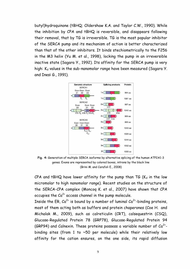
In mammals, three genes (human nomenclature ATP2A1-3) encode three
main SERCA proteins. Each of the three transcripts undergoes tissue-
dependent alternative splicing, increasing the number of pump variants
(fig. 4). The changes in the expression pattern of the variants during
development and tissue differentiation indicate that each isoform is
adapted to specific functions.
SERCA1a and SERCA1b variants are expressed in adult and neonatal fast-
twitch skeletal muscles, respectively. The SERCA2a variant is selectively
expressed in heart and slow-twitch skeletal muscles, whereas the
SERCA2b variant is expressed nearly ubiquitously and is thus considered
the housekeeping isoform. SERCA3 is instead expressed in a limited
number of non-muscle cells (Brini M. and Carafoli E., 2008).
It also known that the SERCA pump was inhibited by La3+ and
orthovanadate (Szasz I. et al., 1978). Specific inhibitors have also been
discovered: thapsigargin (TG, isolated from the roots of Thapsia
garganica) (Thastrup O. et al., 1990), cyclopiazonic acid (CPA, produced by
Aspergillus and Penicillum) (Seidler N.W. et al., 1989) and 2,5-di(t-
9
butyl)hydroquinone (tBHQ; Oldershaw K.A. and Taylor C.W., 1990). While
the inhibition by CPA and tBHQ is reversible, and disappears following
their removal, that by TG is irreversible. TG is the most popular inhibitor
of the SERCA pump and its mechanism of action is better characterized
than that of the other inhibitors. It binds stechiometrically to the F256
in the M3 helix (Yu M. et al., 1998), locking the pump in an irreversible
inactive state (Sagara Y., 1992). Its affinity for the SERCA pump is very
high: Kd values in the sub-nanomolar range have been measured (Sagara Y.
and Inesi G., 1991).
Fig. 4: Generation of multiple SERCA isoforms by alternative splicing of the human ATP2A1-3
genes. Exons are represented by colored boxes, introns by the black line
(Brini M. and Carafoli E., 2008)
CPA and tBHQ have lower affinity for the pump than TG (Kd in the low
micromolar to high nanomolar range). Recent studies on the structure of
the SERCA-CPA complex (Moncoq K. et al., 2007) have shown that CPA
occupies the Ca2+ access channel in the pump molecule.
Inside the ER, Ca2+ is bound by a number of luminal Ca2+-binding proteins,
most of them acting both as buffers and protein chaperones (Coe H. and
Michalak M., 2009), such as calreticulin (CRT), calsequestrin (CSQ),
Glucose-Regulated Protein 78 (GRP78), Glucose-Regulated Protein 94
(GRP94) and Calnexin. These proteins possess a variable number of Ca2+-
binding sites (from 1 to ~50 per molecule) while their relatively low
affinity for the cation ensures, on the one side, its rapid diffusion
10
through the organelle and, on the other, its prompt release upon opening
of the release channels (Zampese E. and Pizzo P., 2011).
ER Ca2+ release relies on two main gated ion channels: the ubiquitous IP3
receptor (IP3R), and, in many cells, the Ryanodine Receptors (RyRs).
Inositol 1,4,5-trisphosphate (IP3) is a second messenger produced
through phosphoinositide turnover (fig. 5) in response to many
extracellular stimuli (including hormones, growth factors,
neurotransmitters, neutrophins, odorants and light) and controls various
Ca2+-dependent cell functions (including cell proliferation, differentiation,
fertilization, embryonic development, secretion, muscular contraction,
immune responses, brain functions, chemical senses and light
transduction) by inducing Ca2+ release from intracellular Ca2+ stores, such
as from the ER (Berridge M.J., 1993).
Among the various inositol-phosphates and phospholipids, IP3 is unique in
that it is the only one that has a channel as a target molecule, the IP3
receptor (IP3R).
IP3 diffuses in the cytoplasm and binds to the IP3R, which is an
intracellular ligand-gated Ca2+ release channel localized primarily in the
ER membrane (Maeda N. et al., 1991).
IP3Rs are coded by three different genes, and alternatively spliced
isoforms have been identified in mammalian cells; the three full-length
sequences are 60–80% homologous and have distinct and overlapping
patterns of expression that vary during differentiation, with most cells
expressing more than one isoform (Foskett J.K. et al., 2007). The
different isoforms are probably involved in shaping different types of
signaling events (Hattori M. et al., 2004).
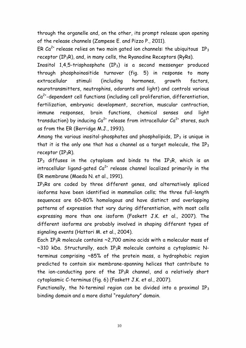
Each IP3R molecule contains ~2,700 amino acids with a molecular mass of
~310 kDa. Structurally, each IP3R molecule contains a cytoplasmic N-
terminus comprising ~85% of the protein mass, a hydrophobic region
predicted to contain six membrane-spanning helices that contribute to
the ion-conducting pore of the IP3R channel, and a relatively short
cytoplasmic C-terminus (fig. 6) (Foskett J.K. et al., 2007).
Functionally, the N-terminal region can be divided into a proximal IP3
binding domain and a more distal “regulatory” domain.

11
Fig.5: IP3 receptor and protein kinase C (PKC) signaling cascade system. Both IP3 and
diacylglycerol (DAG) are produced from phosphatidyl inositol bis-phosphate, but both have
completely different functions: IP3 releases Ca2+ from internal store and DAG activates PKC for
phosphorylation of various proteins.
The IP3R channel is a tetramer of four IP3R molecules (fig. 6).
Approximately 2,000 amino acids separate the IP3-binding domain from
the pore. This intervening region between the IP3 binding domain and the
pore contains consensus sequences for phosphorylation and binding by
nucleotides and various proteins. It may function to integrate, through
allosteric coupling, other signaling pathways or metabolic states with the
gating of the IP3R.
The localization of the IP3 binding region to the N-terminus of the IP3R
was first proposed by Mignery and co-workers based on the discovery
that deletion of the first 410 residues of the protein completely
eliminated IP3 binding (Mignery G.A. et al., 1990).
Binding of IP3 to the receptor is stechiometric (Supattapone S. et al.,
1988) with an apparent Kd usually in the range of 10–80 nM.
IP3 binding to the N-terminus of the channel induces conformational
changes that are transduced to the activation gate that then enables ion
flow through the channel. The molecular identity of the gate is unknown,
and the mechanisms that couple ligand binding to opening and closing of
the gate are unknown as well (Foskett K.J. et al., 2007).

12
Fig.6: The IP3R Ca2+ release channel. Cartoon depicting three of four IP3R molecules (in different
colors) in a single tetrameric channel structure (Foskett J.K. et al., 2007).
As to the structure of the pore, six transmembrane topology of the IP3R
was established by immunocytochemical techniques and N-linked
glycosylation analyses of full-length and truncated proteins. These
studies suggested that C-terminal transmembrane helices are involved in
ion permeation, with helices 5 and 6 and intervening sequences in IP3R
critical for creating the basic pore structure (Michikawa T. et al., 1994)
(fig. 6).
Deletion of the first four transmembrane helices from IP3R, leaving
transmembrane helices 5 and 6, resulted in a channel with normal
conductance and selectivity properties (Ramos-Franco J. et al., 1999),
consistent with this model. Site-directed mutagenesis of two residues
between TM5 and 6, and believed to be located in the putative selectivity
filter (Boehning D. et al., 2001), also suggested that such a model
provides a rational basis for considering the roles of particular residues
that contribute to conductance and selectivity properties of the IP3R
permeation pathway.
The IP3R is, most fundamentally, a Ca2+-activated ion channel. Ca2+ is
instead a critical modulator of IP3R channel function.
The steady-state gating activity of the IP3-liganded channel is regulated
by Ca2+ with a biphasic Ca2+ concentration dependence (Mak D.O.D. et al.,
2001). Modest increases in [Ca2+]c potentiate the responses to IP3, while
more substantial increases inhibit them (Foskett J.K. et al., 2007).
13
The primary functional effect of IP3 is to relieve Ca2+ inhibition of the
channel, enabling Ca2+ activation sites to gate it (Mak D.O.D. et al., 1998).
In essence, Ca2+ is the true channel ligand. Experimental results and
insights that have emerged from patch-clamp studies of the IP3R,
together with molecular modeling, indicate that Ca2+ regulation of the
channel is very complex, involving several distinct Ca2+ binding sites.
Where are these Ca2+ binding sites in the IP3R structure and sequence is
not yet known (Foskett K.J. et al., 2007).
Moreover, the N-terminal regulatory domain contains consensus
sequences for phosphorylation, proteolytic cleavage and binding by
different protein modulators and ATP, integrating different cellular
signaling pathways or metabolic states with the IP3R functionality
(Zampese E. and Pizzo P., 2011).
All these structure-function relationships have been thoroughly
investigated, and several molecules - such as homer, protein phosphatases
(PP1, PP2A), RACK1, chromogranin, Na+/K+-ATPase, carbonic anhydrase-
related protein (CARP) and IRBIT - interact with the IP3R and modulate
its activity, suggesting that the receptor can form multi-molecular
complexes, different from cell type to cell type, and function as a center
for signaling cascades (Mikoshiba K., 2007).
The other major Ca2+-releasing channel in the ER membranes is the RyR.
RyRs, the largest known ion channels (Takeshima et al. 1989), are large
conductance channels capable of creating rapid transient increases of
cytosolic Ca2+.
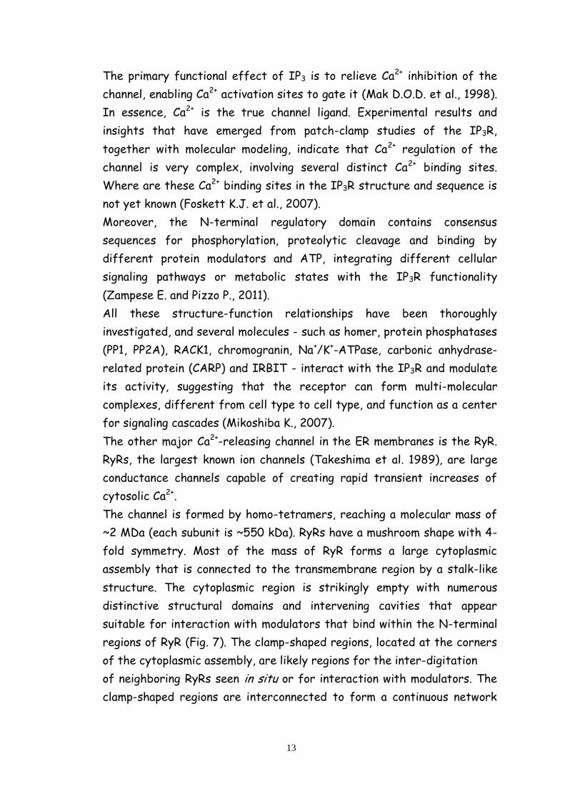
The channel is formed by homo-tetramers, reaching a molecular mass of
~2 MDa (each subunit is ~550 kDa). RyRs have a mushroom shape with 4-
fold symmetry. Most of the mass of RyR forms a large cytoplasmic
assembly that is connected to the transmembrane region by a stalk-like
structure. The cytoplasmic region is strikingly empty with numerous
distinctive structural domains and intervening cavities that appear
suitable for interaction with modulators that bind within the N-terminal
regions of RyR (Fig. 7). The clamp-shaped regions, located at the corners
of the cytoplasmic assembly, are likely regions for the inter-digitation
of neighboring RyRs seen in situ or for interaction with modulators. The
clamp-shaped regions are interconnected to form a continuous network

14
between the central rim and the cytoplasmic stalk-like structure via
several bridging densities (S.L. Hamilton and I.I. Serysheva, 2008).
Fig.7: The RyR macromolecular signaling complex
The pore is thought to be composed of six to eight TM segments (Du G.G.
et al., 2002).
While in skeletal muscle RyR1 opening is primarily (or exclusively) due to
the electromechanical coupling with dihydropyridine receptor type
voltage-dependent calcium channels (DHPR), the major gating mechanism
of RyR2 and RyR3 is the Ca2+ induced Ca2+ release (CICR): in this case, the
opening of RyRs is due to the local Ca2+ increase occurring in the
proximity of plasma membrane Ca2+ channels, as initially demonstrated for
cardiac muscle cells (Fill M. and Copello J.A., 2002); CICR can be also
induced by Ca2+ release from neighboring ER channels, such as IP3Rs or
RyRs, in a regenerative wave.
1.1.2 The Golgi Apparatus (GA) as an intracellular Ca2+ store
The GA represents the central sorting and processing station along the
secretory pathway, ensuring that cargo proteins, which are synthesized in
the ER, are properly modified (e.g. glycosylated, processed and packaged

15
into carrier vesicles) and eventually directed to their final destination
within the cell (P. Pizzo et al., 2011).
In high eukaryotes, the GA can be schematically viewed as being
composed by three main compartments: cis-, medial- and trans-Golgi and
consists of multiple flattened membranous cisternae arranged in close
apposition to each other to form stacks. Stacks are indeed polarized,
consisting of a cis-side associated with a tubular reticular network of
membranes (cis-Golgi network, CGN), a medial area of disc-shaped
flattened cisternae, and a trans-side associated with another tubular
reticular membrane network (trans-Golgi network, TGN) (fig. 8) (P. Pizzo
et al., 2011).
Traditionally, the GA was thought to be a stable, independent structure
able to exchange components with other intracellular organelles, but it is
actually a highly dynamic structure. Indeed, the steady-state structure
of Golgi stacks depends on the balance of anterograde and retrograde
transport through the different cisternae, between ER and the GA and
between this latter and the other cellular compartments (P. Pizzo et al.,
2011).
Functionally, the GA serves mainly as a cell ‘factory’ for the post-
translational modification of proteins and lipids (primarily, but not only,
glycosylation). Thus, the GA contains numerous glycosyl-transferases,
glycosidases, sulphatases and pro-protein convertases (like furin) that
cleave protein precursors into their mature forms (Schafer W. et al.,
1995).
The polarized GA morphology parallels a specific functionality since the
majority of GA glycosyl-transferases act sequentially and, in general,
enzymes acting early in glycanbiosynthetic pathways are known to be
localized in cis- and medial compartments of the GA, whereas enzymes
acting later in the biosynthetic pathway tend to reside within the trans-
Golgi cisternae and the TGN (Breton C. et al., 2001).
In the last two decades, numerous direct and indirect evidence supported
the idea that the GA also plays a key role as intracellular Ca2+ store: (i)
using ion microscopy, Chandra et al. showed that the GA can store up to
5% of the total cellular Ca2+ and that it is substantially more resistant to
Ca2+ depletion than other cellular organelles (Chandra S. et al., 1991); (ii)
using the electron energy loss spectroscopy technique in cryosections of

16
mammalian cells, it was reported a strong Ca2+ labelling extending across
the entire cis/trans axis of the GA (Pezzati R. et al., 1991); (iii)
processing enzymes located in the GA and/or TGN show a strong Ca2+
dependency of their activity (Oda K., 1992); (iv) finally, retrograde
membrane traffic from the Golgi to the ER and selective aggregation of
regulated secretory proteins in the TGN (Chanat E. and Huttner W.B.,
1991) were also critically depended on GA Ca2+ content. Thus, the idea
that variations of the [Ca2+] within the GA could affect cell functions in a
variety of ways was generally accepted, though based on indirect
approaches as no methodology were available to monitor directly the free
Ca2+ concentration in the GA lumen of living cells.
Fig. 8: Three-dimensional tomographic reconstruction of the Golgi complex. The cis-Golgi network
(white) and the stack with the terminal three trans-Golgi cisternae (pink, red and cyan) are shown.
The trans-Golgi network (violet) appears as a tubular network that emerges from the lateral part
of the last trans-cisterna (cyan) (P. Pizzo et al., 2011)
The first direct demonstration that indeed the GA is a dynamic Ca2+
storing organelle capable of releasing Ca2+ into the cytosol upon cell
activation came only when a new aequorin based Ca2+ probe specifically
localized within the GA lumen was developed (Pinton P. et al., 1998). With
this tool, obtained by adding to the aequorin sequence the first 69AA of
the GA resident protein sialyl-transferase, it was shown that a high [Ca2+]
is maintained in the GA lumen in steady state; Ca2+ uptake in the GA
appeared to depend on the combined activity of a typical ER Ca2+ ATPase
(SERCA) and of another Ca2+ pump, with distinct pharmacological

17
properties, the secretory pathway Ca2+ ATPase1, SPCA1. Furthermore,
the Ca2+ content of the GA could be discharged rapidly and extensively
following stimulation with an IP3-generating agonist. Taken together
these studies revealed that the GA could be regarded as a bona fide IP3-
sensitive, rapidly mobilizable, intracellular Ca2+ store and, for its specific
perinuclear location within the cell, of potential strategic importance for
the generation of local cytosolic Ca2+ signals.
Subsequently it has been suggested that Ca2+ handling within the GA is
heterogeneous: in particular, it has been showed that the SPCA1-
containing Ca2+ sub-compartment was insensitive (or mildly sensitive) to
IP3-generating agonist stimulation and therefore not involved in setting
up cytosolic Ca2+ signals (Vanoevelen J. et al., 2004). Furthermore, the
kinetics of Ca2+ release from the GA appeared to be somewhat different
from those of the ER: in particular, while the latencies and initial rates of
Ca2+ release were similar for the two organelles, Ca2+ release from the GA
terminated faster than that from the ER (Missiaen L. et al., 2004).
Altogether these data suggest that the GA could be heterogeneous in
terms of Ca2+ handling properties, with distinct Ca2+ sub-compartments
endowed with different molecular components.
As far as the molecular Ca2+ tool-kit is concerned, the GA contains
several Ca2+ handling proteins known to be expressed in the ER. As
mentioned above, SERCA2 pumps and IP3Rs have been extensively
demonstrated to be present and functional at the GA level (Pizzo P. et al.,
2011). In addition to IP3Rs also the other major intracellular Ca2+
releasing channel, the ryanodine receptor, RyR, has been shown to be
expressed in the GA (Cifuentes F. et al., 2001).
As to the Ca2+ uptake mechanisms, data from different laboratories
clearly indicate that, in addition to SERCA, another ATP-dependent Ca2+
pumping mechanism (not expressed at significant level within the ER)
characterizes this cell compartment: the secretory-pathway Ca2+-
ATPases (SPCAs) (Pizzo P. et al., 2011). The relative contribution of each
of the two types of Ca2+-ATPases, SERCAs and SPCAs, to the total
uptake of Ca2+ into the GA is apparently different in various cell types: in
keratinocytes, for example, SPCA1 knock-down leads to a 67% of
reduction in the average Golgi Ca2+ uptake (as measured with aequorin),

18
while in HeLa cells (using the same method) the effect of reducing SPCA1
level on the Golgi Ca2+ is modest (VanBaelen K. et al., 2003).
The secretory-pathway Ca2+-ATPases (SPCAs) represent a recently
recognized family of phosphorylation-type ATPases that supply the lumen
of the Golgi apparatus with Ca2+ and Mn2+ needed for the normal
functioning of this structure.
The knowledge of the SPCAs is much more limited than that of the well
characterized SERCA (L. Missiaen et al., 2007).
SPCAs are single subunit integral membrane proteins with a large
cytosolic head containing an actuator (A), nucleotide-binding (N), and
phosphorylation (P) domain, and with 10 hydrophobic helices (M1–M10)
displaying membrane-spanning propensity.
The SPCAs contain the typical SDKTGTLT sequence of phosphorylation-
type ATPases with a highly conserved aspartyl residue that is transiently
phosphorylated during the reaction cycle (L. Missiaen et al., 2007).
The transmembrane region of SPCA pumps only have one Ca2+-binding site
and indeed the Hill coefficients obtained from the Ca2+ dependence of
ATP hydrolysis and phosphorylation of human SPCAs are close to 1, also
suggesting that the pump transports only one Ca2+ or Mn2+ ion per each
hydrolyzed ATP (Dode L. et al., 2006).
In human cells, two different genes encoding for SPCA have been cloned:
ATP2C1 (on chromosome 3), that, by alternative processing at the 3’-end,
results in 4 SPCA1 proteins with C-termini differing in length and specific
amino acid sequence (Hu Z. et al., 2000), and ATP2C2 (on chromosome 16)
encoding for a second human SPCA isoform, called SPCA2. As far as
SPCAs distribution and localization is concerned, while SPCA1 is a
housekeeping enzyme with a widespread expression in all cells and
particularly high in human epidermal keratinocytes (Hu Z. et al., 2000),
the tissue and cellular expression of SPCA2 appears to be more
restricted (Vangheluwe P. et al., 2009), suggesting a more specialized
function for this type of pump.
Similarly to the ER, the GA is also equipped with luminal Ca2+-binding
proteins. Notably, the Ca2+ binding proteins of the GA are not found (with
one exception) in the ER lumen and, viceversa, the typical ER Ca2+ binding
proteins are not found in the GA. The most abundant and better
characterized of the GA Ca2+ binding proteins is CALNUC, an EF-hand,
19
Ca2+-binding peripheral membrane protein tightly associated with the
luminal surface of the cis-Golgi Network (CGN) and cis-Golgi cisternae
(Lin P. et al., 1998).
The other two Ca2+-binding proteins present in the GA lumen are
p54/NEFA and calumenin. The first is a luminal protein strongly
associated to GA membranes that shares significant sequence identity
(62%) with CALNUC and, by immunofluorescence and immunoelectron
microscopy, seems to be localized in the medial-Golgi cisternae (Morel-
Huaux V.M. et al., 2002). The second one, calumenin, belongs to the family
of multiple EF-hand proteins and shows a localization not confined to the
Golgi, but also to the ER and throughout the secretory pathway (Yabe D.
et al., 1997).
The GA is thus equipped with all the molecular components necessary for
its function as a Ca2+ store: Ca2+ pumps, Ca2+ channels and luminal Ca2+-
binding proteins. The heterogeneity of these molecules, in part paralleled
by their differential distribution within GA cisternae, suggests, however,
that the GA Ca2+ store does not behave uniformly in terms of Ca2+
handling.
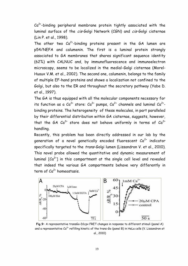
Recently, this problem has been directly addressed in our lab by the
generation of a novel genetically encoded fluorescent Ca2+ indicator
specifically targeted to the trans-Golgi lumen (Lissandron V. et al., 2010).
This novel probe allowed the quantitative and dynamic measurement of
luminal [Ca2+] in this compartment at the single cell level and revealed
that indeed the various GA compartments behave very differently in
term of Ca2+ homeostasis.
Fig.9: A representative transGo-D1cpv FRET changes in response to different stimuli (panel A)
and a representative Ca2+ refilling kinetic of the trans-Go (panel B) in HeLa cells (V. Lissandron et
al., 2010)
20
In particular, it has been demonstrated that the trans-Golgi takes up Ca2+
almost exclusively via SPCA1 (and not by SERCA) and does not release
Ca2+ in response to IP3 generation; rather the trans-Golgi accumulates the
cation as a consequence of the cytoplasmic Ca2+ rises that follow this
latter event (fig. 9).
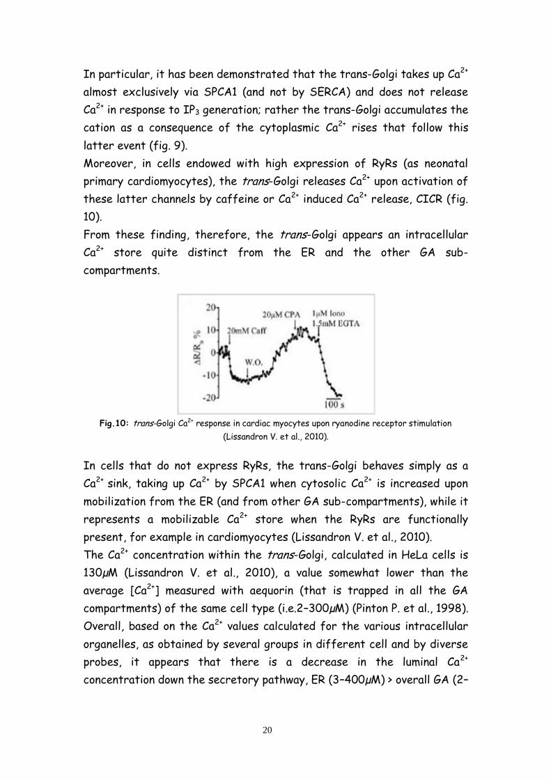
Moreover, in cells endowed with high expression of RyRs (as neonatal
primary cardiomyocytes), the trans-Golgi releases Ca2+ upon activation of
these latter channels by caffeine or Ca2+ induced Ca2+ release, CICR (fig.
10).
From these finding, therefore, the trans-Golgi appears an intracellular
Ca2+ store quite distinct from the ER and the other GA sub-
compartments.
Fig.10: trans-Golgi Ca2+ response in cardiac myocytes upon ryanodine receptor stimulation
(Lissandron V. et al., 2010).
In cells that do not express RyRs, the trans-Golgi behaves simply as a
Ca2+ sink, taking up Ca2+ by SPCA1 when cytosolic Ca2+ is increased upon
mobilization from the ER (and from other GA sub-compartments), while it
represents a mobilizable Ca2+ store when the RyRs are functionally
present, for example in cardiomyocytes (Lissandron V. et al., 2010).
The Ca2+ concentration within the trans-Golgi, calculated in HeLa cells is
130µM (Lissandron V. et al., 2010), a value somewhat lower than the
average [Ca2+] measured with aequorin (that is trapped in all the GA
compartments) of the same cell type (i.e.2–300µM) (Pinton P. et al., 1998).
Overall, based on the Ca2+ values calculated for the various intracellular
organelles, as obtained by several groups in different cell and by diverse
probes, it appears that there is a decrease in the luminal Ca2+
concentration down the secretory pathway, ER (3–400µM) > overall GA (2–

21
300µM) > trans-Golgi(130µM) > secretory vesicles (80µM) (fig. 11) (Pizzo
P. et al., 2011).
The complexity of the GA morphology, the cis-, medial- and trans-Golgi,
and their specific enzymatic repertoire is paralleled by different
mechanisms of the Ca2+ handling machinery in each compartment (in terms
of Ca2+ pumps, channels and buffering proteins). The information on the
cis- and medial-Golgi is still based on indirect data and one of the aim of
my work has been to develop and set up specific targeted Ca2+ probes for
these two sub-compartments, in order to characterized them in term of
Ca2+ handling.
Fig. 11: Ca2+ concentration and molecular tool-kit gradient through the secretory pathway
(P. Pizzo et al., 2011)
1.1.3. Peroxisomes, mithocondria and endolysosomal compartments in
intracellular Ca2+ dynamics
1.1.3.1 Mithocondria
Mitochondria, the cellular organelles responsible primarily for energy
generation, are also intracellular Ca2+ stores. Within the matrix of
mitochondria, Ca2+ is stored not bound to Ca2+ buffering proteins but
instead precipitated out as an insoluble salt, CaPO4. The handling of Ca2+
by mitochondria is very complex , but one aspect worth highlighting is how
22
mitochondria communicate with the ER with respect to Ca2+ fluxes (Prins
D. and Michalak M., 2011).
Release of Ca2+ from the ER results in elevated cytoplasmic Ca2+ levels,
after which mitochondria take up Ca2+ resulting in elevated matrix Ca2+
levels (Rizzuto R. et al., 1992). ER and mitochondria are often tightly
apposed in regions known as mitochondria-associated membranes (MAM);
the interactions between these two organelles are regulated by
cytoplasmic Ca2+ levels (Wang H.J., et al. 2000). Interestingly, resting
cytoplasmic Ca2+ levels promote dissociation of the two organelles
whereas higher Ca2+ levels enhance their association, suggesting that
mitochondria may act as buffers to soak up Ca2+ released from the ER
lumen (Wang et al., 2000).
1.1.3.2 Peroxisomes
Recent results indicate that peroxisomes are capable of taking up and
storing Ca2+ within their lumen at concentrations much higher than those
found in the cytoplasm (Raychaudhury B. et al. 2006; Drago I. et al.
2008). However, it is not yet known how Ca2+ is stored within these
organelles and whether there exist specific peroxisomal Ca2+ buffering
proteins (D. Prins and M. Michalak, 2011).
1.1.3.3 Endolysosomal compartments
Recent results show that nicotinic acid adenine dinucleotide phosphate
(NAADP) acts as a second messenger to mobilize intracellular Ca2+ stores
through actions on two-pore channels (TPCs) in endosomal membranes
(Calcraft P.J. et al. 2009). Endosomes and lysosomes may thus be
considered to be Ca2+ storage organelles; however, the mechanism of
lysosomal Ca2+ buffering has not yet been described (D. Prins and M.
Michalak, 2011).

23
1.2 Ca2+ entry from the extracellular space
1.2.1 Voltage-operated Ca2+ enty
Ca2+ channels in many different excitable cell types activate on membrane
depolarization and mediate Ca2+ influx in response to action potentials and
sub-threshold depolarizing signals.
Ca2+ entering the cell through voltage-gated Ca2+ channels serves as the
second messenger of electrical signaling, initiating many different cellular
events (fig. 12).
Fig.12: Signal transduction by voltage-gated Ca2+ channels.
In cardiac and smooth muscle cells, activation of Ca2+ channels initiates
contraction directly by increasing cytosolic Ca2+ concentration and
indirectly by activating calcium-dependent calcium release by ryanodine-
sensitive Ca2+ release channels in the sarcoplasmic reticulum (W.A.
Catterall, 2011). In skeletal muscle cells, voltage-gated Ca2+ channels in
the transverse tubule membranes interact directly with ryanodine-
sensitive Ca2+ release channels in the sarcoplasmic reticulum and activate
them to initiate rapid contraction (Catterall W.A., 1991).
In endocrine cells, voltage-gated Ca2+ channels mediate Ca2+ entry that
initiates secretion of hormones (Yang S.N. and Berggren P.O., 2006).
In neurons, voltage-gated Ca2+ channels initiate synaptic transmission.

24
In many different cell types, Ca2+ entering the cytosol via voltage-gated
Ca2+ channels regulates enzyme activity, gene expression and other
biochemical processes (W.A. Catterall, 2011).
Thus, voltage-gated Ca2+ channels are the key signal transducers of
electrical excitability, converting the electrical signal of the action
potential in the cell surface membrane to an intracellular Ca2+ transient.
Signal transduction in different cell types involves different molecular
subtypes of voltage-gated Ca2+ channels, which mediate voltage-gated
Ca2+ currents with different physiological, pharmacological and regulatory
properties (W.A. Catterall, 2011).
Since the first recordings of Ca2+ currents in cardiac myocytes, it has
become apparent that there are multiple types of Ca2+ currents (table 1)
as defined by physiological and pharmacological criteria (Tsien R.W. et al.,
1988).
In cardiac, smooth, and skeletal muscle, the major Ca2+ currents are
distinguished by high voltage of activation, large single channel
conductance, slow voltage-dependent inactivation, marked up-regulation
by cAMP-dependent protein phosphorylation pathways, and specific
inhibition by Ca2+ antagonist drugs including dihydropyridines,
phenylalkylamines and benzothiazepines.
These Ca2+ currents have been designated L-type, as they have slow
voltage dependent inactivation and therefore are long lasting when Ba2+ is
the current carrier and there is no Ca2+-dependent inactivation (Tsien
R.W. et al., 1988).
Electrophysiological studies of Ca2+ currents in starfish eggs (Hagiwara S.
et al. 1975) first revealed Ca2+ currents with different properties from
L-type, and these were subsequently characterized in detail in voltage-
clamped dorsal root ganglion neurons (Carbone E. and Lux H.D., 1984). In
comparison to L-type, these novel Ca2+ currents activated at much more
negative membrane potentials, inactivated rapidly, deactivated slowly, had
small single channel conductance and were insensitive to conventional Ca2+
antagonist drugs available at that time. They were designated low-voltage
activated Ca2+ currents for their negative voltage dependence (Carbone
E.and Lux H.D., 1984) or T-type Ca2+ currents for their transient openings
(Nowycky M.C. et al. 1985).
25
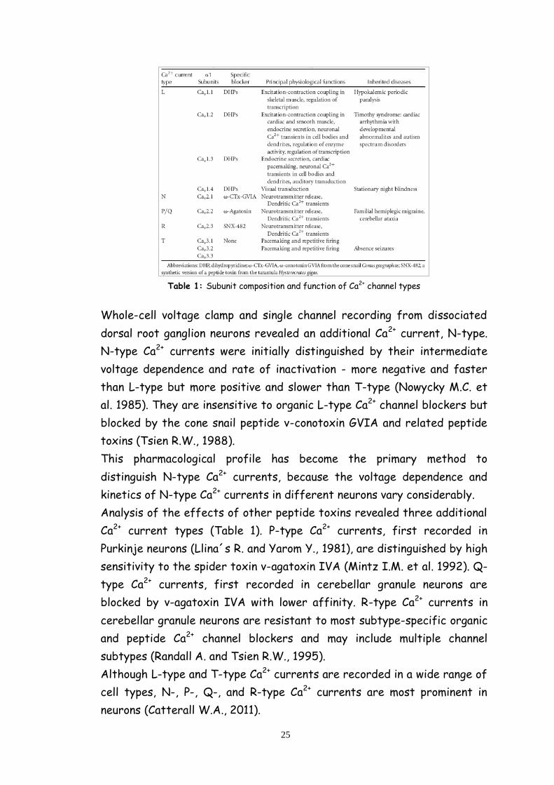
Table 1: Subunit composition and function of Ca2+ channel types
Whole-cell voltage clamp and single channel recording from dissociated
dorsal root ganglion neurons revealed an additional Ca2+ current, N-type.
N-type Ca2+ currents were initially distinguished by their intermediate
voltage dependence and rate of inactivation - more negative and faster
than L-type but more positive and slower than T-type (Nowycky M.C. et
al. 1985). They are insensitive to organic L-type Ca2+ channel blockers but
blocked by the cone snail peptide v-conotoxin GVIA and related peptide
toxins (Tsien R.W., 1988).
This pharmacological profile has become the primary method to
distinguish N-type Ca2+ currents, because the voltage dependence and
kinetics of N-type Ca2+ currents in different neurons vary considerably.
Analysis of the effects of other peptide toxins revealed three additional
Ca2+ current types (Table 1). P-type Ca2+ currents, first recorded in
Purkinje neurons (Llina´s R. and Yarom Y., 1981), are distinguished by high
sensitivity to the spider toxin v-agatoxin IVA (Mintz I.M. et al. 1992). Q-
type Ca2+ currents, first recorded in cerebellar granule neurons are
blocked by v-agatoxin IVA with lower affinity. R-type Ca2+ currents in
cerebellar granule neurons are resistant to most subtype-specific organic
and peptide Ca2+ channel blockers and may include multiple channel
subtypes (Randall A. and Tsien R.W., 1995).
Although L-type and T-type Ca2+ currents are recorded in a wide range of
cell types, N-, P-, Q-, and R-type Ca2+ currents are most prominent in
neurons (Catterall W.A., 2011).

26
Ca2+ channels purified from skeletal muscle transverse tubules are
complexes of 1, 2, , , and subunits (fig. 12).
Analysis of the biochemical properties, glycosylation, and hydrophobicity
of these five subunits led to a model comprising a principal
transmembrane 1 subunit of 190 kDa in association with a disulfide-
linked 2 dimer of 170 kDa, an intracellular phosphorylated subunit of
55 kDa, and a transmembrane subunit of 33 kDa (fig. 12) (Takahashi M.
et al. 1997).
Intensive studies of the structure and function of the related pore-
forming subunits of Na+, Ca2+, and K+ channels have led to identification of
their principal functional domains (Catterall W.A., 2011). Each domain of
the principal subunits consists of six transmembrane helices (S1–S6)
and a membrane-associated loop between S5 and S6 (Fig. 12). The S4
segments of each homologous domain serve as the voltage sensors for
activation, moving outward and rotating under the influence of the
electric field and initiating a conformational change that opens the pore.
Fig. 13: Subunit structure of Ca2+ channels
The S5 and S6 segments and the membrane-associated pore loop between
them form the pore lining of the voltage-gated ion channels. The narrow
external end of the pore is lined by the pore loop, which contains a pair of
glutamate residues in each domain that are required for Ca2+ selectivity, a
structural feature that is unique to Ca2+ channels (Heinemann S.H. et al.,
1992).
27
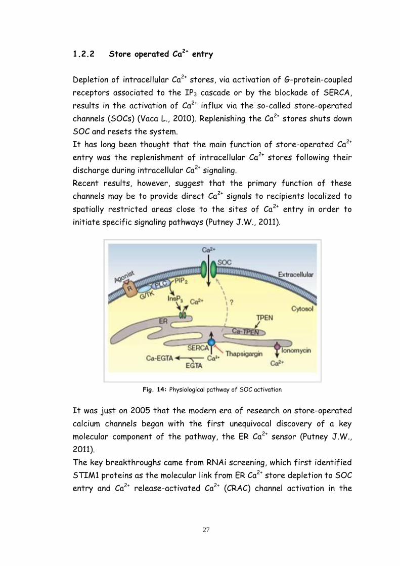
1.2.2 Store operated Ca2+ entry
Depletion of intracellular Ca2+ stores, via activation of G-protein-coupled
receptors associated to the IP3 cascade or by the blockade of SERCA,
results in the activation of Ca2+ influx via the so-called store-operated
channels (SOCs) (Vaca L., 2010). Replenishing the Ca2+ stores shuts down
SOC and resets the system.
It has long been thought that the main function of store-operated Ca2+
entry was the replenishment of intracellular Ca2+ stores following their
discharge during intracellular Ca2+ signaling.
Recent results, however, suggest that the primary function of these
channels may be to provide direct Ca2+ signals to recipients localized to
spatially restricted areas close to the sites of Ca2+ entry in order to
initiate specific signaling pathways (Putney J.W., 2011).
Fig. 14: Physiological pathway of SOC activation
It was just on 2005 that the modern era of research on store-operated
calcium channels began with the first unequivocal discovery of a key
molecular component of the pathway, the ER Ca2+ sensor (Putney J.W.,
2011).
The key breakthroughs came from RNAi screening, which first identified
STIM1 proteins as the molecular link from ER Ca2+ store depletion to SOC
entry and Ca2+ release-activated Ca2+ (CRAC) channel activation in the

28
plasma membrane, and then identified Orai (CRACM) proteins that
comprise the CRAC channel pore-forming subunit (Cahalan M.D., 2009).
STIM1, initially characterised as a phosphoprotein, includes a single
transmembrane domain located in the ER in resting cells (fig. 15). The N-
terminus of STIM1 stretches into the ER lumen, while the C-terminus
extends into the cytosol. Within the former, a single EF-hand Ca2+-
binding motif acts as luminal Ca2+ sensor. A sterile-alpha motif (SAM),
including two N-linked glycosylation sites, is followed by the
transmembrane domain and a cytosolic C-terminus with two coiled-coil
regions overlapping with an ezrin-radixin-moesin (ERM)-like domain.
Subsequent glutamate-, serine/proline-, serine/threonine- and lysine-rich
regions are additionally located at the C-terminus (fig. 15) (Liou J. et al.,
2005).
Fig. 15: STIM1 is localized primarily in the ER membrane and Orai1 is a plasma membrane protein
with four membrane-spanning regions and intracellular N and C termini.
Orai1 contains four predicted transmembrane segments (fig. 15), form
Ca2+ selective plasma membrane channels. Within the cytosolic strands,

29
the N-terminus of only Orai1 includes a proline/arginine-rich region. In
their C-terminus Orai1 contains a putative coiled-coil domain. The
extracellular loop of Orai1 between the third and fourth transmembrane
segment comprises an N-glycosylation site (Cahalan M.D. et al., 2005).
STIM1 mediated robust Orai1 currents are initially stimulated by Ca2+
store-depletion sensed by the STIM1 luminal EF-hand (fig. 16).
In resting cells, STIM1 is uniformly distributed within the ER, displays
tubular structures and binds to the microtubule-plus-end-tracking protein
EB1 at those sites where microtubule ends come in contact with the ER
(fig. 16) (Grigoriev I. et al, 2008).
Fig. 16: The functional units of store-operated Ca2+ entry assemble in response to store
depletion.
The STIM1 EF-hand is adequate to sense a decrease in the ER Ca2+ level
that is approximately 300–500 μM at rest.
Subsequently, STIM1 forms oligomers before it redistributes into
punctuate clusters close to the plasma-membrane (fig. 16) (Liou J. et al.,
2005). It has recently been shown that oligomerisation of about four
STIM1 proteins is the critical process transmitting ER store depletion to
STIM1/Orai1 clusters thereby activating store-operated Ca2+ entry (fig.
30
16). In particular, four Orai1 molecules create the pore-forming subunit
of CRAC that opens upon stimulation by STIM1, with the C-terminus of
STIM1 and N-terminus of Orai1 participating in this process (fig. 16)
(R.S. Lewis, 2007)
31
2. GENETICALLY ENCODED Ca2+ INDICATORS
Ca2+ is one of the most important and versatile second messengers in cell
biology; consequently, there has been enormous effort devoted to
developing tools to image Ca2+ in living cells.
Fluorescent indicators for Ca2+ allow the researcher to monitor Ca2+
signals in living cells and in real time, thus preserving temporal control of
Ca2+ signaling.
Genetically encoded Ca2+ indicators (GECIs) are a subset of fluorescent
indicators that offer the additional advantage of being able to monitor
Ca2+ dynamics in specific sub-cellular locations, thereby maintaining
spatial heterogeneity of Ca2+ transients (McCombs J.E. and Palmer A.E.,
2008).
GECIs are defined as indicators that are produced by translation of a
nucleic acid sequence. Therefore, these indicators are comprised solely
of natural protein or peptide motifs. In order to convert a Ca2+ signal into
an optical readout, GECIs consist of at least one light-emitting protein
and a Ca2+ responsive element, such that Ca2+ binding changes the optical
properties of the protein(s). These protein-based indicators are typically
incorporated into cells by gene transfer techniques (McCombs J.E. and
Palmer A.E., 2008).
There are three classes of GECIs that have been developed which differ
in their overall architecture (fig. 17). The first class are bioluminescent
reporters based on the aequorin photoprotein (Rizzuto R. et al., 1994).
These probes are inherently different than the fluorescent protein-
based probes as light is generated by a chemical reaction that requires
reconstitution of the indicator with a co-factor.
The second class is based on single fluorescent proteins. These indicators
are composed of Ca2+-responsive elements, such as calmodulin (CaM) or
portions there of that are inserted into a fluorescent protein, such that
Ca2+ binding alters the protonation state, and hence spectral properties
of the chromophore. These indicators include the camgaroos, G-CaMPs,
pericams, “Case” sensors and grafted EF-hands (McCombs J.E. and Palmer
A.E., 2008).
The last class of sensors are the “cameleon-type” in which Ca2+-
responsive elements are inserted between two fluorescent proteins so

32
that upon Ca2+ binding, an alteration in the efficiency of fluorescence
resonance energy transfer (FRET) occurs (Miyawaki A. et al., 1997).
Fig. 17: Models of the three classes of GECIs. (a) The aequorin photoprotein is shown in complex
with coelenterazine. Upon binding of Ca2+, the aequorin undergoes a conformational change,
releasing coelenteramide and emitting blue light. (b) Single FP sensors employing the Ca2+-
responsive element CaM and a CaM binding peptide attached to a circularly permutated FP. On
binding Ca2+ , CaM executes a conformational change, interacting with the peptide and altering the
protonation state of the chromophore thus changing the fluorescence intensity of the protein. (c)
Grafted sensors utilizing EF hands or portions of CaM inserted into a fluorescent protein. Binding
of Ca2+ causes a change in protein conformation and a shift in the protonation state of the
chromophore. (d) FRET-based sensors having a Ca2+ binding domain located between two
flurophorescent proteins. As Ca2+ binds, the Ca2+ binding domain undergoes a conformational
change, interacting with its binding peptide. This brings the two FPs closer together, increasing
the efficiency of FRET.
2.1 FRET-based cameleon probes
Cameleons (fig. 17) are tandem repeats of GFP mutants with overlapping
excitation/emission spectra linked together by a Ca2+ sensor based on
Calmodulin (CaM), a glycylglycine linker and the CaM binding peptide of
myosin light chain kinase M13 (Miyawaki A. et al., 1997). In this design,

33
Ca2+ binding promotes the reversible association of CaM and M13,
promoting fluorescence resonance energy transfer (FRET) between the
two GFP variants (Demaurex N., 2005).
The first probes generated, yellow cameleons (YC), used cyan and yellow
fluorescent proteins (CFP and YFP) as FRET donor and acceptors. The
reversible changes in FRET can be detected as changes in the yellow over
cyan emission fluorescence and the probes function as ratiometric
emission Ca2+ indicators. The original YC comprised probes of different
affinities, ranging from 1 to 300µM (YC2, YC3 and YC4), but all probes
suffered from a poor dynamic range and were sensitive to pH and to
chloride ions. YC were subsequently made pH resistant by YFP
mutagenesis (YC2.1 and YC3.1) and brighter by replacing YFP with citrine
(YC3.3) or with Venus, a YFP mutant with fast and efficient maturation
(YC2.12 and VC6.1). Although these cameleons were greatly improved over
the original design, they still displayed insufficient signal-to-noise ratio
when targeted to organelles (Demaurex N., 2005).
To further increase the dynamic range of cameleons, Nagai et al.
generated several circularly permuted YFP mutants in an attempt to
optimize the relative orientation of the two FRET partners (Nagai T. et
al., 2004). By introducing new termini into the surface exposed loop
regions of the β-barrel of Venus, they generated several YC variants with
improved dynamics. The best construct, YC 3.6, which contains a YFP
circularly permutated at position 173 (173cpVenus), matures efficiently,
is resistant to acidification, and displays a monophasic Ca2+ sensitivity
with a Kd of 250 nM. The cp173Venus cameleon has much better dynamics
than all previous cameleons when expressed in cells, with an impressive
5.6-fold increase in ratio between Rmin and Rmax. The expanded dynamic
range of YC3.6 is a clear advantage to image Ca2+ dynamics in cells and
tissues with adequate spatiotemporal resolution.
Moreover A.E. Palmer et al. generated a series of indicators that resist
endogenous CaM and have varying Ca2+ affinities to be useful in
monitoring Ca2+ in distinct sub-cellular locations as well as in micro-
domains of high Ca2+ (Palmer A.E. et al., 2006). Their goal in redesigning
the cameleon was to reengineer the binding interface between CaM and a
target peptide to generate selective and specific binding pairs that could
not be perturbed by wild-type (wt) CaM. They previously reversed the
34
salt bridge interactions between basic residues in the target peptide and
acidic residues in CaM to generate a mutant calmodulin-peptide pair that
was unaffected by large concentrations of excess CaM. This redesigned
pair led to a Ca2+ indicator (D1) with a relatively weak affinity for Ca2+ (Kd
= 60 µM) and has been used to monitor Ca2+ directly in the ER in individual
living cells (Palmer A.E. et al., 2004).
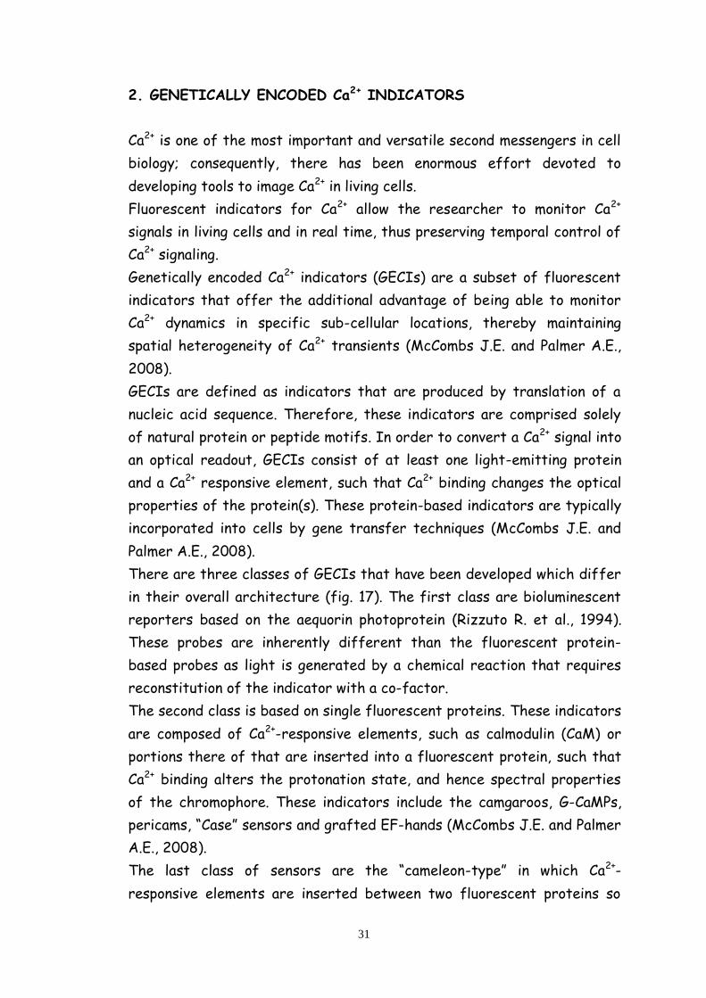
Subsequently, they computationally designed steric bumps in the target
peptide and complementary holes in CaM in order to generate a series of
indicators (D2, D3, and D4) with varying Ca2+ affinities (fig. 18), including
a high-affinity indicator that cannot be perturbed by excess CaM. These
indicators show significant improvements in the ability to monitor Ca2+ in
different sub-cellular locations such as the plasma membrane of neurons
and the mitochondria (Palmet A.E. et al., 2006).
Fig. 18: In Vitro Characterization of the Redesigned Cameleons Ca2+ titration curves of original
and redesigned cameleons. The original cameleons (YC2, YC3, and YC4; black diamonds) are labeled
on the graph. The first redesign, D1, is presented as red circles, and the computational designs
are presented as follows: D2cpv, blue squares; D3cpv, green, upside-down triangles; D4cpv, purple
triangles.
To date, cameleons have been expressed in the cytosol (D1cpv probe)
(Leite M.F. et al., 2003), targeted to the ER (ER-D1 probe) and the
nucleoplasm (H2BD1cpv probe) (Leite M.F. et al., 2003), to the
mitochondria (4mt-D1cpv probe) (Giacomello M., et al., 2010), to the
trans-Golgi (Go-D1cpv probe) (V. Lissandron et al., 2010), to the
peroxisomal matrix (KVK-SKL-D3cpv probe) (Drago I., et al., 2008).
All these probes have been successfully used to monitor Ca2+ dynamics in
their specific sub-cellular compartments.

35
3. ALZHEIMER’S DISEASE
Alzheimer’s disease (AD) was first described little more than 100 years
ago. It is the most common cause of dementia with an estimated
prevalence of 30 million people worldwide, a number that is expected to
quadruple in 40 years.
In the 105 years since Alzheimer’s original case report and particularly in
the past 3 decades, much has been learned about AD. However, there
currently is no effective treatment that delays the onset or slows the
progression of the disease (Holtzman D.M. et al., 2011).
3.1 Clinical and pathological features of AD
Dementia is an acquired syndrome characterized by a loss or decline in
memory and other cognitive abilities. It represents a decline from a
person’s previously established level of intellectual function that is
sufficient to interfere with the everyday performance of that individual.
AD is the most common cause of dementia, estimated to contribute to
about 60–70% of cases (Barker W.W. et al., 2002).
The essential feature of AD, intra-individual decline in cognitive abilities,
has an insidious onset over several months with subsequent gradual but
relentless progression though successive stages of dementia severity.
The time course of AD dementia averages 7–10 years and inevitably the
illness culminates in death. Impaired recent memory usually is an initial
symptom of AD but other cognitive deficits, such as executive
dysfunction manifested by changes in attention and problem solving
abilities, are typically also present. As dementia progresses, language
dysfunction, visuospatial difficulty, loss of insight, and personality
changes (withdrawal, decreased initiative, occasionally depression)
frequently are apparent. There also is impaired ability to function at
routine tasks at home, such that even basic activities of daily living (e.g.,
dressing, bathing, grooming) require supervision or assistance. In the
severe stage of Alzheimer dementia, individuals are totally dependent on
caregivers for all activities of daily living and, in advanced disease, often
become mute, non-ambulatory and unable to swallow or control bladder
and bowel function (Holtzman D.M. et al., 2011).

36
The key neuropathological elements of AD were described by Alois
Alzheimer in 1906 and at about the same time by Oskar Fischer (Goedert
M., 2009). At the macroscopic level, there is gross atrophy of the brain
(Figure 19).
Fig. 19: Postmortem brain section from an AD case (left) compared with that of a cognitively
normal individual (right)
At the microscopic level, the hallmarks of the disease are amyloid
plaques, neurofibrillary tangles (fig. 20) and extensive neuronal loss.
Amyloid plaques are accumulations of molecules in the extracellular space
of the brain (fig. 20). The principal proteinaceous component of plaques is
the amyloid- (A ) peptide, a 38–43 amino acid peptide derived from a
much larger protein, the amyloid precursor protein (APP). Within plaques,
A is present in aggregated (insoluble) forms including fibrils as well as
oligomers (Kayed R. et al., 2003).
Fig. 20: The neurofibrillary tangles and tha amyloid plaques depositions in AD brain
37
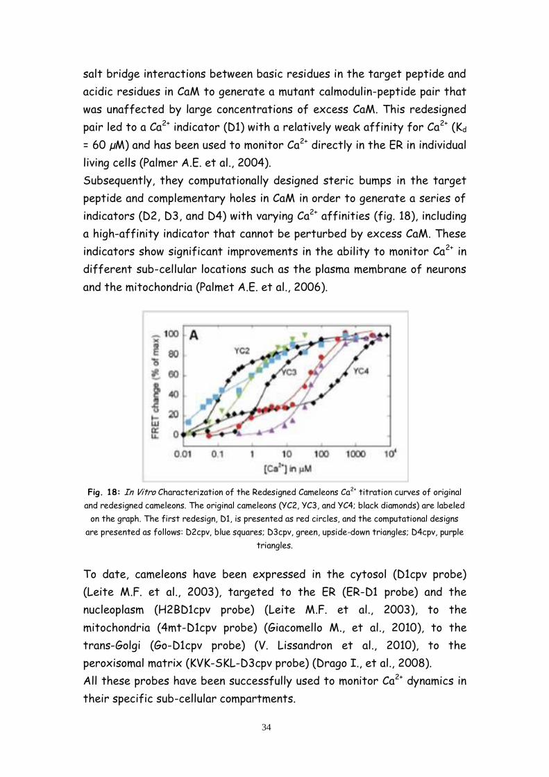
These “neuritic” plaques are surrounded by swollen, degenerating neurites
(axons and dendrites). In areas surrounding neuritic plaques, there is also
“gliosis” with hypertrophy and an alteration of the morphology as well as
the proliferation of astrocytes and microglia (immune cells of the CNS).
It is likely that this inflammatory response contributes to brain injury
although there is evidence that glial cells also play a protective role (Lucin
K.M. and Wyss-Coray T., 2009) (fig. 21).
Fig. 21: Diagram illustrating aspects of the neuropathology of AD.
In addition to the deposition of A in plaques in AD, nerve cell bodies as
well as their processes in specific brain regions develop neurofibrillary
tangles (NFTs) (fig. 20), neuropil threads and neuritic dystrophy (Braak
H., and Braak E., 1997). NFTs (present in neuronal cell bodies) and
neuropil threads (present in neuronal processes) are intracellular
structures composed predominantly of a hyperphosphorylated,
aggregated form of the microtubule binding protein, tau. Tau is
synthesized and produced in all neurons and is also present in glia. The
normal function of tau is to bind to tubulin and stabilize microtubules.
However, in AD, tau becomes hyperphosphorylated and this form of tau
disassociates from microtubules and has a tendency to self-aggregate
forming NFTs in cell bodies and dystrophic neurites.
Data strongly suggest that neurofibrillary pathology contributes to
neuronal dysfunction and correlates with the clinical progression of AD
(Holtzman D.M., 2011).
Clinically, most cases of symptomatic AD begin after age 65 years with
incidence increasing with age. These cases are often referred to as

38
“sporadic” or “late-onset” AD (LOAD). This form of AD accounts for
>99% of all cases. In addition to LOAD, a very small percentage (<1%) of
AD occurs within families and is inherited in an autosomal dominant
fashion and are termed Familial AD (FAD). In these families, dementia
onset is usually between the ages of 30–60 years. APP was identified as
the first gene in which mutations in the coding sequence cause FAD (Levy
E. et al., 1990). These mutations within the APP gene provided an
important clue regarding the likely mechanism leading to increased A
accumulation in FAD brains.
In addition to APP, mutations in 2 other genes have been identified in
which specific mutations result in the rare, autosomal dominant forms of
FAD: presenilin-1 (PSEN1) (Levy-Lahad E. et al., 1995) and presenilin-2
(PSEN2) (Sherrington R. et al., 1990). These genes encode highly
homologous transmembrane proteins in which multiple mutations have
been identified in FAD families. Mutations in PSEN1 are the most common
identified cause of FAD.
A molecular connection between presenilins and A production was
uncovered in the late 1990s. A normal function of presenilins is to form a
-secretase complex with 3 other proteins, APH-1, PEN2, and nicastrin
(Takasugi N. et al., 2003). The -secretase complex directly cleaves the
transmembrane protein Notch, APP and other substrates, and its activity
is required for Aβ formation (Holtzman M.D., 2011).
Thus, AD is thought to be a disorder of protein aggregation in which the
aggregation and accumulation in the brain of A and tau are key players in
AD pathophysiology. However, there are many additional cellular
pathways, processes and molecules involved in AD pathogenesis that have
emerged and will continue to be discovered, that play important roles in
the disease (Holtzman D.M., 2011).
3.2. The molecular pathogenesis of AD
The pathogenesis of AD is complex and involves many molecular, cellular
and physiological phenomenon, firstly the accumulation of amyloid and
the hyperphosphorilation of tau. Although for many years ‘A -centric’
hypotheses dominated AD research (Pritchard S.M. et al., 2011), the
importance of tau in the pathogenesis of AD is now much more

39
appreciated (Small S.A. and Duff K., 2008) and seems to increase as new
findings emerge. In addition, the calcium hypothesis support the proposal
of an early, central role of calcium dysregulation in the pathogenesis of
AD.
3.2.1 The amyloid hypothesis
The amyloid plaques associated with AD were first purified and found to
consist of multimeric aggregates of A polypeptide containing about 40
amino acid residues in the mid-1980s (Glenner G.G. and Wong C.W., 1984).
Cloning of the complementary DNA (cDNA) of A revealed that Aβ is
derived from a larger precursor protein (Tanzi R.E. et al. 1987). The full-
length cDNA of the amyloid precursor protein (APP) was later isolated
and sequenced and APP was predicted to be a glycosylated integral
membrane cell surface receptor protein with 695 amino acids (Kang J. et
al., 1987).
The A peptide was found to be a cleavage product derived from the
transmembrane domain of this large precursor protein, the APP.
The APP protein is a type I integral membrane protein with a large
extracellular portion, a hydrophobic transmembrane domain, and a short
C-terminus designated the APP intracellular domain (AICD).
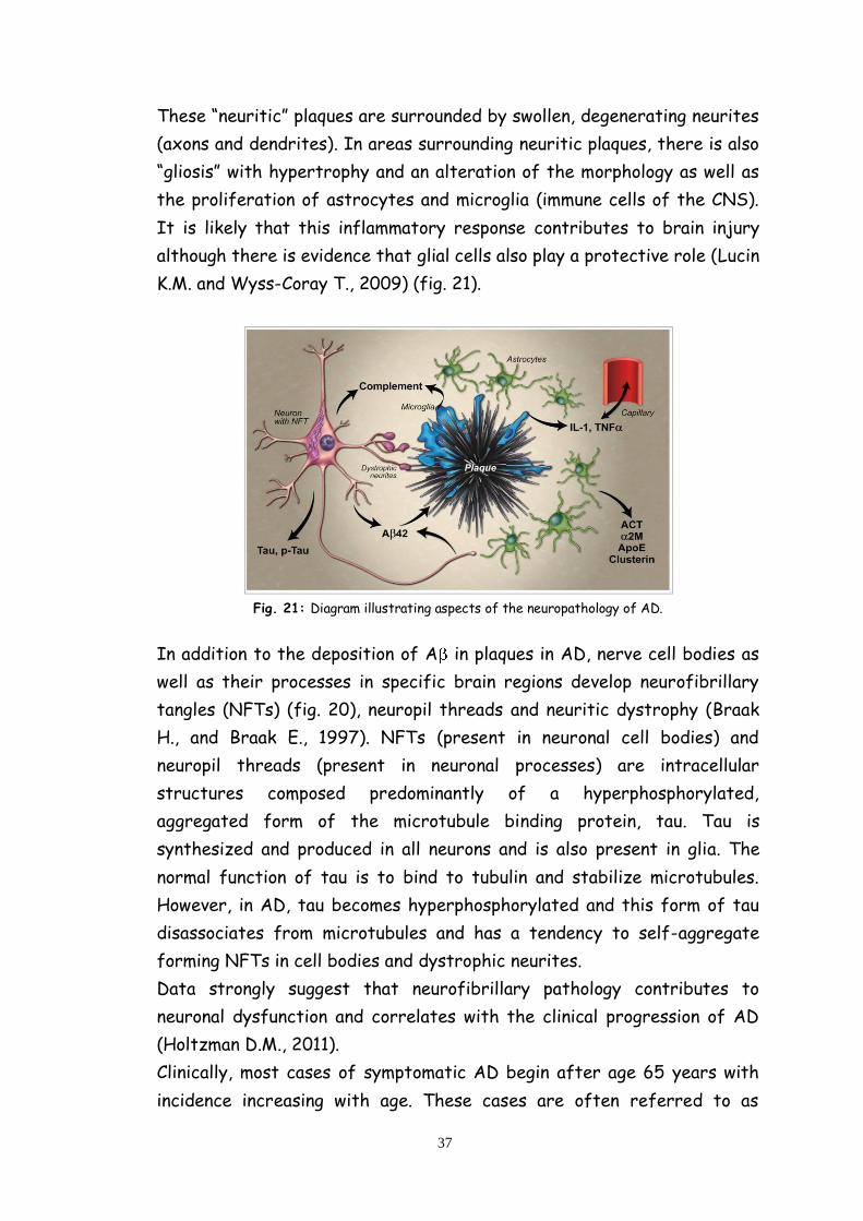
APP undergoes post-translational processing, involving several different
secretases and proteases, via two major pathways. In the non-
amyloidogenic pathway, APP is sequentially cleaved by -secretase and -
secretase. -cleavage, which cuts APP at the 17th amino acid inside the
A peptide sequence (fig. 22), releases a large secreted extracellular
domain (sAPP- ) and a membrane-associated C-terminal fragment
consisting of 83 amino acids (C83). APP C83 is further cleaved by -
secretase to release a P3 peptide and the AICD, both of which are
degraded rapidly (Zhang H. et al., 2012).
In the amyloidogenic pathway, APP is primarily processed by -secretase
at the first residue or at the 11th residue (so called ’ site) of the A
peptide sequence (fig. 22), shedding sAPP and generating a membrane
associated C-terminal fragment consisting of 99 amino acids (C99) (Sarah
C. and Robert V., 2007). -Secretase further cleaves C99 to release
40
AICD and the amyloidogenic A peptide which aggregates and fibrillates
to form amyloid plaques in the brain.
Fig. 22: Proteolytic processing of APP
3.2.1.1 -secretase and -cleavage
As APP was found to be constitutively cleaved at the -site to yield sAPP-
α, three members of the disintegrin and metalloproteinase (ADAMs),
ADAM-10, ADAM-17 and ADAM-9 have been proposed as the -secretase
(Koike H. et al. 1999).
ADAMs are type I integral membrane proteins that belong to the zinc
protease super family and have been implicated in the control of cytokine
and growth factor shedding.
ADAM10 is widely expressed in the brain and in other tissues and a
several fold increase in sAPP-a levels in cell lines overexpressing ADAM10
can be observed (Kojro E. et al. 2001).
Moderate neuronal over-expression of human ADAM10 increases sAPP-α
production while reducing A generation/plaque formation in mice
carrying the human APP V717I mutation, while expression of a
catalytically-inactive form of the ADAM10 mutation increases the size

41
and number of amyloid plaques in mouse brains (Postina R. et al. 2004).
These findings suggest that ADAM10 may be responsible for constitutive
α-cleavage activity. However, although sAPP- generation is not affected
in ADAM9/17 knock-down cell lines nor in mice carrying deficient
ADAM9/17 genes, over-expression of ADAM9/17 does increase the level
of sAPP- under some conditions, suggesting that ADAM9 and ADAM17
are more likely involved in the regulated α-cleavage of APP rather than in
constitutive -cleavage (Zhang H. et al, 2012).
3.2.1.2 -Secretase and -cleavage
A generation is initiated by -cleavage at the ectodomain of APP,
resulting in the generation of an sAPP- domain and the membrane
associated APP C-terminal fragment C99.
The putative -secretase, -site APP cleaving enzyme 1 (BACE1), was first
identified and characterized in 1999 (Sinha S. et al., 1999). BACE1 is a
type I transmembrane aspartyl protease with its active site on the
luminal side of the membrane. The originally identified full length BACE1
has 501 amino acids (BACE1-501) and is predominantly expressed in
perinuclear post-Golgi membranes, vesicular structures throughout the
cytoplasm, as well as on the cell surface (Ehehalt R. et al., 2002).
Knocking out the BACE1 gene prevents A generation and completely
abolishes A pathology in mice expressing the Swedish mutation of human
APP (Cai H. et al. 2001). The expression level and activity of BACE1 were
also found to be elevated in AD patients (Holsinger R.M. et al. 2002).
3.2.1.3 -Secretase and -cleavage
Both -cleavage and -cleavage generate short APP C-terminal fragments
that are further processed by -secretase.
Distinct from -/ -secretases, -activity involves a large proteinase
complex (fig. 23) consisting of at least four major protein components
(Presenilin1 or Presenilin2, Presenilin enhancer 2 (PEN2), Anterior
pharynx-defective1 (APH1) and Nicastrin) (Vetrivel K.S. et al. 2006).

42
Fig. 23: The -secretase complex
Presenilins (PSs) were identified and cloned in the mid-1990s (Levy-Lahad
E. et al., 1995; Sherrington R. et al., 1995). Genetic mutations of PSs are
found in a large portion of FAD cases, indicating its crucial role in AD.
Although other proteins are required for the -secretase complex, PSs
are believed to contain the actual protease activity (Zhang H., 2012).
PSs (fig. 24) are multi-transmembrane proteins and can be cleaved at the
cytoplasmic loop between the sixth and seventh transmembrane regions
to generate an N-terminal and a C-terminal fragment during post-
translational maturation (Thinakaran G. et al., 1996). The two fragments
interact with each other after the cleavage and they are both necessary
for -secretase activity.
Nicastrin, identified as a protein that interacts with PS in 2000 (Yu G. et
al., 2000), is a type I membrane glycoprotein with a large ectodomain.
Nicastrin undergoes a glycosylation/maturation process that causes a
conformation change in its ectodomain, which is crucial for the assembly
and maturation of the -secretase complex and -activity. Mature
nicastrin can bind to the ectodomain of APP C-terminal fragments derived
through -/ -secretase cleavage and may act as a substrate receptor of
-secretase (Shirotani K. et al. 2003).

43
Fig. 24: Schematic representation of Presenilin
PEN2 and APH1 are another two -secretase complex components that
were originally identified as the enhancers of PSs (Francis R. et al.,
2002).
APH1 is a multiple transmembrane protein with seven transmembrane
domains and a cytosolic C terminus. APH1 interacts with immature
nicastrin and PS to form a relatively stable pre-complex which is then
translocated to the trans-Golgi from the ER/cis-Golgi for further
maturation (Lee S.F. et al., 2002).
PEN2 is a hairpin-like protein with two transmembrane domains and with
both ends in the lumen. PEN2 is found to mediate the endoproteolysis of
PS (Luo W.J. et al., 2003).
-Secretase cleaves APP at multiple sites and in sequential steps to
generate A peptides of different lengths (fig. 22). The majority of A
peptides produced are 40 amino acids long, however, peptides ranging
from 38 to 43 amino acids are found in vivo. Besides the dominant -
cleavage site at 40 and 42 residues, ξ-cleavage at 46 and e-cleavage at
49 residues are also thought to be mediated by -secretase. Accordingly,
various AICDs (C50, C53, C57 and C59) can be generated during these
multi-site cleavages executed by -secretase. However, all of the
endogenous AICD forms are rarely detected, probably because of their
very rapid degradation (Zhang H., 2012).
Strong evidence suggests that the -secretase complex resides primarily
in the ER, Golgi/TGN, endocytic and intermediate compartments – most
of which (except the TGN) are not major subcellular localizations for APP

44
(Cupers P. et al., 2001). In addition to cleaving APP, -secretase cleaves a
series of functionally important transmembrane proteins, including Notch,
cadherin, tyrosinase, ErbB4, CD44, etc (Zhang Y. et al., 2011).
The cleavage of various substrates appears to be dependent on the sub-
cellular compartment; APP is mainly cleaved in the TGN and early
endosomal domains whereas Notch is primarily cleaved at the plasma
membrane (Tarassishin L. et al., 2004). Thus a disturbance in the
localization of the -secretase complex may play some role in abnormal A
generation and AD pathogenesis (Zhang Y. et al., 2011).
3.2.2 The hyperphosphorylated tau hypothesis
AD belongs to a group of neurodegenerative diseases collectively
designated as ‘‘tauopathies’’, because they are characterized by the
aggregation of abnormally phosphorylated tau protein. The mechanisms
responsible for tau aggregation and its contribution to neurodegeneration
are still unknown. Thereby, understanding the modes of regulation of tau
is of high interest in the determination of the possible causes at the
origin of the formation of tau aggregates and to elaborate protection
strategies to cope with these pathological lesions (Martin L. et al., 2011).
Tau protein (tubulin-associated unit) was identified in 1975 (Weingarten
M.D. et al., 1975). Tau is a microtubule-associated protein highly
conserved and exclusively found in higher eukaryotes. Tau is mainly
expressed in neuron and its primary role, by interacting with
microtubules, is to stabilize neuronal cytoskeleton.
Tau protein is subdivided into four regions (fig. 25): (1) an acidic region in
the N-terminal part; (2) a proline-rich region; (3) a region responsible for
tau binding with microtubules that contains four repeat domains R1, R2,
R3 and R4 also called MBDs (microtubule-binding domains); and (4) a C-
terminal region. Each repeat domain contains a conserved consensus motif
KXGS, which can be phosphorylated at serine (Ozer R.S. and Halpain S.,
2000). Serine phosphorylation at KXGS motifs, belonging to MBD region,
decreases tau affinity for microtubules and consequently prevents its
binding to microtubules which results in the destabilization of the
neuronal cytoskeleton (Sengupta A. et al., 1998).

45
Fig. 25: Tau protein
Cytoskeleton destabilization is well known to cause disruption of tau-
dependent cellular functions including axonal growth, vesicle and organelle
transport as well as nervous signal propagation along the nerve network
formed by microtubules (LaPointe N.E. et al., 2009).
Fig. 26: Model for multistep process of tau aggregation
46
Phosphorylation specifically modulates tau aggregation by disrupting its
binding to microtubules and subsequently by promoting tau self-
aggregation (fig. 26) (Martin L. et al., 2011).
Various kinases and phosphatases regulate tau phosphorylation. Each tau
phosphorylation site is subjected to the action of one or more protein
kinase. Tau kinases are grouped into three classes: protein kinases PDPKs
(proline-directed protein kinases), protein kinases non-PDPKs and protein
kinases specific for tyrosines. Alterations in the expression and/or the
activity of tau kinases such as GSK3 , CDK5, DYRK1A, p38 and CK1 have
been reported in the brains of AD patients, suggesting that one or
several of them could be involved in the tau hyperphosphorylation (Chung
S.H., 2009).
3.2.3 The Ca2+ hypothesis
A perturbed Ca2+ homeostasis has been demonstrated in AD and, in
particular, mutations in PSs have variably been correlated to alteration of
Ca2+ signaling and different molecular targets have been identified,
suggesting a physiological role for these proteins in multiple intracellular
Ca2+ pathway (Zampese E. et al., 2009).
PS, present as two homologous proteins PS1 and PS1, is an essential
component of the APP cleaving enzymes -secretase. As an integral
membrane protein, it is abundantly expressed with markers of ER, GA,
endo-exocytic vesicles and only a minor fraction is present at the plasma
membrane level (Vetrivel K.S. et al., 2004).
Both PS1 and PS2 are however regarded as a multi-faceted proteins
whose roles can be functionally distinguished and independent of their
enzymatic activity. In particular, several lines of evidence suggest that
PS is also involved in the regulation of cellular Ca2+ homeostasis (E.
Zampese et al., 2009). Since their discovery, different FAD-linked PS
mutants were reported to induce alterations of cellular Ca2+ handling in
several cell models and, in particular, it was observed that the FAD-
mutant PS1-L286V, when expressed in PC12 cells, causes an increased Ca2+
release upon different stimulation (Furukawa K. et al., 1998). Then, other
two FAD mutations in PS1 (A246E and M146V) were described to cause
larger Ca2+ release from intracellular stores and increase excitotoxicity in

47
neurons from transgenic (Tg) mice expressing the mutant protein
(Stutzmann G.E. et al., 2006). The idea that FAD-linked PS mutations are
somehow correlated to altered Ca2+ signaling was further supported by
the fact that these mutations could modify the sensitivity (or
expression) of ER Ca2+ release channels (RyR and IP3R) in different cell
models, in neurons from tg AD mice as well as in isolated brain
microsomes.
According to the popular “Ca2+ overload” hypothesis, PS mutations
increase Ca2+ content of the ER, thus sensitizing neurons to
excitotoxicity and progressive dementia (Zampese E. et al., 2009).
However, different studies reported either no alteration or a reduced
store Ca2+ content in cells expressing wt or FAD-mutant PSs. In
particular, it was demonstrated that the FAD-linked PS2 mutations
M239I and T122R reduce rather than increase Ca2+ release in fibroblasts
from FAD patients and in cell lines stably or transiently expressing the
PS2 mutants (Zatti G. et al., 2004; Giacomello M. et al., 2005;). In
addition, an extended investigation of other FAD-linked PS mutants by
directly monitoring the ER and the Golgi Ca2+ content in different cell
lines confirmed that the FAD-linked PS2 mutations cause a reduction in
the Ca2+ level of these organelles and none of the PS1 mutations cause an
increase (Zatti G. et al., 2006). Similar results have been obtained in rat
cortical neurons transiently expressing FAD-mutant PSs (Zatti G. et al.,
2006) and in neurons from transgenic mice expressing mutant presenilin 2
(Kipanyula M.J., et al., 2012).
The absence of elevated ER [Ca2+] in these models suggests that the
enhanced Ca2+ release observed by other groups, upon expression of
various FAD-linked PS mutants, might depend on different mechanisms,
not necessarily due to an increased amount of Ca2+ within the stores
(Zampese E. et al., 2009).
A dysregulation of store Ca2+ handling seems however the common
feature of a heterogeneous group of FAD-linked PS mutants.
The hypothetical mechanisms through which PS mutants exert their
effect on Ca2+ homeostasis are numerous. For instance, a model that was
suggested in accordance with the Ca2+ overload supports the idea that
PSs represent one of the molecular components of the elusive Ca2+ leak
channel of the ER membrane and the FAD-linked PS mutants, by reducing

48
the leak, increase the ER Ca2+ content (Zampese E., et al., 2009).
Moreover, in addition to the ER Ca2+ release channel and modulation of
CCE, other mechanisms have been implicated, on the basis of either
physical or functional interactions with proteins involved in cellular Ca2+
handling, such as sorcin (Pack-Chung E. et al., 2000), calsenilin (Buxbaum
J.D. et al., 1998), calcindin (Guo Q. et al., 1998). In particular, recent data
by our lab have demonstrated that PS2 mutations linked to FAD reduce
ER and GA Ca2+ levels by inhibiting SERCA pump (Zatti et al., 2006 and
Brunello et al., 2009). Moreover, endogenous PS seems to interact with
SERCA and modulate SERCA function; SERCA pumping is impaired in the
absence of both PSs (Green K.N. et al., 2008).
49
Materials and Methods
Constructs
The first 32 N-terminal amino acids of the resident Golgi enzyme 1,6
N-acetyl glucosaminyl transferase (C2gnT)
(MIHTNLKKKFSCCVLVFLLFAVICVWKEKKK
GSYYDSFKLQTKEFQVLKSLGKLAMGSDSQSVFSSSTQ), was
introduced before the start codon of the D1cpv cDNA (kindly provided
by Dr. R. Tsien) in the HindIII - BamHI restriction sites, after its
isolation from the GFP fused construct kindly provided by Dr. A. El-
Battari (Zerfaoui et al., 2002).
Cell culture and transfection
SH-SY5Y cells and fibroblasts were grown in DMEM containing 10%
(20% for fibroblasts) FCS, supplemented with L-glutamine (2 mM),
penicillin (100 U/ml), and streptomycin (100 µg/ml), in a humidified
atmosphere containing 5% CO2. Cells were seeded onto glass coverslips
(18-mm diameter) and transfection was performed at 60% confluence
using Lipofectamine™ 2000 Transfection Reagent (Life Technologies)
with 2 µg of DNA (0.5 µg of cameleon codifying DNAs and 1.5 µg of
pcDNA3 or PSs codifying DNAs); fibroblasts were transfected by
electroporetion, using the Neon Transfection System (MPK500,
Invitrogen). FRET measurements have been usually performed 24 h
after transfection.
SPCA1 siRNA transfection in SH-SY5Y cells and Western Blot
The day before transfection, SH-SY5Y cells were plated in order to
ensure 50% confluence on the day of transfection. Functionally
validated Stealth™ RNA directed against SPCA1
CATCGAGAAGTAACATTGCCTTTA (SPCA1 Oligo 1) and Stealth™ RNA
negative control were from Invitrogen. SPCA1 siRNAs (100 nM each)
or control siRNA were transfected using Lipofectamine™ 2000
Transfection Reagent (Life Technologies).
Proteins were resolved by SDS-PAGE, blotted onto a PVDF membrane
and probed with selected antibodies (mouse monoclonal anti-ATP2C1,
Sigma-Aldrich); mouse monoclonal anti- -actin (Sigma-Aldrich).

50
Cell imaging
Cells expressing the fluorescent probes were analyzed using a
DM6000 inverted microscope (Leica, Wetzlar, Germany) with a 40X oil
objective (HCX Plan Apo, NA 1.25). Excitation light produced by a
410nm LED (Led Engin #LZ1-00UA00 LED) was filtered at the
appropriate wavelength (425 nm) through a band pass filter, and the
emitted light was collected through a beamsplitter (OES s.r.l., Padua,
Italy) (emission filters HQ 480/40M (for CFP) and HQ 535/30M (for
cpv-YFP) and a dichroic mirror 515 DCXR). The beamsplitter permits
the collection of the two emitted wavelengths at the same time, thus
preventing any artefact due to movement of the organelles. All filters
and dichroics were from Chroma Technologies (Bellow Falls, VT, USA).
Images were acquired using an IM 1.4C cool camera (Jenoptik Optical
Systems) attached to a 12-bit frame grabber. Synchronization of the
excitation source and cool camera was performed through a control
unit ran by a custom-made software package, Roboscope (developed by
Catalin Cubotaru at VIMM, Padua, Italy); this software was also used
for image analysis. Exposure time and frequency of image capture
varied from 200 ms to 400 ms, depending on the intensity of the
fluorescent signal of the cells analyzed and on the speed of
fluorescence changes.
Cells were mounted into an open-topped chamber and maintained in an
extracellular medium (modified Krebs-Ringer Buffer). Classical
experiments started in 1 mM CaCl2; after perfusion with 300 µM
EGTA, cells were stimulated by applying bradykinin (BK, 100nM) and/or
CPA (20 µM); thereafter, the Ca2+ ionophore ionomycin (1 µM) was
applied to completely discharge the stores; finally digitonin (50 µM, to
obtain plasma membrane permeabilization) in Ca2+-free medium and
then a saturating CaCl2 concentration (3 mM) were applied, in order to
verify the dynamic range of the probe.
An intracellular-like medium was used for pH determination [130 mM
KCl, 10 mM NaCl, 1 mM MgCl2, 2 mM succinic acid and 20 mM Pipes at
different pH, at 37°C] and for experiments in permeabilized cells for
NAADP response and medialGo-D1cpv calibration [130 mM KCl, 10 mM
NaCl, 1mM KH2PO4, 1 mM MgSO4, 2 mM succinic acid, 300 µM EGTA

51
and 20 mM Hepes, pH 7.05, at 37 °C] to which different
concentrations of CaCl2 were added.
For Ca2+ pumping experiments a Ca2+-buffer solution was prepared by
adding to the intracellular medium: H-EDTA, pyruvic acid and MgCl2 (1
mM each), EGTA (2 mM) and CaCl2 (350 µM). ATP-Na (100 µM) was
added to this Ca2+-buffered solution. The free [Ca2+] (0.1 µM) was
estimated by MaxChelator2.5 and checked by fluorimetric
measurements with fura-2.
All media were perfused through a temperature controller (Warner
Instruments, TC-324B) set to have a constant temperature of 37°C in
the chamber.
Immunocytochemistry
Cells decorated with anti-GM130 (BD Bioscience Pharmigen, San Jose
CA, USA), anti-TGN46 anti-Giantin (AbCam, Cambridge UK) and anti-
PS2 (Santa Cruz Biotechnology, Dallas Texas, USA) antibodies were
fixed in Phosphate Buffered Saline (PBS) containing 4%
paraformaldehyde for 15 min, incubated with 50mM NH4Cl for 20 min,
permeabilized with 0.1% Triton X-100 in PBS 3 min and then blocked
with 2% BSA and 0,2% gelatine for 30 min; anti-GM130 (1:1000) and
anti-Giantin (1:300) were added for 1 hour at room temperature, anti-
TGN46 (1:200) and anti-PS2 (1:500) were added for 2h at 37°C.
Samples were treated with Alexa Fluor 488 (555, 568 or 647)
conjugated goat anti-mouse, anti-rabbit or rabbit anti-goat IgG
(Molecular Probes Invitrogen, Eugene OR) for 1 hour at room
temperature and mounted with Mowiol (Sigma-Aldrich Saint Louis,
MI). Images were collected at Leica TCS-SP5-II confocal system,
equipped with a PlanApo 100×/1.4 numerical aperture objective. For all
images, pinhole was set to 1 Airy unit. The Argon laser line (488 nm)
was used to excite the Cameleon sensors or AlexaFluor488 the He/Ne
543 nm laser was used to excite the AlexaFluor555/568 and the
AlexaFluor647 was excited by the HeNe 633nm laser line. Video
microscopy was performed at 1024×1024 pixels per image, with a
100Hz acquisition rate. Images were elaborated with ImageJ program
(Wayne Rasband, Bethesda,USA). Z-stacks were acquired with a step
of 0.42 µm.
52
Materials
Restriction and modification enzymes were purchase from NEB Inc.
(Ipswich, MA). CPA, digitonin, BK, caffeine were purchased from
SIGMA-Aldrich; ionomycin from Calbiochem and NAADP from Tocris.
All other materials were analytical or highest available grade.
Statistical analysis
Off-line analysis of FRET experiments was performed with ImageJ
software (Wayne Rasband, Bethesda, USA). Cpv-YFP and CFP images
were subtracted of background signals and distinctly analyzed after
selecting proper regions of interest (ROIs) on each cell; subsequently,
a ratio between cpv-YFP and CFP emissions was calculated
(R=F530/F480). Data are presented as a normalized ΔR/R0 %, where R
is the R value at each time (t) of the experiment and R0 is the R value
at the beginning of the experiment (t0), or as % of FRET.
All the data are representative of at least five different experiments.
Data were analyzed by Origin 7.5 SR5 (OriginLab Corporation).
Averages are expressed as mean ± s.e.m. (n = number of independent
experiments; = p < 0.05, = p < 0.01, = p < 0.001, unpaired
Student’s t test).
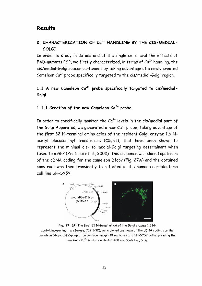
53
Results
2. CHARACTERIZATION OF Ca2+ HANDLING BY THE CIS/MEDIAL-
GOLGI
In order to study in details and at the single cells level the effects of
FAD-mutants PS2, we firstly characterized, in terms of Ca2+ handling, the
cis/medial-Golgi subcompartement by taking advantage of a newly created
Cameleon Ca2+ probe specifically targeted to the cis/medial-Golgi region.
1.1 A new Cameleon Ca2+ probe specifically targeted to cis/medial-
Golgi
1.1.1 Creation of the new Cameleon Ca2+ probe
In order to specifically monitor the Ca2+ levels in the cis/medial part of
the Golgi Apparatus, we generated a new Ca2+ probe, taking advantage of
the first 32 N-terminal amino acids of the resident Golgi enzyme 1,6 N-
acetyl glucosaminyl transferase (C2gnT), that have been shown to
represent the minimal cis- to medial-Golgi targeting determinant when
fused to a GFP (Zerfaoui et al., 2002). This sequence was cloned upstream
of the cDNA coding for the cameleon D1cpv (Fig. 27A) and the obtained
construct was then transiently transfected in the human neuroblastoma
cell line SH-SY5Y.
Fig. 27: (A) The first 32 N-terminal AA of the Golgi enzyme 1,6 N-
acetylglucosaminyltransferase, C32(1-32), were cloned upstream of the cDNA coding for the
cameleon D1cpv. (B) Z-projection confocal image (10 sections) of a SH-SY5Y cell expressing the
new Golgi Ca2+ sensor excited at 488 nm. Scale bar, 5 μm

54
Figure 27B shows that the expressed probe localized in a perinuclear
region that appears composed of cysternae and small vesicles, thus
resembling the Golgi complex.
1.1.2 Localization of the new probe
Figure 28 shows that, when probe-expressing cells (panels A, D, G) were
fixed and immunolabeled with bona fide markers of the cis- (GM130;
panel B), cis/medial- (Giantin; panel E) and trans- (TGN46; panel H) GA
compartments, there was a good overlap of the probe’s fluorescence with
that of the cis/medial-Golgi marker (panel F) while a partial separation
was observed between the signals of GM130 (panel C) and TGN46 (panel
I) and that of the novel probe (although a partial overlapping of the
signals was however evident).
Fig. 28: Confocal fluorescence images corresponding to Ca2+ probe expressing SH-SY5Y cells
either excited at 488 nm (A, D, G) or decorated with antibody against the cis-Golgi marker
GM130 (B), the medial-Golgi marker Giantin (E) and the trans-Golgi marker TGN46 (H), and
excited at 543 nm. (C, F, I) Merging of the two previous images; the yellow colour indicates
overlapping of the green and red signals. Insets in each panel show magnifications of selected
cisternae. Bar, 10 µm.
Confocal Z-stacks of cells labelled as above were statistically analyzed
(by ImageJ) to quantify the signal co-localizations of the new probe
versus the three markers (Fig. 29, panel A): the Pearson’s coefficient (an
index of signal co-localization that varies from -1, no co-localization, to +1,
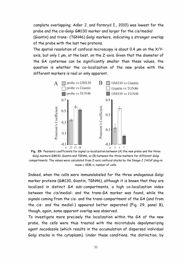
55
complete overlapping; Adler J. and Parmryd I., 2010) was lowest for the
probe and the cis-Golgi GM130 marker and larger for the cis/medial
(Giantin) and trans- (TGN46) Golgi markers, indicating a stronger overlap
of the probe with the last two proteins.
The spatial resolution of confocal microscopy is about 0.4 µm on the X/Y-
axis, but only 1 µm, at the best, on the Z-axis. Given that the diameter of
the GA cysternae can be significantly smaller than these values, the
question is whether the co-localization of the new probe with the
different markers is real or only apparent.
Fig. 29: Pearson’s coefficients for signal co-localization between (A) the new probe and the three
Golgi markers GM130, Giantin and TGN46, or (B) between the three markers for different Golgi
compartments. The values were calculated from Z-axis confocal stacks by the Image-J JACoP plug-in;
mean ± SEM; n, number of cells.
Indeed, when the cells were immunolabeled for the three endogenous Golgi
marker proteins (GM130, Giantin, TGN46), although it is known that they are
localized in distinct GA sub-compartments, a high co-localization index
between the cis/medial- and the trans-GA marker was found, while the
signals coming from the cis- and the trans-compartment of the GA (and from
the cis- and the medial-) appeared better separated (Fig. 29, panel B),
though, again, some apparent overlap was observed.
To investigate more precisely the localization within the GA of the new
probe, the cells were thus treated with the microtubule depolymerizing
agent nocodazole (which results in the accumulation of dispersed individual
Golgi stacks in the cytoplasm). Under these conditions, the distinction, by

56
confocal microscopy, of the different GA sub-compartments should be
facilitated (Thyberg J and Moskalewski S., 1999). In figure 30A, the cells
were thus first treated with nocodazole, fixed and then triple-
immunolabeled for GM130, Giantin and TGN46. In this case, the localization
of the three endogenous markers appears more distinct and, particularly
when the vesicles were image en face, a typical 3 colour staining was often
evident, with the Giantin signal closer to that of partially overlapped to that
of TGN46, while the GM130 signal was more clearly separated from the
other two.
Cells expressing the probe were thus similarly treated, fixed and
immunolabelled with the same two antibodies towards the cis- and the trans-
GA marker (fig. 30, panel B). In some structures, the spatial separation
among the three signals was striking evident. On the contrary, if the same
protocol was applied, but the cells were immunolabelled with antibodies
against GM130 and Giantin (fig. 30, panel C), the probe signal appears almost
completely separated from the first marker, but completely largely
overlapped with the second.
Fig. 30: Confocal fluorescence images corresponding to SH-SY5Y cells treated with nocodazole and
expressing the new Ca2+ probe (and excited at 488 nm; B and C, blue). Cells were decorated with
antibody against the cis-Golgi marker GM130 (and excited at 647 nm; A, B, C, green), the medial-Golgi
marker Giantin (and excited at 488 nm; A, blue; or at 543 nm; C, red) and the trans-Golgi marker
TGN46 (and excited at 543 nm; A and B, red). Insets in each panel show magnifications of en face
vesicles. Bar, 10 µm.
In conclusion, the new probe appears to be almost completely excluded from
the compartment containing primarily localized halfway between the TGN46-
and GM130-positive structures while and well overlapped the best
overlapping was observed with the vesicles to that containing Giantin. A
partial overlapping between the probe distribution and TGN46 was observed,

57
similar to that between the trans-GA marker and Giantin. Accordingly, for
simplicity, the new probe from now on is named medialGo-D1cpv (mGo-D1cpv).
1.1.3 Calibration of the new mGo-D1cpv
In order to determine the absolute value of [Ca2+] within the medial-GA
compartment, the calibration of the probe was carried out, performing
passive Ca2+ loading experiments, permeabilizing the cells with digitonin in an
intracellular-like medium containing different Ca2+ concentrations and no
energy source (fig. 31, panels A and B).
Fig. 31: (A-B) Representative kinetics of the FRET changes (% of maximal) in permeabilized SH-SY5Y
cells transiently transfected with mGo D1cpv. Where indicated, digitonin-permeabilized cells were
bathed with the an intracellular-like medium without energy source and containing different [Ca2+];
3mM CaCl2 was finally added to reach the maximum value. (C) In situ calibration curve for the mGo
D1cpv, obtained perfusing the cells with different [Ca2+]-containing intracellular-like medium as
described in A. Mean ± s.e.m., n ≥ 3 for each Ca2+ concentration.
The calculated apparent Kd for the new probe is 27.4 μM (fig. 31, panel C)
and the mean Ca2+ level that matched the cpv YFP/CFP fluorescence emission
ratio of SH-SY5Y cells at rest before permeabilization was 235 ± 30 μM
(mean ± s.e.m., n = 24).
1.2 Ca2+ handling in the medial-Golgi
Functional experiments were then carried out in living SH-SY5Y cells
transiently transfected with the cDNA encoding mGo-D1cpv. Dcpv-probes
are FRET-based and FRET occours between two mutants of GFP (the CFP and
YFP). It is caused by the interaction between Ca2+ activated Calmodulin
(CaM) and its target peptide M13. Variations in Ca2+ concentration can be
followed as variations in the ratio between YFP and CFP fluorescence.

58
Changes in [Ca2+] can be conveniently expressed as ΔR/R0. A drop in ΔR/R0
indicates a decrease of [Ca2+] within the lumen.
Figure 32 shows typical ΔR/R0 changes in medialGo-D1cpv expressing SH-
SY5Y cells after addition of different stimuli. Perfusion, in a Ca2+-free
(EGTA supplemented) medium, of an IP3-generating stimulus, such as
bradikinin (BK; Fig. 32 B), results typically in small decreases (a drop of 10 ±
0.4%, mean ± s.e.m., n = 14; Fig. 32 E) of the average ΔR/R0 value within the
GA region. The following addition of the Ca2+ ionophore ionomycin causes a
rapid and large decrease (a drop of 70 ± 2.1%, mean ± s.e.m., n = 37; Fig. 32
E) of the Golgi ΔR/R0 down to a level that was not significantly affected by
the subsequent permeabilization of the plasma membrane with digitonin.
We then tested the effect on medial-Golgi Ca2+ handling of the SERCA
inhibitor cyclopiazonic acid (CPA), added alone (Fig. 32 C) or in combination
with BK (Fig. 32 D): the SERCA inhibitor alone resulted in a slow decrease of
ΔR/R0 down to a new steady state value approximately halfway between the
value at rest and the minimum obtained after cell permeabilization with
digitonin in Ca2+-free, EGTA-containing medium (-40 ± 3.4%, mean ± s.e.m., n =
21; Fig. 4 E); however, when the addition of the SERCA inhibitor was rapidly
followed by BK, the slow CPA-induced ΔR/R0 decrease was strongly
accelerated (Fig. 32 D) until a new steady-state value, similar to that
reached upon CPA addition alone was reached (-40 ± 1%, mean ± s.e.m., n = 9;
Fig. 31 E). Addition of ionomycin, after CPA, induced, in both cases, a further
large drop in the ΔR/R0 signal (Fig. 32 C and D). In Fig. 32 E, the averaged
decreases in ΔR/R0 values obtained in SH-SY5Y cells expressing the mGo-
D1cpv upon different stimulations are reported.
The partial depletion of medial-Golgi by SERCA inhibitors suggests that this
Golgi compartment is endowed with two mechanism of Ca2+ uptake, only one
of which sensitive to the drugs. The existence in the medial-Golgi of a Ca2+
uptake mechanism other than the SERCA was then investigated. First, cells
expressing the mGo-D1cpv were treated (in Ca2+ free, EGTA-containing
medium) with ionomycin to completely deplete Ca2+ from the lumen; ionomycin
was then washed away, cells were treated with the irreversible SERCA
inhibitor thapsigargin (Tg, 1 µM) and finally 1mM CaCl2 was added to the
medium. For comparison, the same procedure was employed in parallel cells
expressing an ER targeted Ca2+ probe, ER-D4.
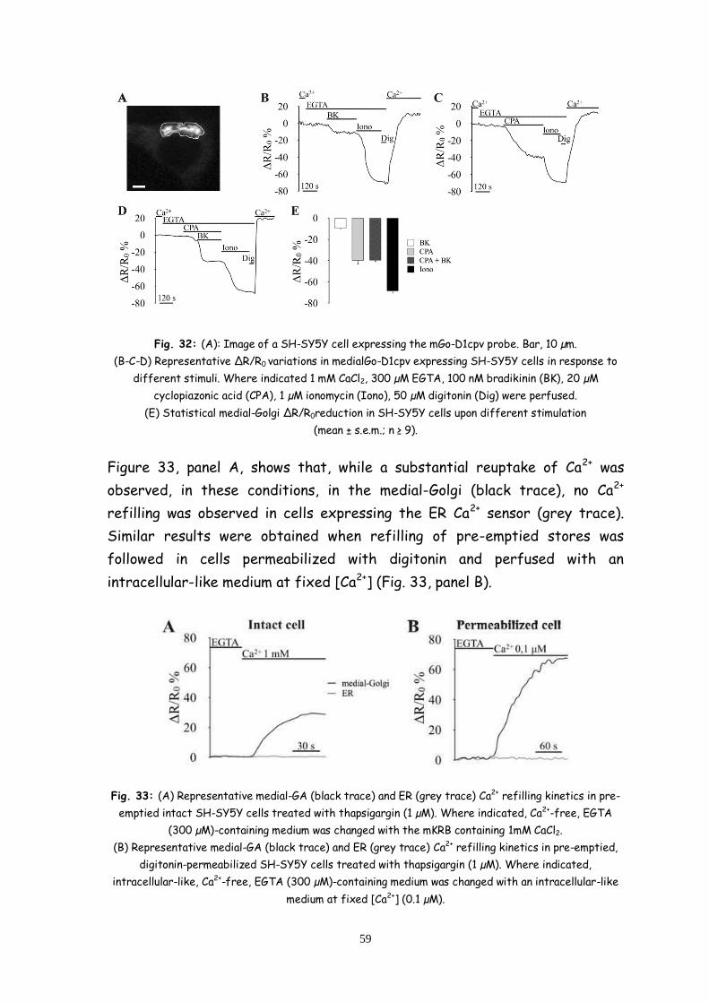
59
Fig. 32: (A): Image of a SH-SY5Y cell expressing the mGo-D1cpv probe. Bar, 10 µm.
(B-C-D) Representative ΔR/R0 variations in medialGo-D1cpv expressing SH-SY5Y cells in response to
different stimuli. Where indicated 1 mM CaCl2, 300 µM EGTA, 100 nM bradikinin (BK), 20 µM
cyclopiazonic acid (CPA), 1 µM ionomycin (Iono), 50 µM digitonin (Dig) were perfused.
(E) Statistical medial-Golgi ΔR/R0reduction in SH-SY5Y cells upon different stimulation
(mean ± s.e.m.; n ≥ 9).
Figure 33, panel A, shows that, while a substantial reuptake of Ca2+ was
observed, in these conditions, in the medial-Golgi (black trace), no Ca2+
refilling was observed in cells expressing the ER Ca2+ sensor (grey trace).
Similar results were obtained when refilling of pre-emptied stores was
followed in cells permeabilized with digitonin and perfused with an
intracellular-like medium at fixed [Ca2+] (Fig. 33, panel B).
Fig. 33: (A) Representative medial-GA (black trace) and ER (grey trace) Ca2+ refilling kinetics in pre-
emptied intact SH-SY5Y cells treated with thapsigargin (1 µM). Where indicated, Ca2+-free, EGTA
(300 µM)-containing medium was changed with the mKRB containing 1mM CaCl2.
(B) Representative medial-GA (black trace) and ER (grey trace) Ca2+ refilling kinetics in pre-emptied,
digitonin-permeabilized SH-SY5Y cells treated with thapsigargin (1 µM). Where indicated,
intracellular-like, Ca2+-free, EGTA (300 µM)-containing medium was changed with an intracellular-like
medium at fixed [Ca2+] (0.1 µM).

60
We thus hypothesized that the medial-Golgi is equipped by both the SERCA
and the SPCA-1 pumps. To further investigate this Ca2+ uptake mechanisms in
these Golgi subcompartment, we verified the effect in SH-SY5Y cells
expressing the medial-Golgi probe of SPCA1 knock-down (Fig. 34). SH-SY5Y
cells treated with specific siRNA against SPCA1 and expressing medialGo-
D1cpv were analyzed by Western blotting with an anti-SPCA1 antibody: a
reduction in the total SPCA1 protein level of 54 ± 9% (n=3) was revealed in
interfered cells, compared to control scramble siRNA-transfected cells (Fig.
34A). Taken into consideration the transfection efficiency in these cells
(~70%), this should correspond to a reduction of about 70% of SPCA1 level
in the transfected cells.
The effects of SPCA1 down-regulation were then tested on medial-GA Ca2+
handling. Fig. 34B shows that SPCA1 knockdown caused a lower steady-state
FRET level with the medial-Golgi at rest and a bigger ΔFRET change upon
CPA addition, compared to controls (Fig. 34C); accordingly, the additional
drop induced by ionomycin was larger in controls compared to SPCA1
knocked-down cells (Fig. 34C).
Fig. 34: (A) Western blot of control (Ctrl, scrambe siRNA-trasfected) and interfered (siSPCA1-
transfected) SH-SY5Y cell extracts visualized with anti SPCA1 and α-actin antibodies. A significant
reduction in the level of SPCA1 was observed in interfered cells (-54 ± 9%, mean ± s.e.m., n = 3).
(B) Representative FRET changes (% of maximal) in medialGo-D1cpv expressing control (grey trace)
and interfered (black trace) SH-SY5Y cells in response to the addition of different stimuli. Where
indicated, 1 mM CaCl2, 300 µM EGTA, 20 µM CPA, 1 µM ionomycin (Iono) were perfused. (C) Statistical
medial-Golgi FRET variations in SH-SY5Y cells treated as in B.
(mean ± s.e.m., n = 25; = p < 0.05; *** = p < 0.001).
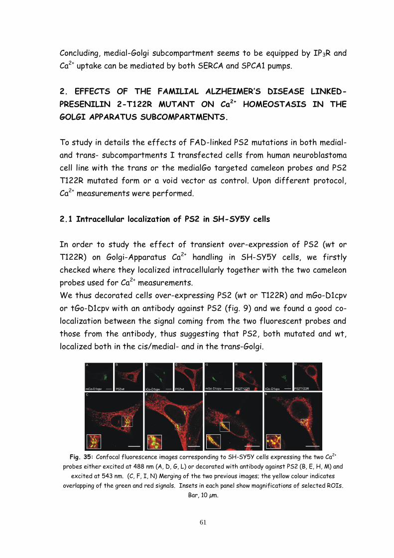
61
Concluding, medial-Golgi subcompartment seems to be equipped by IP3R and
Ca2+ uptake can be mediated by both SERCA and SPCA1 pumps.
2. EFFECTS OF THE FAMILIAL ALZHEIMER’S DISEASE LINKED-
PRESENILIN 2-T122R MUTANT ON Ca2+ HOMEOSTASIS IN THE
GOLGI APPARATUS SUBCOMPARTMENTS.
To study in details the effects of FAD-linked PS2 mutations in both medial-
and trans- subcompartments I transfected cells from human neuroblastoma
cell line with the trans or the medialGo targeted cameleon probes and PS2
T122R mutated form or a void vector as control. Upon different protocol,
Ca2+ measurements were performed.
2.1 Intracellular localization of PS2 in SH-SY5Y cells
In order to study the effect of transient over-expression of PS2 (wt or
T122R) on Golgi-Apparatus Ca2+ handling in SH-SY5Y cells, we firstly
checked where they localized intracellularly together with the two cameleon
probes used for Ca2+ measurements.
We thus decorated cells over-expressing PS2 (wt or T122R) and mGo-D1cpv
or tGo-D1cpv with an antibody against PS2 (fig. 9) and we found a good co-
localization between the signal coming from the two fluorescent probes and
those from the antibody, thus suggesting that PS2, both mutated and wt,
localized both in the cis/medial- and in the trans-Golgi.
Fig. 35: Confocal fluorescence images corresponding to SH-SY5Y cells expressing the two Ca2+
probes either excited at 488 nm (A, D, G, L) or decorated with antibody against PS2 (B, E, H, M) and
excited at 543 nm. (C, F, I, N) Merging of the two previous images; the yellow colour indicates
overlapping of the green and red signals. Insets in each panel show magnifications of selected ROIs.
Bar, 10 µm.
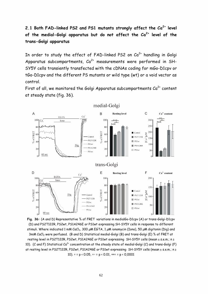
62
2.1 Both FAD-linked PS2 and PS1 mutants strongly affect the Ca2+ level
of the medial-Golgi apparatus but do not affect the Ca2+ level of the
trans-Golgi apparatus
In order to study the effect of FAD-linked PS2 on Ca2+ handling in Golgi
Apparatus subcompartments, Ca2+ measurements were performed in SH-
SY5Y cells transiently transfected with the cDNAs coding for mGo-D1cpv or
tGo-D1cpv and the different PS mutants or wild type (wt) or a void vector as
control.
First of all, we monitored the Golgi Apparatus subcompartments Ca2+ content
at steady state (fig. 36).
Fig. 36: (A and D) Representative % of FRET variations in medialGo-D1cpv (A) or trans-Golgi-D1cpv
(D) and PS2T122R, PS2wt, PS1A246E or PS1wt expressing SH-SY5Y cells in response to different
stimuli. Where indicated 1 mM CaCl2, 300 µM EGTA, 1 µM ionomycin (Iono), 50 µM digitonin (Dig) and
3mM CaCl2 were perfused. (B and D) Statistical medial-Golgi (B) and trans-Golgi (E) % of FRET at
resting level in PS2T122R, PS2wt, PS1A246E or PS1wt expressing SH-SY5Y cells (mean ± s.e.m.; n ≥
10). (C and F) Statistical Ca2+ concentration at the steady state of medial-Golgi (C) and trans-Golgi (F)
at resting level in PS2T122R, PS2wt, PS1A246E or PS1wt expressing SH-SY5Y cells (mean ± s.e.m.; n ≥
10). = p < 0.05, = p < 0.01, = p < 0,0001

63
We found that the overexpression of FAD-linked PS2T122R but not
PS1A246E affects the Ca2+ level of the medial-Golgi apparatus that appears
to be strongly reduced.
In term of FRET (fig. 36, panel B), the effect of PS2T122R over-expression
is from about 82 ± 1.5 % (mean ± s.e.m., n = 24) of FRET in control cells to
about 58 ± 3 % (mean ± s.e.m., n = 15) in PS2T122R expression cells that in
terms of Ca2+ (fig. 36, panel C) reflects a reduction from about 235 ± 30 µM
(mean ± s.e.m., n = 24) of control cells to about 45 ± 6 µM (mean ± s.e.m., n =
15) of PS2T122R expressing cells. This effect is mimicked, in a weaker
manner, by the over-expression of wt PS2, that reduces medial-Golgi Ca2+
content to 64 ± 7 µM (mean ± s.e.m., n = 10) but not wt PS1 (fig. 36, panels A,
B, C).
No differences in Ca2+ content between cells expressing mutated PS1 or PS2
and control cells has been found in trans-Golgi apparatus, whose Ca2+ content
at the steady state were about 146 ± 15 µM (mean ± s.e.m., n = 16) for control
cells, 156 ± 19 µM (mean ± s.e.m., n = 15) for PS2T122R expressing cells, 157
± 14 µM (mean ± s.e.m., n = 17) for PS2wt expressing cells, 173 ± 15 µM (mean
± s.e.m., n = 12) for PS1A246E expressing cells and 167 ± 17 µM (mean ±
s.e.m., n = 12) for wt PS1 expressing cells (fig. 36, panels D, E, F).
Similar results have been obtain in fibroblasts from FAD patients transiently
transfected with mGo-D1cpv (fig. 37, panel A) or tGo-D1cpv (fig. 37, panel
B), whose cells carrying PS2 N141I mutation showed a reduced Ca2+ content
in the medial-Golgi (fig. 37, panels C, D, E) with control cells showing a % of
FRET of about 87 ± 1 % (mean ± s.e.m., n = 12) and PS2N141I cells a % of
FRET of about 69 ± 3 % (mean ± s.e.m., n = 10) at the steady state in the
medial-Golgi. In terms of Ca2+ content, control fibroblasts showed a medial-
Golgi Ca2+ content of about 298 ± 30 µM (mean ± s.e.m., n = 12) whereas
PS2N141I fibroblasts showed a medial-Golgi Ca2+ content of about 84 ± 12
µM (mean ± s.e.m., n = 10).
No alterations in trans-Go Ca2+ content as respect to fibroblasts from
healthy patients have been found in PS2N141I fibroblasts (fig. 37, panels F,
G, H). Control and PS2N141I fibroblasts showed respectively a trans-Golgi
Ca2+ content at the steady state of about 105 ± 7 µM (mean ± s.e.m., n = 10)
and 110 ± 12 µM (mean ± s.e.m., n = 10).
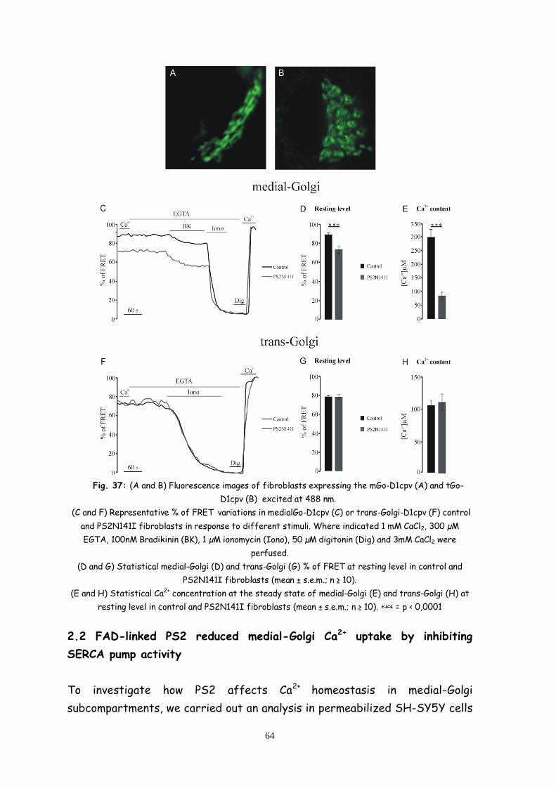
64
Fig. 37: (A and B) Fluorescence images of fibroblasts expressing the mGo-D1cpv (A) and tGo-
D1cpv (B) excited at 488 nm.
(C and F) Representative % of FRET variations in medialGo-D1cpv (C) or trans-Golgi-D1cpv (F) control
and PS2N141I fibroblasts in response to different stimuli. Where indicated 1 mM CaCl2, 300 µM
EGTA, 100nM Bradikinin (BK), 1 µM ionomycin (Iono), 50 µM digitonin (Dig) and 3mM CaCl2 were
perfused.
(D and G) Statistical medial-Golgi (D) and trans-Golgi (G) % of FRET at resting level in control and
PS2N141I fibroblasts (mean ± s.e.m.; n ≥ 10).
(E and H) Statistical Ca2+ concentration at the steady state of medial-Golgi (E) and trans-Golgi (H) at
resting level in control and PS2N141I fibroblasts (mean ± s.e.m.; n ≥ 10). = p < 0,0001
2.2 FAD-linked PS2 reduced medial-Golgi Ca2+ uptake by inhibiting
SERCA pump activity
To investigate how PS2 affects Ca2+ homeostasis in medial-Golgi
subcompartments, we carried out an analysis in permeabilized SH-SY5Y cells

65
transiently transfected with cDNA coding for mGo-D1cpv and the PS2T122R
or wild type (wt) and a void vector as control.
Permeabilized cells were incubated in an EGTA containing medium for 10 min
to induce Ca2+ stores depletion and then Ca2+ uptake was measured at 100nM
of free [Ca2+] in presence of ATP (fig. 38, panels A-E). In cells transiently
expressing mGo-D1cpv, the expression of PS2-T122R significantly reduced
the initial uptake rate (fig. 38, panel B) from 1.64 ± 0.35 Δ FRET/s % (mean ±
s.e.m., n = 13) in control cells to 0.63 ± 0.06 Δ FRET/s % (mean ± s.e.m., n =
15) in PS2T122R expressing cells and the plateau reached after refilling (fig.
38, panel D) from 148 ± 20 µM (mean ± s.e.m., n = 13) in control cells to 35 ± 6
µM (mean ± s.e.m., n = 15).
Medial-Golgi Ca2+ uptake were also affected upon over-expression of wt PS2
even if its effect is weaker than the FAD-mutant effect (fig. 38) with an
the initial uptake rate (fig. 38, panel B) of 0.85 ± 0.07 Δ FRET/s % (mean ±
s.e.m., n = 13) and a plateau reached after refilling (fig. 38, panel D)
corresponding to 54 ± 5 µM (mean ± s.e.m., n = 13).
Similar experiments were then performed in SH-SY5Y cells treated with the
SERCA inhibitor CPA. In this case, Ca2+ uptake were mediated only by SPCA-
1 pump and no effect of PS2-T122R and wt PS2 over-expression was found
(fig. 38, panels D, E and F). All samples showed a similar initial uptake rate
(fig. 38, panel F): 1 ± 0.1 Δ FRET/s % (mean ± s.e.m., n = 9) for control cells,
0.9 ± 0.1 Δ FRET/s % (mean ± s.e.m., n = 10) for PS2T122R expressing cells
and 1.1 ± 0.06 Δ FRET/s % (mean ± s.e.m., n = 9) for wt PS2 expressing cells
and respectively a plateau reached after refilling (fig. 38, panel H)
corresponding to 52 ± 6 µM (mean ± s.e.m., n = 9), 49 ± 7 µM (mean ± s.e.m., n
= 10) and 49 ± 4 µM (mean ± s.e.m., n = 9).
In order to investigate whether PS2 affect also trans-Golgi Ca2+ uptake, we
carried out an analysis in permeabilized SH-SY5Y cells transiently
transfected with cDNA coding for tGo-D1cpv and the PS2T122R, wt PS2 or a
void vector as control. Ca2+ uptake was then measured at 100nM of free
[Ca2+] (fig. 38, panels I-O). Expression of PS2-T122R, and also PS2 wt, does
not alter neither the initial uptake rate (fig. 38, panel L), with 1.4 ± 0.16 Δ
FRET/s % (mean ± s.e.m., n = 10) for control cells, 1.4 ± 0.12 Δ FRET/s %
(mean ± s.e.m., n = 10) for PS2T122R expressing cells and 1.4 ± 0.16 Δ FRET/s
% (mean ± s.e.m., n = 10) for wt PS2 expressing cells neither the plateau
reached after refilling (fig. 38, panel O) with Ca2+ level of 28 ± 5 µM (mean ±

66
s.e.m., n = 10) for control cells, 30 ± 7 µM (mean ± s.e.m., n = 10) for
PS2T122R expressing cells and 27 ± 5 µM (mean ± s.e.m., n = 10) for wt PS2
expressing cells.
Fig. 38: (A, E and I) Representative % of FRET variations in mGo-D1cpv (A and E) or tGo-D1cpv (I)
and PS2-T122R or PS2wt expressing SH-SY5Y cells, in response to different stimuli. Where indicated
ICB 0 Ca2+, ICB 100nM Ca2+, ICB 100nM Ca2+ and 200μM ATP with (E) or without (A and I) CPA and
3mM CaCl2 were perfused. (B, F and L) Statistical medial-Golgi (B and F) and trans-Golgi (L) ΔR/s %
for the first 10 s of Ca2+ uptake level in PS2-T122R, or PS2wt, expressing SH-SY5Y cells (mean ±
s.e.m.; n ≥ 10). (C, G and M) Statistical medial-Golgi (C and G) and trans-Golgi (M) % of FRET reached
after refilling in PS2-T122R, PS2wt expressing or control SH-SY5Y cells (mean ± s.e.m.; n ≥ 9). (D, H
and O) Statistical medial-Golgi (D and H) and trans-Golgi (O) Ca2+ concentration reached after refilling
in PS2-T122R, PS2wt expressing or control SH-SY5Y cells (mean ± s.e.m.; n ≥ 9). *= p < ** = p 0.05.
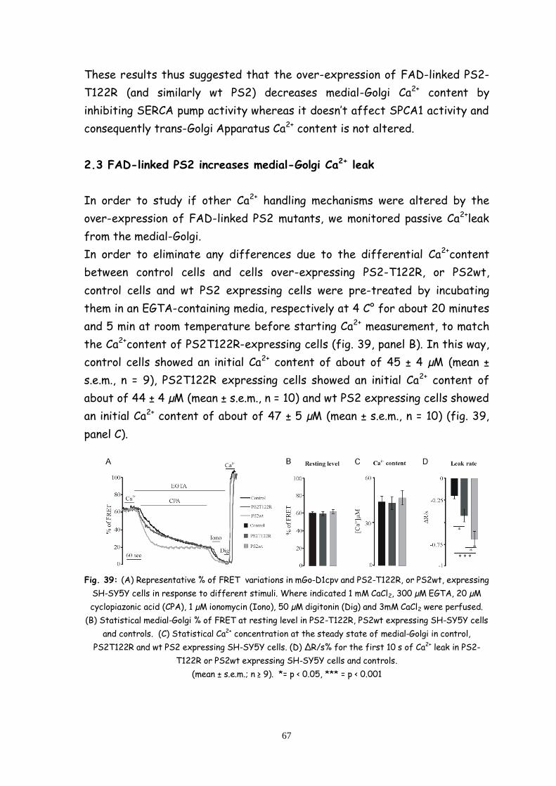
67
These results thus suggested that the over-expression of FAD-linked PS2-
T122R (and similarly wt PS2) decreases medial-Golgi Ca2+ content by
inhibiting SERCA pump activity whereas it doesn’t affect SPCA1 activity and
consequently trans-Golgi Apparatus Ca2+ content is not altered.
2.3 FAD-linked PS2 increases medial-Golgi Ca2+ leak
In order to study if other Ca2+ handling mechanisms were altered by the
over-expression of FAD-linked PS2 mutants, we monitored passive Ca2+leak
from the medial-Golgi.
In order to eliminate any differences due to the differential Ca2+content
between control cells and cells over-expressing PS2-T122R, or PS2wt,
control cells and wt PS2 expressing cells were pre-treated by incubating
them in an EGTA-containing media, respectively at 4 C° for about 20 minutes
and 5 min at room temperature before starting Ca2+ measurement, to match
the Ca2+content of PS2T122R-expressing cells (fig. 39, panel B). In this way,
control cells showed an initial Ca2+ content of about of 45 ± 4 µM (mean ±
s.e.m., n = 9), PS2T122R expressing cells showed an initial Ca2+ content of
about of 44 ± 4 µM (mean ± s.e.m., n = 10) and wt PS2 expressing cells showed
an initial Ca2+ content of about of 47 ± 5 µM (mean ± s.e.m., n = 10) (fig. 39,
panel C).
Fig. 39: (A) Representative % of FRET variations in mGo-D1cpv and PS2-T122R, or PS2wt, expressing
SH-SY5Y cells in response to different stimuli. Where indicated 1 mM CaCl2, 300 µM EGTA, 20 µM
cyclopiazonic acid (CPA), 1 µM ionomycin (Iono), 50 µM digitonin (Dig) and 3mM CaCl2 were perfused.
(B) Statistical medial-Golgi % of FRET at resting level in PS2-T122R, PS2wt expressing SH-SY5Y cells
and controls. (C) Statistical Ca2+ concentration at the steady state of medial-Golgi in control,
PS2T122R and wt PS2 expressing SH-SY5Y cells. (D) ΔR/s% for the first 10 s of Ca2+ leak in PS2-
T122R or PS2wt expressing SH-SY5Y cells and controls.
(mean ± s.e.m.; n ≥ 9). *= p < 0.05, *** = p < 0.001

68
Figure 39, panel D, shows that the over-expression of PS2wt increases
spontaneous Ca2+ leak from the medial-Golgi treated with CPA in an EGTA-
containing media to visualize it (fig. 39, panel A). A similar, but weaker
effect seemed to be caused by the over-expression of PS2T122R. Infact,
the initial leak rate were about -0.2 ± 0.03 Δ FRET/s % (mean ± s.e.m., n = 9)
for control cells, about -0.4 ± 0.07 Δ FRET/s % (mean ± s.e.m., n = 10) for
PS2T122R expressing cells and -0.7 ± 0.08 Δ FRET/s % (mean ± s.e.m., n = 11)
for wt PS2 expressing cells.
2.4 FAD-linked PS2 increase IP3 sensitivity of medial-Golgi Apparatus
We finally evaluated if FAD-linked PS2 mutants affected also the medial-
Golgi sensitivity to the Ca2+ releasing second messenger IP3.
SH-SY5Y transiently transfected with mGo-D1cpv and PS2 (T122R or wt), or
a void vector as control, were treated with the IP3-generating stimulus BK
and the induced drop were measured (Fig. 40, panels A-F). Figure 40 shows
that over-expression of PS2T122R and also PS2wt, increased Ca2+ release
due to IP3 generation. Indeed, in terms of FRET changes, BK induced-drop is
measured in control cells about -12 ± 1.6 Δ FRET % (fig. 40, panel C) from 82
± 3 FRET % at the steady state to 69 ± 3 FRET % after BK (fig. 40, panel B),
in PS2T122R expressing cells about -24 ± 1.8 Δ FRET % (fig. 40, panel C)
from 61 ± 2 FRET % at the steady state to 36 ± 1.5 FRET % after BK (fig.
40, panel B) and in wt PS2 expressing cells about -24 ± 1.9 Δ FRET % (fig.
40, panel C) from 68 ± 3 FRET % at the steady state to 45 ± 13 FRET %
after BK (fig. 40, panel B). Even if the FRET drops were translated in term
of Ca2+ (fig. 40, panels D-E), the increased IP3 sensitivity seemed to be
induced in both PS2T122R and wt PS2 expressing cells, where the 71 ± 3 %
(mean ± s.e.m., n = 12), in PS2T122R expressing cells, and the 73 ± 3 % (mean
± s.e.m., n = 9), in wt PS2 expressing cells, of the Ca2+ store seemed to be
sensitive to IP3, versus the 58 ± 6 % (mean ± s.e.m., n = 10) in control cells
(fig. 40, panel F).

69
Fig. 40: (A-G) Representative % of FRET variations in medialGo-D1cpv and PS2-T122R, or PS2wt,
expressing and control SH-SY5Y cells (A) and in mGo-D1cpv expressing FAD-PS2-N141I or control
human fibroblasts, in response to different stimuli. Where indicated 1 mM CaCl2, 300 µM EGTA,
100nM Bradikinin (BK), 1 µM ionomycin (Iono), 50 µM digitonin (Dig) and 3mM CaCl2 were perfused.

70
(B-H) Statistical medial-Golgi % of FRET at resting level in PS2-T122R, PS2wt expressing and control
SH-SY5Y cells (B) nad in FAD-PS2 and control fibroblasts (H). (C and I) Statistical medial-Golgi Δ
FRET % between resting level and the plateau reached after BK-induced drop in PS2-T122R, PS2wt
expressing and control SH-SY5Y cells (C) and FAD-PS2 or control fibroblasts (I).
(D and L) Statistical Ca2+ concentration at the steady state and at the plateau reached after BK-
induced drop of medial-Golgi in control, PS2T122R and wt PS2 expressing SH-SY5Y cells (D) and in
FAD-PS2 and control fibroblasts (L). (E and M) µM Ca2+ released in response to IP3 generation in PS2-
T122R, PS2wt expressing and control SH-SY5Y cells (E) and FAD-PS2 or control fibroblasts (M).
(F and N) % of medial-Golgi Ca2+ content released in response to IP3 generation in PS2-T122R, PS2wt
expressing and control SH-SY5Y cells (E) and FAD-PS2 or control fibroblasts (M).
(mean ± s.e.m.; n ≥ 9) *= p < 0.05, *** = p < 0.001.
Similar results have been obtained in human fibroblasts (fig. 40, panels G-N).
In control cells, the BK induced-drop is measured about -10 ± 1.8 Δ FRET %
(fig. 40, panel I ) from 87 ± 1 FRET % at the steady state to 77 ± 1.8 FRET %
after BK (fig. 40, panel H) whereas in PS2N141I fibroblasts, the BK induced-
drop is measured (fig. 40, panel I) about -24 ± 3 Δ FRET % from 69 ± 3
FRET % at the steady state to 45 ± 3.7 FRET % after BK (fig. 40, panel H).
These data are confirmed also after Ca2+ translation (fig. 40, panels L-N),
with the 71 ± 3 % (mean ± s.e.m., n = 10) of Ca2+ store sensitive to IP3 in
PS2N141I (fig. 40, panel N) compared to controls cells with a 51 ± 8 % (mean
± s.e.m., n = 12) of Ca2+ store sensitive to IP3. (Fig. 40, panel N).

71
Discussion and conclusions
Direct information on the Ca2+ homeostatic mechanisms in the GA of living
cells has been obtained for the first time only in 90's, using an aequorin
chimeric construct localized in the organelle lumen (Pinton P. et al., 1998). It
was demonstrated that GA Ca2+ handling (as a whole) resembles that of the
ER. Subsequently it has been suggested that Ca2+ handling within the GA is
heterogeneous: in particular it has been showed that the SPCA1-containing
Ca2+ sub-compartment was insensitive (or mildly sensitive) to IP3-generating
agonist stimulation and therefore not involved in setting up cytosolic Ca2+
signals (Vanoevelen J. et al., 2004).
More recently, using a novel GFP-based Ca2+ probe, tGo-D1cpv, selectively
localized in the trans-Golgi, also our laboratory have demonstrated that this
GA sub-compartment is endowed with unique characteristics that differ
completely from those of the ER (only SPCA1 and not SERCA equipped and
IP3 insensitive store) (Lissandron V. et al., 2010).
During my PhD, we generated another Ca2+ probe aimed at measuring the
[Ca2+] in the cis/medial-GA that reveals additional complexities in the Ca2+
handling characteristics of the organelle. In fact, unlike the trans-Golgi that
utilizes solely SPCA1 for Ca2+ uptake, the medial compartment appears to
accumulate Ca2+ within its lumen taking advantage of both SPCA1 and SERCA.
Moreover, the medial-Golgi, but not the trans-Golgi, seems to be sensitive to
IP3.
Thus, in a space of a few microns, and despite its continuous inter-mixing
dynamics, the Golgi-Apparatus appears to be formed by separate functional
entities with clearly distinct Ca2+ handling mechanisms.
The differences between the two GA sub-compartments are confirmed by
the specific effect on Ca2+ homeostasis of the expression of Familial
Alzheimer’s Disease-linked Presenilin-2 mutants. Cells expressing a FAD
mutated form of the protein show a decreased Ca2+ content in the
cis/medial-Golgi, but no effects on trans-Golgi Ca2+ homeostasis: PS2
mutants decrease Ca2+ uptake in the cis/medial-Golgi by inhibiting SERCA
pump activity while do not affect Ca2+ uptake, mediated by SPCA1, in the
trans-Golgi.
Also the over-expression of PS2wt seems to alter Ca2+ handling in the
medial-compartment but in a different way: while the decreased Ca2+ content
72
induced by FAD-linked PS2 mutants is primarily due to SERCA inhibition, the
effect mediated by the over-expression of the wt protein seems to be more
aspecific and caused by the increased passive Ca2+ leak from the organelle.
One of the proposed functions of presenilin is indeed to act as a calcium leak
channel and this function could be exaggerated in PS over-expressing cells.
Finally, we demonstrated that FAD-linked mutants increase medial-Golgi IP3
sensitivity. It has been reported that FAD mutant PS can interact with, and
increase the open probability of IP3R, causing an abnormally exaggerated
calcium signals in cells.
Concluding, FAD-linked presenilin mutants cause dysregulation in medial-Golgi
Ca2+ handling and because the cation regulates many neuronal functions, this
abnormality may contribute to the pathogenesis of the Alzheimer's disease.
Calcium in cells is precisely regulated and chronic abnormal calcium
dysregulation, as a result of mutations in presenilin, may therefore cause
cellular toxicity and eventually cell death. Presenilin-2 seems indeed to
interact with important Ca2+ signaling proteins altering their function, and
altered calcium signaling in turn could affect cell functionality.
These study should thus provide new insights into the molecular mechanisms
of AD and into the development of novel targets for therapeutic
interventions.
73
Abbreviations
A , Amyloid-
AD, Alzheimer’s Disease
ADP, Adenosine Diphosphate
AICD, APP Intracellular Domain
APH1, Anterior Pharynx-defective 1
APP, Amyloid Precursor Protein
ATP, Adenosine-5'-triphosphate
BACE1, -site APP Cleaving Enzyme 1
BK, Bradikin
CaM, calmodulin
CCE, Capacitative Calcium Entry
cDNA, complementary DNA
CFP, Cyan Fluorescent Protein
CIRC, Ca2+ induced Ca2+ release
CNG, cis-Golgi Network
CPA, Ciclopiazonic Acid
CRAC, Ca2+ release-activated Ca2+ channel
Dig, Digitonin
DNA, Desossiribonucleic Acid
EGTA, Ethylene Glycol Tetraacetic Acid
ER, Endoplasmic Reticulum
FAD, Familial Alzheiemer’s Disease
FRET, Fluorescence Resonance Energy Transfer

74
GA, Golgi Apparatus
GECIs, Genetically Encoded Ca2+ Indicators
GFP, Green Fluorescent Protein
InsP3Rs, Inositol 1,4,5-trisphosphate Receptors
Iono, Ionomycin
IP3, Inositol Trisphosphate
LOAD, “late-onset” AD
MBDs, microtubule-binding domains
NAADP, nicotinic acid adenine dinucleotide phosphate
NFTs, neurofibrillary tangles
PEN2, Presenilin Enhancer 2
PS, Presenilin
PSEN1, Presenilin-1
PSEN2, Presenilin-2
R, Ratio
RyRs, Ryanodine Receptors
sAPP- , Secreted Extracellular Domain
SERCA, Sarco/Endoplasmatic Reticulum Calcium ATPasi
siRNA, Small Interference Ribonucleic Acid
SOCs, Store-operated Channels
SPCA1, Secretory Pathway Ca2+-ATPase isoform 1
Tau, Tubulin-associated Unit
tBHQ, 2,5-di(t-butyl)hydroquinone
Tg, Thapsigargin

75
TGN, Trans-Golgi Network
TPCs, Two-pore Channels
wt, wild type
YFP, Yellow Fluorescent Protein

76

77
References
Adler J. and Parmryd I., Quantifying Colocalization by Correlation: The
Pearson Correlation Coefficient is Superior to the Mander’s Overlap
Coefficient. Cytometry, Vol. 77: 733-742, 2010.
Baird G.S., Zacharias D.A., Tsien R.Y., Circular permutation and receptor
insertion within green fluorescent proteins. Proc. Natl. Acad. Sci. U.S.A.,
Vol. 96: 11241–11246, 1999.
Barker W.W., Luis C.A., Kashuba A., Luis M., Harwood D.G., Loewenstein D.,
Waters C., Jimison P., Shepherd E., Sevush S., Graff-Radford N., Newland
D., Todd M., Miller B., Gold M., Heilman K., Doty L., Goodman I., Robinson B.,
Pearl G., Dickson D., Duara R., Relative frequencies of Alzheimer disease,
Lewy body, vascular and frontotemporal dementia, and hippocampal
sclerosis in the State of Florida Brain Bank. Alzheimer Dis. Assoc. Disord.,
Vol. 16:203–212, 2002.
Berridge M.J., Bootman M.D. and Roderick H.L., Calcium signaling: dynamics,
homeostasis and remodeling, Nature Review, Vol. 4: 517-529, 2004.
Berridge M.J., Inositol trisphosphate and calcium signalling. Nature, Vol.
361: 315–325, 1993.
Boehning D., Mak D.O.D., Foskett J.K., Joseph S.K., Molecular determinants
of ion permeation and selectivity in inositol 1,4,5-trisphosphate receptor
Ca2+ channels. J Biol Chem, Vol. 276: 13509–13512, 2001.
Braak H., and Braak E., Frequency of stages of Alzheimer-related lesions
in different age categories. Neurobiol. Aging, Vol. 18: 351–357, 1997.
Breton C., Mucha J., Jeanneau C., Structural and functional features of
glycosyl-transferases. Biochimie, Vol. 83: 713–718, 2001.
Brini M. and Carafoli E., Calcium Pumps in Health and Disease, Physiol Rev,
Vol. 89: 1341–1378, 2009.
Brunello L., Zampese E., Florean C., Pozzan T., Pizzo P., Fasolato C.,
Presenilin-2 dampens intracellular Ca2+ stores by increasing Ca2+ leakage
and reducing Ca2+ uptake. J. Cell. Mol. Med., Vol. 13: 3358-69, 2009.

78
Buxbaum J.D., Choi E.K., Luo Y., Lilliehook C., Crowley A.C., Merriam D.E.,
Wasco W., Calsenilin: a calcium-binding protein that interacts with the
presenilins and regulates the levels of a presenilin fragment. Nat. Med.,
Vol. 4: 1177-81, 1998.
Cahalan M.D., STIMulating store-operated Ca2+ entry., Nat Cell Biol., Vol.
11: 669–677, 2009.
Cahalan M.D., Zhang S.L., Yeromin A.V., et al. Molecular basis of the CRAC
channel. Cell Calcium, Vol. 42: 133-144, 2007.
Cai H., Wang Y., McCarthy D., Wen H., Borchelt D.R., Price D.L. and Wong
P.C., BACE1 is the major beta-secretase for generation of Abeta
peptides by neurons. Nat. Neurosci., Vol. 4: 233–234, 2001.
Calcraft P.J., Ruas M., Pan Z., Cheng X., Arredouani A., Hao X., Tang J.,
Rietdorf K., Teboul L., Chuang K.T., et al., NAADP mobilizes calcium from
acidic organelles through two-pore channels. Nature, Vol. 459: 596–600,
2009.
Carbone E., Lux H.D., A low voltage-activated, fully inactivating Ca channel
in vertebrate sensory neurones. Nature, Vol. 310: 501–502, 1984.
Catterall W.A., Excitation-contraction coupling in vertebrate skeletal
muscle: A tale of two calcium channels. Cell, Vol. 64: 871–874, 1991.
Catterall W.A., Voltage-Gated Calcium Channels. Cold Spring Harb.
Perspect. Biol., Vol. 3: 3947-3960, 2011.
Chanat E. and Huttner W.B., Milieu-induced,selective aggregation of
regulated secretory proteins in the trans-Golgi network. J.Cell Biol., Vol.
115:1505–1519, 1991.
Chandra S., Kable E.P., Morrison G.H., Webb W.W., Calcium sequestration in
the Golgi apparatus of cultured mammalian cells revealed by laser
scanning confocal microscopy and ion microscopy. J.CellSci., Vol. 100: 747–
752, 1991.
Chung S.H., Aberrant phosphorylation in the pathogenesis of Alzheimer’s
disease. BMB Rep., Vol. 42, 467–474, 2009.

79
Cifuentes F., Gonzalez C.E., Fiordelisio T., Guerrero G., Lai F.A., Hernandez-
Cruz A., A ryanodine fluorescent derivative reveals the presence of high-
affinity ryanodine binding sites in the Golgi complex of rat sympathetic
neurons, with possible functional roles in intracellular Ca2+ signaling. Cell
Signal., Vol. 13: 353–362, 2001.
Coe H., Michalak M., Calcium binding chaperones of the endoplasmic
reticulum. Gen Physiol Biophys, Vol. 28 Spec No Focus:F96–F103, 2009.
Csala M., Ba´nhegyi G., Benedetti A., Endoplasmic reticulum: A metabolic
compartment, FEBS Letters, Vol. 580: 2160–2165, 2006.
Cupers P., Bentahir M., Craessaerts K., Orlans I., Vanderstichele H., Saftig
P., De Strooper B., Annaert W., The discrepancy between presenilin
subcellular localization and gamma-secretase processing of amyloid
precursor protein. J. Cell Biol., Vol. 154:731-740, 2001.
de Brito O.M. and Scorrano L., Mitofusin2 tethers endoplasmic reticulum to
mitochondria. Nature, Vol. 456: 605–610, 2008.
Demaurex N., Calcium measurements in organelles with Ca2+-sensitive
fluorescent proteins. Cell Calcium, Vol. 38: 213–222, 2005.
Dode L., Andersen J.P., Vanoevelen J., et al., Dissection of the functional
differences between human secretory pathway Ca2+/Mn2+-ATPase (SPCA)
1 and 2 isoenzymes by steady-state and transient kinetic analyses. J.
Biol. Chem., Vol. 281: 3182–3189, 2006.
Drago I., Giacomello M., Pizzo P., Pozzan T., Calcium dynamics in the
peroxisomal lumen of living cells. J. Biol. Chem., Vol. 283:14384-90, 2008.
Du G.G., Sandhu B., Khanna V.K., Guo X.H., and MacLennan D.H., Topology of
the Ca2+ release channel of skeletal muscle sarcoplasmic reticulum
(RyR1). Proc. Natl. Acad. Sci. U.S.A. Vol: 99, 16725–16730, 2002.
Ehehalt R., Michel B., De Pietri Tonelli D., Zacchetti D., Simons K. and Keller
P., Splice variants of the beta-site APP-cleaving enzyme BACE1 in human
brain and pancreas. Biochem. Biophys. Res. Commun., Vol. 293, 30–37, 2002.
Fill M., Copello J.A., Ryanodine receptor calcium release channels. Physiol
Rev, Vol. 82: 893–922, 2002.
80
Foskett J.K., White C., Cheung K., Mak D.D., Inositol Trisphosphate
Receptor Ca2+ Release Channels. Physiol Rev, Vol. 87: 593–658, 2007;
Foskett J.K., White C., Cheung K.H., Mak D.O., Inositol trisphosphate
receptor Ca2+ release channels. Physiol Rev, Vol. 87: 593–658, 2007.
Francis R., McGrath G., Zhang J. et al., aph-1 and pen-2 are required for
Notch pathway signaling, gamma-secretase cleavage of betaAPP, and
presenilin protein accumulation. Dev. Cell, Vol. 3: 85–97, 2002.
Furukawa K., Guo Q., Schellenberg G.D., Mattson M.P., Presenilin-1 mutation
alters NGF-induced neurite outgrowth, calcium homeostasis, and
transcription factor (AP-1) activation in PC12 cells. J. Neurosci Res., Vol.
52: 618-24, 1998.
Giacomello M., Barbiero L., Zatti G., Squitti R., Binetti G., Pozzan T., Fasolato
C., Ghidoni R., Pizzo P., Reduction of Ca2+ stores and capacitative Ca2+
entry is associated with the familial Alzheimer's disease presenilin-2
T122R mutation and anticipates the onset of dementia. Neurobiol. Dis.,
Vol. 18: 638-48, 2005.
Giacomello M., Drago I., Bortolozzi M., Scorzeto M., Gianelle A., Pizzo P.,
Pozzan T., Ca2+ hot spots on the mitochondrial surface are generated by
Ca2+ mobilization from stores, but not by activation of store-operated
Ca2+ channels. Mol Cell., Vol. 38: 280-90, 2010.
Glenner G.G. and Wong C.W., Alzheimer’s disease: initial report of the
purification and characterization of a novel cerebrovascular amyloid
protein. Biochem. Biophys. Res. Commun. Vol. 120, 885–890, 1984.
Goedert M., Oskar Fischer and the study of dementia. Brain, Vol. 132:
1102–1111, 2009.
Green K.N., Demuro A., Akbari Y., Hitt B.D., Smith I.F., Parker I., LaFerla
F.M., SERCA pump activity is physiologically regulated by presenilin and
regulates amyloid beta production. J. Gen. Physiol., Vol. 132, 2008.
Grigoriev I., Gouveia S.M., van der Vaart B., et al. STIM1 is a MT-plus-
end-tracking protein involved in remodeling of the ER. Curr. Biol., Vol. 18:
177-182, 2008.
81
Guo Q., Christakos S., Robinson N., Mattson M.P., Calbindin D28k blocks the
proapoptotic actions of mutant presenilin 1: reduced oxidative stress and
preserved mitochondrial function. Proc. Natl. Acad. Sci. USA, Vol. 95:
3227-32, 1998.
Hagiwara S., Ozawa S., Sand O, Voltage clamp analysis of two inward
current mechanisms in the egg cell membrane of a starfish. J. Gen.
Physiol., Vol. 65: 617–644, 1975.
Hattori M., Suzuki A.Z., Higo T., Miyauchi H., Michikawa T., Nakamura T.,
Inoue T., Mikoshiba K., Distinct roles ofinositol 1, 4, 5-trisphosphate
receptor types 1 and 3 in Ca2+ signaling. J Biol Chem, Vol. 279: 11967–
11975, 2004.
Heinemann S.H., Terlau H., Stühmer W., Imoto K. Numa S., Calcium channel
characteristics conferred on the sodium channel by single mutations.
Nature, Vol. 356: 441–443, 1992.
Holsinger R.M., McLean C.A., Beyreuther K., Masters C.L. and Evin G.,
Increased expression of the amyloid precursor beta-secretase in
Alzheimer’s disease. Ann. Neurol., Vol. 51, 783–786, 2002.
Holtzman D.M., Morris John C., and Goate A., Alzheimer’s Disease: The
Challenge of the Second Century. Sci. Transl. Med., Vol. 6: 77-112, 2011;
Hu Z., Bonifas J.M., Beech J., Bench G., Shigihara T., Ogawa H., Ikeda S.,
Mauro T., EpsteinJr. E.H., Mutations in ATP2C1, encoding a calcium pump,
cause Hailey–Hailey disease. Nat. Genet., Vol. 24: 61–65, 2000.
Kang J., Lemaire H.G., Unterbeck A., Salbaum J.M., Masters C.L., Grzeschik
K.H., Multhaup G., Beyreuther K. and Muller-Hill B., The precursor of
Alzheimer’s disease amyloid A4 protein resembles a cell-surface
receptor. Nature, Vol. 325: 733–736, 1987.
Kayed R., Head E., Thompson J.L., McIntire T.M., Milton S.C., Cotman C.W.,
Glabe C.G., Common structure of soluble amyloid oligomers implies common
mechanism of pathogenesis. Science, Vol. 300: 486–489, 2003.
Koike H., Tomioka S., Sorimachi H., Saido T.C., Maruyama K., OkuyamaA.,
Fujisawa-Sehara A., Ohno S., Suzuki K. and Ishiura S., Membrane-anchored
82
metalloprotease MDC9 has an alphasecretase activity responsible for
processing the amyloid precursor protein. Biochem. J., Vol. 343: 371–375,
1999.
Kojro E., Gimpl G., Lammich S., Marz W. and Fahrenholz F., Low cholesterol
stimulates the nonamyloidogenic pathway by its effect on the alpha-
secretase ADAM 10. Proc. Natl Acad. Sci. USA, Vol. 98: 5815–5820, 2001.
Kipanyula M.J., Contreras L., Zampese E., Lazzari C., Wong A.K., Pizzo P.,
Fasolato C., Pozzan T., Ca2+ dysregulation in neurons from transgenic mice
expressing mutant presenilin 2. Aging Cell, Vol 11: 885-93, 2012.
LaPointe N.E., Morfini G., Pigino G., Gaisina I.N., Kozikowski A.P., Binder L.I.,
Brady S.T., The amino terminus of tau inhibits kinesin-dependent axonal
transport: implications for filament toxicity. J. Neurosci. Res., Vol. 87:
440–451, 2009.
Lee S.F., Shah S., Li H., Yu C., Han W. and Yu G., Mammalian APH-1
interacts with presenilin and nicastrin and is required for intramembrane
proteolysis of amyloid-beta precursor protein and Notch. J. Biol. Chem.,
Vol. 277: 45013–45019, 2002
Leite M.F., Thrower E.C., Echevarria W., Koulen P., Hirata K., Bennett A.M.,
Ehrlich B.E., Nathanson M.H., Nuclear and Ca2+ homeostasis in sub-cellular
compartments cytosolic calcium are regulated independently. Proc Natl
Acad Sci USA, Vol. 100: 2975–2980, 2003.
Levy E., Carman M.D., Fernandez M.I.J., Power M.D., Lieberburg I., van
Duinen S.G., Bots G.T., Luyendijk W., Frangione B., Mutation of the
Alzheimer’s disease amyloid gene in hereditary cerebral hemorrhage.
Dutch type. Science, Vol. 248: 1124–1126, 1990.
Levy-Lahad E., Wasco W., Poorkag P., Romano D.M., Oshima J., Pettingell
W.H., Yu C-E., Jondor P.D., Schmidt S.D., Wang K., et al. Candidate gene for
the chromosome 1 familial Alzheimer’s disease locus. Science. 1995;
269:973–977.
Lin P., Le-Niculescu H., Hofmeister R., McCaffery J.M., Jin M., Hennemann
H-, McQuistan T., DeVries L., Farquhar M.G., The mammalian calcium-
83
binding protein, nucleobindin (CALNUC) is a Golgi resident protein. J. Cell
Biol., Vol. 141: 1515–1527, 1998.
Liou J., Kim M.L., Heo W.D., et al., STIM is a Ca2+ sensor essential for
Ca2+-store-depletion triggered Ca2+ influx. Curr. Biol., Vol. 15: 1235-1241,
2005.
Lissandron V., Podini P., Pizzo P. and Pozzan T., Unique characteristics of
Ca2+ homeostasis of the trans-Golgi compartment. Proc. Natl. Acad. Sci.
U.S.A., Vol: 107, 9198–9203, 2010.
Llina´s R. and Yarom Y., Electrophysiology of mammalian inferior olivary
neurones in vitro. Different types of voltage-dependent ionic
conductances. J. Physiol. Vol. 315: 569–584, 1981.
Lucin K.M. and Wyss-Coray T., Immune activation in brain aging and
neurodegeneration: too much or too little? Neuron, Vol. 64: 110–122, 2009.
Luo W.J., Wang H., Li H., Kim B.S., Shah S., Lee H.J., Thinakaran G., Kim
T.W., Yu G. and Xu H., PEN-2 and APH-1 coordinately regulate proteolytic
processing of presenilin 1. J. Biol. Chem., Vol. 278: 7850–7854, 2003.
Maeda N., Kawasaki T., Nakade S., Yokota N., Taguchi T., Kasai M., Mikoshiba
K., Structural and functional characterization of inositol 1,4,5-
trisphosphate receptor channel from mouse cerebellum. J Biol Chem, Vol.
266: 1109–1116, 1991.
Mak D.O.D., McBride S., Foskett J.K., Inositol 1,4,5-trisphosphate
activation of inositol trisphosphate receptor Ca2+ channel by ligand tuning
of Ca2+ inhibition. Proc Natl Acad Sci USA, Vol. 95: 15821–15825, 1998.
Mak D.O.D., McBride S., Foskett J.K., Regulation by Ca2+ and inositol
1,4,5-trisphosphate (InsP3) of single recombinant type 3 InsP3 receptor
channels. Ca2+ activation uniquely distinguishes types 1 and 3 InsP3
receptors. J Gen Physiol, Vol. 117: 435–446, 2001.
Martin L., Latypova X., Terro F., Post-translational modifications of tau
protein: Implications for Alzheimer’s disease. Neurochemistry
International, Vol. 58: 458–471, 2011.

84
McCombs J.E. and Palmer A.E., Measuring calcium dynamics in living cells
with Genetically Encodable Calcium Indicators. Methods, Vol. 46: 152–159,
2008.
Meldolesi J. and Pozzan T., The endoplasmic reticulum Ca2+ store: A view
from the lumen, Trends Biochem. Sci. Vol. 23: 10–14, 1998.
Michikawa T., Hamanaka H., Otsu H., Yamamoto A., Miyawaki A., Furuichi T.,
Tashiro Y., Mikoshiba K., Transmembrane topology and sites of N-
glycosylation of inositol 1,4,5 trisphosphate receptor. J Biol Chem, Vol.
269: 9184–9189, 1994.
Mignery G.A., Newton C.L., Archer B.T., Sudhof T.C., Structure and
expression of the rat inositol 1,4,5-trisphosphate receptor. J Biol Chem,
Vol. 265: 12679–12685, 1990.
Mikoshiba K., The IP3 receptor/Ca2+ channel and its cellular function.
Biochem Soc Symp, Vol. 74:9–22, 2007.
Mintz I.M., Adams M.E., Bean B.P., P-type calcium channels in rat central
and peripheral neurons. Neuron, Vol. 9: 85–95, 1992.
Missiaen L., Dode L., Vanoevelen J., Raeymaekers L., Wuytack F., Calcium in
the Golgi apparatus. Cell Calcium, Vol. 41: 405–416, 2007.
Missiaen L., VanAcker K., VanBaelen K., Raeymaekers L., Wuytack F., Parys
J.B., DeSmedt H., Vanoevelen J., Dode L., Rizzuto R., Callewaert G., Calcium
release from the Golgi apparatus and the endoplasmic reticulum in HeLa
cells stably expressing targeted aequorin to these compartments. Cell
Calcium, Vol. 36: 479–487, 2004.
Miyawaki A., Llopis J., Heim R., McCaffery J.M., Adams J.A., Ikura M., Tsien
R.Y., Fluorescent indicators for Ca2+ based on green fluorescent proteins
and calmodulin. Nature, Vol. 388: 882–887, 1997.
Miyawaki A., Llopis J., Heim R., McCaffery J.M., Adams J.A., Ikura M., Tsien
R.Y., Fluorescent indicators for Ca2+ based on green fluorescent proteins
and calmodulin. Nature, Vol. 388: 882–887, 1997.
85
Moncoq K., Trieber C.A., Young H.S., The molecular basis for cyclopiazonic
acid inhibition of the sarcoplasmic reticulum calcium pump. J Biol Chem,
Vol. 282: 9748–9757, 2007.
Morel-Huaux V.M., Pypaert M., Wouters S., Tartakoff A.M., Jurgan U.,
Gevaert K., Courtoy P.J., The calcium-binding protein p54/NEFA is a novel
luminal resident of medial Golgi cisternae that traffics independently of
mannosidase II. Eur. J. Cell Biol., Vol. 81: 87–100, 2002.
Nagai T., Yamada S., Tominaga T., Ichikawa M., Miyawaki A., Expanded
dynamic range of fluorescent indicators for Ca(2+) by circularly permuted
yellow fluorescent proteins. Proc. Natl. Acad. Sci. USA, Vol. 10: 10554–
10559, 2004.
Nowycky M.C., Fox A.P., Tsien R.W., Three types of neuronal calcium
channel with different calcium agonist sensitivity. Nature, Vol. 316: 440–
443, 1985.
Oda K., Calcium depletion blocks proteolytic cleavages of plasmaprotein
precursors which occur at the Golgi and/or trans-Golgi network. Possible
involvement of Ca(2+)-dependent Golgi endo proteases. J.Biol.Chem., Vol.
267: 17465–17471, 1992.
Oldershaw K.A., Taylor C.W., 2,5-Di-(tert-butyl)-1,4-benzohydroquinone
mobilizes inositol 1,4,5-trisphosphate-sensitive and –insensitive Ca2+
stores. FEBS Lett, Vol. 274: 214–216, 1990.
Ozer R.S., Halpain S., Phosphorylation-dependent localization of
microtubule- associated protein MAP2c to the actin cytoskeleton. Mol.
Biol. Cell, Vol. 11, 3573–3587, 2000.
Pack-Chung E., Meyers M.B., Pettingell W.P., Moir R.D., Brownawell A.M.,
Cheng I., Tanzi R.E., Kim T.W., Presenilin 2 interacts with sorcin, a
modulator of the ryanodine receptor. J. Biol. Chem., Vol. 275: 14440-5,
2000.
Palmer A.E., Giacomello M., Kortemme T., Andrew Hires S., Lev-Ram V.,Baker
D. and Tsien R.Y., Ca2+ Indicators Based on Computationally Redesigned
Calmodulin-Peptide Pairs. Chemistry & Biology, Vol. 13: 521–530, 2006.
86
Palmer A.E., Jin C., Reed J.C. and Tsien R.Y., Bcl-2-mediated alterations in
endoplasmic reticulum Ca2+ analyzed with an improved genetically encoded
fluorescent sensor. Proc. Natl. Acad. Sci. USA, Vol. 101: 17404–17409,
2004.
Pezzati R., Bossi M. , Podini P., Meldolesi J., Grohovaz F., High-resolution
calcium mapping of the endoplasmic reticulum-Golgi-exocytic membrane
system. Electron energy loss imaging analysis of quick frozen-freeze
dried PC12cells. Mol. Biol.Cell, Vol. 8: 1501–1512, 1991.
Pinton P., Pozzan T., Rizzuto R., The Golgi apparatus is an inositol 1,4,5-
trisphosphate-sensitive Ca2+ store, with functional properties distinct
from those of the endoplasmic reticulum. EMBO J., Vol. 17: 5298–5308,
1998.
Pizzo P., Lissandron V., Capitanio P., Pozzan T., Ca2+ signalling in the Golgi
apparatus. Cell Calcium, Vol. 50: 184-92, 2011.
Postina R., Schroeder A., Dewachter I. et al., A disintegrin-
metalloproteinase prevents amyloid plaque formation and hippocampal
defects in an Alzheimer disease mouse model. J. Clin. Invest., Vol. 113,
1456–1464, 2004.
Pozzan T., Rizzuto R., Volpe P., Meldolesi J., Molecular and cellular
physiology of intracellular calcium stores, Physiol Rev July Vol. 74: 595-
636, 1994.
Prins D. and Michalak M., Organellar Calcium Buffers, Cold Spring Harb
Perspect Biol, Vol. 3: 1-16, 2011.
Pritchard S.M., Dolan P.J., Vitkus A., Johnson G.V.W, The toxicity of tau in
Alzheimer disease: turnover, targets and potential therapeutics. J. Cell.
Mol. Med., Vol. 15: 1621-1635, 2011.
Putney J.W., The Physiological Function of Store-operated Calcium
Entry., Neurochem. Res., Vol. 36:1157–1165, 2011.
Ramos-Franco J., Galvan D., Mignery G.A., Fill M., Location of the
permeation pathway in the recombinant type 1 inositol 1,4,5-
trisphosphate receptor. J Gen Physiol, Vol. 114: 243–250, 1999.

87
Randall A., Tsien R.W., Pharmacological dissection of multiple types of
calcium channel currents in rat cerebellar granule neurons. J. Neurosci.
Vol. 15: 2995–3012, 1995.
Raychaudhury B., Gupta S., Banerjee S., Datta S.C., Peroxisome is a
reservoir of intracellular calcium. Biochim. Biophys. Acta, Vol. 1760: 989–
992, 2006.
Rizzuto R., Brini M., Pozzan T., Targeting recombinant aequorin to specific
intracellular organelles. Methods Cell. Biol., Vol. 40:339–358, 1994.
Rizzuto R., Simpson A.W., Brini M., Pozzan T., Rapid changes of
mitochondrial Ca2+ revealed by specifically targeted recombinant
aequorin. Nature, Vol. 358: 325–327, 1992.
S.L. Hamilton and I. Serysheva, Ryanodine Receptor Structure: Progress
and Challenges. J Biol Chem, Vol. 284: 4047-4051, 2008.
Sagara Y., Fernandez-Belda F., de Meis L., Inesi G., Characterization of the
inhibition of intracellular Ca2+ transport ATPases by thapsigargin. J Biol
Chem, Vol. 267: 12606–12613, 1992.
Sagara Y., Inesi G., Inhibition of the sarcoplasmic reticulum Ca2+transport
ATPase by thapsigargin at subnanomolar concentrations. J Biol Chem, Vol.
266: 13503–13506, 1991.
Sarah C. and Robert V., The Alzheimer’s disease beta-secretase enzyme,
BACE1. Mol. Neurodegener., Vol. 2: 22, 2007.
Schafer W., Stroh A., Berghofer S., Seiler J., Vey M. , Kruse M.L., Kern H.F.,
Klenk H.D., Garten W., Two independent targeting signals in the
cytoplasmic domain determine trans-Golgi network localization and
endosomal trafficking of the proprotein convertase furin. EMBO J., Vol.
14: 2424–2435, 1995.
Schrader M. and Yoon Y., Mitochondria and peroxisomes: are the ‘big
brother’ and the ‘little sister’ closer than assumed? BioEssays : news and
reviews in molecular. Cell. Dev. Biol., Vol. 29: 1105–1114, 2007.
88
Seidler N.W., Jona I., Vegh M., A. Martonos, Cyclopiazonic acid is a
specific inhibitor of the Ca2+-ATPase of sarcoplasmic reticulum. J Biol
Chem, Vol. 264: 17816–17823, 1989.
Sengupta A., Kabat J., Novak M., Wu Q., Grundke-Iqbal I., Iqbal K.,
Phosphorylation of tau at both Thr 231 and Ser 262 is required for
maximal inhibition of its binding to microtubules. Arch. Biochem. Biophys.,
Vol. 357: 299–309, 1998.
Sherrington R., Rogaev E.I., Liang Y., Rogaeva E.A., Levesque G., Ikeda M., Chi
H., Lin C., Li G., Holman K., Tsuda T., Mar L., Foncin J.F., Bruni A.C., Montesi
M.P., Sorbi S., Rainero I., Pinessi L., Nee L., Chumakov I., Pollen D., Brookes
A., Sanseau P., Polinsky R.J., Wasco W., Da Silva H.A.R., Haines J.L., Pericak-
Vance M.A., Tanzi R.E., Roses A.D., Fraser P.E., Rommens J.M., St George-
Hyslop P.H., Cloning a gene bearing missense mutations in early-onset
familial Alzheimer’s disease. Nature, Vol. 375: 754–760 (1990).
Shirotani K., Edbauer D., Capell A., Schmitz J., Steiner H. and Haass C.,
Gamma-Secretase activity is associated with a conformational change of
nicastrin. J. Biol. Chem., Vol. 278: 16474, 2003.
Sinha S., Anderson J.P., Barbour R. et al., Purification and cloning of
amyloid precursor protein beta-secretase from human brain. Nature, Vol.
402: 537–540, 1999.
Small S.A. and Duff K., Linking Abeta and tau in late-onset Alzheimer’s
disease: a dual pathway hypothesis. Neuron, Vol. 60: 534–42, 2008.
Stutzmann G.E., Smith I., Caccamo A., Oddo S., Laferla F.M., Parker I.,
Enhanced ryanodine receptor recruitment contributes to Ca2+ disruptions
in young, adult, and aged Alzheimer's disease mice. J Neurosci., Vol. 26:
5180-9, 2006.
Supattapone S., Worley P.F., Baraban J.M., Snyder S.H., Solubilization,
purification, characterization of an inositol trisphosphate receptor. J Biol
Chem, Vol. 263: 1530–1534, 1988.
Szasz I., Sarkadi B., Schubert A., Gardos G., Effects of lanthanum on
calcium-dependent phenomena in human red cells, Biochim Biophys Acta
Vol. 512: 331–340, 1978.

89
Takahashi M., Seagar M.J., Jones J.F., Reber B.F., Catterall W.A., Subunit
structure of dihydropyridine-sensitive calcium channels from skeletal
muscle. Proc. Natl. Acad. Sci., Vol. 84: 5478–5482, 1997.
Takasugi N., Tomita T., Hayashi I., Tsuruoka M., Niimura M., Takahashi Y.,
Thinakaran G., Iwatsubo T., The role of presenilin cofactors in the
gamma-secretase complex. Nature, Vol. 422: 438–441 (2003).
Takeshima H., Nishimura S., Matsumoto T., Ishida H., Kangawa K., Minamino
N., Matsuo H., Ueda M., Hanaoka M., Hirose T., et al.. Primary structure and
expression from complementary DNA of skeletal muscle ryanodine
receptor. Nature, Vol. 339: 439–445, 1989.
Tanzi R.E., Gusella J.F., Watkins P.C., Bruns G.A., St George-Hyslop P., Van
Keuren M.L., Patterson D., Pagan S., Kurnit D.M. and Neve R.L., Amyloid beta
protein gene: cDNA, mRNA distribution, and genetic linkage near the
Alzheimer locus. Science, Vol. 235: 880–884, 1987.
Tarassishin L., Yin Y.I., Bassit B., Li Y.M., Processing of Notch and amyloid
precursor protein by gamma-secretase is spatially distinct. Proc. Natl.
Acad. Sci. USA, Vol. 101: 17050-17055, 2004.
Thastrup O., Cullen P.J., Drobak B.K., Hanley M.R., Dawson A.P., Thapsigargin,
a tumor promoter, discharges intracellular Ca2+ stores by specific
inhibition of the endoplasmic reticulum Ca2+-ATPase. Proc Natl Acad Sci
USA, Vol. 87: 2466–2470, 1990.
Thinakaran G., Borchelt D.R., Lee M.K., Slunt H.H., Spitzer L., Kim G.,
Ratovitsky T., Davenport F., Nordstedt C. and Seeger M., Endoproteolysis of
presenilin 1 and accumulation of processed derivatives in vivo. Neuron,
Vol. 17: 181–190, 1996.
Thyberg J., Moskalewski S., Role of microtubules in the organization of
the Golgi complex. Exp. Cell. Res., Vol.246:263-79, 1999.
Tsien R.W., Lipscombe D., Madison D.V., Bley K.R., Fox A.P., Multiple types of
neuronal calcium channels and their selective modulation. Trends
Neurosci., Vol. 11: 431–438, 1988.
90
Vaca L., SOCIC: The store-operated calcium influx complex. Cell Calcium,
Vol. 47: 199–209, 2010.
VanBaelen K., Vanoevelen J., Callewaert G., Parys J.B., DeSmedt H., Raey-
maekers L., Rizzuto R., Missiaen L., Wuytack F., The contribution of the
SPCA1 Ca2+ pump to the Ca2+ accumulation in the Golgi apparatus of
HeLa cells assessed via RNA-mediated interference. Biochem. Biophys.
Res. Commun., Vol. 306:430–436, 2003.
Vangheluwe P., Sepulveda M.R., Missiaen L., Raeymaekers L., Wuytack F.,
Vanoevelen J., Intracellular Ca2+- and Mn2+-transport ATPases.
Chem.Rev., Vol. 109: 4733–4759, 2009.
Vanoevelen J., Raeymaekers L., Parys J.B., DeSmedt H., VanBaelen K., Calle-
waert G., Wuytack F., Missiaen L., Inositol trisphosphate producing agonists
do not mobilize the thapsigargin-insensitive part of the endoplasmic-
reticulum and Golgi Ca2+ store. Cell Calcium, Vol. 35: 115–121, 2004.
Vetrivel K.S., Cheng H., Lin W., Sakurai T., Li T., Nukina N., Wong P.C., Xu H.,
Thinakaran G., Association of gamma-secretase with lipid rafts in post-
Golgi and endosome membranes. J. Biol. Chem., Vol. 279: 44945-54, 2004.
Vetrivel K.S., Zhang Y. W., Xu H. and Thinakaran G. Pathological and
physiological functions of presenilins. Mol. Neurodegener., Vol. 1: 4, 2006.
Wang H.J., Guay G., Pogan L., Sauve R., Nabi I.R., Calcium regulates the
association between mitochondria and a smooth subdomain of the
endoplasmic reticulum. J Cell. Biol., Vol. 150: 1489–1498, 2000.
Weingarten M.D., Lockwood A.H., Hwo S.Y., Kirschner M.W., A protein
factor essential for microtubule assembly. Proc. Natl. Acad. Sci. USA, Vol.
72: 1858–1862, 1975.
Wuytack F., Raeymaekers L., Missiaen L., Molecular physiology of the
SERCA and SPCA pumps, Cell Calcium Vol. 32: 279–305, 2002.
Yabe D., Nakamura T., Kanazawa N., Tashiro K., Honjo T., Calumenin, a Ca2+-
binding protein retained in the endoplasmic reticulum with a novel
carboxyl-terminal sequence, HDEF. J. Biol. Chem., Vol. 272: 18232–18239,
1997.
91
Yang S.N., Berggren P.O., The role of voltage-gated calcium channels in
pancreatic b-cell physiology and pathophysiology. Endocr. Rev. Vol. 27:
621–676, 2006.
Yu G., Nishimura M., Arawaka S., Levitan D., Zhang L., Tandon A., Song Y. Q.,
Rogaeva E., Chen F. and Kawarai T., Nicastrin modulates presenilin-mediated
notch/glp-1 signal transduction and beta-APP processing., Nature, Vol.
407, 48–54, 2000
Yu M., Zhong L., Rishi A.K., Khadeer M., Inesi G., Hussain A., Specific
substitutions at amino acid 256 of the sarcoplasmic/endoplasmic
reticulum Ca2+ transport ATPase mediate resistance to thapsigargin in
thapsigargin-resistant hamster cells. J Biol Chem, Vol. 273: 3542–3546,
1998.
Zampese E. and Pizzo P., Intracellular organelles in the saga of Ca2+
homeostasis: different molecules for different purposes?, Cell. Mol. Life
Sci., Vol. 69: 1077-1104, 2011.
Zampese E., Brunello L., Fasolato C., Pizzo P., Ca2+ dysregulation mediated
by presenilins in Familial Alzheimer’s Disease: Causing or modulating
factor? Current Trends in Neurology, Vol.3: 1-9, 2009.
Zatti G., Burgo A., Giacomello M., Barbiero L., Ghidoni R., Sinigaglia G.,
Florean C., Bagnoli S., Binetti G., Sorbi S., Pizzo P., Fasolato C., Presenilin
mutations linked to familial Alzheimer's disease reduce endoplasmic
reticulum and Golgi apparatus calcium levels. Cell Calcium, Vol. 39: 539-50,
2006.
Zatti G., Ghidoni R., Barbiero L., Binetti G., Pozzan T., Fasolato C., Pizzo P.,
The presenilin 2 M239I mutation associated with familial Alzheimer's
disease reduces Ca2+ release from intracellular stores. Neurobiol. Dis.,
Vol. 15: 269-78, 2004.
Zerfaoui M., Fukuda M., Langlet C., Mathieu S., Suzuki M., Lombardo D., El-
Battari A., The cytosolic and transmembrane domains of the beta 1,6 N-
acetylglucosaminyltransferase (C2GnT) function as a cis to medial/Golgi-
targeting determinant. Glycobiology, Vol. 12: 15-24, 2002.

92
Zhang H., Ma Q., Zhang J., Xu H., Proteolytic processing of Alzheimer’s -
amyloid precursor protein. J. of Neurochemistry, Vol. 120: 9–21, 2012.
Zhang Y., Thompson R., Zhang H., Xu H., APP processing in Alzheimer’s
disease. Molecular Brain, Vol. 4: 3, 2011.