effects of laser energy density on silicon nanoparticles
TRANSCRIPT
Journal of Physics Conference Series
OPEN ACCESS
Effects of Laser Energy Density on SiliconNanoparticles Produced Using Laser Ablation inLiquidTo cite this article Hiroki Kobayashi et al 2013 J Phys Conf Ser 441 012035
View the article online for updates and enhancements
You may also likeColloids in suspenseWilson Poon Peter Pusey and HenkLekkerkerker
-
Measurements of Particles in Liquid UsingShear-Horizontal Surface Acoustic WaveSensorJun Kondoh Taichi Oyama and ShowkoShiokawa
-
Nitridation of titanium surface by theirradiation of YAG laser pulses in N2O2gas mixture and liquid nitrogenN Takada H Ushida and K Sasaki
-
This content was downloaded from IP address 2032326692 on 12022022 at 1149
Effects of Laser Energy Density on Silicon Nanoparticles
Produced Using Laser Ablation in Liquid
Hiroki Kobayashi Pattarin Chewchinda Hiroyuki Ohtani Osamu Odawara
Hiroyuki Wada
Tokyo Institute of Technology 4259 Nagatsuta Midori-ku Yokohama 226-8503
Japan
E-mail kobayashihawmtitechacjp
Abstract We investigated the morphology of silicon nanoparticles prepared using laser
ablation in liquid through varying the energy density and laser irradiation time Silicon
nanoparticles were prepared using laser ablation in liquid A silicon wafer was irradiated in
ethanol using a laser beam (Nd YAGsecond harmonic generation 532 nm) Crystalline
silicon nanoparticles approximately 6 nm in size were observed by TEM observation The
quantity of silicon nanoparticles proportionally increased with an increase in energy density
greater than the laser ablation threshold This quantity also increased with an increase in laser
irradiation time without saturation due to absorption of the nanoparticles in liquid in the light
path
1 Introduction
Silicon nanoparticles are important for applications such as optoelectronics [1] [2] and bioimaging [3]
[4] Silicon nanoparticles have been researched to synthesize or to apply to applications Silicon is
used as the raw material for a semiconductor chip and is easily acquired Small nanoparticles less than
the de Broglie wave of an electron and the hole or Bohr radius of exciton produce a quantum effect
Such particles are ldquoquantum-dotsrdquo A quantum-dot has discrete bands instead of continuous bands and
notably produces impact ionization [5] Impact ionization is also ldquomultiple exciton generationrdquo (MEG)
In general one photon generates an exciton (a pair of electron and hole) However this phenomenon
generates more than 2 exciton pairs from one photon Silicon can produce 26 exciton pairs from one
photon (34 eV) [6] The maximum size of a silicon quantum-dot can be estimated using the Bohr
radius of an exciton in silicon The diameter is 86 nm [7] Therefore silicon nanoparticles with a
diameter 86 nm or lower are necessary for certain applications especially MEG-based applications
Laser ablation in liquid is one method for synthesizing nanoparticles This method produces
nanoparticles by irradiating a target in solution using a laser with high energy density Laser ablation
in the gas phase or under vacuum has been studied since the 1960s [8] while the study of laser
ablation in liquid was started recently [9] ~ [11] In the case of laser ablation in liquid the target
material was metal usually Later organic material [12] and ceramics [13] ~ [17] were used as a target
Laser ablation in liquid is advantageous for producing silicon nanoparticles because silicon
nanoparticles can be stably maintained on a particle surface by using ethanol as solvent [18]
Moreover laser ablation in liquid has other advantages the simplicity of the process and the possible
creation of small nanoparticles with relatively small spatial dispersion [19]
11th APCPST and 25th SPSM IOP PublishingJournal of Physics Conference Series 441 (2013) 012035 doi1010881742-65964411012035
Content from this work may be used under the terms of the Creative Commons Attribution 30 licence Any further distributionof this work must maintain attribution to the author(s) and the title of the work journal citation and DOI
Published under licence by IOP Publishing Ltd 1
Silicon nanoparticles have attracted attention but it is difficult to produce high quantities by laser
ablation in liquid This study explores fabrication of silicon nanoparticles using laser ablation in liquid
for applications such as a quantum-dot-sensitized solar cell Therefore production of silicon
nanoparticles with a diameter less than 86 nm at a certain quantity is the objective for the study herein
Thus the laser energy density and irradiation time were investigated for their dependence on
nanoparticle size and quantity
2 Experimental
A lt100gt p-type silicon wafer 600 m thick with a 115 ~ 155 Ω cm resistance was used as the raw
material The silicon wafer was cut 2 cm by 2 cm Before laser ablation the silicon wafer was cleaned
in ethanol using ultrasonication twice for 10 minutes and then dried The silicon wafer was irradiated
with a laser beam which was not focused with a lens The unpolished side of the silicon wafer was
placed 7 mm beneath the top surface of the ethanol Ethanol was used as the solvent for laser ablation
to prevent oxidation of the silicon during laser ablation The ethanol was stirred using a magnetic
stirrer during laser ablation The laser beam was a second harmonic generation of a NdYAG laser
(wavelength 532 nm pulse duration 13 ns and repetition rate 10 Hz) The diameter of the irradiated
laser beam on the silicon wafer was 14 mm (154 cm2) The energy density varied from 017 to 063
Jcm2 (laser energy 26 - 97 Jpulse) Irradiation time varied from 30 minutes to 3 hours
The prepared nanoparticles were identified using Raman spectroscopy The solution containing silicon
nanoparticles was dropped in an Al pan under N2 atmosphere and then dried This solution drying
procedure was repeated 4 times After this procedure black powder was confirmed on the Al pan To
measure the Raman spectrum the black powder was irradiated with a laser under the atmosphere to
measure Raman spectrum The particle size of the prepared silicon nanoparticles was measured using
a transmission electron microscope (TEM) After laser ablation the solutions were filtered with a
Teflon filter (pore size 100 nm) The solutions were then dropped onto a Cu grid and the grid was
then dried overnight at 70 degC in a vacuum The quantity of silicon nanoparticles was measured using
inductively-coupled plasma mass spectrometry (ICP-MS) Absorption spectra were measured by
spectrometer with an integrating sphere
3 Results and discussion
Figure 1 shows a Raman spectrum of the silicon nanoparticles produced by ablating a silicon wafer in
ethanol under an Ar atmosphere The energy density was 017 Jcm2 and the ablation was performed
for 2 hours Two peaks were observed at 494 cm-1
and 518 cm-1
The peak wavelength of the silicon
crystal is at 520 cm-1
The particle size of silicon estimated by the value of Raman shift is 4 nm [20]
This peak was slightly shifted as an effect of nanosize silicon whereas the sample peak at 494 cm-1
indicates silicon dioxide [21] The Raman peak for silicon dioxide is attributed to the silicon wafer
native oxide layer or oxidation of the prepared silicon nanoparticles during Raman spectra
Figure 1 Raman spectrum of the silicon nanoparticles
11th APCPST and 25th SPSM IOP PublishingJournal of Physics Conference Series 441 (2013) 012035 doi1010881742-65964411012035
2
measurement Therefore the powder in the solution which was prepared using an irradiating green
laser at 017 Jcm2 on a silicon wafer in ethanol comprises silicon nanoparticles with silicon dioxide
We investigated the effect of energy density from the irradiating laser on the morphology of the silicon
nanoparticles The irradiation time was 1 hour Figure 2 shows TEM images of silicon nanoparticles
These images indicate that we produced silicon nanoparticles at approximately 6 nm Moreover the
nanoparticle shapes are almost spherical Lattice fringes were confirmed through a high magnification
image These observations indicate that the silicon nanoparticles are crystallized
Figure 3 shows the particle size distribution of the silicon nanoparticles measured through TEM
images The average sizes were 56 nm 63 nm and 62 nm at energy densities of 017 Jcm2
032 Jcm2 and 045 Jcm
2 respectively Silicon nanoparticles were produced with the desired size less
than the Bohr radius of exciton Given these results the energy density of the irradiating laser does not
significantly influence the silicon nanoparticle size under such experimental conditions
Mechanism of fabrication of crystalline Si nanoparticles in liquid might be recrystallization of liquid
or vapor of silicon in cavitation bubble which would be created by heating target using laser light
Indeed there are many reports of the relation between cavitation bubble and nanoparticle formation [22-25] In our experiment the particle size of silicon nanoparticles was approximately 6 nm This
particle size might be related to lifetime of bubbles which might restrict the growth of silicon
nanoparticles The lifetime of bubbles would not be changed under our experimental conditions because
pressure and temperature of ethanol were almost constant Therefore the particle size of silicon
nanoparticles was not changed At the same time the recrystallization in ethanol might be possible too
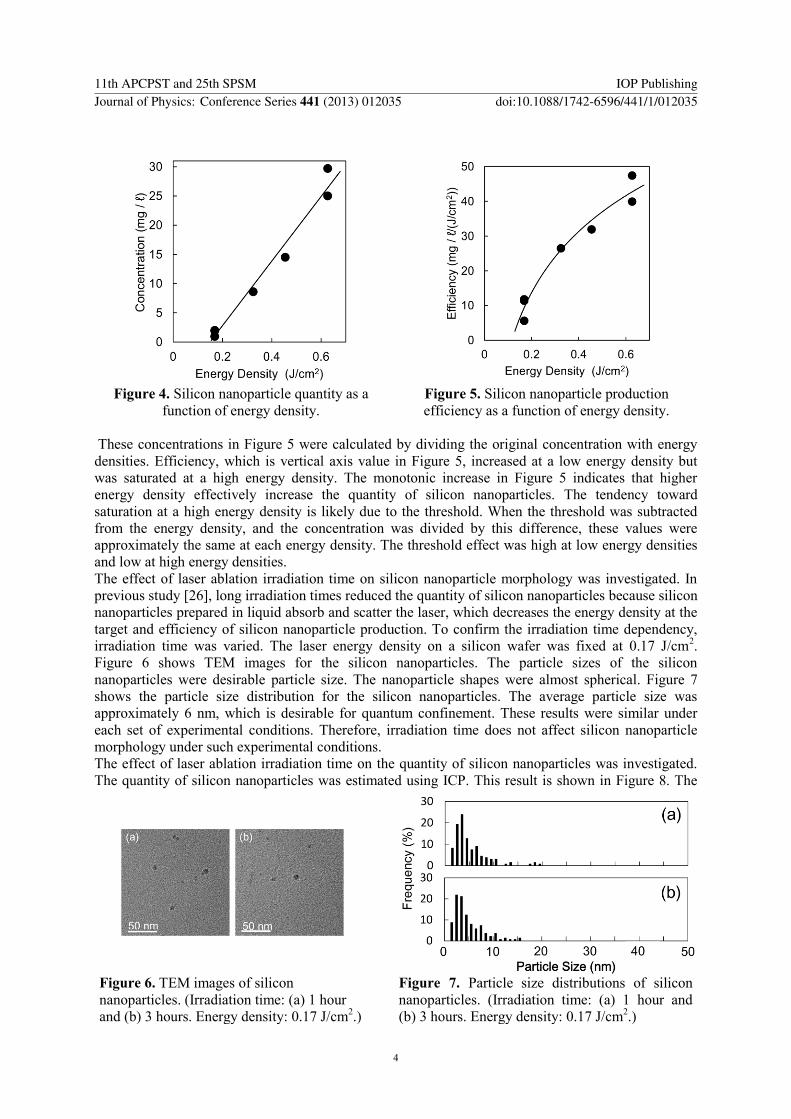
We investigated the effect of the irradiating laser energy density on the quantity of silicon
nanoparticles Figure 4 shows an estimate of the silicon nanoparticle quantity through ICP The
threshold was approximately 015 Jcm2 In general the penetration depth as a function of energy
density is saturated at a high energy density If ablation is consistent with this rule the ablated depth of
a silicon wafer per unit energy density decreases with an increase in energy density However the
experimental results indicate that an increase in laser energy density proportionally increases the
quantity of silicon nanoparticles This result is likely related to the ablated area of the silicon wafer as
an element The ablated area increased with an increase in energy density even in the same irradiated
area To consider the preparation efficiency of silicon nanoparticles as a function of laser power
Figure 4 was deformed Figure 5 shows an estimate for the quantity of silicon nanoparticles per unit
energy density
Figure 2 TEM images of the silicon
nanoparticles (Energy density (a) 017
Jcm2 (b) 032 Jcm
2 (c) 045 Jcm
2 and
(d) high magnification image of 045 Jcm2
Irradiation time 1 hour)
Figure 3 Particle size distributions for the silicon
nanoparticles (Energy density (a) 017 Jcm2 (b)
032 Jcm2 and (c) 045 Jcm
2 Irradiation time
1 hour)
11th APCPST and 25th SPSM IOP PublishingJournal of Physics Conference Series 441 (2013) 012035 doi1010881742-65964411012035
3
These concentrations in Figure 5 were calculated by dividing the original concentration with energy
densities Efficiency which is vertical axis value in Figure 5 increased at a low energy density but
was saturated at a high energy density The monotonic increase in Figure 5 indicates that higher
energy density effectively increase the quantity of silicon nanoparticles The tendency toward
saturation at a high energy density is likely due to the threshold When the threshold was subtracted
from the energy density and the concentration was divided by this difference these values were
approximately the same at each energy density The threshold effect was high at low energy densities
and low at high energy densities
The effect of laser ablation irradiation time on silicon nanoparticle morphology was investigated In
previous study [26] long irradiation times reduced the quantity of silicon nanoparticles because silicon
nanoparticles prepared in liquid absorb and scatter the laser which decreases the energy density at the
target and efficiency of silicon nanoparticle production To confirm the irradiation time dependency
irradiation time was varied The laser energy density on a silicon wafer was fixed at 017 Jcm2
Figure 6 shows TEM images for the silicon nanoparticles The particle sizes of the silicon
nanoparticles were desirable particle size The nanoparticle shapes were almost spherical Figure 7
shows the particle size distribution for the silicon nanoparticles The average particle size was
approximately 6 nm which is desirable for quantum confinement These results were similar under
each set of experimental conditions Therefore irradiation time does not affect silicon nanoparticle
morphology under such experimental conditions
The effect of laser ablation irradiation time on the quantity of silicon nanoparticles was investigated
The quantity of silicon nanoparticles was estimated using ICP This result is shown in Figure 8 The
Figure 4 Silicon nanoparticle quantity as a
function of energy density
Figure 5 Silicon nanoparticle production
efficiency as a function of energy density
Figure 6 TEM images of silicon
nanoparticles (Irradiation time (a) 1 hour
and (b) 3 hours Energy density 017 Jcm2)
Figure 7 Particle size distributions of silicon
nanoparticles (Irradiation time (a) 1 hour and
(b) 3 hours Energy density 017 Jcm2)
11th APCPST and 25th SPSM IOP PublishingJournal of Physics Conference Series 441 (2013) 012035 doi1010881742-65964411012035
4
quantity of silicon nanoparticles increased linearly with irradiation time We observed a small
threshold which is likely related to native silicon dioxide on a target bare silicon wafer Silicon
dioxide has a higher band gap than silicon therefore silicon dioxide has a lower absorbance with the
green laser Further the silicon dioxide melting point is higher than for silicon The lower absorbance
likely decreases laser ablation efficiency In this experiment saturation of the quantity of silicon
nanoparticles prepared was not observed Therefore a longer irradiation time can increase the quantity
of silicon nanoparticles
Figure 9 shows an absorbance spectrum for the silicon nanoparticles dispersed in the solution
Although absorption was observed at 532 nm it has only small influence on particle generation
Therefore the quantity of silicon nanoparticles increased linearly even at a high energy density Figure
10 shows the square root of absorbance as a function of photon energy because silicon is indirect
transition material The intercept of horizontal axis which means bandgap Eg is 22 eV Increase in
bandgap by quantum confinement was observed as a result of nanosize effect
4 Conclusion Silicon nanoparticles were prepared using laser ablation in liquid In general the quantity of
nanoparticles produced from laser ablation in liquid is low To increase this quantity the effect of
energy density from the irradiating laser on a silicon wafer was investigated An increase in energy
density proportionally increased the quantity of silicon nanoparticles The preparation efficiency for
Figure 8 Silicon nanoparticle quantity as a function of irradiation time
Figure 9 Absorbance spectrum for silicon
nanoparticles dispersed in a solution
Figure 10 Absorbance spectrum for silicon
nanoparticles dispersed in a solution
11th APCPST and 25th SPSM IOP PublishingJournal of Physics Conference Series 441 (2013) 012035 doi1010881742-65964411012035
5
the silicon nanoparticles per unit laser power gradually decreased with a decrease in energy density
from the laser ablation threshold Particle size was not influenced by changing the energy density
under such experimental conditions Therefore higher energy density compared with the laser ablation
threshold is necessary for higher nanoparticle preparation efficiency The effect of irradiation time was
also studied The quantity of silicon as a function of irradiation time was linear The effect of silicon
nanoparticle absorption in liquid was not observed under such experimental conditions Conditions for
increasing silicon nanoparticle production were determined herein Such conditions will likely
contribute to silicon nanoparticle applications such as solar cells
5 Acknowledgements
We would like to thank Prof K Nakamura (NdYAG laser) Ms H Tokimori and Mr K Hori (TEM)
and Mr S Nakamura (ICP-MS) in Tokyo Tech This work was supported by the Materials and
Structure Laboratory (Tokyo Tech collaborative research) and JSPS KAKENHI Grant Number
23119506
6 References
[1] Švrček V Turkevych I Hara K and Kondo M 2010 Photovoltaic Specialists Conference
(PVSC) 2010 35th IEEE 001873
[2] Švrček V Sasaki T Shimizu Y and Koshizaki N 2006 Appl Phys Lett 89 213113
[3] Li Z F and Ruckenstein E 2004 Nano Lett 4 1463
[4] Matsui Isao 2005 JOURNAL OF CHEMICAL ENGINEERING OF JAPAN 38 535
[5] Nozik A J 2002 Physica E 14 115
[6] Beard M C Knutsen K P Yu P Luther J M Song Q Metzger W K Ellingson R J and Nozik A
J 2007 Nano Lett 7 2506
[7] Yoffe A D 1993 AdvPhys 42 173
[8] Brech F and Cross L 1962 Applied Spectroscopy 16 59
[9] Neddersen J Chumanov G and Cotton T 1993 Applied Spectroscopy 47 1959
[10] Fojtik A and Henglein A 1993 Berichte der Bunsen-Gesellschaft Physical chemistry chemical
physics 97 252
[11] Mafune F Kohno J Y Takeda Y Kondow T and Sawabe H 2000 J Phys Chem B 104 8333
[12] Tamaki Y Asahi T and Masuhara H 2000 Appl Surf Sci 168 85
[13] Usui H Shimizu Y Sasaki T and Koshizaki N 2005 J Phys Chem B 109 120
[14] Yoshimura F Nakamura K Wakai F Hara M Yoshimoto M Odawara O and Wada H 2011
Appl Surf Sci 257 2170
[15] Yoshimura F Ishizaki M Wakai F Hara M Odawara O and Wada H 2012 Adv Opt Technol
2012 814745
[16] Ishizaki M Katagiri T Sasagawa T Kitamoto Y Odawara O and Wada H 2012 J
Nanotechnol 2012 435205
[17] Nunokawa T Onodera Y Hara M Kitamoto Y Odawara O and Wada H Appl Surf Sci in
press
[18] Yang Shikuan Cai Weiping Liu Guangqiang Zeng Haibo and Liu Peisheng 2009 J Phys
Chem C 113 6480
[19] Yang G W 2007 Progress in Materials Science 52 648
[20] Faraci G et al 2006 Physical Review B 73 33307
[21] Brinker C J Tallant D R Roth E P and Ashley C S 1986 J non-cryst solids 82 117
[22] Takada N Nakano T and Sasaki K 2009 Appl Surf Sci 255 9572
[23] Soliman W Takada N and Sasaki K 2010 Appl Phys Express 3 035201
[24] Soliman W Nakano T Takada N and Sasaki K 2010 Jpn J Appl Phys 49 116202
[25] Sasaki K Takada N Pure 2010 Appl Chem 82 1317
[26] Tsuji Takeshi Iryo Kenzo Watanabe Norihisa and Tsuji Masaharu 2002 Applied Surface
Science 202 80
11th APCPST and 25th SPSM IOP PublishingJournal of Physics Conference Series 441 (2013) 012035 doi1010881742-65964411012035
6
Effects of Laser Energy Density on Silicon Nanoparticles
Produced Using Laser Ablation in Liquid
Hiroki Kobayashi Pattarin Chewchinda Hiroyuki Ohtani Osamu Odawara
Hiroyuki Wada
Tokyo Institute of Technology 4259 Nagatsuta Midori-ku Yokohama 226-8503
Japan
E-mail kobayashihawmtitechacjp
Abstract We investigated the morphology of silicon nanoparticles prepared using laser
ablation in liquid through varying the energy density and laser irradiation time Silicon
nanoparticles were prepared using laser ablation in liquid A silicon wafer was irradiated in
ethanol using a laser beam (Nd YAGsecond harmonic generation 532 nm) Crystalline
silicon nanoparticles approximately 6 nm in size were observed by TEM observation The
quantity of silicon nanoparticles proportionally increased with an increase in energy density
greater than the laser ablation threshold This quantity also increased with an increase in laser
irradiation time without saturation due to absorption of the nanoparticles in liquid in the light
path
1 Introduction
Silicon nanoparticles are important for applications such as optoelectronics [1] [2] and bioimaging [3]
[4] Silicon nanoparticles have been researched to synthesize or to apply to applications Silicon is
used as the raw material for a semiconductor chip and is easily acquired Small nanoparticles less than
the de Broglie wave of an electron and the hole or Bohr radius of exciton produce a quantum effect
Such particles are ldquoquantum-dotsrdquo A quantum-dot has discrete bands instead of continuous bands and
notably produces impact ionization [5] Impact ionization is also ldquomultiple exciton generationrdquo (MEG)
In general one photon generates an exciton (a pair of electron and hole) However this phenomenon
generates more than 2 exciton pairs from one photon Silicon can produce 26 exciton pairs from one
photon (34 eV) [6] The maximum size of a silicon quantum-dot can be estimated using the Bohr
radius of an exciton in silicon The diameter is 86 nm [7] Therefore silicon nanoparticles with a
diameter 86 nm or lower are necessary for certain applications especially MEG-based applications
Laser ablation in liquid is one method for synthesizing nanoparticles This method produces
nanoparticles by irradiating a target in solution using a laser with high energy density Laser ablation
in the gas phase or under vacuum has been studied since the 1960s [8] while the study of laser
ablation in liquid was started recently [9] ~ [11] In the case of laser ablation in liquid the target
material was metal usually Later organic material [12] and ceramics [13] ~ [17] were used as a target
Laser ablation in liquid is advantageous for producing silicon nanoparticles because silicon
nanoparticles can be stably maintained on a particle surface by using ethanol as solvent [18]
Moreover laser ablation in liquid has other advantages the simplicity of the process and the possible
creation of small nanoparticles with relatively small spatial dispersion [19]
11th APCPST and 25th SPSM IOP PublishingJournal of Physics Conference Series 441 (2013) 012035 doi1010881742-65964411012035
Content from this work may be used under the terms of the Creative Commons Attribution 30 licence Any further distributionof this work must maintain attribution to the author(s) and the title of the work journal citation and DOI
Published under licence by IOP Publishing Ltd 1
Silicon nanoparticles have attracted attention but it is difficult to produce high quantities by laser
ablation in liquid This study explores fabrication of silicon nanoparticles using laser ablation in liquid
for applications such as a quantum-dot-sensitized solar cell Therefore production of silicon
nanoparticles with a diameter less than 86 nm at a certain quantity is the objective for the study herein
Thus the laser energy density and irradiation time were investigated for their dependence on
nanoparticle size and quantity
2 Experimental
A lt100gt p-type silicon wafer 600 m thick with a 115 ~ 155 Ω cm resistance was used as the raw
material The silicon wafer was cut 2 cm by 2 cm Before laser ablation the silicon wafer was cleaned
in ethanol using ultrasonication twice for 10 minutes and then dried The silicon wafer was irradiated
with a laser beam which was not focused with a lens The unpolished side of the silicon wafer was
placed 7 mm beneath the top surface of the ethanol Ethanol was used as the solvent for laser ablation
to prevent oxidation of the silicon during laser ablation The ethanol was stirred using a magnetic
stirrer during laser ablation The laser beam was a second harmonic generation of a NdYAG laser
(wavelength 532 nm pulse duration 13 ns and repetition rate 10 Hz) The diameter of the irradiated
laser beam on the silicon wafer was 14 mm (154 cm2) The energy density varied from 017 to 063
Jcm2 (laser energy 26 - 97 Jpulse) Irradiation time varied from 30 minutes to 3 hours
The prepared nanoparticles were identified using Raman spectroscopy The solution containing silicon
nanoparticles was dropped in an Al pan under N2 atmosphere and then dried This solution drying
procedure was repeated 4 times After this procedure black powder was confirmed on the Al pan To
measure the Raman spectrum the black powder was irradiated with a laser under the atmosphere to
measure Raman spectrum The particle size of the prepared silicon nanoparticles was measured using
a transmission electron microscope (TEM) After laser ablation the solutions were filtered with a
Teflon filter (pore size 100 nm) The solutions were then dropped onto a Cu grid and the grid was
then dried overnight at 70 degC in a vacuum The quantity of silicon nanoparticles was measured using
inductively-coupled plasma mass spectrometry (ICP-MS) Absorption spectra were measured by
spectrometer with an integrating sphere
3 Results and discussion
Figure 1 shows a Raman spectrum of the silicon nanoparticles produced by ablating a silicon wafer in
ethanol under an Ar atmosphere The energy density was 017 Jcm2 and the ablation was performed
for 2 hours Two peaks were observed at 494 cm-1
and 518 cm-1
The peak wavelength of the silicon
crystal is at 520 cm-1
The particle size of silicon estimated by the value of Raman shift is 4 nm [20]
This peak was slightly shifted as an effect of nanosize silicon whereas the sample peak at 494 cm-1
indicates silicon dioxide [21] The Raman peak for silicon dioxide is attributed to the silicon wafer
native oxide layer or oxidation of the prepared silicon nanoparticles during Raman spectra
Figure 1 Raman spectrum of the silicon nanoparticles
11th APCPST and 25th SPSM IOP PublishingJournal of Physics Conference Series 441 (2013) 012035 doi1010881742-65964411012035
2
measurement Therefore the powder in the solution which was prepared using an irradiating green
laser at 017 Jcm2 on a silicon wafer in ethanol comprises silicon nanoparticles with silicon dioxide
We investigated the effect of energy density from the irradiating laser on the morphology of the silicon
nanoparticles The irradiation time was 1 hour Figure 2 shows TEM images of silicon nanoparticles
These images indicate that we produced silicon nanoparticles at approximately 6 nm Moreover the
nanoparticle shapes are almost spherical Lattice fringes were confirmed through a high magnification
image These observations indicate that the silicon nanoparticles are crystallized
Figure 3 shows the particle size distribution of the silicon nanoparticles measured through TEM
images The average sizes were 56 nm 63 nm and 62 nm at energy densities of 017 Jcm2
032 Jcm2 and 045 Jcm
2 respectively Silicon nanoparticles were produced with the desired size less
than the Bohr radius of exciton Given these results the energy density of the irradiating laser does not
significantly influence the silicon nanoparticle size under such experimental conditions
Mechanism of fabrication of crystalline Si nanoparticles in liquid might be recrystallization of liquid
or vapor of silicon in cavitation bubble which would be created by heating target using laser light
Indeed there are many reports of the relation between cavitation bubble and nanoparticle formation [22-25] In our experiment the particle size of silicon nanoparticles was approximately 6 nm This
particle size might be related to lifetime of bubbles which might restrict the growth of silicon
nanoparticles The lifetime of bubbles would not be changed under our experimental conditions because
pressure and temperature of ethanol were almost constant Therefore the particle size of silicon
nanoparticles was not changed At the same time the recrystallization in ethanol might be possible too
We investigated the effect of the irradiating laser energy density on the quantity of silicon
nanoparticles Figure 4 shows an estimate of the silicon nanoparticle quantity through ICP The
threshold was approximately 015 Jcm2 In general the penetration depth as a function of energy
density is saturated at a high energy density If ablation is consistent with this rule the ablated depth of
a silicon wafer per unit energy density decreases with an increase in energy density However the
experimental results indicate that an increase in laser energy density proportionally increases the
quantity of silicon nanoparticles This result is likely related to the ablated area of the silicon wafer as
an element The ablated area increased with an increase in energy density even in the same irradiated
area To consider the preparation efficiency of silicon nanoparticles as a function of laser power
Figure 4 was deformed Figure 5 shows an estimate for the quantity of silicon nanoparticles per unit
energy density
Figure 2 TEM images of the silicon
nanoparticles (Energy density (a) 017
Jcm2 (b) 032 Jcm
2 (c) 045 Jcm
2 and
(d) high magnification image of 045 Jcm2
Irradiation time 1 hour)
Figure 3 Particle size distributions for the silicon
nanoparticles (Energy density (a) 017 Jcm2 (b)
032 Jcm2 and (c) 045 Jcm
2 Irradiation time
1 hour)
11th APCPST and 25th SPSM IOP PublishingJournal of Physics Conference Series 441 (2013) 012035 doi1010881742-65964411012035
3
These concentrations in Figure 5 were calculated by dividing the original concentration with energy
densities Efficiency which is vertical axis value in Figure 5 increased at a low energy density but
was saturated at a high energy density The monotonic increase in Figure 5 indicates that higher
energy density effectively increase the quantity of silicon nanoparticles The tendency toward
saturation at a high energy density is likely due to the threshold When the threshold was subtracted
from the energy density and the concentration was divided by this difference these values were
approximately the same at each energy density The threshold effect was high at low energy densities
and low at high energy densities
The effect of laser ablation irradiation time on silicon nanoparticle morphology was investigated In
previous study [26] long irradiation times reduced the quantity of silicon nanoparticles because silicon
nanoparticles prepared in liquid absorb and scatter the laser which decreases the energy density at the
target and efficiency of silicon nanoparticle production To confirm the irradiation time dependency
irradiation time was varied The laser energy density on a silicon wafer was fixed at 017 Jcm2
Figure 6 shows TEM images for the silicon nanoparticles The particle sizes of the silicon
nanoparticles were desirable particle size The nanoparticle shapes were almost spherical Figure 7
shows the particle size distribution for the silicon nanoparticles The average particle size was
approximately 6 nm which is desirable for quantum confinement These results were similar under
each set of experimental conditions Therefore irradiation time does not affect silicon nanoparticle
morphology under such experimental conditions
The effect of laser ablation irradiation time on the quantity of silicon nanoparticles was investigated
The quantity of silicon nanoparticles was estimated using ICP This result is shown in Figure 8 The
Figure 4 Silicon nanoparticle quantity as a
function of energy density
Figure 5 Silicon nanoparticle production
efficiency as a function of energy density
Figure 6 TEM images of silicon
nanoparticles (Irradiation time (a) 1 hour
and (b) 3 hours Energy density 017 Jcm2)
Figure 7 Particle size distributions of silicon
nanoparticles (Irradiation time (a) 1 hour and
(b) 3 hours Energy density 017 Jcm2)
11th APCPST and 25th SPSM IOP PublishingJournal of Physics Conference Series 441 (2013) 012035 doi1010881742-65964411012035
4
quantity of silicon nanoparticles increased linearly with irradiation time We observed a small
threshold which is likely related to native silicon dioxide on a target bare silicon wafer Silicon
dioxide has a higher band gap than silicon therefore silicon dioxide has a lower absorbance with the
green laser Further the silicon dioxide melting point is higher than for silicon The lower absorbance
likely decreases laser ablation efficiency In this experiment saturation of the quantity of silicon
nanoparticles prepared was not observed Therefore a longer irradiation time can increase the quantity
of silicon nanoparticles
Figure 9 shows an absorbance spectrum for the silicon nanoparticles dispersed in the solution
Although absorption was observed at 532 nm it has only small influence on particle generation
Therefore the quantity of silicon nanoparticles increased linearly even at a high energy density Figure
10 shows the square root of absorbance as a function of photon energy because silicon is indirect
transition material The intercept of horizontal axis which means bandgap Eg is 22 eV Increase in
bandgap by quantum confinement was observed as a result of nanosize effect
4 Conclusion Silicon nanoparticles were prepared using laser ablation in liquid In general the quantity of
nanoparticles produced from laser ablation in liquid is low To increase this quantity the effect of
energy density from the irradiating laser on a silicon wafer was investigated An increase in energy
density proportionally increased the quantity of silicon nanoparticles The preparation efficiency for
Figure 8 Silicon nanoparticle quantity as a function of irradiation time
Figure 9 Absorbance spectrum for silicon
nanoparticles dispersed in a solution
Figure 10 Absorbance spectrum for silicon
nanoparticles dispersed in a solution
11th APCPST and 25th SPSM IOP PublishingJournal of Physics Conference Series 441 (2013) 012035 doi1010881742-65964411012035
5
the silicon nanoparticles per unit laser power gradually decreased with a decrease in energy density
from the laser ablation threshold Particle size was not influenced by changing the energy density
under such experimental conditions Therefore higher energy density compared with the laser ablation
threshold is necessary for higher nanoparticle preparation efficiency The effect of irradiation time was
also studied The quantity of silicon as a function of irradiation time was linear The effect of silicon
nanoparticle absorption in liquid was not observed under such experimental conditions Conditions for
increasing silicon nanoparticle production were determined herein Such conditions will likely
contribute to silicon nanoparticle applications such as solar cells
5 Acknowledgements
We would like to thank Prof K Nakamura (NdYAG laser) Ms H Tokimori and Mr K Hori (TEM)
and Mr S Nakamura (ICP-MS) in Tokyo Tech This work was supported by the Materials and
Structure Laboratory (Tokyo Tech collaborative research) and JSPS KAKENHI Grant Number
23119506
6 References
[1] Švrček V Turkevych I Hara K and Kondo M 2010 Photovoltaic Specialists Conference
(PVSC) 2010 35th IEEE 001873
[2] Švrček V Sasaki T Shimizu Y and Koshizaki N 2006 Appl Phys Lett 89 213113
[3] Li Z F and Ruckenstein E 2004 Nano Lett 4 1463
[4] Matsui Isao 2005 JOURNAL OF CHEMICAL ENGINEERING OF JAPAN 38 535
[5] Nozik A J 2002 Physica E 14 115
[6] Beard M C Knutsen K P Yu P Luther J M Song Q Metzger W K Ellingson R J and Nozik A
J 2007 Nano Lett 7 2506
[7] Yoffe A D 1993 AdvPhys 42 173
[8] Brech F and Cross L 1962 Applied Spectroscopy 16 59
[9] Neddersen J Chumanov G and Cotton T 1993 Applied Spectroscopy 47 1959
[10] Fojtik A and Henglein A 1993 Berichte der Bunsen-Gesellschaft Physical chemistry chemical
physics 97 252
[11] Mafune F Kohno J Y Takeda Y Kondow T and Sawabe H 2000 J Phys Chem B 104 8333
[12] Tamaki Y Asahi T and Masuhara H 2000 Appl Surf Sci 168 85
[13] Usui H Shimizu Y Sasaki T and Koshizaki N 2005 J Phys Chem B 109 120
[14] Yoshimura F Nakamura K Wakai F Hara M Yoshimoto M Odawara O and Wada H 2011
Appl Surf Sci 257 2170
[15] Yoshimura F Ishizaki M Wakai F Hara M Odawara O and Wada H 2012 Adv Opt Technol
2012 814745
[16] Ishizaki M Katagiri T Sasagawa T Kitamoto Y Odawara O and Wada H 2012 J
Nanotechnol 2012 435205
[17] Nunokawa T Onodera Y Hara M Kitamoto Y Odawara O and Wada H Appl Surf Sci in
press
[18] Yang Shikuan Cai Weiping Liu Guangqiang Zeng Haibo and Liu Peisheng 2009 J Phys
Chem C 113 6480
[19] Yang G W 2007 Progress in Materials Science 52 648
[20] Faraci G et al 2006 Physical Review B 73 33307
[21] Brinker C J Tallant D R Roth E P and Ashley C S 1986 J non-cryst solids 82 117
[22] Takada N Nakano T and Sasaki K 2009 Appl Surf Sci 255 9572
[23] Soliman W Takada N and Sasaki K 2010 Appl Phys Express 3 035201
[24] Soliman W Nakano T Takada N and Sasaki K 2010 Jpn J Appl Phys 49 116202
[25] Sasaki K Takada N Pure 2010 Appl Chem 82 1317
[26] Tsuji Takeshi Iryo Kenzo Watanabe Norihisa and Tsuji Masaharu 2002 Applied Surface
Science 202 80
11th APCPST and 25th SPSM IOP PublishingJournal of Physics Conference Series 441 (2013) 012035 doi1010881742-65964411012035
6