effects of preterm birth on the...
TRANSCRIPT
3
Effects of Preterm Birth on the Kidney
Mary Jane Black, Megan R. Sutherland and Lina Gubhaju Department of Anatomy and Developmental Biology, Monash University
Australia
1. Introduction
Preterm birth is the leading cause of morbidity and mortality in the neonatal period (Ward and Beachy 2003) and in childhood overall (McCormick, 1985). Over recent decades both the incidence of preterm birth and survival rates of preterm infants has increased, with babies born as early as 25 weeks gestation now having about an 80% chance of survival (Kutz et al., 2009). Preterm birth is defined as birth prior to 37 weeks of gestation; it can be further sub-classified as moderately preterm (birth between 32 and 37 weeks gestation) very preterm (birth < 32 weeks gestation) and extremely preterm (birth at < 28 weeks gestation) (Tucker and McGuire, 2004). Due to the immaturity of the organs at the time of birth, preterm infants exhibit an increased risk of developing a number of postnatal complications including renal insufficiency and in severe cases renal failure (Drukker and Guignard, 2002; Choker and Gouyon, 2004); the mortality rate in these infants is very high (Drukker and Guignard, 2002; Andreoli, 2004). There is also evidence that preterm birth adversely affects nephrogenesis (the formation of nephrons) in the developing kidney; if this is the case, this has the potential to not only adversely affect renal function in the early postnatal period but to also increase the risk of renal disease later in life. Certainly, there are many studies linking a reduced nephron endowment early in life with hypertension (Keller et al., 2003; Luyckx and Brenner, 2005) and vulnerability to secondary renal insults in adulthood (Nenov et al., 2000; Zimanyi et al., 2006; Hoppe et al., 2007). In this regard, there is substantial recent epidemiological evidence linking preterm birth with an increase in blood pressure in adulthood (Siewert-Delle and Ljungman, 1998; Kistner et al., 2000; Kistner et al., 2002; Doyle et al., 2003; Bonamy et al., 2005; Hack et al., 2005; Johansson et al., 2005; Dalziel et al., 2007; Cooper et al., 2008; Keijzer-Veen et al., 2010b); these observations may be due to a reduced nephron endowment in preterm individuals. In this chapter, we review the current knowledge of the effects of preterm birth on nephrogenesis in the developing kidney and on renal function postnatally.
2. The effects of preterm birth on nephrogenesis
The human kidney develops from a ridge of mesodermal tissue (known as the nephrogenic cord) which is found along the posterior wall of the abdominal cavity on either side of the primitive aorta (Blackburn, 2003). Development of the permanent kidney involves the formation of the pronephros and mesonephros (transitory organs) and the metanephros (the permanent kidney) (Saxen, 1987; Clark and Bertram, 1999; Sweeney and Avner, 2004; Moritz
www.intechopen.com
Basic Nephrology and Acute Kidney Injury
62
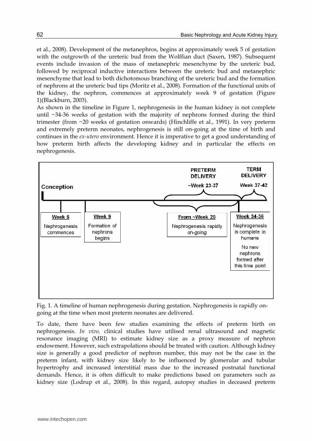
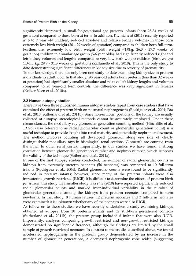
et al., 2008). Development of the metanephros, begins at approximately week 5 of gestation with the outgrowth of the ureteric bud from the Wolffian duct (Saxen, 1987). Subsequent events include invasion of the mass of metanephric mesenchyme by the ureteric bud, followed by reciprocal inductive interactions between the ureteric bud and metanephric mesenchyme that lead to both dichotomous branching of the ureteric bud and the formation of nephrons at the ureteric bud tips (Moritz et al., 2008). Formation of the functional units of the kidney, the nephron, commences at approximately week 9 of gestation (Figure 1)(Blackburn, 2003). As shown in the timeline in Figure 1, nephrogenesis in the human kidney is not complete until ~34-36 weeks of gestation with the majority of nephrons formed during the third trimester (from ~20 weeks of gestation onwards) (Hinchliffe et al., 1991). In very preterm and extremely preterm neonates, nephrogenesis is still on-going at the time of birth and continues in the ex-utero environment. Hence it is imperative to get a good understanding of how preterm birth affects the developing kidney and in particular the effects on nephrogenesis.
Fig. 1. A timeline of human nephrogenesis during gestation. Nephrogenesis is rapidly on-going at the time when most preterm neonates are delivered.
To date, there have been few studies examining the effects of preterm birth on nephrogenesis. In vivo, clinical studies have utilised renal ultrasound and magnetic resonance imaging (MRI) to estimate kidney size as a proxy measure of nephron endowment. However, such extrapolations should be treated with caution. Although kidney size is generally a good predictor of nephron number, this may not be the case in the preterm infant, with kidney size likely to be influenced by glomerular and tubular hypertrophy and increased interstitial mass due to the increased postnatal functional demands. Hence, it is often difficult to make predictions based on parameters such as kidney size (Lodrup et al., 2008). In this regard, autopsy studies in deceased preterm
www.intechopen.com
Effects of Preterm Birth on the Kidney
63
neonates have provided insight into how preterm birth affects the structure of the kidney and the number of glomerular generations formed within the kidney. As well, carefully controlled experimental studies in the nonhuman primate provide valuable insight into the effects of preterm birth on nephrogenesis and on the total number of nephrons formed.
2.1 Clinical in vivo studies
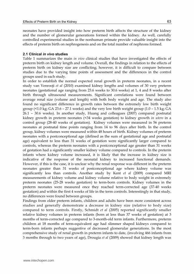
Table 1 summarizes the main in vivo clinical studies that have investigated the effects of preterm birth on kidney length and volume. Overall, the findings in relation to the effects of preterm birth on kidney size are conflicting; however, it is difficult to compare between studies due to the varying time points of assessment and the differences in the control groups used in each study. In order to establish the normal expected renal growth in preterm neonates, in a recent
study van Venrooji et al (2010) examined kidney lengths and volumes of 30 very preterm
neonates (gestational age ranging from 23.6 weeks to 30.6 weeks) at 1, 4 and 8 weeks after
birth through ultrasound measurements. Significant correlations were found between
average renal size (volume and length) with both body weight and age. The study also
found no significant difference in growth rates between the extremely low birth weight
group (<1.0 kg; GA 23.6 – 27.1 weeks) and the very low birth weight group (1.0 – 1.5 kg; GA
26.1 – 30.6 weeks). In another study, Huang and colleagues (2007) compared postnatal
kidney growth in preterm neonates (<34 weeks gestation) to kidney growth in utero in a
control group (28-40 weeks of gestation). Kidney volumes were measured in 56 preterm
neonates at postnatal time points ranging from 14 to 96 days after birth. In the control
group, kidney volumes were measured within 48 hours of birth. Kidney volumes of preterm
neonates with a postconceptional age (defined as the sum of gestational age and postnatal
age) equivalent to less than 31 weeks of gestation were significantly larger compared to
controls, whereas the preterm neonates with a postconceptional age greater than 31 weeks
of gestation had a significantly smaller kidney volume compared to controls. In the preterm
infants where kidney volume increased, it is likely that the increase in kidney size is
indicative of the response of the neonatal kidney to increased functional demands.
However, if this is the case, it is unclear why the renal response was different in the preterm
neonates greater than 31 weeks of postconceptional age where kidney volume was
significantly less than controls. Another study by Kent et al (2009) compared MRI
measurements of kidney volume and kidney volume relative to body weight in extremely
preterm neonates (25-28 weeks gestation) to term-born controls. Kidney volumes in the
preterm neonates were measured once they reached term-corrected age (37-40 weeks
gestation) and within the first 4 weeks of life in the term controls. Interestingly in that study,
no differences were found between groups.
Findings from older preterm infants, children and adults have been more consistent across
studies and generally demonstrate a decrease in kidney size (relative to body size)
compared to term controls. Firstly, Schmidt et al (2005) reported significantly smaller
relative kidney volumes in preterm infants (born at less than 37 weeks of gestation) at 3
months of term-corrected age compared to 3-month-old term infants. Furthermore, preterm
children at 18 months of term-equivalent age had slimmer shaped kidneys compared to
term-born infants perhaps suggestive of decreased glomerular generations. In the most
comprehensive study of renal growth in preterm infants to date, (involving 466 infants from
3 months through to two years of age), Drougia et al (2009) showed that kidney length was
www.intechopen.com
Basic Nephrology and Acute Kidney Injury
64
Author & Year
Gestational age at birth
(weeks), range
Age at assessment
Study Groups and sample size
Outcomes on renal size
Huang (2007) 24-36 14-96 days Preterm (n=56)
Gestational controls (n=44)
Larger relative volume in preterm <31 weeks PCA
Smaller relative volume in preterm >31 weeks PCA
Van Venrooij (2010)
23-30 1, 4, 8 weeks ELBW (n=14) VLBW (n=16)
No differences in volume or length
Kent (2009) 25-28 37-40 weeks
(term equivalent)
Preterm (n=17) Term (n=13)
No differences in volume
Drougia (2008) 28-41
36 and 40 weeks (term equivalent)
3, 6, 12 and 24 months
Preterm 28-34 weeks
(SGA: n=100 AGA: n=54)
Preterm 34-36 weeks (SGA:
n=80 AGA: n=61) Term (AGA: n=90
SGA: n=81)
Smaller right kidney length in preterm SGA 28-34 week
group compared to AGA group at all postnatal time
points
Schmidt (2005) <37 40 weeks 3 and 18 months
Preterm (n=59) Term (n=801)
Smaller relative volumes at 3 and 18 months
Zaffanello (2010) 26-31 5-6 years ELBW (n=36) VLBW (n=43)
ELBW have smaller volumes
(Right, Left, Total) ELBW have smaller length
(Right and Left)
Kwinta (2011) 26-29 6-7 years ELBW (n=78) Term (n=38)
ELBW have smaller volume
Rakow (2008) <32 (mean=27) 9-12 years Preterm (n=33)
Term (n=37)
Smaller absolute volume; not significant when
adjusted for body surface area
Rodriguez-Soriano (2005)
23-35 6-12 years Preterm (n=27)
Length and volume appeared to be in normal range; no
comparison group
Keijzer-Veen (2010)
<32 (mean=31) 20 years
Preterm SGA (n=22)
Preterm AGA (n=29)
Term AGA (n=30)
Smaller length and volume in preterm (SGA and AGA) female group compared to term
Table 1. The main studies that have examined renal size (volume and length) in preterm neonates, children and adults (PCA=post-conceptional age, ELBW=extremely low birth weight, VLBW=very low birth weight, SGA=small for gestational age, AGA=appropriately grown for gestational age)
www.intechopen.com
Effects of Preterm Birth on the Kidney
65
significantly decreased in small-for-gestational age preterm infants (born 28-34 weeks of
gestation) compared to those born at term. In addition, Kwinta et al (2011) recently reported
in 6 to 7 year old children, reduced absolute and relative kidney volumes in those born
extremely low birth weight (26 – 29 weeks of gestation) compared to children born full-term.
Furthermore, extremely low birth weight (birth weight <1.0kg; 26.3 - 27.7 weeks of
gestation) children in a similar age group (5-6 year olds), had significantly reduced right and
left kidney volumes and lengths compared to very low birth weight children (birth weight
1.0-1.5 kg; 29.9 - 31.3 weeks of gestation) (Zaffanello et al., 2010). This is the only study to
date demonstrating significant differences in kidney size due to severity of prematurity.
To our knowledge, there has only been one study to date examining kidney size in preterm individuals in adulthood. In that study, 20-year-old adults born preterm (less than 32 weeks of gestation) had significantly smaller absolute and relative left kidney lengths and volumes compared to 20 year-old term controls; the difference was only significant in females (Keijzer-Veen et al., 2010a).
2.2 Human autopsy studies
There have been three published human autopsy studies (apart from case studies) that have examined the effect of preterm birth on postnatal nephrogenesis (Rodriguez et al., 2004; Faa et al., 2010; Sutherland et al., 2011b). Since non-uniform portions of the kidney are usually collected at autopsy, stereological methods cannot be accurately employed. Under these circumstances, the medullary ray glomerular generation counting method (Hinchliffe et al., 1992b) (also referred to as radial glomerular count or glomerular generation count) is a useful technique to provide insight into renal maturity and potentially nephron endowment. The method involves counting all developed glomeruli along one side of clearly distinguishable medullary rays in histological renal sections. Glomeruli are counted from the inner to outer renal cortex. Importantly, in our studies we have found a strong correlation between glomerular generation number and nephron number, which supports the validity of the technique (Sutherland et al., 2011a). In one of the first autopsy studies conducted, the number of radial glomerular counts in
kidneys from extremely preterm neonates (56 neonates) was compared to 10 full-term
infants (Rodriguez et al., 2004). Radial glomerular counts were found to be significantly
reduced in preterm infants; however, since many of the preterm infants were also
intrauterine growth restricted (IUGR) it is difficult to determine the effects of preterm birth
per se from this study. In a smaller study, Faa et al (2010) have reported significantly reduced
radial glomerular counts and marked inter-individual variability in the number of
glomerular generations among the kidneys from preterm neonates compared to term
newborns. In that study, 8 human fetuses, 12 preterm neonates and 3 full-term neonates
were examined; it is unknown whether any of the neonates were also IUGR.
As follow on to these studies, we have recently undertaken a study examining kidneys
obtained at autopsy from 28 preterm infants and 32 still-born gestational controls
(Sutherland et al., 2011b); the preterm group included 6 infants that were also IUGR.
Importantly, analyses comparing growth restricted and non-growth restricted kidneys
demonstrated no significant differences, although the findings are limited by the small
sample of growth restricted neonates. In contrast to the studies described above, we found
accelerated nephrogenesis in the preterm group demonstrated by an increase in the
number of glomerular generations, a decreased nephrogenic zone width (suggesting
www.intechopen.com
Basic Nephrology and Acute Kidney Injury
66
earlier cessation of nephrogenesis postnatally) and a decreased proportion of glomeruli in
the immature V (vesicle) -stage of maturation compared to still-born gestational controls.
Furthermore, mean renal corpuscle cross sectional area was significantly larger in the
preterm kidneys. Of particular concern, kidneys from preterm infants had a higher
percentage of structurally abnormal glomeruli compared to the gestational controls with
up to 13.7% of glomeruli affected. These abnormal glomeruli exhibited a dilated
Bowman’s space and shrunken glomerular tuft. The factors associated with the
development of abnormal glomeruli are yet unknown and this is an important area of
future research.
2.3 Nonhuman primate animal studies
We have shown that the baboon is an ideal model to study human kidney development, as the ontogeny of the kidney very closely matches that of the human (Gubhaju and Black, 2005). Similar to the human, nephrogenesis in the baboon commences at approximately 30 days of gestation (Hendrickx et al., 1971) and ceases prior to term by 175 days gestation (Term = 185 days gestation) (Gubhaju and Black, 2005). Similar to the wide range in nephron number found in human kidneys, in the kidneys we examined total nephron number ranged from 193,983 to 334,316 in baboons delivered at term. In collaboration with researchers at the Southwest Foundation for Biomedical Research (San Antonio, Texas, U.S.A) we have examined the kidneys from fetal baboons that have
been prematurely delivered and ventilated after birth in a neonatal intensive care unit (NICU) in a similar manner to human preterm babies (Gubhaju et al., 2009). These
appropriate weight-for-gestational age baboons were delivered extremely preterm (125 days of gestation); equivalent to approximately 27 weeks gestation in humans. After birth,
all preterm neonates were intubated, administered 100 mg/kg surfactant (Survanta; donated by Ross Products, Columbus, OH), and ventilated with pressure limited infant
ventilators (InfantStar; donated by Infrasonics, San Diego, CA). All preterm neonates were also treated with ampicillin and gentamicin for the first 7–10 days of life (Thomson et al.,
2004). Further doses of antibiotics were only administered in cases of clinically suspected infection. Following birth, the baboon neonates were ventilated in the NICU for a
maximum period of 21 days. In this model, kidney volume, nephron number and size of the renal corpuscle were
estimated using unbiased stereology, the gold standard method for the determination of
nephron number (Bertram, 2001; Sutherland et al., 2011a). One of the most significant
findings from the nonhuman primate studies was the clear evidence that nephrogenesis was
on-going in the extrauterine environment following preterm birth. There was structural
evidence of on-going nephrogenesis in the outer renal cortex (branching of the ureteric bud,
metanephric mesenchyme and Comma and S-shaped bodies) and this was accompanied by
a significant increase in the number of glomerular generations and nephron number in the
postnatal environment by postnatal day 21 (Gubhaju et al., 2009). Furthermore, kidney
weight and volume relative to body weight were significantly higher in the preterm baboon
neonates compared to gestational age-matched controls; a finding that has been previously
reported in human studies (Huang et al., 2007). There was a significant decrease in
glomerular density (glomeruli/gram of kidney) in the kidney from preterm baboon
neonates compared to gestational controls suggestive of altered renal growth and
potentially an increase in tubular mass.
www.intechopen.com

Effects of Preterm Birth on the Kidney
67
Fig. 2. A representative photomicrograph of a histological renal section from a preterm baboon showing abnormal glomeruli in the outer renal cortex; these morphologically immature glomeruli exhibited a shrunken glomerular tuft and enlarged Bowman’s space.
Scale bar = 100 m
Similar to the findings from the human autopsy studies, morphologically abnormal glomeruli were also found in kidneys from preterm baboon neonates; with up to 18% of glomeruli affected (Figure 2). High proportions of abnormal glomeruli were only found in those kidneys from preterm baboons, whereas in gestational controls the proportion of abnormal glomeruli was negligible. The observed abnormal glomeruli were only present in the superficial outer cortex of the preterm kidney suggesting that it is the glomeruli that are recently formed (possibly those formed in the extrauterine environment) that are ‘at risk.’ Further immunohistochemical analyses demonstrated that the abnormal glomeruli were poorly vascularised (lack of endothelial cell marker, CD31 immunostaining). In addition, immunostaining with the podocyte marker, WT-1, revealed that the abnormal glomeruli were in a relatively immature stage of development since the glomerular tuft contained WT-1 positive cells surrounding a mass of relatively undifferentiated cells. Importantly, there were a large number of parietal epithelial cells surrounding the Bowman’s capsule; previous human studies have reported a similar morphology in atubular glomeruli (Gibson et al., 1996; Bariety et al., 2006). If the abnormal glomeruli in the preterm kidneys are atubular, then they will never be functional. Further studies are required to determine whether this is the case. Certainly, a large proportion of non-functional glomeruli in the preterm kidney is likely to have adverse consequences on renal function both in the neonatal period and in the long-term (by reducing the functional reserve of nephrons).
www.intechopen.com

Basic Nephrology and Acute Kidney Injury
68
3. Renal function in the preterm neonate
There have been a number of studies that have examined the effects of preterm birth on renal function. However, it must be kept in mind when interpreting the data from these studies that the function of the immature preterm kidney is likely to be quite different to that of the term infant, which in turn is likely to be quite different to the adult. Hence, although the ‘normal’ levels of the standard markers of renal function (such as serum creatinine and urinary albumin) have been well-established for the adult population, the standard levels in the neonate, especially those of the preterm neonate, are not clearly defined. This often makes the clinical assessment of renal function in the preterm neonate difficult. In future research, it is necessary to establish the ‘normal’ levels of renal function in the preterm infant and to identify robust biomarkers for the early diagnosis of renal injury in the neonatal period, which may in turn prevent long-term renal dysfunction.
3.1 Fluid and electrolyte homeostasis An imbalance of fluid and electrolyte intake versus excretion is very common in premature neonates, and can lead to significant morbidity and mortality (Bhatia, 2006); hypernatraemia, for example, can result in severe neurological injury (Moritz and Ayus, 2005). Insensible fluid loss is a major factor (Bhatia, 2006), and is primarily transcutaneous due to the developmental immaturity of the skin and a high body surface area to body water mass ratio (Baumgart and Costarino, 2000). Equally, the delayed loss of extracellular fluid volume following preterm birth is also associated with an increased risk of morbidity, in particular bronchopulmonary dysplasia (Oh et al., 2005) and patent ductus arteriosus (Bell and Acarregui, 2008). Three phases of fluid and electrolyte homeostasis have been observed in the immediate period following preterm birth; these phases occurred similarly in extremely low birth weight infants and those at older gestational ages (Lorenz et al., 1982; Lorenz et al., 1995). As described by Lorenz et al., (1982; 1995) in the first 24 hours following birth, a period known as the pre-diuretic stage, urine output is minimal and sodium excretion is low. On postnatal days 2-3, termed the diuretic phase, sodium excretion and urine output significantly increase, which occurs independently of fluid intake. From approximately days 4-5 of life, the post-diuretic phase, urine output changes in response to fluid intake (Lorenz et al., 1982; Lorenz et al., 1995). Importantly, however, the postnatal time-point that these phases occur, and their duration, differ between individual neonates (Lorenz et al., 1995), as does the amount of insensible fluid loss; together, this highlights the need for an individualised approach to fluid therapy in preterm neonates. Urine output is the most commonly and easily measured indicator of renal function in the preterm neonate. Urine output less than 0.5 ml/kg/h, known as oliguria, can be indicative of acute kidney injury (AKI). AKI, however, can also be non-oliguric, therefore urine output is not a very specific indicator of renal function. Furthermore, from the post-diuretic phase of fluid homeostasis urine output is highly dependent upon fluid intake; high intakes may artificially increase urine output, while not accurately reflecting renal functional capacity. The most common measure of electrolyte balance in the neonate is the calculation of the fractional excretion of sodium (FENa), which is the percentage of sodium that is excreted and not taken up through tubular reabsorption. The calculation of FENa takes into account the levels of both serum and urine sodium, and it is corrected for serum and urine creatinine levels. Therefore, high urine sodium levels may be indicative of structural immaturity of the
www.intechopen.com
Effects of Preterm Birth on the Kidney
69
renal tubule (short length of the tubules, and changes in the density and structure of transporter proteins) (Jones and Chesney, 1992), or due to renal injury (Ueda and Shah, 2000; Bonventre, 2007). Studies that have assessed FENa during the neonatal period have determined that sodium excretion is significantly higher in preterm neonates compared to term controls (Siegel and Oh, 1976; Aperia et al., 1981), and significantly decreases with increasing gestational (Gallini et al., 2000) and postnatal age (Ross et al., 1977; Sulyok et al., 1979; Aperia et al., 1981; Gallini et al., 2000; Giapros et al., 2007). Therefore, with increasing renal maturity a positive sodium balance (low FENa) is achieved, which is essential for the growth and development of the neonate and the maintenance of fluid homeostasis (Engle, 1986).
3.2 Glomerular filtration rate Endogenous creatinine is the most practical and commonly used marker of renal function, with calculated creatinine clearance widely used as an estimate of glomerular filtration rate (GFR). In the clinical setting, repeated serum creatinine levels are used to gauge renal function in neonates; this is an easily obtainable measure via routine blood collection and does not rely on timed urine samples or additional invasive procedures. This method does, however, have significant limitations. Immediately following birth, serum creatinine levels are equivalent to the fetal levels, which during the third trimester of gestation rise from 42 µmol/L at 23 weeks to 47 µmol/L at term; the increase likely reflecting an increase in muscle mass (Moniz et al., 1985). In the first forty-eight hours following birth, however, serum creatinine levels significantly increase (Bueva and Guignard, 1994; Miall et al., 1999). This is considered to be due, in part, to tubular creatinine reabsorption, as has been evidenced in a neonatal animal model (Matos et al., 1998), and also due to the inadequacy of glomerular filtration during the early postnatal period (Miall et al., 1999). Peak serum creatinine levels are reached at postnatal day 2-4 of life, with the highest levels and most delayed timing of the peak creatinine level seen in neonates at the lowest gestational ages (Miall et al., 1999). During the first week of life following preterm birth, GFR is significantly lower in preterm neonates than in term-born controls (Siegel and Oh, 1976; Finney et al., 2000; Schreuder et al., 2009), and is significantly positively correlated with both gestational age at birth, and postnatal age (Clark et al., 1989; Gordjani et al., 1998; Iacobelli et al., 2009). Compared to term neonates, the rate of increase in GFR after birth is slower in neonates born preterm (Gordjani et al., 1998). Up until two months of age there are similar findings, with a number of studies observing an increase in GFR concurrent to increasing gestational and postnatal ages (Ross et al., 1977; Fawer et al., 1979; Sulyok et al., 1979; Aperia et al., 1981; Wilkins, 1992; Bueva and Guignard, 1994; Gallini et al., 2000; Cuzzolin et al., 2006; Thayyil et al., 2008). Although a number of studies have now been performed in this area, there is still a lack of clear definition regarding expected GFR values in the preterm neonate. Recently published standard curves of GFRs in neonates born at 27-31 weeks gestational age, from 7 to 28 days of life, will go some way in aiding in the clinical interpretation of renal function in this particular group of neonates (Vieux et al., 2010). Given that age has been found to be a strong determinant of GFR, the low GFR observed in the preterm neonate after birth is likely the result of renal immaturity (a low number of filtering glomeruli), and it is also likely to be influenced by differences in renal blood flow and vascular resistance. It is essential that GFR is monitored in the postnatal period following preterm birth, as a very low GFR is likely to impair renal drug clearance, leading to nephrotoxicity.
www.intechopen.com

Basic Nephrology and Acute Kidney Injury
70
3.3 Acute kidney injury Acute kidney injury (AKI; previously referred to as acute renal failure) is reported to occur in 8% to 24% of preterm neonates admitted to neonatal intensive care units (Stapleton et al., 1987; Hentschel et al., 1996). The current diagnosis of AKI is primarily based on the RIFLE system, which categorises the stages of increasing AKI severity: Risk, Injury, Failure, Loss and End-stage kidney disease (ESKD) (Bellomo et al., 2004). This system was further modified following recommendations from the acute kidney injury network (AKIN) (Mehta et al., 2007). The initial clinical indication of AKI risk includes a 50% increase in serum creatinine (or ≥ 0.3 mg/dl within a 48 hour period), and/or a urine output less than 0.5 mg/kg/hr for a period of six hours (Bellomo et al., 2004; Mehta et al., 2007), which are changes indicative of a significantly reduced GFR. Classifications for the definition of AKI in a neonatal specific population, however, have not been developed. The causes of AKI in the preterm neonate are primarily pre-renal in origin, arising from conditions which affect renal perfusion such as hypotension, hypoxia and sepsis (Stapleton et al., 1987; Cataldi et al., 2005). These in turn lead to apoptotic, necrotic and inflammatory processes within the kidney (Ueda and Shah, 2000; Bonventre, 2007). Importantly, AKI in the preterm neonate may subsequently lead to long-term chronic renal disease (Abitbol et al., 2003). In a study involving 172 preterm neonates by Cataldi et al. 2005, the risk factors for AKI were found to be maternal and neonatal drug administration (non-steroidal anti-inflammatory drugs (NSAIDs) and antibiotics, especially ceftazidime), a low Apgar score, and a patent ductus arteriosus. Interestingly, gestational age did not affect risk of AKI, however, the majority of AKI cases (79%) weighed < 1.5 kg at birth (Cataldi et al., 2005). In a larger study by Cuzzolin et al. (2006), involving 281 preterm neonates, a number of risk factors for AKI were also identified. These included maternal NSAID administration, low Apgar score, respiratory distress syndrome, neonatal drug administration (antibiotics and NSAIDs), and a number of clinical interventions (intubation at birth, catheterization, phototherapy, and mechanical ventilation). Given the importance of the early diagnosis and treatment of AKI, there has been much recent focus on the discovery of novel urinary biomarkers. The expectation of a new biomarker is to enable the diagnosis of cellular injury before a decline in renal function occurs. For example, serum creatinine is not elevated until 48-72 hours after an acute injury has occurred (Moran and Myers, 1985); such a prolonged delay before diagnosis and treatment likely results in further renal injury. As Rosner (2009) describes, it would be optimal if a biomarker could be developed to: 1) assess the response to, and any adverse effects of therapeutic interventions 2) indicate the severity of renal injury 3) inform on the etiology of the injury and 4) identify the location of injured cells. In a systematic review of the current literature, Parikh et al. (2010) determined that the molecules with the most promise for the diagnosis of established AKI include interleukin-18 (IL-18), kidney injury molecule-1 (KIM-1), N-acetyl-beta-D-glucosaminidase (NAG) and neutrophil gelatinase-associated lipocalin (NGAL). NGAL, IL-18, fatty acid binding protein (FABP), and cystatin-C are the most encouraging biomarkers for the early diagnosis of AKI, given that the upregulation of these molecules following injury onset precedes the rise in serum creatinine by many hours (Parikh et al., 2010). In the preterm neonate, a small number of studies have been conducted for the assessment of urinary NGAL levels, with mixed results. These studies have shown that the highest NGAL levels are evident in the neonates that are critically ill, with and without evidence of
www.intechopen.com
Effects of Preterm Birth on the Kidney
71
renal dysfunction (Lavery et al., 2008; Parravicini, 2010); in particular, NGAL shows potential as a promising biomarker of late-onset sepsis (Lavery et al., 2008; Parravicini et al., 2010). Urinary NGAL levels also strongly correlated with gestational and postnatal age (Lavery et al., 2008; Huynh et al., 2009), perhaps reflecting the renal production of NGAL during nephrogenesis (Gwira et al., 2005) which is often still ongoing during the early postnatal period. Normative values for urinary NGAL in preterm neonates with uncomplicated clinical courses have also been published, with the results indicating a greater variation in females than males (Huynh et al., 2009).
3.4 Proteinuria Proteinuria, the presence of high levels of protein in the urine, may be of glomerular and/or
tubular origin. The number of different proteins that have been identified in the adult
urinary proteome is 1,543, and these are primarily of membrane, extracellular and lysosomal
origin (Adachi et al., 2006). Despite this large number, unless renal function is impaired,
proteins are normally only present at very low levels in urine, due to the function of the
glomerular filtration barrier and tubular reabsorption capabilities.
Presence of high molecular weight (HMW) proteins in the urine, such as albumin
traditionally indicates a disruption in the integrity of the glomerular filtration barrier.
Recent debate, however, has suggested that the contribution of tubular reabsorption of
albumin from the filtrate may be greater than previously considered (Comper et al., 2008).
In general, albuminuria is a strong marker for renal and cardiovascular disease, and a risk
factor for mortality (Matsushita et al., 2010; Methven et al., 2011). Normally, adults excrete
less than 30 mg of albumin per 24 hours (Mathieson, 2004). Urinary albumin levels between
30 – 300 mg in 24 hours is considered microalbuminuria, with levels greater than 300 mg
classified as macroalbuminuria (Mathieson, 2004). Traditionally, 24 hour urine samples
were required for reliable estimates of urinary protein. However, single random spot
samples with protein levels corrected for urine creatinine, have been shown to be
significantly correlated with results from 24 hour collections, and are equally effective in the
prediction of outcomes (Ralston et al., 1988; Methven et al., 2011). In neonates, 24 hour urine
collection is difficult, therefore analysis of urinary protein levels are undertaken using spot
urine samples obtained using urine collection bags.
Low molecular weight (LMW) proteins, such as ┙1-microglobulin, ┚2-microglobulin and
retinol binding protein pass freely through the glomerular filter and undergo reuptake via
proximal tubule cells (Tomlinson, 1992). Megalin and cubulin have been identified as
important receptors involved in tubular protein uptake, with mutations in the receptors
resulting in proteinuria (Christensen and Birn, 2001). To date, LMW protein levels in the
urine are not routinely measured in the clinical setting. Importantly, however, amongst the
LMW proteins there may be potential novel biomarkers of tubular cell injury and this
requires further research (Rosner, 2009; Parikh et al., 2010).
In the preterm neonate, few studies have been conducted to examine urine protein excretion. In general, there is a high variability in urine albumin levels between individual neonates (Clark et al., 1989; Fell et al., 1997), with the highest levels exhibited by those with a low gestational age at birth and those that are clinically unstable (Galaske, 1986; Clark et al., 1989; Tsukahara et al., 1994; Fell et al., 1997; Awad et al., 2002b). The majority of studies have only been conducted during the first week of life following preterm birth. However, in a study by Tsukahara et al. (1994) urine albumin levels were assessed in preterm and term
www.intechopen.com
Basic Nephrology and Acute Kidney Injury
72
neonates over the first 28 days of life. Urine albumin levels were found to remain relatively stable postnatally over the one month period in the term neonates, whereas in the preterm neonates, urine albumin was seen to decrease with increasing postnatal age. These findings suggest that the glomerular filtration barrier following preterm birth is structurally immature, until beyond one month of age. Urinary ┚2-microglobulin levels have also been shown to be significantly greater in the preterm infant compared to term-born infants throughout the first month of life (Aperia et al., 1981; Tsukahara et al., 1990; Tsukahara et al., 1994), and are decreased with increasing gestational and postnatal age (Takieddine et al., 1983). Similarly, levels of 1-microglobulin and RBP are higher in preterm neonates than neonates born at term (Clark et al., 1989; Fell et al., 1997; Awad et al., 2002a). To date, however, it remains unclear whether the increased urinary high- and low- molecular weight protein levels reported in preterm neonates are associated with renal immaturity and/or injury. The high variability in urinary protein levels may also reflect differences in the postnatal clinical course in preterm neonates; further studies are necessary to verify whether this is the case.
3.5 Long-term effects of preterm birth on renal function Renal function in preterm-born children and adults, has to date only been investigated in a small number of studies, with inconclusive results. In school-aged children, Rakow et al. (2008) found no difference in GFR or urinary levels of both HMW and LMW proteins between children born less than 32 weeks gestational age, and those that were born at term. Similarly, Vanpee et al. (1992) determined no difference in renal function in preterm and term-born children at 8 years of age, despite lower GFR and higher urine albumin levels being evident in the preterm group at 9 months of age. In contrast, however, a study by Rodriguez-Soriano and colleagues (2005) reported that GFR was significantly reduced in preterm-born children compared to term controls, with impairments in electrolyte excretion also evident. Furthermore, in children examined at 6-8 years of age, Iacobelli et al. (2007) demonstrated microalbuminuria in 8.3% of the preterm neonates, which was associated with postnatal factors such as neonatal hypotension and increased catch-up growth. Increased risk of renal demise was also evident in individuals born preterm who were obese during childhood (Abitbol et al., 2009). Two studies have also been conducted to examine renal function in young adults (20-30 years of age), with both Keijzer-Veen et al. (2007) and Kistner et al. (Kistner et al., 2000) finding no effect of preterm birth on GFR or albuminuria. To the contrary, in a cohort of 19 year old young adults, those who were born preterm as well as IUGR, there was a significant reduction in GFR (Keijzer-Veen et al., 2005). Given these results in preterm-born children and adults, there is some suggestion that preterm birth adversely affects the growth and functional capacity of the kidney and may result in progressive renal failure later in life. Importantly, adverse consequences appear to be more likely to occur in combination with other insults. Therefore, future research must be directed towards identifying these insults and their effects on the structure and function of the kidney.
4. Preterm birth leads to glomerular abnormalities – Areas of future research
One of the most important findings we have shown thus far, is the presence of abnormal glomeruli in both the human and nonhuman primate (baboon) preterm kidney. These abnormal glomeruli are located in the outer renal cortex and are in the most immature stage of development (stage 1); they are composed of an undifferentiated glomerular anlage of cells
www.intechopen.com

Effects of Preterm Birth on the Kidney
73
(foundation group of cells) surrounded by a layer of podocytes with scant, if any, capillarisation. Our findings thus strongly suggest that it is the very immature glomeruli (possibly those formed in the extrauterine environment) that are particularly vulnerable to preterm birth. Given the gross abnormalities observed in these glomeruli, it is unlikely that these glomeruli will ever be functional and thus, it is expected that they will be subsequently resorbed into the surrounding tissue. In the short-term, such abnormalities will likely lead to marked impairment of renal function in the neonate if a high proportion of the nephrons are affected, or to minor impairment if only a small proportion are abnormal. When the kidney is severely affected this will adversely impact on the number of functional nephrons at the beginning of life and thus reduce the long-term functional reserve of the kidney, rendering it vulnerable to hypertension and secondary life style insults. The cause(s) of the glomerular abnormalities in the preterm infant is currently unknown. Importantly in this regard, we have shown that there is a wide variation in the proportion of abnormal glomeruli within the kidneys of preterm infants, with the kidneys of some preterm infants appearing morphologically normal whereas in others a large proportion of the glomeruli appear abnormal (Sutherland et al., 2011b). Given the wide variation in the proportion of abnormal glomeruli within the kidneys of preterm infants, this suggests that it is not preterm birth per se that leads to the glomerular abnormalities; instead they are likely due to factors often associated with preterm birth as shown in Figure 3. It is likely that these deleterious effects may relate to: 1) adverse factors in the in utero environment that have led to premature delivery, 2) factors in the neonatal care of the preterm infant and 3) pharmacological interventions/therapies administered to mothers prior to birth and/or the infant after birth. There are many factors which apply to each of these categories; below we have selected some that we consider are important for future research.
Fig. 3. Depiction of the potential factors that may contribute to impaired nephrogenesis in the preterm kidney and consequently lead to a reduced nephron endowment and vulnerability to renal disease in adulthood
www.intechopen.com

Basic Nephrology and Acute Kidney Injury
74
4.1 Adverse factors in the in utero environment
Two potential factors in the in utero environment that may render the preterm kidney vulnerable are IUGR and/or exposure to chorioamnionitis.
4.1.1 Intrauterine growth restriction
IUGR (growth below the 10th percentile for gestational age) is often a co-morbidity of
preterm birth. Certainly, it is well described in both human and experimental models that
IUGR leads to a reduced nephron endowment at birth (Hinchliffe et al., 1992a; Merlet-
Benichou et al., 1994; Manalich et al., 2000; Zimanyi et al., 2004; Zohdi et al., 2007). This is
likely due to the reduced growth of the fetal kidney (with the number of nephrons directly
proportional to kidney size). In this regard, there is often redistribution of blood flow in the
growth-restricted fetus, leading to preferential blood flow to the brain (termed brain
sparing) and reduced blood flow to organs such as the kidneys (Behrman et al., 1970;
Gunnarsson et al., 1998). To our knowledge the impact of reduced blood flow to the
formation of nephrons in the IUGR fetal kidney has not been investigated. Given the
dramatic change in hemodynamics at the time of birth (elevation in blood pressure and
increased renal blood flow) it is conceivable that the recently formed glomeruli in the IUGR
kidney (with a reduced renal blood flow prenatally) may be particularly vulnerable to the
haemodynamic transition at birth. This is an important area of research that needs to be
thoroughly investigated.
4.1.2 Chorioamnionitis
Chorioamnionitis (a bacterial infection of the chorion and amnion) is a common antecedent of preterm birth (Romero et al., 2006; Goldenberg et al., 2008), especially in births prior to 30 weeks of gestation (Lahra and Jeffery, 2004); it is often complicated by IUGR. Chorioamnionitis may manifest as either a clinical or subclinical condition. When severe, it can ultimately give rise to the fetal inflammatory response syndrome which is characterised by funisitis, fetal vasculitis and an increase in of pro-inflammatory cytokines in fetal blood and amniotic fluid (Romero et al., 1998; Romero et al., 2003; Gotsch et al., 2007). It is likely that the fetal systemic inflammatory response will lead to renal inflammation in chorioamnionitis-exposed infants; conceivably if present in utero this will have deleterious effects on nephrogenesis, and if present at the time of delivery, it will adversely impact on renal function. In this regard, we have recent evidence in fetal sheep to demonstrate that exposure to chorioamnionitis late in gestation does adversely affect nephrogenesis, leading to a 23% and 18% reduction in nephron endowment in singleton and twin-exposed fetuses, respectively (Galinsky et al., 2011). It is important to note that we did not observe any morphological abnormalities in the glomeruli of these fetal kidneys. However, it is conceivable, that if renal inflammation is present at the time of birth, that this will further compromise postnatal nephrogenesis and renal function in the neonate. Following the hemodynamic transition at birth, it may be then that glomerular morphological abnormalities develop in these already compromised kidneys. Importantly in this regard, a multiple logistic regression analysis of 2508 preterm neonates, treated with indomethacin, showed a significant correlation between intrauterine inflammation and prevalence of renal and electrolyte abnormalities (Itabashi et al., 2003), thus suggesting that intrauterine inflammation in concert with postnatal indomethacin treatment can lead to renal dysfunction. Certainly our results support the idea that prematurity, when complicated with
www.intechopen.com
Effects of Preterm Birth on the Kidney
75
chorioamnionitis, is likely to exacerbate postnatal renal dysfunction and further studies are required to determine whether renal inflammation at the time of birth is associated with the formation of abnormal glomeruli in the neonatal period.
4.2 Factors in the neonatal care of the infant 4.2.1 Hyperoxia
The administration of supplemental oxygen to preterm infants experiencing respiratory distress is standard therapy, however, the effects of high levels of oxygen in the bloodstream on the development of organs is not well understood. Although the levels of supplemental oxygen administered to the preterm infant have been substantially reduced in recent years, the levels of oxygen in the bloodstream remain elevated above normal; hence it is important that research is conducted into the effects of hyperoxia on nephrogenesis in the preterm infant. Certainly, experimental studies in the lung have shown that hyperoxia can lead to the generation of oxygen free radicals, infiltration of inflammatory cells, collagen deposition, cellular apoptosis and subsequent tissue injury (McGrath-Morrow and Stahl, 2001; Dieperink et al., 2006; Alejandre-Alcazar et al., 2007; Chen et al., 2007; Chetty et al., 2008). Whether this is also the case in other tissues has not been examined.
4.2.2 Ventilation
The mode of ventilation of the preterm infant also has the potential to impact on the developing kidney, since alterations in airway pressure are reported to lead to significant cardiopulmonary haemodynamic changes (Polglase et al., 2009) which may subsequently affect renal perfusion. For instance, mechanical ventilation has been shown to alter renal hemodynamics by leading to an increase in intrathoracic pressure, therefore decreasing cardiac output, leading to renal vasoconstriction and decreased GFR (Arant, 1987). It is likely that changes in renal blood flow will directly influence the growth of the kidney. Hence, it is important in future research to determine the effects of altered renal hemodynamics on growth of the developing kidney and how this is influenced by different modes of ventilation.
4.2.3 Extrauterine growth / nutrition
In general, postnatal growth of the preterm infant is markedly attenuated when compared to the term infant and when compared to the normal rate of growth in utero for the same post-conceptional age (Ehrenkranz, 2000; Clark et al., 2003). Extrauterine growth restriction
in premature neonates (defined as growth below the 10th percentile of intrauterine growth
expectation) is likely to directly influence nephrogenesis. In support of this concept,
Bacchetta et al (2009) reported lower GFRs (albeit in the normal range) in 7 year old children
who had been born very preterm (< 30 weeks gestation) and who were either IUGR or
extrauterine growth restricted. However, it is important to note that in some preterm infants
there can be a disproportional increase in kidney size (relative to body weight) after birth
(Huang et al., 2007; Sutherland et al., 2011b) most probably due to the increased functional
demands on the kidney. Importantly we have shown in our preterm baboon studies that
under these circumstances, there remains a significant correlation between kidney size and
nephron number (Gubhaju et al., 2009).
Taken together, these studies highlight the importance that neonatal nutrition can potentially have on kidney development in the preterm infant. Since nephrogenesis is
www.intechopen.com
Basic Nephrology and Acute Kidney Injury
76
ongoing after birth in the preterm infant, this provides a window of opportunity whereby early postnatal nutrition in the intensive care unit may be able to directly influence the number of nephrons formed within the kidney. Hence optimising nutrition in the neonatal period, with an aim to maximising nephron endowment in the preterm newborn, is an important area for future research. At this stage, there is no known maternal nutrient supplementation that can improve renal outcomes in the fetus; it is critical to investigate this.
4.3 Pharmacological treatments to the mother prior to birth and / or the infant after birth
There are a number of medications routinely administered to women during pregnancy and to preterm infants. Some of the most commonly used are: 1) antenatal glucocorticoids which are routinely administered either to the mother ‘at risk’ of preterm delivery (Vidaeff et al., 2003) or to the preterm infant immediately after delivery to accelerate lung maturation in the infant, 2) antibiotics, often administered to the mother with chorioamnionitis and to the infant with postnatal conditions such as necrotising enterocolitis (Gortner et al., 1991) 3) non steroidal anti-inflammatory drugs (such as ibuprofen and indomethacin) which are often administered to close a patent ductus arteriosus (Ellison et al., 1983) and 4) inotropes (such as dopamine and dobutamine) which are administered in cases of hypotension and poor blood flow (Osborn et al., 2002). Importantly, and of concern, all these medications have the potential to adversely impact on nephrogenesis.
4.3.1 Glucocorticoids
In addition to leading to lung maturation in the preterm infant, administration of
glucocorticoids has been favourably reported to accelerate renal maturation thus
establishing an adequate GFR and efficient tubular reabsorption (Ervin et al., 1996; Ervin et
al., 1998; Petershack et al., 1999). However, the question remains: ‘Does this acceleration in
renal maturation lead to an abnormal and rapid cessation of nephrogenesis which would
ultimately affect nephron endowment? In support of this, a number of experimental studies
(conducted in animal models) have shown that exposure of the fetus to maternal
glucocorticoids can lead to reduced nephron endowment in the offspring (Celsi et al., 1998;
Ortiz et al., 2001; Moritz et al., 2002). As follow up to these studies, in a carefully controlled
study in our baboon model we have examined the effect of administration of antenatal
betamethasone (intramuscular injection of 6mg at 48 hours and 24 hours prior to preterm
delivery) on nephrogenesis in the neonatal baboon kidney (Gubhaju et al., 2009). We found
that although there was acceleration of glomerular maturation in the betamethasone-treated
baboons, the total number of nephrons was within the normal range and importantly we
demonstrated that fetal exposure to maternal glucocorticoids was not the cause of the
glomerular abnormalities associated with preterm birth.
4.3.2 Antibiotics
The use of antibiotics is often essential in the treatment of the mother during pregnancy and of the preterm infant after birth. Alarmingly, it has been shown that antibiotics, such as the aminoglycosides, can be nephrotoxic in the newborn with the preterm infant most vulnerable (Giapros et al., 2003). This is of concern, given that aminoglycosides are not metabolised in the body and thus accumulate in the kidney where they are eventually
www.intechopen.com
Effects of Preterm Birth on the Kidney
77
eliminated (Nagai and Takano, 2004). In experimental studies antibiotics are also linked with impairment of nephrogenesis (Gilbert et al., 1990; Gilbert et al., 1994; Cullen et al., 2000; Nathanson et al., 2000). For instance, it has been shown that incubation of metanephroi in culture with gentamicin leads to decreased branching morphogenesis of the ureteric tree; this is a likely mediator of the reduction in the number of nephrons formed (Cullen et al., 2000). Given that antibiotics can readily cross the placenta, plus their wide use in the neonatal care of the preterm infant, it is imperative in future studies to gain a more precise understanding of the dose, duration and class of antibiotic treatment that leads to adverse effects in the neonatal kidney; such information would likely influence the care in the neonatal intensive care unit.
4.3.3 Non steroidal anti-inflammatory drugs
NSAIDs, such as ibuprofen and indomethacin are usually administered to preterm neonates to stimulate closure of a patent ductus arteriosus; this can occur in up to 80% of extremely preterm infants (Ellison et al., 1983). Importantly, there have been a number of studies that have reported adverse effects on both the structure and function of the preterm kidney following treatment with NSAIDs. For example, renal insufficiency, demonstrated by a significant increase in serum creatinine has been reported in infants following either antenatal or postnatal exposure to NSAIDs (Kang et al., 1999; Butler-O'Hara and D'Angio, 2002). Of concern, in a case-controlled study where renal impairment was reported in preterm neonates that had received indomethacin treatment for a patent ductus arteriosus, 24% of the babies suffered acute renal failure (Akima et al., 2004). In addition, in the rat model where, similar to the preterm infant, nephrogenesis is ongoing after birth, exposure to indomethacin, ibuprofen and gentamicin have all been shown to lead to renal injury in the immature kidneys. There was evidence of vacuolization of epithelium and loss of microvilli in proximal tubules, effacement of podocyte foot processes and irregularities of the basement membrane in the glomeruli and edema within the interstitium (Kent et al., 2007).
4.3.4 Inotropes
At birth, there is a marked change in hemodynamics, with a subsequent rise in blood pressure and heart rate (Teitel et al., 1987; Louey et al., 2000). Preterm birth causes an abrupt and premature shift in the circulation from the fetal to postnatal configuration at a time when the cardiovascular system is still relatively immature; as a result, it is often necessary for inotropes to be administered to preterm neonates when blood pressure remains abnormally low after birth (Kluckow and Evans, 2001; Osborn et al., 2002). Given the importance of renal blood flow to growth of the kidney it is important that future research examines how the hemodyamic transition at birth affects the development of the immature renal vasculature and/or nephrogenesis and what effect the administration of inotropes have on the developing kidney.
5. Conclusion
Over the past decade, considerable advances have been made in our understanding of the effects of preterm birth on the developing kidney. Encouragingly, it has clearly been demonstrated that nephrogenesis continues after birth in the preterm neonate, however, glomerular abnormalities are commonly observed. Future research should be directed into the causes of these abnormalities, so that strategies can be implemented to maximise the
www.intechopen.com

Basic Nephrology and Acute Kidney Injury
78
number of functional nephrons at the beginning of life in the preterm infant in order to ensure long-term renal health. It is important that renal clinicians are made aware of the potential deleterious effects of preterm birth on developing glomerui, so they are aware of the renal vulnerability in subjects that are born preterm.
6. References
Abitbol, CL, Bauer, CR, Montane, B, Chandar, J, Duara, S & Zilleruelo, G. (2003). Long-term follow-up of extremely low birth weight infants with neonatal renal failure. Pediatr Nephrol, Vol. 18, No. 9, (Sep 2003), pp. 887-893, 0931-041X (Print)
Abitbol, CL, Chandar, J, Rodriguez, MM, Berho, M, Seeherunvong, W, Freundlich, M & Zilleruelo, G. (2009). Obesity and preterm birth: additive risks in the progression of kidney disease in children. Pediatr Nephrol, Vol. 24, No. 7, (Jul 2009), pp. 1363-1370, 1432-198X (Electronic)
Adachi, J, Kumar, C, Zhang, Y, Olsen, JV & Mann, M. (2006). The human urinary proteome contains more than 1500 proteins, including a large proportion of membrane proteins. Genome Biol, Vol. 7, No. 9, 2006), pp. R80, 1465-6914 (Electronic)
Akima, S, Kent, A, Reynolds, G, Gallagher, M & Falk, M. (2004). Indomethacin and renal impairment in neonates. Pediatr Nephrol, Vol. 19, No. 5, (May 2004), pp. 490-493, 0931-041X (Print)
Alejandre-Alcazar, MA, Kwapiszewska, G, Reiss, I, Amarie, OV, Marsh, LM, Sevilla-Perez, J, Wygrecka, M, Eul, B, Kobrich, S, Hesse, M, Schermuly, RT, Seeger, W, Eickelberg, O & Morty, RE. (2007). Hyperoxia modulates TGF-beta/BMP signaling in a mouse model of bronchopulmonary dysplasia. Am J Physiol Lung Cell Mol Physiol, Vol. 292, No. 2, (Feb 2007), pp. L537-549, 1040-0605 (Print)
Andreoli, SP. (2004). Acute renal failure in the newborn. Semin Perinatol, Vol. 28, No. 2, (Apr 2004), pp. 112-123, 0146-0005 (Print)
Aperia, A, Broberger, O, Elinder, G, Herin, P & Zetterstrom, R. (1981). Postnatal development of renal function in pre-term and full-term infants. Acta Paediatr Scand, Vol. 70, No. 2, (Mar 1981), pp. 183-187, 0001-656X (Print)
Arant, BS, Jr. (1987). Postnatal development of renal function during the first year of life. Pediatr Nephrol, Vol. 1, No. 3, (Jul 1987), pp. 308-313, 0931-041X (Print)
Awad, H, el-Safty, I, el-Barbary, M & Imam, S. (2002a). Evaluation of renal glomerular and tubular functional and structural integrity in neonates. American Journal of the Medical Sciences, Vol. 324, No. 5, (Nov 2002a), pp. 261-266,
Awad, H, el-Safty, I, el-Barbary, M & Imam, S. (2002b). Evaluation of renal glomerular and tubular functional and structural integrity in neonates. Am J Med Sci, Vol. 324, No. 5, (Nov 2002b), pp. 261-266, 0002-9629 (Print)
Bacchetta, J, Harambat, J, Dubourg, L, Guy, B, Liutkus, A, Canterino, I, Kassai, B, Putet, G & Cochat, P. (2009). Both extrauterine and intrauterine growth restriction impair renal function in children born very preterm. Kidney Int, Vol. 76, No. 4, (Aug 2009), pp. 445-452, 1523-1755 (Electronic)
Bariety, J, Mandet, C, Hill, G & Bruneval, P. (2006). Parietal podocytes in normal human glomeruli. J Am Soc Nephrol, Vol. 17, No. (Oct 2006), pp. 2770-2780, 1046-6673 (Print)
Baumgart, S & Costarino, AT. (2000). Water and electrolyte metabolism of the micropremie. Clin Perinatol, Vol. 27, No. 1, (Mar 2000), pp. 131-146, vi-vii, 0095-5108 (Print)
www.intechopen.com
Effects of Preterm Birth on the Kidney
79
Behrman, RE, Lees, MH, Peterson, EN, De Lannoy, CW & Seeds, AE. (1970). Distribution of the circulation in the normal and asphyxiated fetal primate. Am J Obstet Gynecol, Vol. 108, No. 6, (Nov 15 1970), pp. 956-969, 0002-9378 (Print)
Bell, EF & Acarregui, MJ. (2008). Restricted versus liberal water intake for preventing morbidity and mortality in preterm infants. Cochrane Database Syst Rev, Vol. No. 1, 2008), pp. CD000503, 1469-493X (Electronic)
Bellomo, R, Ronco, C, Kellum, JA, Mehta, RL & Palevsky, P. (2004). Acute renal failure - definition, outcome measures, animal models, fluid therapy and information technology needs: the Second International Consensus Conference of the Acute Dialysis Quality Initiative (ADQI) Group. Crit Care, Vol. 8, No. 4, (Aug 2004), pp. R204-212, 1466-609X (Electronic)
Bertram, JF. (2001). Counting in the kidney. Kidney Int, Vol. 59, No. 2, (Feb 2001), pp. 792-796, 0085-2538 (Print)
Bhatia, J. (2006). Fluid and electrolyte management in the very low birth weight neonate. J Perinatol, Vol. 26 Suppl 1, No. (May 2006), pp. S19-21, 0743-8346 (Print)
Blackburn, S. 2003. Renal system and fluid and electrolyte homeostasis. In: Maternal, fetal and neonatal physiology, 2 ed: Saunders.
Bonamy, A, Bendito, A, Martin, H, Andolf, E, Sedin, G & Norman, M. (2005). Preterm Birth Contributes to Increased Vascular Resistance and Higher Blood Pressure in Adolescent Girls. Pediatr Res, Vol. 58, No. 5, 2005), pp. 845–849, 0031-3998 (Print)
Bonventre, JV. (2007). Pathophysiology of acute kidney injury: roles of potential inhibitors of inflammation. Contrib Nephrol, Vol. 156, No. 2007), pp. 39-46, 0302-5144 (Print)
Bueva, A & Guignard, J. (1994). Renal Function in the Preterm Neonates. Pediatr Res, Vol. 36, No. 5, 1994), pp. 572-577, 0031-3998 (Print)
Butler-O'Hara, M & D'Angio, CT. (2002). Risk of persistent renal insufficiency in premature infants following the prenatal use of indomethacin for suppression of preterm labor. J Perinatol, Vol. 22, No. 7, (Oct-Nov 2002), pp. 541-546, 0743-8346 (Print)
Cataldi, L, Leone, R, Moretti, U, De Mitri, B, Fanos, V, Ruggeri, L, Sabatino, G, Torcasio, F, Zanardo, V, Attardo, G, Riccobene, F, Martano, C, Benini, D & Cuzzolin, L. (2005). Potential risk factors for the development of acute renal failure in preterm newborn infants: a case-control study. Arch Dis Child Fetal Neonatal Ed, Vol. 90, No. 2005), pp. F514-519, 1359-2998 (Print)
Celsi, G, Kistner, A, Aizman, R, Eklof, AC, Ceccatelli, S, de Santiago, A & Jacobson, SH. (1998). Prenatal dexamethasone causes oligonephronia, sodium retention, and higher blood pressure in the offspring. Pediatr Res, Vol. 44, No. 3, (Sep 1998), pp. 317-322, 0031-3998 (Print)
Chen, CM, Wang, LF, Chou, HC, Lang, YD & Lai, YP. (2007). Up-regulation of connective tissue growth factor in hyperoxia-induced lung fibrosis. Pediatr Res, Vol. 62, No. 2, (Aug 2007), pp. 128-133, 0031-3998 (Print)
Chetty, A, Cao, GJ, Severgnini, M, Simon, A, Warburton, R & Nielsen, HC. (2008). Role of matrix metalloprotease-9 in hyperoxic injury in developing lung. Am J Physiol Lung Cell Mol Physiol, Vol. 295, No. 4, (Oct 2008), pp. L584-592, 1040-0605 (Print)
Choker, G & Gouyon, JB. (2004). Diagnosis of acute renal failure in very preterm infants. Biol Neonate, Vol. 86, No. 3, 2004), pp. 212-216, 0006-3126 (Print)
Christensen, EI & Birn, H. (2001). Megalin and cubilin: synergistic endocytic receptors in renal proximal tubule. Am J Physiol Renal Physiol, Vol. 280, No. 4, (Apr 2001), pp. F562-573, 1931-857X (Print)
www.intechopen.com

Basic Nephrology and Acute Kidney Injury
80
Clark, AT & Bertram, JF. (1999). Molecular regulation of nephron endowment. Am J Physiol, Vol. 276, No. 4 Pt 2, (Apr 1999), pp. F485-497, 0002-9513 (Print)
Clark, PM, Bryant, TN, Hall, MA, Lowes, JA & Rowe, DJ. (1989). Neonatal renal function assessment. Arch Dis Child, Vol. 64, No. 9, (Sep 1989), pp. 1264-1269, 1468-2044 (Electronic)
Clark, R, Thomas, P & Peabody, J. (2003). Extrauterine growth restriction remains a serious problem in prematurely born neonates. Pediatrics, Vol. 111, No. 2003), pp. 986-990, 1098-4275 (Electronic)
Comper, WD, Haraldsson, B & Deen, WM. (2008). Resolved: normal glomeruli filter nephrotic levels of albumin. J Am Soc Nephrol, Vol. 19, No. 3, (Mar 2008), pp. 427-432, 1533-3450 (Electronic)
Cooper, R, Atherton, K & Power, C. (2008). Gestational age and risk factors for cardiovascular disease: evidence from the 1958 British birth cohort followed to mid-life. Int J Epidemiol, Vol. No. (Jul 25 2008), pp. 1464-3685 (Electronic)
Cullen, L, Young, R & Bertram, J. (2000). Studies on the effects of gentamicin on rat metanephric development in vitro. Nephrology, Vol. 5, No. 2000), pp. 115-123, 1440-1797
Cuzzolin, L, Fanos, V, Pinna, B, di Marzio, M, Perin, M, Tramontozzi, P, Tonetto, P & Cataldi, L. (2006). Postnatal renal function in preterm newborns: a role of diseases, drugs and therapeutic interventions. Pediatr Nephrol, Vol. 21, No. 7, 2006), pp. 931-938, 0931-041X (Print)
Dalziel, S, Parag, V, Rodgers, A & Harding, J. (2007). Cardiovascular risk factors at age 30 following pre-term birth. Int J Epidemiol, Vol. 36, No. 4, 2007), pp. 907-915, 0300-5771 (Print)
Dieperink, HI, Blackwell, TS & Prince, LS. (2006). Hyperoxia and apoptosis in developing mouse lung mesenchyme. Pediatr Res, Vol. 59, No. 2, (Feb 2006), pp. 185-190, 0031-3998 (Print)
Doyle, LW, Faber, B, Callanan, C & Morley, R. (2003). Blood pressure in late adolescence and very low birth weight. Pediatrics, Vol. 111, No. 2, (Feb 2003), pp. 252-257, 1098-4275 (Electronic)
Drougia, A, Giapros, V, Hotoura, E, Papadopoulou, F, Argyropoulou, M & Andronikou, S. (2009). The effects of gestational age and growth restriction on compensatory kidney growth. Nephrol Dial Transplant, Vol. 24, No. 1, (Jan 2009), pp. 142-148, 1460-2385 (Electronic)
Drukker, A & Guignard, JP. (2002). Renal aspects of the term and preterm infant: a selective update. Curr Opin Pediatr, Vol. 14, No. 2, (Apr 2002), pp. 175-182, 1040-8703 (Print)
Ehrenkranz, RA. (2000). Growth outcomes of very low-birth weight infants in the newborn intensive care unit. Clin Perinatol, Vol. 27, No. 2, (Jun 2000), pp. 325-345, 0095-5108 (Print)
Ellison, RC, Peckham, GJ, Lang, P, Talner, NS, Lerer, TJ, Lin, L, Dooley, KJ & Nadas, AS. (1983). Evaluation of the preterm infant for patent ductus arteriosus. Pediatrics, Vol. 71, No. 3, (Mar 1983), pp. 364-372, 0031-4005 (Print)
Engle, W. (1986). Development of fetal and neonatal renal function. Semin Perinatol, Vol. 10, No. 1986), pp. 113-124, 0146-0005 (Print)
Ervin, MG, Berry, LM, Ikegami, M, Jobe, AH, Padbury, JF & Polk, DH. (1996). Single dose fetal betamethasone administration stabilizes postnatal glomerular filtration rate and alters endocrine function in premature lambs. Pediatr Res, Vol. 40, No. 5, (Nov 1996), pp. 645-651, 0031-3998 (Print)
www.intechopen.com
Effects of Preterm Birth on the Kidney
81
Ervin, MG, Seidner, SR, Leland, MM, Ikegami, M & Jobe, AH. (1998). Direct fetal glucocorticoid treatment alters postnatal adaptation in premature newborn baboons. Am J Physiol, Vol. 274, No. 4 Pt 2, (Apr 1998), pp. R1169-1176, 0002-9513 (Print)
Faa, G, Gerosa, C, Fanni, D, Nemolato, S, Locci, A, Cabras, T, Marinelli, V, Puddu, M, Zaffanello, M, Monga, G & Fanos, V. (2010). Marked interindividual variability in renal maturation of preterm infants: lessons from autopsy. J Matern Fetal Neonatal Med, Vol. No. (Sep 14 2010), pp. 1476-4954 (Electronic)
Fawer, CL, Torrado, A & Guignard, JP. (1979). Maturation of renal function in full-term and premature neonates. Helv Paediatr Acta, Vol. 34, No. 1, (Feb 1979), pp. 11-21, 0018-022X (Print)
Fell, JM, Thakkar, H, Newman, DJ & Price, CP. (1997). Measurement of albumin and low molecular weight proteins in the urine of newborn infants using a cotton wool ball collection method. Acta Paediatr, Vol. 86, No. 5, (May 1997), pp. 518-522, 0803-5253 (Print)
Finney, H, Newman, DJ, Thakkar, H, Fell, JM & Price, CP. (2000). Reference ranges for plasma cystatin C and creatinine measurements in premature infants, neonates, and older children. Arch Dis Child, Vol. 82, No. 1, (Jan 2000), pp. 71-75, 1468-2044 (Electronic)
Galaske, RG. (1986). Renal functional maturation: renal handling of proteins by mature and immature newborns. Eur J Pediatr, Vol. 145, No. 5, (Oct 1986), pp. 368-371, 0340-6199 (Print)
Galinsky, R, Moss, TJ, Gubhaju, L, Hooper, SB, Black, MJ & Polglase, GR. (2011). Effect of intra-amniotic lipopolysaccharide on nephron number in preterm fetal sheep. Am J Physiol Renal Physiol, Vol. No. (May 18 2011), pp. 1522-1466 (Electronic)
Gallini, F, Maggio, L, Romagnoli, C, Marrocco, G & Tortorolo, G. (2000). Progression of renal function in preterm neonates with gestational age < or = 32 weeks. Pediatr Nephrol, Vol. 15, No. 1-2, (Nov 2000), pp. 119-124, 0931-041X (Print)
Giapros, V, Papadimitriou, P, Challa, A & Andronikou, S. (2007). The effect of intrauterine growth retardation on renal function in the first two months of life. Nephrol Dial Transplant, Vol. 22, No. 1, (Jan 2007), pp. 96-103, 0931-0509 (Print)
Giapros, VI, Andronikou, SK, Cholevas, VI & Papadopoulou, ZL. (2003). Renal function and effect of aminoglycoside therapy during the first ten days of life. Pediatr Nephrol, Vol. 18, No. 1, (Jan 2003), pp. 46-52, 0931-041X (Print)
Gibson, I, Downie, T, More, I & Lindop, G. (1996). Atubular glomeruli and glomerular cysts - a possible pathway for nephron loss in the human kidney? J Pathol, Vol. 179, No. (Aug 1996), pp. 421-426, 0022-3417 (Print)
Gilbert, T, Gaonach, S, Moreau, E & Merlet-Benichou, C. (1994). Defect of nephrogenesis induced by gentamicin in rat metanephric organ culture. Lab Invest, Vol. 70, No. 5, (May 1994), pp. 656-666, 0023-6837 (Print)
Gilbert, T, Lelievre-Pegorier, M & Merlet-Benichou, C. (1990). Immediate and long-term renal effects of fetal exposure to gentamicin. Pediatr Nephrol, Vol. 4, No. 4, (Jul 1990), pp. 445-450, 0931-041X (Print)
Goldenberg, RL, Culhane, JF, Iams, JD & Romero, R. (2008). Epidemiology and causes of preterm birth. Lancet, Vol. 371, No. 9606, (Jan 5 2008), pp. 75-84, 1474-547X (Electronic)
Gordjani, N, Burghard, R, Leititis, J & Brandis, M. (1998). Serum creatinine and creatinine clearance in healthy neonates and prematures during the first 10 days of life. Eur J Pediatr, Vol. 148, No. 1998), pp. 143-145, 0340-6199 (Print)
www.intechopen.com

Basic Nephrology and Acute Kidney Injury
82
Gortner, L, Bernsau, U, Brand, M, Hellwege, HH, Hieronimi, G, Jorch, G, Reiter, HL & Versmold, H. (1991). Drug utilization in very premature infants in neonatal intensive care units. Dev Pharmacol Ther, Vol. 17, No. 3-4, 1991), pp. 167-171, 0379-8305 (Print)
Gotsch, F, Romero, R, Kusanovic, J, Mazaki-Tovi, S, Pineles, B, Erez, O, Espinoza, J & Hassan, S. (2007). The fetal inflammatory response syndrome. Clin Obstet Gynecol, Vol. 50, No. 2007), pp. 652-683, 0009-9201 (Print)
Gubhaju, L & Black, MJ. (2005). The baboon as a good model for studies of human kidney development. Pediatr Res, Vol. 58, No. 2005), pp. 505-509, 0031-3998 (Print)
Gubhaju, L, Sutherland, MR, Yoder, BA, Zulli, A, Bertram, JF & Black, MJ. (2009). Is nephrogenesis affected by preterm birth? Studies in a non-human primate model. Am J Physiol Renal Physiol, Vol. 297, No. 6, (Dec 2009), pp. F1668-1677, 1522-1466 (Electronic)
Gunnarsson, GO, Gudmundsson, S, Hokegard, K, Stale, H, Kjellmer, I, Hafstrom, O & Marsal, K. (1998). Cerebral Doppler blood flow velocimetry and central hemodynamics in the ovine fetus during hypoxemia-acidemia. J Perinat Med, Vol. 26, No. 2, 1998), pp. 107-114, 0300-5577 (Print)
Gwira, JA, Wei, F, Ishibe, S, Ueland, JM, Barasch, J & Cantley, LG. (2005). Expression of neutrophil gelatinase-associated lipocalin regulates epithelial morphogenesis in vitro. J Biol Chem, Vol. 280, No. 9, (Mar 4 2005), pp. 7875-7882, 0021-9258 (Print)
Hack, M, Schluchter, M, Cartar, L & Rahman, M. (2005). Blood pressure among very low birth weight (<1.5 kg) young adults. Pediatr Res, Vol. 58, No. 4, (Oct 2005), pp. 677-684, 0031-3998 (Print)
Hendrickx, A, Bollert, J & Houston, M. (1971). Embryology of the Baboon The University of Chicago Press, 0226327124/0-226-32712-4, London.
Hentschel, R, Lodige, B & Bulla, M. (1996). Renal insufficiency in the neonatal period. Clin Nephrol, Vol. 46, No. 1, (Jul 1996), pp. 54-58, 0301-0430 (Print)
Hinchliffe, SA, Lynch, MR, Sargent, PH, Howard, CV & Van Velzen, D. (1992a). The effect of intrauterine growth retardation on the development of renal nephrons. Br J Obstet Gynaecol, Vol. 99, No. 4, (Apr 1992a), pp. 296-301, 0306-5456 (Print)
Hinchliffe, SA, Sargent, PH, Chan, YF, van Velzen, D, Howard, CV, Hutton, JL & Rushton, DI. (1992b). "Medullary ray glomerular counting" as a method of assessment of human nephrogenesis. Pathol Res Pract, Vol. 188, No. 6, (Aug 1992b), pp. 775-782, 0344-0338 (Print)
Hinchliffe, SA, Sargent, PH, Howard, CV, Chan, YF & van Velzen, D. (1991). Human intrauterine renal growth expressed in absolute number of glomeruli assessed by the disector method and Cavalieri principle. Laboratory Investigation, Vol. 64, No. 6, (Jun 1991), pp. 777-784, 0023-6837 (Print)
Hoppe, CC, Evans, RG, Moritz, KM, Cullen-McEwen, LA, Fitzgerald, SM, Dowling, J & Bertram, JF. (2007). Combined prenatal and postnatal protein restriction influences adult kidney structure, function, and arterial pressure. Am J Physiol Regul Integr Comp Physiol, Vol. 292, No. 1, (Jan 2007), pp. R462-469, 0363-6119 (Print)
Huang, HP, Tsai, IJ, Lai, YC, Cheng, CH & Tsau, YK. (2007). Early postnatal renal growth in premature infants. Nephrology, Vol. 12, No. 6, (Dec 2007), pp. 572-575, 1320-5358 (Print)
Huynh, TK, Bateman, DA, Parravicini, E, Lorenz, JM, Nemerofsky, SL, Sise, ME, Bowman, TM, Polesana, E & Barasch, JM. (2009). Reference values of urinary neutrophil gelatinase-associated lipocalin in very low birth weight infants. Pediatr Res, Vol. 66, No. 5, (Nov 2009), pp. 528-532, 1530-0447 (Electronic)
www.intechopen.com

Effects of Preterm Birth on the Kidney
83
Iacobelli, S, Bonsante, F, Ferdinus, C, Labenne, M & Gouyon, JB. (2009). Factors affecting postnatal changes in serum creatinine in preterm infants with gestational age <32 weeks. J Perinatol, Vol. 29, No. 3, (Mar 2009), pp. 232-236, 1476-5543 (Electronic)
Iacobelli, S, Loprieno, S, Bonsante, F, Latorre, G, Esposito, L & Gouyon, JB. (2007). Renal function in early childhood in very low birthweight infants. Am J Perinatol, Vol. 24, No. 10, (Nov 2007), pp. 587-592, 0735-1631 (Print)
Itabashi, K, Ohno, T & Nishida, H. (2003). Indomethacin responsiveness of patent ductus arteriosus and renal abnormalities in preterm infants treated with indomethacin. J Pediatr, Vol. 143, No. 2, (Aug 2003), pp. 203-207, 0022-3476 (Print)
Johansson, S, Iliadou, A, Bergvall, N, Tuvemo, T, Norman, M & Cnattingius, S. (2005). Risk of high blood pressure among young men increases with the degree of immaturity at birth. Circulation, Vol. 112, No. 2005), pp. 3430-3436, 1524-4539 (Electronic)
Jones, DP & Chesney, RW. (1992). Development of tubular function. Clin Perinatol, Vol. 19, No. 1, (Mar 1992), pp. 33-57, 0095-5108 (Print)
Kang, NS, Yoo, KH, Cheon, H, Choi, BM, Hong, YS, Lee, JW & Kim, SK. (1999). Indomethacin treatment decreases renal blood flow velocity in human neonates. Biol Neonate, Vol. 76, No. 5, (Nov 1999), pp. 261-265, 0006-3126 (Print)
Keijzer-Veen, M, Schrevel, M, Finken, M, Dekker, F, Nauta, J, Hille, E, Frolich, M & van der Heijden, B. (2005). Microalbuminuria and lower glomerular filtration rate at young adult age in subjects born very premature and after intrauterine growth retardation. J Am Soc Nephrol, Vol. 16, No. 9, 2005), pp. 2762-2768, 1046-6673 (Print)
Keijzer-Veen, MG, Devos, AS, Meradji, M, Dekker, FW, Nauta, J & van der Heijden, BJ. (2010a). Reduced renal length and volume 20 years after very preterm birth. Pediatr Nephrol, Vol. 25, No. (Mar 2010a), pp. 499-507, 1432-198X (Electronic)
Keijzer-Veen, MG, Dulger, A, Dekker, FW, Nauta, J & van der Heijden, BJ. (2010b). Very preterm birth is a risk factor for increased systolic blood pressure at a young adult age. Pediatr Nephrol, Vol. 25, No. 3, 2010b), pp. 509-516, 1432-198X (Electronic)
Keijzer-Veen, MG, Kleinveld, HA, Lequin, MH, Dekker, FW, Nauta, J, de Rijke, YB & van der Heijden, BJ. (2007). Renal function and size at young adult age after intrauterine growth restriction and very premature birth. Am J Kidney Dis, Vol. 50, No. 4, (Oct 2007), pp. 542-551, 1523-6838 (Electronic)
Keller, G, Zimmer, G, Mall, G, Ritz, E & Amann, K. (2003). Nephron number in patients with primary hypertension. N Engl J Med, Vol. 348, No. 2, (Jan 9 2003), pp. 101-108, 1533-4406 (Electronic)
Kent, AL, Jyoti, R, Robertson, C, Gonsalves, L, Meskell, S, Shadbolt, B & Falk, MC. (2009). Does extreme prematurity affect kidney volume at term corrected age? J Matern Fetal Neonatal Med, Vol. 22, No. 5, (May 2009), pp. 435-438, 1476-4954 (Electronic)
Kent, AL, Maxwell, LE, Koina, ME, Falk, MC, Willenborg, D & Dahlstrom, JE. (2007). Renal glomeruli and tubular injury following indomethacin, ibuprofen, and gentamicin exposure in a neonatal rat model. Pediatr Res, Vol. 62, No. 3, (Sep 2007), pp. 307-312, 0031-3998 (Print)
Kistner, A, Celsi, G, Vanpee, M & Jacobson, SH. (2000). Increased blood pressure but normal renal function in adult women born preterm. Pediatr Nephrol, Vol. 15, No. 3-4, (Dec 2000), pp. 215-220, 0931-041X (Print)
Kistner, A, Jacobson, L, Jacobson, S, Svensson, E & Hellstrom, A. (2002). Low gestational age associated with abnormal retinal vascularization and increased blood pressure in adult women. Pediatr Res, Vol. 51, No. 6, 2002), pp. 675–680, 0031-3998 (Print)
www.intechopen.com
Basic Nephrology and Acute Kidney Injury
84
Kluckow, M & Evans, N. (2001). Low systemic blood flow in the preterm infant. Semin Neonatol, Vol. 6, No. 1, (Feb 2001), pp. 75-84, 1084-2756 (Print)
Kutz, P, Horsch, S, Kuhn, L & Roll, C. (2009). Single-centre vs. population-based outcome data of extremely preterm infants at the limits of viability. Acta Paediatr, Vol. 98, No. 9, (Sep 2009), pp. 1451-1455, 1651-2227 (Electronic)
Kwinta, P, Klimek, M, Drozdz, D, Grudzien, A, Jagla, M, Zasada, M & Pietrzyk, JJ. (2011). Assessment of long-term renal complications in extremely low birth weight children. Pediatr Nephrol, Vol. 26, No. 7, (Jul 2011), pp. 1095-1103, 1432-198X (Electronic)
Lahra, MM & Jeffery, HE. (2004). A fetal response to chorioamnionitis is associated with early survival after preterm birth. Am J Obstet Gynecol, Vol. 190, No. 1, (Jan 2004), pp. 147-151, 0002-9378 (Print)
Lavery, AP, Meinzen-Derr, JK, Anderson, E, Ma, Q, Bennett, MR, Devarajan, P & Schibler, KR. (2008). Urinary NGAL in premature infants. Pediatr Res, Vol. 64, No. 4, (Oct 2008), pp. 423-428, 1530-0447 (Electronic)
Lodrup, AB, Karstoft, K, Dissing, TH, Nyengaard, JR & Pedersen, M. (2008). The association between renal function and structural parameters: a pig study. BMC Nephrol, Vol. 9, No. 2008), pp. 18, 1471-2369 (Electronic)
Lorenz, JM, Kleinman, LI, Ahmed, G & Markarian, K. (1995). Phases of fluid and electrolyte homeostasis in the extremely low birth weight infant. Pediatrics, Vol. 96, No. 3 Pt 1, (Sep 1995), pp. 484-489, 0031-4005 (Print)
Lorenz, JM, Kleinman, LI, Kotagal, UR & Reller, MD. (1982). Water balance in very low-birth-weight infants: relationship to water and sodium intake and effect on outcome. J Pediatr, Vol. 101, No. 3, (Sep 1982), pp. 423-432, 0022-3476 (Print)
Louey, S, Cock, ML, Stevenson, KM & Harding, R. (2000). Placental insufficiency and fetal growth restriction lead to postnatal hypotension and altered postnatal growth in sheep. Pediatr Res, Vol. 48, No. 6, (Dec 2000), pp. 808-814, 0031-3998 (Print)
Luyckx, VA & Brenner, BM. (2005). Low birth weight, nephron number, and kidney disease. Kidney Int Suppl, Vol. No. 97, (Aug 2005), pp. S68-77, 0098-6577 (Print)
Manalich, R, Reyes, L, Herrera, M, Melendi, C & Fundora, I. (2000). Relationship between weight at birth and the number and size of renal glomeruli in humans: a histomorphometric study. Kidney Int, Vol. 58, No. 2, (Aug 2000), pp. 770-773, 0085-2538 (Print)
Mathieson, P. (2004). The cellular basis of albuminuria. Clin Sci, Vol. 107, No. 2004), pp. 533 - 538,
Matos, P, Duarte-Silva, M, Drukker, A & Guignard, JP. (1998). Creatinine reabsorption by the newborn rabbit kidney. Pediatr Res, Vol. 44, No. 5, (Nov 1998), pp. 639-641, 0031-3998 (Print)
Matsushita, K, van der Velde, M, Astor, BC, Woodward, M, Levey, AS, de Jong, PE, Coresh, J & Gansevoort, RT. (2010). Association of estimated glomerular filtration rate and albuminuria with all-cause and cardiovascular mortality in general population cohorts: a collaborative meta-analysis. Lancet, Vol. 375, No. 9731, (Jun 12 2010), pp. 2073-2081, 1474-547X (Electronic)
McCormick, MC. (1985). The contribution of low birth weight to infant mortality and childhood morbidity. N Engl J Med, Vol. 312, No. 2, (Jan 10 1985), pp. 82-90, 0028-4793 (Print)
www.intechopen.com

Effects of Preterm Birth on the Kidney
85
McGrath-Morrow, SA & Stahl, J. (2001). Apoptosis in neonatal murine lung exposed to hyperoxia. Am J Respir Cell Mol Biol, Vol. 25, No. 2, (Aug 2001), pp. 150-155, 1044-1549 (Print)
Mehta, RL, Kellum, JA, Shah, SV, Molitoris, BA, Ronco, C, Warnock, DG & Levin, A. (2007). Acute Kidney Injury Network: report of an initiative to improve outcomes in acute kidney injury. Crit Care, Vol. 11, No. 2, 2007), pp. R31, 1466-609X (Electronic)
Merlet-Benichou, C, Gilbert, T, Muffat-Joly, M, Lelievre-Pegorier, M & Leroy, B. (1994). Intrauterine growth retardation leads to a permanent nephron deficit in the rat. Pediatr Nephrol, Vol. 8, No. 2, (Apr 1994), pp. 175-180, 0931-041X (Print)
Methven, S, MacGregor, MS, Traynor, JP, Hair, M, O'Reilly, DS & Deighan, CJ. (2011). Comparison of urinary albumin and urinary total protein as predictors of patient outcomes in CKD. Am J Kidney Dis, Vol. 57, No. 1, (Jan 2011), pp. 21-28, 1523-6838 (Electronic)
Miall, LS, Henderson, MJ, Turner, AJ, Brownlee, KG, Brocklebank, JT, Newell, SJ & Allgar, VL. (1999). Plasma creatinine rises dramatically in the first 48 hours of life in preterm infants. Pediatrics, Vol. 104, No. 6, (Dec 1999), pp. e76, 1098-4275 (Electronic)
Moniz, CF, Nicolaides, KH, Bamforth, FJ & Rodeck, CH. (1985). Normal reference ranges for biochemical substances relating to renal, hepatic, and bone function in fetal and maternal plasma throughout pregnancy. J Clin Pathol, Vol. 38, No. 4, (Apr 1985), pp. 468-472, 0021-9746 (Print)
Moran, SM & Myers, BD. (1985). Course of acute renal failure studied by a model of creatinine kinetics. Kidney Int, Vol. 27, No. 6, (Jun 1985), pp. 928-937, 0085-2538 (Print)
Moritz, KM, Johnson, K, Douglas-Denton, R, Wintour, EM & Dodic, M. (2002). Maternal glucocorticoid treatment programs alterations in the renin-angiotensin system of the ovine fetal kidney. Endocrinology, Vol. 143, No. 11, (Nov 2002), pp. 4455-4463, 0013-7227 (Print)
Moritz, KM, Wintour, EM, Black, MJ, Bertram, JF & Caruana, G. (2008). Factors influencing mammalian kidney development: implications for health in adult life. Adv Anat Embryol Cell Biol, Vol. 196, No. 2008), pp. 1-78, 0301-5556 (Print)
Moritz, ML & Ayus, JC. (2005). Preventing neurological complications from dysnatremias in children. Pediatr Nephrol, Vol. 20, No. 12, (Dec 2005), pp. 1687-1700, 0931-041X (Print)
Nagai, J & Takano, M. (2004). Molecular aspects of renal handling of aminoglycosides and strategies for preventing the nephrotoxicity. Drug Metab Pharmacokinet, Vol. 19, No. 3, (Jun 2004), pp. 159-170, 1347-4367 (Print)
Nathanson, S, Moreau, E, Merlet-Benichou, C & Gilbert, T. (2000). In utero and in vitro exposure to beta-lactams impair kidney development in the rat. J Am Soc Nephrol, Vol. 11, No. 5, (May 2000), pp. 874-884, 1046-6673 (Print)
Nenov, VD, Taal, MW, Sakharova, OV & Brenner, BM. (2000). Multi-hit nature of chronic renal disease. Curr Opin Nephrol Hypertens, Vol. 9, No. 2, (Mar 2000), pp. 85-97, 1062-4821 (Print)
Oh, W, Poindexter, BB, Perritt, R, Lemons, JA, Bauer, CR, Ehrenkranz, RA, Stoll, BJ, Poole, K & Wright, LL. (2005). Association between fluid intake and weight loss during the first ten days of life and risk of bronchopulmonary dysplasia in extremely low birth weight infants. J Pediatr, Vol. 147, No. 6, (Dec 2005), pp. 786-790, 0022-3476 (Print)
Ortiz, LA, Quan, A, Weinberg, A & Baum, M. (2001). Effect of prenatal dexamethasone on rat renal development. Kidney Int, Vol. 59, No. 5, (May 2001), pp. 1663-1669, 0085-2538 (Print)
www.intechopen.com

Basic Nephrology and Acute Kidney Injury
86
Osborn, D, Evans, N & Kluckow, M. (2002). Randomized trial of dobutamine versus dopamine in preterm infants with low systemic blood flow. J Pediatr, Vol. 140, No. 2, (Feb 2002), pp. 183-191, 0022-3476 (Print)
Parikh, CR, Lu, JC, Coca, SG & Devarajan, P. (2010). Tubular proteinuria in acute kidney injury: a critical evaluation of current status and future promise. Ann Clin Biochem, Vol. 47, No. Pt 4, (Jul 2010), pp. 301-312, 1758-1001 (Electronic)
Parravicini, E. (2010). The clinical utility of urinary neutrophil gelatinase-associated lipocalin in the neonatal ICU. Curr Opin Pediatr, Vol. 22, No. 2, (Apr 2010), pp. 146-150, 1531-698X (Electronic)
Parravicini, E, Nemerofsky, SL, Michelson, KA, Huynh, TK, Sise, ME, Bateman, DA, Lorenz, JM & Barasch, JM. (2010). Urinary neutrophil gelatinase-associated lipocalin is a promising biomarker for late onset culture-positive sepsis in very low birth weight infants. Pediatr Res, Vol. 67, No. 6, (Jun 2010), pp. 636-640, 1530-0447 (Electronic)
Petershack, JA, Nagaraja, SC & Guillery, EN. (1999). Role of glucocorticoids in the maturation of renal cortical Na+-K+-ATPase during fetal life in sheep. Am J Physiol, Vol. 276, No. 6 Pt 2, (Jun 1999), pp. R1825-1832, 0002-9513 (Print)
Polglase, GR, Hooper, SB, Gill, AW, Allison, BJ, McLean, CJ, Nitsos, I, Pillow, JJ & Kluckow, M. (2009). Cardiovascular and pulmonary consequences of airway recruitment in preterm lambs. J Appl Physiol, Vol. 106, No. 4, (Apr 2009), pp. 1347-1355, 8750-7587 (Print)
Rakow, A, Johansson, S, Legnevall, L, Sevastik, R, Celsi, G, Norman, M & Vanpee, M. (2008). Renal volume and function in school-age children born preterm or small for gestational age. Pediatr Nephrol, Vol. 23, No. 8, (Aug 2008), pp. 1309-1315, 0931-041X (Print)
Ralston, SH, Caine, N, Richards, I, O'Reilly, D, Sturrock, RD & Capell, HA. (1988). Screening for proteinuria in a rheumatology clinic: comparison of dipstick testing, 24 hour urine quantitative protein, and protein/creatinine ratio in random urine samples. Ann Rheum Dis, Vol. 47, No. 9, (Sep 1988), pp. 759-763, 0003-4967 (Print)
Rodriguez-Soriano, J, Aguirre, M, Oliveros, R & Vallo, A. (2005). Long-term renal follow-up of extremely low birth weight infants. Pediatr Nephrol, Vol. 20, No. 2005), pp. 579-584, 0931-041X (Print)
Rodriguez, MM, Gomez, AH, Abitbol, CL, Chandar, JJ, Duara, S & Zilleruelo, GE. (2004). Histomorphometric Analysis of Postnatal Glomerulogenesis in Extremely Preterm Infants. Pediatr Dev Pathol, Vol. 7, No. 2004), pp. 17-25, 1093-5266 (Print)
Romero, R, Chaiworapongsa, T & Espinoza, J. (2003). Micronutrients and intrauterine infection, preterm birth and the fetal inflammatory response syndrome. J Nutr, Vol. 133, No. 5 Suppl 2, (May 2003), pp. 1668S-1673S, 0022-3166 (Print)
Romero, R, Espinoza, J, Goncalves, LF, Kusanovic, JP, Friel, LA & Nien, JK. (2006). Inflammation in preterm and term labour and delivery. Semin Fetal Neonatal Med, Vol. 11, No. 5, (Oct 2006), pp. 317-326, 1744-165X (Print)
Romero, R, Gomez, R, Ghezzi, F, Yoon, BH, Mazor, M, Edwin, SS & Berry, SM. (1998). A fetal systemic inflammatory response is followed by the spontaneous onset of preterm parturition. Am J Obstet Gynecol, Vol. 179, No. 1, (Jul 1998), pp. 186-193, 0002-9378 (Print)
Rosner, MH. (2009). Urinary biomarkers for the detection of renal injury. Adv Clin Chem, Vol. 49, No. 2009), pp. 73-97, 0065-2423 (Print)
www.intechopen.com
Effects of Preterm Birth on the Kidney
87
Ross, B, Cowett, R & Oh, W. (1977). Renal functions of low birth weight infants during the first two months of life. Pediatr Res, Vol. 11, No. 1977), pp. 1162-1164, 0031-3998 (Print)
Saxen, L. (1987). Organogenesis of the Kidney Cambridge University Press, Cambridge. Schmidt, I, Chellakooty, M, Boisen, K, Damgaard, I, Kai, C, Olgaard, K & Main, K. (2005).
Impaired kidney growth in low-birth-weight children: Distinct effects of maturity and weight for gestational age. Kidney Int, Vol. 68, No. 2005), pp. 731-740, 0085-2538 (Print)
Schreuder, MF, Wilhelm, AJ, Bokenkamp, A, Timmermans, SM, Delemarre-van de Waal, HA & van Wijk, JA. (2009). Impact of gestational age and birth weight on amikacin clearance on day 1 of life. Clin J Am Soc Nephrol, Vol. 4, No. 11, (Nov 2009), pp. 1774-1778, 1555-905X (Electronic)
Siegel, SR & Oh, W. (1976). Renal function as a marker of human fetal maturation. Acta Paediatr Scand, Vol. 65, No. 4, (Jul 1976), pp. 481-485, 0001-656X (Print)
Siewert-Delle, A & Ljungman, S. (1998). The impact of birth weight and gestational age on blood pressure in adult life. A population-based study of 49-year-old men. Am J Hypertens, Vol. 11, No. 1998), pp. 946-953, 0895-7061 (Print)
Stapleton, F, Jones, D & Green, R. (1987). Acute renal failure in neonates: Incidence, etiology and outcome. Pediatr Nephrol, Vol. 1, No. 1987), pp. 314-320, 0931-041X (Print)
Sulyok, E, Varg, F, Gyory, E, Jobst, K & Czaba, I. (1979). Postnatal development of renal sodium handling in premature infants. J Pediatr, Vol. 95, No. 1979), pp. 787-792, 0022-3476 (Print)
Sutherland, MR, Gubhaju, L & Black, MJ. (2011a). Stereological assessment of renal development in a baboon model of preterm birth. Am J Nephrol, Vol. 33 Suppl 1, No. 2011a), pp. 25-33, 1421-9670 (Electronic)
Sutherland, MR, Gubhaju, L, Moore, L, Kent, AL, Dahlstrom, JE, Horne, RS, Hoy, WE, Bertram, JF & Black, MJ. (2011b). Accelerated Maturation and Abnormal Morphology in the Preterm Neonatal Kidney. J Am Soc Nephrol, Vol. No. (Jun 2 2011b), pp. 1533-3450 (Electronic)
Sweeney, W & Avner, E. 2004. The kidney - Embryogenesis and anatomic development of the kidney. In: Polin R, Fox W, Abman S, editors. Fetal and Nenatal Physiology, 3 ed: Saunders (Elsevier).
Takieddine, F, Tabbara, M, Hall, P, Sokol, RJ & King, KC. (1983). Fetal renal maturation. Studies on urinary beta 2 microglobulin the neonate. Acta Obstet Gynecol Scand, Vol. 62, No. 4, 1983), pp. 311-314, 0001-6349 (Print)
Teitel, DF, Iwamoto, HS & Rudolph, AM. (1987). Effects of birth-related events on central blood flow patterns. Pediatr Res, Vol. 22, No. 5, (Nov 1987), pp. 557-566, 0031-3998 (Print)
Thayyil, S, Sheik, S, Kempley, ST & Sinha, A. (2008). A gestation- and postnatal age-based reference chart for assessing renal function in extremely premature infants. J Perinatol, Vol. 28, No. 3, (Mar 2008), pp. 226-229, 0743-8346 (Print)
Thomson, MA, Yoder, BA, Winter, VT, Martin, H, Catland, D, Siler-Khodr, TM & Coalson, JJ. (2004). Treatment of immature baboons for 28 days with early nasal continuous positive airway pressure. Am J Respir Crit Care Med, Vol. 169, No. 9, (May 1 2004), pp. 1054-1062, 1073-449X (Print)
Tomlinson, PA. (1992). Low molecular weight proteins in children with renal disease. Pediatr Nephrol, Vol. 6, No. 6, (Nov 1992), pp. 565-571, 0931-041X (Print)
www.intechopen.com

Basic Nephrology and Acute Kidney Injury
88
Tsukahara, H, Fujii, Y, Tsuchida, S, Hiraoka, M, Morikawa, K, Haruki, S & Sudo, M. (1994). Renal handling of albumin and beta-2-microglobulin in neonates. Nephron, Vol. 68, No. 2, 1994), pp. 212-216, 0028-2766 (Print)
Tsukahara, H, Yoshimoto, M, Saito, M, Sakaguchi, T, Mitsuyoshi, I, Hayashi, S, Nakamura, K, Kikuchi, K & Sudo, M. (1990). Assessment of tubular function in neonates using urinary beta 2-microglobulin. Pediatr Nephrol, Vol. 4, No. 5, (Sep 1990), pp. 512-514, 0931-041X (Print)
Tucker, J & McGuire, W. (2004). Epidemiology of preterm birth. BMJ, Vol. 329, No. 7467, (Sep 18 2004), pp. 675-678, 1468-5833 (Electronic)
Ueda, N & Shah, SV. (2000). Tubular cell damage in acute renal failure-apoptosis, necrosis, or both. Nephrol Dial Transplant, Vol. 15, No. 3, (Mar 2000), pp. 318-323, 0931-0509 (Print)
van Venrooij, NA, Junewick, JJ, Gelfand, SL, Davis, AT, Crumb, TL & Bunchman, TE. (2010). Sonographic assessment of renal size and growth in premature infants. Pediatr Radiol, Vol. 40, No. 9, (Sep 2010), pp. 1505-1508, 1432-1998 (Electronic)
Vanpee, M, Blennow, M, Linne, T, Herin, P & Aperia, A. (1992). Renal function in very low birth weight infants: normal maturity reached during early childhood. J Pediatr, Vol. 121, No. 5 Pt 1, (Nov 1992), pp. 784-788, 0022-3476 (Print)
Vidaeff, AC, Doyle, NM & Gilstrap, LC, 3rd. (2003). Antenatal corticosteroids for fetal maturation in women at risk for preterm delivery. Clin Perinatol, Vol. 30, No. 4, (Dec 2003), pp. 825-840, vii, 0095-5108 (Print)
Vieux, R, Hascoet, JM, Merdariu, D, Fresson, J & Guillemin, F. (2010). Glomerular filtration rate reference values in very preterm infants. Pediatrics, Vol. 125, No. 5, (May 2010), pp. e1186-1192, 1098-4275 (Electronic)
Wilkins, BH. (1992). Renal function in sick very low birthweight infants: 1. Glomerular filtration rate. Arch Dis Child, Vol. 67, No. 10 Spec No, (Oct 1992), pp. 1140-1145, 1468-2044 (Electronic)
Zaffanello, M, Brugnara, M, Bruno, C, Franchi, B, Talamini, G, Guidi, G, Cataldi, L, Biban, P, Mella, R & Fanos, V. (2010). Renal function and volume of infants born with a very low birth-weight: a preliminary cross-sectional study. Acta Paediatr, Vol. 99, No. 8, (Aug 2010), pp. 1192-1198, 1651-2227 (Electronic)
Zimanyi, MA, Bertram, JF & Black, MJ. (2004). Does a nephron deficit in rats predispose to salt-sensitive hypertension? Kidney Blood Press Res, Vol. 27, No. 4, 2004), pp. 239-247, 1420-4096 (Print)
Zimanyi, MA, Denton, KM, Forbes, JM, Thallas-Bonke, V, Thomas, MC, Poon, F & Black, MJ. (2006). A developmental nephron deficit in rats is associated with increased susceptibility to a secondary renal injury due to advanced glycation end-products. Diabetologia, Vol. 49, No. 4, (Apr 2006), pp. 801-810, 0012-186X (Print)
Zohdi, V, Moritz, KM, Bubb, KJ, Cock, ML, Wreford, N, Harding, R & Black, MJ. (2007). Nephrogenesis and the renal renin-angiotensin system in fetal sheep: effects of intrauterine growth restriction during late gestation. Am J Physiol Regul Integr Comp Physiol, Vol. 293, No. 3, (Sep 2007), pp. R1267-1273, 0363-6119 (Print)
www.intechopen.com

Basic Nephrology and Acute Kidney InjuryEdited by Prof. Manisha Sahay
ISBN 978-953-51-0139-0Hard cover, 226 pagesPublisher InTechPublished online 02, March, 2012Published in print edition March, 2012
InTech EuropeUniversity Campus STeP Ri Slavka Krautzeka 83/A 51000 Rijeka, Croatia Phone: +385 (51) 770 447 Fax: +385 (51) 686 166www.intechopen.com
InTech ChinaUnit 405, Office Block, Hotel Equatorial Shanghai No.65, Yan An Road (West), Shanghai, 200040, China
Phone: +86-21-62489820 Fax: +86-21-62489821
The first section of the book covers the basics of nephrology and second section focuses on acute kidneyinjury. This easy to reference text examines the physiological and biochemical aspects of renal diseases - all inone convenient resource. Experts in the field discuss topics of increasing concern in nephrology includingnewer methods of assessing renal function. The field of acute kidney injury in nephrology is a rapidly evolvingone with research translating into clinical guidelines and standards. This text brings together experts to providean authoritative reference for management of AKI in various clinical settings. Pregnancy related AKI is animportant entity which has also been discussed in detail. The recent advances in the field of critical care AKIhave been incorporated as well and help the reader to update their knowledge.
How to referenceIn order to correctly reference this scholarly work, feel free to copy and paste the following:
Mary Jane Black, Megan R. Sutherland and Lina Gubhaju (2012). Effects of Preterm Birth on the Kidney, BasicNephrology and Acute Kidney Injury, Prof. Manisha Sahay (Ed.), ISBN: 978-953-51-0139-0, InTech, Availablefrom: http://www.intechopen.com/books/basic-nephrology-and-acute-kidney-injury/effects-of-preterm-birth-on-the-kidney

© 2012 The Author(s). Licensee IntechOpen. This is an open access articledistributed under the terms of the Creative Commons Attribution 3.0License, which permits unrestricted use, distribution, and reproduction inany medium, provided the original work is properly cited.