effects of ultraviolet radiation (uv) in domestic animals ... · understanding the effects of uv ....
TRANSCRIPT

416
https://doi.org/10.22319/rmcp.v10i2.4648
Review
Effects of ultraviolet radiation (UV) in domestic animals. Review
Maricela Olarte Saucedo a*
Sergio Hugo Sánchez Rodríguez b
Carlos Fernando Aréchiga Flores a
Rómulo Bañuelos Valenzuela a
María Argelia López Luna c
a Universidad Autónoma de Zacatecas. Unidad Académica de Medicina Veterinaria y
Zootecnia. Zacatecas, México.
b Universidad Autónoma de Zacatecas. Unidad Académica de Ciencias Biológicas,
Zacatecas, México.
c Universidad Autónoma de Zacatecas. Unidad Académica de Ciencias Químicas.
Zacatecas, México.
* Corresponding author: [email protected]
Abstract:
Solar radiation is necessary for life on Earth. Environmental pollution is contributing to
global climate change, in ways such as degrading the atmospheric ozone layer, vital to
controlling the type and amount of ultraviolet (UV) radiation reaching the surface.
Domestic animals are constantly directly exposed to solar radiation and can consequently
develop skin lesions, optical tumors and thermal stress, or even die. UV light produces
oxidative stress of the skin due to excessive production of reactive oxygen species (ROS),
which can damage cells, causing cell aging or cancer. Antioxidants neutralize these
harmful agents, but their activity decreases with organism age and metabolic state. A
review was done of the histology and physiology of the skin, and the effects of UV
radiation on domestic animals using bibliographic databases (PubMed/MEDLINE,
Science) as well as journals available on the Internet. Understanding the effects of UV

Rev Mex Cienc Pecu 2019;10(2):416-432
417
radiation on the health of domestic animals is vital since it can have substantial financial
impacts on producers, compromise animal welfare and the quality and safety of animal-
origin products.
Key words: Ultraviolet radiation, Domestic animals, Skin, Cancer.
Received: 30/09/2017
Accepted: 16/04/2018
Introduction
Solar energy is necessary for all living beings on the planet. Climate change, global
warming, gas emissions and the greenhouse effect have modified the atmosphere, which
mediates the sun’s rays. These modifications have altered the ozone layer(1, 2), leading to
more direct entry of ultraviolet (UV) radiation to the Earth’s surface. Those environments
and animal species which receive direct solar radiation have been changed as a result.
Livestock species are particularly vulnerable since excess exposure to solar radiation can
cause skin lesions, optic tumors, caloric stress or even death, with substantial consequent
financial losses in the industry(3).
Types of radiation
Radiation can be defined as energy that travels from one point to another, as well as any
energy that propagates in wave or particle form through space(4). The electromagnetic
radiation emitted by the Sun is generally characterized by frequency and wavelength
(Figure 1), and can be classified based on two criteria:
1) By its nature:
There are electromagnetic radiations(5,6), such as wave-propagated radiations (gamma
rays, X-rays); ultraviolet radiations (UVA, UVB, UVC); visible radiation (violet, blue,

Rev Mex Cienc Pecu 2019;10(2):416-432
418
green, yellow, orange, red); infrared radiation; and radio frequencies (radar, microwave).
And there are corpuscular radiations such as subatomic particles (α particles, β particles,
neutrons, cosmic radiations); these move at high speeds and transport large amounts of
energy(4,5,6).
2) By its biological effect:
Radiation that carries enough energy to cause ionization in the mediums it crosses is
known as ionizing radiation, while radiation that cannot separate electrons from atoms or
alter molecular structures is called non-ionizing radiation(7). Photon energy is too weak to
break chemical bonds but it has biological effects such as heating and induction of
electrical currents in tissues and cells(8). The ionizing or non-ionizing character of a
radiation is independent of its corpuscular or electromagnetic nature(9). Ionizing
radiations include alpha and beta radiation, cosmic rays, gamma rays, X rays, and a
portion of the UV spectrum, among others. Examples of non-ionizing radiations are UV,
visible and infrared rays and radio, TV or mobile telephony waves(7,8,9).
Figure 1: Electromagnetic radiation spectrum showing the different wavelengths
emitted by the Sun(10)
Structures Humans Insects Grains of Sand Human cells Protozoaria Molecules Atoms Atomic
nuclei
Subatomic
particles
Wavelength
Radio Microwaves Submillimeter
s
Infrared Visible Ultraviolet X-rays Gamma rays
10 m 10 cm 1 mm 0.3mm 780 nm 380 nm 10 nm 0.01 nm 0.000001 nm
103 109 1011 1012 1014 10161015 10271019 1020
Frequency (Hz)
Photon energy
10-8 10-5 10-3 10-2 1 10010 105 106 109 1012
Rev Mex Cienc Pecu 2019;10(2):416-432
419
Ultraviolet (UV) light
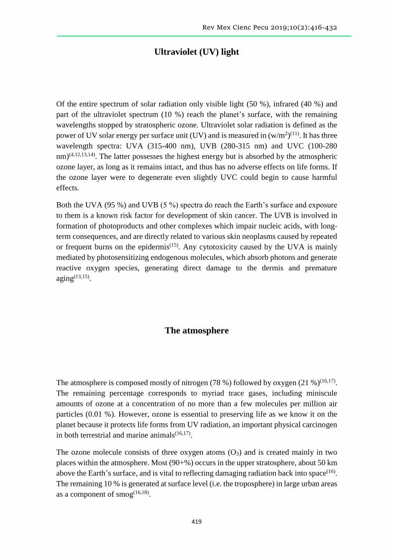
Of the entire spectrum of solar radiation only visible light (50 %), infrared (40 %) and
part of the ultraviolet spectrum (10 %) reach the planet’s surface, with the remaining
wavelengths stopped by stratospheric ozone. Ultraviolet solar radiation is defined as the
power of UV solar energy per surface unit (UV) and is measured in (w/m2)(11). It has three
wavelength spectra: UVA (315-400 nm), UVB (280-315 nm) and UVC (100-280
nm)(4,12,13,14). The latter possesses the highest energy but is absorbed by the atmospheric
ozone layer, as long as it remains intact, and thus has no adverse effects on life forms. If
the ozone layer were to degenerate even slightly UVC could begin to cause harmful
effects.
Both the UVA (95 %) and UVB (5 %) spectra do reach the Earth’s surface and exposure
to them is a known risk factor for development of skin cancer. The UVB is involved in
formation of photoproducts and other complexes which impair nucleic acids, with long-
term consequences, and are directly related to various skin neoplasms caused by repeated
or frequent burns on the epidermis(15). Any cytotoxicity caused by the UVA is mainly
mediated by photosensitizing endogenous molecules, which absorb photons and generate
reactive oxygen species, generating direct damage to the dermis and premature
aging(13,15).
The atmosphere
The atmosphere is composed mostly of nitrogen (78 %) followed by oxygen (21 %)(16,17).
The remaining percentage corresponds to myriad trace gases, including miniscule
amounts of ozone at a concentration of no more than a few molecules per million air
particles (0.01 %). However, ozone is essential to preserving life as we know it on the
planet because it protects life forms from UV radiation, an important physical carcinogen
in both terrestrial and marine animals(16,17).
The ozone molecule consists of three oxygen atoms (O3) and is created mainly in two
places within the atmosphere. Most (90+%) occurs in the upper stratosphere, about 50 km
above the Earth’s surface, and is vital to reflecting damaging radiation back into space(16).
The remaining 10 % is generated at surface level (i.e. the troposphere) in large urban areas
as a component of smog(16,18).
Rev Mex Cienc Pecu 2019;10(2):416-432
420
Since at least the mid-20th Century human activity has altered the ozone layer’s ecological
equilibrium by production and emission of what are known as “ozone depleting
substances” (ODS) into the atmosphere(1,19). The best known ODS are
chlorofluorocarbons (CFC), which were used in the manufacture of aerosols, refrigerators
and air conditioning equipment until banned in 1989. The CFC are extremely reactive;
for example, a single chlorine molecule can destroy a thousand ozone molecules(1,19).
Formation of ozone molecules is a slow process and as ODS concentrations have
increased in the atmosphere overall ozone concentration decreases until a new
equilibrium is reached between formation speed and degradation(16,17,19).
Solar radiation is one of the main environmental factors affecting life on the planet. It
governs the functioning of terrestrial and aquatic ecosystems through control of
photobiological processes (e.g. photosynthesis, photoperiod, phototropisms). These in
turn influence other environmental factors such as temperature, humidity and natural
cycles (daily, annual and hydric cycles) which finally affect organism distribution(19,20).
This makes life possible on Earth but can be detrimental to it at high intensities or when
the proportion of shortwave radiation surpasses certain limits. High intensity radiation
and changes in the spectral composition can affect significant processes in
organisms(19,21).
The quantity and quality of the radiation that reaches the Earth’s surface depends both on
the solar energy emitted and atmospheric characteristics at a given site. A wide range of
the electromagnetic spectrum reaches the surface with approximately 40 % being visible
light or radiation. These wavelengths range from 400 to 700 nm and are used by plants in
photosynthesis. Another range is the photobiological range, from 280 to 1,000 nm(21).
Both UVA and UVB penetrate the biosphere but only UVB is absorbed by atmospheric
ozone, meaning a decrease in ozone concentrations will allow a greater amount of UVB
to reach the surface. Only 1.3 % of the UV radiation emitted by the sun which reaches
the earth passes through the atmosphere, of which 98 % is UVA and 2 % is UVB.
Animal skin physiology
In animals the skin covers the organism surface and is in direct contact with the external
environment. It consists of three strata that harbor additional structures such as sweat and
sebaceous glands(22). Different species have developed supplementary forms of protection
(hair, wool, feathers) and/or keratinized tissues (nails, hooves). The skin protects against
mechanical, physical and chemical threats from the environment(22,23); for example, skin
thickness often increases at points regularly subjected to mechanical compression (e.g.
Rev Mex Cienc Pecu 2019;10(2):416-432
421
hooves, paws, hands and feet)(22). It is also relatively impervious to microorganisms and
many poisonous and noxious substances.
The skin protects against radiation(23), mainly solar radiation of different wavelengths.
For this reason in many animal species its superficial stratum, the epidermis, produces
pigments (melanin granules) that impede the penetration of radiations to deeper tissues.
An example is the skin of the polar bear which has white (refractory) fur and black
(protective) skin as an adaptation to an intense luminous environment subject to direct
solar radiation and indirect radiation reflected from ice and snow(24).
Sweat and sebaceous glands in the skin reach the surface through glandular ducts, making
this an excretory tissue(24). Water excretion (sweat) is a means of thermoregulation
unrelated to maintenance of organism hydric equilibrium, but rather controls animal
thermal conditions vis-à-vis the environment. Cutaneous sebum is a mixture of lipids
secreted by the sebaceous glands aimed at protecting the skin from moisture and
conferring it pliability and resistance(24).
The skin plays an important role in animal growth or somatic body development because
it is the primary storage area and activation site of vitamin D(23). Entering the organism
as D2 (ergocalciferol) or D3 (cholecalciferol), depending on its source, Vitamin D reaches
skin tissues via the blood. Here it is stored as calciferol or a precursor, and is transformed
into cholecalciferol by UV rays from the Sun. The cholecalciferol returns to the
circulatory system, passes through the liver and finally arrives in the kidneys where the
parathormone (PTH) effect transmutes it into the vitamin D hormone (1, 25-
dihydroxycholecalciferol). This hormone acts in the intestinal mucosa by stimulating
facultative absorption of calcium, thus preventing rickets(22).
Skin histology
Total cutaneous area varies by animal species; for example, in adult humans it is estimated
to be up to 2 m2. Certain portions of the skin of different animals generate specialized
formations such as hair, feathers, nails, horns, or hooves, and the presence of sweat and
sebaceous glands can range from numerous, to scarce or absent(25).
Skin thickness in a given organism can vary but is generally thicker on body dorsal
surfaces and limb lateral surfaces, and thinner on body ventral surfaces and limb medial
surfaces. These general trends can differ by species, breed and sex(22). In mammals, the
thinnest skin areas average from 0.4 mm in mice to 2.4 mm in Holstein dairy cows (Bos

Rev Mex Cienc Pecu 2019;10(2):416-432
422
taurus), while the thickest areas average from 1.9 mm in the domestic cat to 10.7 mm in
male horses(24).
The skin is divided into three strata: epidermis, the epithelial or surface stratum; dermis,
the connective or deep intermediate stratum; and hypodermis, the subcutaneous cellular
tissue (Figure 2)(25,26).
Figure 2: Layers of thick rat skin
Epidermis: consists of keratinized stratified layered epithelium; Dermis: connective tissue;
Hypodermis: fatty tissue. Technique: paraffin, hematoxilin-eosine(24).
Epidermis. This consists of keratinized stratified layered epithelium, and is generally
divided into five strata: stratum germinativum, stratum spinosum, stratum granulosum,
stratum lucidum and stratum corneum(25,26,27).
Dermis. This connective layer is divided into two regions, the papillary immediately
below the epidermis, and the deeper reticular. Named for the numerous papillae
projecting from it into the epidermis, the papillary region consists of a dense weave of
irregular lax fibrous connective tissue with trophic functions. Its thickness varies widely
between species, being thicker in ungulates than in carnivores(25-28).
Hypodermis. This layer is mostly connective tissue that adheres the skin to the bones and
muscles. Its primary function is to dampen external pressures and allow free movement
of the skin over underlying structures. Adipose tissue is present in this layer, from small
groups of cells to large masses in the form of pillows or fat pads. In temperate climes, the
hypodermis carries out a thermoregulatory function by increasing in thickness in the
winter to retain heat(24,25). Ancillary structures such hooves, nails, horns and spurs

Rev Mex Cienc Pecu 2019;10(2):416-432
423
originate in keratinization processes in the stratum corneum and have different
thicknesses and consistencies(24,25).
Hair. Hairs are epidermal formations that, in most mammals, are present over the entire
skin surface save on specialized skin tissues such as the pads of paws, palms of hands,
hooves, fingernails, part of the lips of the mouth, the glans, the inner surface of the
foreskin, the vulvar labia, the nipples and the contact surface of the limbs. A hair has a
root, a stem and a tip, which protrudes from the skin. Hair roots are surrounded by an
invagination of epidermal strata spinosum and germinativum into the dermis that reaches
the papillary region and thus accesses the blood vessels therein(25).
The cortical and medullary layers of hair occur in different proportions in different
species. For instance, the hair lining the skin of horses, bovines, dogs and pigs has a
thicker cortical layer than the hair of goats and cats. Also, the fine curly hairs of sheep
and pigs, manifest in hedgehogs or porcupines as sharp hairs known as thorns or barbs.
Hair is generally very fine in young animals and practically lacks marrow(24,25,28,29,30).
Adaptations of the skin in response to environmental conditions
Evolutionary, morphophysiological adaptation of skin to environmental conditions
involves the morphological peculiarities of the skin and its ability to allow thermal
adjustments to environmental variables and thus regulate organism temperature(28). An
excellent example is variation in skin thickness among and between bovine breeds.
Histological studies comparing Zebu cattle (Bos indicus), and Holstein breeds (Bos
Taurus) have found that skin thickness is not homogeneous across the body surface in the
same areas among animals of the same species but different breeds, and can even vary
with age. One study of 21 regions on the skin of Holstein-Friesian cattle (Bos taurus)
found that skin thickness as measured by skin fold changed within the same area and that
overall thickness increased with age. An analysis of different macro- and microscopic
skin structures in Holstein and Zebu cows found the Zebu to be better adapted to high
temperatures(28,31). This breed has shorter, thicker hair, greater overall skin thickness with
a thinner epidermis and a deeper reticulate dermis, a larger number of sweat glands
exhibiting dermal implantation, and consequently a greater excretory surface and
glandular density per area(28). In contrast Holstein-Friesian is a dairy breed characterized
by thinner skin with a thicker epidermis and finer reticulate dermis. Even the shape of
sweat glands differs between the breeds with sweat glands in the Holstein having a tubular
shape with varying degrees of torsion and those in the Zebu a more sack-like shape. In
the latter breed they are also more concentrated, ensuring more efficient heat dissipation
and thus greater tolerance to tropical temperatures(28,32).

Rev Mex Cienc Pecu 2019;10(2):416-432
424
How solar radiation affects animals
Animals exposed to solar radiation for long periods, that live at high altitudes and/or in
the tropics tend to lack pigment in the epidermis, have little hair or suffer hair loss and/or
are at higher risk of skin diseases(33-36). This occurs because UV rays damage cell DNA(33),
which induces the cyclobutane pyrimidine dimers (CPD), pyrimidine (6,4) and
pyrimidinone (6,4 PP), which cause negative effects such as inhibition of replication and
transcription, increased mutations, halting of the cell cycle and cell death(37). One disease
associated with these factors is squamous cell carcinoma (SCC) -also known as
epidermoid carcinoma(34)- a malignant tumor affecting keratinocytes in the
epidermis(35,36), that is locally invasive but not necessarily metastatic(33), but can
compromise the dermis(38).
Found mainly in bovine species, these tumors are most frequent in the Hereford,
Simmental and Holstein breeds, all of which have white skin without pigmentation,
particularly in the eyes(34,35). This condition causes heavy financial losses due to eye
cancer, known also as pink eye, which is common in these breeds(39). It is genetic in origin
but associated with UV exposure. Older individuals are most affected although younger
ones can also develop it, especially those with a white face and little pigment(39). Felids
and canids are also at risk(40,41), but it is uncommon in sheep and pigs(33,35,36). The most
sensitive horse breeds are Belgian, Clydesdale, Shire and Appaloosa, in which lesions
appear largely in muco-cutaneous regions (e.g. conjunctiva, vulva, perineum)(34).
Squamous cell carcinoma (SCC) occurs in 20 to 30 % of canids and 70 % of felids with
no differences by sex, mostly in large breeds and animals older than 10 yr(42). Lesions in
canids occur most frequently on the trunk, limbs, scrotum, lips and nail bed(38), while in
felids they are mostly on the face and ears of white-haired individuals(42).
Exposure to UV light can also cause melanocytomas, formed in melanocytes in the
epidermis, the cells that provide pigmentation to the skin, eyelashes and hairs(43). In
bovines, from 80 to 90 % of these tumors are benign, they are located mainly in the skin
of the extremities, are not age- or sex-dependent and are more prevalent in dark-colored
animals (grey, red and black)(43). Called melanomas in other domestic animals, they are
usually malignant, are common in canids and equids, and are rare in cats and other
species(41,43,44).
In dogs melanomas account for 4.7 % of all neoplasms and more than 7 % of malignant
tumors(44,45). They are most common in the mouth (56 %), followed by the lips (23 %),
skin (11 %), toes (8 %) and other locations (2 %), including the eyes(46). Cutaneous
melanomas are relatively frequent, but only 10 % of malignant melanomas are cutaneous
and these are largely found on the head and scrotum. Melanoma incidence in canids also
Rev Mex Cienc Pecu 2019;10(2):416-432
425
varies by breed, being more frequent in those with marked cutaneous pigmentation, such
as the Schnauzer or Scottish Terrier(45,46). The Irish Setter and Golden Retriever exhibit a
higher incidence of subnail melanomas, and the Irish Setter, Chihuahua, Golden Retriever
and Cocker Spaniel have a greater risk of labial melanomas(45,46). The German Shepherd
and Boxer have a higher risk of developing oral melanomas(47,48). Age at melanoma
appearance ranges from 1 to 17 yr with an average of 10. Incidence is higher in males
than females(47).
Melanoma is uncommon in cats (<1% of oral neoplasms and about 0.5 % of cutaneous
neoplasms)(49,50,51). Ocular and cutaneous melanomas are more common than intraoral
ones(52,53). The most common locations on the skin are the head, tail, distal extremities
and lumbar area(46,53,54). Prognosis is often poor given that half of cases exhibit recurrence
and regional metastases(46,54,55). Affected animals range in age from 2 to 18 yr, with a peak
between 8 and 12 yr(54,55). There is no apparent effect of sex or breed on frequency(54,55).
Another UV-related ailment is appearance of hemangiosarcomas, malignant tumors most
common in middle-aged and elderly dogs, especially large breeds such as the greyhound.
It affects mainly the spleen, the right atrium of the heart, the subcutaneous/dermal tissue
and the liver(56). Hemangiomas are also related to UV light exposure. These are relatively
benign neoplasms in the skin capillaries of the trunk, limbs and soft tissues, but are
frequently precursors to hemangiosarcomas(57).
Pathological effects of ultraviolet radiation
UVA radiation can induce erythema, immediate or delayed pigmentation, alterations of
the dermal connective tissue, release of vasoactive mediators, and photo-oxidative stress.
It can also exacerbate UVB erythema, carcinogenesis and elastosis, causing alterations in
DNA and other structures such as elastic fibers(58). Exposure to UVA is responsible for
many drug photosensitivity reactions, and plays a significant role in diseases such as
polymorphic light eruption, chronic actinic dermatitis, actinic reticuloid, lupus
erythematosus, solar urticaria, persistent reaction to light and xeroderma pigmentosum
(XP)(59,60). Experimental and clinical evidence have established a close causal relationship
between prolonged exposure to UV light and skin cancer, primarily malignant melanoma
(MM), squamous cell carcinoma (SCC) and basal cell carcinoma (BCC)(59,60).
Photosensitivity in animals is classified into three main types. Type I or primary
photosensitivity is caused by fluorescent compounds deposited undisturbed on the skin
after ingestion since a normal liver is unable to process and excrete the original
fluorescent compound. Photosensitizing compounds include hypericin, fagopyrin and

Rev Mex Cienc Pecu 2019;10(2):416-432
426
chemical products such as phenothiazine (sulfoxide phenothiazine). Type II
photosensitivity, also known as abnormal synthesis of endogenous pigment or congenital
porphyria is caused by accumulation of endogenous pigments from abnormal porphyrin
metabolism. Photodynamic agents include uroporphyrin I, coproporphyrin I, and
protoporphyrin III. These accumulate in the blood and tissues in response to dysfunction
in heme group biosynthesis due to an enzymatic deficiency. For example, congenital
protoporphyria in bovines is caused by a deficiency in uroporphyrinogen III cosynthetase,
a key enzyme in heme group biosynthesis. Type III or hepatotoxic photosensitivity is
more common and can have financial impacts. Animals become sensitized due to
accumulation of phylloerythryn, a product of chlorophyll digestion in the peripheral
circulatory system. Phylloerythryn is usually excreted in bile by the liver, but in certain
types of diffuse lesions, the liver is associated with a variety of vegetable, fungal, and
chemical hepatotoxins that are gradually absorbed by the circulatory system until
attaining levels that generate photosensitivity. This toxic matter acts directly on the cells
of the liver and small bile ducts, causing them to inflame and preventing passage of bile,
causing jaundice or yellow coloring(61).
Beneficial effects of ultraviolet radiation
One of the principal benefits of solar radiation is that it allows some homeothermic
animals to maintain proper internal body temperature for metabolism(62). Another well-
known benefit, particularly of UVB, is enabling vitamin D metabolism, indeed,
insufficient exposure can lead to vitamin D deficiency(62). This can have immediate
effects on the skeletal system by increasing the risk of fractures since vitamin D3 is
produced daily to control absorption, transport and deposit of calcium (and to a lesser
extent phosphorus), a vital function in bone maintenance and growth regulation. Vitamin
D is also necessary for hormonal functioning, organ development and embryogenesis(62).
Ultraviolet B radiation is important for animal health because it is required for the
photochemical processes involved in vitamin D synthesis(62). In other words, even if
animals receive an adequate diet and are in an optimum temperature, they will be unable
to correctly incorporate many nutrients if not provided with the radiation needed for
vitamin D production(62).
Irradiation is vital to food safety in that it contributes to preservation by preventing food
from degrading and spoiling, and the appearance of undesired conditions such as
emergence of tubers. It can also destroy some insects, fungi and bacteria(63).

Rev Mex Cienc Pecu 2019;10(2):416-432
427
Conclusions
Domestic animals are almost constantly exposed to ultraviolet radiation but changes in
climate may increase UVB radiation exposure with possible negative health
consequences. Sensitivity to exposure varies between species and even between breeds
within the same species. Many may develop cutaneous pathologies, including skin cancer,
which cause significant financial losses in the agricultural sector, undermine animal
health and well-being, and compromise the quality and safety of animal products intended
for human consumption.
Literature cited:
1. Toca TC. Impacto ambiental empresarial y fallas de la acción pública: una realidad
de las localidades bogotanas. Argos 2011;28:244-269.
2. Albanil E, Ramírez R, Jiménez B. Reporte del Clima en México http://smn.
cna.gob.mx/climatologia/analisis/reporte/Anual2014.pdf, Consultado 26 de Feb, 2015.
3. Alonso S, Ramírez N, Taylor PJ. El cambio climático y su impacto en la producción
de alimentos de origen animal. http: //www.veterinaria.org/revistas/redvet/
n111112.html, Consultado 29 de May, 2015.
4. Ferramola SA, Sancovich HA. Interacciones de las radiaciones electromagnéticas y
especies reactivas del oxígeno sobre la piel. Rev Argent Dermatol 2006;87:113-120.
5. Rivas AM, Rojas EE, Cortés NJ, Santander GE. Efecto de la altura en la radiación
solar ultravioleta en Arica Norte de Chile. Revista Facultad de Ingeniería, U.T.A.
2002;10:59-62.
6. Bohórquez BJ, Pérez JF. Radiación ultravioleta. Ciencia & Tecnología para la salud
visual y ocular. Rev Lasalle 2007;9:97-104.
7. Navas G, Jairo T, Rodrigo C. Non-Ionizing Radiation Detection. Dyna 2009;76:71-
81.
8. Guerreo AJ, Peréz AJ. Las radiaciones no ionizantes y su efecto sobre la salud
humana. Rev Cubana Med Milit 2006;35:3-11.

Rev Mex Cienc Pecu 2019;10(2):416-432
428
9. Pérez A, Miranda LR. Radiaciones electromagnéticas y salud en la investigación
médica. Rev Cubana Med Militar 2010;39:35-43.
10. Mignone C, Barnes R. More than meets the eye: the exotic, high-energy Universe.
Sci in School 2012;24:53-58.
11. Rivas A, Rojas E, Madronich S. Aumento del índice solar ultravioleta con la altura.
Rev Chilena Ing 2008;16:383-388.
12. Gonzáles PM, Vernhes TM, Sánchez LA. La radiación ultravioleta. Su efecto dañino
y consecuencias para la salud humana. Theoria 2009;18:69-80.
13. Vallejo EO, Vargas N, Martínez LM, Agudelo CA, Ortiz IC. Perspectiva genética
de los rayos UV y las nuevas alternativas de protección solar. http://www.scielo.
org.ar/scielo.php?script=sci_arttext&pid=S1851300X2013000300002Rev, Consultado
17 Sep, 2015.
14. Luigi PG, Rightler AL, Heronc M, Gabbuttc CD. Extending human perception of
electromagneticradiation to the UV region through biologically inspired photochromic
fuzzy logic (BIPFUL) systems. Chem Commun 2016;52:1474-1477.
15. Byrne SN, Hammond KJ, Chan CY, Rogers LJ, Beaugie C, Rana S, Marsh-
Wakefield F, Thurman JM, Halliday GM. The alternative complement component factor
B regulates UV-induced oedema, systemic suppression of contact and delayed
hypersensitivity, and mast cell infiltration into the skin. Photochem Photobiol Sci
2015;14:801-806.
16. Sánchez C. Consideraciones sobre la capa de ozono y su relación con el cáncer de
piel. Rev Méd Chile 2006;134:1185-1190.
17. Erickson DJ, Sulzberger B, Zeppc RG, Austind AT. Effects of stratospheric ozone
depletion, solar UV radiation, and climate change on biogeochemical cycling:
interactions and feedbacks. Photochem Photobiol 2015;14:127-148.
18. Environment Canada. The UV Index and Ozone http://www.ec.gc.ca/uv/
Default.asp?lang=En&n=C74058DD, Accessed Feb 16, 2015.
19. Bornman JF, Barnes PW, Robinson SA, Ballaré CL, Flinte SD, Caldwell MM. Solar
ultraviolet radiation and ozone depletion driven climate change: effects on terrestrial
ecosystems. Photochem Photobiol 2015;14:88-10.
20. Baiz AF, McKenzie RL, Bernhard G, Aucamp PJ, Ilyas M, Madronich S, Tourpali
K. Ozone depletion and climate change: impacts on UV radiation Photochem Photobiol.
Sci 2015;14:19-52.
21. Carrasco RL. Efecto de la radiación ultravioleta–B en plantas. IDESIA Chile
2009;3:59-76.

Rev Mex Cienc Pecu 2019;10(2):416-432
429
22. Castellanos I, Clarena G, Rodríguez T, Iregui G. Estructura histológica normal de la
piel del perro. Rev Med Vet 2005;10:104-122.
23. Vega PC. Manual de Histología Esquemática. 1ra ed. La Habana, Cuba: Editorial
Pueblo y Educación; 1980.
24. Dellmann HDB. Histología veterinaria. 2a ed. España: Editorial Acribia, Zaragoza;
1994.
25. Megías M, Molist P, Pombal MA. Atlas de histología vegetal y animal. Órganos
animales tegumento, https://mmegias.webs.uvigo.es/descargas/o-ategumento.pdf,
Consultado 24 Feb, 2016.
26. Paniagua R, Nistal M, Sesma P, Álvarez U, Fraile M, Anadón B, Sáez RFS, De
Miguel MP. Citología e histología vegetal y animal. 2a ed. España: Editorial McGraw-
Hill-Interamericana de España; 1997.
27. Ross MH, Pawlina W. Histología Texto y Atlas. 7ª ed. Barcelona, España: Editorial
Wolters Kluwer; 2016.
28. Esquivel VC. La raza, el pelo y la piel en función del bienestar animal. Mundo
Pecuario 2012;1:73-85.
29. Suro RJ, Gutiérrez FL, Ruiz AJ, Bouhann P. El pelo. Generalidades y funciones.
Dermatología CMQ 2007;4:218-223.
30. William J. Applied Veterinary. 2a ed. USA: Editorial Mosby; 1993.
31. Kamwanja LA, Chase CC, Gutierrez JA, Guerriero V, Olson TA, StatusHammond
AC, Hansen PJ. Responses of bovine lymphocytes to heat shock as modified by breed
and antioxidan. J Anim Sci 1994;72:438-444.
32. Pérez EH. Fisiología Animal II. http://cenida.una.edu.ni/relectronicos/RENL
50P438.pdf. Consultado 28 Sep, 2016.
33. Rosolem MC, Moroz LR, Rodigheri SM. Carcinoma de células escamosas. Pubvet
2012;6:6-14.
34. Ramos AT, Mollerke ND, Elias FM, Gevehr FC. Carcinoma de células escamosas
em bovinos, ovinos e eqüinos: estudo de 50 casos no sul do Rio Grande do Sul. Braz Vet
Res Anim Sci 2007;44:5-13.
35. Fabricio KL, Dantas AF, Riet CF, De Miranda NE, Simões SV, Azevedo SS. Fatores
de risco associados à ocorrência de carcinoma de células escamosas em ruminantes e
equinos no semiárido da Paraíba. Pesq Vet Bras 2012;32:881-886.

Rev Mex Cienc Pecu 2019;10(2):416-432
430
36. Targino J, Almeida S, Macedo E, Almeida BE, Santana de OR, Anjos FE, Ocampos
PPM. Squamous cell carcinoma in the frontal region of the head in a goat. Acta Scient
Vet 2013 41:4-8.
37. Tafurt Y, Marin MA. Principales mecanismos de reparación de daños en la molécula
de ADN. Rev Biosalud 2014;132: 95-110.
38. Costa C, Ramos VD, Huppes R, Bardoza DA, Gaspar RA, Rivera CL, Ramirez UR.
Cryosurgery in the treatment of squamous cell carcinoma in dog. Rev Colomb Cienc
Anim 2013;5:213-221.
39. Heather ST. Combata el cáncer de ojo. Hereford, Bs.As. 1999;(64):38-40.
40. Olinda RG, Frade MT, Soares GS, Aguiar GM, De Costa VM, Lucena RB, De
Dantas AF. Bovine demodicosis associated with squamous cell carcinoma of the vulva.
Acta Scient Vet 2013;41:29-32.
41. Mukaratirwaa S, Chipunzaa J, Chitangaa S, Chimonyoa M, Bhebhea E. Canine
cutaneous neoplasms: prevalence and influence of age, sex and site on the presence and
potential malignancy of cutaneous neoplasms in dogs from Zimbabwe. J S Afr Vet Ass
2005;76:59–62.
42. Revista de la Asociación Madrileña de Veterinarios de Animales de Compañía.
https://es.scribd.com/document/26893256/Revista-de-La-Asociacion-Madrilena-de-
Veterinarios, Consultada 14 Ene, 2010.
43. Chávez VC, Astaiza MJ, Benavides MC, Vallejo TD. Tumor maligno derivado de
melanocitos en piel de un bovino de presentación inusual: estudio de caso. Rev Med Vet
2015;29:63-72.
44. Iakatos I, Mirela E, Cadar, Ali B. Cutaneous tumors’ incidence in dog. Lucrări
Stiinłifice Medicină Veterinară 2009;13:475-381.
45. Silva HG, Juárez BF, López VM, Dávila PM. Squamous cell carcinoma in dogs from
Culiacan, Sinaloa, Mexico Retrospective Rev (2006-2014). Rev Cient, FCV-LUZ
2015;25:573-584.
46. Goldschmidt MH, Shofer FS. Skin tumors of the dog and cat. Oxford, UK:
Butterworth Heinemann; 1992;2:142-151.
47. Todoroff RJ, Brodey RS. Oral and pharyngeal neoplasia in the dog: a retrospective
survey of 361 cases. J Am Vet Med Assoc 1979;175:567-571.
48. Oakes MG, Lewis DD, Hedlund CS, Hosgood G. Canine oral neoplasia. Contin Educ
Pract Vet Comp 1993;15:15-30.

Rev Mex Cienc Pecu 2019;10(2):416-432
431
49. Patnaik AK, Mooney S. Feline melanoma: a comparative study of ocular, oral and
dermal neoplasms. Vet Pathol 1988;25:105-112.
50. Stebbins KE, Morse CC, Goldschmidt MH. Feline oral neoplasia: a ten-year survey.
Vet Pathol 1989;26:121-128.
51. Miller WH, Scott DW, Anderson WI. Feline cutaneous melanocytic neoplasms: a
retrospective analysis of 43 cases (1979-1991). Vet Dermatol 1993;4:19-26.
52. Engle CG, Brodey RS. A retrospective study of 395 feline neoplasms. J AM Anim
Hosp Assoc 1969;5:21-31.
53. Day MJ, Lucke VM. Melanocytic neoplasia in the cat. J Small Anim Pract
1995;36:207-213.
54. Van Der- Linde SJS, De- Wit MML, Van- Garderen E, Molenbeek RF, Van Der-
Velde ZD, De- Weger RA. Cutaneous malignant melanomas in 57 cats: identification of
(amelanotic) signet-ring and balloon cell types and verification of their origin by
immunohistochemistry, electron microscopy, and in situ hybridization. Vet Pathol
1997;34:31-38.
55. Luna LD, Higginbottham ML, Henry CJ, Turnquist SE. Feline non-ocular
melanoma: a retrospective study of 23 cases (1991-1999). J Feline Med Surg 2000; 2:173-
181.
56. Weinborn AR, Issotta CC, Agurto MM, Lara LJ. Descripción clínica de
hemangiosarcoma (HSA) cutáneo metastásico en un canino galgo: estudio clínico de un
caso. Rev Med Vet 2015;2:107-116.
57. Aita N. Hemangioma of the ileum in a dog. J Vet Med Sci 2010;72:1071-1073.
58. López CC, Aréchiga OE, López CM. Protein kinases and transcription factors
activation in response to UV-radiation of skin: Implications for carcinogenesis. Int J Mol
Cienc 2012;13:142-172.
59. Duro ME, Campillos P, Causín S. El sol y los filtros solares. Medifam 2003;13: 159-
165.
60. Alvarez FE. Consecuencias del estrés oxidativo de la piel por radiaciones
ultravioleta.http://scielo.sld.cu/scielo.php?script=sci_arttext&pid=S0864030019950001
00004, Consultado 10 Nov, 2016.
61. Cruz CF. Fotosensibilización. http://www.ammveb.net/clinica/fotosensibilizacion.
pdf, Consultado 20 Oct, 2016.

Rev Mex Cienc Pecu 2019;10(2):416-432
432
62. Fernández L, Poletta GL, Imhof A, Siroski PA. Efectos de la radiación ultravioleta
natural y artificial (UVA/ UVB) sobre la concentración plasmática de calcio y fósforo y
el crecimiento en crías de Caiman latirostris. InVet 2013;15:75-82.
63. Ross LI, Watson D, Escandarani S, Miranda A, Troncoso A. La radiación a la mesa.
Rev Chilena Infect 2009;26:318-330.