effectsof momordica charantia extractontheexpressionof mdr ... · drug efflux analysis was carried...
TRANSCRIPT
WIMJ Open 2014; 1 (3): 92
ORIGINALARTICLE
Effects of Momordica charantia Extract on the Expression of MDR 1 Gene in HumanLung Cancer Cells
T Yuan, H-Y Li
ABSTRACT
Objectives: Multi-drug resistance (MDR) is a major hurdle in treatment of cancer, contributing to thefailure of chemotherapy. Drug resistance is found to be linked to the overexpression of ATP-bindingcassette (ABC) drug transporter proteins that include P-glycoprotein (P-gp), causing a reduction indrug accretion inside the cancer cells. In the present study, the effect of the extracts from the fruit peeland pulp of Momordica charantia (MCFPE) fruit in modulating the function of P-gp in human small-cell lung cancer (SCLC) cell lines was assessed.Methods: The effects of MCFPE were tested on drug-sensitive (H69) and multi-drug resistant(H69/LX4) human SCLC cells. The cell survival percentage was assessed by MTT cytotoxicity assay.The percentage of drug accumulation and drug efflux were assessed by using [3H]-paclitaxel. Theexpression of MDR1 gene was analysed by reverse transcription polymerase chain reaction (RT-PCR),and P-gp by western blot analysis.Results: The extract was able to induce death of cancer cells as measured by cell survival percentageas well as improve drug accumulation, as evidenced by intracellular paclitaxel retention. Priorexposure of cells to MCFPE reversed resistance to paclitaxel. Treatment with MCFPE was found tohave a significant impact on MDR 1 gene expression in H69/LX4 cell line by decreasing its expression.The extract had no influence on expression of MDR 1 gene in the drug-sensitive SCLC cell lines.Western blot analysis of P-gp protein in H69 and H69/LX4 cells revealed that the treatment with theextract modulates the expression of MDR 1 in H69/LX4 and had negligible effect on H69 cells.Conclusion: The results indicate that MCFPE was able to effectively reverse multi-drug resistance andimprove cancer chemotherapy.
Keywords: Lung cancer, Momordica charantia, multi-drug resistance, P-glycoprotein
WIMJ Open 2014; 1 (3): 92
INTRODUCTIONLung cancer is one of the most common type of cancers anda major cause of cancer-related deaths globally. Small-celllung cancer (SCLC), an aggressive cancer, accounts fornearly 10−15% of all lung cancers (1). Chemotherapy withcombinations involving etoposide, doxorubicin, cisplatin andvincristine are employed in the treatment of patients withSCLC (2). The dearth of chemotherapy regimen is the de-velopment of multi-drug resistance (MDR) that leads toineffectiveness of the therapy. In recent years, cancer re-
From: Department of Respiratory Diseases, the Fifth Affiliated Hospital ofZhengzhou University, Zhengzhou 450052, China.
Correspondence: Dr H-Y Li, Department of Respiratory Diseases, the FifthAffiliated Hospital of Zhengzhou University, No. 3 Kangfu Road,Zhengzhou, Henan 450052, China. E-mail: [email protected]
DOI: 107727/wimjopen.2014.147
search has been focussed on the understanding of the funda-mental mechanisms that impart drug resistance.
Multi-drug resistance has been observed to be medi-ated by several mechanisms such as alterations in the apop-totic proteins and pathways, change in the activity of specificenzyme systems involved, active efflux of the chemothera-peutic drugs by ATP-binding cassette (ABC) transporters andothers (3). Among the mechanisms that are proposed toinitiate MDR, overexpression of ABC transporter proteinshas received extensive attention (4).
P-glycoprotein (P-gp), a 170-KD transmembrane gly-coprotein encoded by the human MDR 1 (ABCB1) gene, is amember of the family of ABC transporters. It acts as anATP-driven efflux pump that decreases intracellular drugaccumulation, thereby decreasing the effectiveness of manychemotherapeutic agents (5, 6).

93
The ATPase domains of P-gp are induced by the pre-sence of transport substrates such as vinca alkaloids (vin-cristine, vinblastine), anthracyclines (doxorubicin, daunoru-bicin, epirubicin), epipodophyllotoxins (etoposide, tenipo-side) and taxanes (7, 8). Thus it would be effective if theactivity of P-gp mediated drug efflux could be inhibitedwhich may resensitize MDR. As the P-gp efflux pump is anATP-dependent transport system (9), compounds that areeffective inhibitors of ATP-dependent drug transport shouldmediate the inhibition of drug resistance and increaseintracellular accumulation.
Recent research is now focussing on natural productsand plant-derived compounds as MDR-reversing agents. Anumber of studies demonstrate that natural products derivedfrom plants are potential drugs to overcome MDR in manymulti-drug resistant cells (10−12). These studies suggest thatplant extracts and derived compounds could be potent agentsin reversing MDR and also be chemotherapeutic.
Momordica charantia L (MCFPE; commonly knownas bitter melon) is widely consumed as a vegetable. It is alsoconsumed as a folk medicine in Asia. Extracts of bittermelon have been reported to possess antioxidant activities(13), antiviral (14), antidiabetic and immunomodulating pro-perties (15). The present study was undertaken to investigatethe effect of the hot water extracts of the peel and pulp of thefruit of Momordica charantia on human lung cancer celllines.
MATERIALS AND METHODSChemicals[3H]-paclitaxel (37.9 Ci/mmol) was purchased from MoravekBiochemicals, Inc (Brea, CA, USA). Verapamil, RoswellPark Memorial Institute (RPMI) medium, fetal bovine serum(FBS), glutamine, 3-(4,5-dimethylthiazol-2-yl)-2,5-diphenyltetrazolium bromide (MTT) HRP-conjugated goat anti-mouse IgG and the mono-clonal mouse antibody against P-gp, agarose were obtained from Sigma Aldrich (St Louis,MO, USA). All other chemicals and reagents used were ofanalytical grade.
Preparation of bitter melon extractsThe whole fruit of the freshly harvested bitter melon from alocal farm was collected, washed well and cleaned. The peeland the pulp alone were collected. The peel and pulp of thebitter melon were shade dried at 30−40 °C. The thoroughlydried samples were finely powdered and extracted with hotwater. The powdered sample of 100 g was extracted rigor-ously with two litres water at 100 °C for six hours. Theconcoction was filtered and re-extracted with four litres ofwater. The combined filtrate was re-filtered and concentratedby rotary evaporation and lyophilized to dryness. TheMCFPE extract was dissolved in dimethyl sulfoxide(DMSO) and used for experimental purposes, with the finalDMSO concentration being 0.1% (v/v) in the culturemedium.
Cells and cell cultureDrug-sensitive H69 and multi-drug resistant cell line NCI-H69/LX4 (procured from Sigma Aldrich) were cultured inRPMI medium supplemented with 10% FBS, 2 mMglutamine and 0.4 µg/mL doxorubicin (only for NCI-H69/LX4) at 37 °C in suspension culture in an atmospherewith 5% CO2. NCI-H69/LX4 has been established byexposure of the parent line H69 to doxorubicin. The cell linehyper-expresses P-glycoprotein and is resistant to doxo-rubicin and taxanes. When the cells reached around 80%confluence, they were harvested and plated for subsequentpassages or for drug treatments.
Cytotoxicity assayH69 and H69/LX4 cells were plated at 3.0 × 10³ cells perwell in 96-well plates. After 24 hours, various concentrationsof MCFPE and paclitaxel were added and the cells wereincubated for 72 hours at 37 °C. The cell growth wasassessed by means of an MTT colorimetric assay (16). Ineach experiment, cytotoxicity assessment was carried out intriplicates.
Measurement of [3H]-paclitaxel accumulationAccretion of paclitaxel in H69 and H69/LX4 cells weremeasured using [3H]-paclitaxel as described previously (17,18). The confluent cells in 24-well plates were pre-incubatedwith verapamil (standard compound that is a known inhibitorof P-gp) and MCFPE for one hour at 37 °C. In order toevaluate the drug accumulation, the cells were then incubatedwith 0.1 µM [3H]-paclitaxel in the presence as well as ab-sence of 50 µM verapamil and MCFPE for about two hoursat 37 °C. The cells were then washed with ice-cold phos-phate buffered saline (PBS), trypsinized and the accumu-lation of paclitaxel was measured in terms of measure ofradioactivity. Radioactivity was assessed by using PackardTRI-CARB 1900CA liquid scintillation counter (PackardInstrument, Inc).
Analysis of [3H]-paclitaxel effluxTo measure drug efflux, the cells were treated in a similarmanner as in the drug accumulation experiment; the cellswere then incubated for 0, 60 and 120 minutes in the freshmedium at 37 °C in the presence or absence of the reversalagents. After 120 minutes, the cells were then washed thricewith ice-cold PBS followed by trypsinization. The cells werethen placed in scintillation fluid to measure the radioactivityin the same way as measurement of drug accumulation.Simultaneously treated, a duplicate set of 24-well plates wereused for cell counting.
RNA extraction and reverse transcription polymerasechain reactionTotal RNA was isolated from H69 and H69/LX4 cells treatedfor 72 hours with MCFPE as well as from untreated control
Yuan and Li
94
cells. RNA was isolated using TRIzol (Sigma Aldrich, USA).The isolated RNA was quantified by spectrophotometry andanalysed in 1% agarose gel.
Reverse transcription (RT) reactions using 2.5 µg totalRNA were performed with oligo-dT primers with M-MLVreverse transcriptase (Sigma Aldrich, USA). The primersspecific for MDR 1 were employed (19−21). The referencegene for the normalization of target gene expression wasGAPDH (glyceraldehyde-3-phosphate dehydrogenase).
Polymerase chain reaction (PCR) reactions were per-formed in GeneAmp® PCR System 9700 (AppliedBiosystems, USA). The products were separated in 2%agarose gel and were stained with ethidium bromide. Thegels were visualized under ultraviolet (UV) light and thedensitometry analysis was carried out using Bio-Rad GelDoc 1000 systems.
Western blot analysis of P-glycoproteinPlasma membrane from the cells treated with MCFPE at0.1% for 72 hours was prepared according to the methoddescribed by Anuchapreeda et al (5). The protein contentwas measured by the method of Lowry et al (22), usingbovine serum albumin (BSA) as a standard. Equal amountsof total cell lysates (20 µg of protein/lane) were separated ina 7.5% SDS-PAGE and electrophoretically transferred ontonitrocellulose filters. The filters were incubated sequentiallywith mouse monoclonal anti-P-gp (23) at dilution of 1:5000and HRP conjugated goat anti-mouse IgG at a dilution of1:20 000. Proteins were visualized using a SuperSignal pro-tein detection kit and quantified by scanning densitometry.
Statistical analysisThe results observed are presented as mean ± SD from threeindividual experiments. Differences between the means wereanalysed by one-way analysis of variance (ANOVA) andStudent t-test. Statistical significance was considered whenp < 0.05. The statistical analyses were performed using SPSS10.0 software.
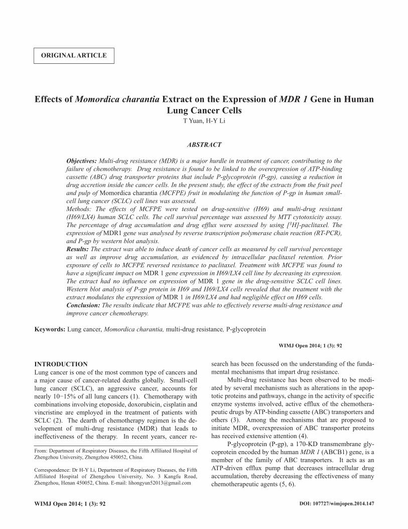
RESULTSEffect of MCFPE on cytotoxicity of paclitaxel in H69 andH69/LX4 cellsTo investigate the efficacy of MCFPE on reversing MDR, theeffect of MCFPE on the SCLC cells treated with paclitaxelwas determined by quantifying the growth inhibition on thecells. The results showed that MCFPE at 0.05%, 0.1% and0.15% v/v increased sensitivity of H69/LX4 cells to pacli-taxel as indicated by a decrease in the cell survival per-centage, while similar treatment on H69 cells provided nosignificant modulating effect. The results obtained weresimilar on treatment of the cells with 20 µM verapamil (Fig.1A and B). The highest concentration of the extract was mosteffective in inhibiting cell survival in SCLC cells.
Measurement of [3H]-paclitaxel accumulationTo determine the effects of MCFPE on the function of P-gpas a drug-efflux pump, the accumulation of P-gp substrate[3H]-paclitaxel in the presence or absence of plant extractwas measured. The data obtained indicated that the intra-cellular concentration of [3H]-paclitaxel in P-gp-overex-pressing H69/LX4 cells was significantly lower than that inH69 cells (Fig. 2) in the absence of MCFPE or verapamil.After the cells were incubated with MCFPE at 0.05% and0.1% in DMSO and the standard P-gp inhibitor verapamil fortwo hours, the intracellular [3H]-paclitaxel accumulation wassignificantly increased in drug resistant cell line, and MCFPEat 0.1% concentration resulted in highest increase in theintracellular [3H]-paclitaxel which was comparable to that of10 µM of verapamil. However, the intracellular level of [3H]-paclitaxel as observed in H69 cells was not significantlyaltered by either MCFPE or verapamil (Fig. 2).
Effects of Momordica charantia Extract on MDR in Human Lung Cancer Cells
Fig. 1: Effect of Momordica charantia (MCFPE) on paclitaxel cyto-toxicity in the H69 (A) and H69/LX4 (B) cell lines. The small-celllung cancer (SCLC) cells were incubated with variousconcentrations of paclitaxel in the presence or absence of MCFPEand verapamil (VPL). The cell survival percentage with varioustreatments is marked. The values are expressed as mean ± SD fromthree individual experiments.

95
Effects of MCFPE on the efflux of [3H]-paclitaxelDrug efflux analysis was carried out in order to establish ifMCFPE was able to increase the drug accumulation via inhi-biting P-gp. H69/LX4 cells were observed to release intra-cellular [3H]-paclitaxel, with increasing time, compared toH69 cells. Upon incubation of the cells with verapamil and0.1% MCPFE, the drug efflux was significantly reducedwhen measured at different time intervals (0, 30, 60, 120minutes) as against the cells not treated with MCFPE orverapamil.
The percentage efflux of the drug [3H]-paclitaxel attime zero minutes was 0%. The percentages of the drugefflux at 30, 60 and 120 minutes were 31.31 ± 1.66%, 43.32± 2.14 % and 62.08 ± 1.26%, respectively, in the H69/LX4cells in the absence of plant extract or verapamil. H69/LX4on incubation with MCFPE at 0.1% of the efflux of the drugwas found to be 20.12 ± 21%, 31.19 ± 2.04% and 39.88 ±2.10% at 30, 60 and 120 minutes, respectively (Fig. 3B). Ontreatment with verapamil, the drug efflux in the H69/LX4cells was observed as 11.21 ± 0.52%, 15.39 ± 0.94%, and19.12 ± 0.70% at 30, 60 and 120 minutes, respectively. InH69 SCLC cells, there was no significant modulation in theaccumulation of paclitaxel in the presence or absence of plantextract or verapamil (Fig. 3A).
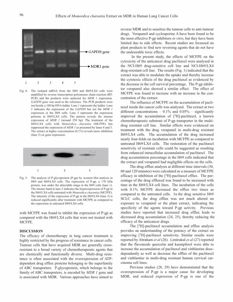
Effect of MCPFE on the expression of MDR 1 gene in theSCLC cellsMDR 1 gene was amplified in order to study the level ofexpression in the SCLC cells. Treatment with MCFPE wasfound to have a significant impact on MDR 1 expression inthe H69/LX4 cell line, decreasing its expression (Fig. 4).
Yuan and Li
Fig. 2: Effect of Momordica charantia (MCFPE) on the accumulation of[3H]-paclitaxel in NCI-H69 and NCI-H69/LX4cells. The accumu-lation of [3H]-paclitaxel was measured after preincubation with orwithout the reversal agents (verapamil (VPL) and MCFPE) for onehour at 37 °C and then incubation with 0.1 µM [3H]-paclitaxel inthe presence or absence of the reversal agents for two hours at 37°C. Verapamil was used as a positive control. The values arepresented as mean ± SD from three individual experiments.*indicates statistical significance at p < 0.05 for values versus thosein the control group in H69/LX4 cells.
Fig. 3: Effect of Momordica charantia (MCFPE) on the efflux of [3H]-paclitaxel from H69 (A) and H69/LX4 (B) cells. The small-celllung cancer (SCLC) cells were treated with MCFPE and verapamil(VPL) and the levels of [3H]-paclitaxel that was effluxed out weredetermined at time periods of 0, 30, 60 and 120 minutes. Controlrepresents SCLC cells untreated with MCPFE or VPL. Values arerepresented as mean ± SD from three individual experiments.*indicates statistical significance at p < 0.05 for values versus thosein the control group in H69/LX4 cells.
Each reaction was performed in duplex since a reference gene GAPDH (glyceraldehyde-3-phosphate dehydrogen-ase) gene expression was included. The extract had no influence on the expression of the MDR 1 gene in the drug-sensitive SCLC cell lines.
Influence of MCFPE on P-gpReversal of P-gp-mediated MDR could be achieved by eitherdecreasing P-gp expression or by inhibiting P-gp function. Toevaluate the effect of MCFPE on P-gp expression, H69/LX4cells were treated with MCFPE for 72 hours. Westernblotting analysis was performed after separation of theproteins by SDS-PAGE. The P-gp antibody was employed todetect the expression levels of P-gp. The result shown in Fig.5 indicates that the extract had no effect on the proteinexpression levels in the drug-sensitive H69 cells. Treatment
96
with MCFPE was found to inhibit the expression of P-gp ascompared with the H69/LX4 cells that were not treated withMCFPE.
DISCUSSIONThe efficacy of chemotherapy in lung cancer treatment ishighly restricted by the progress of resistance in cancer cells.Tumour cells that have acquired MDR are generally cross-resistant to a broad variety of chemotherapeutic agents thatare chemically and functionally diverse. Multi-drug resis-tance is often associated with the overexpression of ATP-dependent drug efflux proteins belonging to the superfamilyof ABC transporters. P-glycoprotein, which belongs to thefamily of ABC transporters, is encoded by MDR 1 gene andis associated with MDR. Various approaches have aimed to
reverse MDR and to sensitize the tumour cells to anti-tumourdrugs. Verapamil and cyclosporine A have been found to bethe most effective P-gp inhibitors in vitro, but they have beenlimited due to side effects. Recent studies are focussed onplant products to find new reversing agents that do not havethe undesirable toxic effects.
In the present study, the effects of MCFPE on thecytoxicity of the anticancer drug paclitaxel were analysed inthe NCI-H69 drug-sensitive cell line and NCI-H69/LX4drug-resistant cell line. The results (Fig. 1) indicated that theextract was able to modulate the uptake and thereby increasethe cytotoxic effects of the drug paclitaxel as evidenced bythe decrease in the cell survival percentage. The P-gp inhibi-tor verapamil also showed a similar effect. The effect ofMCFPE was found to increase with an increase in the con-centration of the extract.
The influence of MCFPE on the accumulation of pacli-taxel inside the cancer cells was analysed. The extract at twodifferent concentrations – 0.1% and 0.05% – significantlyimproved the accumulation of [3H]-paclitaxel, a knownchemotherapeutic substrate of P-gp transporter in the multi-drug resistant cell line. Similar effects were evidenced ontreatment with the drug verapamil in multi-drug resistantH69/LX4 cells. The accumulation of the drug increasednearly four-folds on incubation with MCFPE as compared tountreated H69/LX4 cells. The restoration of the paclitaxelsensitivity of resistant cells could be suggested as resultingfrom enhanced intracellular accumulation of paclitaxel. Thedrug accumulation percentage in the H69 cells indicated thatthe extract and verapamil had negligible effects on the cells.
The drug efflux analysis at different time intervals (30, 60 and 120 minutes) were calculated as a measure of MCFPE efficacy in inhibition of the [3H]-paclitaxel efflux. The per-centage of the drug effluxed was found to be increased with time in the H69/LX4 cell lines. The incubation of the cells with 0.1% MCFPE decreased the efflux two times as compared to the untreated cells. In the drug sensitive H69 SCLC cells, the drug efflux was not much altered on exposure to verapamil or the plant extract, indicating the specificity of the agents toward P-gp activity. Previous studies have reported that increased drug efflux leads to decreased drug accumulation (24, 25), thereby reducing the efficacy of the anticancer drugs.
The [3H]-paclitaxel accumulation and efflux analysisprovides an understanding of the potency of the extract onimproving [3H]-paclitaxel sensitivity. Similar results werereported by Abraham et al (26). Limtrakul et al (27) reportedthat the flavonoids quercetin and kaempferol were able toincrease the accumulation of paclitaxel and vinblastine dose-dependently as well as decrease the efflux of the paclitaxeland vinblastine in multi-drug resistant human cervical car-cinoma cell lines.
Previous studies (28, 29) have demonstrated that theoverexpression of P-gp is a major cause for developingMDR, and reduced expression of P-gp is one of the
Effects of Momordica charantia Extract on MDR in Human Lung Cancer Cells
Fig. 4: The isolated mRNA from the H69 and H69/LX4 cells wereamplified by reverse transcription polymerase chain reaction (RT-PCR) and the products were analysed for MDR 1 expression.GADPH gene was used as the reference. The PCR products wererun beside a 100 bp DNA ladder. Lane 1 represents the ladder. Lane2 indicates the expression of the GAPDH but not the MDR 1expression in the H69 cells. Lane 3 represents the expressionpatterns in H69/LX4 cells. The pattern reveals the intenseexpression of MDR 1 (around 250 bp). The treatment of theH69/LX4 cells with Momordica charantia (MCFPE) hassuppressed the expression of MDR 1 as presented by lanes 4 and 5.The extract at higher concentration (0.1%) reveals more inhibition(lane 5) on gene expression.
Fig. 5: The analysis of P-glycoprotein (P-gp) by western blot analysis inH69 and H69/LX4 cells. The expression of P-gp, a 170 kDaprotein, was under the detectable range in the H69 cells (lane 1).The intense band in lane 2 indicates the hyperexpression of P-gp inthe H69/LX4 cells untreated with Momordica charantia (MCFPE).The intensity of the expression of P-gp in the H69/LX4 (lane 3) isreduced significantly after treatment with MCFPE as compared tothe expression in untreated H69/LX4 cells.

97
modalities in reversing multi-drug resistance. In this study, the expression levels of P-gp were analysed both at the transcriptional and at the protein levels. The mRNA isolated from the SCLC cell lines was amplified by RT-PCR and the result of the analysis, presented in Fig. 4, reveals the expression patterns. The band density was observed to be significantly reduced in the H69/LX4 cells on exposure to MCFPE as compared to the untreated H69/LX4 cells. The band in lane 2 reveals the nearly nil expression of P-gp mRNA in the H69 cells that further supports that MDR 1 gene overexpression contributes to MDR. The decrease in the intensity of the expression band in lanes 4 and 5 (H69/LX4 cells treated with MCFPE) suggests that the extract harbours the potency to reduce the expression of MDR 1 gene. The results are in line with the recent studies with the flavonoid quercetin. Quercetin is capable of decreasing P-gp expression in a dose-dependent manner (27).
Western blot analysis showed that the 170 kDa P-gpwas expressed in H69/LX4 cells untreated with MCFPE(lane 2). The expression of P-gp in the H69 cells was farbelow the detectable level (lane 1). Upon exposure toMCFPE, the P-gp expression of the H69/LX4 cells wasremarkably reduced as seen in lane 3 (Fig. 5). The observedresults further support the capacity of the extract inmodulating MDR by targeting the expression and activity ofP-gp.
The reduction of MDR 1 expression probably at bothtranslational and transcriptional levels has been put forth asone of the possible means by which some compounds reverseMDR (3).
Thus, in this study, our results showed that MCFPE distinctly raised the cytotoxicity of paclitaxel in paclitaxel-resistant P-gp overexpression observed in H69/LX4 cells but faintly in paclitaxel-sensitive H69 cells. This indicates that MCFPE may act by reverses MDR by mediating over-expressed P-gp that is further supported by the results of [3H]-paclitaxel accumulation and efflux analysis. Further-more, as reported by Krishna and Mayer (3), the results of the RT-PCR and western blotting suggest that MCPFE acts at inhibiting the expression of MDR 1 and thus reverses MDR. Similar results were obtained on treatment of drug-resistant cancer cells by cucurmin (30).
Much effort is being taken recently toward identifying natural compounds from plant origins that inhibit P-gp, reverse the MDR phenotype, and sensitize cancer cells to conventional chemotherapy (31, 32). The present study thus is an attempt to suggest that phytochemicals could suppress MDR. In this study, it could be suggested that the secondary metabolites present in the extracts of Momordica charantia were able to potentially modulate MDR.
In conclusion, the extract of the peel and pulp of Momordica charantia fruits represent potential reversal agents for the treatment of MDR in P-gp-overexpressing SCLC tumours.
REFERENCES1. Govindan R, Page N, Morgensztern D. Changing epidemiology of
small-cell lung cancer in the United States over the last 30 years:analysis of the surveillance, epidemiologic, and end results database. JClin Oncol 2006; 24: 4539−44.
2. Pass HI, Carbone DP, Johnson DH, Minna JD, Scagliotti G, Turrisi AT,eds. Principles and Practice of Lung Cancer. Philadelphia: LippincottWilliams Wilkins; 2010.
3. Krishna R, Mayer LD. Multidrug resistance (MDR) in cancer—mechanisms, reversal using modulators of MDR and the role of MDRmodulators in influencing the pharmacokinetics of anticancer drugs.European J Pharma Sci 2000; 11: 265–83.
4. Litman T, Druley TE, Stein WD, Bates SE. From MDR to MXR: newunderstanding of multidrug resistance systems, their properties andclinical significance. Cell Mol Life Sci 2001; 58: 931–59.
5. Anuchapreeda S, Leechanachai P, Smith MM, Ambudkar SV, LimtrakulPN. Modulation of P-glycoprotein expression and function bycurcumin in multidrug-resistant human KB cells. Biochem Pharmacol2002; 64: 573–82.
6. Larsen AK, Escargueil AE, Skladanowski A. Resistance mechanismsassociated with altered intracellular distribution of anticancer agents.Pharmacol Ther 2000; 85: 217–29.
7. Ambudkar SV, Dey S, Hrycyna CA, Ramachandra M, Pastan I,Gottesman MM. Biochemical, cellular, and pharmacological aspects ofthe multidrug transporter. Ann Rev Pharmacol Toxicol 1999; 39:361−98.
8. Szakacs G, Paterson JK, Ludwig JA, Booth-Genthe C, Gottesman MM.Targeting multidrug resistance in cancer. Nat Rev Drug Discov 2006; 5:219−34.
9. Horio M, Lovelace E, Pastan I, Gottesman MM. Agents which reversemultidrug-resistance are inhibitors of [3H] vinblastine transport byisolated vesicles. Biochim Biophys Acta 1991; 1061: 106−10.
10. Duraj J, Zazrivcova K, Bodo J, Sulikova M, Sedlak J. Flavonoidquercetin, but not apigenin or luteolin, induced apoptosis in humanmyeloid leukemia cells and their resistant variants. Neoplasma 2005;52: 273–9.
11. Skupien K, Osmianski J, Kostrzewa-Nowak D, Tarasiuk J. In vitroantileukaemic activity of extracts from berry plant leaves againstsensitive and multidrug resistant HL60 cells. Cancer Lett 2006; 236:282−91.
12. Skupien K, Kostrzewa-Nowak D, Oszmianski J, Tarasiuk J. In vitroantileukaemic activity of extracts from chokeberry (Aroniamelanocarpa [Michx] Elliott) and mulberry (Morus alba L.) leavesagainst sensitive and multidrug resistant HL60 cells. Phytother Res2008; 22: 689−94.
13. Shi H, Hiramatsu M, Komatsu M, Kayama T. Antioxidant property ofFructus Momordicae extract. Biochem Mol Biol Int 1996; 40: 1111–21.
14. Lee-Huang S, Huang PL, Chen HC, Bourinbaiar A, Huang HI, KungHF. Anti-HIV and anti-tumor activities of recombinant MAP30 frombitter melon. Gene 1995; 161: 151–6.
15. Cunnick JE, Sakamoto K, Chapes SK, Fortner GW, Takemoto DJ.Induction of tumor cytotoxic immune cells using a protein from thebitter melon (Momordica charantia). Cell Immunol 1990; 126: 278–89.
16. Limtrakul P, Khantamat O, Pintha K. Inhibition of P-glycoproteinactivity and reversal of cancer multidrug resistance by Momordicacharantia extract. Cancer Chemother Pharmacol 2004; 54: 525−30.
17. Aoki S, Chen ZS, Higasiyama K, Setiawan A, Akiyama A, KobayashiM. Reversing effect of agosterol A, a spongean sterol acetate, onmultidrug resistance in human carcinoma cells. Jpn J Cancer Res 2001;92: 886−95.
18. Shi Z, Tiwari AK, Shukla S, Robey RW, Kim IW, Parmar S et al.Inhibiting the function of ABCB1 and ABCG2 by the EGFR tyrosinekinase inhibitor AG1478. Biochem Pharmacol 2009; 77: 781−93.
19. O’Driscoll L, Daly C, Saleh M, Clynes M. The use of reversetranscriptase-polymerase chain reaction (RT-PCR) to investigatespecific gene expression in multidrug-resistant cells. Cytotechnology1993; 12: 289−314.
Yuan and Li

98
20. Bosch S, Siavoshian S, Jacquot C, Tomasoni C, Dabouis G,Elanbaloussi Y et al. Correlation between multidrug resistance and thedegree of differentiation of non-small-cell bronchopulmonarycarcinoma (NSCLC) in vitro and in vivo. Anticancer Res 1997; 17:4595−8.
21. Chadderton T, Wilson C, Bewick M, Gluck S. Evaluation of three rapidRNA extraction reagents: relevance for use in RT-PCR’s andmeasurement of low level gene expression in clinical samples. Cell MolBiol (Noisy-le-grand) 1997; 43: 1227−34.
22. Lowry OH, Rosebrough NJ, Farr AL, Randall RJ. Protein measurementwith the Folin phenol reagent. J Biol Chem 1951; 193: 265–75.
23. Chu TM, Lin TH, Kawinski E. Detection of soluble P-glycoprotein inculture media and extracellular fluids. Biochem Biophys Res Commun1994; 203: 506−12.
24. Ejendal KF, Hrycyna CA. Multidrug resistance and cancer: the role ofthe human ABC transporter ABCG2. Curr Protein Peptide Sci 2002; 3:503−11.
25. Ambudkar SV, Kimchi-Sarfaty C, Sauna ZE, Gottesman MM. P-glycoprotein: from genomics to mechanism. Oncogene 2003; 22: 7468–85.
26. Abraham I, Jain S, Wu CP, Khanfar MA, Kuang Y, Dai CL et al. Marinesponge-derived sipholane triterpenoids reverse P-glycoprotein(ABCB1)-mediated multidrug resistance in cancer cells. BiochemPharmacol 2010; 80: 1497−506.
27. Limtrakul P, Khantamat O, Pintha K. Inhibition of P-glycoproteinfunction and expression by kaempferol and quercetin. J Chemotherapy2005; 17: 86−95.
28. Sampson KE, Wolf CL, Abraham I. Staurosporine reduces P-glyco-protein expression and modulates multidrug resistance. Cancer Lett1993; 68: 7−14.
29. Muller C, Bailly JD, Goubin F, Laredo J, Jaffrezou JP, Bordier C et al.Verapamil decreased P-glycoprotein expression in multidrug-resistanthuman leukemic cell lines. Int J Cancer 1994; 56: 749−54.
30. Andjelkovic T, Pesic M, Bankovic J, Tanic N, Markovic ID, Ruzdijic S.Synergistic effects of the purine analog sulfinosine and curcumin on themultidrug resistant human non-small cell lung carcinoma cell line(NCI-H460/R). Cancer Biol Ther 2008; 7: 1024−32.
31. Chavez ML, Jordan MA, Chavez PI. Evidence-based drug-herbalinteractions. Life Sci 2006; 78: 2146−57.
32. Zhou SF, Zhou ZW, Li CG, Chen X, Yu X, Xue CC et al. Identificationof drugs that interact with herbs in drug development. Drug DiscovToday 2007; 12: 664−73.
Submitted 17 Jun 2014Accepted 30 Jun 2014Published 24 Oct 2014Online: http://myspot.mona.uwi.edu/wimjopen/article/1599© Yuan and Li 2014.This is an open access article made freely available under Creative Commons Attribution 4.0 International (CC BY 4.0). Users are free to share, copy and adapt this work as long as the copyright holder (author) is appropriately and correctly credited. See http://creativecommons.org/licences/by/4.0/deed.en_us for more information.
Effects of Momordica charantia Extract on MDR in Human Lung Cancer Cells