efficacy of escherichia coli-derived recombinant human
TRANSCRIPT
Clinical Study
Efficacy of Escherichia coli-derived recombinant human bonemorphogenetic protein-2 in posterolateral lumbar fusion: an open,
active-controlled, randomized, multicenter trialJae Hwan Cho, MDa, Jae Hyup Lee, MD, PhDb,*, Jin Sup Yeom, MD, PhDc,
Bong-Soon Chang, MD, PhDd, Jae Jun Yang, MD, PhDe, Ki Hyoung Koo, MD, PhDe,Chang Ju Hwang, MD, PhDa, Kwang Bok Lee, MD, PhDf, Ho-Joong Kim, MD, PhDc,Choon-Ki Lee, MD, PhDd, Hyoungmin Kim, MD, PhDd, Kyung-Soo Suk, MD, PhDg,
Woo Dong Nam, MD, PhDh, Jumi Han, MSi
aDepartment of Orthopedic Surgery, Asan Medical Center, University of Ulsan College of Medicine, Seoul, South KoreabDepartment of Orthopedic Surgery, Seoul National University College of Medicine, SMG-SNU Boramae Medical Center, Seoul, South Korea
cSpine Center and Department of Orthopaedic Surgery, Seoul National University College of Medicine, Seoul National University Bundang Hospital,Sungnam, South Korea
dDepartment of Orthopaedic Surgery, Seoul National University College of Medicine, Seoul National University Hospital, Seoul, South KoreaeDepartment of Orthopedic Surgery, Dongguk University Ilsan Hospital, Dongguk University College of Medicine, Goyang, South Korea
fDepartment of Orthopaedic Surgery, School of Medicine, Research Institute of Clinical Medicine of Chonbuk National University Hospital-BiomedicalResearch Institute of Chonbuk National University Hospital, Jeonju, South Korea
gDepartment of Orthopaedic Surgery, Gangnam Severance Hospital, Yonsei University College of Medicine, Seoul, South KoreahDepartment of Orthopaedic Surgery, Kangwon National University Hospital, Kangwon National University School of Medicine, Chuncheon, South Korea
iClinical Development Center, Daewoong Pharmaceutical Co., Ltd., Seoul, South Korea
Received 7 March 2017; revised 1 June 2017; accepted 20 June 2017
Abstract BACKGROUND CONTEXT: The efficacy and safety of recombinant human bone morphoge-netic protein-2 (rhBMP-2) as a bone graft substitute in spinal fusion has been widely researched.However, no study of the efficacy and safety of Escherichia coli-derived rhBMP-2 (E.BMP-2) witha hydroxyapatite (HA) carrier has been proposed.PURPOSE: This study aimed to compare the efficacy and safety of fusion materials between E.BMP-2and autogenous iliac bone graft in posterolateral fusion (PLF).STUDY DESIGN/SETTING: An open, active-controlled, randomized, multicenter trial was carriedout.PATIENT SAMPLE: This study included 93 patients who underwent single-level lumbar or lum-bosacral PLF.OUTCOME MEASURES: The primary outcome measure was computed tomography (CT)-based fusion rate at 12 and 24 weeks. Secondary outcome measures were fusion grade by radiographs
FDA device/drug status: Investigational (Novosis, E. coli-derivedrhBMP-2).
Author disclosures: JHC: Grant: CG Bio/BioAlpha Inc (E, Paid direct-ly to institution), pertaining to the submitted work. JHL: Grant: Bio alph(D, Paid directly to institution), pertaining to the submitted work; Stock Own-ership: Bio alpha (D), outside the submitted work. JSY: Grant: CG Bio/BioAlpha Inc (D, Paid directly to institution), pertaining to the submittedwork. BSC: Grant: CG Bio/BioAlpha Inc (E, Paid directly to institution),pertaining to the submitted work. JJY: Nothing to disclose. KHK: Grant:CG Bio/BioAlpha Inc (D, Paid directly to institution), pertaining to the sub-mitted work. CJH: Grant: CG Bio/BioAlpha Inc (E, Paid directly toinstitution), pertaining to the submitted work. KBL: Grant: Bio alph (E, Paiddirectly to institution), pertaining to the submitted work. HJK: Nothing to
disclose. CKL: Grant: CG Bio/BioAlpha Inc (D, Paid directly to institu-tion), pertaining to the submitted work. HK: Nothing to disclose. KSS: Grant:Bio alpha (D, Paid directly to institution), pertaining to the submitted work.WDN: Grant: Bio alph (E, Paid directly to institution), pertaining to the sub-mitted work. JH: Nothing to disclose.
The disclosure key can be found on the Table of Contents and atwww.TheSpineJournalOnline.com.
* Corresponding author. Department of Orthopedic Surgery, SeoulNational University College of Medicine, SMG-SNU Boramae MedicalCenter, 20 Boramae-ro 5-gil, Seoul 07061, South Korea. Tel.: (82) 2-870-2314; fax: (82) 2-870-3863.
E-mail address: [email protected] (J.H. Lee)
https://doi.org/10.1016/j.spinee.2017.06.0231529-9430/© 2017 The Authors. Published by Elsevier Inc. This is an open access article under the CC BY-NC-ND license (http://creativecommons.org/licenses/by-nc-nd/4.0/).
The Spine Journal 17 (2017) 1866–1874
and CT at 12 and 24 weeks and changes in Oswestry Disability Index (ODI), Short Form-36 (SF-36)Health Survey, and visual analogue scale (VAS).METHODS: Patients who underwent 1-level PLF (between L1 and S1) for severe spinal stenosisor grade 1 spondylolisthesis were randomized to receive E.BMP-2 with an HA carrier (E.BMP-2group) or autogenous iliac bone graft (AIBG group). Thin-section CT (<2 mm), VAS, ODI, and SF-36 were obtained pre- and postoperatively at 12 and 24 weeks. Outcome measures were comparedbetween the groups.RESULTS: A total of 100 patients were enrolled in this trial. Among them, 93 patients underwentplanned surgery. Preoperative demographic and clinical data showed no difference between groups.CT-based fusion rates were 100.0% (41/41) for the E.BMP-2 group and 90.2% (46/51) for the AIBGgroup (p=.062) at 12 weeks and 100.0% (41/41) and 94.1% (48/51) (p=.251) at 24 weeks, respec-tively. Fusion grade based on radiographs and CT showed non-inferiority of the E.BMP-2 groupcompared with the AIBG group. All clinical parameters improved postoperatively. However, therewas no difference in changes in VAS, ODI, or SF-36 between the groups. No serious adverse eventrelated to E.BMP-2 was found.CONCLUSIONS: The fusion rate of E.BMP-2 was comparable with that of AIBG following PLF.Good clinical efficacy and safety of E.BMP-2 in spinal fusion were also revealed. It was also sug-gested that HA shows suitability as a carrier for E.BMP-2. Thus, E.BMP-2 with an HA carrier canbe an alternative bone graft material in spinal fusion. © 2017 The Authors. Published by ElsevierInc. This is an open access article under the CC BY-NC-ND license (http://creativecommons.org/licenses/by-nc-nd/4.0/).
Keywords: Carrier; Clinical trial; E. coli; Hydroxyapatite; Iliac bone graft; Lumbar; Posterolateral fusion; rhBMP-2
Introduction
Posterior lumbar interbody fusion or posterolateral fusion(PLF) is a frequently used procedure following wide decom-pression caused by spinal stenosis or spondylolisthesis.Traditionally, iliac crest bone graft was used to achieve solidbone fusion in spinal surgery. However, there were severalproblems, such as donor-site morbidity and insufficient volumein cases of osteoporosis or PLF [1]. To avoid the disadvan-tages of iliac bone graft, various bone graft substitutes,including local bone, allograft, or demineralized bone matrix,have been attempted and studied [2,3].
Recently, recombinant human bone morphogenetic protein-2(rhBMP-2) has been widely researched as a bone graft sub-stitute, which is known to have an osteoinductive activity [4,5].Previously, mammalian origin cell lines, such as ChineseHamster Ovary cells, were used to purify rhBMP-2 [6].However, this method incurred low yield and high cost forobtaining sufficient amounts of rhBMP-2 because of a post-translational problem [7]. To overcome this problem,Escherichia coli-derived rhBMP-2 (E.BMP-2) has been re-searched as an alternative, and comparable efficacy has beenreported [8,9]. Regardless of the economic advantage withlarge quantity production, the efficacy of E.BMP-2 has beenquestioned because dimerization does not occur in the finalstructure. In fact, it was reported that the osteoblastic differ-entiation by E.BMP-2 in mesenchymal stem cells was inferiorto that in Chinese Hamster Ovary cell rhBMP-2 [10]. However,it has been reported that the efficacy of both forms of rhBMP-2showed no difference for in vivo studies [8,11]. Osteoinductivityof E.BMP-2 has also been reported in many studies [9,11,12].Furthermore, high purity has been suggested by dimeriza-tion through biochemical processing [11,13].
Application of rhBMP-2 requires carriers. Previously, acollagen carrier was frequently used [14]. However, it ex-hibits poor osteoconductivity and poor affinity for rhBMP2.Subsequently, calcium phosphate-based ceramics were sug-gested to overcome these disadvantages [15]. Additionally,osteoinductive activity by E.BMP-2 with a hydroxyapatite(HA) carrier was proposed in an animal model [16].
Therefore, we attempted to reveal the efficacy and safetyof E.BMP-2 with an HA carrier when applied to lumbar pos-terolateral fusion. Although there have been several studiescomparing clinical outcomes and safety profiles betweenrhBMP-2 and autogenous iliac bone graft (AIBG) in lumbarfusion surgery, this is the first study to analyze the efficacyand safety of E.BMP-2 with an HA carrier compared withAIBG in spinal fusion. Thus, this study aims to compare clin-ical efficacy and safety of E.BMP-2 with an HA carrier andAIBG as bone graft substitutes in lumbar PLF.
Materials and methods
Study design
This study was an open, active-controlled, randomized, mul-ticenter trial. Patients were enrolled competitively in eightinstitutions from March 2013 to March 2016 after approvalfrom the institutional review board at each institution. Thistrial was registered in ClinicalTrials.gov (NCT01764906) andwas conducted following the principles of the Declaration ofHelsinki and guidelines of Good Clinical Practice.
Inclusion criteria were as follows: (1) 18–80 years old and(2) patients requiring one-level posterior decompression andL1 and S1 fusion because of severe spinal stenosis, grade 1spondylolisthesis, or spondylolysis. Exclusion criteria were
1867J.H. Cho et al. / The Spine Journal 17 (2017) 1866–1874

as follows: (1) average spine T-score <−3.0 on dual-energyX-ray absorptiometry, (2) history of cancer (<5-year disease-free state is confirmed), (3) serum calcium and phosphorouslevel below −30% of the normal lower limit or above 30%of the normal upper limit, (4) patients who cannot stop an-ticoagulation therapy, (5) diabetes with serious complications,(6) female patients in their childbearing years who do not agreewith contraception during the clinical trial period, and (7) spe-cific conditions including psychological problems, drugintoxication, liver disease, kidney disease, respiratory disease,or metabolic disease.
History, vital signs, and informed consent were obtainedduring the screening period. Patients were regularly followedup at 2, 12, and 24 weeks postoperatively. Plain radiographswere obtained and laboratory tests were conducted at everyvisit, and three-dimensional computed tomography (CT, thincut, <2 mm) was obtained at 12 and 24 weeks postopera-tively. Clinical outcomes were evaluated by the visual analoguescale (VAS) concerning back and leg pain, Oswestry Disabil-ity Index (ODI), and Short Form-36 (SF-36) Health Surveypreoperatively and 12 and 24 weeks postoperatively.
Randomization
Enrolled patients were randomized to two groups in a 1:1ratio. Randomization was conducted through an interactiveweb response system. To minimize bias, stratified block ran-domization by each institution was used. Randomized
allocation codes were generated by PROC PLAN proce-dure using the Statistical Analysis System (SAS Institute Inc,Cary, NC, USA). Surgeons were blinded until the operationday, and could not identify randomization codes for pa-tients in advance.
Intervention
Lumbar PLF was performed as a routine matter. After pos-terior midline approach, decompression with laminectomy andflavectomy was performed. Pedicle screw fixation in the in-volved level and assigned bone graft materials were appliedbetween two transverse processes. In the E.BMP-2 group, weused Novosis (Bioalpha Inc, Gyeonggi-do, Korea), which wasE. coli-derived rhBMP-2 with an HA carrier. About 3 g (8 cc)of HA was soaked with 1 vial (3.0 mg) of E.BMP-2 andapplied in the intertransverse space with caution to avoidleaking into the neural structure. This process was repeatedin the contralateral side. In the AIBG group, about 8 cc ofiliac bone graft was used in each side. The bone graft fromlaminectomy was not used in both groups. Then, woundclosure was performed after applying suction drainage.
Outcome measures
The primary outcome measure was CT-based fusion rateat 12 and 24 weeks. The fusion status was assessed by bonebridging in coronal reconstruction images of CT scans. Sec-ondary outcome measures were fusion grade by radiographsand CT at 12 and 24 weeks, and percent change from base-line of ODI, SF-36, and VAS. Fusion grade was defined asfollows: grade 1—no fusion; grade 2—partial or limited uni-lateral; grade 3—partial or limited bilateral; grade 4—solidunilateral; grade 5—solid bilateral [17]. Fusion grades 2, 3,4, and 5 were defined as “fusion.” Radiological outcomes wereassessed twice at a one-month interval by two independentradiologists who were not involved with any other aspectsof the study.
Percent change from baseline of ODI, SF-36, and VAS wascalculated as (ODI, SF-36, and VAS at each visit − Baseline)/Baseline × 100 (%). In case of SF-36, mean score was usedin the calculation after converting the score of each item toa scale of 0–100.
Safety evaluation
Safety of E.BMP-2 was evaluated by occurrence and se-verity of all adverse events. Treatment-emergent adverse eventswere analyzed by each group and each part of the body. Eachevent was assessed for a relationship with E.BMP-2.
Statistical analysis
Sample size was estimated using the study of Glassmanet al. [17]. In this study, fusion grades at 24 weeks were4.35±1.11 and 3.16±1.44 in the rhBMP-2 group and AIBGgroup, respectively. In this regard, the limit of non-inferiority
ContextThe authors performed an RCT looking at fusion rates andclinical outcomes between E. coli-derived rhBMP-2 in anHA carrier and ICBG for single-level instrumented pos-terolateral fusions.
ContributionThey found no statistically significant differences in fusionrates, functional and pain outcomes, and complication rates.
ImplicationsThe basic methodology in this study is solid, but cautionis worthwhile. Concerns include the short-term follow-up (24 weeks); the ability to accurately assess fusion whenHA hasn’t yet resorbed; no mention of costs/value (ICBGfused and had equal postop pain—so the older argu-ments for BMPs that included differences in need forrevision and morbidities don’t apply); and financial con-flicts of interest. Previous studies of rhBMP-2 inposterolateral fusion have shown increased risk of earlyradicular pain and seroma (which were not directly as-sessed in this study), and the need for high doses to obtainfusion that likely increase the risk potential.
1868 J.H. Cho et al. / The Spine Journal 17 (2017) 1866–1874
was established as 1.1. The null hypothesis was that the in-feriority of E.BMP-2 to autogenous iliac bone graft, basedon CT-based fusion grade at 24 weeks, would be greater thanthe non-inferiority limit (fusion grade in the E.BMPgroup − fusion grade in the AIBG group >−1.1). To obtaina power of 90% with an alpha of 0.05, 40 patients were re-quired for each group with a 1:1 randomization ratio. Finally,50 patients were to be enrolled in each group in anticipa-tion of a 20% follow-up loss.
Demographic data were analyzed descriptively. Compar-ative analysis between the groups was performed using thetwo-sample t test or Wilcoxon rank sum test for continuousvariables and chi-square test or Fischer exact test for cate-gorical variables. Intraobserver and interobserver agreementswere assessed by calculating intraclass correlation coeffi-cients (ICCs), with ICCs of 0.8 to 1.0, 0.6–0.79, and <0.6defined as good, moderate, and poor, respectively. All sta-tistical analyses were performed using the Statistical AnalysisSystem (SAS Institute Inc). p-Values <.05 were consideredstatistically significant.
Results
Patients
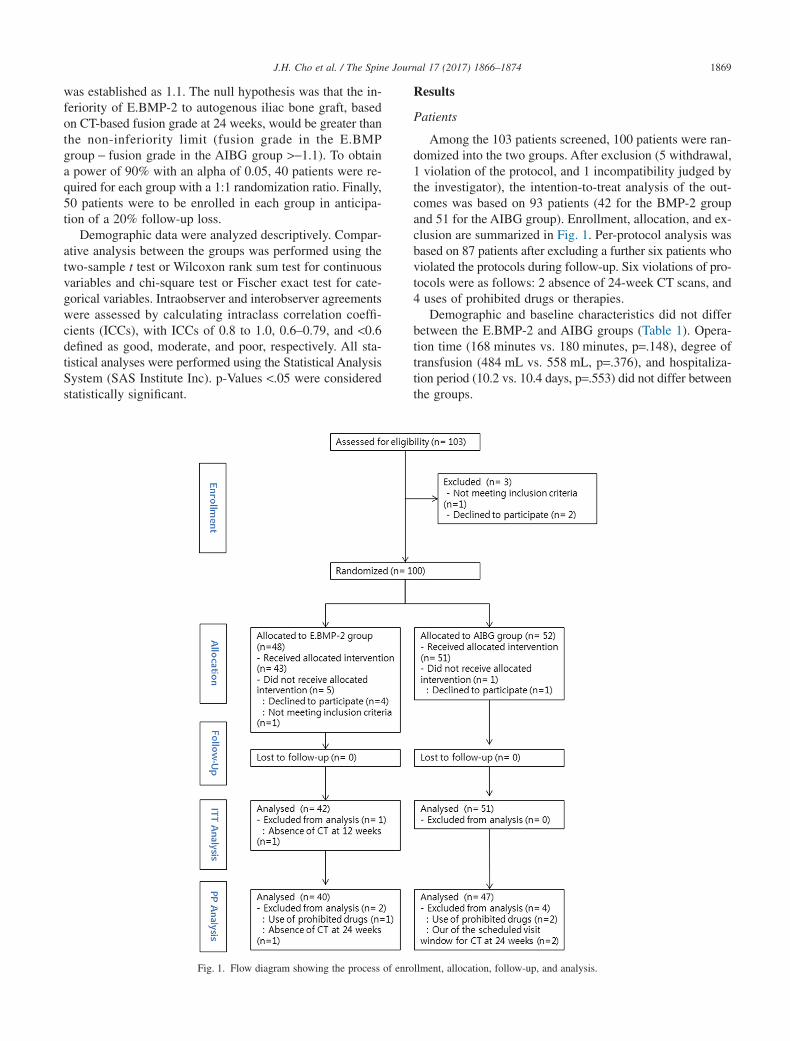
Among the 103 patients screened, 100 patients were ran-domized into the two groups. After exclusion (5 withdrawal,1 violation of the protocol, and 1 incompatibility judged bythe investigator), the intention-to-treat analysis of the out-comes was based on 93 patients (42 for the BMP-2 groupand 51 for the AIBG group). Enrollment, allocation, and ex-clusion are summarized in Fig. 1. Per-protocol analysis wasbased on 87 patients after excluding a further six patients whoviolated the protocols during follow-up. Six violations of pro-tocols were as follows: 2 absence of 24-week CT scans, and4 uses of prohibited drugs or therapies.
Demographic and baseline characteristics did not differbetween the E.BMP-2 and AIBG groups (Table 1). Opera-tion time (168 minutes vs. 180 minutes, p=.148), degree oftransfusion (484 mL vs. 558 mL, p=.376), and hospitaliza-tion period (10.2 vs. 10.4 days, p=.553) did not differ betweenthe groups.
Fig. 1. Flow diagram showing the process of enrollment, allocation, follow-up, and analysis.
1869J.H. Cho et al. / The Spine Journal 17 (2017) 1866–1874

Primary outcome measure
CT-based fusion rates showed no difference between thegroups. Fusion rates at 12 weeks were 100.0% (42/42) in theE.BMP-2 group and 90.2% (46/51) in the AIBG group(p=.062). Fusion rates at 24 weeks were 100.0% (41/41) inthe E.BMP-2 group and 94.1% (48/51) in the AIBG group(p=.251).
Characteristics of fusion in CT images were slightly dif-ferent between the groups. Although fusion mass was detectedin both groups, HA carriers remained without resorption inthe E.BMP-2 group. However, continuity of fused mass wasmore uniformly observed in the AIBG group than the E.BMP-2group (Fig. 2).
Secondary outcome measures and adverse events
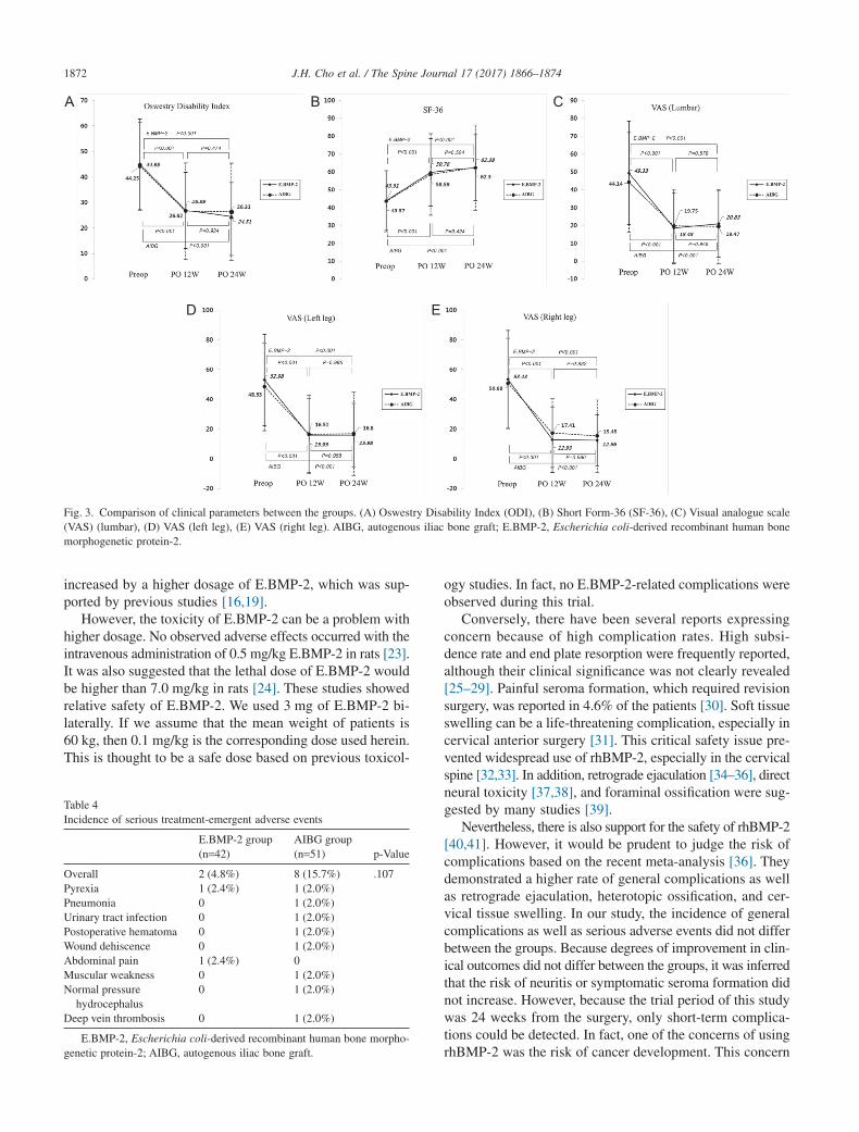
Fusion grade based on radiographs and CT at 12 and 24weeks are compared in Table 2. In all analyses, lower limitof the 95% confidence interval was greater than the non-inferiority limit (−1.1). Intraobserver agreements were good(ICC=0.836 for rater 1 and 0.802 for rater 2). Interobserveragreements were moderate (ICC=0.785 for the first rating and0.748 for the second rating). Clinical parameters showed im-provement postoperatively in both groups. VAS (lumbar, rightleg, and left leg), ODI, and SF-36 at baseline, 12 weeks, and24 weeks are described in Fig. 3. No differences for each clin-ical parameter were observed between the groups at baseline,12 weeks, and 24 weeks. In addition, percent change from
baseline of VAS, ODI, and SF-36 showed no difference at12 and 24 weeks between the groups (Table 3).
The most frequently observed adverse events were con-stipation (11 patients in the E.BMP-2 group and 10 patientsin the AIBG group) and pyrexia (9 patients in the E.BMP-2group and 13 patients in the AIBG group). However, therewas no difference for overall adverse events between thegroups (p=.975). Serious treatment-emergent adverse eventswere detected in 9 patients (10 cases): two for the E.BMP-2group and eight for the AIBG group (Table 4, p=.173).However, no events were related to the medical device. Nodeaths or serious complications leading to trial termination
Table 1Demographic and baseline characteristics
E.BMP-2 group(n=42)
AIBG group(n=51) p-Value
Age (y) 64.9±8.4 62.0±9.2 .121Gender .533
Male 20 (47.6%) 21 (41.2%)Female 22 (52.8%) 30 (58.8%)
Height (cm) 160.4±9.2 160.1±9.0 .982Weight (kg) 64.4±9.6 66.4±10.8 .354BMI (kg/m2) 25.0±3.1 25.9±3.3 .214Smoking .345
Current smoker 4 (9.5%) 6 (11.8%)Ex-smoker 6 (14.3%) 13 (25.5%)Non-smoker 32 (76.2%) 32 (62.7%)
Drinking 1.000Current drinker 13 (40.0%) 17 (33.4%)Ex-drinker 4 (9.5%) 4 (7.8%)Non-drinker 25 (59.5%) 30 (58.8%)
BMD (T-score) −0.4±1.5 −0.2±1.7 .555Radiological findings
Grade 1 spondylolisthesis 22 (52.4%) 25 (49.0%)Spinal stenosis 31 (73.8%) 43 (84.3%)Spondylolysis 0 2 (3.9%)Herniated intervertebral disc 7 (16.7%) 14 (27.5%)
E.BMP-2, Escherichia coli-derived recombinant human bone morpho-genetic protein-2; AIBG, autogenous iliac bone graft; BMI, body mass index;BMD, bone mineral density.
Table 2Fusion grade based on radiographs and CT scans at 12 and 24 weeks
E.BMP-2 group(n=42)
AIBG group(n=51) p-Value
Fusion grade by radiographs(12 wk)
4.86±0.47 4.20±1.00 <.001
Mean difference (95% CI) 0.66 (0.33, 0.99)Fusion grade by radiographs
(24 wk)4.98±0.16 4.04±1.09 <.001
Mean difference (95% CI) 0.93 (0.60, 1.27)Fusion grade by CT (12 wk) 4.48±0.83 4.02±0.93 .013Mean difference (95% CI) 0.46 (0.09, 0.82)Fusion grade by CT (24 wk) 4.56±0.81 3.98±0.94 <.001Mean difference (95% CI) 0.61 (0.25, 0.97)
E.BMP-2, Escherichia coli-derived recombinant human bone morpho-genetic protein-2; AIBG, autogenous iliac bone graft; CT, computedtomography; CI, confidence interval.
}Table 3Percent changes from baseline of VAS, ODI, and SF-36
E.BMP-2group(n=42)
AIBGgroup(n=51) p-Value
VAS (lumbar) at 12 wk −66.4±33.3 −37.8±97.7 .288Mean difference (95% CI) −28.62 (−59.42, 2.18)VAS (lumbar) at 24 wk −56.4±37.9 −42.4±88.3 .814Mean difference (95% CI) −14.08 (−42.91, 14.75)VAS (left leg) at 12 wk −70.1±43.5 −47.0±106.3 .819Mean difference (95% CI) −23.08 (−57.78, 11.62)VAS (left leg) at 24 wk −67.3±40.9 −56.1±77.8 .247Mean difference (95% CI) −11.17 (−37.92, 15.59)VAS (right leg) at 12 wk −47.6±124.5 −68.4±46.7 .651Mean difference (95% CI) 20.75 (−22.72, 64.22)VAS (right leg) at 24 wk −38.4±142.6 −64.3±81.2 .404Mean difference (95% CI) 25.83 (−27.46, 79.12)ODI at 12 wk −29.8±48.7 −36.1±44.8 .480Mean difference (95% CI) 6.35 (−12.92, 25.62)ODI at 24 wk −39.1±44.6 −38.3±43.6 .960Mean difference (95% CI) −0.79 (−19.13, 17.56)SF-36 at 12 wk 50.2±62.8 50.3±87.1 .881Mean difference (95% CI) −0.10 (−31.05, 30.86)SF-36 at 24 wk 60.3±74.3 60.0±87.4 .684Mean difference (95% CI) 0.29 (−33.82, 34.40)
E.BMP-2, Escherichia coli-derived recombinant human bone morpho-genetic protein-2; AIBG, autogenous iliac bone graft; VAS, visual analoguescale; ODI, Oswestry Disability Index; SF-36, Short Form-36 Health Survey;CI, confidence interval.
1870 J.H. Cho et al. / The Spine Journal 17 (2017) 1866–1874

were found. In addition, no difference of laboratory tests wasfound between two groups.
Discussion
The efficacy of rhBMP-2 in spinal surgery has beenwidely researched. Many studies revealed comparable fusionrates and clinical outcomes for rhBMP-2 as a bone graftsubstitute in different types of spinal fusion. More rapidincorporation and formation of fusion mass was suggestedwhen rhBMP-2 was used as a bone graft substitute in PLF[17]. In another multicenter trial, the fusion rate was higherin the rhBMP-2 group than in the autograft group (94% vs.69%, p=.007), although clinical outcomes were not differ-
ent [18]. The efficacy of E.BMP-2 as an alternative tomammalian cell origin rhBMP-2 was also suggested in animalstudies [19,20]. Additionally, osteoinductivity of E.BMP-2was comparable with that of mammalian cell BMP-2 [21,22].Based on our study, efficacy of E.BMP-2 with an HA carrierin spinal fusion was comparable with that of an autograft.Moreover, there was a trend of early fusion in the E.BMP-2group compared with the AIBG group (100.0% vs. 90.2%,at 3 months), although it did not reach statistical signifi-cance (p=.062). This difference disappeared 6 monthspostoperatively (p=.251). This means that more rapid fusionmight be induced by E.BMP-2. Osteoinductive activity toinduce rapid fusion will be critical for patients with specificconditions, such as osteoporosis. This activity could be
A B
C
D
Fig. 2. Characteristics of fusion mass taken in postoperative 12 weeks. (A) Fusion mass in a radiograph in the E.BMP-2 group (arrows). (B) Remaining HAcarrier without resorption in the E.BMP-2 group. (C) Fusion mass in a radiograph in the AIBG group (arrows) (D) Continuously fused mass in the AIBGgroup. AIBG, autogenous iliac bone graft; E.BMP-2, Escherichia coli-derived recombinant human bone morphogenetic protein-2; HA, hydroxyapatite.
1871J.H. Cho et al. / The Spine Journal 17 (2017) 1866–1874
increased by a higher dosage of E.BMP-2, which was sup-ported by previous studies [16,19].
However, the toxicity of E.BMP-2 can be a problem withhigher dosage. No observed adverse effects occurred with theintravenous administration of 0.5 mg/kg E.BMP-2 in rats [23].It was also suggested that the lethal dose of E.BMP-2 wouldbe higher than 7.0 mg/kg in rats [24]. These studies showedrelative safety of E.BMP-2. We used 3 mg of E.BMP-2 bi-laterally. If we assume that the mean weight of patients is60 kg, then 0.1 mg/kg is the corresponding dose used herein.This is thought to be a safe dose based on previous toxicol-
ogy studies. In fact, no E.BMP-2-related complications wereobserved during this trial.
Conversely, there have been several reports expressingconcern because of high complication rates. High subsi-dence rate and end plate resorption were frequently reported,although their clinical significance was not clearly revealed[25–29]. Painful seroma formation, which required revisionsurgery, was reported in 4.6% of the patients [30]. Soft tissueswelling can be a life-threatening complication, especially incervical anterior surgery [31]. This critical safety issue pre-vented widespread use of rhBMP-2, especially in the cervicalspine [32,33]. In addition, retrograde ejaculation [34–36], directneural toxicity [37,38], and foraminal ossification were sug-gested by many studies [39].
Nevertheless, there is also support for the safety of rhBMP-2[40,41]. However, it would be prudent to judge the risk ofcomplications based on the recent meta-analysis [36]. Theydemonstrated a higher rate of general complications as wellas retrograde ejaculation, heterotopic ossification, and cer-vical tissue swelling. In our study, the incidence of generalcomplications as well as serious adverse events did not differbetween the groups. Because degrees of improvement in clin-ical outcomes did not differ between the groups, it was inferredthat the risk of neuritis or symptomatic seroma formation didnot increase. However, because the trial period of this studywas 24 weeks from the surgery, only short-term complica-tions could be detected. In fact, one of the concerns of usingrhBMP-2 was the risk of cancer development. This concern
A B
D E
C
Fig. 3. Comparison of clinical parameters between the groups. (A) Oswestry Disability Index (ODI), (B) Short Form-36 (SF-36), (C) Visual analogue scale(VAS) (lumbar), (D) VAS (left leg), (E) VAS (right leg). AIBG, autogenous iliac bone graft; E.BMP-2, Escherichia coli-derived recombinant human bonemorphogenetic protein-2.
Table 4Incidence of serious treatment-emergent adverse events
E.BMP-2 group(n=42)
AIBG group(n=51) p-Value
Overall 2 (4.8%) 8 (15.7%) .107Pyrexia 1 (2.4%) 1 (2.0%)Pneumonia 0 1 (2.0%)Urinary tract infection 0 1 (2.0%)Postoperative hematoma 0 1 (2.0%)Wound dehiscence 0 1 (2.0%)Abdominal pain 1 (2.4%) 0Muscular weakness 0 1 (2.0%)Normal pressure
hydrocephalus0 1 (2.0%)
Deep vein thrombosis 0 1 (2.0%)
E.BMP-2, Escherichia coli-derived recombinant human bone morpho-genetic protein-2; AIBG, autogenous iliac bone graft.
1872 J.H. Cho et al. / The Spine Journal 17 (2017) 1866–1874
resulted from the possible activation of BMP receptors invarious cancer types. However, the risk of developing a newcancer was not likely to be higher than expected. In one ret-rospective cohort study of 527 patients, the standardizedincidence ratio for cancer was 0.84 [0.56–1.21] [42]. In ad-dition, no correlation was reported between the use of rhBMP-2and development of cancer (hazard ratio=0.99 [0.95–1.02])in another large-scale retrospective cohort study [43].
The applicability of rhBMP-2 is another important issue.Stable carriers with high osteoconductive activity and goodaffinity for rhBMP-2 are required to enhance the osteoinductiveactivity of rhBMP-2. Although collagen carriers were fre-quently used in the past, HA has been suggested as analternative. The HA granules existed in CT images 6 monthspostoperatively without resorption, which means stability ofHA as a carrier. Its higher affinity with E.BMP-2 has beenproposed by several studies [20,44]. The suitability of HAas a carrier for E.BMP-2 was also confirmed in this study.
This study has a few limitations. First, drop-out rate (13%)due to the violation of the protocol or withdrawal was notlow, even though follow-up loss was absent. This was mainlycaused by the strict regulation protocol. Second, the numberof enrolled patients were not equal among the institutionsbecause enrollment was conducted in a competitive manner.However, no differences of outcomes were found among theinstitutions. Third, quality of bone fusion was not assessed.In a previous study, quality of bone fusion by rhBMP-2 wasreported to be inferior to that of AIBG in anterior lumbarinterbody fusion [45]. We did not assess the quality of bonefusion due to remaining HA granules in the E.BMP-2 group,while continuous bone fusion mass was obviously found forthe AIBG group in CT images. Fourth, the follow-up periodwas not adequate to evaluate long-term clinical outcomes andsafety. Regardless of the above limitations, this prospectiverandomized controlled trial is thought to be worthy of noticebased on the solid study design with reliable sample size es-timation and strict study protocol. However, this is the firststudy to compare the efficacy and safety of bone graft sub-stitutes between E.BMP-2 with an HA carrier and AIBG inPLF.
In conclusion, the fusion rate with E.BMP-2 was compa-rable with AIBG following PLF. Good clinical efficacy andsafety of E.BMP-2 in spinal fusion were also revealed in thisstudy. It was also suggested that HA showed suitability as acarrier for E.BMP-2. Thus, E.BMP-2 with an HA carrier canbe an alternative bone graft material in spinal fusion.
Acknowledgments
This study was supported by a research grant for clinicalstudies from CGBio Inc/BioAlpha Inc (Gyeonggi-do, Korea).
References
[1] Sasso RC, LeHuec JC, Shaffrey C. Iliac crest bone graft donor site painafter anterior lumbar interbody fusion: a prospective patient satisfactionoutcome assessment. J Spinal Disord Tech 2005;18(Suppl.):S77–81.
[2] Fu TS, Wang IC, Lu ML, Hsieh MK, Chen LH, Chen WJ. The fusionrate of demineralized bone matrix compared with autogenous iliac bonegraft for long multi-segment posterolateral spinal fusion. BMCMusculoskelet Disord. 2016;17:3.
[3] Tilkeridis K, Touzopoulos P, Ververidis A, Christodoulou S, KazakosK, Drosos GI. Use of demineralized bone matrix in spinal fusion. WorldJ Orthop. 2014;18:30–7.
[4] Agarwal R, Williams K, Umscheid CA, Welch WC. Osteoinductivebone graft substitutes for lumbar fusion: a systematic review. JNeurosurg Spine 2009;11:729–40.
[5] Carlisle E, Fischgrund JS. Bone morphogenetic proteins for spinalfusion. Spine J 2005;5(6 Suppl.):240S–9S.
[6] Israel DI, Nove J, Kerns KM, Moutsatsos IK, Kaufman RJ. Expressionand characterization of bone morphogenetic protein-2 in Chinesehamster ovary cells. Growth Factors 1992;7:139–50.
[7] Vallejo LF, Brokelmann M, Marten S, et al. Renaturation andpurification of bone morphogenetic protein-2 produced as inclusionbodies in high-cell-density cultures of recombinant Escherichia coli.J Biotechnol 2002;94:185–94.
[8] Bessho K, Konishi Y, Kaihara S, Fujimura K, Okubo Y, Iizuka T. Boneinduction by Escherichia coli-derived recombinant human bonemorphogenetic protein-2 compared with Chinese hamster ovarycell-derived recombinant human bone morphogenetic protein-2. Br JOral Maxillofac Surg 2000;38:645–9.
[9] Bessa PC, Pedro AJ, Klosch B, et al. Osteoinduction in humanfat-derived stem cells by recombinant human bone morphogeneticprotein-2 produced in Escherichia coli. Biotechnol Lett 2008;30:15–21.
[10] Kimura M, Zhao M, Zellin G, Linde A. Bone-inductive efficacy ofrecombinant human bone morphogenetic protein-2 expressed inEscherichia coli: an experimental study in rat mandibular defects. ScandJ Plast Reconstr Surg Hand Surg 2000;34:289–99.
[11] Yano K, Hoshino M, Ohta Y, et al. Osteoinductive capacity and heatstability of recombinant human bone morphogenetic protein-2 producedby Escherichia coli and dimerized by biochemical processing. J BoneMiner Metab 2009;27:355–63.
[12] Lee JH, Kim CS, Choi KH, et al. The induction of bone formation inrat calvarial defects and subcutaneous tissues by recombinant humanBMP-2, produced in Escherichia coli. Biomaterials 2010;31:3512–19.
[13] Vallejo LF, Rinas U. Optimized procedure for renaturation ofrecombinant human bone morphogenetic protein-2 at high proteinconcentration. Biotechnol Bioeng 2004;85:601–9.
[14] Geiger M, Li RH, Friess W. Collagen sponges for bone regenerationwith rhBMP-2. Adv Drug Deliv Rev 2003;55:1613–29.
[15] Lee JH, Ryu MY, Baek HR, et al. Effects of porous beta-tricalciumphosphate-based ceramics used as an E. coli-derived rhBMP-2 carrierfor bone regeneration. J Mater Sci Mater Med 2013;24:2117–27.
[16] Lee JH, Yu CH, Yang JJ, et al. Comparative study of fusion rate inducedby different dosages of Escherichia coli-derived recombinant humanbone morphogenetic protein-2 using hydroxyapatite carrier. Spine J2012;12:239–48.
[17] Glassman SD, Dimar JR, Carreon LY, Campbell MJ, Puno RM, JohnsonJR. Initial fusion rates with recombinant human bone morphogeneticprotein-2/compression resistant matrix and a hydroxyapatite andtricalcium phosphate/collagen carrier in posterolateral spinal fusion.Spine 2005;30:1694–8.
[18] Hurlbert RJ, Alexander D, Bailey S, et al. rhBMP-2 for posterolateralinstrumented lumbar fusion: a multicenter prospective randomizedcontrolled trial. Spine 2013;38:2139–48.
[19] Hwang CJ, Lee JH, Baek H-R, Chang B-S, Lee CK. Evaluation of theefficacy of Escherichia coli-derived recombinant human bonemorphogenetic protein-2 in a mini-pig spinal anterior interbody fusionmodel. Bone Joint J 2013;95-B:217–23.
[20] Kong CB, Lee JH, Baek HR, Lee CK, Chang BS. Posterolateral lumbarfusion using Escherichia coli-derived rhBMP-2/hydroxyapatite in themini pig. Spine J 2014;14:2959–67.
1873J.H. Cho et al. / The Spine Journal 17 (2017) 1866–1874
[21] Lee J, Lee EN, Yoon J, et al. Comparative study of Chinese hamsterovary cell versus Escherichia coli-derived bone morphogenetic protein-2using the critical-size supraalveolar peri-implant defect model. JPeriodontol 2013;84:415–22.
[22] Kim IS, Lee EN, Cho TH, et al. Promising efficacy of Escherichia colirecombinant human bone morphogenetic protein-2 in collagen spongefor ectopic and orthotopic bone formation and comparison withmammalian cell recombinant human bone morphogenetic protein-2.Tissue Eng Part A. 2011;17:337–48.
[23] Lee JH, Lee EN, Nam SH. The short-term effects of repetitive E.coli-derived rhBMP-2 administration through intravenous injection inrats. Drug Chem Toxicol 2014;37:40–7.
[24] Lee JH, Lee EN, Chang BS, Lee CK. Acute intravenous injectiontoxicity study of Escherichia coli-derived recombinant human bonemorphogenetic protein-2 in rat. Asian Spine J 2014;8:113–18.
[25] Michielsen J, Sys J, Rigaux A, Bertrand C. The effect of recombinanthuman bone morphogenetic protein-2 in single-level posterior lumbarinterbody arthrodesis. J Bone Joint J Am. 2013;95:873–80.
[26] Fox MG, Goldberg JM, Gaskin CM, et al. MRI of transforaminal lumbarinterbody fusion: imaging appearance with and without the use of humanrecombinant bone morphogenetic protein-2 (rhBMP-2). Skeletal Radiol2014;43:1247–55.
[27] Singh K, Nandyala SV, Marquez-Lara A, et al. Clinical sequelae afterrhBMP-2 use in a minimally invasive transforaminal lumbar interbodyfusion. Spine J 2013;13:1118–25.
[28] Lewandrowski KU, Nanson C, Calderon R. Vertebral osteolysis afterposterior interbody lumbar fusion with recombinant human bonemorphogenetic protein 2: a report of five cases. Spine J 2007;7:609–14.
[29] Vaidya R, Weir R, Sethi A, Meisterling S, Hakeos W, Wybo CD.Interbody fusion with allograft and rhBMP-2 leads to consistent fusionbut early subsidence. J Bone Joint Surg Br 2007;89:342–5.
[30] Garrett MP, Kakarla UK, Porter RW, Sonntag VK. Formation of painfulseroma and edema after the use of recombinant human bonemorphogenetic protein-2 in posterolateral lumbar spine fusions.Neurosurgery 2010;66:1044–9.
[31] Carragee EJ, Hurwitz EL, Weiner BK. A critical review of recombinanthuman bone morphogenetic protein-2 trials in spinal surgery: emergingsafety concerns and lessons learned. Spine J 2011;11:471–91.
[32] Schroeder GD, Hsu WK, Kepler CK, et al. Use of recombinant humanbone morphogenetic protein-2 in the treatment of degenerativespondylolisthesis. Spine 2016;41:445–9.
[33] Poeran J, Opperer M, Rasul R, et al. Change in off-label use of bonemorphogenetic protein in spine surgery and associations with adverseoutcome. Global Spine J. 2016;6:650–9.
[34] Carragee EJ, Mitsunaga KA, Hurwitz EL, Scuderi GJ. Retrogradeejaculation after anterior lumbar interbody fusion using rhBMP-2: acohort controlled study. Spine J 2011;11:511–16.
[35] Comer GC, Smith MW, Hurwitz EL, Mitsunaga KA, Kessler R,Carragee EJ. Retrograde ejaculation after anterior lumbar interbodyfusion with and without bone morphogenetic protein-2 augmentation:a 10-year cohort controlled study. Spine J 2012;12:881–90.
[36] Vavken J, Mameghani A, Vavken P, Schaeren S. Complications andcancer rates in spine fusion with recombinant human bonemorphogenetic protein-2 (rhBMP-2). Eur Spine J 2016;25:3979–89.
[37] Rowan FE, O’Malley N, Poynton A. RhBMP-2 use in lumbar fusionsurgery is associated with transient immediate post-operative leg pain.Eur Spine J 2012;21:1331–7.
[38] Lykissas MG, Aichmair A, Sama AA, et al. Nerve injury and recoveryafter lateral lumbar interbody fusion with and without bonemorphogenetic protein-2 augmentation: a cohort-controlled study. SpineJ 2014;14:217–24.
[39] Hoffmann MF, Jones CB, Sietsema DL. Complications of rhBMP-2utilization for posterolateral lumbar fusions requiring reoperation: asingle practice, retrospective case series report. Spine J 2013;13:1244–52.
[40] Faundez A, Tournier C, Garcia M, Aunoble S, Le Huec JC. Bonemorphogenetic protein use in spine surgery-complications and outcomes:a systematic review. Int Orthop 2016;40:1309–19.
[41] Lubelski D, Abdullah KG, Steinmetz MP, et al. Adverse eventswith the use of rhBMP-2 in thoracolumbar and lumbar spinefusions: a 9-year institutional analysis. J Spinal Disord Tech 2015;28:E277–83.
[42] Malham GM, Giles GG, Milne RL, Blecher CM, Brazenor GA. Bonemorphogenetic proteins in spinal surgery: what is the fusion rate anddo they cause cancer? Spine 2015;40:1737–42.
[43] Cooper GS, Kou TD. Risk of cancer after lumbar fusion surgery withrecombinant human bone morphogenic protein-2 (rh-BMP-2). Spine2013;38:1862–8.
[44] Chung CH, Kim YK, Lee JS, Jung UW, Pang EK, Choi SH. Rapidbone regeneration by Escherichia coli-derived recombinant human bonemorphogenetic protein-2 loaded on a hydroxyapatite carrier in the rabbitcalvarial defect model. Biomater Res. 2015;19:17.
[45] Flouzat-Lachaniette CH, Ghazanfari A, Bouthors C, Poignard A,Hernigou P, Allain J. Bone union rate with recombinant humanbone morphogenic protein-2 versus autologous iliac bone in PEEKcages for anterior lumbar interbody fusion. Int Orthop 2014;38:2001–7.
1874 J.H. Cho et al. / The Spine Journal 17 (2017) 1866–1874