efficacy of intravenous immunoglobulin in neurological ... · review efficacy of intravenous...
TRANSCRIPT
REVIEW
Efficacy of Intravenous Immunoglobulin in Neurological Diseases
Jan D. Lünemann1,2& Isaak Quast1 & Marinos C. Dalakas3,4
Published online: 23 September 2015# The American Society for Experimental NeuroTherapeutics, Inc. 2015
Abstract Owing to its anti-inflammatory efficacy in var-ious autoimmune disease conditions, intravenous immu-noglobulin (IVIG)—pooled IgG obtained from the plas-ma of several thousands individuals—has been used fornearly three decades and is proving to be efficient in agrowing number of neurological diseases. IVIG therapyhas been firmly established for the treatment ofGuillain–Barré syndrome, chronic inflammatory demye-linating polyneuropathy, and multifocal motor neuropa-thy, either as first-line therapy or adjunctive treatment.IVIG is also recommended as rescue therapy in patientswith worsening myasthenia gravis and is beneficial as asecond-line therapy for dermatomyositis and stiff-personsyndrome. Subcutaneous rather than intravenous admin-istration of IgG is gaining momentum because of itseffectiveness in patients with primary immunodeficiencyand the ease with which it can be administered indepen-dently from hospital-based infusions. The demand forIVIG therapy is growing, resulting in rising costs andsupply shortages. Strategies to replace IVIG with
recombinant products have been developed based onproposed mechanisms that confer the anti-inflammatoryactivity of IVIG, but their efficacy has not been testedin clinical trials. This review covers new developmentsin the immunobiology and clinical applications of IVIGin neurological diseases.
Keywords Intravenous immunoglobulin . Immunotherapy .
Neurology
Introduction
The success story of the clinical application of pooled IgGpreparations from thousands of donors, so-called intravenousIgG (IVIG) therapy, to patients with autoimmune disease con-ditions began more than 30 years ago. In 1980, Imbach et al.[1] started the first IVIG administration at an empirical dose of0.4 g/kg bodyweight as a replacement therapy in a boy withimmune thrombocytopenia ( ITP) and secondaryhypogammaglobulinemia due to long-term immunosuppres-sive treatment. After the first administration of IVIG, his plate-let counts dramatically increased and IVIG therapy was con-tinued at the same dose for a total of 5 days. Twelve consec-utive children with ITP but without hypogammaglobulinemiashowed the same phenomenon [1]. Later studies confirmedthese results in adult patients with ITP, initiating the wide-spread use of IVIG preparations as an immunomodulatorytherapy for the treatment of other autoimmune diseases [2].
Today, IVIG therapy is used in the treatment of a widevariety of diseases, with >75 % of the IVIG in the USA ad-ministered to patients with autoimmune or inflammatory con-ditions [3]. The Food and Drug Administration (FDA)-ap-proved indications for immune globulin therapy are limited,but a large number of diseases, especially those seen by
* Jan D. Lü[email protected]
1 Institute of Experimental Immunology, Laboratory ofNeuroinflammation, University of Zürich, Winterthurerstrasse 190,Zürich, Switzerland
2 Department of Neurology, University Hospital of Basel,Basel, Switzerland
3 Neuroimmunology Unit, University of Athens Medical School,Athens, Greece
4 Department of Neurology, Thomas Jefferson University,Philadelphia, PA, USA
Neurotherapeutics (2016) 13:34–46DOI 10.1007/s13311-015-0391-5
neurologists, have shown potentially beneficial responses toIVIG. Based on controlled clinical trials IVIG is now recog-nized as first-line therapy in patients with Guillain–Barré syn-drome (GBS), chronic inflammatory demyelinatingpolyneuropathy (CIDP), and multifocal motor neuropathy(MMN). It is an effective rescue therapy in some patients withworsening myasthenia gravis (MG) and beneficial as second-line therapy in patients with dermatomyositis and stiff-personsyndrome [4]. Despite its widespread use and broad efficacy,the mechanisms that confer the anti-inflammatory orimmunomodulating activity in autoimmune disease condi-tions are poorly understood. This review provides on over-view of the immunobiology of the proposed mechanisms ofaction of IVIG and its clinical efficacy in neurologicaldiseases.
Immunobiology of IVIG
The production of IVIG follows the general guidelinesestablished by the International Union of ImmunologicalSocieties and the World Health Organization. The source ma-terial should be plasma obtained from a pool of at least 1000donors. Final products should be free of any potentially harm-ful contaminants, which includes testing for HIV 1 and 2,hepatitis C virus antibodies, hepatitis B surface antigen, andhigh titers of ABO antibodies, to reduce the risk for hemolyticreactions. The levels of IgG complexes and concentrations ofIgA should be kept to a minimum to avoid overt immunestimulation and anaphylactic reactions in patients with IgAdeficiency and anti-IgA antibodies, respectively. The finalproduct should contain at least 90 % intact IgG and subclassesshould be present in similar proportions to those in normalblood plasma. There are 4 subclasses in humans (IgG1,IgG2, IgG3, and IgG4), named in order of their abundancein serum, with IgG1 being the most abundant (60–70 %)followed by IgG2 (20 %), IgG3 (7 %), and IgG4 (3 %).These subclasses differ in their ability to induce complementactivation (IgG3 > IgG1 > IgG2) and their affinity for IgG-Fcreceptors (IgG1 > IgG3 > IgG4 > IgG2).
IgG molecules are glycoproteins that can be divided into 2functional components: 1) the C-terminal immunoglobulindomains CH3 and CH2 of the antibody heavy chain constitutethe Bfragment crystallizable^ (Fc) domain; and 2) the N-terminal CH1 and VH domains together with the antibodylight chain constitute the Bfragment antigen binding^ domain(Fab) (Fig. 1A). These 2 domains are joined by hinge regionsproviding flexibility to the antibody. Fab domains containhighly variable Bcomplementarity determining regions^,which are responsible for antigen recognition. The purposeof the Fc region is to provide stability to the antibody and tocombine the specificity of the Fab with cellular and humoraleffector mechanisms.
IgG has evolved to fulfill its functions using many mecha-nisms of the immune systems, which, in turn, requires a broadspectrum of regulatory mechanisms to tightly control and co-ordinate the properties of IgG. This is mainly achieved bystructural diversification of the Fc resulting either from theusage of 1 of the 4 distinct IgG isotypes or by alteration ofthe Fc-linked glycan (Fig. 1B). The Fc-linked glycan com-prises a single, highly conserved canonical glycosylation mo-tif present in both of the 2 Fc polypeptide chains, each ofwhich is composed of a nonvariable structure consisting of 2N-acetylglucosamine residues followed by 3 branched man-nose residues. The vast majority of glycans also contain N-acetylglucosamine on the alpha 1–3 and alpha 1–6 arm, and asmall fraction additionally contains a so-called bisecting N-acetylglucosamine on the alpha 1–4 arm. The presence ofthe core heptasaccharide is a key component for antibodyfunctionality and essential for IgG to pass post-translationalquality control. In contrast, the presence or absence of fucose,terminal galactose, and sialic acid on the glycan is not essen-tial and can therefore be used by the immune system to fine-tune effector functions of IgG.
Receptors binding the Fc region of IgG, so called Fcgamma-receptors (FcγRs) are crucial in mediating the effectorfunctions of antibodies. Traditionally, these receptors are divid-ed into 2 groups, based on their activating or inhibitory prop-erties reflected by the presence of either immune thyrosin-inhibitory molecule or immune thyrosin-activating moleculesequences in their intracellular domain or their signaling adap-tor molecules (Fig. 2). The receptor’s affinity for IgG deter-mines whether IgG binds in a monomeric form and cells ex-pressing the respective FcγR are therefore constantly associat-ed with surface-bound antibodies, or only bind if the avidity isincreased by the recognition of an antigen and the formation ofan immune complex. Monocytes, macrophages, and dendriticcells (DCs) express several FcγRs on their cell surface and canco-express activating FcγRs (FcγRI, FcγRIIA(C) FcγRIIIA)with the inhibitory FcγRIIB. Relative expression levels of ac-tivating versus inhibitory receptors have been shown to influ-ence the response towards immune complexes and set thethreshold for DC activation [5]. In addition to its function inmyeloid cells, FcγRIIB negatively regulates signaling via theB-cell receptor complex, resulting in decreased antigen-induced proliferation and antibody production [6, 7]. Its expres-sion on plasma cells controls their persistence in the bone mar-row [8], while deletion of FcγRIIB leads to increased frequen-cies of autoreactive plasma cells [9]. FcγRIIIA is the mainFcγR found on natural killer cells, where it is essential for thelysis of tumor or virus-transformed cells by antibody-dependent cell-mediated cytotoxicity.
Fc-receptors also contribute to the long half-life of IgG an-tibodies of approximately 21 days for the most abundant sub-classes IgG1 and IgG2. The neonatal FcR (FcRn), similar instructure to major histocompatibility complex class I
Efficacy of Intravenous Immunoglobulin in Neurological Diseases 35
molecules, cycles between the cell surface and acidicendosomal compartments where a low pH allows its bindingto internalized IgG (Fig. 2). FcRn–IgG complexes are thentransported back to the cell surface and physiological pH leadsto subsequent dissociation of the FcRn–IgG complex [10]. Thisprocess increases the half-life of IgG molecules up to 5-fold[11, 12].
Potential Mechanisms of IVIG Efficacy
IVIG preparations contain antibodies directed against abroad range of pathogens, as well as against numerous
foreign and self antigens. Based on the architecture of theirFc domain, antibodies differ in their ability to induce Fc-mediated effector functions, such as activation of innateimmune cells via Fc receptor binding or complement acti-vation, as discussed above. Given the heterogeneity of thevarious autoimmune disease conditions that respond toIVIG, it seems likely that different disease-specific path-ways mediate the clinical efficacy of this agent for a givendisease. Hence, it has been difficult to identify a generalmechanism for the anti-inflammatory or immunomodulatingefficacy of IVIG. Potential mechanisms identified so farhave mainly been attributed to the F(ab′)2 domain, the Fcdomain, and/or the Fc-linked N-glycan.
Fig. 1 Structure of IgG and itsFc-linked glycans. (A) Schematicdepiction of a prototypic IgG1antibody composed of 2 heavyand light chains and 2 glycans.Structural and functionalcomponents are indicated in gray.Fab = fragment antigen binding;Fc = fragment crystallizable; CH =constant domain heavy chain; CL
= constant domain light chain; VH
= variable domain heavy chain;VL = variable domain light chain.(B) Composition of a fullyprocessed N297-glycan(galactosylated, sialylated, andcontaining a bisecting N-acetylglucosamine)
36 Lünemann et al.
F(ab′)2-mediated Mechanisms
An example for F(ab′)2-mediated mechanisms, Fas–FasL (al-so called CD95–CD95L) receptor–ligand mediated apoptosisof keratinocytes, an early morphologic feature in patients withtoxic epidermal necrolysis (Lyell syndrome), was shown to beinhibited by blockade through CD95-binding antibodies inIVIG preparations, and patients with toxic epidermalnecrolysis showed a clinical benefit from IVIG therapy [13].Additional specificities found in IVIG and implicated as po-tentially important therapeutic targets are anti-idiotype anti-bodies that might neutralize pathogenic antibodies such asglycolipid-specific antibodies in GBS or acetylcholinereceptor-targeting antibodies in patients with MG [14–17].Moreover, the presence of antibodies against T-cell receptorswithin IVIG preparations might shape the T-cell repertoireduring inflammatory responses [18].
Fc-mediated Mechanisms
Unlike F(ab′)2-mediated anti-inflammatory properties, whichhave so far only been demonstrated in vitro or in animalmodels, a clinical trial demonstrated that infusion of Fc frag-ments alone is an efficient treatment of acute ITP in children[19], suggesting that the beneficial clinical effects of IVIG inITP are Fc-mediated. Potential mechanisms include FcγRblocking [20], modulation of FcγR expression and signaling[21, 22], increased autoantibody clearance by FcRn saturation[23], suppression of immunoglobulin production [24], modu-lation of antigen-presenting cell activation by induction ofinhibitory FcγRIIB [25], and blockade of complement pro-teins [26, 27]. Other potential mechanisms are induction ofregulatory T cells by peptide sequences called Btregitopes^
contained within the IgG constant regions [28], and inhibitionof differentiation and maturation of dendritic cells [29].
The presence of sialic acid in IgG Fc glycans has also beenimplicated in mediating the anti-inflammatory properties ofIVIG. The significance of IgG glycosylation is highlighted bythe loss of therapeutic activity of deglycosylated IVIG prepara-tions [30]. Conversely, IVIG preparations, as well as isolated Fcfragments enriched for terminal sialic acid residues, have a morethan 10-fold higher anti-inflammatory activity in an antibody-mediated animal model of rheumatoid arthritis (K/BxN model)[30, 31]. The requirement of sialic acid for the protective effectof IVIG was confirmed for several but not all autoimmune dis-ease models investigated [32–35].
Clinical Efficacy of IVIG in Neurological Diseases
A number of randomized controlled trials have shown thatIVIG is effective in acute and chronic demyelinating neurop-athies, in worsening MG as short-term therapy, in certain in-flammatory myopathies, and in stiff-person syndrome(Table 1). In large number of case series but not controlledstudies, IVIG is also promising in treating variousneuroinflammatory, painful, or even neurodegenerative disor-ders. For these diseases, however, the use of IVIG is largelyBoff-label^, even when efficacy has been established with ran-domized trials. As a result, and because of the high cost or attimes unwise use, insurance carriers, healthcare organizations,and government agencies many times do not approve IVIGtherapy, even for disorders where there is evidence-based ef-ficacy or approved indications. A judicious use of the drug istherefore necessary, with periodic assessments of its continu-ous effectiveness to avoid unnecessary use or overtreatment,
Fig. 2 The human FcγR family. Schematic depiction of human FcγRsα-chains and the major signaling adaptor molecule for FcγRI andFcγRIIIA, the common gamma chain dimer (γ2). Immune tyrosine-based activation (ITAM, green) or inhibition (ITIM, red) sequences are
indicated. FcRn is a heterodimeric major histocompatibility chain-I likemolecule associated with β2 microglobulin (β2m). The receptor’saffinity for IgG and their function in upon receptor cross-linking areindicated
Efficacy of Intravenous Immunoglobulin in Neurological Diseases 37
as discussed [36]. However, as clinicians, we need to have anopen mind to help patients with autoimmune neurologicalsyndromes that do not always fit into a predefined or restrictedcategory for which there is an approved indication. In suchcircumstances, if a patient does not demonstrate any objectiveclinical benefit after 2–3 infusions, it is unlikely that IVIGwillbe useful and should not be continued to avoid unwise use andunnecessarily high cost. The diseases in which IVIG has beentested in controlled trials include the following.
GBS
Based on at least 2 randomized trials, one dose of IVIG (5-dayregimen of 0.4 g/kg/day) was comparable to plasmapheresis(PE) in outcome measures, including time to unaided walkingand discontinuation of ventilation (class I evidence) [37, 38].Combining IVIG with PE or with 500 mg intravenousmethylprednisone produces no incremental response. InGBS variants, such as the acute axonal motor or motor senso-ry forms, Miller–Fisher syndrome, or acute dysautonomia,IVIG appears to be helpful, but controlled studies have notbeen conducted. IVIG remains the treatment of choice inchildhood GBS, based on observations attesting to a fasterrecovery and reduced morbidity, but controlled studies arenot available and may never be conducted. In mechanically
ventilated children, however, PE may be superior to IVIG[39]. Whether a second IVIG infusion may offer additionalbenefit when improvement has either not occurred or is inad-equate 3 weeks after the first infusion, remains unclear in spiteof anecdotal evidence [40]. Because a small increase in serumIgG level 2 weeks after a single IVIG infusion was indepen-dently associated with a significantly slower recovery andmore disability at 6 months [41], a low ΔIgG 2 weeks afterthe initial infusion was considered a sign that a higher dosageor a second course may be helpful for patients who exhibitpoor outcome. On this basis, a controlled study assessing thebenefit of a second IVIG infusion is currently ongoing.
CIDP
Controlled studies have shown that steroids, PE, and IVIG areequally effective on a short-term basis [42–44]. The ICE trial,the largest ever conducted in CIDP, has showed that IVIG issafe and effective not only in the short term, but also in thelong term, leading to the first FDA-approved indication for abrand of IVIG (class I evidence) [45]. A strong and positiveeffect on quality of life and improvement in some electrophys-iological measurements were also noted [46, 47]. In mostpatients, IVIG becomes effective after 6 weeks, necessitatingthe need for at least 2 infusions before it can be concluded thatIVIG is ineffective [48]. Although IVIG is generally consid-ered as first-line therapy, based on the ICE trial, the choice ofhow best to initiate therapy (choosing between prednisone,IVIG, or PE, which are all effective in controlled trials) isjudged against cost, long-term side effects, patient age, venousaccess, disease severity, and concurrent illnesses. In practice,we have also noticed, for reasons that still remain unclear, thatsome patients respond predominantly to IVIG, others to pred-nisone, and still others to PE. Patients more likely to respondto IVIG appear those with disease duration <1 year, a relaps-ing course, and electrophysiological signs of demyelinationwith conduction block [49, 50]. At the molecular level, singlenucleotide polymorphisms corresponding to transient axonalglycoprotein 1 were significantly associated with improve-ment from IVIG [51], suggesting that, if confirmed, respon-siveness to IVIG may be genetically determined. FcγRIIBexpression was reported to be decreased in treatment-naïvepatients with CIDP and upregulated upon clinically effectiveIVIG therapy [21], suggesting that the effect on FcγRIIB maybe a factor predicting the patients more likely to respond toIVIG. Further, the effect of IVIG on glycosylation of immu-noglobulin and FcγRIIB is currently being explored as a po-tentially biomarker predicting IVIG responsiveness.
MMN
Unlike CIDP and GBS, MMN does not respond to steroids orPE, only to IVIG. Efficacy has been established with a number
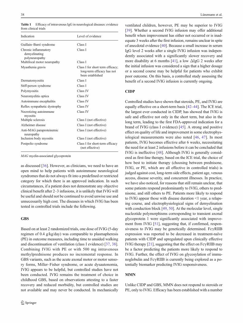
Table 1 Efficacy of intravenous IgG in neurological diseases: evidencefrom clinical trials
Indication Level of evidence
Guillain–Barré syndrome Class I
Chronic inflammatorydemyelinatingpolyneuropathy
Class I
Multifocal motor neuropathy Class I
Myasthenia gravis Class I for short term efficacy;long-term efficacy has notbeen established
Dermatomyositis Class I
Stiff-person syndrome Class I
Polymyositis Class IV
Neuromyelitis optica Class IV
Autoimmune encephalitis Class IV
Reflex sympathetic dystrophy Class IV
Necrotizing autoimmunemyositis
Class IV
Multiple sclerosis Class I (not effective)
Alzheimer disease Class I (not effective)
Anti-MAG paraproteinemicneuropathy
Class I (not effective)
Inclusion body myositis Class I (not effective)
Postpolio syndrome Class I for short-term efficacy(not effective)
MAG myelin-associated glycoprotein
38 Lünemann et al.
of controlled trials [52–54]. As a result, regulatory agencies inthe US approved the IVIG product fromBaxter (Deerfield, IL,USA) for the treatment of MMN. The improvement lasts from3 to 6 weeks, requiring reinfusion at almost predictable timeperiods. As symptoms diminish, the electrophysiologic con-duction block may resolve [53]. Therapy starts with 2 g/kg butthe response can be maintained with 1 g/kg.
Other Neuropathies
IVIG has been unsuccessful in anti-myelin-associated glyco-protein demyelinating, neuropathy based on 2 controlled trials[55, 56]. It has been anecdotally tried in diabetic amyotrophy[57, 58], vasculitic neuropathy [59], and in some painful sen-sory neuropathies, such as those associated with Sjögren syn-drome [60], with variable results. At this point, the evidence ofefficacy is overall insufficient to earn recommendation for useof IVIG in any of these conditions.
MG
The use of IVIG in MG has been examined in randomizedtrials for treating exacerbations in lieu of plasmapheresis. In 2randomized trials, IVIG was as effective as plasmapheresis atday 15 [61]. In one of the studies [62], there was no differencebetween patients randomized to 1 g/kg for 1 day versus 2 g/kgfor 2 days. IVIG was also superior to placebo, 14 days aftertherapy, in patients with moderate-to-severe MG andBworsening weakness^ [63]. Although IVIG may be effectiveon a short-term basis, its role in the chronic management of thedisease or as a steroid-sparing drug has not yet beenestablished [64] necessitating 2 ongoing trials, the first to es-tablish long-term efficacy and the second to explore thesteroid-sparing effect. The efficacy of IVIG has also not beentested in seronegarive or muscle-specific tyrosine kinase-positive MG. At present, IVIG may be justified in lieu ofplasmapheresis for acutely worsening disease to prevent orminimize impending bulbar or respiratory failure or preparea weak patient for thymectomy. IVIG may be also effective inLambert–Eaton myasthenic syndrome, based on a smallplacebo-controlled study that showed a statistically significantincrease inmuscle strength comparedwith placebo, 2–4weeksafter therapy [65].
Inflammatory Myopathies
They comprise 4 subsets: dermatomyositis, polymyositis, in-clusion body myositis, and necrotizing autoimmune myositis.In a double-blind, placebo-controlled study conducted in der-matomyositis, compared with those treated with placebo, thepatients treated with IVIG experienced a significant improve-ment in strength and muscle function, and a marked improve-ment of the active rash or the chronic scaly eruptions [66].
Repeat muscle biopsies demonstrated significant improve-ment in the muscle cytoarchitecture, including increased mus-cle fiber diameter, revascularization, reduction of inflamma-tion, interception of complement deposition, resolution of im-munopathology, and downregulation of inflammatory media-tors at the protein, mRNA, and gene level [67–70]. This studywas important to demonstrate that an effective action of IVIGis via inhibition of complement. In dermatomyositis, earlydeposition of membranolytic attack complex on theendomysial capillaries is the fundamental process leading tocapillary destruction, muscle ischemia, and inflammation. Inimproved patients, IVIG inhibited complement consumptionin serum and intercepted the complement-mediated destruc-tion of capillaries with resolution of immunopathology. IVIGseems effective in some patients with polymyositis and nec-rotizing autoimmune myositis [71, 72], but a controlled studyhas never been performed. IVIG is ineffective in inclusion-body myositis, based on 2 controlled studies [68, 73, 74].Combination of IVIG with prednisone was also ineffectiveand did not exert a synergistic effect in patients withinclusion-body myositis [75]. The reason for such inefficacywas recently explored in the repeated biopsies of these patientsand found that while there was downregulation in the expres-sion of some inflammatory molecules in the patients’muscles,several crucial markers of inflammation, cell stress, and de-generation remain unchanged [76].
Stiff-person Syndrome
Stiff-person syndrome is a disabling autoimmune disordercharacterized by muscle rigidity, episodic muscle spasms,and antibodies to glutamic acid decarboxylase 65 [77]. In aplacebo-controlled, crossover study [78], IVIG significantlydecreased stiffness scores, and substantially increased walkingand functions of daily activities, concluding that IVIG is aneffective immunotherapy in stiff-person syndrome.
Multiple Sclerosis
Two initial randomized, double-blind, placebo-controlledclinical trials in patients with relapsing–remitting multiplesclerosis (MS) demonstrated that IVIG treatment reduces theexacerbation rate and clinical progression, as defined by theexpanded disability status score, over an observation period of2 years [79, 80]. However, subsequent multicenter random-ized, double-blind, placebo-controlled clinical trials did notconfirm these findings. As an add-on treatment to methylpred-nisolone, IVIG did not further ameliorate MS relapses [81,82]. No significant reduction of relapse rate and expandeddisability status score progression was observed in patientswith relapsing–remitting MS compared with those receivingplacebo [83]. In secondary progressive MS, compared withplacebo, IVIG had no beneficial effects on clinical disease
Efficacy of Intravenous Immunoglobulin in Neurological Diseases 39
progression or T2-lesion load [84]. These divergent results onthe efficacy of IVIG in MS might result from differences instudy populations, dosing regimens, and clinical trial design,but led to the conclusion that IVIG is ineffective and notrecommended for patients with relapsing–remitting or second-ary progressive MS. However, a retrospective study that ex-plored safety and efficacy of IVIG in pregnant women withMS showed that IVIG therapy during pregnancy until12 weeks postpartum significantly reduced the frequency ofMS exacerbations compared with untreated patients [85, 86].Based on these findings and owing to safety concerns withcurrently licensed MS drugs during pregnancy and lactation,IVIG is occasionally considered as an optional treatment toreduce the incidence of pregnancy-related or postpartumrelapses.
Novel Applications
Neuromyelitis Optica
Neuromyelitis optica (Devic syndrome), long considered aclinical variant of MS, is now regarded as a distinct clinicalentity. In a retrospective study, IVIG was described to be ef-fective in some patients who experienced acute relapses anddid not benefit from steroids or plasma exchange therapy [87],and might also be effective as long-term treatment [88]. Whilethese data suggest that IVIG therapy could be an alternativefor patients with contraindication to immunosuppressive pro-phylactic treatments such as azathioprine and rituximab or,particularly, in children, randomized controlled trials are need-ed to establish the efficacy of IVIG for treatment of relapsesand prevention of disease progression in patients with thiscondition.
Autoimmune Encephalitis
Autoimmune encephalitis comprises an expanding group ofpotentially treatable disorders that occurs in association withantibodies to neuronal cell surface or synaptic proteins. In caseseries, but not controlled studies, patients with these condi-tions have been shown to benefit from IVIG therapy, as wellas from corticosteroids and plasma exchange, but they oftenrequire more aggressive, immunosuppressive therapies withrituximab or cyclophosphamide [89].
Postpolio Syndrome
This is clinically characterized by new muscle weakness, fa-tigue, and pain that develop many years after an initial attackof acute paralytic poliomyelitis. It is thought to be due toattrition of the surviving motor neurons [90]. Lymphocyticinfiltrates in the spinal cords of have, however, been observed
even 30 years after the original infection, and upregulation ofRNA for tumor necrosis factor, interferon-γ, interleukin-4,and interleukin-10 has been observed in the cerebrospinal flu-id (CSF), suggesting the possibility of a persistent smolderinginflammatory response. Following IVIG treatment,interferon-γ and tumor necrosis factor mRNA levels werereduced in the CSF, prompting a controlled trial performedin 135 patients. Although the results were essentially negative,some significant differences, of uncertain clinical importance,were observed in some physical activity and quality-of-lifescores [91]. On this basis, a new FDA-approved multicenterclinical trial has started and is currently ongoing.
Reflex Sympathetic Dystrophy
In the pathogenesis of reflex sympathetic dystrophy and otherchronic pain syndromes, proinflammatory cytokines, or anti-bodies to potassium channels have been implicated. On thisbasis, IVIG has been tried, with variable results [92]. In arandomized, double-blind, placebo-controlled crossover trialthe efficacy of low-dose IVIG 0.5 g/kg was examined in 12patients 6–19 days after IVIG. Although a reduction in painscales was noted, the study was too small to conclude on theoverall efficacy of IVIG not only in RSD, but also in otherpain syndromes. However, the concept is of interest and re-quires vigorously controlled large-scale trials.
The Emerging Role of Subcutaneous IgG
The subcutaneous route of administering IgG, instead of in-travenously, is gaining momentum because of its effectivenessin primary immunodeficiency and ease of self-administrationat home on a weekly basis. Subcutaneous IgG (SCIG) is alsowell tolerated, can be used in patients with poor venous ac-cess, has fewer systemic side effects, and is probably lesscostly. As a result, a number of controlled studies are emerg-ing in CIDP, MMN, and dermatomyositis, especially withnew high-concentration products that allow the self-infusionof high IgG quantities. In a small CIDP trial, 30 patients wererandomized to SCIG or placebo at home. After 12 weeks, theSCIG group had a statistically improved strength and stabilityof electrophysiology compared with placebo [93]. Large-scalecontrolled studies are ongoing, examining the safety, formulaof transitioning from IVIG to SCIG, and efficacy and patients’preference.
Pharmacokinetics of IVIG versus SCIG
An infusion of 2 g/kg IVIG increases the serum IgG level>4-fold, from pretreatment means of 700–1060 mg/dl to apeak well over 3000 mg/dl [94], dropping to about 50 %over 48–72 h. The infused IgG is also seen in the CSF,
40 Lünemann et al.
where it peaks by 2 days to almost twice the baseline [95].The infused IVIG is distributed in the extracellular space,which is about double the intravascular space [96]; it is thencatabolized slowly compared with other plasma proteins viaFcRn, which recycles IgG and protects it from lysosomaldegradation [97]. As a result, the half-life of IgG is main-tained at approximately 21 days, and repeated infusions areneeded after 1 month. The saturation of the FcRn with highconcentrations of normal IgG from the infused exogenousIVIG keeps the endogenous pathologic IgG from this path-way and increases its degradation [98]. In contrast, SCIG isfirst transported through the lymphatic systems and entersthe bloodstream via the thoracic duct, with equilibration ofIgG derived from SCIG into the intravascular space requir-ing up to 72 h [96]. The peak serum IgG concentrationachieved with SCIG is, on average, 61 % of the peakachieved with intravenous infusions of the same dose [99].This probably accounts for the fewer adverse events com-pared with IVIG. With weekly SCIG, only a few days elapsebetween the peak serum level from 1 intravenous dose andadministration of the next subcutaneous dose; this clearlyobviates the low Btrough^ serum IgG levels experienced3–4 weeks after a large bolus of IVIG [100]. Of interest,pooled data have shown that serum IgG levels are higherby 10–20 % (mean 13 %) with weekly SCIG compared withmonthly IVIG [96]. After 6–12 weekly infusions, SCIG re-sults in near steady-state IgG levels, with peak trough dif-ferences only about 5 % of the overall mean. In contrast,with IVIG the trough-to-peak difference is often >100 % ofthe overall mean. With the weekly SCIG, the levels of IgGat 7–10 days is the same as the one achieved by IVIG in thatperiod; however, the difference is that with IVIG the IgGdrops thereafter while with SCIG it remains constant, indi-cating a good means of sustaining response. Whether theseinteresting kinetics translate into the same degree of efficacyof SCIG as that of IVIG in the treatment of autoimmuneneurological disorders is currently being explored withincontrolled trials.
IVIG Therapy: Practical Issues
Administration
As discussed earlier, the therapeutic dose of IVIG is empiri-cally set at 2 g/kg, as originally applied by Imbach et al. [1].This dose is given over 2–5 days, and followed by a mainte-nance dose of 1–2 g/kg every 3–4 weeks; although the major-ity of clinicians prefer to infuse the total dose over 2 days andmost studies have used this formula, in older people and thosewith impaired renal or cardiovascular function it is prudent toadminister the dose over 3–5 days.
Adverse Reactions
Common Infusion-related Reactions
These are usually minor and occur in not more than 10 % ofpatients. The most common include mild-to-moderate head-ache that responds to nonsteroidal anti-inflammatory medi-cations, fever, chills, myalgia, and chest or back pain thatoccur during the first few hours of the infusion and usuallyrespond by stopping the infusion for 30 min and resuming itat a slower rate. Postinfusion fatigue, fever, or nausea mayalso occur and last for up to 24 h. In a large retrospectiveevidence-based study of all trials performed in neuromuscu-lar disorders, headaches occurred in 16.6 % of patients, feverin 6.6 %, hypertension in 4.6 %, chills in 3.3 %, and nauseain 3.2 % [101].
Rare, More Serious Reactions
Rare, more serious reactions include thromboembolic events,such as strokes, pulmonary embolism, or myocardial infarc-tion [102–107]. Patients with recent deep vein thrombosis, orimmobilized patients who may have a subclinical thrombosis,may be at higher risk. The main causative factor may be anincrease in serum viscosity, especially in patients with riskfactors, such as cryoglobulinemia, hypercholesterolemia, orhypergammaglobulinemia [106]. A reversible cerebral vaso-spasm has also been noted [108].
Severe headache due to aseptic meningitis can also beseen. This is unrelated to the proprietary product, the rateof infusion, or the underlying disease [73, 95, 109, 110].Prophylaxis with intravenous steroids can be occasionallyhelpful. The symptoms respond to strong analgesia and sub-side in 24–48 h. Additional diagnostic testing is rarely nec-essary [109, 110]. The occurrence of aseptic meningitis ishigher in migrainous patients [95], and IVIG may also trig-ger a migraine attack in patients with a prior history ofmigraines.
Skin reactions can develop 2–5 days after the infusions andmay last up to 30 days. They include urticaria, lichenoid cu-taneous lesions, pruritus of the palms, and petechiae of theextremities [73, 109, 111]; they have occurred in 7 of the120 patients we have serially treated [73, 109, 111].
Rare anaphylactic reaction due to absence or severe defi-ciency of IgA in patients who also have anti-IgA antibodies,rarely seen in patients with common variable immunodefi-ciency, can occur [112]. IgA deficiency is common in thegeneral population (prevalence ~1:1000), but it is asymptom-atic and not a risk factor by itself; about 29 % of these indi-viduals have anti-IgA antibodies but the presence of theseantibodies does not necessarily predict the development ofallergic reaction to IVIG [113].
Efficacy of Intravenous Immunoglobulin in Neurological Diseases 41
Acute renal tubular necrosis, mostly reversible, may rare-ly occur in patients with pre-existing kidney disease andvolume depletion, especially the elderly and those with dia-betes or poor hydration. It is more often associated with thehigh concentration of sucrose in some proprietary IVIGproducts. Osmotically induced tubular injury andvacuolization are the common histopathological findings up-on renal biopsy. It is usually reversible, but rare fatalitieshave been noted. Is should be noted that serum creatininemay rise 1–10 days after the infusion but returns to baselinewithin 2–60 days of discontinuation. In patients with pre-existing kidney disease, close monitoring of creatinine andblood urea nitrogen are essential, while slowing the rate ofinfusion or selecting products with low osmolality mini-mizes that risk.
Laboratory AbnormalitiesAfter IVIG therapy, the erythro-cyte sedimentation rate increases 6-fold owing to enhancedrouleux formation and reduced surface area caused by theinfused gamma globulin [111, 114]. This false increase canpersist for 2–3 weeks, and should not be considered a signof a developing vasculitis. Hyponatremia, as low as 130 mg/l (normal 135–145 mg/l), after IVIG therapy can be detect-ed. This is also due to the assay method used to measureNa+ because additional dilution of the sample is requiredowing to the high serum protein concentration that followsIVIG infusion. A mild and inconsequential leukopenia canbe also observed.
Differences in Products and Pharmacoeconomics
Among the various IVIG preparations on the market, thereis no documented evidence that one is more efficaciousthan the other for a given disorder. Their cost is also sim-ilar. However, some products may be preferable for high-risk patients because these products are low in osmolality,sodium, or sucrose, and presumably have fewer side effectsin high-risk patients [3]. Although, IVIG is considered safefor long-term administration, compared with other effectivetherapeutic modalities such as PE, corticosteroids, or immu-nosuppressants used in autoimmune neuromuscular disor-ders, there are no comparative long-term data. The samealso applies to pharmacoeconomics, where no long-termcomparative studies between these therapies have been per-formed, except for some limited data being obtained forCIDP [115].
Future Prospects
Evidence from controlled clinical trials has establishedIVIG as a first-line therapy for GBS, CIDP, and MMN.
IVIG might be considered as short-term therapy formoderate-to-severe and worsening MG, and it is effectiveas second-line therapy for treating patients with dermato-myositis, stiff-person syndrome and Lambert–Eaton myas-thenic syndrome. The therapeutic benefit of IVIG is shortlived and probably explained by the half-life of IgG mole-cules in vivo. Evolving studies are now exploring the ef-fects and superiority of subcutaneous IgG compared withIVIG in chronic maintenance therapy once induction bene-fit has been established with IVIG. As with other immuno-modulatory treatments, a subset of patients does not benefitfrom IVIG therapy and, at present, we are unable to predictwhich patients will respond to IVIG. Thus, surrogate pa-rameters that could predict from the outset which patientsare more likely to respond are needed. Progress towardsthis goal is profiling expression levels of Fcγ receptors.Expression of the inhibitory FcγRIIB on B cells and mye-loid cells is impaired in patients with CIDP, but upregulatedfollowing clinically effective IVIG therapy [21, 116].Upregulation of the FcγRIIB is also observed in animalmodels of autoimmune disease conditions following effec-tive IVIG treatment [30, 117]. In addition, single nucleotidepolymorphisms from genes encoding for the transient axo-nal glycoprotein 1 and the C-type lectin domain family 10,member A are reported to be associated with the clinicaloutcome of IVIG therapy in patients with CIDP [51]. Thesecandidate biomarkers require validation in larger cohortsand it will be useful to implement a biomarker identifica-tion and validation component to define therapeutic re-sponses to IVIG in future clinical trials.
The demand for IVIG therapy is ever-growing but itsavailability is limited, resulting in supply shortages. Thus,the development of a possible replacement for IVIG in theform of a recombinant product, even if only for a particulardisease, would have a major effect. Several companies arecurrently working towards developing IVIG replacementsbased on a variety of critical pathways targeted by IVIGtherapy. Such developments include recombinant antibodiesblocking neonatal Fc receptors for the reduction of autoan-tibody half-lives, multimeric IgG Fc preparations that blockimmune complex binding to activating Fcγ receptors, andIVIG preparations with enhanced levels of anti-inflammatory sialic acid-rich IgG glycovariants. Such re-placement therapies might not be effective in all IVIG-responsive autoimmune diseases as they target more specificdisease mechanisms than intact native IVIG, while manyconditions that respond to IVIG therapy have differing path-ological profiles. Clinical studies are therefore needed toevaluate which of the aforementioned recombinant IVIGreplacements is effective and for which type of autoimmunedisease. In addition, advances in our understanding of howIVIG exerts its beneficial effects in specific neurologicaldiseases might also enable us to develop novel therapeutics
42 Lünemann et al.
with immunomodulatory activities similar to those of nativeIgG for targeted immunotherapeutic interventions.
Acknowledgments Isaak Quast was supported by a DOC scholarshipprovided by the Austrian Academy of Sciences.
Required Author Forms Disclosure forms provided by the authors areavailable with the online version of this article.
References
1. Imbach P, Barandun S, d'Apuzzo V, et al. High-dose intravenousgammaglobulin for idiopathic thrombocytopenic purpura in child-hood. Lancet 1981;1:1228-1231.
2. Fehr J, Hofmann V, Kappeler U. Transient reversal of thrombocy-topenia in idiopathic thrombocytopenic purpura by high-dose in-travenous gamma globulin. N Engl J Med 1982;306:1254-1258.
3. Gelfand EW. Intravenous immune globulin in autoimmune andinflammatory diseases. N Engl J Med 2012;367:2015-2025.
4. Lunemann JD, Nimmerjahn F, Dalakas MC. Intravenous immu-noglobulin in neurology–mode of action and clinical efficacy. NatRev Neurol 2015;11:80-89.
5. Boruchov AM, Heller G, Veri MC, Bonvini E, Ravetch JV, YoungJW. Activating and inhibitory IgG Fc receptors on human DCsmediate opposing functions. J Clin Invest 2005;115:2914-2923.
6. Yuasa T, Kubo S, Yoshino T, et al. Deletion of fcgamma receptorIIB renders H-2(b)mice susceptible to collagen-induced arthritis. JExp Med 1999;189:187-194.
7. Daeron M, Latour S, Malbec O, et al. The same tyrosine-basedinhibition motif, in the intracytoplasmic domain of Fc gammaRIIB, regulates negatively BCR-, TCR-, and FcR-dependent cellactivation. Immunity 1995;3:635-646.
8. Xiang Z, Cutler AJ, Brownlie RJ, et al. FcgammaRIIb controlsbone marrow plasma cell persistence and apoptosis. Nat Immunol2007;8:419-429.
9. Fukuyama H, Nimmerjahn F, Ravetch JV. The inhibitoryFcgamma receptor modulates autoimmunity by limiting the accu-mulation of immunoglobulin G+ anti-DNA plasma cells. NatImmunol 2005;6:99-106.
10. Ober RJ,Martinez C, Vaccaro C, Zhou J,Ward ES. Visualizing thesite and dynamics of IgG salvage by the MHC class I-relatedreceptor, FcRn. J Immunol 2004;172:2021-2029.
11. Chaudhury C, Mehnaz S, Robinson JM, et al. The major histo-compatibility complex-related Fc receptor for IgG (FcRn) bindsalbumin and prolongs its lifespan. J Exp Med 2003;197:315-322.
12. Dickinson BL, Badizadegan K, Wu Z, et al. Bidirectional FcRn-dependent IgG transport in a polarized human intestinal epithelialcell line. J Clin Invest 1999;104:903-911.
13. Viard I, Wehrli P, Bullani R, et al. Inhibition of toxic epidermalnecrolysis by blockade of CD95 with human intravenous immu-noglobulin. Science 1998;282:490-493.
14. Rossi F, Kazatchkine MD. Antiidiotypes against autoantibodies inpooled normal human polyspecific Ig. J Immunol 1989;143:4104-4109.
15. Shoenfeld Y, Rauova L, Gilburd B, et al. Efficacy of IVIG affinity-purified anti-double-stranded DNA anti-idiotypic antibodies in thetreatment of an experimental murine model of systemic lupuserythematosus. Int Immunol 2002;14:1303-1311.
16. Fuchs S, Feferman T, Meidler R, et al. A disease-specific fractionisolated from IVIG is essential for the immunosuppressive effectof IVIG in experimental autoimmune myasthenia gravis. JNeuroimmunol 2008;194:89-96.
17. Buchwald B, Ahangari R, Weishaupt A, Toyka KV. Intravenousimmunoglobulins neutralize blocking antibodies in Guillain-Barrésyndrome. Ann Neurol 2002;51:673-680.
18. Marchalonis JJ, Kaymaz H, Dedeoglu F, Schluter SF, Yocum DE,Edmundson AB. Human autoantibodies reactive with syntheticautoantigens from T-cell receptor beta chain. Proc Natl Acad SciU S A 1992;89:3325-3329.
19. Debre M, Bonnet MC, Fridman WH, et al. Infusion of Fc gammafragments for treatment of children with acute immune thrombo-cytopenic purpura. Lancet 1993;342:945-949.
20. Nagelkerke SQ, Dekkers G, Kustiawan I, et al. Inhibition ofFcgammaR-mediated phagocytosis by IVIg is independent ofIgG-Fc sialylation and FcgammaRIIb in human macrophages.Blood 2014;124:3709-3718.
21. Tackenberg B, Jelcic I, Baerenwaldt A, et al. Impaired inhibitoryFcgamma receptor IIB expression on B cells in chronic inflamma-tory demyelinating polyneuropathy. Proc Natl Acad Sci U S A2009;106:4788-4792.
22. Samuelsson A, Towers TL, Ravetch JV. Anti-inflammatory activ-ity of IVIG mediated through the inhibitory Fc receptor. Science2001;291:484-486.
23. Hansen RJ, Balthasar JP. Intravenous immunoglobulin mediatesan increase in anti-platelet antibody clearance via the FcRn recep-tor. Thromb Haemost 2002;88:898-899.
24. Kondo N, Kasahara K, Kameyama T, et al. Intravenous immuno-globulins suppress immunoglobulin productions by suppressingCa(2+)-dependent signal transduction through Fc gamma recep-tors in B lymphocytes. Scand J Immunol 1994;40:37-42.
25. Anthony RM, Kobayashi T, Wermeling F, Ravetch JV.Intravenous gammaglobulin suppresses inflammation through anovel T(H)2 pathway. Nature 2011;475:110-113.
26. Mollnes TE, Hogasen K, Hoaas BF, Michaelsen TE, Garred P,Harboe M. Inhibition of complement-mediated red cell lysis byimmunoglobulins is dependent on the IG isotype and its C1 bind-ing properties. Scand J Immunol 1995;41:449-456.
27. Quast I, Keller CW, Maurer MA, Giddens JP, Tackenberg B,Wang LX, Münz C, Nimmerjahn F, Dalakas MC, LünemannJD. Sialylation of IgG Fc domain impairs complement-dependent cytotoxicity. J Clin Invest 2015; in press.
28. De Groot AS, Moise L, McMurry JA, et al. Activation of naturalregulatory T cells by IgG Fc-derived peptide BTregitopes^. Blood2008;112:3303-3311.
29. Bayry J, Lacroix-Desmazes S, Carbonneil C, et al. Inhibition ofmaturation and function of dendritic cells by intravenous immu-noglobulin. Blood 2003;101:758-765.
30. Kaneko Y, Nimmerjahn F, Ravetch JV. Anti-inflammatory activityof immunoglobulin G resulting from Fc sialylation. Science2006;313:670-673.
31. Anthony RM, Nimmerjahn F, Ashline DJ, Reinhold VN,Paulson JC, Ravetch JV. Recapitulation of IVIG anti-inflammatory activity with a recombinant IgG Fc. Science2008;320:373-376.
32. Schwab I, Mihai S, Seeling M, Kasperkiewicz M, Ludwig RJ,Nimmerjahn F. Broad requirement for terminal sialic acid residuesand FcgammaRIIB for the preventive and therapeutic activity ofintravenous immunoglobulins in vivo. Eur J Immunol 2014;44:1444-1453.
33. Leontyev D, Katsman Y, Ma XZ, Miescher S, Kasermann F,Branch DR. Sialylation-independent mechanism involved in theamelioration of murine immune thrombocytopenia using intrave-nous gammaglobulin. Transfusion 2012;52:1799-1805.
34. Leontyev D, Katsman Y, Branch DR. Mouse background andIVIG dosage are critical in establishing the role of inhibitoryFcgamma receptor for the amelioration of experimental ITP.Blood 2012;119:5261-5264.
Efficacy of Intravenous Immunoglobulin in Neurological Diseases 43
35. Othy S, Topcu S, Saha C, et al. Sialylation may be dispensable forreciprocal modulation of helper T cells by intravenous immuno-globulin. Eur J Immunol 2014;44:2059-2063.
36. Dalakas MC, Medscape. Advances in the diagnosis, pathogenesisand treatment of CIDP. Nat Rev Neurol 2011;7:507-517.
37. van der Meche FG, Schmitz PI. A randomized trial comparingintravenous immune globulin and plasma exchange in Guillain–Barré syndrome. Dutch Guillain-Barré Study Group. N Engl JMed 1992;326:1123-1129.
38. Plasma Exchange/Sandoglobulin Guillain-Barré Syndrome TrialGroup. Randomised trial of plasma exchange, intravenous immu-noglobulin, and combined treatments in Guillain-Barré syndrome.Lancet 1997;349:225-230.
39. El-Bayoumi MA, El-Refaey AM, Abdelkader AM, El-AssmyMM, Alwakeel AA, El-Tahan HM. Comparison of intravenousimmunoglobulin and plasma exchange in treatment of mechani-cally ventilated children with Guillain Barré syndrome: a random-ized study. Crit Care 2011;15:R164.
40. Farcas P, Avnun L, Frisher S, Herishanu YO, Wirguin I. Efficacyof repeated intravenous immunoglobulin in severe unresponsiveGuillain-Barré syndrome. Lancet 1997;350:1747.
41. Kuitwaard K, de Gelder J, Tio-Gillen AP, et al. Pharmacokineticsof intravenous immunoglobulin and outcome in Guillain-Barrésyndrome. Ann Neurol 2009;66:597-603.
42. Hughes R, Bensa S,Willison H, et al. Randomized controlled trialof intravenous immunoglobulin versus oral prednisolone in chron-ic inflammatory demyelinating polyradiculoneuropathy. AnnNeurol 2001;50:195-201.
43. Dyck PJ, Litchy WJ, Kratz KM, et al. A plasma exchange versusimmune globulin infusion trial in chronic inflammatory demyelin-ating polyradiculoneuropathy. Ann Neurol 1994;36:838-845.
44. Mendell JR, Barohn RJ, Freimer ML, et al. Randomized con-trolled trial of IVIg in untreated chronic inflammatory demyelin-ating polyradiculoneuropathy. Neurology 2001;56:445-449.
45. Hughes RA, Donofrio P, Bril V, et al. Intravenous immune glob-ulin (10% caprylate-chromatography purified) for the treatment ofchronic inflammatory demyelinating polyradiculoneuropathy(ICE study): a randomised placebo-controlled trial. LancetNeurol 2008;7:136-144.
46. Merkies IS, Bril V, Dalakas MC, et al. Health-related quality-of-life improvements in CIDP with immune globulin IV 10%: theICE Study. Neurology 2009;72:1337-1344.
47. Bril V, Katzberg H, Donofrio P, et al. Electrophysiology in chronicinflammatory demyelinating polyneuropathy with IGIV. MuscleNerve 2009;39:448-455.
48. Latov N, Deng C,DalakasMC, et al. Timing and course of clinicalresponse to intravenous immunoglobulin in chronic inflammatorydemyelinating polyradiculoneuropathy. Arch Neurol 2010;67:802-807.
49. Vermeulen M, van Doorn PA, Brand A, Strengers PF, JennekensFG, Busch HF. Intravenous immunoglobulin treatment in patientswith chronic inflammatory demyelinating polyneuropathy: a dou-ble blind, placebo controlled study. J Neurol Neurosurg Psychiatry1993;56:36-39.
50. Hahn AF, Bolton CF, Pillay N, et al. Plasma-exchange therapy inchronic inflammatory demyelinating polyneuropathy. A double-blind, sham-controlled, cross-over study. Brain 1996;119:1055-1066.
51. Iijima M, Tomita M, Morozumi S, et al. Single nucleotide poly-morphism of TAG-1 influences IVIg responsiveness of Japanesepatients with CIDP. Neurology 2009;73:1348-1352.
52. Chaudhry V, Corse AM, Cornblath DR, et al. Multifocal motorneuropathy: response to human immune globulin. Ann Neurol1993;33:237-242.
53. Federico P, Zochodne DW, Hahn AF, Brown WF, Feasby TE.Multifocal motor neuropathy improved by IVIg: randomized,
double-blind, placebo-controlled study. Neurology 2000;55:1256-1262.
54. Van den Berg-Vos RM, Franssen H,Wokke JH, Van den Berg LH.Multifocal motor neuropathy: long-term clinical and electrophys-iological assessment of intravenous immunoglobulin maintenancetreatment. Brain 2002;125:1875-1886.
55. Dalakas MC, Quarles RH, Farrer RG, et al. A controlled study ofintravenous immunoglobulin in demyelinating neuropathy withIgM gammopathy. Ann Neurol 1996;40:792-795.
56. Comi G, Roveri L, Swan A, et al. A randomised controlled trial ofintravenous immunoglobulin in IgM paraprotein associated demy-elinating neuropathy. J Neurol 2002;249:1370-1377.
57. Jann S, Bramerio MA, Facchetti D, Sterzi R. Intravenous immu-noglobulin is effective in patients with diabetes and with chronicinflammatory demyelinating polyneuropathy: long term follow-up. J Neurol Neurosurg Psychiatry 2009;80:70-73.
58. Kawagashira Y, Watanabe H, Morozumi S, et al. Differential re-sponse to intravenous immunoglobulin (IVIg) therapy amongmultifocal and polyneuropathy types of painful diabetic neuropa-thy. J Clin Neurosci 2010;17:1003-1008.
59. Levy Y, Uziel Y, Zandman G, et al. Response of vasculitic periph-eral neuropathy to intravenous immunoglobulin. Ann N YAcadSci 2005;1051:779-786.
60. Rist S, Sellam J, Hachulla E, et al. Experience of intravenousimmunoglobulin therapy in neuropathy associated with primarySjogren's syndrome: a national multicentric retrospective study.Arthritis Care Res (Hoboken) 2011;63:1339-1344.
61. Gajdos P, Chevret S, Clair B, Tranchant C, Chastang C. Clinicaltrial of plasma exchange and high-dose intravenous immunoglob-ulin in myasthenia gravis. Myasthenia Gravis Clinical StudyGroup. Ann Neurol 1997;41:789-796.
62. Gajdos P, Tranchant C, Clair B, et al. Treatment of myastheniagravis exacerbation with intravenous immunoglobulin: a random-ized double-blind clinical trial. Arch Neurol 2005;62:1689-1693.
63. Zinman L, Ng E, Bril V. IV immunoglobulin in patients withmyasthenia gravis: a randomized controlled trial. Neurology2007;68:837-841.
64. Gajdos P, Chevret S, Toyka K. Intravenous immunoglobulin formyasthenia gravis. CochraneDatabase Syst Rev 2008;CD002277.
65. Bain PG, Motomura M, Newsom-Davis J, et al. Effects of intra-venous immunoglobulin on muscle weakness and calcium-channel autoantibodies in the Lambert–Eaton myasthenic syn-drome. Neurology 1996;47:678-683.
66. DalakasMC, Illa I, Dambrosia JM, et al. A controlled trial of high-dose intravenous immune globulin infusions as treatment for der-matomyositis. N Engl J Med 1993;329:1993-2000.
67. Basta M, Dalakas MC. High-dose intravenous immunoglobulinexerts its beneficial effect in patients with dermatomyositis byblocking endomysial deposition of activated complement frag-ments. J Clin Invest 1994;94:1729-1735.
68. Dalakas MC. Controlled studies with high-dose intravenous im-munoglobulin in the treatment of dermatomyositis, inclusion bodymyositis, and polymyositis. Neurology 1998;51(6 Suppl. 5):S37-S45.
69. Amemiya K, Semino-Mora C, Granger RP, Dalakas MC.Downregulation of TGF-beta1 mRNA and protein in the musclesof patients with inflammatory myopathies after treatment withhigh-dose intravenous immunoglobulin. Clin Immunol 2000;94:99-104.
70. Raju R, Dalakas MC. Gene expression profile in the muscles ofpatients with inflammatory myopathies: effect of therapy withIVIg and biological validation of clinically relevant genes. Brain2005;128:1887-1896.
71. Kampylafka EI, Kosmidis ML, Panagiotakos DB, Dalakas M,Moutsopoulos HM, Tzioufas AG. The effect of intravenous
44 Lünemann et al.
immunoglobulin (IVIG) treatment on patients with dermatomyo-sitis: a 4-year follow-up study. Clin Exp Rheumatol 2012;30:397-401.
72. Wang DX, Shu XM, Tian XL, et al. Intravenous immunoglobulintherapy in adult patients with polymyositis/dermatomyositis: asystematic literature review. Clin Rheumatol 2012;31:801-806.
73. Dalakas MC, Sonies B, Dambrosia J, Sekul E, Cupler E,Sivakumar K. Treatment of inclusion-body myositis with IVIg:a double-blind, placebo-controlled study. Neurology 1997;48:712-716.
74. Walter MC, Lochmuller H, Toepfer M, et al. High-dose immuno-globulin therapy in sporadic inclusion body myositis: a double-blind, placebo-controlled study. J Neurol 2000;247:22-28.
75. Dalakas MC, Koffman B, Fujii M, Spector S, Sivakumar K,Cupler E. A controlled study of intravenous immunoglobulincombined with prednisone in the treatment of IBM. Neurology2001;56:323-327.
76. Zschuntzsch J, Voss J, Creus K, et al. Provision of an explanationfor the inefficacy of immunotherapy in sporadic inclusion bodymyositis: quantitative assessment of inflammation and beta-amyloid in the muscle. Arthritis Rheum 2012;64:4094-4103.
77. Dalakas MC, Fujii M, Li M, McElroy B. The clinical spectrum ofanti-GAD antibody-positive patients with stiff-person syndrome.Neurology 2000;55:1531-1535.
78. Dalakas MC, Fujii M, Li M, Lutfi B, Kyhos J, McElroy B. High-dose intravenous immune globulin for stiff-person syndrome. NEngl J Med 2001;345:1870-1876.
79. Fazekas F, Deisenhammer F, Strasser-Fuchs S, Nahler G, MamoliB. Randomised placebo-controlled trial of monthly intravenousimmunoglobulin therapy in relapsing-remitting multiple sclerosis.Austrian Immunoglobulin in Multiple Sclerosis Study Group.Lancet 1997;349:589-593.
80. Achiron A, Gabbay U, Gilad R, et al. Intravenous immunoglobu-lin treatment in multiple sclerosis. Effect on relapses. Neurology1998;50:398-402.
81. Sorensen PS, Haas J, Sellebjerg F, Olsson T, Ravnborg M, GroupTS. IV immunoglobulins as add-on treatment to methylpredniso-lone for acute relapses in MS. Neurology 2004;63:2028-2033.
82. Visser LH, Beekman R, Tijssen CC, et al. A randomized, double-blind, placebo-controlled pilot study of i.v. immune globulins incombination with i.v. methylprednisolone in the treatment of re-lapses in patients with MS. Mult Scler 2004;10:89-91.
83. Fazekas F, Lublin FD, Li D, et al. Intravenous immunoglobulin inrelapsing-remitting multiple sclerosis: a dose-finding trial.Neurology 2008;71:265-271.
84. Hommes OR, Sorensen PS, Fazekas F, et al. Intravenous immu-noglobulin in secondary progressive multiple sclerosis:randomised placebo-controlled trial. Lancet 2004;364:1149-1156.
85. Achiron A, Kishner I, Dolev M, et al. Effect of intravenous im-munoglobulin treatment on pregnancy and postpartum-related re-lapses in multiple sclerosis. J Neurol 2004;251:1133-1137.
86. Haas J, Hommes OR. A dose comparison study of IVIG in post-partum relapsing-remitting multiple sclerosis. Mult Scler 2007;13:900-908.
87. Elsone L, Panicker J, Mutch K, Boggild M, Appleton R, Jacob A.Role of intravenous immunoglobulin in the treatment of acuterelapses of neuromyelitis optica: experience in 10 patients. MultScler 2014;20:501-504.
88. Magraner MJ, Coret F, Casanova B. The effect of intravenousimmunoglobulin on neuromyelitis optica. Neurologia 2013;28:65-72.
89. Titulaer MJ, McCracken L, Gabilondo I, et al. Treatment andprognostic factors for long-term outcome in patients with anti-NMDA receptor encephalitis: an observational cohort study.Lancet Neurol 2013;12:157-165.
90. Dalakas MC. Pathogenetic mechanisms of post-polio syndrome:morphological, electrophysiological, virological, and immunolog-ical correlations. Ann N YAcad Sci 1995;753:167-185.
91. Gonzalez H, Sunnerhagen KS, Sjoberg I, Kaponides G, Olsson T,Borg K. Intravenous immunoglobulin for post-polio syndrome: arandomised controlled trial. Lancet Neurol 2006;5:493-500.
92. Goebel A, Baranowski A,Maurer K, Ghiai A,McCabe C, AmblerG. Intravenous immunoglobulin treatment of the complex regionalpain syndrome: a randomized trial. Ann Intern Med 2010;152:152-158.
93. Markvardsen LH, Harbo T, Sindrup SH, et al. Subcutaneous im-munoglobulin preserves muscle strength in chronic inflammatorydemyelinating polyneuropathy. Eur J Neurol 2014;21:1465-1470.
94. Vlam L, Cats EA, Willemse E, et al. Pharmacokinetics of intrave-nous immunoglobulin in multifocal motor neuropathy. J NeurolNeurosurg Psychiatry 2014;85:1145-1148.
95. Sekul EA, Cupler EJ, Dalakas MC. Aseptic meningitis associatedwith high-dose intravenous immunoglobulin therapy: frequencyand risk factors. Ann Intern Med 1994;121:259-262.
96. Berger M, Allen JA. Optimizing IgG therapy in chronic autoim-mune neuropathies: a hypothesis driven approach. Muscle Nerve2015;51:315-326.
97. Roopenian DC, Akilesh S. FcRn: the neonatal Fc receptor comesof age. Nat Rev Immunol 2007;7:715-725.
98. Yu Z, Lennon VA. Mechanism of intravenous immune globulintherapy in antibody-mediated autoimmune diseases. N Engl JMed1999;340:227-228.
99. Berger M. Choices in IgG replacement therapy for primary im-mune deficiency diseases: subcutaneous IgG vs. intravenous IgGand selecting an optimal dose. Curr Opin Allergy Clin Immunol2011;11:532-538.
100. Berger M, RojavinM, Kiessling P, Zenker O. Pharmacokinetics ofsubcutaneous immunoglobulin and their use in dosing of replace-ment therapy in patients with primary immunodeficiencies. ClinImmunol 2011;139:133-141.
101. Patwa HS, Chaudhry V, Katzberg H, Rae-Grant AD, So YT.Evidence-based guideline: intravenous immunoglobulin in thetreatment of neuromuscular disorders: report of the Therapeuticsand Technology Assessment Subcommittee of the AmericanAcademy of Neurology. Neurology 2012;78:1009-1015.
102. Woodruff RK, Grigg AP, Firkin FC, Smith IL. Fatal thromboticevents during treatment of autoimmune thrombocytopenia withintravenous immunoglobulin in elderly patients. Lancet 1986;2:217-218.
103. Hague RA, Eden OB, Yap PL, Mok JY, Rae P. Hyperviscosity inHIV infected children–a potential hazard during intravenous im-munoglobulin therapy. Blut 1990;61:66-67.
104. Schiff RI. Intravenous gammaglobulin, 2: Pharmacology, clinicaluses and mechanisms of action. Pediatr Allergy Immunol 1994;5:127-156.
105. Schiff RI. Intravenous gammaglobulin: pharmacology, clinicaluses and mechanisms of action. Pediatr Allergy Immunol1994;5:63-87.
106. Dalakas MC. High-dose intravenous immunoglobulin and serumviscosity: risk of precipitating thromboembolic events. Neurology1994;44:223-226.
107. Dalakas MC, Clark WM. Strokes, thromboembolic events, andIVIg: rare incidents blemish an excellent safety record.Neurology 2003;60:1736-1737.
108. Voltz R, Rosen FV, Yousry T, Beck J, Hohlfeld R. Reversibleencephalopathy with cerebral vasospasm in a Guillain-Barré syn-drome patient treated with intravenous immunoglobulin.Neurology 1996;46:250-251.
109. DalakasMC. Intravenous immune globulin therapy for neurologicdiseases. Ann Intern Med 1997;126:721-730.
Efficacy of Intravenous Immunoglobulin in Neurological Diseases 45
110. Dalakas MC. Update on the use of intravenous immune globulinin the treatment of patients with inflammatory muscle disease. JClin Immunol 1995;15(6 Suppl.):70S-75S.
111. Dalakas MC. Intravenous immunoglobulin in the treatment of au-toimmune neuromuscular diseases: present status and practicaltherapeutic guidelines. Muscle Nerve 1999;22:1479-1497.
112. Bjorkander J, Hammarstrom L, Smith CI, Buckley RH,Cunningham-Rundles C, Hanson LA. Immunoglobulin prophy-laxis in patients with antibody deficiency syndromes and anti-IgAantibodies. J Clin Immunol 1987;7:8-15.
113. Burks AW, Sampson HA, Buckley RH. Anaphylactic reactionsafter gamma globulin administration in patients withhypogammaglobulinemia. Detection of IgE antibodies to IgA. NEngl J Med 1986;314:560-564.
114. Koffman BM, Dalakas MC. Effect of high-dose intravenous im-munoglobulin on serum chemistry, hematology, and lymphocytesubpopulations: assessments based on controlled treatment trialsin patients with neurological diseases. Muscle Nerve 1997;20:1102-1107.
115. McCrone P, Chisholm D, Knapp M, et al. Cost-utility analysis ofintravenous immunoglobulin and prednisolone for chronic inflam-matory demyelinating polyradiculoneuropathy. Eur J Neurol2003;10:687-694.
116. Quast I, Cueni F, Nimmerjahn F, Tackenberg B, Lunemann JD.Deregulated Fcγ Receptor Expression in Patients with CIDP.Neurol Neuroimmunol Neuroinflamm 2015; in press.
117. Schwab I, Nimmerjahn F. Intravenous immunoglobulin therapy:how does IgG modulate the immune system? Nat Rev Immunol2013;13:176-189.
46 Lünemann et al.