ehpenvironmental health perspectives · spread of waterborne disease (craun et al. 2010; kozicki et...
TRANSCRIPT

ENVIRONMENTALHEALTH PERSPECTIVES
This article will be available in its final, 508-conformant form 2–4 months after Advance Publication. If you need assistance accessing this article before then, please contact [email protected]. Our staff will work with you to assess and meet your accessibility needs within 3 working days.
http://www.ehponline.org
ehpEpidemiology and Ecology of Opportunistic Premise
Plumbing Pathogens: Legionella pneumophila, Mycobacterium avium, and Pseudomonas aeruginosa
Joseph O. Falkinham III, Elizabeth D. Hilborn, Matthew J. Arduino, Amy Pruden, and Marc A. Edwards
http://dx.doi.org/10.1289/ehp.1408692
Received: 14 May 2014Accepted: 17 March 2015
Advance Publication: 20 March 2015

1
Epidemiology and Ecology of Opportunistic Premise Plumbing
Pathogens: Legionella pneumophila, Mycobacterium avium, and
Pseudomonas aeruginosa
Joseph O. Falkinham III1, Elizabeth D. Hilborn2, Matthew J. Arduino3, Amy Pruden,4 and Marc
A. Edwards4
1Department of Biological Sciences, Virginia Tech, Blacksburg, Virginia, USA; 2U.S. Environ-
mental Protection Agency, MD-58 A, Research Triangle Park, North Carolina, USA; 3Centers
for Disease Control and Prevention, Atlanta, Georgia, USA; 4Department of Civil and Environ-
mental Engineering, Virginia Tech, Blacksburg, Virginia, USA
Address correspondence to J.O. Falkinham III, Department of Biological Sciences, Virginia
Tech, Blacksburg, VA 24061-0406 USA. Telephone: 1-540-231-5931. Fax: 1-540-231-9307. E-
mail: [email protected]
Running head: Opportunistic premise plumbing pathogens
Acknowledgments: A multidisciplinary workshop expert panel authored this review based on
the proceedings of the WRF-sponsored Workshop. The workshop was organized and co-chaired
by A.P, M.E. and JOF III and sponsored by a grant from the Water Research Foundation (Project
4379).
Disclaimers: The views expressed in this report are those of the individual authors and do not
necessarily reflect the views and policies of the U.S. Environmental Protection Agency (EDH) or
the Centers for Disease Control and Prevention/the Agency for Toxic Substances and Disease
Registry (MJA). Mention of trade names or commercial products does not constitute endorse-
ment or recommendation for use.

2
Competing financial interests: The authors declare they have no actual or potential competing
financial interests.

3
Abstract
Background: Legionella pneumophila, Mycobacterium avium, and Pseudomonas aeruginosa
are opportunistic premise plumbing pathogens (OPPPs) that persist and grow in household
plumbing, habitats they share with humans. Infections caused by these OPPPs involve individu-
als with pre-existing risk factors and frequently require hospitalization.
Objectives: To alert professionals of the impact of OPPPs, the fact that 30 % of the population
may be exposed to OPPPs, and the need to develop means to reduce OPPP exposure, we herein
present a review of the epidemiology and ecology of these three bacterial OPPPs; specifically to
identify common and unique features.
Methods: A Water Research Foundation-sponsored workshop gathered experts from across the
United States to review the characteristics of OPPPs, identify problems, and develop a list of re-
search priorities to address critical knowledge gaps with respect to increasing OPPP-associated
disease.
Discussion: OPPPs share the common characteristics of disinfectant-resistance and growth in
biofilms in water distribution systems or premise plumbing. As such they share a number of hab-
itats with humans (e.g., showers) that can lead to exposure and infection. The frequency of
OPPP-infected individuals is rising and will likely continue to rise as the number of at-risk indi-
viduals is increasing. Improved reporting of OPPP-disease and increased understanding of the
genetic, physiologic, and structural characteristics governing the persistence and growth of
OPPPs in drinking water distribution systems and premise plumbing is needed.
Conclusions: As broadly effective community-level engineering interventions for the control of
OPPPs have yet to be identified, and as the number of at-risk individuals will continue to rise, it
is likely that OPPP-related infections will continue to increase. However, it is possible that indi-
viduals can take measures (e.g., raise hot water heater temperatures and filter water) to reduce
home-exposures.

4
Introduction
As community water systems supply water to over 95 % of the approximately 300 million people
in the United States, there is a need for a firm understanding of their precise contribution to the
spread of waterborne disease (Craun et al. 2010; Kozicki et al. 2012). Community water systems
deliver water to premise plumbing, which is the portion of the water distribution system beyond
the property line and includes households, office buildings, and hospitals. As such, premise
plumbing serves as the interface of exposure of people to the microbes inhabiting their water
supply.
Several unique features of premise plumbing can increase risk of microbial infection. High sur-
face to volume ratio, intermittent stagnation, low disinfectant residual, and warming cycles can
stimulate growth of waterborne pathogens. High surface to volume ratios of premise plumbing
means that there is a large area conducive for biofilm formation. Biofilms can be attractive habi-
tats for pathogens and offer protection from disinfectants. Opportunistic pathogens have been
found to grow in shower heads, faucets, along pipe walls and in water heaters. The long resi-
dence time of water in premise plumbing enhances biofilm formation, including growth of resi-
dent pathogens. While longer water ages are thought to enhance attenuation of traditional enteric
pathogens, opportunistic pathogens can adapt and grow at low oxygen levels characteristic of
stagnation in premise plumbing.
Herein we review the epidemiology and ecology of opportunistic premise plumbing pathogens
(OPPPs), focusing on three of the most commonly tracked bacterial agents: Legionella pneu-
mophila, Mycobacterium avium, and Pseudomonas aeruginosa. Infections by all three have
been linked to human exposure via premise plumbing (Table 1). It has been estimated that the

5
costs of the estimated 29,636 cases of OPPP disease per year is approximately $ 850 million
(Collier et al. 2013).
Until recently [e.g., 1976 (Legionella), 1980 (Mycobacterium), 1997 (Pseudomonas aeruginosa)],
there was no consideration of pathogenic microorganisms that were natural inhabitants of water
and drinking water systems. In the past, the term “waterborne” pathogens referred to those
agents in human or animal waste that entered water as contaminants with ingestion of drinking
water the principle route of exposure and infection. Classically, these included polio virus, Shi-
gella, Salmonella, and other enteric bacteria, all examples of pathogens with a fecal-oral route of
infection. Those pathogens were not normal inhabitants of water, but were contaminants, and
generally are not capable of reproduction in the water supply. Operationally, one can identify
the point source for such “waterborne pathogens” by moving up stream as their numbers increase
as they get closer to the point source. In contrast, the numbers of OPPPs increase as the distance
from the treatment plant increases because they can multiply in pipes and plumbing systems
(Falkinham et al. 2001; Haralo and Edberg 1997; Hsu et al. 1984; Lin et al. 1998; Squier et al.,
2000; States et al. 1987). Further, OPPP numbers do not correlate with fecal coliform numbers
(Falkinham et al. 2001).
Increasingly, the role of biofilms within water distribution systems and premise plumbing is rec-
ognized as important for the establishment and maintenance of the chronic colonization associat-
ed with L. pneumophila, M. avium, and P. aeruginosa. Biofilms and amoebic host organisms
can protect these pathogens from effective treatment with disinfectants, such as chlorine (Donlon
and Costerton, 2002; Høiby et al., 2010; Murga et al., 2001; Simoes et al., 2010; 2010; Steed and
Falkinham, 2006; Suman et al., 2008).

6
In March 25-27, 2012, a Water Research Foundation-sponsored expert workshop “Research
Needs for Opportunistic Pathogens in Premise Plumbing” was held at the Virginia Tech North-
ern Virginia Center, Falls Church, Virginia to document the widespread prevalence and impact
of OPPPs on humans and to review their epidemiology and ecology and identify common fea-
tures contributing to the infective process. The two-day Workshop assembled over 50 experts in
drinking water and water borne pathogens to (1) review the state of knowledge of OPPPs, (2)
identify gaps in knowledge of OPPPs, and (3) identify and prioritize research objectives (Pruden
et al.2013). This literature review serves to summarize the state of the knowledge with respect to
epidemiology and ecology of key bacterial OPPPs, which may better inform the development of
approaches to reduce human exposure and infection. Key knowledge gaps identified at the ex-
pert workshop that should be prioritized for future research are also described.
Legionella Epidemiology
There are three major presentations of Legionella spp. infection; (1) Pontiac Fever and (2) either
community-acquired or (3) outbreak associated Legionnaires’ Disease. Pontiac Fever is an in-
fluenza-like, mild illness that is spontaneously resolved without therapy and is caused by a varie-
ty of Legionella spp. from either water (Castor et al., 2005) or soil (Cramp et al., 2010). The fo-
cus here will be on L. pneumophila, the causative agent of serious, life-threatening pneumonia
(“Legionnaires’ Disease”), often requiring hospitalization (CDC, 2011b; Yoder et al., 2008). In
the United States, the most recent estimated number of hospitalized cases, based on a population
based study (1997), is 8000-18,000 (CDC 2011b). Moreover, the number of reported Legionello-
sis cases increased 3.5-fold between 2000 and 2011 (Table 1); (CDC, 2011b, CDC, 2013b;
Yoder et al. 2008). Legionellosis is an acute illness and, in its most severe form, is generally re-

7
sponsive to timely and appropriate antimicrobial therapy (Bruin et al. 2012; Niederman et al.
2001).
In addition to identification of air-conditioning systems and cooling towers as sources (Nguyen
et al. 2006; Sabria et al. 2006), it is now understood that drinking water is an important source of
L. pneumophila (Benin 2002; Boccia 2006; CDC, 2013a; Neil and Berkelmann 2008; Phares et
al. 2007; Yoder et al. 2008). L. pneumophila recently became the single most common cause of
reported disease outbreaks involving drinking water (CDC, 2013a; Craun et al. 2010; Yoder et al.
2008), in part because of improved detection and diagnosis and also likely because it is the first
OPPP -associated disease that must be reported to the National Notifiable Diseases Surveillance
System (http://wwwn.cdc.gov/nndss/ ).
Community-Acquired L. pneumophila Pneumonia
The prevalence of community-acquired and healthcare-associated legionellosis are both increas-
ing. One-quarter (25%) of Legionella spp. infections are healthcare-associated (Neil and
Berkelmann 2008). As it has been shown that L. pneumophila is present in drinking water distri-
bution systems and household water (Arnow et al. 1985; Bollin et al. 1985; Borella et al. 2005;
Donohue et al. 2014), persons at risk for Legionnaires’ disease should take precautions. Risk
factors for Legionnaires’ disease include: reduced immune competence, smoking, alcoholism,
and older age (CDC, 2011b). Case reports are highest in summer and in the mid-Atlantic region
of the United States (CDC, 2011b).
Outbreak-Associated L. pneumophila Pneumonia
Legionnaires’ Disease was first described as an outbreak of pneumonia amongst attendees of an
American Legion convention in Philadelphia, Pennsylvania in 1976 (Fraser et al., 1977). In the
8
absence of evidence of person-to-person transmission, it was hypothesized that the infective
agent originated from the environment (Fraser et al., 1977). Since that first report, outbreaks of
L. pneumophila disease have been linked to water sources in hospitals, hotels, cruise ships, in-
dustrial facilities, and multiple and single family residences (Arnow et al. 1985; Borella et al.
2005; Hung et al. 1993; Kusnetsov 2003; O’Loughlin et al. 2007; Polverino et al. 2010). The
British Communicable Disease Surveillance Centre reported that 19 of 20 hospital outbreaks of
Legionnaires' disease in the United Kingdom from 1980 to 1992 were primarily attributed to
hospital water systems (Joseph et al. 1994). Legionella spp. in hospital drinking water samples
have been linked to patient isolates by DNA-fingerprinting methods (den Boer et al. 2008;
Kozak-Muiznieks et al., 2014; Ragull et al. 2007; Sabrina et al. 2002; Stout et al. 1988). Conse-
quently, Legionnaires’ disease should be considered for all pneumonia cases with prior hospital
exposure, particularly the elderly, smokers, immunosuppressed and those with chronic lung dis-
ease.
Legionella Ecology
The natural habitat for Legionella appears to be aquatic bodies including rivers, streams, and
thermally-polluted waters (Brooks et al. 2004; Colbourne and Dennis 1989; Hsu et al. 1984; Lin
et al. 1998; Stout et al. 1985). Legionella bacteria have been detected in all segments of water
distribution – from the source water (rivers and ground water) to the tap. Natural aquatic bodies
contain only small numbers of Legionella. The presence of Legionella in a water distribution
system is not necessarily an indication that the system is poorly maintained, as this bacterium
may be a normal constituent of the microbial population of water distribution systems. It has
been estimated that Legionella are found in approximately 50% of large building water systems
and 10-30% of home water systems in the U.S. (Kool 1999; Stout and Yu 2011) and detection
9
methods are becoming increasingly sensitive. Depending on the study and methods, a range of
12-70% of hospital water systems are estimated to be colonized with Legionella (Stout and Yu
2011). Recent publications have demonstrated the presence of ‘non-culturable’ cells of L. pneu-
mophila and methods for their resuscitation to ensure that colony counts are not underestimated
(Ducret et al., 2014). In the first national study to use more sensitive molecular techniques Le-
gionella genetic material was detected in 50% of cold water samples (Donohue et al. 2014).
Cooling towers and, to a lesser degree, evaporative condensers were implicated in the earlier
outbreaks prior to recognition of potable water as a reservoir (Bentham 2000; Nguyen et al.,
2006). The emphasis of cooling towers in the dissemination of Legionella has been challenged
(Stout and Muder 2004). Reports of cooling towers as reservoirs for legionellosis have dwindled
in comparison to those linked to building water distribution systems.
Legionella are not completely eliminated from drinking water by standard water treatment prac-
tices. For example, Legionella are comparatively more resistant to chlorine than Escherichia
coli (Garcia and Pelaz 2008; Hosein 2005; Kim 2002; Zhang et al. 2007). Legionella are also
known to be sheltered within encysted amoebae; indeed, after phagocytosis by amoebae, whose
cells are relatively chlorine-resistant, Legionella can survive up to 50 ppm of chlorine (Kilving-
ton and Price 1990). Legionella growth and proliferation occur in engineered habitats, especially
water distribution systems, which provide favorable water temperatures (25°-42°C), surfaces for
biofilm formation, and nutrients (Arnow et al. 1985; Donlon and Costerton 2002; Lin et al. 1998;
Murga et al. 2001). One important factor appears to be water temperature. Buildings with recir-
culating hot water distribution systems colonized with L. pneumophila were significantly more
likely to have lower hot water heater temperatures (< 60° C) than systems that were not colo-
nized (Arnow et al. 1985; Darelid 2002). The microorganism is readily found in biofilm and de-

10
tritus at the bottom of hot water tanks. Bacteria, protozoa, and amoebae also colonize water pipe
surfaces, some of which have been shown to promote Legionella replication (Buse et al. 2014;
Kilvington and Price 1990). Legionella and other microorganisms attach to surfaces and form
biofilms on pipes throughout the water distribution system. Cold-water sources such as ice from
ice machines and water from fountains with stable, biofilm colonized surfaces have also been
implicated as a source of infection (Hoebe et al. 1998; O’Loughlin et al. 2007; Stout et al. 1985).
Sources of Legionella Exposure and Transmission
Multiple modes have been identified for transmission of Legionella to humans; there is evidence
for aerosolization, aspiration, or even instillation into the lung during respiratory tract manipula-
tion. Because one of the first environmental isolations of L. pneumophila was from a showerhead
(Stout 2003), it has been widely thought that aerosols from showers may be an important means
for dissemination of this microorganism. However, as Legionella are prevalent in home water
systems, any shower or faucets can be sources of infection (Stout and Muder 2004) and detecta-
ble airborne Legionella aerosols have been detected in proximity to faucets (Bollin et al. 1985).
Aspiration of contaminated water or oropharyngeal secretions appears to be the major mode of
transmission in the hospital setting (Blatt et al. 1993; Yu 1993). Colonization of oropharyngeal
flora by L. pneumophila is a theoretical possibility. The evidence for aspiration has accumulated.
Nasogastric tube placement has been shown to be a significant risk factor for healthcare-
associated legionellosis in intubated patients; microaspiration of contaminated water was the pre-
sumed mode of entry (Blatt et al. 1993). It is possible that ingestion of water also can play a role.
During the original 1976 outbreak, consumption of water and possible aspiration at the implicat-
ed hotel was associated with acquisition of disease—an association that has been generally over-
looked.

11
Healthcare personnel frequently use tap water to rinse respiratory apparatus and tubing used for
ventilators. If the tap water contains L. pneumophila, the bacteria could possibly be instilled di-
rectly into the lung of a patient (Tablan et al. 2004). In numerous studies, the risk of Legion-
naires' disease was significantly greater for patients who underwent endotracheal tube placement
more often or had a significantly longer duration of intubation than for patients who had other
causes of pneumonia. Use of sterile water for all nasogastric suspensions, for humidifiers in
breathing circuits of mechanical ventilators, and for flushing tubes has been recommended to
prevent Legionella infection (Tablan et al. 2004).
Mycobacterium avium Epidemiology
M. avium infections are known to originate from environmental sources (Falkinham 1996). It is
one amongst 175 species of the genus Mycobacterium that do not belong to the Mycobacterium
tuberculosis complex (Tortoli, 2003) and thus are called nontuberculous mycobacteria (NTM).
Our focus is on M. avium, as the causal agent of the majority of NTM infections in the United
States (Falkinham, 1996) and it is the most prevalent Mycobacterium in drinking water (Falkin-
ham et al., 2001; Falkinham, 2011).
There are three major presentations of M. avium infection: (1) bacteremia in HIV-infected indi-
viduals; (2) cervical lymphadenitis in young children; and (3) community-acquired M. avium in-
fection in adults. There have been few reports of M. avium disease outbreaks and these tend to
be associated with contamination of solutions and instruments in hospitals (Wallace et al., 1998).
Reports have also linked isolation of M. avium from bronchoscopes, ice, whirlpool tubs, pools,
footbaths, and prepared cleaning and irrigation solutions (Gubler et al. 1992; Kahana et al. 1997;
Winthrop et al. 2002). M. avium bacteremia among HIV-infected and immunosuppressed per-
sons emerged during the 1980s (Horsburgh and Selik 1989). At one time, approximately 50 %

12
of late stage AIDs patients had M. avium bacteremia (Horsburgh and Selik, 1989), but with the
implementation of highly active antiretroviral therapy, the number of HIV-infected patients with
M. avium infections has fallen dramatically. Although there are no national statistics document-
ing numbers of M. avium-associated cervical lymphadenitis in children, there has been no pub-
lished evidence of this manifestation disappearing or increasing (Wolinsky, 1995). In a report
published in 1995 by a physician who had seen mostly all cases of cervical lymphadenitis in the
United States over the period of 1958-1990, only 105 cases were found (Wolinsky, 1995). The
age of the infected children (median age 3 years), suggests that exposure to water or soil contain-
ing M. avium, coupled with gum trauma due to erupting teeth, led to M. avium infection of the
lymph nodes of the head and neck.
Community-Acquired M. avium Infection
Currently, the majority of M. avium cases are community-acquired. As disease caused by M.
avium is not nationally notifiable in the United States (CDC 2011a), population-based studies are
uncommon and the public health burden of M. avium disease is difficult to measure. Two recent
population-based studies in the United States have described M. avium disease rates as increasing
among older persons and women (Table 1, Prevots et al. 2010; Winthrop et al. 2010). Estimated
prevalence of M. avium lung disease vary by study, location, age, and susceptible population,
and increasing with age and highest among patients with AIDS at 647 cases/100,000 persons
(Marras and Daly 2002; Prevots et al. 2010).
Among studies that evaluated numbers of M. avium isolates recovered from clinical specimens,
rates of positive cultures appear to be increasing. These include reports from: the United States
(du Moulin et al. 1985; Prevots et al. 2010), Japan (Tsukamura et al. 1988), Canada (Al Houqani
et al. 2011; Marras et al, 2013), Taiwan (Chen et al. 2011), South Korea (Ryoo et al. 2008), and

13
China (Wang et al. 2010). The increase in M. avium isolation and disease frequency has also co-
incided with a shift in the observed epidemiology, from disease occurring principally in older
men with reduced lung function due to smoking or occupational dust exposure, to tall, slender,
and older women (Prince et al. 1989). Among studies where age is reported, older age was asso-
ciated with a higher prevalence of disease. However, factors (e.g., improved detection) other than
age alone may be associated with increased rates reported per year in many studies, even in
countries with aging populations (Al Houqani et al. 2012).
Both pulmonary and extrapulmonary M. avium infections have been described (Bodle et al. 2008;
Maras and Daley 2002; Winthrop et al. 2002). Extrapulmonary sites of M. avium infection in-
clude skin and soft tissue, catheter-related bacteremia, exit-site infection, surgical site infections
and infections such as peritonitis, lymphadenitis, keratitis, and osteomyelitis (Piersimoni and
Scarparo 2009). M. avium disease typically involves sub acute to chronic infections that are dif-
ficult to treat and are frequently resistant to antimicrobials (Brown-Elliot et al. 2012; Griffith et
al., 2007; Philley and Griffith 2013). Susceptibility to M. avium disease is poorly characterized;
however, multiple host risk factors have been associated with M. avium infection. Competent
cell-mediated (innate) immunity is an essential first line of defense against mycobacterial infec-
tions, defects in these pathways or in T-cell function, may be a risk factor for infection (Collins
1989; Holland 2007). Chronic lung disease such as chronic obstructive pulmonary disease,
bronchiectasis (Fowler et al. 2006; Maugein et al. 2005), silicosis (Armen and Morrow 1956;
Bailey et al. 1974), cystic fibrosis (Olivier et al. 2003); alveolar proteinosis (Witty et al. 1994),
pneumoconiosis (Fujita et al. 2004) and previous tuberculosis (Sonnenberg et al. 2000) have all
been identified as risk factors for NTM-associated pulmonary disease. Other risk factors for

14
NTM pulmonary disease include: connective tissue disorders and abnormal cystic fibrosis geno-
types (Iseman et al. 1991; Kim et al. 2008).
Multiple reports have linked M. avium disease with exposure to drinking water using molecular
typing methods. Closely related M. avium isolates have been recovered from hospitalized pa-
tients and their water (Tobin-D’Angelo et al. 2004; von Reyn et al. 1994), from residents and
their household water (Falkinham et al. 2008; Falkinham 2011) and from a resident and his/her
municipal water (Hilborn et al. 2008). Strains of M. avium isolated from humans have been
shown to be competent biofilm formers (Carter et al. 2003; Mullis and Falkinham 2013; Wil-
liams et al. 2009). An in vitro study suggests that a M. avium strain’s ability to form biofilm is
associated with pathogenicity and invasion of bronchial epithelial cells (Yamazaki et al. 2006).
Mycobacterium avium Ecology
Many factors appear to interact in complex and poorly characterized ways to support or inhibit M.
avium growth in water pipes. Factors include: temperature, water flow, nutrients, pipe material
and condition, residual disinfectant, free-living phagocytic amoebae, mycobacteriophage, and
other bacteria. Reports implicate some of these risk factors, but none alone predict M. avium
concentration at the point of use. Concentrations of M. avium in water were significantly corre-
lated with organic carbon concentrations (Falkinham et al. 2001), with hot water plumbing lines
(du Moulin et al. 1988; Falkinham 2011), and plastic pipe material (Schultze-Robbecke et al.
1992), although Norton et al. (2004) reported significant M. avium concentrations in water inde-
pendent of pipe material.
M. avium has been isolated from multiple environmental sources, including from water and bio-
film (Schultze-Robbecke et al. 1992; Tsintzou et al. 2000). Reports of M. avium in water distri-

15
bution and treatment plants suggest that biofilms that form in the distribution system act as an
important niche for the survival of M. avium (Falkinham et al. 2001; Feazel et al. 2009; Hilborn
et al. 2006). Further, M. avium, like L. pneumophila and P. aeruginosa, is an amoeba-resisting
microorganism, able to grow and survive in amoebae (Cirillo et al., 1997; Thomas and Ashbolt,
2011). M. avium is approximately 500-times more resistant to chlorine than Escherichia coli
(Taylor et al 2000) and 40-times more tolerant to chlorine than P. aeruginosa (Grobe et al. 2001).
Further, M. avium survives and multiplies in distribution systems despite ambient chlorine resid-
ual concentrations (Falkinham et al. 2001). M. avium grown in water is more chlorine resistant
than the same strains grown in culture medium and most strains are more resistant to chloramine
compared to free chlorine (Taylor et al. 2000).
Drinking water is a known environmental source of NTM and has been extensively studied in an
attempt to characterize the risk of human exposure to NTM. Quantitative interpretation of the
results of these studies is problematic as isolation methods vary and decontamination steps to
prevent the growth of more rapidly growing microorganisms are known to reduce concentrations
of M. avium in water samples (Thomson et al. 2008). Unfortunately, there are no selective or dif-
ferential media for the cultivation of NTM from samples containing other microorganisms.
Therefore, observed and reported occurrence in water samples should be interpreted as conserva-
tive estimates of true occurrence and abundance. M. avium isolation from drinking water has
been documented at points of use within both public or private buildings (Falkinham et al. 2008;
Hilborn et al. 2006; Perkins et al. 2009).
Sources of M. avium Exposure and Transmission
Water is a well-documented source of M. avium exposure. M. avium isolation from hospital wa-
ter supplies is of particular concern due to the potential for exposure of immunosuppressed pa-

16
tients (Baird et al 2011; du Moulin et al. 1988). The persistence of a single clone of M. avium
(up to 18 months) in hospital (von Reyn et al. 1994) and distributed municipal drinking water
(Hilborn et al. 2006) as a potential chronic source of human exposure is a major challenge. It is
important to point out that identification of identical M. avium clones in patients and their house-
hold plumbing does not necessarily indicate that the tap water is the original source; especially if
the patients collected the samples. It could be that patients continually re-infect their own taps.
Filters have been recommended as a means to reduce M. avium exposure from water. However,
some types of filters, namely granular activated charcoal filters, have been shown to be colonized
by M. avium and support their growth (Holinger et al. 2014; Rodgers et al. 1999; Williams et al.
2011. Consequently, the GAC filter becomes a source of M. avium and likely the other OPPPs.
Epidemiology of Pseudomonas aeruginosa
There are four major presentations of P. aeruginosa infection (Table 1): (1) bacteremia in im-
munocompromised, (2) pneumonia in cystic fibrosis (CF) patients, (3) community-acquired ear
and pneumonia infections, and (4) hospital-acquired outbreaks, principally associated with con-
taminated solutions or medical devices amongst general patients or those in intensive care units
(Fujitani et al., 2011). In all four presentations, water containing P. aeruginosa is the source of
infection.
Community-Acquired Pneumonia
Very little data are available about the occurrence of P. aeruginosa disease outside of the
healthcare setting. Much of what is known about infections in the community setting is from
reports in the published literature from case reports and outbreak investigations. Infections pro-
duced by P. aeruginosa are also not nationally notifiable so the burden of disease is difficult to

17
assess. The primary infections (ear and skin) acquired in the community involve the use of
swimming pools, hot tubs, and whirlpools where there has been a failure to maintain the equip-
ment or maintain sufficient residual disinfectant (Hlavsa et al. 2014). P. aeruginosa is rarely car-
ried by healthy individuals (2-10% of individuals, likely in the ear), but can be recovered from
50-60% of hospitalized patients (Cholley, et al. 2008). P. aeruginosa is a major cause of otitis
externa (“Swimmers’ ear”) with a magnitude of 2.4 million cases per year and an estimated out-
patient cost of ~ $ 500 million (CDC, 2011a).
Hospital-Acquired Infections
In a meta-analysis of 43 water-associated outbreaks in hospitals covering 35 years (1966-2001),
it was estimated that there were approximately 1,400 nosocomial pneumonia deaths per year in
the United States caused by P. aeruginosa (Anaissie et al, 2002). Among Gram-negative
healthcare-associated pathogens, P. aeruginosa is the second most frequent pathogen causing
ventilator-associated pneumonia, and the third or fourth most frequent pathogen causing septi-
cemia, urinary tract infections, and surgical wound infections (Trautmann et al. 2009). A 10-
year molecular epidemiological study attempted to determine the respective roles of exogenous
and endogenous flora and time on infection and the effect of P. aeruginosa infection in ICU pa-
tients (Cuttelod et al. 2011). Isolates fell into three types: (1) identical patient and faucet isolates,
(2) identical patient isolates, but none in faucets, and (3) unrelated patient and faucet isolates.
Higher levels of faucet contamination with P. aeruginosa were correlated with higher numbers
of cases in group 1; namely 34 per 1,000 patient admissions (Cuttelod et al. 2011). The number
of type (3) or “endogenous” cases was considerably lower and stable over time (Cuttelod et al.
2011). Two studies of Pseudomonas aeruginosa transmission amongst ICU patients established
that patient isolates contaminated faucets using either pulsed field gel electrophoresis (PFGE) or

18
arbitrary-primed PCR (AP-PCR) (Reuter et al., 2002; Rogues et al., 2007). This information
should be considered when hypothesizing transmission pathways involving other OPPPs; for ex-
ample, are M. avium-infected patients the source of identical clones of M. avium in household
plumbing?
P. aeruginosa in Cystic Fibrosis
Cystic fibrosis patients become colonized early in life with P. aeruginosa, and the prevalence of
colonization increases with age (Rajan and Saiman 2002). There are 30,000 cystic fibrosis pa-
tients in the United States and 1,000 new patients appear annually (Aaron et al., 2010). P. aeru-
ginosa is a major pathogen of CF patients; 60-80 % of CF patients are infected (Aaron et al.,
2010). Although a proportion of CF patients are infected with P. aeruginosa from other patients
(cohabitating hospital wards), the major source of infection is water (Geddes, 2008; Hayes et
al.,2010).
Pseudomonas aeruginosa Ecology
Pseudomonas aeruginosa is a ubiquitous Gram-negative bacillus commonly associated with soil
and water with minimal nutritional requirements that enable it to survive in many different envi-
ronments and especially known for possessing a characteristically large genome with vast meta-
bolic capabilities (Klockgether et al. 2011) and also as a model biofilm-forming organism
(Masak et al. 2014). In addition to the ability of pseudomonads to grow on a wide variety of or-
ganic compounds, P. aeruginosa is resistant to chlorine and other disinfectants used in water
treatment (Grobe et al., 2001; Seyfried and Fraser, 1980). P. aeruginosa has been isolated from
water (Favero et al. 1971; Mena and Gerba 2009), antimicrobial soaps (Bertrand et al. 2000; La-
nini et al. 2011), and disinfectant solutions and chlorine-based sanitizing solutions (Russell 1999).
P. aeruginosa is widely detected in a variety of aquatic environments, including tap water, and

19
are notoriously subject to multiple antibiotic resistance and can therefore their infections can be
very difficult to treat (Vaz-Moreira et al. 2012).
Sources of P. aeruginosa Exposure and Transmission
The role of tap water as a source of P. aeruginosa disease has been established in a number of
published studies (Crivaro et al. 2009; Trautmann et al. 2001; 2005; 2009). The modes of trans-
mission have included direct contact with water and aerosols, aspiration, indirect transfer from
moist environmental surfaces, and via healthcare worker hands (Döring et al. 1993; Hollyoak et
al. 1995). A number of such studies used molecular markers to demonstrate relatedness of tap
water and patient isolates. Tap water samples from sinks within in the intensive care (ICUs)
have been found to contain P. aeruginosa strains that were identical by molecular typing to those
obtained from infected and colonized patients (Crivaro et al. 2009). Tap water faucets were colo-
nized with the same P. aeruginosa strain for more than 2 years, even though P. aeruginosa was
not recovered from the mains supplying the sinks (Reuter et al. 2002). Tap water and outlets ap-
pear to be the reservoir for P. aeruginosa within healthcare facilities (Trautmann et al. 2001;
2005; 2009). However, in a surveillance study hospitalized patients, most were colonized before
admission (Cholley et al. 2008). P. aeruginosa in tap water was shown to be the source of infec-
tion in 1 of 14 patients based on the identity of water and patient isolates (Cholley et al. 2008).
Future of OPPPs
Information available for disease incidences for each bacterial OPPPs focused on in this review
are listed in Table 1, with two amoebal OPPPs also indicated for comparison as emerging and
serious health threats. In particular, Naegleria fowleri, a brain-eating amoeba that prefers warm
aquatic environments, was recently detected in premise plumbing and linked to high-profile

20
deaths in the southern U.S., with investigations ongoing (Bartrand et al. 2014). Overall, it is ex-
pected that the number of people susceptible to infection with OPPPs will grow in the U.S.
Cystic fibrosis, transplant recipient and immunosuppressed patients are living longer lives, re-
sulting in more time to become colonized or infected. For example, the U.S. population is aging
and it is estimated that the proportion of individuals over 60 years will increase from 16.1 % in
2000 to 24.8 % in 2025 (UN Population Division, 2002). Such an increase in individuals over 60
years coupled with higher rates of infection by OPPPs in that age group strongly suggests that
the prevalence of OPPP disease will rise. Our public water systems are also deteriorating and in
desperate need of maintenance and replacement, with increased opportunities for intrusion of
soil-associated OPPPs and for biofilm formation, which favors their growth. At the same time,
exposure occurs via premise plumbing, and knowledge of the interaction between the chemistry
and microbiology of the municipal water and the premise plumbing is needed in order to inform
risk management strategies.
Common Features of OPPPs
There are a number of traits shared by the three bacterial OPPPs that selects for their presence
and persistence in premise plumbing. Those traits include: disinfectant-resistance, biofilm-
formation, survival to high temperatures, and growth in free-living phagocytic amoebae. The
concentrations of disinfectants used to treat water (e.g., chlorine, chloramine) required to kill
99.9 % of the bacterial OPPPs are greater than that needed to result in a 3 log reduction in Giar-
dia lamblia cysts [See 40 CFR 141.72 (a) (4) & (b) (3), U.S. EPA 2010], the standard used for
water disinfection. The presence of residual disinfectant provides these resistant OPPPs a com-
petitive advantage (Grobe et al., 2001; Seyfried and Fraser, 1980; Taylor et al. 2000). One factor
likely leading to the occurrence and persistence of the bacterial OPPPs in premise plumbing and
21
distributions systems is their ability to adhere to surfaces and form biofilms. Bacteria in biofilms
are also more resistant to disinfectants (Simões et al. 2010). Residence in biofilms also makes
bacterial OPPPs more accessible to free-living, phagocytic amoebae [e.g., Acanthamoeba and
Vermamoeba (nee Hartmanella; Smirov et al. 2005)], which can actually enhance their prolifera-
tion in drinking water (Thomas and Ashbolt 2011). All three bacterial OPPPs belong to the cat-
egory of amoebae-resisting-microorganisms; they are not necessarily killed by amoebae follow-
ing phagocytosis but can actually survive and grow. Finally, relative resistance to high tempera-
tures that are encountered in hot water pipes (e.g., 35°- 45° C) means that the numbers of these
OPPPs actually increase in the hot water heater and premise plumbing.
Remediation and Control of OPPPs
Currently, there are no documented broadly effective community-level engineering control strat-
egies for OPPPs in municipal drinking water or premise plumbing. Identification and use of
methods to effectively and economically control OPPPs is in its infancy. Preventative measures
could be imposed by: (1) water utilities, (2) healthcare operators, and (3) homeowners (Table 2).
Although a simple increase in disinfectant concentration is possible, it would likely be counter-
productive in that it would create taste and odor problems, and additional chlorination may result
in potentially carcinogenic disinfection byproducts. Also, it might be ineffective, as the OPPPs
are very resistant to disinfectants, particularly if they are encased within amoebae cysts (Haralo
and Edberg 1997). The Workshop participants took the view that novel approaches are needed;
for example through manipulation of the chemistry or microbiome of drinking water distribution
systems and premise plumbing. A recent review identifies specific challenges for in-building
control of Legionella, much of which is driven by limitations of water chemistry, scaling and
corrosion control (Rhoads et al. 2014). Even if possible measures are identified, there may be

22
wide variance in outcomes because of differences in types of disinfectant, organic carbon levels,
pipe composition, system design, water chemistry and even bacterial species and strains. At the
level (and responsibility) of the water provider, reduction of turbidity and organic carbon levels
and the employment of biofilm-discouraging pipes (e.g., antimicrobial coated or impregnated)
might prove useful (Table 2). For the hydrophobic M. avium and other mycobacteria, turbidity
reductions do reduce numbers (Falkinham et al. 2001); most likely as the cells enter the treat-
ment plant adhered to soil particulates. Organic carbon reduction might also be expected to re-
duce OPPP concentrations, as all three of the species discussed herein are heterotrophs, requiring
organic carbon for growth. Although speculative, it might be possible to reduce OPPP biofilm
numbers by pre-treating distribution and plumbing pipes with agents (e.g., antimicrobials or sur-
factants) that reduce adherence and biofilm-growth.
Individuals or building managers can also employ measures to reduce OPPP numbers in premise
plumbing (Rhoads et al. 2014) (Table 2). First, temporary high disinfectant concentrations or
high temperature water can be applied to a building plumbing system to kill viable OPPPs. Alt-
hough that has been performed successfully, the OPPPs at least occasionally return, sometimes
in higher numbers than were present before the temporary treatment, likely due to necrophilic
growth (Temmerman et al. 2006). Point-of-use microbiological filters have been used in some
healthcare settings to prevent exposure of patients to OPPPs or to prevent contamination of solu-
tions that may come in contact with patients (e.g., dye and disinfecting solutions). However,
those filters can, over time, become sources of OPPPs (Rodgers et al. 1999).
Research Needs for OPPPs
One outcome of the 2012 Expert Workshop sponsored by the Water Research Foundation was a
listing of research gaps in our understanding and knowledge of the bacterial OPPPs (Pruden et al.,

23
2013). Those research gaps specifically focused on epidemiology and ecology of OPPPs are
listed in Appendix 1 and are ordered by priority as judged by the experts participating in the
workshop. Of single importance above all research needs identified at the workshop was the de-
termination of the prevalence, incidence, and trends of disease caused by OPPPs. As disease
caused by M. avium and P. aeruginosa is not required to be reported, the full impact of disease
caused by these OPPPs can only be estimated; and those are under-estimates. Second, it will be
important to determine the relationship between OPPP densities (infectious dose) and disease.
Two factors make such risk-analysis calculation difficult is that the OPPPs vary widely in viru-
lence (even within single species) and individuals vary widely in susceptibility.
Although the focus of the Workshop and this review is on three OPPPs, it is to be understood
that there are other opportunistic pathogens in premise plumbing. As mentioned above, amoebal
OPPPs, such as N. fowleri and Acanthamoeba, are of growing concern both as pathogens and as
hosts for bacterial OPPPs. Other important bacterial OPPPs include Acinetobacter spp. Sten-
trophomonas spp. Brevundimonas spp. Sphingomonas spp. and Chryseobacterium spp. (Baron et
al., 2014). In addition, the slow-growing, lipid-rich, mycobacterial-like Segniliparus spp. isolat-
ed from cystic fibrosis patients (Butler et al., 2007), are also waterborne OPPPs.
One novel research concept that was identified by the Workshop was the possibility that the mi-
crobiome of a water distribution system or premise plumbing could be manipulated to influence
the presence of OPPPs. Such studies might provide understanding of the distribution system mi-
crobiome to predict when colonization by OPPPs would be favored. This idea was described in
detail in a recent review (Wang et al. 2013). Putting such an idea into practice might not only
require study of the role of other bacteria, but better understanding the role of bacterial viruses
(bacteriophage) and amoebae. Evidence that a switch from chlorine disinfection to chloramine
24
resulted in an absence of Legionella but an increase in M. avium (Baron et al., 2014; Pryor et al.
2004; Williams et al., 2005) illustrates the complexity of the challenge presented by OPPPs and
deserves further exploration and research. Studies of the impact of disinfection methods should
include the use of ultraviolet irradiation, an agent that not only results in killing, but also muta-
tions. Some studies also indicate that overuse of chlorination can also enhance selection of anti-
biotic-resistant pathogens (Karumathi et al. 2014; Shrivastava et al. 2004). As the OPPPs belong
to the category of amoebae-resisting microorganisms, a thorough investigation of the role of
free-living, phagocytic amoebae in supporting the persistence of OPPPs in premise plumbing is
warranted. There is a dynamic changing relationship between OPPPs and amoebae over time
(Buse et al., 2014a). As such, the interaction between OPPPs and free-living phagocytic amoe-
bae and disinfectant regulation needs careful examination (Buse et al., 2014b; Revetta et al.,
2013) to provide a guide for remedial measures. As discussed above concerning remediation and
control of OPPPs, it will be important to determine whether microbial ecologic controls could
effectively reduce human exposure to OPPPs.

25
References
Aaron SD, Vandemheen KL, Ramotar K, Giesbrecht-Lewis T, Tullis E, Freitag A, Paterson N,
Jackson M, Lougheed MD, Dowson C, Kumar V, Ferris W, Chan W, Chan F, Doucette S,
Fergusson D. 2010. Infection with transmissible strains of Pseudomonas aeruginosa and
clinical outcomes in adults with cystic fibrosis. J Am Med Assoc 304: 2145-2153.
Al Houqani M, Jamieson F, Chedore P, Mehta M, May K, Marras TK. 2011. Isolation preva-
lence of pulmonary nontuberculous mycobacteria in Ontario in 2007. Can Respir J 18:19-24.
Al-Houqani M, Jamieson F, Mehta M, Chedore P, May K, Marras TK. 2012. Aging, COPD and
other risk factors do not explain the increased prevalence of pulmonary Mycobacterium avi-
um complex in Ontario. Chest 141:190-197.
Anaissie EJ, Penzak SR, Dignani MC. 2002. The hospital water supply as a source of nosocomi-
al infections: a plea for action. Arch Intern Med 162:1483-1492.
Armen RN, Morrow CS. 1956. Nontuberculous pulmonary cavitation in anthracosilicosis. Ann
Intern Med 45:598-613.
Arnow P M, Weil D, Para MF. 1985. Prevalence and significance of Legionella pneumophila a
contamination of residential hot-tap water systems. J Infect Dis 152:145-151.
Bailey WC, Brown M, Buechner HA, Weill H, Ichinose H, Ziskind M. 1974. Silico-
mycobacterial disease in sandblasters. Am Rev Respir Dis 110:115-125.
Baird SF, Taori SK, Dave J, Willocks LJ, Roddie H, Hanson M. 2011. Cluster of non-
tuberculous mycobacteraemia associated with water supply in a haemato-oncology unit. J
Hosp Infect 79:339-343.
Baron J, Vikram A. Duda S, Stout JE, Bibby K. 2014. Shift in the microbial ecology of a hospital
hot water system following the introduction of an on-site monochloramine disinfection sys-
tem. PLoS ONE 9: e102679.
Bartrand TA, Causey JJ, Clancy JL. 2014. Naegleria fowleri, an emerging drinking water patho-
gen. JAWWA 106(10): http://dx.doi.org/10.5942/jawwa.2014.106.0140.
Benin AL. 2002. Trends in Legionnaires' disease, 1980-1998: declining mortality and new pat-
terns of diagnosis. Clin Infect Dis 35:1039-1046.
Bentham RH. 2000. Routine sampling and the control of Legionella spp. in cooling tower water
systems. Curr Microbiol 41:271-275.

26
Bertrand X, Bailly P, Blasco G, Balvay P, Boillot A, Talon D. 2000. Large outbreak in a surgical
intensive care unit of colonization or infection with Pseudomonas aeruginosa that overex-
pressed an active efflux pump. Clin Infect Dis 31:E9-E14.
Blatt, SP, Parkinson MD, Pace E, Hoffman P, Dolan D, Lauderdale P, Zajac RA, Melcher GP
1993. Nosocomial Legionnaires' disease: aspiration as a primary mode of transmission. Am
J Med 95:16-22.
Boccia, S. 2006. Prospective three-year surveillance for nosocomial and environmental Legionel-
la: implications for infection control. Infect Control Hosp 27:459-465.
Bodle EE, Cunningham JA, Della-Latta P, Schluger NW, Saiman L. 2008. Epidemiology of non-
tuberculous mycobacteria in patients without HIV infection, New York City. Emerg Infect
Dis 14:390-396.
Bollin GE, Plouffe JF, Para MF, Hackman B. 1985. Aerosols containing Legionella pneumophila
generated by shower heads and hot-water faucets. Appl Environ Microbiol. 50:1128–1131.
Borella P, Montagna MT, Stampi S, Stancanelli G, Romano-Spica V, Triassi M, et al. 2005. Le-
gionella contamination in hot water of Italian hotels. Appl Environ Microbiol 71: 5805-5813.
Bruin JP, Ijzerman EP, den Boer JW, Mouton JW, Diederen BM. 2012. Wild-type MIC distribu-
tion and epidemiological cut-off values in clinical Legionella pneumophila serogroup 1 iso-
lates. Diagn Microbiol Infect Dis 72:103-108.
Brooks T, Osicki RA, Springthorpe VS, Satter SA, Filion L, Abrial D, Riffard S. 2004. Detection
and identification of Legionella species from groundwaters. J Toxicol Environ Health
67:1845-1859.
Brown-Elliott BA, Nash KA, Wallace RJ Jr. 2012. Antimicrobial susceptibility testing, drug re-
sistance mechanisms, and therapy of infections with nontuberculous mycobacteria. Clin Mi-
crobiol Rev 25:545-582.
Buse HY, Lu J, Lu X, Mou X, Ashbolt NJ. 2014a. Microbial diversities (16S and 18S rRNA
gene pyrosequencing) and environmental pathogens within drinking water biofilms grown
on the common premise plumbing materials unplasticized polyvinylchloride and copper.
FEMS Microbiol Ecol 88: 280-295.
Buse HY, Lu J, Struewing IT, Ashbolt NJ. 2014b. Preferential colonization and release of Le-
gionella pneumophila from mature drinking water biofilms grown on copper versus unplas-
ticized polyvinylchloride coupons. Int J Hyg Environ Hlth 217:219–225

27
Butler WR, Sheils CA, Brown-Elliott BA, Charles N, Colin AA, Gant MJ, Goodill J, Hindman D,
Toney SR, Wallace RJ Jr, Yakrus MA. 2007. First isolations of Segniliparus rugosus from
patients with cystic fibrosis. J Clin Microbiol 45 : 3449-3452.
Carter G, Wu M, Drummond DC, Bermudez LE. 2003. Characterization of biofilm formation by
clinical isolates of Mycobacterium avium. J Med Microbiol 52:747-752.
Castor ML, Wagstrom EA, Danila RN, Smith KE, Naimi TS Besser JM, Peacock KA, Juni BA,
Hunt JM, Bartkus JM,1 Kirkhorn SR, Lynfield R. 2005. An Outbreak of Pontiac Fever with
Respiratory Distress among Workers Performing High-Pressure Cleaning at a Sugar-Beet
Processing Plant. J Infect Dis 191:1530-1537.
Centers for Disease Control and Prevention (CDC). 2011a. Estimated burden of acute otitis ex-
terna – United States, 2003-2007. Morb Mortal Wkly Rep. 60:605-609.
Centers for Disease Control and Prevention (CDC). 2011b. Legionellosis – United States, 2000-
2009. Morb Mortal Wkly Rep. 60:1083-1086.
Centers for Disease Control and Prevention (CDC). 2013a. Surveillance for waterborne disease
outbreaks associated with drinking water and other nonrecreational water – United States,
2009-2010. Morb Mortal Wkly Rep. 62:714-720.
Centers for Disease Control and Prevention (CDC). 2013b. Summary of Notifiable Diseases –
United States, 2011. Morb Mortal Wkly Rep. 60:1-117.
Chen CY, Chen HY, Chou CH, Huang CT, Lai CC, Hsueh PR. 2011. Pulmonary infection
caused by nontuberculous mycobacteria in a medical center in Taiwan, 2005-2008. Diagn
Microbiol Infect Dis 72:47-51.
Cholley P, Thouverez M, Floret N, Bertrand X, Talon D. 2008. The role of water fittings in in-
tensive care rooms as reservoirs for the colonization of patients with Pseudomonas aeru-
ginosa. Intensive Care Med 34:1428-1433.
Cirillo JD, Falkow S, Tompkins LS, Bermudez LE. 1997. Interaction of Mycobacterium avium
with environmental amoebae enhances virulence. Infect Immun 65:3759-3767.
Colbourne J S, Dennis PJ. 1989. The ecology and survival of Legionella pneumophila. Thames
Water Authority J 3:345-350.
Collier SA, Stockman LJ, Hicks LA, Garrison LE, Zhou FJ, Beach MJ. 2013. Direct healthcare
costs of selected diseases primarily or partially transmitted by water. Epidemiol Infect 140:
2003-2013.

28
Collins FM. 1989. Mycobacterial disease, immunosuppression, and acquired immunodeficiency
syndrome. Clin Microbiol Rev 2:360-377.
Cramp GJ, Harte D, Douglas NM, Graham F, Schusboe M, Sykes K. 2010. An outbreak of Pon-
tiac fever due to Legionella longbeachae serogroup 2 found in potting mix in a horticultural
nursery in New Zealand. Epidemiol Infect 138: 15-20.
Craun GF, Brunkard JM, Yoder JS, Roberts VA, Carpenter J, Wade T, et al.2010. Causes of
outbreaks associated with drinking water in the United States from 1971 to 2006. Clin Mi-
crobiol Rev 23:507-528.
Crivaro, V., Di Popolo, A., Caprio, A., Lambiase, A., Di Resta, M., Borriello, T., Scarcella, A.,
Triassi, M., and Zarrilli, R. 2009. Pseudomonas aeruginosa in a neonatal intensive care unit:
molecular epidemiology and infection control measures. BMC Infectious Dis. 9:70.
Cuttelod M, Senn L, Terletskiy V, Nahimana I, Petignat C, Eggimann P, Bille J, Prod'hom G,
Zanetti G, Blanc DS. 2011. Molecular epidemiology of Pseudomonas aeruginosa in inten-
sive care units over a 10-year period (1998-2007). Clin Microbiol Infect 17:57-62.
Darelid J. 2002. Control of nosocomial Legionnaires’' disease by keeping the circulating hot wa-
ter temperature above 55°C: experience from a 10-year surveillance programme in a district
general hospital. J Hosp Infect 50:213-219.
den Boer JW, Bruin JP, Verhoef LP, Van der Zwaluw K, Jansen R, Yzerman EPF. 2008. Geno-
typic comparison of clinical Legionella isolates and patient-related environmental isolates in
the Netherlands, 2002-2006. Clin Microbiol Infect 14:459-466.
Donlan R, Costerton JW. 2002. Biofilms: survival mechanisms of clinically relevant microorgan-
isms. Clin Microbiol Rev 15:167-193.
Donohue MJ, O’Connell K, Vesper SJ, Mistry JH, King D, Kostich M, Pfaller S. 2014. Wide-
spread molecular detection of Legionella pneumophila serogroup 1 in cold water taps across
the United States. Env Sci Technol 48:3145-3152.
Döring G, Hörz M, Ortelt J, Grupp H, Wolz C. 1993. Molecular epidemiology of Pseudomonas
aeruginosa in an intensive care unit. Epidemiol Infect 110:427-436.
Ducret A, Chabalier M, Dukan S. 2014. Characterization and resuscitation of ‘non-culturable’
cells of Legionella pneumophila. BMC Microbiol 14:3.
du Moulin GC, Sherman IH, Hoaglin DC, Stottmeier KD. 1985 Mycobacterium avium complex,
an emerging pathogen in Massachusetts. J Clin Microbiol 22:9-12.

29
du Moulin GC, Stottmeier KD, Pelletier PA, Tsang AY, Hedley-Whyte J. 1988. Concentration of
Mycobacterium avium by hospital hot water systems. JAMA 260:1599-1601.
Falkinham JO III. 1996. Epidemiology of infection by nontuberculous mycobacteria. Clin Mi-
crobiol Revs 9:177-215.
Falkinham JO III, Norton CD, LeChevallier MW. 2001. Factors influencing numbers of Myco-
bacterium avium, Mycobacterium intracellulare, and other mycobacteria in drinking water
distribution systems. Appl Environ Microbiol 67:1225-1231.
Falkinham JO III, Iseman MD, de Haas P, van Soolingen D. 2008. Mycobacterium avium in a
shower linked to pulmonary disease. J Water Health. 6:209-213.
Falkinham JO III. 2011. Nontuberculous mycobacteria from household plumbing of patients
with nontuberculous mycobacteria disease. Emerg Infect Dis 17:419-424.
Favero MS, Carson LA, Bond WW, Petersen NJ. 1971. Pseudomonas aeruginosa: growth in dis-
tilled water from hospitals. Science 173:836-838.
Feazel LM, Baumgartner LK, Peterson KL, Frank DK, Harris JK, Pace NR. 2009. Opportunistic
pathogens enriched in showerhead biofilms. Proc Nat Acad Sci USA. 106:16393-16399.
Fowler SJ, French J, Screaton NJ, Foweracker J, Condiffe A, Haworth CS et al. 2006. Nontuber-
culous mycobacteria in bronchiectasis: prevalence and patient characteristics. Eur Respir J
28:1204–1210.
Fraser DW, Tsai TR, Orenstein W, Parkin WE et al. 1977. Legionnaires’ disease. N Eng J Med
297: 1189-1197.
Fujita J, Kishimoto T, Ohtsuki Y, Shigito E, Ohnishi T, Shiode M, et al. 2004. Clinical features
of eleven cases of Mycobacterium avium–intracellulare complex pulmonary disease associ-
ated with pneumoconiosis. Respir Med 98:721–725.
Fujitani S, Sun H-Y, Yu VL, Weingarten JA. 2011. Pneumonia due to Pseudomonas aeruginosa.
Part 1: Epidemiology, clinical diagnosis, and source. Chest 139: 909-919.
Garcia MT, Pelaz C. 2008. Effectiveness of disinfectants used in cooling towers against Le-
gionella. Chemother 54:107-116.
Gaynes R, Edwards JR, National Nosocomial Infections Surveillance System. 2005. Overview of
nosocomial infections caused by Gram-negative bacilli. Clin Infect Dis 41: 848-854.
Geddes D. 2008. Segregation is not good for patients with cystic fibrosis. J Roy Soc Med 101:
S36-S38.

30
Griffith DE, Aksamit T, Brown-Elliott BA, Catanzaro A, Daley C, Gordin F, Holland SM.,
Horsburgh R, Huitt G, Iademarco M F, Iseman M, Olivier K, Ruoss S, von Reyn CF, Wal-
lace RJ Jr. Winthrop K. 2007. An official ATS/IDSA statement: diagnosis, treatment, and
prevention of nontuberculous mycobacterial diseases. Am J Respir Crit Care Med 175: 367-
416.
Grobe S, Wingender J, Flemming H-C. 2001. Capability of mucoid Pseudomonas aeruginosa to
survive in chlorinated water. Int J Hyg Environ Health 204:139-142.
Gubler JG, Salfinger M, von Graevenitz A. 1992. Pseudoepidemic of nontuberculous mycobac-
teria due to a contaminated bronchoscope cleaning machine. Report of an outbreak and re-
view of the literature. Chest 101:1245-1249.
Haralo C, Edberg SC. 1997. Pseudomonas aeruginosa: assessment of risk from drinking water.
Crit Rev Microbiol 23:47-75
Hayes D, West SE, Rock MJ, Li ZL, Splaingard ML, Farrell PM. 2010. Pseudomonas aeruginosa
in children with cystic fibrosis diagnosed through newborn screening: assessment of clinic
exposures and microbial genotypes. Pediatr Pulmonol 45: 708-716.
Hilborn ED, Covert TC, Yakrus MA, Harris SI, Donnelly SF, Rice EW, Toney S, Bailey SA,
Stelma GN Jr. 2006. Persistence of nontuberculous mycobacteria in a drinking water system
after addition of filtration treatment. Appl Environ Microbiol 72:5864-5869.
Hilborn ED, Yakrus MA, Covert TC, Harris SI, Donnely SF, Schmitt MT, Toney S, Bailey SA,
Stelma GN Jr. 2008. Molecular comparison of Mycobacterium avium isolates from clinical
and environmental sources. Appl Environ Microbiol 74:4966-4968.
Hlavsa MC, Roberts VA, Kahler Am, Hilborn ED, Wade TJ, Backer LC, Yoder JS. 2014. Recre-
ational water-associated disease outbreaks – United States, 2009-2010. Morb Mortal Wkly
Rep. 63:6-10.
Hoebe CJP, Cluitmanans JJM, Wagenvoort JHT. 1998. Two fatal cases of nosocomial Legionel-
la pneumophila pneumonia associated with a contaminated cold water supply. Eur J Clin
Microbiol Infect Dis 17:740-749.
Høiby N, Ciofu O, Bjarnsholt T. 2010. Pseudomonas aeruginosa biofilms in cystic fibrosis. Fu-
ture Microbiol 5:1663-1674.
Holinger EP, Ross KA, Robertson CE, Stevens MJ, Harris JK, Pace NR. 2014. Molecular analy-
sis of point-of-use municipal drinking water microbiology. Water Res 49:225-235.

31
Holland SM. 2007. Interferon gamma, IL-12, IL-12R and STAT-1 immunodeficiency diseases:
disorders of the interface of innate and adaptive immunity. Immunol Res 38:342-346.
Hollyoak V, Boyd P, Freeman R. 1995. Whirlpool baths in nursing homes: use, maintenance,
and contamination with Pseudomonas aeruginosa. CDR Review 5:R102-104.
Horsburgh CR Jr, Selik RM. 1989. The epidemiology of disseminated nontuberculous mycobac-
terial infection in the acquired immunodeficiency syndrome (AIDS). Am Rev Respir Dis
139:4-7.
Hosein K. 2005. Point-of care controls for nosocomial Legionellosis combined with chlorine di-
oxide potable water decontamination: a two year survey at a Welsh teaching hospital. J Hosp
Infect 61:100-106.
Hsu SC, Martin R, Wentworth BB. 1984. Isolation of Legionella species from drinking water.
Appl Environ Microbiol 48:830-832.
Hung LL, Copperthite DC, Yang CS, Lewis FA, Zampiello FA. 1993. Environmental Legionella
assessment in office buildings of continental United States. Indoor Air 349:349-353.
Iseman MD, Buschman DL, Ackerson LM. 1991. Pectus excavatum and scoliosis. Thoracic
anomalies associated with pulmonary disease caused by Mycobacterium avium complex.
Am Rev Respir Dis 144:914-916.
Joseph CA, Watson JM, Harrison TG, Bartlett CLR. 1994. Nosocomial Legionnaires’’ disease in
England and Wales, 1980-1992. Epidemiol Infect 112:329-345.
Kahana LM, Kay JM, Yakrus MA, Waserman S. 1997. Mycobacterium avium complex infection
in an immunocompetent young adult related to hot tub exposure. Chest 111:242-245.
Karumathi DP, Yin H-B, Kollanoor-Johny A, Venkitanarayanan K. 2014. Effect of chlorine ex-
posure on the survival and antibiotic gene expression of multidrug resistant Acinetobacter
baumannii in water. Int J Environ Res Public Health 11):1844-1854;
Kilvington S, Price J. 1990. Survival of Legionella pneumophila within cysts of Acanthamoeba
polyphaga following chlorine exposure. J Appl Bacteriol 68:519-525
Kim B.R. 2002. Literature review-efficacy of various disinfectants against Legionella in water
systems. Water Res 36:4433-4444.
Kim RD, Greenberg DE, Ehrmantraut ME, Guide SV, Ding L, Shea Y, et al. 2008. Pulmonary
nontuberculous mycobacterial disease: prospective study of a distinct preexisting syndrome.
Am J Respir Crit Care Med 178:1066-1074.

32
Klockgether J, Cramer N, Wiehlmann L, Davenport CF, Tummler B. 2011. Pseudomonas aeru-
ginosa genomic structure and diversity. Front Microbiol 2: 0.3389/fmicb.2011.00150.
Kool JL. 1999. Hospital characteristics associated with colonization of water systems by Le-
gionella and risk of nosocomial Legionnaires' disease: a cohort study of 15 hospitals. Infect
Control Hosp Epidemiol 20:798-805.
Kozak-Muiznieks NA, Lucas CE, Brown E, Pondo T, Taylor TH Jr, Frace M, Miskowski D,
Winchell JM. 2014. Prevalence of sequence types among clinical and environmental isolates
of Legionella pneumophila serogroup 1 in the United States from 1982 to 2012. J Clin Mi-
crobiol 52:201-211.
Kozicki AA, Cwiek MA, Lopes JE Jr, Rodabaugh G, Tymes N Jr, Baiyasi-Kozicki JS. 2012.
Waterborne pathogens: a public health risk in US hospitals. J Am Water Works Assoc
104:52-56.
Kusnetsov J. 2003. Colonization of hospital water systems by legionellae, mycobacteria and oth-
er heterotrophic bacteria potentially hazardous to risk group patients. APMIS 111:546-556.
Lanini S, D'Arezzo S, Puro V, Martini L, Imperi F, Piselli P, Montanaro M, Paoletti S, Visca P,
Ippolito G. 2011. Molecular epidemiology of a Pseudomonas aeruginosa hospital outbreak
driven by a contaminated disinfectant-soap dispenser. PloS One 6: e17064
Lin, YE, Vidic RD, Stout JE, Yu VL. 1998. Legionella in water distribution systems. J Am Wa-
ter Works Assoc 90:112-121.
Marras TK, Daley CL. 2002. Epidemiology of human pulmonary infection with nontuberculous
mycobacteria. Clin Chest Med 23:553-67.
Marras TK, Mendelson D, Marchand-Austin A, May K, Jamieson FB. 2013. Pulmonary nontu-
berculous mycobacterial disease, Ontario, Canada, 1998-2010. Emerg Infect Dis 19:1889-
1981.
Masak J, Cejkova A, Schreiberova O, Rezanka T. 2014. Pseudomonas biofilms: possibilities of
their control. FEMS Microbiol Ecol 89(1): 1-14.
Maugein J, Dailloux M, Carbonnelle B, Vincent V, Grosset J. 2005. French mycobacteria study
group. sentinel-site surveillance of Mycobacterium avium complex pulmonary disease. Eur
Respir J 26:1092-1096.
Mena KD, Gerba CP. 2009. Pseudomonas aeruginosa: assessment of risk from drinking water.
Rev Environ Contam Toxicol 201:71-115.

33
Mullis SN, Falkinham JO III. 2013. Adherence and biofilm formation of Mycobacterium avium,
Mycobacterium intracellulare and Mycobacterium abscessus to household plumbing materi-
als. J Appl Microbiol 115:908-914.
Murga R, Forster TS, Brown E, Pruckler JM, Fields BS, Donlan RM. 2001. Role of biofilms in
the survival of Legionella pneumophila in a model potable water system. Microbiol
147:3121-3126.
Neil K, Berkelman R. 2008. Increasing incidence of legionellosis in the United States, 1990-
2005: changing epidemiological trends. Clin Infect Dis 47:591-599.
Niederman MS, Mandell LA, Anzueto A, Bass JB, Broughton WA, Campbell GD et al. 2001.
Guidelines for the management of adults with community-acquired pneumonia. Am J Respir
Crit Care Med 163:1730-1754.
Nguyen TMN, Ilef D, Jarraud S, Rouil L, Campese C, Che D, et al. 2006. A community-wide
outbreak of Legionnaires' disease linked to industrial cooling towers - how far can contami-
nated aerosols spread? J Infect Dis 193:102-111.
Norton CD, LeChevallier MW, Falkinham JO III. 2004. Survival of Mycobacterium avium in a
model distribution system. Water Res 38:1457-1466.
Olivier KN, Weber DJ, Wallace RJ Jr, Faiz AR, Lee J-H, Zhang Y, et al., 2003. Nontuberculous
Mycobacteria. I. Multicenter prevalence study in cystic fibrosis. Am J Respir Crit Care Med
167:828-834.
O'Loughlin RE, Kightlinger L, Werpy MC, Brown E, Stevens V, Hepper C, et al. 2007. Restau-
rant outbreak of Legionnaires' disease associated with a decorative fountain: an environ-
mental and case-control study. BMC Infect Dis 7:93.
Perkins SD, Mayfield J, Fraser V, Angenent LT. 2009. Potentially pathogenic bacteria in shower
water and air of a stem cell transplant unit. Appl Environ Microbiol 75:5363-5372.
Phares CR, Russell E, Thigpen MC, Service W, Crist MB, Salyers M, et al. 2007. Legionnaires'
disease among residents of a long-term care facility: the sentinel event in a community out-
break. Am J Infect Cont 35:319-323.
Philley JV, Griffith DE. 2013. Management of nontuberculous mycobacterial (NTM) lung dis-
ease. Semin Respir Crit Care Med 34:135-142.
Piersimoni C, Scarparo C. 2009. Extrapulmonary infections associated with nontuberculous my-
cobacteria in immunocompetent persons. Emerg Infect Dis 15:1351-1358;
34
Polverino E, Dambrava P, Cillóniz C, Balasso V, Marcos MA, Esquinas C, Mensa J, Ewig S,
Torres A. 2010. Nursing home-acquired pneumonia: a 10 year single-centre experience.
Thorax 65:354-359.
Prevots DR, Shaw PA, Strickland D, Jackson, LA, Raevel MA, Blosky MA, et al. 2010. Nontu-
berculous mycobacterial lung disease prevalence at four integrated healthcare delivery sys-
tems. Am. J. Resp. Crit. Care Med. 182:970-976.
Prince DS, Peterson DD, Steiner RM, Gottlieb JE, Scott R, Israel HL, Figueroa WG, Fish JE.
1989 Infection with Mycobacterium avium complex in patients without predisposing condi-
tions. N Engl J Med 321:863-868.
Pruden A, Edwards MA, Falkinham JO III, Arduino M, Bird J, Birdnow R, Bédard E, Camper A,
Clancy J, Hilborn E, Hill V, Martin A, Masters S, Pace NR, Prevost M, Rosenblatt A,
Rhoads W, Stout JE, Zhang Y. 2013. Research Needs for Opportunistic Pathogens in Prem-
ise Plumbing: Methodology, Microbial Ecology, and Epidemiology. Water Research Foun-
dation Project 4379 Final Report. Water Research Foundation. Denver, CO, 188 pages.
http://www.waterrf.org/Pages/Projects.aspx?PID=4379.
Pryor M, Springthorpe S, Riffard S, Brooks T, Huo Y, Davis G, Sattar SA. 2004. Investigation of
opportunistic pathogens in municipal drinking water under different supply and treatment
regimes. Water Sci Technol 50:83-90.
Ragull S, Garcia-Nunez M, Pedro-Botet ML, Sopena N, Ragull S, Garcia-Nuñez M, et al. 2007.
Legionella pneumophila in cooling towers: fluctuations in counts, determination of genetic
variability by pulsed-field gel electrophoresis (PFGE) and persistence of PFGE patterns.
Appl Environ Microbiol 73:5382-5384.
Rajan S, Saiman L. 2002. Pulmonary infections in patients with cystic fibrosis. Sem Respir In-
fect 17:47-56.
Reuter S, Sigge A, Wiedeck H, Trautmann M. 2002. Analysis of transmission pathways of Pseu-
domonas aeruginosa between patients and tap water outlets. Crit Care Med 30:2222-2228.
Revetta RP, Gomez-Alvarez V, Gerke TL, Curioso1 C, Santo Domingo JW, Ashbolt NJ. 2013.
Establishment and early succession of bacterial communities in monochloramine-treated
drinking water biofilms. FEMS Microbiol Ecol 86: 404-414.
Rhoads W, Pruden A, Edwards MA. 2014. Anticipating challenges with in-building disinfection
for control of opportunistic pathogens. Water Environ. Res. 86:540-549.
35
Rogues A-M, Boulestreau H, Lashéras A, Boyer A, Gruson D, Merle, C, Castaing Y, Bébear CM,
Gachie J_P. 2007. Contribution of tap water to patient colonisation with Pseudomonas ae-
ruginosa in a medical intensive care unit. J Hosp Infect 67: 72-78.
Rodgers MR, Blackstone BJ, Reyes AL, Covert TC. 1999, Colonisation of point of use water
filters by silver resistant non-tuberculous mycobacteria. J Clin Pathol 52:629.
Russell AD. 1999. Bacterial resistance to disinfectants: present knowledge and future problems.
J Hosp Infection43:S57-S68.
Ryoo SW, Shin S, Shim MS, Park YS, Lew WJ, Park SN, Park YK, Kang S. 2008. Spread of
nontuberculous mycobacteria from 1993 to 2006 in Koreans. J Clin Lab Anal 22:415-420.
Sabria M, Alvarez J, Dominguez A, Pedrol A, Sauca G, Salleras L, et al. 2006. A community
outbreak of Legionnaires' disease: evidence of a cooling tower as the source. Clin Microbiol
Infect 12:642-647.
Sabria M, Garcia-Nunez M, Pedro-Botet ML, Sopena N, Gimeno JM, Reynaga E, et al. 2002.
Presence and chromosomal subtyping of Legionella spp in potable water systems in 20 hos-
pitals of Catalonia, Spain. Infect Control Hosp Epidemiol 22:673-676.
Schultze-Robbecke R, Janning B, Fischeder R. 1992. Occurrence of mycobacteria in biofilm
samples. Tubercle Lung Dis 73:141-144.
Schuster FL, Visvesvara GS. 2004. Free-living amoebae as opportunistic and non-opportunistic
pathogens of humans and animals. Int J Parasitol 34 :1001-1027.
Seyfried PL, Fraser DJ. 1980. Persistence of Pseudomonas aeruginosa in chlorinated swimming
pools. Canad J Microbiol 26 : 350-355.
Simões LC, Simões M, Vieira MJ. 2010. Influence of the diversity of bacterial isolates from
drinking water on resistance of biofilms to disinfection. Appl Environ Microbiol 76:6673-
6679.
Smirnov, A., Nassonova, E., Berney, C., Fahri, J., Bolivar, I, and Pawlowski, J. 2005. Molecular
phylogeny and classification of the lobose amoebae. Protist 156:129-142.
Sonnenberg P, Murray J, Glynn JR, Thomas RG, Godfrey-Faussett P, Shearer S. 2000. Risk fac-
tors for pulmonary disease due to culture-positive M. tuberculosis or nontuberculous myco-
bacteria in South African gold miners. Eur Respir J 15:291-296.
Squier C, Yu VL, Stout JE. 2000. Waterborne nosocomial infections. Curr Infect Dis Rep 2:490-
496.
36
Shrivastava R, Upreti RK, Jain SR, Prasad KN, Seth PK, Chaturvedi UC. 2004. Suboptimal
chlorine treatment of drinking water leads to selection of multidrug-resistant Pseudomonas
aeruginosa. Ecotoxicol Environ Saf 58: 277–283.
States SJ, Conley LF, Kuchta JM, Oleck BM, Lipovich MJ, Wolford RS, et al. 1987. Survival
and multiplication of Legionella pneumophila in municipal drinking water systems. Appl
Environ Microbiol 53:979-986.
Steed KA, Falkinham JO III. 2006. Effect of growth in biofilms on chlorine susceptibility of My-
cobacterium avium and Mycobacterium intracellulare. Appl Environ Microbiol 72:4007-
4100.
Stout JE. 2003. Hospital-acquired Legionnaires' disease: new developments. Curr Opin Infect
Dis 16:337-341.
Stout, J.E. and Muder, R.R. 2004. Legionella in residential water systems. ASHRAE J. 52-54.
Stout JE, Yu VL. 2011. Legionellosis. In: Hospital Epidemiology and Infection Control, 4th ed.
(Mayhall GC, ed), Lippincott Williams and Wilkins, New York.
Stout, J. E., V. L. Yu, Best MG. 1985. Ecology of Legionella pneumophila a within water distri-
bution systems. Appl Env Microbiol 49:221-228.
Suman E, Singh S, Koltian S. 2008. Pseudomonas aeruginosa biofilms in hospital water systems
and the effect of sub-inhibitory concentrations of chlorine. J Hosp Infect 70:199-201.
Tablan OC, Anderson LJ, Besser R, Bridges C, Haijeh R, 2004. CDC, Healthcare Infection Con-
trol Advisory Committee. Guideline for preventing health-care—associated pneumonia,
2003: recommendations of the CDC and the Healthcare Infections Control Practices Adviso-
ry Committee. MMWR Recommend Rep 53:1-26.
http://www.cdc.gov/mmwr/preview/mmwrhtml/rr5303a1.
Taylor RH, Falkinham JO III, Norton CD, LeChevallier MW. 2000. Chlorine, chloramine, chlo-
rine dioxide, and ozone susceptibility of Mycobacterium avium. Appl Environ Microbiol
66:1702-1705.
Temmerman R, Vervaeren H, Noseda B, Boon N, Verstraete W. 2006. Necrophilic growth of
Legionella pneumophila. Appl. Environ. Microbiol. 72:4323-4328.
Thomas JM, Ashbolt NJ. 2011. Do free-living amoebae in treated drinking water systems present
an emerging health risk? Environ Sci Technol 45: 860-869.

37
Thomson R, Carter R, Gilpin C, Coulter C, Hargreaves M. 2008. Comparison of methods for
processing drinking water samples for the isolation of Mycobacterium avium and Mycobac-
terium intracellulare. Appl Environ Microbiol 74:3094-3098.
Tobin-D'Angelo MJ, Blass MA, del Rio C, Halvosa JS, Blumberg HM, Horsburgh CR Jr. 2004.
Hospital water as a source of Mycobacterium avium complex isolates in respiratory speci-
mens. J Infect Dis 189:98-104.
Tortoli E. 2003. Impact of genotype studies on mycobacterial taxonomy: the new mycobacteria
of the 1990s. 2003. Clin Microbiol Revs 16: 319-354.
Trautmann M, Bauer C, Schumann C, Hahn P, Höher P, Haller M, Lepper PM. 2009. Common
RAPD pattern of Pseudomonas aeruginosa from patients and tap water in a medical inten-
sive care unit. Int J Hyg Environ Health 209:325-331.
Trautmann M, Lepper PM, Haller M. 2005. Ecology of Pseudomonas aeruginosa in the inten-
sive care unit and the evolving role of water outlets as a reservoir of the organism. Am J In-
fect Control 33(5 Suppl 1):S41-49.
Trautmann M, Michalsky T, Wiedeck H, Radosavljevic V, Ruhnke M. 2001. Tap water coloni-
zation with Pseudomonas aeruginosa in a surgical intensive care unit (ICU) and relation to
Pseudomonas infections of ICU patients. Infect Control Hosp Epidemiol 22:49-52.
Tsintzou A, Vantarakis A, Pagonopoulou O, Athanassiadou A, Papapetropoulou M. 2000. Envi-
ronmental mycobacteria in drinking water before and after replacement of the water distri-
bution network. Water Air Soil Pollut 120:273–282.
Tsukamura M, Kita N, Shimoide H, Arakawa H, Kuze A. 1988. Studies on the epidemiology of
nontuberculous mycobacteriosis in Japan. Am Rev Respir Dis 137:1280-1284.
United Nations Population Division. 2002. World Population Ageing: 1950-2050. Page 462.
United Nations Population Division, United Nations, New York.
http://www.globalaging.org/ruralaging/world/ageingo/htm
U.S. Environmental Protection Agency (EPA). 2010. 40 CFR Ch. I (7–1–10 Edition) § 141.72
http://www.gpo.gov/fdsys/pkg/CFR-2010-title40-vol22/pdf/CFR-2010-title40-vol22-
sec141-72.pdf
Vaz-Moreira I, Nunes OC, Manaia, CM. 2012. Diversity and antibiotic resistance in Pseudomo-
nas spp. from drinking water. Sci Tot Environ 426: 366-374.

38
von Reyn CF, Maslow JN, Barber TW, Falkinham JO III, Arbeit RD. 1994. Persistent colonisa-
tion of potable water as a source of Mycobacterium avium infection in AIDS. Lancet
343:1137–1141.
Wallace RJ Jr, Brown BA, Griffith DE. 1998. Nosocomial outbreaks/pseudo-outbreaks caused
by nontuberculous mycobacteria. Annu Rev Microbiol 52: 453-490.
Wang HX, Yue J, Han M, Yang JH, Gao RL, Jing LJ, Yang SS, Zhao YL. 2010. Nontuberculous
mycobacteria: susceptibility pattern and prevalence rate in Shanghai from 2005 to 2008.
Chin Med J 123:184-187.
Wang H, Edwards M, Falkinham JO III, Pruden A. 2013. Probiotic approach to pathogen control
in premise plumbing systems? A review. Environ Sci Technol 47:10117–10128.
Williams MM, Yakrus MA, Arduino MJ, Cooksey RC, Crane CB, Banerjee SN, Hilborn ED,
Donlan RM. 2009. Structural analysis of biofilm formation by rapidly and slowly growing
nontuberculous mycobacteria. Appl Environ Microbiol 75:2091-2098.
Williams MM, Santo Domingo JW, Meckes MC. 2005. Population diversity in model water bio-
films receiving chlorine or chloramine residual. Biofouling 21: 279-288.
Williams MM, Chen T-H, Keane T, Toney N, Toney S, Armbruster CR, Butler WR, Arduino MJ.
2011. Point-of-use membrane filtration and hyperchlorination to prevent patient exposure to
rapidly growing mycobacteria in the potable water supply of a skilled nursing facility. Infect
Contr Hosp Epidemiol 32:837-844.
Winthrop KL, Abrams M, Yakrus M, Schwartz I, Ely J, Gillies D, Vugia DJ. 2002. An outbreak
of mycobacterial furunculosis associated with footbaths at a nail salon. N Engl J Med
346:1366-1371.
Winthrop KL, McNelley E, Kendall B, Marshall-Olson A, Morris C, Cassidy M, Saulson A,
Hedberg K. 2010. Pulmonary nontuberculous mycobacterial disease prevalence and clinical
features: an emerging public health disease. Am J Respir Crit Care Med 182:977-982.
Witty LA, Tapson VF, Piantadosi CA. 1994. Isolation of mycobacteria in patients with pulmo-
nary alveolar proteinosis. Medicine (Baltimore) 73:103–109.
Wolinsky E. 1995. Mycobacterial lymphadenitis in children: a prospective study of 105 nontu-
berculous cases with long-term follow-up. Clin Infect Dis 20: 954-963.

39
Yamazaki Y, Danelishvili L, Wu M, Hidaka E, Katsuyama T, Stang B, Petrofsky M, Bildfell R,
Bermudez LE. 2006. The ability to form biofilm influences Mycobacterium avium invasion
and translocation of bronchial epithelial cells. Cell Microbiol 8:806-814.
Yoder JS, Hlavsa MC, Craun GF, Hill V, Roberts V, Yu PA, et al. 2008. Surveillance for water-
borne disease and outbreaks associated with recreational water use and other aquatic facili-
ty-associated health events--United States, 2005-2006. MMWR Surveill Summ 57:1-29.
Yu VL. 1993. Could aspiration be the major mode of transmission for Legionella? Am J Med
95:13-15.
Zhang Z, McCann C, Stout JE, Piescznski S, Hawks R, Vidic R, Yu VL. 2007. Safety and effi-
cacy of chlorine dioxide for Legionella control in a hospital water system. Infect Control
Hosp Epidemiol 28:1009-1012.

40
Table 1. Estimated OPPP Disease Occurrence.
Disease (ICD-Code) Measurement Source Disease Prevalence/100,000 Legionnaires’ (A48.1) 0.39 (2000) to 1.36 (2011) CDC (2011b; 2013b) NTMa –US ()31.0) Men 3.5 (94-96) to 4.9 (04-06) Prevots et al. (2010) Women 4.5 (94-96) to 7.5 (04-06) Prevots et al. (2010) NTMa-Ontario (031.0) 29.3 (98-02) to 41.3 (06-10) Marras et al. (2013) Isolation Prevalence/year P. aeruginosa (J15.1) 2,025 (1986) to 5,205 (2000) Gaynes et al. (2005) Acanthamoeba > 3,000 global cases (2004) Schuster and Visvesvara (2004) N. fowleri 111 cases (1962-2008) Yoder et al. (2008) aNTM = Nontuberculous Mycobacteria
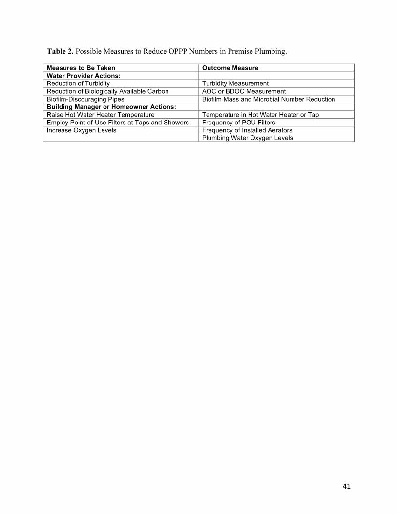
41
Table 2. Possible Measures to Reduce OPPP Numbers in Premise Plumbing.
Measures to Be Taken Outcome Measure Water Provider Actions: Reduction of Turbidity Turbidity Measurement Reduction of Biologically Available Carbon AOC or BDOC Measurement Biofilm-Discouraging Pipes Biofilm Mass and Microbial Number Reduction Building Manager or Homeowner Actions: Raise Hot Water Heater Temperature Temperature in Hot Water Heater or Tap Employ Point-of-Use Filters at Taps and Showers Frequency of POU Filters Increase Oxygen Levels Frequency of Installed Aerators
Plumbing Water Oxygen Levels

42
Appendix 1. Recommended Research Projects Related to Epidemiology and Ecology of OPPPs
1. Determine the prevalence, incidence, and trends of disease caused by OPPPs
2. Determine the dose-response relationship between OPPP numbers and disease
3. Determine whether the microbiome of a distribution system is a determinant of premise plumbing microbiome
4. Determine the role of free-living phagocytic amoebae species on the prevalence, persistence, growth, and survival of OPPPs
5. Determine whether microbial ecologic controls could reduce exposure to OPPPs
6. Determine the contributions of bacterial, viral, and eukaryotic microorganisms to the persis-tence of OPPPs
7. Determine the impact of disinfection methods, including ultraviolet irradiation, on the emer-gence of antibiotic-resistant OPPPs