elderly: musculoskeletal pain in the
TRANSCRIPT

Musculoskeletal Pain in the Elderly: Challenges in Evaluation and Diagnosis
Matt Schlough MDAssistant Professor, UNM Dept of Family and Community Medicine2/17/2021

Goals and ObjectivesGoal:
● Describe common changes and pathologies of the musculoskeletal system with age and
their evaluative challenges
Objectives:
● Outline normal physiologic musculoskeletal changes with age
● Review common musculoskeletal etiologies
● Discuss common pitfalls of diagnostic imaging and labs
● Identify red flags and outline an approach to multifactoral causes

Outline
1. Case Presentation
2. MSK changes with age
3. Challenges in HPI and exam
4. Challenges in laboratory and imaging evaluation
5. General Red flags
6. Case Presentation revisited

Case Presentation
CC: R foot pain
HPI: 79 yo F new patient. Not a great historian.
● Hasn’t been to doctor in >2 yrs ● R foot has been bothering for about a month. Hurts to walk on it. No pain at rest. No
trauma. No new swelling. ● Chronic knee pain R>L. ● Chronic low back pain. Back hurts to lie flat or walk. Feels better sitting. Back pain
interfering with sleep in last few months. ● Lower extremities feel more stiff particularly in the morning and she has been feeling
more tired. ● 15 lbs weight loss in last year● Years of hand pain, having trouble preparing food now. ● Denies any f/c or otherwise feeling ill.

Case Presentation - Cont’d
PMHx: ● DMII, hypothyroid, HTN, chronic low back pain, DJD
Meds: ● metformin, glipizide, amlodipine, levothyroxine, atorvastatin, naprosyn,
MVI, ASA
Soc Hx: ● Recently moved to the area to be closer to her children for more help.
Lives independently in an apt. Widowed. Former smoker. Drinks 1 beer/day.

Case Presentation - Cont’d
Exam
AF, BP 145/90, HR 78, weight 160lbs, BMI 29MSK: UE - prominent DIP joint nodesBack - paralumbar tenderness, mild lumbar spine tendernessLE - 4/5 strength hip flex, 5/5 strength remainder. medial knee joint pain on palpation R>L. moderate R knee effusion, mildly warmFeet - pes planus and R>L hallux valgus. R 1st MTP a little red medially, ankle w/ no focal swelling/warmth/redness, pain posterior/inferior to medial malleolus on palpationNeuro: 1+ DTRs throughout, neg babinski, neg straight leg raise b/l, abnormal monofilament testingExt: 1+ edema in b/l LEs up to mid shins, palpable DP pulsesSkin: diffuse red/brown discoloration on b/l shinsGait: favoring R leg a bit

Pair up 1. Discuss possible differential2. What labs and/or imaging would you want

Case Presentation - Cont’dDifferential:
MSK:● Mechanical ● OA ● RA ● Gout/pseudogout● Tendinopathy● PMR● Spinal stenosis● Compression fracture.
Non-MSK:● Venous insufficiency● PAD● Thyroid ● Malignancy● Cognition

Outline
1. Case Presentation
2. MSK changes with age
3. Challenges in HPI and exam
4. Challenges in laboratory and imaging evaluation
5. General Red flags
6. Case Presentation revisited

MSK Changes with Age
Sarcopenia
1

MSK Changes with Age
● Muscle mass 30% of weight at 30 yrs old. 15% at 75 years old. 2
● Muscle twitch strength loss of 20% at age 60 and 50% at age 80 compared to age 30. 2
● 1.5% loss of muscle/day of inactivity as elderly 2
● Cartilage thins and is more frail 3
● Ligaments and tendons more rigid and brittle 3
● Bone density decreases 3

MSK Changes with Age
4

Common etiologies
● Osteoarthritis
● Tendonitis and bursitis
● Gout and Pseudogout
● Spinal Stenosis
● Osteoporotic fractures
● Rheumatoid arthritis
● Polymyalgia Rheumatica
● Complications from CVA, Parkinsons, etc
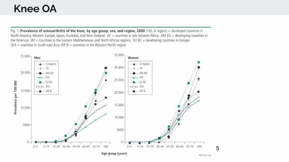
Knee OA
5
Knee OA

Low Back Pain
Low Back Pain
5

Osteoporotic Fractures in Women
5

RA
RA
5

Outline
1. Case Presentation
2. MSK changes with age
3. Challenges in HPI and exam
4. Challenges in laboratory and imaging evaluation
5. General Red flags
6. Case Presentation revisited

Challenges in HPI and Exam
● Underreporting of pain symptoms 6● Atypical symptoms 7● Cognitive impairment 8
○ Undertreatment○ Alternate pain assessment
● Function, Function, Function 9● Difficult exam with mobility issues● Difficult to locate exact pain

Outline
1. Case Presentation
2. MSK changes with age
3. Challenges in HPI and exam
4. Challenges in laboratory and imaging evaluation
5. General Red flags
6. Case Presentation revisited

Case Presentation - Cont’d
Labs: hct 38, BUN 12, Cr 1.3, A1c 8.1, TSH 2.0, WBC 7, ESR 38, CRP 2.8, RF 1:64, ANA 1:120, uric acid 7.4
Imaging:
B/l knee xray - severe OA
Lumbar spine xray - moderate degenerative changes, severe neural foraminal narrowing and facet arthropathy at many levels, no lytic lesions, no compression fracture
Right foot and ankle xray - moderate degenerative changes, no fractures
Differential: Mechanical, OA, RA, gout, pseudogout, tendinopathy, PMR, malignancy, spinal stenosis, compression fracture. Non-MSK - venous insufficiency, PAD, thyroid, malignancy, cognition

Pair up 1. How do you interpret these findings?2. What would you like to do for this patient?

Challenges in Laboratory Evaluation
● Inflammatory markers 10, 11
○ ESR, CRP
● Rheumatalogic markers 10
○ RF, ANA
● Metabolic markers○ Uric Acid 12

Challenges in Imaging Evaluation
13

Challenges in Imaging Evaluation
Back● Pittsburgh Xray study of adults >65 yrs of age w/out chronic back pain vs with back pain 14
○ 95% had disc disease, 93% had facet disease, no correlation of severity with pain● MRI of asymptomatic adults >60 yrs old 15
○ ~100% had DJD, ~21% radiographic evidence of spinal stenosis
Knee● NEJM ~1000 patients age 50-90 received knee MRI. Majority of meniscal tears seen in
asymptomatic patients, more than 50% of age 50-90 had meniscal tears/destruction 16

Challenges in Imaging Evaluation
16

Outline
1. Case Presentation
2. MSK changes with age
3. Challenges in HPI and exam
4. Challenges in laboratory and imaging evaluation
5. General Red flags
6. Case Presentation revisited

Red Flags
● Night pain
● Systemic signs (weight loss, f/c, sweats, etc)
● Neurologic findings
● Concern for specific etiologies: GCA/PMR, Charcot foot

Outline
1. Case Presentation
2. MSK changes with age
3. Challenges in HPI and exam
4. Challenges in laboratory and imaging evaluation
5. General Red flags
6. Case Presentation revisited

Case revisited
● Red flags: Weight loss, painful diabetic foot
● Inflammatory vs non-inflammatory
○ ?Consider trial prednisone, consider Rheumatology referral
● Triage multiple areas:
○ Low back pain - PT, pain control
○ Knee pain - tap knee, consider steroid injection
○ Foot pain - inserts and possible podiatry referral
● Control co-morbidities: compression stockings, change CCB, family assistance

Conclusions
● Red flags?● Inflammatory vs non-inflammatory● Importance of History and Exam (and thus time with patient)● Judicious use of imaging and cautious interpretation of labs● Prevention with activity!

References1. https://www.gmjournal.co.uk/sarcopenia-an-emerging-geriatric-giant2. Gorevic PD. Osteoarthritis: A review of musculoskeletal aging and treatment issues in geriatric patients. Geriatrics 59 (Aug) 28-32. 2004.
3. Merck manual - Effects of aging on the musculoskeletal system.
http://www.merckmanuals.com/home/bone_joint_and_muscle_disorders/biology_of_the_musculoskeletal_system/effects_of_aging_on_the_musculoskeletal_system.html
4. Salaffi F, Farah S, Di Carlo M. Frailty syndrome in rheumatoid arthritis and symptomatic osteoarthritis: an emerging concept in rheumatology. Acta Biomed. 2020;91(2):274-296. Published
2020 May 11. doi:10.23750/abm.v91i2.9094
5. Woolf, Anthony D and Pfleger B. Burden of major musculoskeletal conditions. Bulletin of the World Health Organization 2003;81:646-656.
6. Hershkovitz A, Rothschild B et al. Medical Care Perceptions in Elderly Patients with Musculoskeletal Complaints. IMAJ 2001;3:822±827.
7. National Health and Medical Research Council. Musculoskeletal disorders in the elderly. Australia 1994. https://www.nhmrc.gov.au/_files_nhmrc/publications/attachments/ac4.pdf
8. Cornali C, Franzoni S, Gatti S, Trabucchi M. Diagnosis of chronic pain caused by osteoarthritis and prescription of analgesics in patients with cognitive impairment. J Am Med Dir Assoc. 2006
Jan;7(1):
9. Brown S, Kirkpatrick M, et al. Pain Experience of the Elderly. Pain Management Nursing, Vol 12, No 4 (December), 2011: pp 190-196.
10. Calkins E and Vladutiu A. Practice of Geriatrics. CHAPTER 39 MUSCULOSKELETAL DISORDERS. http://medtextfree.wordpress.com/2010/10/16/chapter-39-musculoskeletal-disorders/
11. Woloshin S, Schwartz LM. Distribution of C-reactive protein values in the United States. N Engl J Med. 2005 Apr 14;352(15):1611-3.
12. Campion E, Glynn R, et al. Asymptomatic hyperuricemia. Risks and consequences in the normative aging study. The American Journal of Medicine. Volume 82, Issue 3, March 1987, Pages
421–426.
13. https://inertiamedical.com/treat-the-patient-not-the-image/14. Hicks G, Morone N, et al. Degenerative Lumbar Disc and Facet Disease in Older Adults:Prevalence and Clinical Correlates. Spine (Phila Pa 1976). 2009 May 20; 34(12): 1301–1306.
15. Kalff R, Ewald C, et al. Degenerative lumbar spinal stenosis in older people—current treatment options. Dtsch Arztebl Int 2013; 110(37): 613–24.
16. Englund et al. Incidental Meniscal Findings on Knee MRI in Middle-Aged and Elderly Persons N Engl J Med. Sep 11, 2008; 359(11): 1108–1115.