electrocardiographic methods for diagnosis and risk stratification in the brugada syndrome
TRANSCRIPT

P.O. Box 2925 Riyadh – 11461KSATel: +966 1 2520088 ext 40151Fax: +966 1 2520718Email: [email protected]
REV
IEW A
RTICLE
Disclosure: Authors have nothing to disclose with regard to commercial support.
Received 1 May 2014; revised 2 June 2014; accepted 26 June 2014.Available online 3 July 2014
⇑ Corresponding author. Address: Cardiac and Vascular SciencesResearch Centre, St. George’s University of London Cranmer Terrace,London SW17 0RE, United Kingdom. Tel.: +44 (0)208 725 3708.E-mail address: [email protected] (V.N. Batchvarov).
1016–7315 � 2014 Production and hosting by Elsevier B.V. on beha
Peer review under responsibility of King Saud University.
URL: www.ksu.edu.sa
http://dx.doi.org/10.1016/j.jsha.2014.06.004
URL: www.sha.org.sa
Electrocardiographic methods for diagnosisand risk stratification in the Brugadasyndrome
lf of King Saud University.
Production and hosting by Elsevier
Abdulrahman Naseef a,b, Elijah R. Behr b, Velislav N. Batchvarov b,⇑
a Center for Health Studies, Prince Sultan Military Medical City, Riyadhb Cardiac and Vascular Sciences Research Centre, St. George’s University of London, London
a Saudi Arabiab United Kingdom
The Brugada syndrome (BrS) is a malignant, genetically-determined, arrhythmic syndrome manifesting as syncope orsudden cardiac death (SCD) in individuals with structurally normal hearts. The diagnosis of the BrS is mainly based onthe presence of a spontaneous or Na + channel blocker induced characteristic, electrocardiographic (ECG) pattern (type1 or coved Brugada ECG pattern) typically seen in leads V1 and V2 recorded from the 4th to 2nd intercostal (i.c.) spaces.This pattern needs to be distinguished from similar ECG changes due to other causes (Brugada ECG phenocopies). Thisreview focuses mainly on the ECG-based methods for diagnosis and arrhythmia risk assessment in the BrS. Presently,the main unresolved clinical problem is the identification of those patients at high risk of SCD who need implantablecardioverter-defibrillator (ICD), which is the only therapy with proven efficacy. Current guidelines recommend ICDimplantation only in patients with spontaneous type 1 ECG pattern, and either history of aborted cardiac arrest ordocumented sustained VT (class I), or syncope of arrhythmic origin (class IIa) because they are at high risk of recurrentarrhythmic events (up to 10% or more annually for those with aborted cardiac arrest). The majority of BrS patients areasymptomatic when diagnosed and considered to have low risk (around 0.5% annually) and therefore not indicated forICD. The majority of SCD victims in the BrS, however, had no symptoms prior to the fatal event and therefore were notprotected with an ICD. While some ECG markers such as QRS fragmentation, infero-lateral early repolarisation, andabnormal late potentials on signal-averaged ECG are known to be linked to increased arrhythmic risk, they are notsufficiently sensitive or specific. Potential novel ECG-based strategies for risk stratification are discussed based oncomputerised methods for depolarisation and repolarisation analysis, a composite approach targeting several majorcomponents of ventricular arrhythmogenesis, and the collection of large digital ECG databases in genotyped BrSpatients and their relatives.
� 2014 Production and hosting by Elsevier B.V. on behalf of King Saud University.
Keywords: Brugada syndrome, Electrocardiogram, Sudden cardiac death, Risk stratification, Genetic arrhythmic
syndromes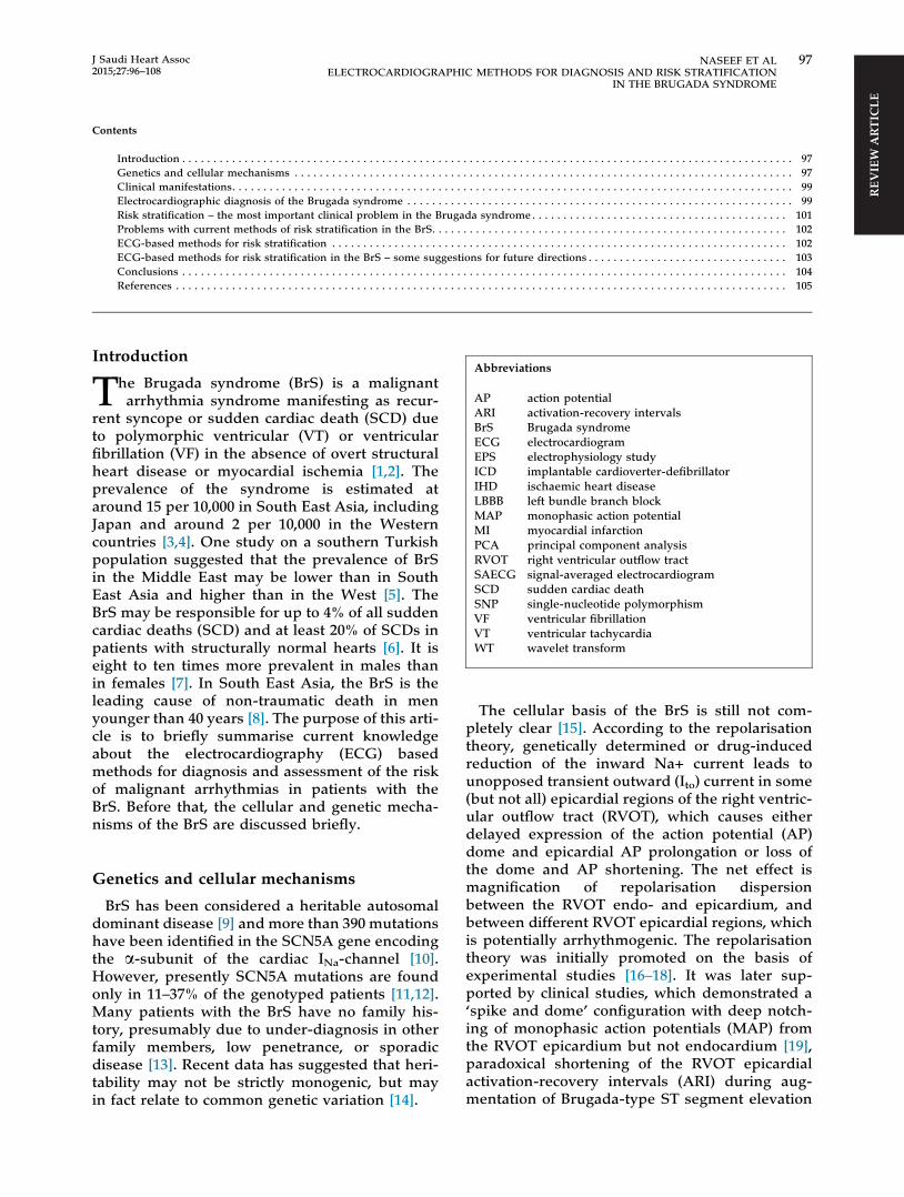
RTIC
LE
J Saudi Heart Assoc2015;27:96–108
NASEEF ET AL 97ELECTROCARDIOGRAPHIC METHODS FOR DIAGNOSIS AND RISK STRATIFICATION
IN THE BRUGADA SYNDROME
Contents
REV
IEW
A
Introduction . . . . . . . . . . . . . . . . . . . . . . . . . . . . . . . . . . . . . . . . . . . . . . . . . . . . . . . . . . . . . . . . . . . . . . . . . . . . . . . . . . . . . . . . . . . . . . . . . . 97Genetics and cellular mechanisms . . . . . . . . . . . . . . . . . . . . . . . . . . . . . . . . . . . . . . . . . . . . . . . . . . . . . . . . . . . . . . . . . . . . . . . . . . . . . . . . 97Clinical manifestations. . . . . . . . . . . . . . . . . . . . . . . . . . . . . . . . . . . . . . . . . . . . . . . . . . . . . . . . . . . . . . . . . . . . . . . . . . . . . . . . . . . . . . . . . . 99Electrocardiographic diagnosis of the Brugada syndrome . . . . . . . . . . . . . . . . . . . . . . . . . . . . . . . . . . . . . . . . . . . . . . . . . . . . . . . . . . . . . . 99Risk stratification – the most important clinical problem in the Brugada syndrome . . . . . . . . . . . . . . . . . . . . . . . . . . . . . . . . . . . . . . . . . 101Problems with current methods of risk stratification in the BrS. . . . . . . . . . . . . . . . . . . . . . . . . . . . . . . . . . . . . . . . . . . . . . . . . . . . . . . . . 102ECG-based methods for risk stratification . . . . . . . . . . . . . . . . . . . . . . . . . . . . . . . . . . . . . . . . . . . . . . . . . . . . . . . . . . . . . . . . . . . . . . . . . 102ECG-based methods for risk stratification in the BrS – some suggestions for future directions . . . . . . . . . . . . . . . . . . . . . . . . . . . . . . . . 103Conclusions . . . . . . . . . . . . . . . . . . . . . . . . . . . . . . . . . . . . . . . . . . . . . . . . . . . . . . . . . . . . . . . . . . . . . . . . . . . . . . . . . . . . . . . . . . . . . . . . . 104References . . . . . . . . . . . . . . . . . . . . . . . . . . . . . . . . . . . . . . . . . . . . . . . . . . . . . . . . . . . . . . . . . . . . . . . . . . . . . . . . . . . . . . . . . . . . . . . . . . 105
Abbreviations
AP action potentialARI activation-recovery intervalsBrS Brugada syndromeECG electrocardiogramEPS electrophysiology studyICD implantable cardioverter-defibrillatorIHD ischaemic heart diseaseLBBB left bundle branch blockMAP monophasic action potentialMI myocardial infarctionPCA principal component analysisRVOT right ventricular outflow tractSAECG signal-averaged electrocardiogramSCD sudden cardiac deathSNP single-nucleotide polymorphismVF ventricular fibrillationVT ventricular tachycardiaWT wavelet transform
Introduction
The Brugada syndrome (BrS) is a malignantarrhythmia syndrome manifesting as recur-
rent syncope or sudden cardiac death (SCD) dueto polymorphic ventricular (VT) or ventricularfibrillation (VF) in the absence of overt structuralheart disease or myocardial ischemia [1,2]. Theprevalence of the syndrome is estimated ataround 15 per 10,000 in South East Asia, includingJapan and around 2 per 10,000 in the Westerncountries [3,4]. One study on a southern Turkishpopulation suggested that the prevalence of BrSin the Middle East may be lower than in SouthEast Asia and higher than in the West [5]. TheBrS may be responsible for up to 4% of all suddencardiac deaths (SCD) and at least 20% of SCDs inpatients with structurally normal hearts [6]. It iseight to ten times more prevalent in males thanin females [7]. In South East Asia, the BrS is theleading cause of non-traumatic death in menyounger than 40 years [8]. The purpose of this arti-cle is to briefly summarise current knowledgeabout the electrocardiography (ECG) basedmethods for diagnosis and assessment of the riskof malignant arrhythmias in patients with theBrS. Before that, the cellular and genetic mecha-nisms of the BrS are discussed briefly.
Genetics and cellular mechanisms
BrS has been considered a heritable autosomaldominant disease [9] and more than 390 mutationshave been identified in the SCN5A gene encodingthe a-subunit of the cardiac INa-channel [10].However, presently SCN5A mutations are foundonly in 11–37% of the genotyped patients [11,12].Many patients with the BrS have no family his-tory, presumably due to under-diagnosis in otherfamily members, low penetrance, or sporadicdisease [13]. Recent data has suggested that heri-tability may not be strictly monogenic, but mayin fact relate to common genetic variation [14].
The cellular basis of the BrS is still not com-pletely clear [15]. According to the repolarisationtheory, genetically determined or drug-inducedreduction of the inward Na+ current leads tounopposed transient outward (Ito) current in some(but not all) epicardial regions of the right ventric-ular outflow tract (RVOT), which causes eitherdelayed expression of the action potential (AP)dome and epicardial AP prolongation or loss ofthe dome and AP shortening. The net effect ismagnification of repolarisation dispersionbetween the RVOT endo- and epicardium, andbetween different RVOT epicardial regions, whichis potentially arrhythmogenic. The repolarisationtheory was initially promoted on the basis ofexperimental studies [16–18]. It was later sup-ported by clinical studies, which demonstrated a‘spike and dome’ configuration with deep notch-ing of monophasic action potentials (MAP) fromthe RVOT epicardium but not endocardium [19],paradoxical shortening of the RVOT epicardialactivation-recovery intervals (ARI) during aug-mentation of Brugada-type ST segment elevation

Figure 2. Resting ECG in a 45-year-old asymptomatic man with BrS,with simultaneous recording of leads V1 and V2 from the 4th, 3rd and2nd intercostal (i.c.) space (leads V1, V2, V13, V23, V12 and V22,respectively) as well as lead V3 in standard position and one (V33)and two (V32) i.c. spaces higher. Note that for all three leads (V1, V2and V3), Brugada type 1 pattern appears either only or more clearlyin the ‘high’ positions (3rd and 2nd i.c. spaces) compared to theirstandard locations. For example, lead V3 shows no Brugada typepattern in the standard position, clear type 2 pattern is one i.c. spacehigher, and marked type 1 pattern, when the electrode is moved, istwo i.c. spaces higher. See the text for details.
REV
IEW A
RTICLE
98 NASEEF ET ALELECTROCARDIOGRAPHIC METHODS FOR DIAGNOSIS AND RISK STRATIFICATIONIN THE BRUGADA SYNDROME
J Saudi Heart Assoc2015;27:96–108
[20], steep AP duration restitution (slope > 1) inthe RVOT[21–23] (both clinically and experimen-tally), and longer ARI in the RVOT epicardiumrecorded from the conus branch of the right coro-nary artery than in the endocardium of patientswith BrS and type 1 ECG pattern, but not incontrols [24].
There is also mounting evidence from experi-mental [22], histopathological [25], computational[25], clinical electrophysiological [23,26], andimaging [27] studies for the presence of conduc-tion abnormalities in the RVOT and their impor-tance for the genesis of ventricular arrhythmiasin BrS [22,23] (depolarisation theory). A mecha-nism explaining the Brugada ECG type solely bydelay of the RVOT activation relative to the restof the RV has also been proposed [28]. The pres-ence of late potentials and prolonged filteredQRS duration on signal-averaged ECG (SAECG)as well as increased notching and fragmentationof the QRS on the standard ECG are linked toincreased arrhythmic risk in BrS [29–32]. A thirdhypothesis unifying the above two explains theBrS with abnormal expression of the neural crestcells during the embryological development ofthe RVOT. This defect in the embryogenesis ofthe RVOT leads to both abnormally augmentedelectrical gradients during repolarisation as wellas to delayed activation of the RVOT [33].
From the electrocardiographic point of view, thecharacteristic elevation of the J point and STsegment of the diagnostic Brugada type 1 pattern(see below) results from early relative (intracellu-lar) positivity of the unaffected zone (RVOT
Figure 1. Leads V1–V3 from a resting 12 lead ECG in a 32-year-oldman with the BrS. Note the typical type 1 pattern in leads V1 and V2and type 2 patterns in lead V3. In this one and in all subsequentfigures, ECGs are displayed at 25 mm/s, 1 cm/mV, unless statedotherwise. All ECGs presented on this one and all subsequent figureshave been originally acquired with high-pass filter of 0.05 Hz.
Figure 3. Snapshot from a positive diagnostic ajmaline test in anasymptomatic 50-year-old man with BrS and non-diagnostic resting12-lead ECG. Lead V1 and V2 leads from the 3rd and 2nd i.c. spacewere not recorded because the test was performed before thediagnostic value of the ‘high’ right precordial leads was established.The ECG on the figure was recorded four minutes after the start of thetest. The standard unipolar lead V2 still displays non-diagnostic type2 pattern, whereas the bipolar leads between the V2 electrode(positive pole) and V4, V5 or V6 electrode (leads V2_4, V_5, V2_6,respectively) display typical type 1 pattern. One minute later,diagnostic type 1 pattern appeared in lead V2 as well (not shown).
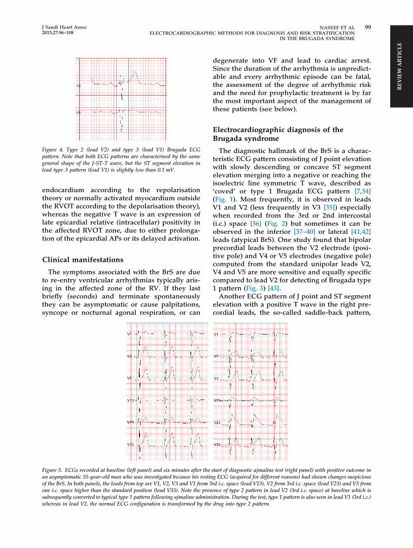
Figure 4. Type 2 (lead V2) and type 3 (lead V1) Brugada ECGpattern. Note that both ECG patterns are characterised by the samegeneral shape of the J-ST-T wave, but the ST segment elevation inlead type 3 pattern (lead V1) is slightly less than 0.1 mV.
REV
IEW
ART
ICLE
J Saudi Heart Assoc2015;27:96–108
NASEEF ET AL 99ELECTROCARDIOGRAPHIC METHODS FOR DIAGNOSIS AND RISK STRATIFICATION
IN THE BRUGADA SYNDROME
endocardium according to the repolarisationtheory or normally activated myocardium outsidethe RVOT according to the depolarisation theory),whereas the negative T wave is an expression oflate epicardial relative (intracellular) positivity inthe affected RVOT zone, due to either prolonga-tion of the epicardial APs or its delayed activation.
Clinical manifestations
The symptoms associated with the BrS are dueto re-entry ventricular arrhythmias typically aris-ing in the affected zone of the RV. If they lastbriefly (seconds) and terminate spontaneouslythey can be asymptomatic or cause palpitations,syncope or nocturnal agonal respiration, or can
Figure 5. ECGs recorded at baseline (left panel) and six minutes after thean asymptomatic 35-year-old man who was investigated because his restinof the BrS. In both panels, the leads from top are V1, V2, V3 and V1 from 3one i.c. space higher than the standard position (lead V33). Note the pressubsequently converted to typical type 1 pattern following ajmaline adminiwhereas in lead V2, the normal ECG configuration is transformed by the
degenerate into VF and lead to cardiac arrest.Since the duration of the arrhythmia is unpredict-able and every arrhythmic episode can be fatal,the assessment of the degree of arrhythmic riskand the need for prophylactic treatment is by farthe most important aspect of the management ofthese patients (see below).
Electrocardiographic diagnosis of theBrugada syndrome
The diagnostic hallmark of the BrS is a charac-teristic ECG pattern consisting of J point elevationwith slowly descending or concave ST segmentelevation merging into a negative or reaching theisoelectric line symmetric T wave, described as‘coved’ or type 1 Brugada ECG pattern [7,34](Fig. 1). Most frequently, it is observed in leadsV1 and V2 (less frequently in V3 [35]) especiallywhen recorded from the 3rd or 2nd intercostal(i.c.) space [36] (Fig. 2) but sometimes it can beobserved in the inferior [37–40] or lateral [41,42]leads (atypical BrS). One study found that bipolarprecordial leads between the V2 electrode (posi-tive pole) and V4 or V5 electrodes (negative pole)computed from the standard unipolar leads V2,V4 and V5 are more sensitive and equally specificcompared to lead V2 for detecting of Brugada type1 pattern (Fig. 3) [43].
Another ECG pattern of J point and ST segmentelevation with a positive T wave in the right pre-cordial leads, the so-called saddle-back pattern,
start of diagnostic ajmaline test (right panel) with positive outcome ing ECG (acquired for different reasons) had shown changes suspiciousrd i.c. space (lead V13), V2 from 3rd i.c. space (lead V23) and V3 fromence of type 2 pattern in lead V2 (3rd i.c. space) at baseline which isstration. During the test, type 1 pattern is also seen in lead V1 (3rd i.c.)drug into type 2 pattern.
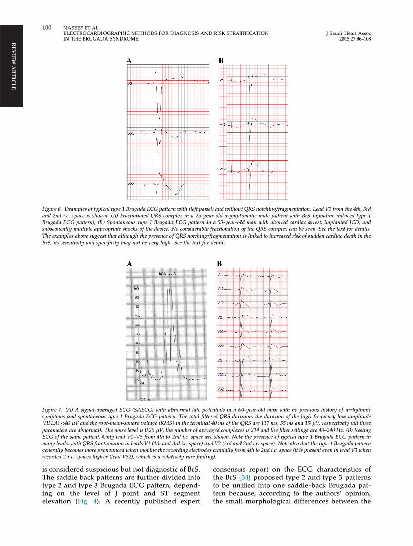
Figure 6. Examples of typical type 1 Brugada ECG pattern with (left panel) and without QRS notching/fragmentation. Lead V1 from the 4th, 3rdand 2nd i.c. space is shown. (A) Fractionated QRS complex in a 25-year-old asymptomatic male patient with BrS (ajmaline-induced type 1Brugada ECG pattern); (B) Spontaneous type 1 Brugada ECG pattern in a 53-year-old man with aborted cardiac arrest, implanted ICD, andsubsequently multiple appropriate shocks of the device. No considerable fractionation of the QRS complex can be seen. See the text for details.The examples above suggest that although the presence of QRS notching/fragmentation is linked to increased risk of sudden cardiac death in theBrS, its sensitivity and specificity may not be very high. See the text for details.
REV
IEW A
RTICLE
Figure 7. (A) A signal-averaged ECG (SAECG) with abnormal late potentials in a 60-year-old man with no previous history of arrhythmicsymptoms and spontaneous type 1 Brugada ECG pattern. The total filtered QRS duration, the duration of the high frequency low amplitude(HFLA) <40 lV and the root-mean-square voltage (RMS) in the terminal 40 ms of the QRS are 137 ms, 55 ms and 15 lV, respectively (all threeparameters are abnormal). The noise level is 0.25 lV, the number of averaged complexes is 214 and the filter settings are 40–240 Hz. (B) RestingECG of the same patient. Only lead V1–V3 from 4th to 2nd i.c. space are shown. Note the presence of typical type 1 Brugada ECG pattern inmany leads, with QRS fractionation in leads V1 (4th and 3rd i.c. space) and V2 (3rd and 2nd i.c. space). Note also that the type 1 Brugada patterngenerally becomes more pronounced when moving the recording electrodes cranially from 4th to 2nd i.c. space (it is present even in lead V3 whenrecorded 2 i.c. spaces higher (lead V32), which is a relatively rare finding).
100 NASEEF ET ALELECTROCARDIOGRAPHIC METHODS FOR DIAGNOSIS AND RISK STRATIFICATIONIN THE BRUGADA SYNDROME
J Saudi Heart Assoc2015;27:96–108
is considered suspicious but not diagnostic of BrS.The saddle back patterns are further divided intotype 2 and type 3 Brugada ECG pattern, depend-ing on the level of J point and ST segmentelevation (Fig. 4). A recently published expert
consensus report on the ECG characteristics ofthe BrS [34] proposed type 2 and type 3 patternsto be unified into one saddle-back Brugada pat-tern because, according to the authors’ opinion,the small morphological differences between the
REV
IEW
ART
ICLE
J Saudi Heart Assoc2015;27:96–108
NASEEF ET AL 101ELECTROCARDIOGRAPHIC METHODS FOR DIAGNOSIS AND RISK STRATIFICATION
IN THE BRUGADA SYNDROME
two patterns had no diagnostic or prognosticsignificance. In the latest HRS/EHRA/APHRSExpert Consensus Statement on the Diagnosisand Management of Patients with InheritedPrimary Arrhythmia Syndromes published inDecember 2013 [7], however, type 2 and type 3Brugada ECG patterns are mentioned separately.Up to 40% of patients with the BrS present withnormal or non-diagnostic, resting ECG [44]. Inthese patients, the diagnostic ‘coved’ ECG patterncan be elicited by IV administration of sodiumchannel blocker (ajmaline, procainamide, flecai-nide) [45,46] (Fig. 5). It is important to distinguishbetween type 2 Brugada ECG pattern and ther0-pattern (incomplete right bundle branch block(IRBBB) pattern) in leads V1 and V2 (especiallywhen recorded from the 3rd or 2nd i.c. space),which can be observed in healthy subjects, andfrequently in athletes [47]. It has been reportedthat a broader angle between the ascending anddescending limb of the r0-wave [48] or a broaderbase of the triangle formed by the two limbs ofthe r0-wave measured at 5 mm from the highestpoint [49,50] can reliably distinguish type 2Brugada ECG pattern from IRBBB pattern.
Currently, the diagnosis of BrS is definite whentype 1 pattern is observed in at least one of leadsV1 and V2 recorded from the 4th, 3rd or 2nd i.c.space, either spontaneously or following adminis-tration of Na+ channel [7]. The presence of genemutations is not considered essential for thediagnosis [2,7].
The classical diagnostic type 1 Brugada ECGpattern needs to be distinguished from similarBrugada-like patterns caused by atypical rightbundle branch block, septal hypertrophy, arrhyth-mogenic right ventricular cardiomyopathy(ARVC), pectus excavatum and other conditions,and also from the transient appearance of the typ-ical Brugada pattern in the cause of various acuteprocesses such as acute ischaemia, Prinzmetalangina, pulmonary embolism, pericarditis, meta-bolic disorders, various medications and others(the so-called Brugada phenocopies) [51].
The use of inappropriate high-pass filters dur-ing ECG acquisition (e.g. non-linear phase, high-pass filter of 0.5 Hz instead of the recommended0.05 Hz) [52] can cause considerable ST segmentdistortion which can mimic type 1 or 2 Brugadapattern [34,53,54].
The ECG in BrS shows considerable dynamicvariability; it can be completely normal at one timeand demonstrate diagnostic type 1 pattern atanother [55]. Vagal influences (slow heart rate,postprandial state, night-time) tend to augment
the J point and ST segment elevation and the type1 pattern [56], whereas exercise and catecholamineinfusion have the opposite effect (in selected BrSpatients the ST segment elevation might becomemore prominent during exercise) [57]. Autonomicinfluences also play important roles in the genesisof malignant arrhythmias because most arrhyth-mic events in BrS occur at night [2], and long RRintervals often precede episodes of VT/VF [58],whereas catecholamine infusion is used as a firstline treatment of such episodes [59]. Patients withBrS have increased incidence (10–53%) of atrialfibrillation (AF) [60,61].
In asymptomatic patients with spontaneous orinduced by Na-channel blockers type 1 pattern,the diagnosis of BrS is supported by the presenceof atrial fibrillation, atrio-ventricular or intraven-tricular conduction abnormalities (first degreeA-V block, left axis deviation of the QRS complex,fragmented QRS (Fig. 6), late potentials on thesignal-averaged ECG (SAECG) (Fig. 7), HVinterval >60 ms), ventricular ectopic beats with leftbundle branch block (LBBB) morphology, andshort (<200 ms) ventricular effective refractoryperiod during electrophysiology study (EPS) [7].
Risk stratification – the most importantclinical problem in the Brugada syndrome
Currently, this is by far the most important andyet unresolved clinical problem in the BrS. It issimilar to one of the main (and also unresolved)problems of modern cardiology – the identifica-tion of patients with ischaemic heart disease(IHD) at high risk of dying suddenly who need aprophylactic implantable cardioverter-defibrilla-tor (ICD).
In BrS patients with a previous history ofarrhythmic syncope or aborted cardiac arrest, theannual event rate of sustained VT or VF is rela-tively high – between 1.9% [62] and 8.8% [63],and between 7.7% [62] and 13.8% [63], respec-tively. They are indicated for ICD, which is thesingle therapy with proven efficacy (class I indica-tion for those with aborted cardiac arrest orspontaneous sustained VT and class IIa for thosewith syncope) [7]. However, the majority of BrSpatients (64% in the largest reported series of1029 BrS patients, the FINGER study [62]) haveno symptoms at the time of establishment of diag-nosis. The annual rate of SCD or sustained VT inthese patients is low – between 0% [64,65] and0.8 [66] (0.5% in the FINGER study [62], 0.4–1%in three Japanese studies [67–69]) and cannot jus-tify ICD implantation in all of them. On the other
REV
IEW A
RTICLE
102 NASEEF ET ALELECTROCARDIOGRAPHIC METHODS FOR DIAGNOSIS AND RISK STRATIFICATIONIN THE BRUGADA SYNDROME
J Saudi Heart Assoc2015;27:96–108
hand, the majority of these patients have structur-ally normal hearts and are young or middle agedwhen diagnosed (median age 45 years in theFINGER study), and therefore the low annual risktranslates into considerable cumulative arrhyth-mic risk for the next several decades of their lifeexpectancy. In fact, the majority of victims ofSCD in BrS come from this ‘low risk’ (accordingto current standards) population. Among patientswith the BrS who have died suddenly, 68% had noprevious history of arrhythmia-related symptomsand therefore had not been protected by ICD[13]. Currently, there are no reliable methods foridentification of these patients (see below). Simi-larly, the largest absolute number of patients withIHD who die suddenly also comes from a patientpopulation considered to have low risk (i.e. withrelatively preserved ejection fraction) [70].
On the other hand, the decision to offer an ICDeven to a BrS patient with a syncope is often diffi-cult because unlike IHD patients, most of themare relatively young, apparently healthy, andwithout any previous awareness of cardiac prob-lems. In addition, it is often very difficult toexclude non-arrhythmic cause of the syncope.Finally, the rate of ICD-related complications(20–30% annually including inappropriate shocks)is higher than the rate of appropriate activation ofthe device (2.6–8% annually) [71–73]. This suggeststhat novel, better methods of risk-stratification areneeded even in the group of patients with cur-rently accepted indications for ICD therapy (thosewith aborted cardiac arrest or syncope).
Problems with current methods of riskstratification in the BrS
While two studies [74,75] reported increasedoccurrence of arrhythmic events in BrS patientswith SCN5A mutations, these findings have notbeen confirmed by other studies [76,77]. Somespecific mutations or common single-nucleotidepolymorphism (SNP) might have prognostic sig-nificance [7]. The genetic analysis is expensive,time-consuming, and available only in specialisedcentres. Male gender [11] and presence of AF[60,61] are linked to increased risk of malignantventricular arrhythmias. The role of programmedventricular stimulation (PVS) during EPS forinduction of VT and/or assessment of the ventric-ular effective refractory period for the purpose ofrisk stratification have been an object of contro-versy and debate since the first descriptions ofthe BrS. While some studies support its value forrisk stratification mainly due to its high negativepredictive value [78,79], most recent studies have
failed to confirm its independent predictive value[80,81,71]. Likewise, in the FINGER study [62],inducibility during EPS did not predict arrhythmicevents in multivariate analysis, whereas in themulticentre PRELUDE study which tested uni-form protocol of PVS during EPS in all 308patients, the rate of arrhythmic events during anaverage follow-up of three years showed no differ-ence between the 126 inducible (3.9%) and the 182non-inducible patients (4.9%) [82]. Currently, itsrole of EPS for risk stratification is accepted onlyas a Class IIb (‘may be considered’) indication[7,83]. The method is inherently limited by itsinvasive character and probably also by the labilenature of the underlying electrophysiologic sub-strate [84].
ECG-based methods for risk stratification
Apart from the spontaneous (as opposed todrug-induced) appearance of the diagnostic type1 pattern, currently no other ECG feature has con-sistently shown an indication for increasedarrhythmic risk.
In contrast with acute myocardial infarction(MI), the number of leads displaying type 1 pat-tern and the degree of J point or ST-segment ele-vation do not seem to correlate with thearrhythmic risk [35,36]. The high (3rd or 2nd i.c.space) positions of leads V1 and V2 are diagnosti-cally more sensitive than their standard positionsin the 4th i.c. space [85–87], but, prognostically,their value is the same [88]. Similar to other dis-eases with intraventricular conduction abnormali-ties such as IHD and cardiomyopathies, thepresence of notched or fragmented QRS [82,32],(Fig. 6) and abnormal late potentials on the sig-nal-averaged ECG (SAECG) [31] (Fig. 7) seems toindicate increased arrhythmic risk in the BrS.However, currently, the presence of notching/fractionation is assessed only visually with arbi-trary descriptive terms (e.g. number of QRSpeaks). The role of the standard time-domainSAECG in BrS is limited by (a) inability to detectconduction abnormalities within the QRS complex,(b) uncertain value in patients with bundle branchblock, and (c) the use of a single-lead ECG com-plex, which is derived from the XYZ orthogonalleads and does not contain any regional informa-tion. Limited data suggest that a number of ECGparameters may indicate BrS patients at higherrisk of malignant arrhythmias: changes in repolar-isation dynamics (QT/RR and Tpeak � Tend/RRintervals relations); [89] deep negative T wave inlead V1; [90] QTc interval more than 460 ms in

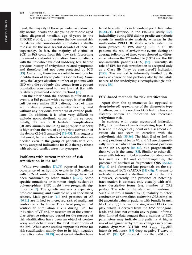
Figure 8. Principal component analysis (PCA) of the ST-T wave(from the J point to the end of the T wave) in the right precordial leadsof ECGs recorded during diagnostic ajmaline test. (A) Positiveajmaline test in a 15-year-old girl with the BrS and a past history ofsyncope of presumable arrhythmic origin. (B) Negative ajmaline testin a 67-year-old asymptomatic man with a family history of BrS andsudden cardiac death. Each bar represents the PCA value (ratio of the2nd/1st eigenvalue) from analysis of one 10-s ECG recording. Two tofive 10-s ECGs were recorded during the test. On the X-axis, time isshown in minutes from the beginning of the test. ECGs were recordedat baseline (b) as well as up to 15 min after the start of the drugadministration. PCA has been applied to the V1–V3 leads (bluediamonds) and to leads V1–V3 recorded from the 3rd i.c. space (red
REV
IEW
ART
ICLE
J Saudi Heart Assoc2015;27:96–108
NASEEF ET AL 103ELECTROCARDIOGRAPHIC METHODS FOR DIAGNOSIS AND RISK STRATIFICATION
IN THE BRUGADA SYNDROME
lead V2 and prolonged Tpeak � Tend interval; [91]dynamic alterations in the amplitude of the STelevation; [92] presence of infero-lateral earlyrepolarisation; [93,94] the presence of horizontal(as opposed to rapidly ascending) ST segmentafter the J point; [68] prolonged PR-interval; [90]the presence of atrial arrhythmias; [95] and aug-mentation of the ST segment elevation duringthe early recovery phase of exercise test [96].
However, none of the above ECG-based param-eters has consistently shown sufficiently high riskpredictive value. As a result, currently the onlyclass I indications for ICD implantation (‘ICD isrecommended’) in patients diagnosed with theBrS endorsed by the 2013 HRS/EHRA/APHRSExpert Consensus Statement [7] is history ofaborted cardiac arrest or documented spontaneoussustained VT (with or without syncope). TheConsensus Statement-endorsed history of syncopeis judged to be likely caused by ventriculararrhythmias only as a Class IIa indication (‘ICDcan be useful’) which reflects the difficulty ofexcluding a non-cardiac origin of syncope. Theguidelines of the Japanese Cardiac Society of2011 accept practically the same Class I indications,whereas for Class IIa indications they require thepresence of at least two of the following risk factors:history of syncope, family history of suddencardiac death, and inducible VF during EPS [97].
In summary, the current indications for ICDimplantation in the BrS are based solely on thepresence of arrhythmia-related symptoms, spon-taneous appearance of type 1 (‘coved’) BrugadaECG pattern and, in some institutions, inducibilityof sustained VT/VF during EPS. While studieshave demonstrated the link between severalECG parameters and increased arrhythmic risk,none of these parameters seem to possess consis-tent and sufficient sensitivity, specificity and pre-dictive value to be used as a risk-stratifier in theBrS. As a result, an unknown number of asymp-tomatic BrS patients are likely to experience theirfirst and potentially lethal arrhythmic event with-out being protected by ICD. Therefore, there is apressing need to develop new, sufficiently sensi-tive and specific, easily applicable methods forrisk stratification in the BrS. ECG-based methodsseem to be the most suitable for this task.
diamonds). Data are presented as mean ± standard deviation (SD) ofall complexes within one 10-s ECG. Generally, higher values reflectmore heterogeneous (and, hence, more abnormal and potentially morearrhythmogenic) ventricular repolarisation. The figures show that theappearance of diagnostic type 1 Brugada ECG pattern during thepositive test (A) is accompanied by a striking increase in the PCAratio, whereas during a negative test, there is practically no change inPCA (the SD deviation bars are hidden within the diamond bars).Adapted from [100].
ECG-based methods for risk stratificationin the BrS – some suggestions for futuredirections
Obvious obstacles to the development of novelmethods for risk stratification in the BrS are the
low rate of arrhythmic events (i.e. endpoint eventsin prospective follow-up), the small number ofpatients in the individual centres (since the prev-alence of the disease outside South East Asia isgenerally low), difficult organisation of big multi-centre prospective studies and, possibly, inherent
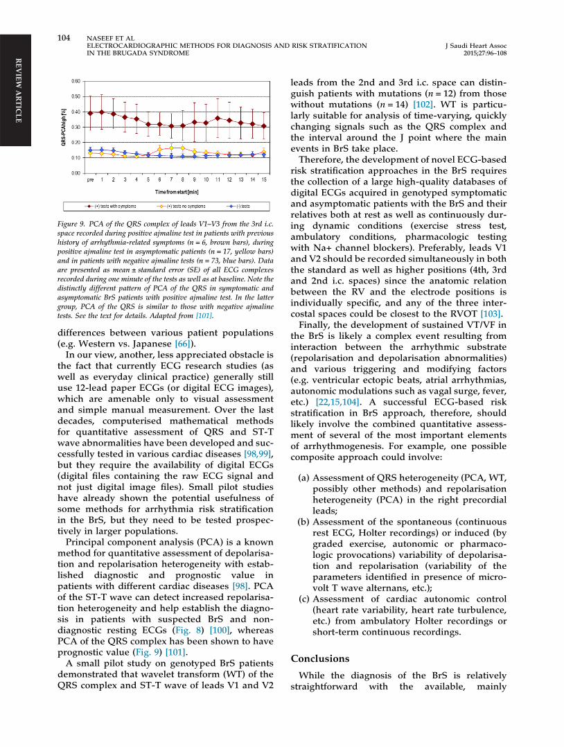
Figure 9. PCA of the QRS complex of leads V1–V3 from the 3rd i.c.space recorded during positive ajmaline test in patients with previoushistory of arrhythmia-related symptoms (n = 6, brown bars), duringpositive ajmaline test in asymptomatic patients (n = 17, yellow bars)and in patients with negative ajmaline tests (n = 73, blue bars). Dataare presented as mean ± standard error (SE) of all ECG complexesrecorded during one minute of the tests as well as at baseline. Note thedistinctly different pattern of PCA of the QRS in symptomatic andasymptomatic BrS patients with positive ajmaline test. In the lattergroup, PCA of the QRS is similar to those with negative ajmalinetests. See the text for details. Adapted from [101].
REV
IEW A
RTICLE
104 NASEEF ET ALELECTROCARDIOGRAPHIC METHODS FOR DIAGNOSIS AND RISK STRATIFICATIONIN THE BRUGADA SYNDROME
J Saudi Heart Assoc2015;27:96–108
differences between various patient populations(e.g. Western vs. Japanese [66]).
In our view, another, less appreciated obstacle isthe fact that currently ECG research studies (aswell as everyday clinical practice) generally stilluse 12-lead paper ECGs (or digital ECG images),which are amenable only to visual assessmentand simple manual measurement. Over the lastdecades, computerised mathematical methodsfor quantitative assessment of QRS and ST-Twave abnormalities have been developed and suc-cessfully tested in various cardiac diseases [98,99],but they require the availability of digital ECGs(digital files containing the raw ECG signal andnot just digital image files). Small pilot studieshave already shown the potential usefulness ofsome methods for arrhythmia risk stratificationin the BrS, but they need to be tested prospec-tively in larger populations.
Principal component analysis (PCA) is a knownmethod for quantitative assessment of depolarisa-tion and repolarisation heterogeneity with estab-lished diagnostic and prognostic value inpatients with different cardiac diseases [98]. PCAof the ST-T wave can detect increased repolarisa-tion heterogeneity and help establish the diagno-sis in patients with suspected BrS and non-diagnostic resting ECGs (Fig. 8) [100], whereasPCA of the QRS complex has been shown to haveprognostic value (Fig. 9) [101].
A small pilot study on genotyped BrS patientsdemonstrated that wavelet transform (WT) of theQRS complex and ST-T wave of leads V1 and V2
leads from the 2nd and 3rd i.c. space can distin-guish patients with mutations (n = 12) from thosewithout mutations (n = 14) [102]. WT is particu-larly suitable for analysis of time-varying, quicklychanging signals such as the QRS complex andthe interval around the J point where the mainevents in BrS take place.
Therefore, the development of novel ECG-basedrisk stratification approaches in the BrS requiresthe collection of a large high-quality databases ofdigital ECGs acquired in genotyped symptomaticand asymptomatic patients with the BrS and theirrelatives both at rest as well as continuously dur-ing dynamic conditions (exercise stress test,ambulatory conditions, pharmacologic testingwith Na+ channel blockers). Preferably, leads V1and V2 should be recorded simultaneously in boththe standard as well as higher positions (4th, 3rdand 2nd i.c. spaces) since the anatomic relationbetween the RV and the electrode positions isindividually specific, and any of the three inter-costal spaces could be closest to the RVOT [103].
Finally, the development of sustained VT/VF inthe BrS is likely a complex event resulting frominteraction between the arrhythmic substrate(repolarisation and depolarisation abnormalities)and various triggering and modifying factors(e.g. ventricular ectopic beats, atrial arrhythmias,autonomic modulations such as vagal surge, fever,etc.) [22,15,104]. A successful ECG-based riskstratification in BrS approach, therefore, shouldlikely involve the combined quantitative assess-ment of several of the most important elementsof arrhythmogenesis. For example, one possiblecomposite approach could involve:
(a) Assessment of QRS heterogeneity (PCA, WT,possibly other methods) and repolarisationheterogeneity (PCA) in the right precordialleads;
(b) Assessment of the spontaneous (continuousrest ECG, Holter recordings) or induced (bygraded exercise, autonomic or pharmaco-logic provocations) variability of depolarisa-tion and repolarisation (variability of theparameters identified in presence of micro-volt T wave alternans, etc.);
(c) Assessment of cardiac autonomic control(heart rate variability, heart rate turbulence,etc.) from ambulatory Holter recordings orshort-term continuous recordings.
Conclusions
While the diagnosis of the BrS is relativelystraightforward with the available, mainly
REV
IEW
ART
ICLE
J Saudi Heart Assoc2015;27:96–108
NASEEF ET AL 105ELECTROCARDIOGRAPHIC METHODS FOR DIAGNOSIS AND RISK STRATIFICATION
IN THE BRUGADA SYNDROME
ECG-based methods, the identification of allpatients at high risk of dying suddenly who needICD implantation is still a largely unresolvedproblem. With the current methods for risk strat-ification, the majority of potential victims of SCDremain unidentified and hence unprotected byICD before the fatal event. While some ECGmarkers of depolarisation and repolarisationabnormalities are known to be linked to increasedarrhythmic risk, their sensitivity or specificityseems low, and they have not been tested in largeprospective studies. The development of novelECG-based methods for risk stratification wouldlikely require the availability of large digital ECGdatabases in patients with BrS and their relatives,application of new computerised methods forquantitative assessment of depolarisation andrepolarisation abnormalities, and the design ofcomposite risk-stratification approaches targetingall major elements of ventricular arrhythmogene-sis in the BrS.
References
[1] Brugada P, Brugada J. Right bundle branch block,persistent ST segment elevation and sudden cardiacdeath: a distinct clinical and electrocardiographicsyndrome. A multicenter report. J Am Coll Cardiol1992;20(6):1391–6.
[2] Antzelevitch C, Brugada P, Borggrefe M, Brugada J,Brugada R, Corrado D, et al. Brugada syndrome: reportof the second consensus conference: endorsed by theHeart Rhythm Society and the European Heart RhythmAssociation. Circulation 2005;111(5):659–70.
[3] Kamakura S. Epidemiology of Brugada syndrome inJapan and rest of the world. J Arrhythm 2013;29(2):52–5.
[4] Juang JM, Huang SK. Brugada syndrome—an under-recognized electrical disease in patients with suddencardiac death. Cardiology 2004;101(4):157–69.
[5] Bozkurt A, Yas D, Seydaoglu G, Acartürk E. Frequency ofBrugada-type ECG pattern (Brugada sign) in SouthernTurkey. Int Heart J 2006;47(4):541–7.
[6] Derval N, Simpson CS, Birnie DH, Healey JS, Chauhan V,Champagne J, et al. Prevalence and characteristics ofearly repolarization in the CASPER registry: cardiacarrest survivors with preserved ejection fractionregistry. J Am Coll Cardiol 2011;58(7):722–8.
[7] Priori SG, Wilde AA, Horie M, Cho Y, Behr ER, Berul C,et al. HRS/EHRA/APHRS expert consensus statement onthe diagnosis and management of patients with inheritedprimary arrhythmia syndromes. Heart Rhythm 2013;10(12):1932–63.
[8] Nademanee K, Veerakul G, Nimmannit S, Chaowakul V,Bhuripanyo K, Likittanasombat K, et al. Arrhythmogenicmarker for the sudden unexplained death syndrome inThai men. Circulation 1997;96(8):2595–600.
[9] Horie M, Ohno S. Genetic basis of Brugada syndrome. JArrhythm 2013;29(2):71–6.
[10] The Gene Connection for the Heart. Genetic mutationsand inherited arrhythmias. <http://www.fsm.it/cardmoc/> [accessed 24.03.14].
[11] Gehi AK, Duong TD, Metz LD, Gomes JA, Mehta D. Riskstratification of individuals with the Brugadaelectrocardiogram: a meta-analysis. J CardiovascElectrophysiol 2006;17(6):577–83.
[12] Kapplinger JD, Tester DJ, Alders M, Benito B, Berthet M,Brugada J, et al. An international compendium ofmutations in the SCN5A-encoded cardiac sodiumchannel in patients referred for Brugada syndromegenetic testing. Heart Rhythm 2010;7(1):33–46.
[13] Raju H, Papadakis M, Govindan M, Bastiaenen R,Chandra N, O’Sullivan A, et al. Low prevalence of riskmarkers in cases of sudden death due to Brugadasyndrome. Relevance to risk stratification in Brugadasyndrome. J Am Coll Cardiol 2011;57(23):2340–5.
[14] Bezzina CR, Barc J, Mizusawa Y, Remme CA, GourraudJB, Simonet F, et al. Common variants at SCN5A-SCN10Aand HEY2 are associated with Brugada syndrome, a raredisease with high risk of sudden cardiac death. Nat Genet2013;45:1044–9.
[15] Morita H, Zipes DP, Wu J. Brugada syndrome: insights ofST elevation, arrhythmogenicity, and risk stratificationfrom experimental observations. Heart Rhythm 2009;6(11Suppl):S34–43.
[16] Yan GX, Antzelevitch C. Cellular basis for the Brugadasyndrome and other mechanisms of arrhythmogenesisassociated with ST-segment elevation. Circulation1999;100(15):1660–6.
[17] Yan GX, Shimizu W, Antzelevitch C. Characteristics anddistribution of M cells in arterially perfused canine leftventricular wedge preparations. Circulation1998;98(18):1921–7.
[18] Antzelevitch C. The Brugada syndrome: ionic basis andarrhythmia mechanisms. J Cardiovasc Electrophysiol2001;12(2):268–72.
[19] Kurita T, Shimizu W, Inagaki M, Suyama K, Taguchi A,Satomi K, et al. The electrophysiologic mechanism of ST-segment elevation in Brugada syndrome. J Am CollCardiol 2002;40(2):330–4.
[20] Simizu W, Aiba T, Kurita T, Kamakura S. Paradoxicabbreviation of repolarisation in epicardium of the rightventricular outflow tract during augmentation ofBrugada-type ST segment elevation. J CardiovascElectrophysiol 2001;12(12):1418–21.
[21] Narayan SM, Kim J, Tate C, Berman BJ. Steep restitutionof ventricular action potential duration and conductionslowing in human Brugada syndrome. Heart Rhythm2007;4(8):1087–9.
[22] Aiba T, Shimizu W, Hidaka I, Uemura K, Noda T, ZhengC, et al. Cellular basis for trigger and maintenance ofventricular fibrillation in the Brugada syndrome model:high-resolution optical mapping study. J Am Coll Cardiol2006;47(10):2074–85.
[23] Lambiase PD, Ahmed AK, Ciaccio EJ, Brugada R, LizotteE, Chaubey S, et al. High-density substrate mapping inBrugada syndrome: combined role of conduction andrepolarization heterogeneities in arrhythmogenesis.Circulation 2009;120(2):106–17.
[24] Nagase S, Kusano KF, Morita H, Nishii N, Banba K,Watanabe A, et al. Longer repolarization in theepicardium at the right ventricular outflow tract causestype 1 electrocardiogram in patients with Brugadasyndrome. J Am Coll Cardiol 2008;51(12):1154–61.
[25] Coronel R, Casini S, Koopmann TT, Wilms-Schopman FJ,Verkerk AO, de Groot JR, et al. Right ventricular fibrosisand conduction delay in a patient with clinical signs ofBrugada syndrome: a combined electrophysiological,genetic, histopathologic, and computational study.Circulation 2005;112(18):2769–77.
[26] Nagase S, Kusano KF, Morita H, Fujimoto Y, Kakishita M,Nakamura K, et al. Epicardial electrogram of the rightventricular outflow tract in patients with the Brugadasyndrome. J Am Coll Cardiol 2002;39(12):1992–5.
[27] Tukkie R, Sogaard P, Vleugels J, de Groot IK, Wilde AA,Tan HL. Delay in right ventricular activation contributesto Brugada syndrome. Circulation 2004;109(10):1272–7.
[28] Meregalli PG, Wilde AA, Tan HL. Pathophysiologicalmechanisms of Brugada syndrome: depolarization

REV
IEW A
RTICLE
106 NASEEF ET ALELECTROCARDIOGRAPHIC METHODS FOR DIAGNOSIS AND RISK STRATIFICATIONIN THE BRUGADA SYNDROME
J Saudi Heart Assoc2015;27:96–108
disorder, repolarization disorder, or more? CardiovascRes 2005;67(3):367–78.
[29] Ikeda T, Sakurada H, Sakabe K, Sakata T, Takami M,Tezuka N, et al. Assessment of noninvasive markers inidentifying patients at risk in the Brugada syndrome:insight into risk stratification. J Am Coll Cardiol2001;37(6):1628–34.
[30] Furushima H, Chinushi M, Hirono T, Sugiura H,Watanabe H, Komura S, et al. Relationship betweendominant prolongation of the filtered QRS duration inthe right precordial leads and clinical characteristics inBrugada syndrome. J Cardiovasc Electrophysiol2005;16(12):1311–7.
[31] Huang Z, Patel C, Li W, Xie Q, Wu R, Zhang L, et al. Roleof signal-averaged electrocardiograms in arrhythmic riskstratification of patients with Brugada syndrome: aprospective study. Heart Rhythm 2009;6(8):1156–62.
[32] Morita H, Kusano KF, Miura D, Nagase S, Nakamura K,Morita ST, et al. Fragmented QRS as a marker ofconduction abnormality and a predictor of prognosis ofBrugada syndrome. Circulation 2008;118(17):1697–704.
[33] Elizari MV, Levi R, Acunzo RS, Chiale PA, Civetta MM,Ferreiro M, et al. Abnormal expression of cardiac neuralcrest cells in heart development: a different hypothesisfor the etiopathogenesis of Brugada syndrome. HeartRhythm 2007;4(3):359–65.
[34] Bayés de Luna A, Brugada J, Baranchuk A, Borggrefe M,Breithardt G, Goldwasser D, et al. Currentelectrocardiographic criteria for diagnosis of Brugadapattern: a consensus report. J Electrocardiol 2012;45(5):433–42.
[35] Richter S, Sarkozy A, Paparella G, Henkens S, Boussy T,Chierchia GB, et al. Number of electrocardiogram leadsdisplaying the diagnostic coved-type pattern in Brugadasyndrome: a diagnostic consensus criterion to be revised.Eur Heart J 2010;31(11):1357–64.
[36] Govindan M, Batchvarov VN, Raju H, Shanmugam N,Bizrah M, Bastiaenen R, et al. Utility of high and standardright precordial leads during ajmaline testing for thediagnosis of Brugada syndrome. Heart 2010;96(23):1904–8.
[37] Takagi M, Aihara N, Takaki H, Taguchi A, Shimizu W,Kurita T, et al. Clinical characteristics of patients withspontaneous or inducible ventricular fibrillation withoutapparent heart disease presenting with J wave and STsegment elevation in inferior leads. J CardiovascElectrophysiol 2000;11(8):844–8.
[38] Chinushi M, Izumi D, Furushima H, Watanabe H, AizawaY. Multiple premature beats triggered ventriculararrhythmias during pilsicainide infusion in a patientwith inferior ST-segment elevation. Pacing ClinElectrophysiol 2006;29(12):1445–8.
[39] Kalla H, Yan GX, Marinchak R. Ventricular fibrillation ina patient with prominent J (Osborn) waves and STsegment elevation in the inferior electrocardiographicleads: a Brugada syndrome variant? J CardiovascElectrophysiol 2000;11(1):95–8.
[40] Batchvarov VN, Govindan M, Camm AJ, Behr ER.Brugada-like changes in the peripheral leads duringdiagnostic ajmaline test in patients with suspectedBrugada syndrome. Pacing Clin Electrophysiol 2009;32(6):695–703.
[41] van den Berg MP, Wiesfeld AC. Brugada syndrome withST-segment elevation in the lateral leads. J CardiovascElectrophysiol 2006;17(9):1035.
[42] Bonakdar H, Haghjoo M, Sadr-Ameli MA. Brugadasyndrome manifested by the typical electrocardiographicpattern both in the right precordial and the high lateralleads. Indian Pacing Electrophysiol J 2008;8(2):137–40.
[43] Batchvarov VN, Govindan M, Macfarlane P, Camm AJ,Behr ER. Diagnostic utility of bipolar precordial leadsduring ajmaline testing for suspected Brugada syndrome.Heart Rhythm 2010;7(2):208–15.
[44] Rivero-Ayerza M, Brugada R, Brugada J, Geelen P,Brugada P. Electrocardiogram of Brugada syndromeand its dynamic patterns. In: Malik M, Camm AJ(editors), Dynamic Electrocardiography. Futura/Blackwell Publishing, NY, 2004; pp. 417–424.
[45] Brugada R, Brugada J, Antzelevitch C, Kirsch GE, PotenzaD, Towbin JA, et al. Sodium channel blockers identify riskfor sudden death in patients with ST-segment elevationand right bundle branch block but structurally normalhearts. Circulation 2000;101(5):510–5.
[46] Rolf S, Bruns HJ, Wichter T, Kirchof P, Ribbing M,Wasmer K, et al. The ajmaline challenge in Brugadasyndrome: diagnostic impact, safety, and recommendedprotocol. Eur Heart J 2003;24(12):1104–12.
[47] Chung EH, McNeely 3rd DE, Gehi AK, Brickner T, EvansS, Pryski E, et al. Brugada-type patterns are easilyobserved in high precordial lead ECGs in collegiateathletes. J Electrocardiol 2014;47(1):1–6.
[48] Chevallier S, Forclaz A, Tenkorang J, Ahmad Y, Faouzi M,Graf D, et al. New electrocardiographic criteria fordiscriminating between Brugada types 2 and 3 patternsand incomplete right bundle branch block. J Am CollCardiol 2011;58(22):2290–8.
[49] Serra G, Baranchuk A, Bayés de Luna A, Brugada J,Goldwasser D, Capulzini L, et al. Newelectrocardiographic criteria to differentiate the Type-2Brugada pattern from electrocardiogram of healthyathletes with r0-wave in leads V1/V2. Europace 2014[Epub ahead of print].
[50] García-Niebla J, Baranchuck A, de Luna AB. TrueBrugada pattern or only high V1–V2 electrodeplacement? J Electrocardiol 2014 [Epub ahead of print].
[51] Baranchuk A, Nguyen T, Ryu MH, Femenía F, Zareba W,Wilde AA, et al. Brugada phenocopy: new terminologyand proposed classification. Ann NoninvasiveElectrocardiol 2012;17(4):299–314.
[52] Kligfield P, Gettes LS, Bailey JJ, Childers R, Deal BJ,Hancock EW, et al. Recommendations for thestandardization and interpretation of theelectrocardiogram part I: the electrocardiogram and itstechnology: a scientific statement from the AmericanHeart Association Electrocardiography and ArrhythmiasCommittee, Council on Clinical Cardiology; theAmerican College of Cardiology Foundation; and theHeart Rhythm Society endorsed by the InternationalSociety for Computerized Electrocardiology. J Am CollCardiol 2007;49(10):1109–27.
[53] García-Niebla J, Serra-Autonell G, Bayés de Luna A.Brugada syndrome electrocardiographic pattern as aresult of improper application of a high pass filter. Am JCardiol 2012;110(2):318–20.
[54] García-Niebla J, Serra-Autonell G, Fiol M, Bayés de LunaA. Brugada electrocardiographic pattern: reality orfiction? J Electrocardiol 2014;47(3):362–3.
[55] <http://www.brugada.org/about/disease-chngsinheartrateexercise.html> [accessed 13.04.14].
[56] Mizumaki K, Fujiki A, Nishida K, Sakabe M, Tsuneda T,Sugao M, et al. Bradycardia-dependent ECG changes inBrugada syndrome. Circ J 2006;70(7):896–901.
[57] Amin AS, de Groot EA, Ruijter JM, Wilde AA, Tan HL.Exercise-induced ECG changes in Brugada syndrome.Circ Arrhythm Electrophysiol 2009;2(5):531–9.
[58] Matsuo K, Shimizu W, Kurita T, Inagaki M, Aihara N,Kamakura S. Dynamic changes of 12-leadelectrocardiograms in a patient with Brugada syndrome.J Cardiovasc Electrophysiol 1998;9(5):508–12.
[59] Veerakul G, Nademanee K. Treatment of electricalstorms in Brugada syndrome. J Arrhythm 2013;29(2):117–24.
[60] Morita H, Kusano-Fukushima K, Nagase S, Fujimoto Y,Hisamatsu K, Fujio H, et al. Atrial fibrillation and atrialvulnerability in patients with Brugada syndrome. J AmColl Cardiol 2002;40(8):1437–44.
REV
IEW
ART
ICLE
J Saudi Heart Assoc2015;27:96–108
NASEEF ET AL 107ELECTROCARDIOGRAPHIC METHODS FOR DIAGNOSIS AND RISK STRATIFICATION
IN THE BRUGADA SYNDROME
[61] Kusano KF, Taniyama M, Nakamura K, Miura D, BanbaK, Nagase S, et al. Atrial fibrillation in patients withBrugada syndrome relationships of gene mutation,electrophysiology, and clinical backgrounds. J Am CollCardiol 2008;51(12):1169–75.
[62] Probst V, Veltmann C, Eckardt L, Meregalli PG, Gaita F,Tan HL, et al. Long-term prognosis of patients diagnosedwith Brugada syndrome: results from the FINGERBrugada syndrome registry. Circulation 2010;121(5):635–43.
[63] Brugada J, Brugada R, Antzelevitch C, Towbin J,Nademanee K, Brugada P. Long-term follow-up ofindividuals with the electrocardiographic pattern ofright bundle-branch block and ST-segment elevation inprecordial leads V1 to V3. Circulation 2002;105(1):73–8.
[64] Letsas KP, Weber R, Efremidis M, Korantzopoulos P,Astheimer K, Charalampous C, et al. Long-termprognosis of asymptomatic individuals with spontaneousor drug-induced type 1 electrocardiographic phenotype ofBrugada syndrome. J Electrocardiol 2011;44(3):346–9.
[65] Champagne J, Philippon F, Gilbert M, Molin F, Blier L,Nault I, et al. The Brugada syndrome in Canada: a uniqueFrench–Canadian experience. Can J Cardiol2007;23(Suppl. B):71B–5B.
[66] Eckardt L, Probst V, Smits JP, Bahr ES, Wolpert C,Schimpf R, et al. Long-term prognosis of individuals withright precordial ST-segment-elevation Brugadasyndrome. Circulation 2005;111(3):257–63.
[67] Atarashi H, Ogawa S. Idiopathic Ventricular FibrillationInvestigators. New ECG criteria for high-risk Brugadasyndrome. Circ J 2003;67(1):8–10.
[68] Takagi M, Aonuma K, Sekiguchi Y, Yokoyama Y, AiharaN, Hiraoka M, et al. The prognostic value of earlyrepolarization (J wave) and ST-segment morphologyafter J wave in Brugada syndrome: multicenter study inJapan. Heart Rhythm 2013;10(4):533–9.
[69] Yokokawa M, Noda T, Okamura H, Satomi K, Suyama K,Kurita T, et al. Comparison of long-term follow-up ofelectrocardiographic features in Brugada syndromebetween the SCN5A-positive probands and theSCN5A-negative probands. Am J Cardiol 2007;100(4):649–55.
[70] Huikuri HV, Castellanos A, Myerburg RJ. Sudden deathdue to cardiac arrhythmias. N Engl J Med 2001;345(20):1473–82.
[71] Sacher F, Probst V, Iesaka Y, Jacon P, Laborderie J,Mizon-Gérard F, et al. Outcome after implantation of acardioverter-defibrillator in patients with Brugadasyndrome: a multicenter study. Circulation 2006;114(22):2317–24.
[72] Sarkozy A, Boussy T, Kourgiannides G, Chierchia GB,Richter S, De Potter T, et al. Long-term follow-up ofprimary prophylactic implantable cardioverter-defibrillator therapy in Brugada syndrome. Eur Heart J2007;28(3):334–44.
[73] Rosso R, Glick A, Glikson M, Wagshal A, Swissa M,Rosenhek S, et al. Outcome after implantation ofcardioverter defibrillator [corrected] in patients withBrugada syndrome: a multicenter Israeli study(ISRABRU). Isr Med Assoc J 2008;10(6):435–9.
[74] Sommariva E, Pappone C, Martinelli Boneschi F, Di RestaC, Rosaria Carbone M, Salvi E, et al. Genetics cancontribute to the prognosis of Brugada syndrome: apilot model for risk stratification. Eur J Hum Genet2013;21(9):911–7.
[75] Nishii N, Ogawa M, Morita H, Nakamura K, Banba K,Miura D, et al. SCN5A mutation is associated with earlyand frequent recurrence of ventricular fibrillation inpatients with Brugada syndrome. Circ J2010;74(12):2572–8.
[76] Priori SG, Napolitano C, Gasparini M, Pappone C, DellaBella P, Giordano U, et al. Natural history of Brugadasyndrome: insights for risk stratification andmanagement. Circulation 2002;105(11):1342–7.
[77] Meregalli PG, Tan HL, Probst V, Koopmann TT, TanckMW, Bhuiyan ZA, et al. Type of SCN5A mutationdetermines clinical severity and degree of conductionslowing in loss-of-function sodium channelopathies.Heart Rhythm 2009;6(3):341–8.
[78] Brugada J, Brugada R, Brugada P. Electrophysiologictesting predicts events in Brugada syndrome patients.Heart Rhythm 2011;8(10):1595–7.
[79] Sarkozy A, Paparella G, Boussy T, Casado-Arroyo R,Yazaki Y, Chierchia GB, et al. The usefulness of theconsensus clinical diagnostic criteria in Brugadasyndrome. Int J Cardiol 2013;167(6):2700–4.
[80] Gasparini M, Priori SG, Mantica M, Coltorti F, GalimbertiP, Ceriotti C, et al. Provocative tests in the Brugadasyndrome: do we have the right tools? Circulation2000;102(Suppl. 2):677.
[81] Eckardt L, Kirchhof P, Schulze-Bahr E, Rolf S, Ribbing M,Loh P, et al. Electrophysiologic investigation in Brugadasyndrome; yield of programmed ventricular stimulationat two ventricular sites with up to three premature beats.Eur Heart J 2002;23(17):1394–401.
[82] Priori SG, Gasparini M, Napolitano C, Della Bella P,Ottonelli AG, Sassone B, et al. Risk stratification inBrugada syndrome: results of the PRELUDE(PRogrammed ELectrical stimUlation preDictive valuE)registry. J Am Coll Cardiol 2012;59(1):37–45.
[83] Zipes DP, Camm AJ, Borggrefe M, Buxton AE, ChaitmanB, Fromer M, et al. ACC/AHA/ESC 2006 Guidelines forManagement of Patients with Ventricular Arrhythmiasand the Prevention of Sudden Cardiac Death: a report ofthe American College of Cardiology/American HeartAssociation Task Force and the European Society ofCardiology Committee for Practice Guidelines.Circulation 2006;114(10):e385–484.
[84] Wichter T, Matheja P, Eckardt L, Kies P, Schäfers K,Schulze-Bahr E, et al. Cardiac autonomic dysfunction inBrugada syndrome. Circulation 2002;105(6):702–6.
[85] Shimizu W, Matsuo K, Takagi M, Tanabe Y, Aiba T,Taguchi A, et al. Body surface distribution and responseto drugs of ST segment elevation in Brugada syndrome:clinical implication of eighty-seven-lead body surfacepotential mapping and its application to twelve-leadelectrocardiograms. J Cardiovasc Electrophysiol2000;11(4):396–404.
[86] Sangwatanaroj S, Prechawat S, Sunsaneewitayakul B,Sitthisook S, Tosukhowong P, Tungsanga K. Newelectrocardiographic leads and the procainamide testfor the detection of the Brugada sign in suddenunexplained death syndrome survivors and theirrelatives. Eur Heart J 2001;22(24):2290–6.
[87] Teijeiro R, Garro HA, Acunzo RS, Albino E, Chiale PA.Recording of high V1–V3 precordial leads may beessential to the diagnosis of Brugada syndrome duringthe ajmaline test. J Cardiovasc Pharmacol Ther2006;11(2):153–5.
[88] Miyamoto K, Yokokawa M, Tanaka K, Nagai T, OkamuraH, Noda T, et al. Diagnostic and prognostic value of atype1 Brugada electrocardiogram at higher (third orsecond) V1 to V2 recording in men with Brugadasyndrome. Am J Cardiol 2007;99(1):53–7.
[89] Sangawa M, Morita H, Nakatsu T, Nishii N, Miura D,Miura A, et al. Abnormal transmural repolarizationprocess in patients with Brugada syndrome. HeartRhythm 2009;6(8):1163–9.
[90] Miyamoto A, Hayashi H, Makiyama T, Yoshino T,Mizusawa Y, Sugimoto Y, et al. Risk determinants inindividuals with a spontaneous type 1 Brugada ECG. CircJ 2011;75(4):844–51.
[91] Castro Hevia J, Antzelevitch C, Tornés Bárzaga F,Dorantes Sánchez M, Dorticós Balea F, Zayas Molina R,et al. Tpeak � Tend and Tpeak � Tend dispersion as riskfactors for ventricular tachycardia/ventricular fibrillationin patients with the Brugada syndrome. J Am Coll Cardiol2006;47(9):1828–34.
REV
IEW A
RTICLE
108 NASEEF ET ALELECTROCARDIOGRAPHIC METHODS FOR DIAGNOSIS AND RISK STRATIFICATIONIN THE BRUGADA SYNDROME
J Saudi Heart Assoc2015;27:96–108
[92] Take Y, Morita H, Wu J, Nagase S, Morita S, Toh N, et al.Spontaneous electrocardiogram alterations predictventricular fibrillation in Brugada syndrome. HeartRhythm 2011;8(7):1014–21.
[93] Kamakura S, Ohe T, Nakazawa K, Aizawa Y, Shimizu A,Horie M, et al. Long-term prognosis of probands withBrugada-pattern ST-elevation in leads V1–V3. CircArrhythm Electrophysiol 2009;2(5):495–503.
[94] Sarkozy A, Chierchia GB, Paparella G, Boussy T, DeAsmundis C, Roos M, et al. Inferior and lateralelectrocardiographic repolarization abnormalities inBrugada syndrome. Circ Arrhythm Electrophysiol2009;2(2):154–61.
[95] Bordachar P, Reuter S, Garrigue S, Caï X, Hocini M, JaïsM, et al. Incidence, clinical implications and prognosis ofatrial arrhythmias in Brugada syndrome. Eur Heart J2004;25(10):879–84.
[96] Makimoto H, Nakagawa E, Takaki H, Yamada Y,Okamura H, Noda T, et al. Augmented ST-segmentelevation during recovery from exercise predicts cardiacevents in patients with Brugada syndrome. J Am CollCardiol 2010;56(19):1576–84.
[97] Shimizu A. Indication of ICD in Brugada syndrome. JArrhythm 2013;29(2):110–6.
[98] Okin PM, Devereux RB, Fabsitz RR, Lee ET, Galloway JM,Howard BV. Principal component analysis of the T waveand prediction of cardiovascular mortality in American
Indians: the strong heart study. Circulation2002;105(6):714–9.
[99] Addison PS. Wavelet transforms and the ECG: a review.Physiol Meas 2005;26(5):R155–99.
[100] Batchvarov VN, Christov II, Bortolan G, Govindan M,Camm AJ, Behr ER. Automatic assessment of rightventricular repolarisation dispersion during diagnosticajmaline test for suspected Brugada syndrome. ComputCardiol 2009;36:297–300.
[101] Batchvarov VN, Christov II, Bortolan G, Behr E. Principalcomponent analysis of the qrs complex during diagnosticajmaline test for suspected Brugada syndrome. ComputCardiol 2010;37:501–4.
[102] Batchvarov VN, Bortolan G, Christov II, Bastiaenen R,Raju H, Naseef A, et al. ECG wavelet analysis for thedetection of gene mutations in patients with BrugadaSyndrome. Comput Cardiol 2011;38:785–8.
[103] Veltmann C, Papavassiliu T, Konrad T, Doesch C,Kuschyk J, Streitner F, et al. Insights into the location oftype I ECG in patients with Brugada syndrome:correlation of ECG and cardiovascular magneticresonance imaging. Heart Rhythm 2012;9(3):414–21.
[104] Morita H, Zipes DP, Lopshire J, Morita ST, Wu J. T wavealternans in an in vitro canine tissue model of Brugadasyndrome. Am J Physiol Heart Circ Physiol2006;291(1):H421–8.