electrochemical and antioxidant properties of biogenic
TRANSCRIPT
Int. J. Electrochem. Sci., 10 (2015) 7905 - 7916
International Journal of
ELECTROCHEMICAL SCIENCE
www.electrochemsci.org
Electrochemical and Antioxidant Properties of Biogenic Silver
Nanoparticles
Arif Ullah Khan
1, Yun Wei
1, Zia Ul Haq
Khan
1, Kamran Tahir
1, Shahab Ullah Khan
2, Aftab Ahmad
1,
Faheem Ullah Khan1, Li Cheng
1, Qipeng Yuan
1*
1*
State Key Laboratory of Chemical Resource Engineering, Beijing University of Chemical
Technology, Beijing, PR China 2Department of Physics, University of Peshawar, 25000, Pakistan
*E-mail: [email protected]
Received: 15 June 2015 / Accepted: 27 July 2015 / Published: 26 August 2015
The Present Bio-synthesis is an efficient, fast, cheap, nontoxic, simple and eco-friendly protocol for
silver nanoparticles synthesis, which is more beneficial than commonly used physiochemical methods.
Here in silver nanoparticles are biosynthesized using Citrus sinensis var. Kozan yerly fruit juice as
reducing and stabilizing agent. The X-ray diffraction (XRD) pattern confirms the crystalline nature of
silver nanoparticles. The high resolution transmission electron microscopy confirmed that the silver
nanoparticles ere of average 4-10 nm particle size. The UV–vis absorption spectrum shows a
characteristic surface plasmon resonance absorption peak of silver nanoparticles at 443 nm. FTIR
confirmed phytochemicals of Citrus sinenses var kazan yerly fruit juice involved in the synthesis and
stabilization of silver nanoparticles. Furthermore, the silver nanoparticles modified electrode (Ag/GC)
exhibited an excellent electro-catalytic activity toward the red-ox reaction of phenolic compounds
(Catechol) and DPPH free radical scavenging activity. The synthesized silver nanoparticles are stable
and comparable in size. These silver nanoparticles showed potential applications in the fields of
electrochemistry, sensors, catalysis, nanodevices and medical.
Keywords: Electro-catalytic, Bio-syntheses, Silver nanoparticles, Citrus sinensis var. Kozan yerly,
Antioxidant
1. INTRODUCTION
The synthesis of noble-metal (Au, Ag and Cu) nanoparticles with controllable particle size and
shape, is of great interest because such nanoparticles patterns provide the possibility of developing
different functional devices with optical, sensing, and electrical properties [1]. Nanoparticles have
great importance than their bulk materials because of their unique mechanical, physical, optical and
Int. J. Electrochem. Sci., Vol. 10, 2015
7906
electromagnetic properties which are based upon size, dispersion and morphology of Nanoparticles.
Nanoparticles exhibit a high surface to volume ratio with decreasing size, which plays an important
role in catalytic activity and other related properties such as antimicrobial activities of Ag
nanoparticles. Chemical method is the most important among the different methods employed for the
syntheses of nano particles [2]. However, the use of lethal chemicals cannot be avoided by these
methods of synthesis of nanoparticles. Platinum, Gold and silver nano particles are broadly used in
daily life such as soaps, shampoos, detergents, cosmetic products, shoes, and toothpastes as well as
medical and pharmaceutical fields [3]. Therefore, it is the need of time to develop green or eco-benign
protocols for nanoparticles synthesis that do not require toxic chemicals and depend upon the naturally
occurring nonlethal compounds. Nanoparticles can be synthesized by biological methods using
microorganisms [4, 5], enzymes [6] and plant extracts (natural compounds) which are considered as
possible green or eco-benign alternatives to chemical and physical methods. The use of plant extracts
for nanoparticles synthesis is favorable and beneficial over other biological methods such as microbial
protocol, because it does not require the complex process of maintaining cell cultures and can also be
suitable to scale up for large scale synthesis [7].
Citrus fruits and derived products from them have been well known to have beneficial effects
on human health owing to their high concentration of vitamin C and bioactive compounds such as
phenolic acid, flavonoid, limonoid, carotenoid and fiber [8, 9]
Plants have a number of reducing agents such as poly phenols and flavonoids etc. which takes
place the reduction of Ag+ ions. These poly phenols and flavonoids are used as antimicrobial and
antioxidant agents by the plants to protect themselves from various pathological conditions. Citrus
sinensis var. kozan yerly fruit juice has been investigated for phytochemical investigation and it was
reported that plant is rich in several poly phenols and other bioactive compounds [10]. In this study we
synthesized Ag nanoparticles using fruit extract of Citrus sinensis var. kozan yerly on time scale (≤
100 min), which is faster or equivalent to some of the currently used chemical methods. The less time
processing, well dispersion, spherical, small sized (4-10nm) nano particles synthesis, efficient
electrocatalytic and antioxidant properties ensures the study prominent than other biological methods
for silver nanoparticles synthesis.
2. MATERIALS AND METHODS
2.1. Preparation of Orange extract
Citrus sinensis var. kozan yerly was purchased from market and peeled out .The juice was
quenched using juice extractor and filtered with whatman No:1 filter paper. The filtrate was
centrifuged again at 10000 rpm for 10 minutes at 4°C to remove the remaining solid materials. The
supernatant was used for the synthesis and stabilization of silver nanoparticles.

Int. J. Electrochem. Sci., Vol. 10, 2015
7907
2.2. Synthesis of Ag nanoparticles
For the synthesis of Ag nanoparticles, 20 ml of Citrus sinensis var. kozan yerly fruit extract
was added to 50 ml of 6×10⁻³
M aqueous solution of AgNO3 (sigma Aldrich) in 100ml beaker. The
beaker was placed on magnetic stirrer and stirred at 25 °C to ensure the complete mixing.
The Ag nanoparticles suspension thus obtained was centrifuged at 10,000 rpm for 15 min
followed by re-dispersion of the pellet in de-ionized water. Then the supernatant was discarded and the
pallet (Ag nanoparticles) was freeze dried using VirTis freeze mobile 6ES freeze drier.
2.3. Characterization studies
UV-2450 spectrophotometer (Shimadzu) at a resolution of 1nm in the wavelength range of
350-800 nm was used to monitor the Biosynthesis of silver nanoparticles. The X-ray diffraction (XRD)
pattern was studied by Rigaku Miniflex X-ray diffracto meter at 10–70°. A Hitachi EDX elemental
microanalysis system and JEOL3010 high resolution transmission electron microscope were used to
study the morphology, size and crystalline nature of the Ag nanoparticles. Infra red (IR) spectrum of
Ag nanoparticle was obtained using the KBr pellet technique on an ABB MB3000 spectrophotometer
where it was scanned between 2000 and 500 cm−1 at a resolution of 4 cm−1 in transmittance mode.
2.4. Assembling of AgNPs modified glassy carbon electrode
The bare glassy carbon electrode (GCE) was polished into a mirror-like surface with 0.5 and
0.05 mm alpha Al2O3 and then rinsed ultrasonically with water bath, so that any physically adsorbed
species is removed. The cleaned GC electrode was modified by dip coating with silver nanoparticles,
using immersion times (2 h) in the colloidal nanoparticles solutions.
2.5. Electrocatlytic activity of AgNPs modified glassy carbon electrode
The modified electrode was electrochemically characterized by cyclic voltammetry (CV) in
0.15M sodium acetate solution as electrolyte aqueous medium. Electrocatalytic activity of biogenic
AgNPs was determined by cyclic voltammetry (CV). The CV responses of phenolic compound
catechol C6H6O2 in 0.15M sodium acetate solution at the glassy carbon electrode (GCE) vs saturated
calomel electrode (SCE) at room temperature and AgNPs assembled GC electrode vs SCE was
monitored.
2.5. Antioxidant assay
Antioxidant assay for Ag nanoparticles was performed as previously reported [11] with a slight
modification. Different concentrations (0.031. 0.062, 0.125, 0.250, 0.5 and 1mg/ml) of Ag
nanoparticles were individually mixed with 0.5ml of 1mM DPPH and incubated in dark for 30
Int. J. Electrochem. Sci., Vol. 10, 2015
7908
minutes. After incubation the absorbance of the samples was determined by UV 1100 spectro-
photometer (MAPADA instruments) at 517nm against methanol as a blank. DPPH methanol reagent
without sample was used as control and Vit.C was used as standard. The percentage of inhibition was
calculated according to the following formula.
% of inhibition= [(Absorbancecontrol -Absorbancetest) / Absorbancecontrol]×100
3. RESULTS AND DISCUSSION
The present work was aimed to investigate the biosynthesis, subsequent characterization,
elecrocatalytic and antioxidant properties of silver nanoparticles. The biosynthesis of silver
nanoparticles was observed based on visual observation of the color change from yellow to dark black
within 30 min, followed by monitoring the rate of biosynthesis using UV spectrophotometer
(Shimadzu 2450). The fruit extract of Citrus sinensis var. kozan yerly in the absence of AgNO3 was
used as control and no color change was observed in control.
This color appears due to SPR in Ag nanoparticles. The characteristic SPR peak was not
observed at the initial stage but after 20 minutes the free electrons of silver nanoparticles give rise to
SPR peak [12].
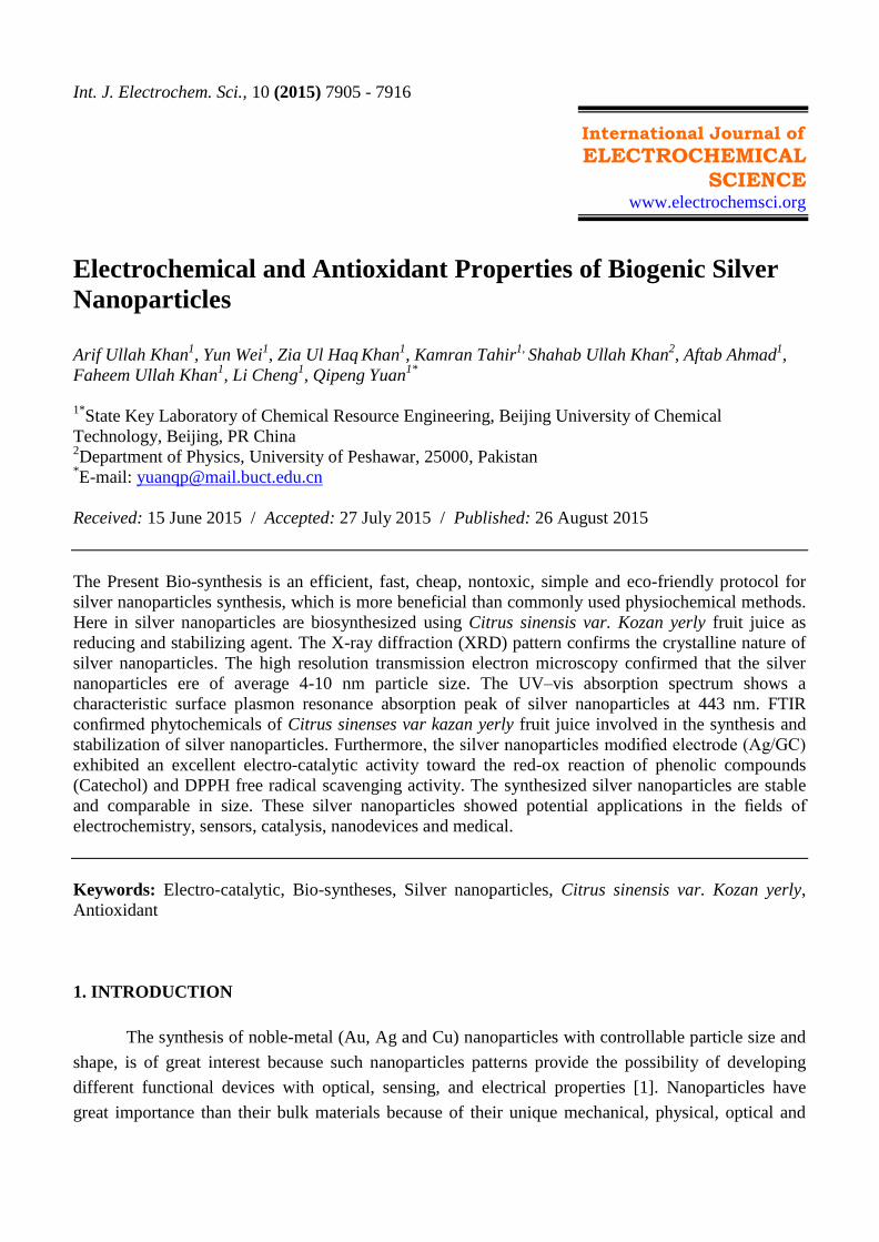
3.1 UV-Vis spectroscopy
The rate of Ag nanoparticles biosynthesis was monitored using UV- visible spectroscopy. The
monoitoring of UV- vis spectra was time dependant (Fig.1). Fig.1 clearly shows that with increase in
contact time the intensity and sharpness of SPR peak increases. SPR showed the maximum absorbance
at 447 nm. SPR peak depends upon the size, shape dispersion and the surrounding media of silver
nanoparticles. SPR gives rise to peak which is well-documented for various metallic nano particles
ranging from 2 nm to 100 nm [13].
Figure 1. Time dependent UV-Vis Spectra of Ag nanoparticles
Int. J. Electrochem. Sci., Vol. 10, 2015
7909
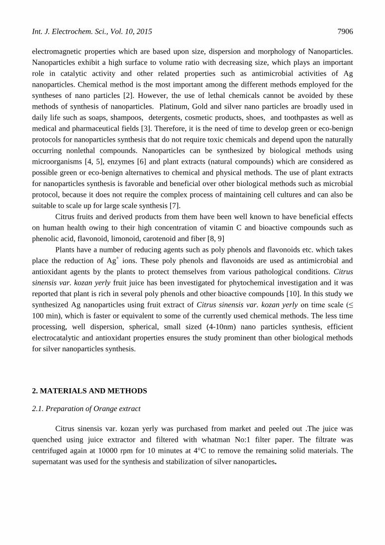
3.2 XRD pattern
X-ray diffraction analysis at 10°-70
° (Fig.2) confirmed the crystalline nature of Ag
nanoparticles. Different numbers of Bragg reflection with 2theta values of 38.03°, 46.18
°, 63.43
°
represent to the (111), (200) and (220) set of lattice planes (Fig.2). Which are in agreement with
JCPDS file number 00-004-0783 and may be indexed to the face centered cubic (fcc) structure of Ag
nanoparticles. The peak corresponding to (111) is more intense than the other planes suggesting that
(111) is the predominant orientation as confirmed by the HRTEM measurements. The peaks which are
unassigned may be due to the crystallization of bioorganic materials that capped on the surface of Ag
nano particles.
Figure 2. XRD pattern of Ag nanoparticles
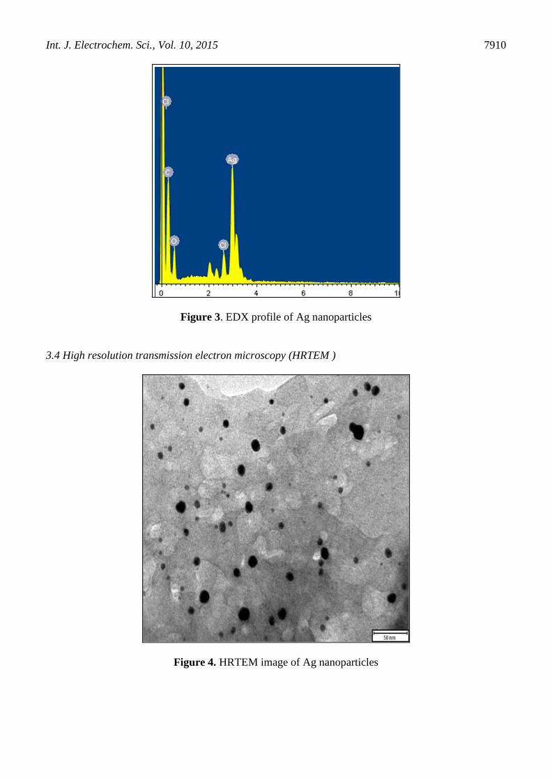
3.3 EDX profile
The EDX profile confirmed the elemental composition of Ag nanoparticles and indicated
strong signals for Silver atoms in the range of 3keV which is typical signal of the absorption of
metallic and spherical Silver nanocrystals due to surface plasmon resonance [14, 15] as shown in fig.3.
There is no ionic silver (Ag+) peak which confirms that all the Ag
+ are reduced to Ag
0 resulting in good
yield of silver nanoparticles The EDX pattern clearly shows that the Ag nano particles are crystalline
in nature, which is caused by the reduction of silver ions using Citrus sinensis var kozan yerly fruit
extract.
Int. J. Electrochem. Sci., Vol. 10, 2015
7910
Figure 3. EDX profile of Ag nanoparticles
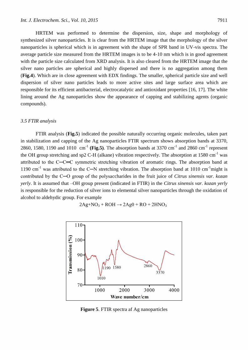
3.4 High resolution transmission electron microscopy (HRTEM )
Figure 4. HRTEM image of Ag nanoparticles
Int. J. Electrochem. Sci., Vol. 10, 2015
7911
HRTEM was performed to determine the dispersion, size, shape and morphology of
synthesized silver nanoparticles. It is clear from the HRTEM image that the morphology of the silver
nanoparticles is spherical which is in agreement with the shape of SPR band in UV-vis spectra. The
average particle size measured from the HRTEM images is to be 4-10 nm which is in good agreement
with the particle size calculated from XRD analysis. It is also cleared from the HRTEM image that the
silver nano particles are spherical and highly dispersed and there is no aggregation among them
(Fig.4). Which are in close agreement with EDX findings. The smaller, spherical particle size and well
dispersion of silver nano particles leads to more active sites and large surface area which are
responsible for its efficient antibacterial, electrocatalytic and antioxidant properties [16, 17]. The white
lining around the Ag nanoparticles show the appearance of capping and stabilizing agents (organic
compounds).
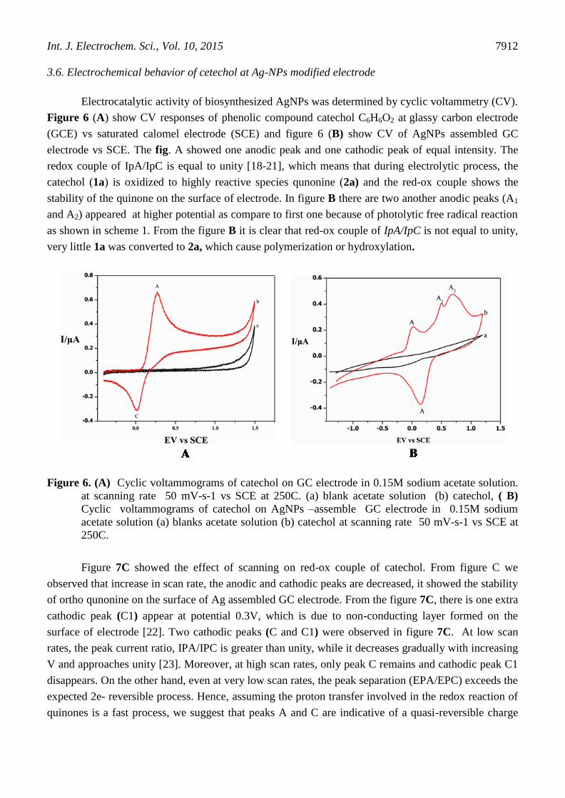
3.5 FTIR analysis
FTIR analysis (Fig.5) indicated the possible naturally occurring organic molecules, taken part
in stabilization and capping of the Ag nanoparticles FTIR spectrum shows absorption bands at 3370,
2860, 1580, 1190 and 1010 cm-1
(Fig.5). The absorption bands at 3370 cm-1
and 2860 cm-1
represent
the OH group stretching and sp2 C-H (alkane) vibration respectively. The absorption at 1580 cm-1
was
attributed to the C─C═C symmetric stretching vibration of aromatic rings. The absorption band at
1190 cm-1
was attributed to the C─N stretching vibration. The absorption band at 1010 cm-1
might is
contributed by the C─O group of the polysaccharides in the fruit juice of Citrus sinensis var. kozan
yerly. It is assumed that –OH group present (indicated in FTIR) in the Citrus sinensis var. kozan yerly
is responsible for the reduction of silver ions to elemental silver nanoparticles through the oxidation of
alcohol to aldehydic group. For example
2Ag+NO3 + ROH → 2Ag0 + RO + 2HNO3
Figure 5. FTIR spectra af Ag nanoparticles
Int. J. Electrochem. Sci., Vol. 10, 2015
7912
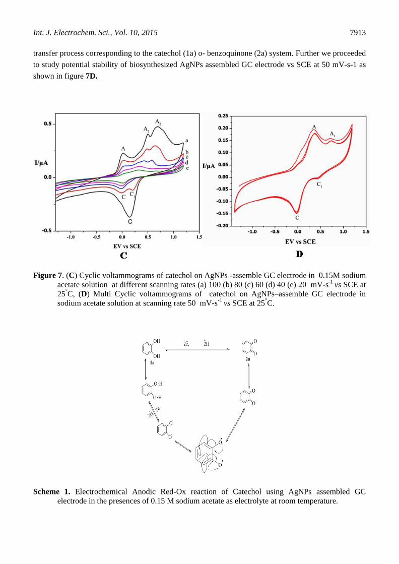
3.6. Electrochemical behavior of cetechol at Ag-NPs modified electrode
Electrocatalytic activity of biosynthesized AgNPs was determined by cyclic voltammetry (CV).
Figure 6 (A) show CV responses of phenolic compound catechol C6H6O2 at glassy carbon electrode
(GCE) vs saturated calomel electrode (SCE) and figure 6 (B) show CV of AgNPs assembled GC
electrode vs SCE. The fig. A showed one anodic peak and one cathodic peak of equal intensity. The
redox couple of IpA/IpC is equal to unity [18-21], which means that during electrolytic process, the
catechol (1a) is oxidized to highly reactive species qunonine (2a) and the red-ox couple shows the
stability of the quinone on the surface of electrode. In figure B there are two another anodic peaks (A1
and A2) appeared at higher potential as compare to first one because of photolytic free radical reaction
as shown in scheme 1. From the figure B it is clear that red-ox couple of IpA/IpC is not equal to unity,
very little 1a was converted to 2a, which cause polymerization or hydroxylation.
Figure 6. (A) Cyclic voltammograms of catechol on GC electrode in 0.15M sodium acetate solution.
at scanning rate 50 mV-s-1 vs SCE at 250C. (a) blank acetate solution (b) catechol, ( B)
Cyclic voltammograms of catechol on AgNPs –assemble GC electrode in 0.15M sodium
acetate solution (a) blanks acetate solution (b) catechol at scanning rate 50 mV-s-1 vs SCE at
250C.
Figure 7C showed the effect of scanning on red-ox couple of catechol. From figure C we
observed that increase in scan rate, the anodic and cathodic peaks are decreased, it showed the stability
of ortho qunonine on the surface of Ag assembled GC electrode. From the figure 7C, there is one extra
cathodic peak (C1) appear at potential 0.3V, which is due to non-conducting layer formed on the
surface of electrode [22]. Two cathodic peaks (C and C1) were observed in figure 7C. At low scan
rates, the peak current ratio, IPA/IPC is greater than unity, while it decreases gradually with increasing
V and approaches unity [23]. Moreover, at high scan rates, only peak C remains and cathodic peak C1
disappears. On the other hand, even at very low scan rates, the peak separation (EPA/EPC) exceeds the
expected 2e- reversible process. Hence, assuming the proton transfer involved in the redox reaction of
quinones is a fast process, we suggest that peaks A and C are indicative of a quasi-reversible charge
Int. J. Electrochem. Sci., Vol. 10, 2015
7913
transfer process corresponding to the catechol (1a) o- benzoquinone (2a) system. Further we proceeded
to study potential stability of biosynthesized AgNPs assembled GC electrode vs SCE at 50 mV-s-1 as
shown in figure 7D.
Figure 7. (C) Cyclic voltammograms of catechol on AgNPs -assemble GC electrode in 0.15M sodium
acetate solution at different scanning rates (a) 100 (b) 80 (c) 60 (d) 40 (e) 20 mV-s-1
vs SCE at
25°C, (D) Multi Cyclic voltammograms of catechol on AgNPs–assemble GC electrode in
sodium acetate solution at scanning rate 50 mV-s-1
vs SCE at 25°C.
Scheme 1. Electrochemical Anodic Red-Ox reaction of Catechol using AgNPs assembled GC
electrode in the presences of 0.15 M sodium acetate as electrolyte at room temperature.
Int. J. Electrochem. Sci., Vol. 10, 2015
7914
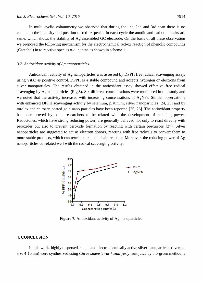
In multi cyclic voltammetry we observed that during the 1st, 2nd and 3rd scan there is no
change in the intensity and position of red-ox peaks. In each cycle the anodic and cathodic peaks are
same, which shows the stability of Ag assembled GC electrode. On the basis of all these observation
we proposed the following mechanism for the electrochemical red-ox reaction of phenolic compounds
(Catechol) in to reactive species o-qunonine as shown in scheme 1.
3.7. Antioxidant activity of Ag nanoparticles
Antioxidant activity of Ag nanoparticles was assessed by DPPH free radical scavenging assay,
using Vit.C as positive control. DPPH is a stable compound and accepts hydrogen or electrons from
silver nanoparticles. The results obtained in the antioxidant assay showed effective free radical
scavenging by Ag nanoparticles (Fig.8). Six different concentrations were monitored in this study and
we noted that the activity increased with increasing concentrations of AgNPs. Similar observations
with enhanced DPPH scavenging activity by selenium, platinum, silver nanoparticles [24, 25] and by
torolex and chitosan coated gold nano particles have been reported [25, 26]. The antioxidant property
has been proved by some researchers to be related with the development of reducing power.
Reductones, which have strong reducing power, are generally believed not only to react directly with
peroxides but also to prevent peroxide formation by reacting with certain precursors [27]. Silver
nanoparticles are suggested to act as electron donors, reacting with free radicals to convert them to
more stable products, which can terminate radical chain reaction. Moreover, the reducing power of Ag
nanoparticles correlated well with the radical scavenging activity.
Figure 7. Antioxidant activity of Ag nanoparticles
4. CONCLUSION
In this work, highly dispersed, stable and electrochemically active silver nanoparticles (average
size 4-10 nm) were synthesized using Citrus sinensis var kozan yerly fruit juice by bio-green method, a
Int. J. Electrochem. Sci., Vol. 10, 2015
7915
cost effective and eco-friendly protocol. The new application of silver nanoparticles onto the GC
electrode is proposed. The uniform and high surface areas onto the GC electrode facilitate its’ use for
electro-catalytic applications. The resulting silver nanoparticles assembled GC electrode show an
excellent electro-catalytic response towards the red-ox reaction of phenolic compounds. Silver
nanoparticles showed very good electro-catalytic performance with low cost and sensitivity. Thus the
AG/GC has attractive electro-catalytic properties for the red-ox reaction of phenolic compounds and
other red-ox reactions or applications. Furthermore silver nanoparticles are found to have significant
DPPH free radical scavenging properties. This could be result as an eco-friendly for electronic
applications, cancer treatment, sensors, drug delivery and other medical applications. The outcomes of
this study illustrate a broad range of applications of electrochemically and bioactive silver
nanoparticles.
ACKNOWLEDGMENTS
The authors are thankful to China Scholarship Council (CSCNo: 2013GXZ036) for support of this
research work.
References
1. Renáta Orináková, Lenka Škantárová, Andrej Orinák, Jakub Demko, Miriam Kupková, Jan T.
Andersson, Int. J. Electrochem. Sci., 8 (2013) 80 - 99
2. S. Sundarrajan, A.R. Chandrasekaran, S. Ramakrishna, J Am Ceram Soc., 93 (2010) 3955-3975.
3. D.R. Bhumkar, H.M. Joshi, M. Sastry, V.B. Pokharkar, Pharm res., 24 (2007) 1415-1426.
4. V.C. Verma, R.N. Kharwar, A.C. Gange, Nanomedicine., 5 (2010) 33-40.
5. Y. Konishi, K. Ohno, N. Saitoh, T. Nomura, S. Nagamine, H. Hishida, Y. Takahashi, T. Uruga, J
Biotech, 128 (2007) 648-653.
6. I. Willner, R. Baron, B. Willner, Adv Mater., 18 (2006) 1109-1120.
7. S.S. Shankar, A. Rai, A. Ahmad, M. Sastry, J. Colloid Interface Sci., 275 (2004) 496-502.
8. . Gorinstein, O. Mart n- elloso, .- . ark, R. Haruenkit, A. Lojek, M. , A. Caspi, I. Libman,
S. Trakhtenberg, Food Chem., 74 (2001) 309-315.
9. M.A. Anagnostopoulou, P. Kefalas, E. Kokkalou, A.N. Assimopoulou, V.P. Papageorgiou, Biomed.
Chromatogr., 19 (2005) 138-148.
10. E. Agcam, A. Aky ld z, G.A. Evrendilek, Food Chem., 143 (2014) 354-361.
11. C.W. Choi, S.C. Kim, S.S. Hwang, B.K. Choi, H.J. Ahn, M.Y. Lee, S.H. Park, S.K. Kim, Plant Sci.,
163 (2002) 1161-1168.
12. M. Noginov, G. Zhu, M. Bahoura, J. Adegoke, C. Small, B. Ritzo, V. Drachev, V. Shalaev, Appl.
Phys. B., 86 (2007) 455-460.
13. M. Gutierrez, A. Henglein, J Phy Chem., 97 (1993) 11368-11370.
14. P. Magudapathy, P. Gangopadhyay, B. Panigrahi, K. Nair, S. Dhara, Phy B: Cond Mat., 299 (2001)
142-146.
15. S. Muthukrishnan, S. Bhakya, T.S. Kumar, M. Rao, Ind Crop and Prod., 63 (2015) 119-124.
16. K. Mogyorósi, N. alázs, D. rankó, E. Tombácz, I. Dékány, A. Oszkó, . ipos, A. Dombi, Appl
Cat B: Env., 96 (2010) 577-585.
17. S. Saha, A. Pal, S. Kundu, S. Basu, T. Pal, Langmuir, 26 (2009) 2885-2893.
18. Nematollahi, D.; Golabi, S. M. J. Electroanal. Chem., 405 (1996) 133-140.
19. Zia Ul Haq Khan, Yongmei Chen, Shafiullah Khan Dandan Kong Mo Heng Liang Pingyu Wan,
Int. J. Electrochem. Sci., Vol. 10, 2015
7916
Int. J. Electrochem. Sci., 9 (2014) 4665 - 4674
20. Nematollahi, D.; Golabi, S. M. J. Electroanal. Chem., 481 (2000) 208-214.
21. Zia Ul Haq Khan, Arif Ullah Khan, Yongmei Chen, Shafiullah Khan, Dandan Kong , Karman
Tahir, Faheem Ullah Khan, Pingyu Wan and Jin Xin. Tetrahedron., 71 (2105) 1674-1678
22. Silverstein R, Webster M FM. Spectrometric., Wiley: NewYork. 6th ed (1998). 217-249
23. D. Nematollahi, S.M. Golabib. J. Electroanal. Chem., 481 (2000) 208–214
24. X. Gao, J. Zhang, L. Zhang, Adv Mater., 14 (2002) 290.
25. Z. Nie, K.J. Liu, C.-J. Zhong, L.-F. Wang, Y. Yang, Q. Tian, Y. Liu, Free Radical Biol. Med., 43
(2007) 1243-1254.
26. D. Raghunandan, M.D. Bedre, S. Basavaraja, B. Sawle, S. Manjunath, A. Venkataraman, Colloids
Surf., B, 79 (2010) 235-240.
27. D.-Z. Xia, X.-F. Yu, Z.-Y. Zhu, Z.-D. Zou, Nat Prod Res., 25 (2011) 1893-1901.
© 2015 The Authors. Published by ESG (www.electrochemsci.org). This article is an open access
article distributed under the terms and conditions of the Creative Commons Attribution license
(http://creativecommons.org/licenses/by/4.0/).