electroencephalographic responses to laserneedle and ... · electroencephalographic responses to...
TRANSCRIPT
The Internet Journal of Laserneedle Medicine TM
Electroencephalographic Responses to Laserneedle and Punctual Stimulation Quantified by Bispectral (BIS) Monitoring: A Pilot Study to Evaluate Methods and Instrumentation
Gerhard Litscher M.Sc., Ph.D., M.D.Sc. Research Unit of Biomedical Engineering Anesthesia and Intensive Care Medicine Medical University of Graz
Lu Wang M.D. Research Unit of Biomedical Engineering Anesthesia and Intensive Care Medicine Medical University of Graz
Ingrid Gaischek M.Sc. Research Unit of Biomedical Engineering Anesthesia and Intensive Care Medicine Medical University of Graz
Citation:
Gerhard Litscher, Lu Wang & Ingrid Gaischek: Electroencephalographic Responses to Laserneedle and Punctual Stimulation Quantified by Bispectral (BIS) Monitoring: A Pilot Study to Evaluate Methods and Instrumentation: The Internet Journal of Laserneedle Medicine. 2007; Volume 1, Number 1.
Abstract
Laserneedle is a new optical stimulation method. Electrical punctual stimulation (P-Stim) also represents a new stimulation method in body and auricular acupuncture. Whereas laserneedle stimulation has been extensively investigated within scientific literature, only few studies exist in referenced journals regarding P-Stim. Fifteen healthy volunteers (mean age ± SD: 29.1 ± 2.8 years; range: 24 - 33 years) were examined in a randomized cross-over study using two different acupuncture schemes, including a placebo stimulation. The EEG-bispectral index (BIS), heart rate (HR) and non-invasive measurement of mean arterial pressure (MAP) were evaluated before, during and after stimulation of sedating acupoints and conditions of placebo. During optical (I) and electrical (II) activation significant (p < 0.05; ANOVA, Tukey test) stimulation induced reductions in BIS-values occurred, however, the combination of both methods (III) and placebo stimulation (IV) resulted in insignificant changes of BIS values. HR and MAP did not show any significant changes during all stimulation procedures (I - IV). Laserneedle- as well as punctual stimulation of sedating acupoints led to significant changes in bioelectric brain activity. However, the response patterns were different and the
combination of both methods did not intensify the effects quantified by BIS values. These neuro-modulating effects require further investigation in a larger population sample.
Introduction
Laserneedle- and punctual stimulation (P-Stim) represent two new methods of stimulation that are based on different modalities (either optical or electrical).
Whereas laserneedle stimulation has been extensively investigated within scientific literature [1,2,3,4,5,6,7], only few studies exist in referenced journals regarding punctual stimulation [8,9,10,11]. Both methods provide new techniques that allow continuous and specific stimulation of body and auricular acupoints.
Goal of this study is to quantify possible neuromodulation of spontaneous bioelectrical brain activity using a bioelectric neuromonitoring method (EEG-bispectralindex = BIS) in healthy volunteers and to compare possible interactions between both stimulation methods in regard to possible effects on the named index.
Methods
Volunteers
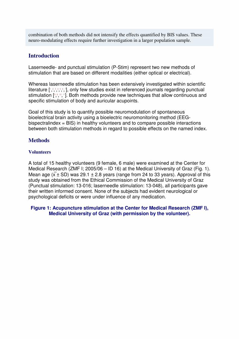
A total of 15 healthy volunteers (9 female, 6 male) were examined at the Center for Medical Research (ZMF I; 2005/06 – ID 16) at the Medical University of Graz (Fig. 1). Mean age (x� ± SD) was 29.1 ± 2.8 years (range from 24 to 33 years). Approval of this study was obtained from the Ethical Commission of the Medical University of Graz (Punctual stimulation: 13-016; laserneedle stimulation: 13-048), all participants gave their written informed consent. None of the subjects had evident neurological or psychological deficits or were under influence of any medication.
Figure 1: Acupuncture stimulation at the Center for Medical Research (ZMF I), Medical University of Graz (with permission by the volunteer).
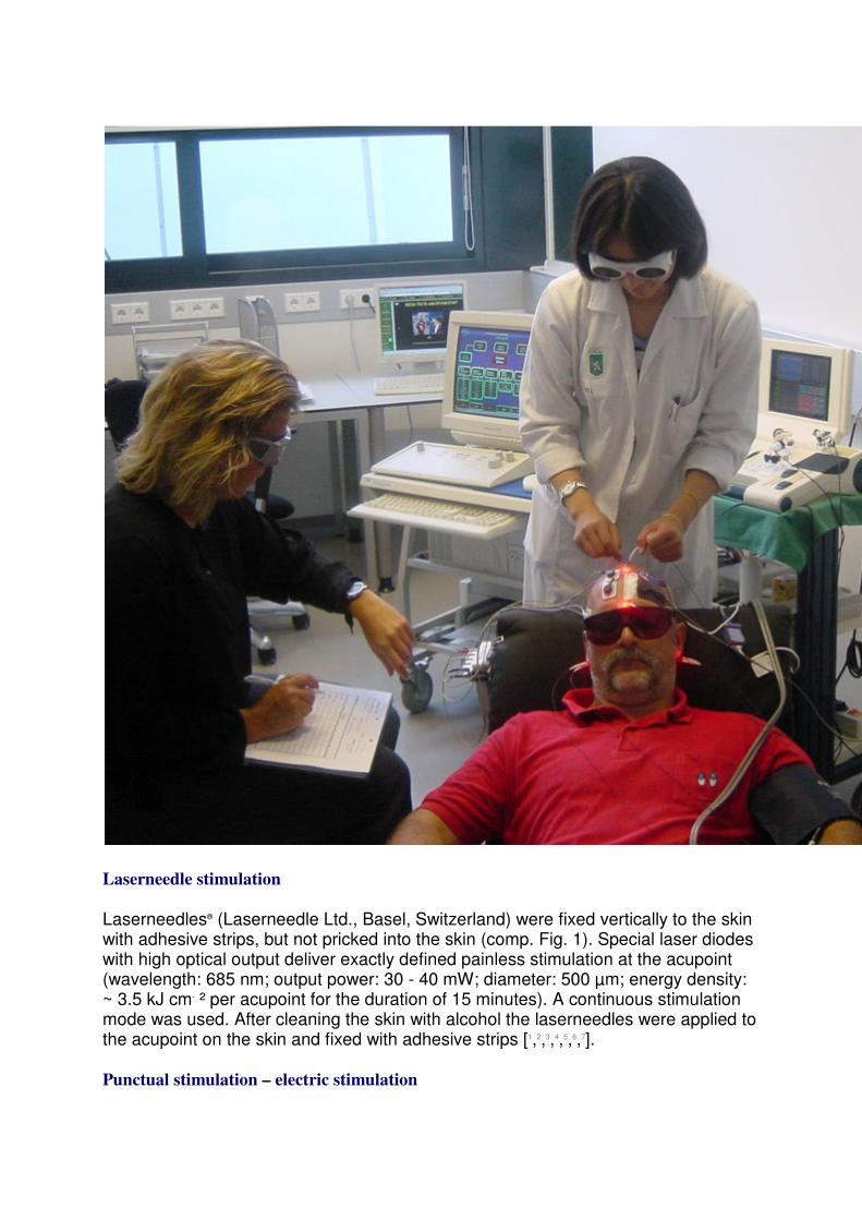
Laserneedle stimulation
Laserneedles® (Laserneedle Ltd., Basel, Switzerland) were fixed vertically to the skin with adhesive strips, but not pricked into the skin (comp. Fig. 1). Special laser diodes with high optical output deliver exactly defined painless stimulation at the acupoint (wavelength: 685 nm; output power: 30 - 40 mW; diameter: 500 µm; energy density: ~ 3.5 kJ cm- ² per acupoint for the duration of 15 minutes). A continuous stimulation mode was used. After cleaning the skin with alcohol the laserneedles were applied to the acupoint on the skin and fixed with adhesive strips [1,2,3,4,5,6,7].
Punctual stimulation – electric stimulation
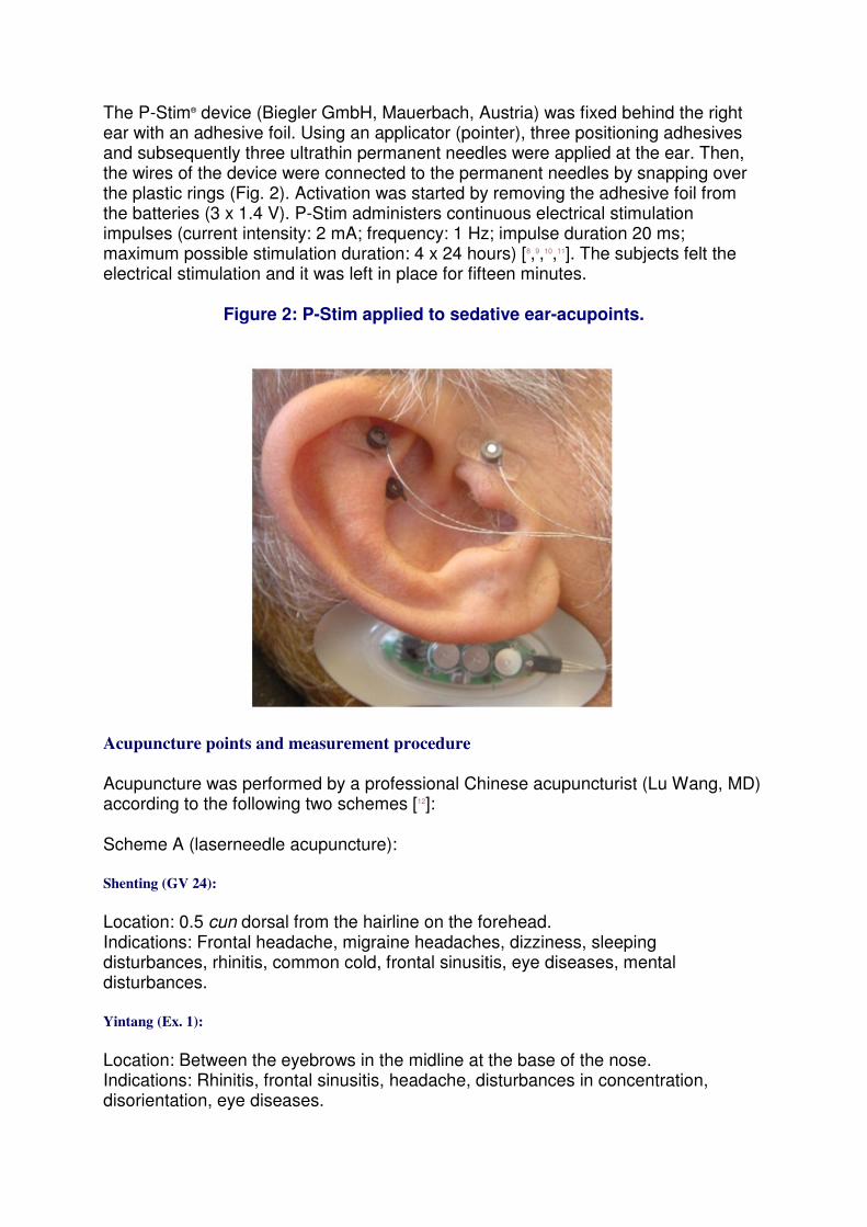
The P-Stim® device (Biegler GmbH, Mauerbach, Austria) was fixed behind the right ear with an adhesive foil. Using an applicator (pointer), three positioning adhesives and subsequently three ultrathin permanent needles were applied at the ear. Then, the wires of the device were connected to the permanent needles by snapping over the plastic rings (Fig. 2). Activation was started by removing the adhesive foil from the batteries (3 x 1.4 V). P-Stim administers continuous electrical stimulation impulses (current intensity: 2 mA; frequency: 1 Hz; impulse duration 20 ms; maximum possible stimulation duration: 4 x 24 hours) [8,9,10,11]. The subjects felt the electrical stimulation and it was left in place for fifteen minutes.
Figure 2: P-Stim applied to sedative ear-acupoints.
Acupuncture points and measurement procedure
Acupuncture was performed by a professional Chinese acupuncturist (Lu Wang, MD) according to the following two schemes [12]:
Scheme A (laserneedle acupuncture):
Shenting (GV 24):
Location: 0.5 cun dorsal from the hairline on the forehead. Indications: Frontal headache, migraine headaches, dizziness, sleeping disturbances, rhinitis, common cold, frontal sinusitis, eye diseases, mental disturbances.
Yintang (Ex. 1):
Location: Between the eyebrows in the midline at the base of the nose. Indications: Rhinitis, frontal sinusitis, headache, disturbances in concentration, disorientation, eye diseases.

Anmian I (Ex. 8):
Location: Between SJ.17 (Yifeng) and Ex.7 (Yiming), 0.5 cun dorsal from SJ.17. Indication: Sleeping disturbances.
Anmian II (Ex.9):
Location: In the middle between Ex.7 (Yiming) and GB.20 (Fengchi). Indication: Sleeping disturbances.
Shenmen (He.7):
Location: At the flexor fold of the wrist, radial from the tendon of M. flexor carpi ulnaris. Indications: Sleeping disturbances, phobic conditions, mental disturbances such as agitation, nervousness, inner imbalance; According to the traditional view, this point calms the Shen or mind and harmonizes the heart.
Scheme B (electrical acupuncture; P-Stim):
Auricular point 55 (Shenmen):
Location: In the Fossa triangularis. Indications: Generally effective sedative and analgesic point.
Auricular point 95 (kidney):
Location: With the earpoints 84 - 104 in the Cacum conchae semicircular around the Crus helicis [12] Indications [13] Psychosomatic disorders with anxiety (existential, sexual), lack in willpower; the kidneys rule willpower.
Auricular point 12 (analgesia):
Location: On the tragus with the earpoints 12 – 21 [12]. Indications: Sedating and analgesic point.
The subjects lay relaxed on a bed with their eyes closed during the entire procedure. In four separate experiments (I – IV), either laserneedle stimulation (I) or electrical stimulation (II) or both (III; Fig. 3) were activated in each case for the duration of 15 minutes (Fig. 4). The fourth condition was a placebo stimulation (IV) which was performed in the same manner as laserneedle stimulation, however, the optical stimulation was not activated in this case. Which method of examination was applied first was selected at random. The resting period between examinations was at least 20 minutes. All subjects underwent all four stimulation modules.
Figure 3: A combination of optical laserneedle stimulation and electrical punctual stimulation performed in this study.
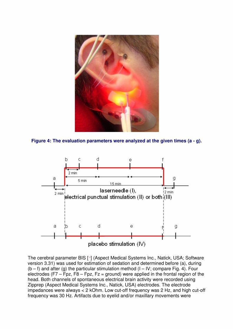
Figure 4: The evaluation parameters were analyzed at the given times (a - g).
The cerebral parameter BIS [14] (Aspect Medical Systems Inc., Natick, USA; Software version 3.31) was used for estimation of sedation and determined before (a), during (b – f) and after (g) the particular stimulation method (I – IV; compare Fig. 4). Four electrodes (F7 – Fpz, F8 – Fpz, Fz = ground) were applied in the frontal region of the head. Both channels of spontaneous electrical brain activity were recorded using Zipprep (Aspect Medical Systems Inc., Natick, USA) electrodes. The electrode impedances were always < 2 kOhm. Low cut-off frequency was 2 Hz, and high cut-off frequency was 30 Hz. Artifacts due to eyelid and/or maxillary movements were
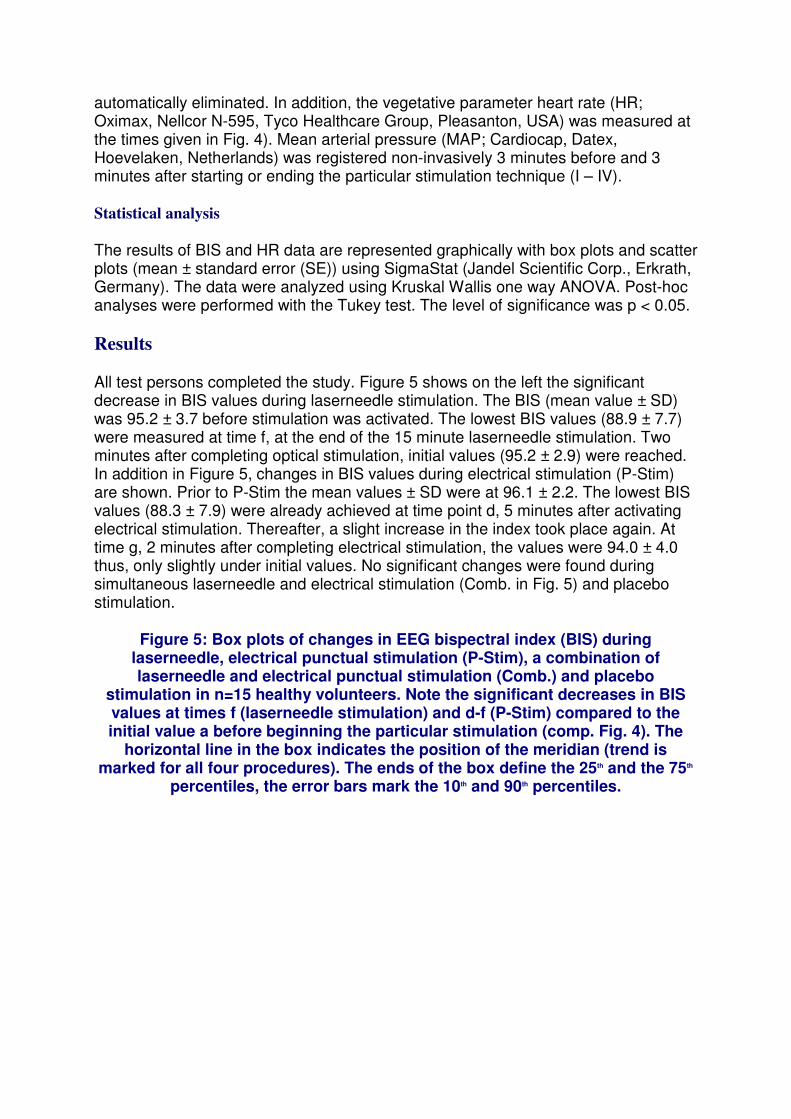
automatically eliminated. In addition, the vegetative parameter heart rate (HR; Oximax, Nellcor N-595, Tyco Healthcare Group, Pleasanton, USA) was measured at the times given in Fig. 4). Mean arterial pressure (MAP; Cardiocap, Datex, Hoevelaken, Netherlands) was registered non-invasively 3 minutes before and 3 minutes after starting or ending the particular stimulation technique (I – IV).
Statistical analysis
The results of BIS and HR data are represented graphically with box plots and scatter plots (mean ± standard error (SE)) using SigmaStat (Jandel Scientific Corp., Erkrath, Germany). The data were analyzed using Kruskal Wallis one way ANOVA. Post-hoc analyses were performed with the Tukey test. The level of significance was p < 0.05.
Results
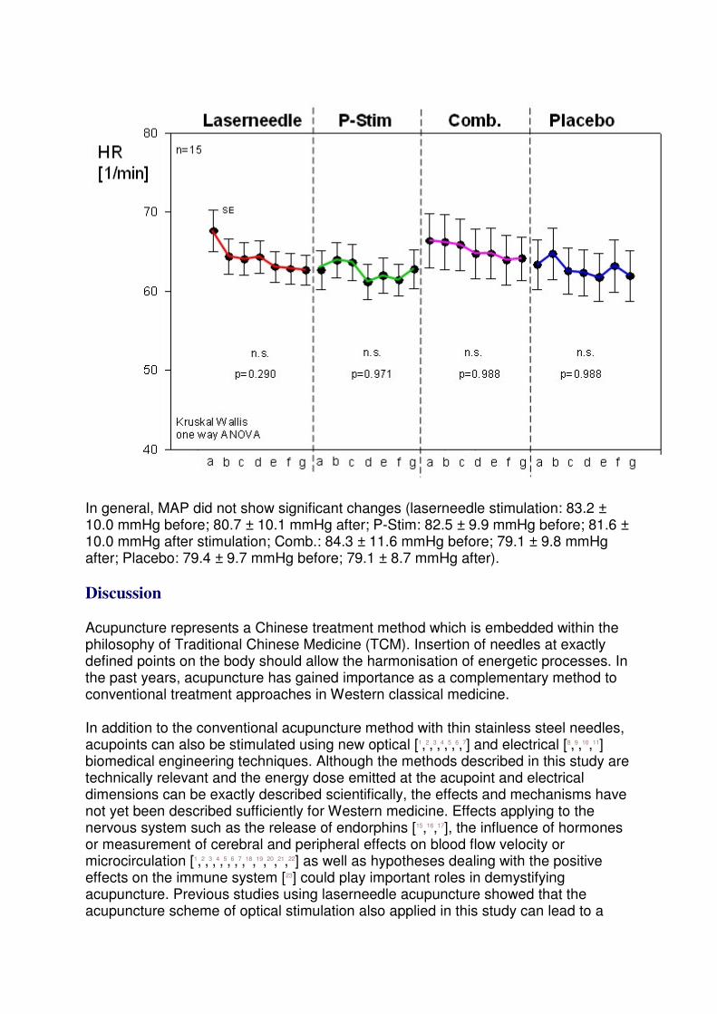
All test persons completed the study. Figure 5 shows on the left the significant decrease in BIS values during laserneedle stimulation. The BIS (mean value ± SD) was 95.2 ± 3.7 before stimulation was activated. The lowest BIS values (88.9 ± 7.7) were measured at time f, at the end of the 15 minute laserneedle stimulation. Two minutes after completing optical stimulation, initial values (95.2 ± 2.9) were reached. In addition in Figure 5, changes in BIS values during electrical stimulation (P-Stim) are shown. Prior to P-Stim the mean values ± SD were at 96.1 ± 2.2. The lowest BIS values (88.3 ± 7.9) were already achieved at time point d, 5 minutes after activating electrical stimulation. Thereafter, a slight increase in the index took place again. At time g, 2 minutes after completing electrical stimulation, the values were 94.0 ± 4.0 thus, only slightly under initial values. No significant changes were found during simultaneous laserneedle and electrical stimulation (Comb. in Fig. 5) and placebo stimulation.
Figure 5: Box plots of changes in EEG bispectral index (BIS) during laserneedle, electrical punctual stimulation (P-Stim), a combination of laserneedle and electrical punctual stimulation (Comb.) and placebo
stimulation in n=15 healthy volunteers. Note the significant decreases in BIS values at times f (laserneedle stimulation) and d-f (P-Stim) compared to the initial value a before beginning the particular stimulation (comp. Fig. 4). The
horizontal line in the box indicates the position of the meridian (trend is marked for all four procedures). The ends of the box define the 25th and the 75th
percentiles, the error bars mark the 10th and 90th percentiles.

The HR data are shown in Figure 6. Whereas the mean value of HR sank within an interval of 2 minutes during laserneedle stimulation from 67.7 ± 10.2 min-1 to 64.4 ± 8.7 min-1 , it increased during electrical stimulation from 62.7 ± 9.6 min-1 to 63.9 ± 8.5 min-1 . Altogether, the changes were not significant in the test subjects.
Figure 6: Means ± standard error (SE) of changes in heart rate (HR) resulting from (from left to right): laserneedle stimulation, electrical punctual stimulation
(P-Stim), a combination of laserneedle and electrical punctual stimulation (Comb.) and placebo stimulation.
In general, MAP did not show significant changes (laserneedle stimulation: 83.2 ± 10.0 mmHg before; 80.7 ± 10.1 mmHg after; P-Stim: 82.5 ± 9.9 mmHg before; 81.6 ± 10.0 mmHg after stimulation; Comb.: 84.3 ± 11.6 mmHg before; 79.1 ± 9.8 mmHg after; Placebo: 79.4 ± 9.7 mmHg before; 79.1 ± 8.7 mmHg after).
Discussion
Acupuncture represents a Chinese treatment method which is embedded within the philosophy of Traditional Chinese Medicine (TCM). Insertion of needles at exactly defined points on the body should allow the harmonisation of energetic processes. In the past years, acupuncture has gained importance as a complementary method to conventional treatment approaches in Western classical medicine.
In addition to the conventional acupuncture method with thin stainless steel needles, acupoints can also be stimulated using new optical [1,2,3,4,5,6,7] and electrical [8,9,10,11] biomedical engineering techniques. Although the methods described in this study are technically relevant and the energy dose emitted at the acupoint and electrical dimensions can be exactly described scientifically, the effects and mechanisms have not yet been described sufficiently for Western medicine. Effects applying to the nervous system such as the release of endorphins [15,16,17], the influence of hormones or measurement of cerebral and peripheral effects on blood flow velocity or microcirculation [1,2,3,4,5,6,7,18,19,20,21,22] as well as hypotheses dealing with the positive effects on the immune system [23] could play important roles in demystifying acupuncture. Previous studies using laserneedle acupuncture showed that the acupuncture scheme of optical stimulation also applied in this study can lead to a

reduction in required anaesthetics [24]. It turned out that state entropy as well as response entropy, both EEG parameters used for estimating the depth of anaesthesia, could be reduced significantly [24]. The scheme mentioned in this present study led to a significant reduction in EEG-BIS values. In a sense, acupuncture can be interpreted as an effective method with practically no side effects which has relaxing and stabilising functional aspects. Thereby possible disadvantages of sedation with drugs such as suppressive effects on the respiratory system can be reduced.
Complementary to laserneedle acupuncture a new type of continuous electrical stimulation at the ear was used in this current study. This miniature device which is fixed behind the ear was developed in Vienna and combines electrical acupuncture with auricular acupuncture by Paul Nogier [25]. According to his theory, specific regions of the body should be influenced with this kind of acupuncture. Repeated stimulation (frequency 1 Hz) should increase the effects of stimulation. First results from pain studies showed that this device can achieve an obvious decrease in pain [8,9].
In principle, the BIS index is an appropriate instrument to assess the depth of anaesthesia or deep stages of hypnosis. Particularly in conditions of sedation, classification of indices of depth of anaesthesia into sedation depths was only performed in few studies [26,27,28,29,30]. BIS values are correlated directly with the majority of the commonly used sedation scores. These are the Ramsay sedation score [28,29], the sedation-agitation scale [27], the Richmond-agitation sedation scale [30] and the Comfort scale [29,30]. In our study, the inclusion of such clinical scores for the assessment of the depth of sedation was not performed in order not to distort the objective data obtained by technical equipment.
Some authors describe that a BIS value of 75 - 85 should indicate that a patient shows no reaction when his name is called loudly with a probability of 96 % [26]. Thus, BIS values within "Conscious sedation” should lie over 80 since based on its definition, communicative contact should be maintained [26]. The lowest BIS mean values of 88.3 (P-Stim) or 88.9 (laserneedle stimulation) determined in our study would fulfil this criterion.
In the present study, a placebo measurement was necessary in order to estimate the influences of the study conditions (lying position, eyes closed) on the level of awareness. In this context it was very interesting that the median BIS value of the placebo measurement was almost constant during the whole recording session (comp. Fig. 5).
The two stimulation methods (optical and electrical stimuli) are based on completely different physical principles and the stimulus intensities are not directly comparable. However, both methods induce similar effects in spontaneous bioelectrical brain activities as shown in this study. Furthermore it should be mentioned that the simultaneous stimulation with both methods did not increase the effects on BIS values. Although the combination did not have an effect which was significant, there is a trend to a similar effect as with the two individual treatments. This fact has to be investigated in further studies.
Acknowledgments
The authors thank Andreas Tiran, Prof. MD, head of the Center for Medical Research (ZMF I), Medical University of Graz, and his team for providing the optimum conditions for our study.
We also thank Detlef Schikora, PhD, from the University of Paderborn, Germany, Mr. Alessandro Visconti (Laserneedle Ltd., Basel, Switzerland) for their competent support regarding the laserneedle system and Mr. Gabriel Landl (Gabriel Medizintechnik, Rohrbach, Austria) for the P-Stim system.
The study was supported in part by the “Zukunftsfonds” of the Styrian Government (project 4071).
Correspondence to
Gerhard Litscher, Prof. MSc PhD MDsc Research Unit of Biomedical Engineering in Anesthesia and Intensive Care Medicine Medical University of Graz Auenbruggerplatz 29 A-8036 Graz / Austria Tel. ++43 316 385-3907 Fax ++43 316 385-3908 E-mail: [email protected] Websites: http://litscher.info http://litscher.at
References
1. Litscher G, Schikora D (Eds): Laserneedle-Acupuncture. Science and Practice. Lengerich, Berlin, Bremen, Miami: Pabst Science Publishers; 2005.
2. Litscher G, Schikora D (Eds): Lasernadel-Akupunktur. Wissenschaft und Praxis. Lengerich, Berlin, Bremen: Pabst Science Publishers; 2004.
3. Litscher G, Schikora D: Cerebral effects of noninvasive laserneedles measured by transorbital and transtemporal Doppler sonography. Lasers Med Sci 2002, 17:289-295.
4. Litscher G, Schikora D: Near-infrared spectroscopy for objectifying cerebral effects of needle and laserneedle acupuncture. Spectroscopy 2002, 16:335-342.
5. Litscher G: Cerebral and peripheral effects of laserneedle-stimulation. Neurol Res 2003, 25:722-728.
6. Litscher G: Effects of acupressure, manual acupuncture and laserneedle acupuncture on EEG bispectral index (BIS) and spectral edge frequency (SEF) in healthy volunteers. Eur J Anaesthesiol 2004, 21:13-19.
7. Litscher G, Rachbauer D, Ropele S, Wang L, Schikora D, Fazekas F, Ebner F: Acupuncture using laser needles modulates brain function: first evidence from functional transcranial Doppler sonography and functional magnetic resonance imaging. Lasers Med Sci 2004, 19:6-11.
8. Sator-Katzenschlager SM, Scharbert G, Kozek-Langenecker SA, Szeles JC, Finster G, Schiesser AW, Heinze G, Kress HG: The short- and long-term benefit in chronic low back pain through adjuvant electrical versus manual auricular acupuncture. Anesth Analg 2004, 98:1359-1364.
9. Sator-Katzenschlager SM, Széles JC, Scharbert G, Michalek-Sauberer A, Kober A, Heinze G, Kozek-Langenecker SA: Electrical stimulation of auricular acupuncture points is more effective than conventional manual auricular acupuncture in chronic cervical pain: a pilot study. Anesth Analg 2003, 97:1469-1473.
10. Széles JC, Litscher G: Objectivation of cerebral effects with a new continuous electrical auricular stimulation technique for pain management. Neurol Res 2004, 26:797-800.
11. Széles JC, Litscher G: Zerebrale Effekte der Punktualstimulation. Schmerz & Akupunktur 2003, 4:4-10.
12. Stux G, Stiller N, Pomeranz B: Akupunktur. Lehrbuch und Atlas. Berlin, Heidelberg, New York: Springer; 1999.
13. Hecker U, Steveling A, Peuker E, Kastner J: Lehrbuch und Repetitorium Akupunktur. Stuttgart: Hippokrates; 2001.
14. Rampil IJ: A primer for EEG signal processing in anesthesia. Anesthesiology 1998, 89:980-1002.
15. Cheng RS, Pomeranz B: Electroacupuncture analgesia could be mediated by at least two pain-relieving mechanisms; endorphin and non-endorphin systems. Life Sci 1979, 25:1957-1962.
16. Han JS, Terenius L: Neurochemical basis of acupuncture analgesia. Annu Rev Pharmacol Toxicol 1982, 22:193-220.
17. Han JS: Opioid and antiopioid peptides: a model of Yin-Yang balance in acupuncture mechanisms of pain modulation. In Clinical Acupuncture: Scientific Basis. Edited by Stux G and Hammerschlag R. Berlin Heidelberg New York: Springer; 2001:51-68.
18. Litscher G, Wang L, Yang NH, Schwarz G: Computer-controlled acupuncture®. Quantification and separation of specific effects. Neurol Res 1999, 21:530-534.
19. Litscher G, Wang L, Yang NH, Schwarz G: Ultrasound-monitored effects of acupuncture on brain and eye. Neurol Res 1999, 21:373-377.
20. Litscher G, Wang L, Wiesner-Zechmeister M: Specific effects of laserpuncture on the cerebral circulation. Lasers Med Sci 2000, 15:57-62.
21. Litscher G, Wang L, Huber E, Nilsson G: Changed skin blood perfusion in the fingertip following acupuncture needle introduction as evaluated by laser Doppler imaging. Lasers Med Sci 2002, 17:19-25.
22. Litscher G: Computer-based objectivation of traditional Chinese-, ear-, and Korean hand acupuncture: Needle-induced changes of regional cerebral blood flow velocity. Neurol Res 2002, 24:377-380.
23. Gollub RL, Hui KK, Stefano GB: Acupuncture: pain management coupled to immune stimulation. Zhongguo Yao Li Xue Bao 1999, 20:769-777.
24. Litscher G: Electroencephalogram-entropy and acupuncture. Anesth Analg 2006, 102:1745-1751.
25. Helling R, Feldmeier M: Aurikulomedizin nach Nogier. Stuttgart: Hippokrates; 1999.
26. Weixler D: Sedierungszustände - Sedierungseffekte. In Praxis der Sedierung. Edited by Weixler D and Paulitsch K. Wien: Facultas Universitätsverlag; 2002:21-38.
27. Simmons L, Riker R, Prato S, Fraser GL: Assessing sedation during intensive care unit mechanical ventilation with the Bispectral Index and the Sedation-Agitation Scale. Crit Care Med 1999, 27(8):1499-1504.
28. Consales G, Chelazzi C, Rinaldi S, De Gaudio AR: Bispectral Index compared to Ramsay score for sedation monitoring in intensive care units. Minerva Anestesiol 2006, 72(5):329-336.
29. Roustan JP, Valette S, Aubas P, Rondouin G, Capdevila X: Can electroencephalographic analysis be used to determine sedation levels in critically ill patients? Anesth Analg 2005. 101(4):1141-1151.
30. Turkmen A, Altan A, Turgut N, Vatansever S, Gokkaya S: The correlation between the richmond agitation-sedation scale and bispectral index during dexmedetomidine sedation. Eur J Anaesthesiol 2006, 23(4):300-304.
References