electron tomography of frozen-hydrated isolated triad junctions · 2017-01-01 · ogy. electron...
TRANSCRIPT

Electron Tomography of Frozen-Hydrated Isolated Triad Junctions
T. Wagenknecht,*† C.-E. Hsieh,* B. K. Rath,* S. Fleischer,‡ and M. Marko**Wadsworth Center, New York State Department of Health, Empire State Plaza, Albany, New York 12201-0509 USA; †Department ofBiomedical Sciences, State University of New York, Empire State Plaza, Albany, New York 12201-0509 USA; and ‡Department ofBiological Sciences, Vanderbilt University, Nashville, Tennessee 37235 USA
ABSTRACT Cryoelectron microscopy and tomography have been applied for the first time to isolated, frozen-hydratedskeletal muscle triad junctions (triads) and terminal cisternae (TC) vesicles derived from sarcoplasmic reticulum. Isolatedtriads were selected on the basis of their appearance as two spherical TC vesicles attached to opposite sides of a flattenedvesicle derived from a transverse tubule (TT). Foot structures (ryanodine receptors) were resolved within the gap between theTC vesicles and TT vesicles, and some residual ordering of the receptors into arrays was apparent. Organized dense layers,apparently containing the calcium-binding protein calsequestrin, were found in the lumen of TC vesicles underlying the footstructures. The lamellar regions did not directly contact the sarcoplasmic reticulum membrane, thereby creating an �5-nm-thick zone that potentially constitutes a subcompartment for achieving locally elevated [Ca2� ] in the immediate vicinity of theCa2�-conducting ryanodine receptors. The lumen of the TT vesicles contained globular mass densities of unknown origin,some of which form cross-bridges that may be responsible for the flattened appearance of the transverse tubules whenviewed in cross-section. The spatial relationships among the TT membrane, ryanodine receptors, and calsequestrin-containing assemblage are revealed under conditions that do not use dehydration, heavy-metal staining, or chemical fixation,thus exemplifying the potential of cryoelectron microscopy and tomography to reveal structural detail of complex subcellularstructures.
INTRODUCTION
In striated muscle, action potentials, initiated at neuromus-cular junctions, stimulate contraction by a signal-transduc-tion process known as excitation contraction (EC) coupling.EC coupling occurs at specialized regions where the intra-cellular sarcoplasmic reticulum forms junctions with thesarcolemma or, more frequently, with tubular invaginationsof the sarcolemma known as transverse tubules (Franzini-Armstrong and Jorgensen, 1994; Flucher and Franzini-Arm-strong, 1996). In skeletal muscle, a region of transversetubule (TT) forms junctions with two terminal cisternaeregions of the sarcoplasmic reticulum (SR), hence the termtriad junction for these regions.
Two multisubunit protein complexes are thought to bepreeminent in the mechanism of EC coupling. The dihydro-pyridine receptor (DHPR), a voltage-activated calciumchannel, in the sarcolemmal/TT system functions as thesensor of transmembrane voltage fluctuations, particularlythe depolarization wave associated with an action potential.Upon receiving a signal from the DHPR, the ryanodinereceptor (RyR) releases Ca2� from the SR into the cyto-plasm. Recent studies show that communication betweenthe RyR and DHPR is bidirectional (Nakai et al., 1996,1998; Grabner et al., 1999). The increased cytoplasmicCa2� binds to troponin, a component of the muscle fila-ments that acts as a switch and stimulates muscle contrac-tion. Other protein components occur at triad junctions, and
more continue to be discovered; ostensibly they play eitherregulatory or structural roles (Mackrill, 1999). For example,calsequestrin, a Ca2�-binding protein that is enriched in thelumen of the SR, probably interacts with RyRs either di-rectly or indirectly via the integral membrane proteins,triadin and junctin. Recently, two additional proteins havebeen implicated as serving structural roles in the formationor maintenance of TT:SR junctions, mitsugumin(Takeshima et al., 1998; Brandt and Caswell, 1999) andjunctophilin (Takeshima et al., 2000).
Current understanding of the three-dimensional ultra-structure of triad junctions from skeletal muscle has comelargely from electron microscopy (EM) studies of thin-sectioned or freeze-fractured muscle (Franzini-Armstrongand Jorgensen, 1994; Flucher and Franzini-Armstrong,1996) and of isolated sarcoplasmic reticulum/triad prepara-tions, which preserve interactions among vesicles derivedfrom SR and TTs (Caswell et al., 1976; Mitchell et al.,1983; Kim et al., 1990). From these studies a structuralmodel of the triad junction in vertebrates has been proposed(Block et al., 1988) in which RyRs are arranged on junc-tional regions of the SR in a two-rowed lattice of variablelength, with their large cytoplasmic regions (“feet”) locatedin the gap between the SR and TT membranes. In the TT,DHPRs are arranged in groups of four, called tetrads(Takekura et al., 1994). Each tetrad aligns with an RyR,which itself is a tetrameric assembly. However, only everysecond RyR is mated with an apposing tetrad. Calsequestrinoccupies the lumen of the SR and appears to be attached tothe SR membrane by thin cables of uncertain molecularidentity.
A more detailed structural model is needed for under-standing the mechanism of EC coupling, but progress to-
Submitted September 28, 2001, and accepted for publication July 12, 2002.
Address reprint requests to Dr. Terence Wagenknecht, Wadsworth Center,New York State Department of Health, Albany, NY 12201-0509. Tel.:518-474-2450; Fax: 518-474-7992; E-mail: [email protected].
© 2002 by the Biophysical Society
0006-3495/02/11/2491/11 $2.00
2491Biophysical Journal Volume 83 November 2002 2491–2501

ward this goal has been slow due to the complexity of thetriad junction and the lack of suitable experimental technol-ogy. Electron microscopy of triads, either in situ or inisolation, by conventional techniques such as thin section-ing or negative staining fails to resolve reliably structuraldetails in the junctional regions (i.e., between the SR and theTT) other than RyRs. Also, these techniques are prone towell-documented artifacts, associated mainly with dehydra-tion and chemical treatment of the specimen. Recently, ithas become feasible to apply electron tomography to fro-zen-hydrated subcellular structures such as organelles andeven whole bacterial cells (Koster et al., 1997; Grimm et al.,1998; Baumeister et al., 1999; Mannella et al., 1999; Nicas-tro et al., 2000). This approach preserves native structure byrapidly freezing the specimen in a thin aqueous layer byplunging into cryogen, without the use of fixatives or stains(Dubochet et al., 1988). Then, the grid is tilted incremen-tally over as large a range as possible (typically �60°) usinga suitably equipped transmission electron microscope, andthe images obtained at each angle in the series are combinedcomputationally to produce a three-dimensional reconstruc-tion. Currently, resolutions of 5 to 10 nm are feasible, butsignificant improvements are predicted (Bohm et al., 2000;McEwen and Marko, 2001). Nevertheless, even with thecurrent technology, novel insights into subcellular organi-zation are possible. Here we describe our initial character-ization of isolated triad junctions and terminal cisternae(TC) vesicles by cryoelectron tomography.
MATERIALS AND METHODS
Preparation of specimens for cryomicroscopy
TC vesicles, a fraction of SR-derived vesicles that is enriched in junctionalregions of the SR membrane), were isolated as described previously (Saitoet al., 1984). Preparations of TC vesicles are also enriched in associatedvesicles comprising a TT vesicle and one TC vesicle (dyads) or a TT andtwo TC vesicles present on opposing faces of the TT (triads). Isolated triadswere also prepared by the method of Ikemoto et al. (1988). At the currentstage of our analyses, the triads/dyads obtained by the two methods do notdisplay significant differences.
To prepare specimen grids for cryomicroscopy a 3- to 5-�L aliquot ofTC vesicles or isolated triads was applied to a 300-mesh grid containing aholey carbon film, blotted with Whatman #40 filter paper, and plunged intoliquid ethane (Dubochet et al., 1988; Wagenknecht et al., 1988). Colloidalgold (�15-nm diameter) particles were added as fiduciary markers.
Electron tomography
Tilt series (�60 to �60°) were collected at 2°-intervals at an electron doseof 0.5 to 1 electron/Å2 per image (30–80 electron/Å2 total estimated doseper reconstruction). Data were collected using a JEOL JEM4000FX trans-mission electron microscope operated at 200 kV with objective lens under-focused to 10 �m. At this defocus the first zero of the contrast transferfunction is at (5 nm)�1. Both the defocus level and the tilt-angle incrementlimit the best attainable resolution to 5 to 6 nm. Semiautomated datacollection was performed with a TVIPS imaging system implemented asdescribed by Rath et al. (1997). A pixel size of 1 nm was used. Alignmentof the projections and three-dimensional reconstruction were carried out
using software functions contained within the SPIDER image processingsoftware system (Penczek et al., 1995). No corrections for the contrasttransfer function of the microscope were attempted for this study. Six triadswere reconstructed for this study. The tomographic volumes were seg-mented manually using the program STERECON (Marko and Leith, 1996)and rendered as surfaces using IRIS Explorer.
RESULTS
CryoEM of terminal cisternae vesicles and triads
Fig. 1 A shows selected micrograph images of frozen-hydrated SR-derived vesicles (see Materials and Methods).Most of the vesicles contain dense material in their lumens,indicating that they are derived from the terminal cisternaeregions of the SR, which are those regions that form junc-tions with TTs (Saito et al., 1984). We refer to these vesiclesas TC vesicles. Micrographs of frozen-hydrated TC vesiclesshow many of the same structural features that were docu-mented previously by negative staining and thin sectioningof resin-embedded, fixed, and stained specimens(Kawamoto et al., 1988). The lumenal density has beenattributed to the Ca2�-binding protein, calsequestrin (Jor-gensen et al., 1983; Saito et al., 1984). Often the calseques-trin does not fill the entire lumen and is asymmetricallydistributed (e.g., left panel, Fig. 1 A).
At the exterior edges of the vesicles two types of surfacefeatures are visible. Much of the surface has a serratedappearance (Fig. 1 A, left and middle panels), which arisesfrom the cytoplasmic regions of the sarcoplasmic Ca2�-ATPase/Ca2� pump, the most abundant transmembraneprotein of the SR (Stewart and MacLennan, 1974). Inter-spersed among the Ca2�-ATPase molecules are rectangularstructures (�120 Å normal to the membrane and 300 Ålaterally), which often occur in clusters. The rectangularstructures are known to correspond to the cytoplasmic re-gions of RyRs (arrowheads in Fig. 1 A) based upon theirmorphology and immunolabeling (Saito et al., 1988;Kawamoto et al., 1988). These structures are often referredto as junctional “feet” (Franzini-Armstrong, 1980), a termthat was coined before their identification as RyRs (Inui etal., 1987). Frequently, the dense lumenal material (calse-questrin) appears to concentrate near regions of the SRjunctional face membrane that are enriched in RyRs (e.g.,Fig. 1 A, left panel).
In a small population of TC vesicles, the dense lumenalmaterial appears paracrystalline (Fig. 1, right panel). The spac-ing between the rows of dense cables that form these arrays is�120 Å. Saito et al. (1984) first observed these arrays in TCvesicles that had been prepared for microscopy by noncryotechniques and interpreted them as representing a polymerizedpolymorphic variant of calsequestrin. Another small popula-tion of vesicles has a rather smooth surface and contains littlelumenal density material in the interior. These vesicles appearto be derived from the sarcolemma/TT system and are some-times associated with TC vesicles.
2492 Wagenknecht et al.
Biophysical Journal 83(5) 2491–2501
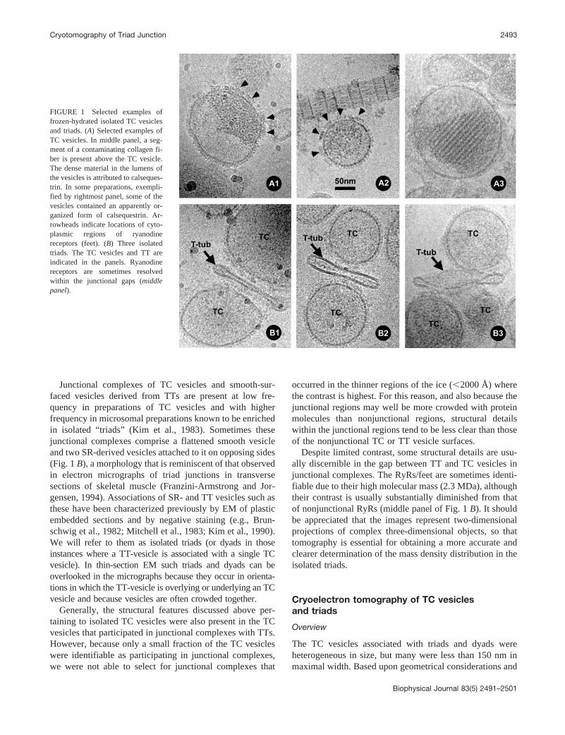
Junctional complexes of TC vesicles and smooth-sur-faced vesicles derived from TTs are present at low fre-quency in preparations of TC vesicles and with higherfrequency in microsomal preparations known to be enrichedin isolated “triads” (Kim et al., 1983). Sometimes thesejunctional complexes comprise a flattened smooth vesicleand two SR-derived vesicles attached to it on opposing sides(Fig. 1 B), a morphology that is reminiscent of that observedin electron micrographs of triad junctions in transversesections of skeletal muscle (Franzini-Armstrong and Jor-gensen, 1994). Associations of SR- and TT vesicles such asthese have been characterized previously by EM of plasticembedded sections and by negative staining (e.g., Brun-schwig et al., 1982; Mitchell et al., 1983; Kim et al., 1990).We will refer to them as isolated triads (or dyads in thoseinstances where a TT-vesicle is associated with a single TCvesicle). In thin-section EM such triads and dyads can beoverlooked in the micrographs because they occur in orienta-tions in which the TT-vesicle is overlying or underlying an TCvesicle and because vesicles are often crowded together.
Generally, the structural features discussed above per-taining to isolated TC vesicles were also present in the TCvesicles that participated in junctional complexes with TTs.However, because only a small fraction of the TC vesicleswere identifiable as participating in junctional complexes,we were not able to select for junctional complexes that
occurred in the thinner regions of the ice (�2000 Å) wherethe contrast is highest. For this reason, and also because thejunctional regions may well be more crowded with proteinmolecules than nonjunctional regions, structural detailswithin the junctional regions tend to be less clear than thoseof the nonjunctional TC or TT vesicle surfaces.
Despite limited contrast, some structural details are usu-ally discernible in the gap between TT and TC vesicles injunctional complexes. The RyRs/feet are sometimes identi-fiable due to their high molecular mass (2.3 MDa), althoughtheir contrast is usually substantially diminished from thatof nonjunctional RyRs (middle panel of Fig. 1 B). It shouldbe appreciated that the images represent two-dimensionalprojections of complex three-dimensional objects, so thattomography is essential for obtaining a more accurate andclearer determination of the mass density distribution in theisolated triads.
Cryoelectron tomography of TC vesiclesand triads
Overview
The TC vesicles associated with triads and dyads wereheterogeneous in size, but many were less than 150 nm inmaximal width. Based upon geometrical considerations and
FIGURE 1 Selected examples offrozen-hydrated isolated TC vesiclesand triads. (A) Selected examples ofTC vesicles. In middle panel, a seg-ment of a contaminating collagen fi-ber is present above the TC vesicle.The dense material in the lumens ofthe vesicles is attributed to calseques-trin. In some preparations, exempli-fied by rightmost panel, some of thevesicles contained an apparently or-ganized form of calsequestrin. Ar-rowheads indicate locations of cyto-plasmic regions of ryanodinereceptors (feet). (B) Three isolatedtriads. The TC vesicles and TT areindicated in the panels. Ryanodinereceptors are sometimes resolvedwithin the junctional gaps (middlepanel).
Cryotomography of Triad Junction 2493
Biophysical Journal 83(5) 2491–2501
assuming that the vesicles lie in regions of the embeddingice whose thickness approximates the diameter of the larg-est vesicles, micrographs should be recorded at 2-degreeintervals covering 180° to achieve an isotropic resolution of5 to 7 nm (Crowther et al., 1970). In practice, the tilt rangeachievable with the specimen holders used in this study islimited to � 60°, so we collected tilt series at 2° intervalsover this range for fields of vesicles that appeared to containat least one triad. The main effect of the resulting “missingwedge” in the data is that the resolution in the Z-direction(defined as the direction along the normal to the plane of thenon-tilted specimen) will be reduced. The total electrondose used to collect the series ranged from 30 to 80 elec-trons/Å2, a range for which damage from radiation exposureis not expected to be resolution limiting.
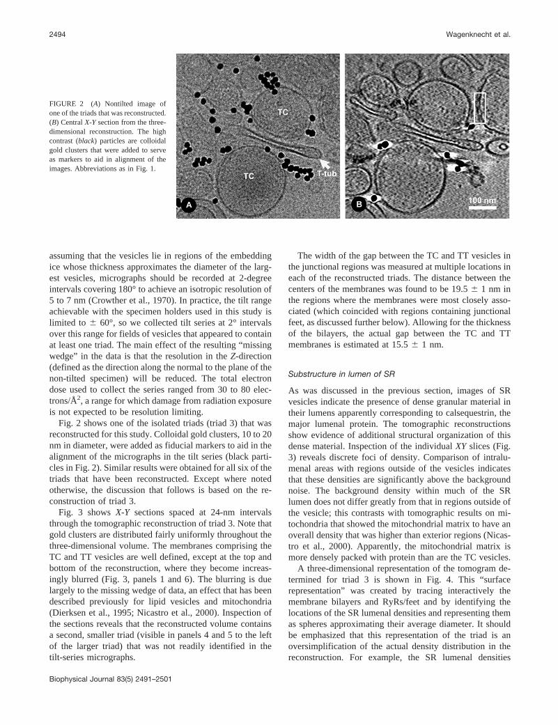
Fig. 2 shows one of the isolated triads (triad 3) that wasreconstructed for this study. Colloidal gold clusters, 10 to 20nm in diameter, were added as fiducial markers to aid in thealignment of the micrographs in the tilt series (black parti-cles in Fig. 2). Similar results were obtained for all six of thetriads that have been reconstructed. Except where notedotherwise, the discussion that follows is based on the re-construction of triad 3.
Fig. 3 shows X-Y sections spaced at 24-nm intervalsthrough the tomographic reconstruction of triad 3. Note thatgold clusters are distributed fairly uniformly throughout thethree-dimensional volume. The membranes comprising theTC and TT vesicles are well defined, except at the top andbottom of the reconstruction, where they become increas-ingly blurred (Fig. 3, panels 1 and 6). The blurring is duelargely to the missing wedge of data, an effect that has beendescribed previously for lipid vesicles and mitochondria(Dierksen et al., 1995; Nicastro et al., 2000). Inspection ofthe sections reveals that the reconstructed volume containsa second, smaller triad (visible in panels 4 and 5 to the leftof the larger triad) that was not readily identified in thetilt-series micrographs.
The width of the gap between the TC and TT vesicles inthe junctional regions was measured at multiple locations ineach of the reconstructed triads. The distance between thecenters of the membranes was found to be 19.5 � 1 nm inthe regions where the membranes were most closely asso-ciated (which coincided with regions containing junctionalfeet, as discussed further below). Allowing for the thicknessof the bilayers, the actual gap between the TC and TTmembranes is estimated at 15.5 � 1 nm.
Substructure in lumen of SR
As was discussed in the previous section, images of SRvesicles indicate the presence of dense granular material intheir lumens apparently corresponding to calsequestrin, themajor lumenal protein. The tomographic reconstructionsshow evidence of additional structural organization of thisdense material. Inspection of the individual XY slices (Fig.3) reveals discrete foci of density. Comparison of intralu-menal areas with regions outside of the vesicles indicatesthat these densities are significantly above the backgroundnoise. The background density within much of the SRlumen does not differ greatly from that in regions outside ofthe vesicle; this contrasts with tomographic results on mi-tochondria that showed the mitochondrial matrix to have anoverall density that was higher than exterior regions (Nicas-tro et al., 2000). Apparently, the mitochondrial matrix ismore densely packed with protein than are the TC vesicles.
A three-dimensional representation of the tomogram de-termined for triad 3 is shown in Fig. 4. This “surfacerepresentation” was created by tracing interactively themembrane bilayers and RyRs/feet and by identifying thelocations of the SR lumenal densities and representing themas spheres approximating their average diameter. It shouldbe emphasized that this representation of the triad is anoversimplification of the actual density distribution in thereconstruction. For example, the SR lumenal densities
FIGURE 2 (A) Nontilted image ofone of the triads that was reconstructed.(B) Central X-Y section from the three-dimensional reconstruction. The highcontrast (black) particles are colloidalgold clusters that were added to serveas markers to aid in alignment of theimages. Abbreviations as in Fig. 1.
2494 Wagenknecht et al.
Biophysical Journal 83(5) 2491–2501

(calsequestrin) are not always spherical in shape as indi-cated, and frequently there appears to be additional den-sity that interconnects these densities that is too faint totrace reliably and is therefore not indicated in the surfacerepresentation.
Interestingly, the lumenal high-density regions in the TCvesicles appear much more densely packed and organizednear the membrane surface, specifically at regions wheremultiple RyRs are present. Several of these regions are
outlined by dotted lines in Fig. 3 (panels 3–5). Sometimes anearly continuous sheet of density, several tens of nanome-ters in thickness, underlies the SR membrane. The detailedstructural features in these regions are difficult to discernfully, so in the surface-rendered representation of the triadwe indicate only the approximate boundaries of these re-gions (Fig. 4, yellow shading).
These regions of organized lumenal density appear not tocontact the surface of the SR membrane. Rather, there is a
FIGURE 3 Selected XY-slices from to-mographic reconstruction of triad 3. TheZ-coordinate of each slice is indicated inlower right corner. Orange dots denotelocations of RyRs (feet); yellow dottedlines are drawn to indicate regions of or-ganized calsequestrin located between thedotted line and the SR membrane. The TCvesicle at upper right in panels 4 and 5,which is not part of the triad, containsparticularly well-resolved RyRs and un-derlying organized calsequestrin. Noticethat the colloidal gold clusters are presentin all slices, indicating that the clusters aredistributed uniformly throughout the layerof vitreous buffer in which the triads areembedded.
Cryotomography of Triad Junction 2495
Biophysical Journal 83(5) 2491–2501

zone of reduced density �5 nm in width between thelumenal density and the SR membrane (this is particularlywell-resolved in the nonjunctional TC vesicle on the rightside of panel 4 in Fig. 3). This “clear” zone is apparently notan electron-optical artifact, because it is much less promi-nent on the exterior surface of the SR membrane. Further-more, cryotomograms of mitochondria, determined by sim-ilar procedures to those we have used, do not show such lowdensity regions associated with their inner membrane andcristae even though the mitochondrial matrix is denselypacked with protein (Mannella et al., 1999; see also Nicas-tro et al. 2000). Densities that bridge the gap between theregion of high density and the SR membrane sporadicallyinterrupt the clear zone.
T-tubule
Although structural details are not reproducibly resolved onthe exterior membrane surfaces of the TT vesicles, thelumens of the vesicles contain particulate densities, partic-ularly in the widest portions, outside of the junctional re-gions. The locations and approximate sizes of the TT lume-nal densities are indicated in the surface-rendered model oftriad 3 (purple areas in Fig. 4). In some regions the densitiesappear to form cross-bridges that extend across the width ofthe TT (e.g., panels 2 and 5 of Fig. 3). Perhaps thesestructures are involved in establishing the distinctive flat-tened shape of the TT vesicles.
Exterior surface of TC vesicles: nonjunctional andjunctional regions
The X-Y sections shown in Fig. 3 show TC vesicles con-taining high-density regions on their exterior (cytoplasmic-facing) surfaces whose size and shape indicate that theycorrespond to the cytoplasmic domains of RyRs (i.e., feet,
indicated by orange dots in Fig. 3) that have been observedin previous EM studies of sectioned muscle and isolatedSR-derived vesicles (Caswell et al., 1976; Mitchell et al.,1983; Kim et al., 1990; Franzini-Armstrong and Jorgensen,1994; Flucher and Franzini-Armstrong, 1996). Frequently,the feet appear disconnected from the SR membrane towhich they are apparently attached via the RyR’s transmem-brane region. In junctional regions, visible connections be-tween the feet and TT membrane are also infrequent (how-ever, see below), and consequently the feet often appear tobe “floating” between the SR and TT membranes. Thisappearance of the feet is also conveyed in the surfacerendered representation of the triad (blue regions in Fig. 4).The feet that occur outside of junctional regions probablyarose during the homogenization step of the isolation pro-cedure, as most RyRs are thought to be associated with theTT/sarcolemma membrane system in situ.
Individual Ca2�-ATPase molecules are not well re-solved from one another in the reconstructed triads, butthe nonjunctional regions of the TC appear thicker,denser, and less smooth on their exterior faces, which isconsistent with these regions being densely packed withCa2�-ATPase.
The junctional regions between TC and TT vesicles are ofparticular interest. However, besides the RyRs, little sub-structure is reliably resolved in these regions. The cytoplas-mic domains of RyRs are readily identifiable in the gapbetween the vesicles, although they are less well contrastedthan in nonjunctional regions of the TC vesicles, probablydue to the presence of other proteins in the junctional gap.The locations of the RyRs that were identified on the twoTC vesicles in triad 3 are indicated in Fig. 4 (blue struc-tures). Most of the RyRs in this reconstruction are localizedat or near regions of the TC vesicles that interact with TTvesicle, but not all appear close enough to the surface of the
FIGURE 4 Surface-rendered representa-tion of triad 3 in stereo. SR- and TT-de-rived vesicles in green and red, respec-tively. Yellow spheres in lumen of SRrepresent calsequestrin and continuous yel-low slabs near junctional surfaces of SRrepresent the condensed calsequestrin (seeFig. 3). Blue structures correspond to feet/RyRs. Magenta particles in lumen of TTare of unknown identity.
2496 Wagenknecht et al.
Biophysical Journal 83(5) 2491–2501

TT vesicle to directly interact with it. Most of the RyRs ineach of the two junctional regions are constrained to anarrow band that may represent the remnant of a two-rowedarray of the type observed in electron micrographs offreeze-fractured or thin sectioned images of intact muscle(Ferguson et al., 1984).
A clearer example of RyRs forming a two-rowed array isfound in one of the nonjunctional TC vesicles that werepresent adjoining triad 3 (upper right in Fig. 3, panels 4 and5). A subvolume extracted from this region, when viewed inprojection, shows apparently three well-resolved RyRs (Fig.5). When this region of the three-dimensional volume isviewed at a 90° angle, onto the exterior surface of the TCvesicle, it becomes apparent that six RyRs are actuallypresent and that they are arranged in a two-rowed array ofthe type observed previously by EM of intact muscle (Fig.5B). The handedness of the array is also revealed by thereconstruction, and it agrees with the handedness that weobserved previously for oligomers comprising two or threeRyRs that sometimes occur in preparations of purified RyRs(Wagenknecht et al., 1997).
In junctional regions, there often appears to be a gap ofseveral nanometers between the distal face of the feet andthe surface of the TT vesicle (see RyRs in junctional regionsin Fig. 3, panels 2–4). Because the nature of the linkagebetween the RyRs and TT-associated voltage sensor (theDHPR) is thought to be critical for understanding the mech-anism of EC coupling, we have examined all of the junc-tional RyRs in the reconstructions. Some of the RyRs ap-peared to be associated with density linking them to the TTvesicle (Fig. 6). The number of connections per RyR variesbetween zero and three, with zero or one being the mostcommon. In some cases the density occurs in regions wherethe gap between TC and TT vesicle is greater that 16 nm(lower rightmost panel in Fig. 6). The bridging densitiesmay correspond to cytoplasmic regions of the DHPRs, butfurther studies are required to determine their molecularidentity.
DISCUSSION
We have described here the first results of cryo-EM andtomography of triad junctions. Since gaining popularity inthe 1980s, EM of frozen-hydrated solutions of isolatedmacromolecules has become the standard method for char-acterizing the ultrastructure of large multisubunit biologicalstructures such as ribosomes, viruses, and membrane-asso-ciated proteins (e.g., Agrawal and Frank, 1999; Chiu et al.,1999; Baumeister and Steven, 2000; Nogales and Grigor-ieff, 2001). There is widespread agreement that this tech-nique, which does not require the use of stains, fixatives, ordehydration, is capable of preserving macromolecular struc-ture to atomic resolution. However, one drawback of thetechnique is the poor contrast exhibited by ice-embeddedspecimens. For isolated multisubunit protein or nucleic ac-
id/protein complexes this problem can be overcome byaveraging over images obtained from thousands of particles,but averaging is not feasible for subcellular structures, suchas the isolated triads, which are not homogeneous withregard to their overall sizes and shapes. In electron tomog-raphy a limited number of images of a single subcellularstructure or region is obtained by tilting incrementally thespecimen, and these are combined to produce a three-di-mensional reconstruction. Although the contrast of the re-construction is improved over that present in the unproc-essed images, it is still not optimal for making detailedinterpretation of the density maps.
Here, isolated triads were chosen for tomography ratherthan triad-containing regions of intact muscle because sec-tioning frozen tissue is technically difficult and producescompression artifacts (Ruiz et al., 1994; Dubochet andSartori Blanc, 2001; Hsieh et al., 2002). Further, the contrastof triads in frozen-hydrated sections would doubtless be
FIGURE 5 Evidence for arrays of ryanodine receptors. (A) Enlargedview of the cluster of ryanodine receptors shown on right side of Fig. 3(panels 4 and 5) as seen in projection in a subvolume extracted from thetomographic reconstruction. For comparison, at the bottom is shown areconstructed ryanodine receptor as determined previously by single-par-ticle image processing of purified receptors (Radermacher et al., 1994).Note that contrast is reversed from previous figure: protein density is white.(B) En face view of the cluster of ryanodine receptors. When the subvol-ume in the orientation shown in A is projected following rotation of 90°about the vertical, it becomes clear that the cluster of receptors comprisestwo rows of receptors (six in total). A single-particle reconstruction of thereceptor in the en face view is at the bottom. Double-rowed arrays of thistype have been detected previously by the freeze-fracture technique (Blocket al., 1988).
Cryotomography of Triad Junction 2497
Biophysical Journal 83(5) 2491–2501

even lower than that found for isolated triads. Moreover,observations showing that isolated triads retain the basicstructural features of the triad junction and are functional, inthe sense that chemical-induced depolarization of the TTsinduces release of Ca2� from the associated SR vesicles,indicate that isolated triads represent a valid model systemfor elucidating the mechanism of EC coupling (Andersonand Meissner, 1995; Corbett et al., 1992; Ikemoto et al.,1984; Kramer and Corbett, 1996). Isolated triads offer sev-eral properties that are favorable for cryoelectron micros-copy and tomography: 1) contrast is maximized becausethey can be imaged in thin (150–300 nm) films of bufferand in the absence of nontriad cytoplasmic proteins; 2)many of the triad’s protein components are readily accessi-ble for immuno-labeling; 3) experimental conditions can bevaried readily and tested for functional consequences inparallel with structural characterization. As discussed indetail below, our initial reconstructions reveal some newultrastructural features that point to the promise of cryoelec-tron tomography.
Comparison with previous results on theultrastructure of the triad junction
A model for the three-dimensional architecture of the ver-tebrate skeletal muscle triad junction has been proposed byFranzini-Armstrong and her colleagues (Block et al., 1988).This model is based upon results of numerous morphologicstudies using various EM techniques (for reviews, see Fran-zini-Armstrong and Jorgensen, 1994; Franzini-Armstrong,1994; Flucher and Franzini-Armstrong, 1996). In thismodel, RyRs are arranged on the junctional-face membrane
of the SR as an ordered, two-rowed array whose length isvariable. An intriguing feature of the model is that theDHPRs in the TT are disposed as groups of four (calledtetrads) whose members align with the subunits of thefour-fold symmetric RyRs in the apposed SR membrane(Block et al., 1988). However, the pairing of tetrads withRyRs appears to occur in an alternating pattern, such thatonly one-half of the RyRs are paired with tetrads. Thisprecise alignment of the two receptor types suggests aphysical interaction between them, which is consistent withthe currently favored “mechanical coupling” mechanism ofEC coupling in skeletal muscle whereby a voltage-depen-dent conformational change in the DHPRs induces openingof those RyRs in direct contact with them. Another featureof the Franzini-Armstrong model is that calsequestrin isconcentrated in the cisternae of the junctional SR, but ap-pears not to extend to the inner leaflet of the SR membrane.Thin strands of material, which could contain one or both ofthe proteins, triadin and junctin (Zhang et al., 1997), anchorthe calsequestrin to the SR membrane. Many additionalproteins are known (Costello et al., 1986) or hypothesized tobe present at the triad junction, but their locations, interac-tion partners, and stoichiometry are unknown (Mackrill,1999). Also, there is considerable uncertainty about some ofthe basic architectural features of the model. For instance,reports of the width of the gap between the SR and TTmembranes range from 10 to 20 nm. The studies describedhere provide a path to refine and extend this model for thetriad junction, which is essential for elucidating the mech-anisms of EC coupling.
The structural organization of the gap between the TTand SR junctional face membranes in the triad junction(triadic gap) is of particular interest, because events associ-ated with communication between the two membrane sys-tems are likely to occur there. The only structural featurethat has been definitively identified in the triadic gap cor-responds to the large (Mr � 2 � 106), fourfold symmetriccytoplasmic region of the RyR, the so-called junctional“feet.” Our reconstructions also show these structures.
One difference exhibited by our three-dimensional recon-structions from previous microscopy studies of skeletalmuscle or isolated triads prepared by thin sectioning is thatthe majority of the feet do not appear to physically contactthe TT. Instead, a gap, several nm in width, exists betweenthe TT and RyR. Consequently, the total width of thejunctional gap is 15 to 16 nm, significantly greater than the12-nm width of the cytoplasmic region of the RyR (Sery-sheva et al., 1995; Radermacher et al., 1994). Previousestimates of the width of the triadic gap have ranged from10 to 20 nm (Franzini-Armstrong, 1994). Some of the feetappear to be connected to the TT via bridging density (Fig.6). Possibly these bridging densities correspond to domainsor subunits of the DHPRs that are believed to directlyinteract with the RyRs. However, we have not observed acase where four symmetrically arranged bridging structures
FIGURE 6 Ryanodine receptor t-tubule interactions. Selected examplesof RyRs that appear to be linked to the TT. Arrows indicate the locationsof density that bridges the gap between the RyR/foot and the TT mem-brane. Dotted lines mark the cytoplasmic regions (feet) of RyRs. Locationsof TT (T) and junctional SR membranes are also indicated. Arrows indicateconnections between ryanodine receptors and TTs, which bridge the �4-nm gap between the RyR and the TT. Regions of high protein ormembrane density are dark.
2498 Wagenknecht et al.
Biophysical Journal 83(5) 2491–2501
interact with a RyR, as would be expected if they corre-sponded to the tetrads that have been observed in micro-graphs of TT derived membrane by the freeze-fracturetechnique (Block et al., 1988).
Although we cannot be certain that the 15- to 16-nmjunctional gap that we have observed is not artifactuallylarge as a consequence of structural rearrangements thatmight occur upon homogenization of muscle tissue andisolation of the triads, several considerations argue againstthis possibility. As mentioned earlier, isolated triads exhibitEC coupling activity in vitro, and this activity is expected tobe sensitive to disruption of the linkage between the TT andSR. Also, biological material prepared for microscopy byconventional thin-sectioning techniques suffers fromshrinkage and other potential artifacts, which could accountfor the more compact appearance of the triad junction insome of these preparations. Finally, recent structural char-acterizations of voltage-gated potassium and sodium chan-nels (Gulbis et al., 2000; Kobertz et al., 2000; Sokolova etal., 2001; Sato et al., 2001), which are related to the DHPR,indicate that folded domains of the channels contributesignificant mass on the cytoplasmic side of the membrane.If similar or analogous cytoplasmic domains exist for theDHPR, then they would suffice to bridge the 3- to 4-nmdistance between the distal face of the RyR and the surfaceof the TT membrane, which is consistent with our observa-tion of density apparently connecting some RyRs to the TT(Fig. 6).
Much of the dense material occupying the lumen of thejunctional SR vesicles is attributable to the protein calse-questrin, the most abundant lumenal protein (Saito et al.1984; Maurer et al., 1985). Our tomographic reconstruc-tions show a dense layer of protein (which we presume to becalsequestrin based on evidence summarized by Franzini-Armstrong and Jorgensen (1994)), just beneath the SRmembrane and primarily in regions that contain RyRs.These subsarcolemmal regions are variable in size, but theydo not extend more that a few tens of nanometers into theinterior of the lumen. Tomography was necessary to revealthis distribution of the density, as it was not apparent in themicrographs themselves. The distribution of globular struc-tures that comprise these regions gives the impression ofstructural organization (Fig. 3). There are also isolatedpunctate regions of high density deeper within the lumen ofthe SR vesicles (small yellow spheres in Fig. 4). Thesedensities may represent monomeric or oligomeric clustersof calsequestrin molecules that co-exist with the larger SRmembrane-associated assemblages.
Interestingly, the calsequestrin-enriched regions do notappear to interact directly with the SR membrane, but rathera zone of reduced density of �5-nm thickness occurs be-tween the SR membrane and the calsequestrin. Sometimes,faintly visible bridges of density between the SR and theorganized calsequestrin interrupt the “clear zone.” In themodel of the triad junction proposed by Franzini-Armstrong
(Block et al., 1988; Franzini-Armstrong and Jorgensen,1994), the calsequestrin also forms a lumenal aggregate thatis linked to the SR membrane by thin strands of protein. Thewidth of the gap has been uncertain, because different EMpreparation methods have yielded discordant results (Brun-schwig et al., 1982; Franzini-Armstrong et al., 1987). Be-cause cryotomography does not require dehydration or fix-ation, we are confident that the �5-nm-thick zone ofreduced density between the SR and the organized calse-questrin reflects the native architecture. This zone could beof functional significance for calcium release from the SR,perhaps by maintaining an appropriately high level of Ca2�
ions in the immediate vicinity of the RyRs’ ion-conductingchannels.
The TTs in frozen-hydrated preparations of triads appearas flattened vesicles that are wider at their perimeter. Theyare most flattened in regions that form junctions with theSR-derived vesicles with the separation between bilayers inthe narrowest zones approaching 10 nm. Mass densitywithin the lumen is lower than for SR vesicles and isconcentrated in the wider, peripheral regions. These find-ings generally agree with observations made by noncryoEM of isolated triads (Brunschwig et al., 1982; Kim et al.,1990; Mitchell et al., 1983). Some high-density regionswere observed in the more flattened regions of the TT,sometimes giving the appearance of forming cross-bridgeslinking apposing bilayers. These structures have not beendescribed in previous ultrastructural studies of isolated tri-ads or in studies of sectioned muscle, except for one reportby Dulhunty (1989) who observed similar structures inlightly fixed and stained specimens and termed them “teth-ers.” In agreement with previous studies, we find that thecytoplasmic surfaces of the TT bilayer (the exterior side inthe isolated triads) appear much smoother than those of theSR vesicles.
Outlook
The isolated triad junctions that we have reconstructed bycryoelectron tomography have likely retained their nativearchitecture to a degree that has not been achieved inprevious microscopy studies. However, the inherent low-contrast and radiation sensitivity of nonstained, nonfixedbiological specimens limit the information content of recon-structions determined by current implementations of thetechnique. The resolution attained for the triads describedhere are not known precisely, but is unlikely to be betterthan 6 nm in the X-Y direction and poorer in the Z-direction(normal to the specimen grid). Even so, as discussed above,several features are present in the tomograms that are eithernovel or support unsubstantiated previous observations.
We are optimistic that future investigations by cryoelec-tron tomography will provide much more detailed structuralinformation on the triad junction, quite possibly at a levelthat cannot be obtained by any other approach. Improve-
Cryotomography of Triad Junction 2499
Biophysical Journal 83(5) 2491–2501

ments and enhancements to electron microscopes, data-collection strategies, and image-processing methods are ex-pected to permit resolutions of 2 to 4 nm to be attainable(Koster et al., 1997; Bohm et al., 2000). In this resolutionrealm it should be possible to identify multisubunit proteinassemblies whose structures are already known (e.g., fromx-ray crystallography or EM of the isolated complexes) onthe basis of their density distribution alone. In the case oftriads, RyRs are already identifiable, and their coordinatesand orientations should be determinable with high reliabilityand precision. This will permit individual RyRs to be “iso-lated” computationally from the tomograms and then to beaveraged. Averaged RyRs will reveal the presence of pro-tein components that interact directly with them. In thismanner a much more detailed picture of the interactions ofthe RyR with TT components will emerge.
Another way to increase the information attainable fromcryotomography would be to label the specimens with elec-tron-dense probes that are specific for triadic components.For example, we have added calmodulin that was labeledwith a 1.4-nm diameter gold cluster (Wagenknecht et al.,1994) to isolated triads and found that the gold cluster couldbe identified in micrographs of frozen-hydrated specimen(unpublished data). Similarly, we envision that gold cluster-labeled antibodies specific for triad components could offera general approach to identify the locations of the compo-nents with a precision that is not possible with conventionalimmunoelectron microscopy.
Supported by the National Institutes of Health Grants AR40615 andRR01219. We also gratefully acknowledge use of the Wadsworth Center’selectron microscopy core facility. We also thank Robert Grassucci forcontributions to imaging triads by cryoelectron microscopy.
REFERENCES
Agrawal, R. K., and J. Frank. 1999. Structural studies of the translationalapparatus. Curr. Opin. Struct. Biol. 9:215–221.
Anderson, K., and G. Meissner. 1995. T-tubule depolarization-induced SRCa2� release is controlled by dihydropyridine receptor- and Ca2�-dependent mechanisms in cell homogenates from rabbit skeletal muscle.J. Gen. Physiol. 105:363–383.
Baumeister, W., R. Grimm, and J. Walz. 1999. Electron tomography ofmolecules and cells. Trends Cell Biol. 9:81–85.
Baumeister, W., and A. C. Steven. 2000. Macromolecular electron micros-copy in the era of structural genomics. Trends Biochem. Sci. 25:624–631.
Block, B. A., T. Imagawa, K. P. Campbell, and C. Franzini-Armstrong.1988. Structural evidence for direct interaction between the molecularcomponents of the transverse tubule/sarcoplasmic reticulum junction inskeletal muscle. J. Cell Biol. 107:2587–2600.
Bohm, J., A. S. Frangakis, R. Hegerl, S. Nickell, D. Typke, and W.Baumeister. 2000. Toward detecting and identifying macromolecules ina cellular context: template matching applied to electron tomograms.Proc. Natl. Acad. Sci. U. S. A. 97:14245–14250.
Brandt, N. R., and A. H. Caswell. 1999. Localization of mitsugumin 29 totransverse tubules in rabbit skeletal muscle. Arch. Biochem. Biophys.371:348–350.
Brunschwig, J. P., N. R. Brandt, A. H. Caswell, and D. S. Lukeman. 1982.Ultrastructural observations of the junctional foot protein and dihydro-pyridine receptor in skeletal muscle by use of tannic acid mordanting.J. Cell Biol. 93:533–542.
Caswell, A. H., Y. H. Lau, and J.-P. Brunschwig. 1976. Ouabain-bindingvesicles from skeletal muscle. Arch. Biochem. Biophys. 176:417–430.
Chiu, W., A. Mcgough, M. B. Sherman, and M. F. Schmid. 1999. High-resolution electron cryomicroscopy of macromolecular assemblies.Trends Cell Biol. 9:154–159.
Corbett, A. M., J. Bian, J. B. Wade, and M. F. Schneider. 1992. Depolar-ization-induced calcium release from isolated triads measured withimpermeant Fura-2. J. Membr. Biol. 128:165–179.
Costello, B., C. Chadwick, A. Saito, A. Chu, A. Maurer, and S. Fleischer.1986. Characterization of the junctional face membrane from terminalcisternae of sarcoplasmic reticulum. J. Cell Biol. 103:741–753.
Crowther, R. A., D. J. DeRosier, and A. Klug. 1970. The reconstruction ofa three-dimensional structure from projections and its application toelectron microscopy. Proc. Roy. Soc. Lond. A. 317:319–340.
Dierksen, K., D. Typke, R. Hegerl, J. Walz, E. Sackmann, and W.Baumeister. 1995. Three-dimensional structure of lipid vesicles embed-ded in vitreous ice and investigated by automated electron tomography.Biophys. J. 68:1416–1422.
Dubochet, J., M. Adrian, J.-J. Chang, J.-C. Homo, J. Lepault, A. W.McDowall, and P. Schultz. 1988. Cryo-electron microscopy of vitrifiedspecimens. Quart. Rev. Biophys. 21:129–228.
Dubochet, J., and N. Sartori Blanc. 2001. The cell in absence of aggrega-tion artifacts. Micron. 32:91–99.
Dulhunty, A. F. 1989. Feet, bridges, and pillars in triad junctions ofmammalian skeletal muscle: their possible relationship to calcium buff-ers in terminal cisternae and T-tubules and to excitation-contractioncoupling. J. Membr. Biol. 109:73–83.
Ferguson, D. G., H. W. Schwartz, and C. Franzini-Armstrong. 1984.Subunit structure of junctional feet in triads of skeletal muscle: afreeze-drying, rotary-shadowing study. J. Cell Biol. 99:1735–1742.
Flucher, B. E., and C. Franzini-Armstrong. 1996. Formation of junctionsinvolved in excitation-contraction coupling in skeletal and cardiac mus-cle. Proc. Natl. Acad. Sci. U. S. A. 93:8101–8106.
Franzini-Armstrong, C. 1980. Structure of the sarcoplasmic reticulum. Fed.Proc. 39:2403–2409.
Franzini-Armstrong, C. 1994. The sarcoplasmic reticulum and the trans-verse tubules. In Myology: Basic and Clinical. A.G. Engel and C.Franzini-Armstrong, editors. McGraw-Hill, Inc., New York. 176–199.
Franzini-Armstrong, C., and A. O. Jorgensen. 1994. Structure and devel-opment of E-C coupling units in skeletal muscle. Annu. Rev. Physiol.56:509–534.
Franzini-Armstrong, C., L. J. Kenney, and E. Varriano-Marston. 1987. Thestructure of calsequestrin in triads of vertebrate skeletal muscle: adeep-etch study. J. Cell Biol. 105:49–56.
Grabner, M., R. T. Dirksen, N. Suda, and K. G. Beam. 1999. The II-III loopof the skeletal muscle dihydropyridine receptor is responsible for thebi-directional coupling with the ryanodine receptor. J. Biol. Chem.274:21913–21919.
Grimm, R., H. Singh, R. Rachel, D. Typke, W. Zillig, and W. Baumeister.1998. Electron tomography of ice-embedded prokaryotic cells. Biophys. J.74:1031–1042.
Gulbis, J. M., M. Zhou, S. Mann, and R. MacKinnon. 2000. Structure ofthe cytoplasmic � subunit-T1 assembly of voltage-dependent K� chan-nels. Science. 289:123–127.
Hsieh, C., M. Marko, J. Frank, and C. Mannella. 2002. Electron tomo-graphic analysis of frozen-hydrated tissue sections. J. Struct. Biol. 138:63–73.
Ikemoto, N., B. Antoniu, and D. H. Kim. 1984. Rapid calcium release fromthe isolated sarcoplasmic reticulum is triggered via the attached trans-verse tubular system. J. Biol. Chem. 259:13151–13158.
Ikemoto, N., D. H. Kim, and B. Antoniu. 1988. Measurement of calciumrelease in isolated membrane systems: coupling between the transversetubule and sarcoplasmic reticulum. Method. Enzymol. 157:469–480.
2500 Wagenknecht et al.
Biophysical Journal 83(5) 2491–2501

Inui, M., A. Saito, and S. Fleischer. 1987. Purification of the ryanodinereceptor and identity with feet structures of junctional terminal cisternaeof sarcoplasmic reticulum from fast skeletal muscle. J. Biol. Chem.262:1740–1747.
Jorgensen, A. O., A. C. Shen, K. P. Campbell, and D. H. MacLennan.1983. Ultrastructural localization of calsequestrin in rat skeletal muscleby immunoferritin labeling of ultrathin frozen sections. J. Cell Biol.97:1573–1581.
Kawamoto, R. M., J.-P. Brunschwig, and A. H. Caswell. 1988. Localiza-tion by immunoelectron microscopy of spanning protein of triad junctionin terminal cisternae/triad vesicles. J. Muscle Res. Cell Motil.9:334–343.
Kim, D. H., S. T. Onhishi, and N. Ikemoto. 1983. Kinetic studies ofcalcium release from sarcoplasmic reticulum in vitro. J. Biol. Chem.258:9662–9668.
Kim, K. C., A. H. Caswell, J.-P. Brunschwig, and N. R. Brandt. 1990.Identification of a new subpopulation of triad junctions isolated fromskeletal muscle, morphological correlations with intact muscle.J. Membr. Biol. 113:221–235.
Kobertz, W. R., C. Williams, and C. Miller. 2000. Hanging gondolastructure of the T1 domain in voltage-gated K� channel. Biochemistry.39:10347–10352.
Koster, A. J., R. Grimm, D. Typke, R. Hegerl, A. Stoschek, J. Walz, andW. Baumeister. 1997. Perspectives of molecular and cellular electrontomography. J. Struct. Biol. 120:276–308.
Kramer, J. W., and A. M. Corbett. 1996. Comparison of Ca2� loading andretention in isolated skeletal muscle triads and terminal cisternae.Am. J. Physiol. 270:C1602–C1610.
McEwen, B. F., and M. Marko. 2001. The emergence of electron tomog-raphy as an important tool for investigating cellular architecture. J. His-tochem. Cytochem. 49:553–563.
Mackrill, J. J. 1999. Protein-protein interactions in intracellular Ca2�-release channel function. Biochem. J. 337:345–361.
Mannella, C. A., C.-E. Hsieh, and M. Marko. 1999. Electron microscopictomography of whole, frozen-hydrated rat-liver mitochondria at 400 kV.Microsc. Microanal. 5:416–417.
Marko, M., and A. Leith. 1996. Sterecon–three-dimensional reconstruc-tions from stereoscopic contouring. J. Struct. Biol. 116:93–98.
Maurer, A., M. Tanaka, T. Ozawa, and S. Fleischer. 1985. Purification andcrystallization of the calcium binding protein of sarcoplasmic reticulumfrom skeletal muscle. Proc. Natl. Acad. Sci. U.S.A. 82:4036–4040.
Mitchell, R. D., A. Saito, P. Palade, and S. Fleischer. 1983. Morphology ofisolated triads. J. Cell Biol. 96:1017–1029.
Nakai, J., R. T. Dirksen, H. T. Nguyen, I. N. Pessah, K. G. Beam, and P. D.Allen. 1996. Enhanced dihydropyridine receptor channel activity in thepresence of ryanodine receptor. Nature. 380:72–75.
Nakai, J., N. Sekiguchi, T. A. Rando, P. D. Allen, and K. G. Beam. 1998.Two regions of the ryanodine receptor involved in coupling with L-typeCa2� channels. J. Biol. Chem. 273:13403–13406.
Nicastro, D., A. S. Frangakis, D. Typke, and W. Baumeister. 2000. Cryo-electron tomography of Neurospora mitochondria. J. Struct. Biol. 129:48–56.
Nogales, E., and N. Grigorieff. 2001. Molecular machines: putting thepieces together. J. Cell Biol. 152:F1–F10.
Penczek, P., M. Marko, K. Buttle, and J. Frank. 1995. Double-tilt tomog-raphy. Ultramicroscopy. 60:393–410.
Radermacher, M., V. Rao, R. Grassucci, J. Frank, A. P. Timerman, S.Fleischer, and T. Wagenknecht. 1994. Cryo-electron microscopy andthree-dimensional reconstruction of the calcium release channel ryano-dine receptor from skeletal muscle. J. Cell Biol. 127:411–423.
Rath, B. K., M. Marko, M. Radermacher, and J. Frank. 1997. Low-doseautomated electron tomography: a recent implementation. J. Struct. Biol.120:210–218.
Ruiz, T., I. Erk, and J. Lepault. 1994. Electron cryo-microscopy of vitrifiedbiological specimens: towards high spatial and temporal resolution. Biol.Cell. 80:203–210.
Saito, A., M. Inui, M. Radermacher, J. Frank, and S. Fleischer. 1988.Ultrastructure of the calcium release channel of sarcoplasmic reticulum.J. Cell Biol. 107:211–219.
Saito, A., S. Seiler, A. Chu, and S. Fleischer. 1984. Preparation andmorphology of sarcoplasmic reticulum terminal cisternae from rabbitskeletal muscle. J. Cell Biol. 99:875–885.
Sato, C., Y. Ueno, K. Asai, K. Takahashi, M. Sato, A. Engel, and Y.Fujiyoshi. 2001. The voltage-sensitive sodium channel is a bell-shapedmolecule with several cavities. Nature. 409:1047–1051.
Serysheva, I. I., E. V. Orlova, W. Chiu, M. B. Sherman, S. L. Hamilton,and M. van Heel. 1995. Electron cryomicroscopy and angular reconsti-tution used to visualize the skeletal muscle calcium release channel.Struct. Biol. 2:18–24.
Sokolova, O., L. Kolmakova-Partensky, and N. Grigorieff. 2001. Three-dimensional structure of a voltage-gated potassium channel at 2.5 nmresolution. Structure. 9:215–220.
Stewart, P. S., and D. H. MacLennan. 1974. Surface particles of sarco-plasmic reticulum membranes: structural features of the adenosinetriphosphatase. J. Biol. Chem. 249:985–993.
Takekura, H., L. Bennett, T. Tanabe, K. G. Beam, and C. Franzini-Armstrong. 1994. Restoration of junctional tetrads in dysgenic myotubesby dihydropyridine receptor cDNA. Biophys. J. 67:793–803.
Takeshima, H., S. Komazaki, M. Nishi, M. Iino, and K. Kangawa. 2000.Junctophilins: a novel family of junctional membrane complex proteins.Mol. Cell. 6:11–22.
Takeshima, H., M. Shimuta, S. Komazaki, K. Ohmi, M. Nishi, M. Iino, A.Miyata, and K. Kangawa. 1998. Mitsugumin29, a novel synaptophysinfamily member from the triad junction in skeletal muscle. Biochem. J.331:317–322.
Wagenknecht, T., J. Berkowitz, R. Grassucci, A. P. Timerman, and S.Fleischer. 1994. Localization of calmodulin binding sites on the ryano-dine receptor from skeletal muscle by electron microscopy. Biophys. J.67:2286–2295.
Wagenknecht, T., R. Grassucci, and J. Frank. 1988. Electron microscopyand computer image averaging of ice-embedded large ribosomal sub-units from Escherichia coli. J. Mol. Biol. 199:137–147.
Wagenknecht, T., M. Radermacher, R. Grassucci, J. Berkowitz, H.-B. Xin,and S. Fleischer. 1997. Locations of calmodulin and FK506-bindingprotein on the three-dimensional architecture of the skeletal muscleryanodine receptor. J. Biol. Chem. 272:32463–32471.
Zhang, L., J. Kelley, G. Schmeisser, Y. M. Kobayashi, and L. R. Jones.1997. Complex formation between junctin, triadin, calsequestrin, and theryanodine receptor: proteins of the cardiac junctional sarcoplasmic re-ticulum membrane. J. Biol. Chem. 272:23389–23397.
Cryotomography of Triad Junction 2501
Biophysical Journal 83(5) 2491–2501