electronic structure of the water dimer cationiopenshell.usc.edu/pubs/pdf/jpca-112-6159.pdf ·...
TRANSCRIPT
Electronic Structure of the Water Dimer Cation
Piotr A. Pieniazek,† Joost VandeVondele,‡ Pavel Jungwirth,*,§ Anna I. Krylov,*,† andStephen E. Bradforth†,*Department of Chemistry, UniVersity of Southern California, Los Angeles, California 90089-0482, PhysicalChemistry Institute, Zurich UniVersity, Winterthurerstrasse 190, CH-8057 Zurich, Switzerland, and Institute ofOrganic Chemistry and Biochemistry, Academy of Sciences of the Czech Republic, and Center for Biomoleculesand Complex Molecular Systems, FlemingoVo nam. 2, 16610 Prague 6, Czech Republic
ReceiVed: March 11, 2008; ReVised Manuscript ReceiVed: April 29, 2008
The spectroscopic signatures of proton transfer in the water dimer cation were investigated. The six lowestelectronic states were characterized along the reaction coordinate using the equation-of-motion coupled-cluster with single and double substitutions method for ionized systems. The nature of the dimer states wasexplained in terms of the monomer states using a qualitative molecular orbital framework. We found thatproton transfer induces significant changes in the electronic spectrum, thus suggesting that time-resolvedelectronic femtosecond spectroscopy is an effective strategy to monitor the dynamics following ionization.The electronic spectra at vertical and proton-transferred configurations include both local excitations (featuressimilar to those of the monomers) and charge-transfer bands. Ab initio calculations were used to test theperformance of a self-interaction correction for density functional theory (DFT). The corrected DFT/BLYPmethod is capable of quantitatively reproducing the proper energetic ordering of the (H2O)2
+ isomers and thusis a reasonable approach for calculations of larger systems.
1. Introduction
Ionized states in condensed media, for example, thoseproduced by radiation in biological tissue, or encountered inthe storage and reprocessing of fissioned nuclear materials arenot yet fully understood.1–3 Even radiolysis of water, apredominant molecular target of the incident radiation in thesesituations, which has also served as the prototypical system fordisentangling the phenomenology of high-energy processes inbulk liquids, has not been completely mapped out. For example,the nature of the initially excited states and sub-50-fs ionization/dissociation dynamics after deposition of energy have not beendirectly observed.3
The major ionization channel in water radiolysis includes thefollowing steps:
nH2Of (H2O)n*fH2O
+(H2O)n-1 + e-f . . . fH3Oaq+ +
OHaq + eaq- (1)
The extent of delocalization of the initial excitation, that is,the number n of water molecules participating in (H2O)n
/, andthe character of the charged species immediately after chargeseparation are poorly defined. Both quantities are likely todepend on the initial energy deposited.4 In addition to reaction1, energy deposited into water can also lead to homolytic bondbreaking yielding H + OH, which resembles the gas phasephotodissociation.5 Besides the practical interest in the yieldsand spatial distribution of reactive radical products, this systemalso presents several fundamental questions on the mechanism
and dynamics of separation and delocalization of both chargeand spin in a partially disordered and rapidly fluctuating system.
Both the electronic structure and molecular dynamics of waterare amenable to simulation. Although the structure, dynamics,and spectroscopy of the excess electron have been investigatedextensively,modelingtheother initialproductofphotoionization,6,7
the ionized hole, has not been attempted in bulk, although clusterstudies were reported.8–11 The delocalization of the initial charge,the timescale, and dynamics of reaction 1, as well as the fateand separation of the products are all important issues that asimulation could help to explain. Our ongoing efforts to simulatethe excess hole in bulk require the benchmarking of electronicstructure methods for ab initio molecular dynamics (AIMD).One of the goals of the present study is to compare reliablecalculations of the simplest fragment of the hole in water, thewater dimer cation, against computationally less demandingmethods suitable for AIMD.12
Moreover, in the laboratories of one of us, photolysis studiesof pure water have been carried out with pulses as short as 25fs. With this time resolution, femtosecond pump-probe spec-troscopy should be able to track the proton-transfer step inreaction 1 and with a dispersed probe covering continuouslythe near UV and visible, the transients should be identifiableby their spectroscopic signatures. However, it is critical toestablish the signature of the H2O + intermediate and distinguishit from the subsequent species such as (OH · · ·H3O+) andOH(aq).13,14 Therefore, another major goal of this work is todescribe the excited states of each of these species accurately,at least in small clusters. This will allow an assessment of whatinformation electronic spectroscopy can provide in tracking theionization chemistry of bulk water. Our theoretical approachparallels a recent study we performed on the electronicspectroscopy of the benzene dimer cation, a core species in theionization of liquid benzene.15
* Corresponding authors. E-mail: [email protected] (P.J.),[email protected] (A.K.), and [email protected] (S.B.).
† University of Southern California.‡ Zurich University.§ Institute of Organic Chemistry and Biochemistry, Academy of Sciences
of the Czech Republic, and Center for Biomolecules and Complex MolecularSystems.
J. Phys. Chem. A 2008, 112, 6159–6170 6159
10.1021/jp802140c CCC: $40.75 2008 American Chemical SocietyPublished on Web 06/18/2008

The results for a gas-phase water dimer establish a foundationfor understanding the ionized water in the condensed phase.Moreover, the gas-phase species is also of interest and hasattracted experimental and theoretical attention in the past, albeitnot in the the context of its electronic spectroscopy. Ng et al.16
determined the adiabatic ionization energy (IE) to be 11.2 eVby photoionization threshold measurements. De Visser17 andcoworkers investigated the reactivity of (H2O)2
+ towards a seriesof substrates, placing the adiabatic IE in the 10.8-10.9 eVbracket. Achiba and co-workers18 measured the photoelectronspectra of (H2O)2
+, providing information about the two lowestelectronic states of the cation. They observed two broad peakscentered at 12.1 and 13.2 eV and determined the onset ofionization to be at 11.1 eV. This work spurred a series oftheoretical studies.19–25 Although the first ionization was as-signed as ionization from the out-of-plane orbital of the donormolecule, there was a controversy as to the site of secondionization. Early study pointed to ionization of the a1 donororbital, but subsequent correlated calculations reassigned it tothe b1 acceptor orbital.23 Also, 2D potential energy surface (PES)cuts for the proton-transfer reaction along the O-O and O-Hcoordinates were calculated.22 Density functional theory (DFT)calculations determined the lowest-energy isomer to be thehemibonded structure,26,27 which later was shown to be anartifact of the self-interaction error (SIE).28 Muller and cowork-ers studied ionization of small water clusters using Green’sfunctions.29 Their work focused on vertical structures and ahinted at the delocalized nature of the hole in the ground andexcited states of water-cluster cations.
This work presents calculations for the water dimer cation atthe geometry of the neutral water dimer and along the proton-transfer reaction coordinate to the OH · · ·H3O+ products. Thecharacter of the ground and the low-lying excited states aredescribed within a Dimer Molecular Orbital-Linear Combinationof Fragment Molecular Orbitals (DMO-LCFMO) framework.We also chart the electronic spectroscopy of the system as itevolves along the reaction coordinate. The predictions of thisstudy will guide ongoing laboratory femtosecond studies andAIMD simulations of ionized bulk water.
2. Theoretical Methods and Computational Details
We begin by characterizing the monomer fragments and thenproceed to the structures and excited states of water dimer cation.Theoretical descriptions of the ground and excited electronicstates of doublet systems, like (H2O)2
+ or OH, are problematicbecause of symmetry breaking and spin-contamination of thedoublet Hartree-Fock (HF) references.30–32 Equation-of-motioncoupled-cluster for ionization energies (EOM-IP-CC)33–36 over-comes these difficulties by describing the problematic open-shell doublet wave functions Ψ(cation) as ionized/excited statesof a well-behaved neutral wave function Ψ(neutral):
Ψ(cation)) RΨ(neutral), (2)
where the Koopmans-like operator R generates all possibleionized, and ionized and simultaneously excited configurationsout of the closed-shell reference determinant, and Ψ(neutral)is a coupled-cluster wave function, typically including onlysingle and double substitutions (CCSD). Truncating R at 1h2pand 2h3p levels gives rise to the EOM-IP-CCSD and EOM-IP-CC(2,3) models, respectively. The latter model includes morecorrelation and is, therefore, more accurate. However, it is notpractical for extensive calculations because of the highercomputational cost, and we employ it in this work for benchmarkpurposes.
The geometries of the closed-shell species [H2O, OH-, H3O+,(H2O)2] were optimized at the second-order Møller-Plessetperturbation theory level with the 6-311++G** basis set (MP2/6-311++G**), whereas those of the open-shell doublet species[OH, proton-transferred and hemibonded (H2O)2
+], at the EOM-IP-CCSD/6-311++G** level. For the proton-transferred struc-ture, both the Cs and the C1 symmetry structures were obtained.
Monomer calculations of IEs were carried out, and theiraccuracy was assessed using the available experimental data.Calculations were performed at the EOM-IP-CCSD and EOM-IP-CC(2,3) levels using the 6-311++G** and aug-cc-pVTZbasis sets. On the basis of these benchmark results, the6-311++G** basis set was selected for production calculations.Excitation energies and transition properties for monomerfragments and the dimer were computed using EOM-IP-CCSDand EOM-CCSD for excitation energies (EOM-EE-CCSD).37–39
EOM-EE-CCSD allows one to investigate the possible presenceof states derived by excitation to virtual orbitals and, therefore,not described by EOM-IP. EOM-EE results are also useful forassessing the accuracy of the excited state’s description basedon the open-shell reference. Additionally, excitation energieswere obtained using EOM-IP-CC(2,3).
The character of the dimer excited states was characterizedusing the natural bond orbital (NBO) analysis40 by calculatingthe natural charge of the fragments. The NBO analysis was alsoapplied to the reference CCSD/6-311++G** wave functions.When appropriate, unrestricted HF (UHF) references were usedin geometry optimizations, and restricted open-shell (ROHF)references were employed in evaluation of excitation energiesand transition properties.
To characterize the proton-transfer reaction from the verticalionized dimer, we computed a PES scan using EOM-IP-CCSD/6-311++G**, under the Cs symmetry constraint. To evaluatethe consequences of this approximation, we compared C1 andCs configurations of the proton-transferred structure. Briefly,excitation energies are insensitive to this constraint, but intensi-ties are sensitive to it. The distance between the oxygen atomswas varied between 2.30 and 3.10 Å, while the distance betweenthe hydrogen of the H-bond donor and the oxygen of theacceptor was varied from 0.90 to 2.20 Å, with 0.05-Å incre-ments. At each point, a constrained geometry optimization wasconducted. Subsequently, the points corresponding to the neutral(A) and proton-transferred geometries (C) were identified onthe grid. A steepest descent path was followed between A andC. Excitation energies and transition properties were computedalong the path using EOM-IP-CCSD/6-311++G**.
All ab initio calculations were performed using the Q-CHEMab initio package.41 The basis sets were obtained from the EMSLrepository.42 The geometries are available as Supporting Infor-mation.
With the goal of ab initio Born-Oppenheimer moleculardynamics simulations of photoionization in bulk water,12 webenchmarked the performance of DFT methods implementedwithin the CP2K/Quickstep software package.43 In CP2K/Quickstep, DFT is implemented using a mixed Gaussian andplane-waves approach. We employed the BLYP functional withthe double-� (DZVP) and triple-� (TZV2P) basis sets, alongwith the Goedecker-Teter-Hutter pseudo-potentials for oxygen1s electrons.44 The energy cutoff for plane waves was set to280 Ry. Calculations for the radical cation were performed usingunrestricted (UKS) and restricted open-shell (ROKS) Kohn-Shamschemes.Toeliminate theerrorsduetoelectronself-interaction,45–47
which are significant for open-shell and charge-transfer systems,we applied a self-interaction correction (SIC) within the ROKS
6160 J. Phys. Chem. A, Vol. 112, No. 27, 2008 Pieniazek et al.
method.48–50 For SIC, we used the recommended values of 0.2for Coulomb term scaling and zero for scaling of the exchange-correlation term.49 For comparison and a stability check,we alsoemployed a value of 0.2 for the latter scaling.
3. Results
3.1. Monomers. The DMO-LCFMO description (see theAppendix) of the dimer is based on the fragment orbitals andrelevant excited states. In the water dimer cation, we areinterested in the states derived by single ionization of the neutral.Thus, at the vertical geometry the DMOs can be expressed interms of the occupied H2O orbitals. The proton-transferred cationorbitals originate from the OH- · · · H3O+ dimer, and the OH-
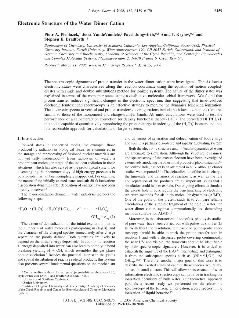
and H3O+ occupied orbitals form a neutral basis. The threehighest occupied molecular orbitals (MOs) of H2O, OH-, andH3O+ are shown in Figure 1, and the corresponding IEs aregiven in Table 1. Overall, the theory and the experiment, whereavailable, agree well.
It is important to determine which of the monomer transitionsare relevant to the dimer cation spectroscopy. The ground-stateelectronic structure at the proton-transferred geometry corre-sponds to the ionization of the hydroxyl part of the OH- · · ·H3O+
dimer, as dictated by the IE considerations. The EOM-IP methodrestricts us to the electronic states derived by single ionizationsof the neutrals. Thus, we only discuss transitions in the ionizedsystem for which the singly occupied MO (SOMO) is the targetorbital. This restricts the types of transitions to (i) the transitionswithin the OH moiety, (ii) the transitions from the H3O+ to theOH moiety, leading to the formation of OH- · · ·H3O2+. The twogroups can be related to the excited states of OH and H3O2+
molecules, respectively. In contrast, excitations at the neutralvertical geometry always correspond to H2O+ · · ·H2O, the chargebeing located on either the H-bond donor or the acceptor. Thus,one needs to consider only the states of H2O+. The excited statesof the monomers are given in Table 2.
There are two possible excitations in H2O+ in the experi-mentally relevant energy range (1-6 eV). The b1r a1 transitionis optically allowed and occurs at 2.3 eV, whereas the dipole-forbidden b1 r b2 excitation is at 6.5 eV. The character of theelectronic states does not change upon structural relaxation fromthe vertical neutral to the cation equilibrium geometry. The OHradical has only one excitation, π r σ, at 4.2 eV. Addition ofa proton to H2O+ produces H3O2+, which features a weak a1
r e transition at 5.8 eV. Overall, addition/subtraction of a protonto/from H2O+ dramatically changes the nature of the MOs andthe electronic transitions. Thus, each species has uniqueelectronic states that cannot be described as perturbed H2O+
states.The EOM-IP-CCSD method treats accurately the dimer cation
states derived by single ionization of the neutral, whereas the
cation states corresponding to excitation to a virtual orbital ofthe neutral are not well-described owing to their significant two-electron character. Neglecting these cation excitations is ac-ceptable only if these states are outside the experimentallyrelevant spectral region. In the context of the water dimer cationat the geometry of the neutral species, we need to consider theexcited states of H2O+ and H2O. The lowest excited state ofthe water monomer in the gas phase is the A1 B1 state at 7.4-7.5eV.51 It is derived by excitation from the 1b1 to the 4a1/Rydberg3s orbital. The first two excitations (2 and 6 eV) in H2O+ areexcitations to the SOMO and are well-described by EOM-IP-CCSD. The next excited state, 2 2B1, occurs at 13.7 eV ascalculated by EOM-EE-CCSD/6-311++G**. At the proton-transferred configuration states of H3O+ and OH are relevant.The lowest excited state of H3O+, 2 1A 1, occurs at 11.8 eV(EOM-EE-CCSD/6-311++G**). The lowest OH excitation isσ r π at 4 eV and is described by EOM-IP-CCSD. The nextone, to the B 2Σ+ state, is at 8.7 eV. Thus, monomer states notdescribed by EOM-IP-CCSD are well outside the region probedin the experiment, at both the neutral and the proton-transferredgeometry.
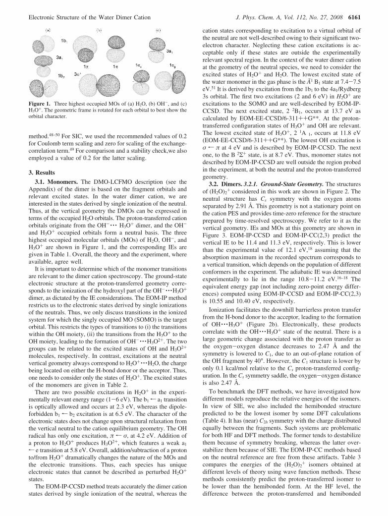
3.2. Dimers. 3.2.1. Ground-State Geometry. The structuresof (H2O)2
+ considered in this work are shown in Figure 2. Theneutral structure has Cs symmetry with the oxygen atomsseparated by 2.91 Å. This geometry is not a stationary point onthe cation PES and provides time-zero reference for the structureprepared by time-resolved spectroscopy. We refer to it as thevertical geometry. IEs and MOs at this geometry are shown inFigure 3. EOM-IP-CCSD and EOM-IP-CC(2,3) predict thevertical IE to be 11.4 and 11.3 eV, respectively. This is lowerthan the experimental value of 12.1 eV,18 assuming that theabsorption maximum in the recorded spectrum corresponds toa vertical transition, which depends on the population of differentconformers in the experiment. The adiabatic IE was determinedexperimentally to lie in the range 10.8-11.2 eV.16–18 Theequivalent energy gap (not including zero-point energy differ-ences) computed using EOM-IP-CCSD and EOM-IP-CC(2,3)is 10.55 and 10.40 eV, respectively.
Ionization facilitates the downhill barrierless proton transferfrom the H-bond donor to the acceptor, leading to the formationof OH · · ·H3O+ (Figure 2b). Electronically, these productscorrelate with the OH · · ·H3O+ state of the neutral. There is alarge geometric change associated with the proton transfer asthe oxygen-oxygen distance decreases to 2.47 Å and thesymmetry is lowered to C1, due to an out-of-plane rotation ofthe OH fragment by 40°. However, the C1 structure is lower byonly 0.1 kcal/mol relative to the Cs proton-transferred config-uration. In the Cs symmetry saddle, the oxygen-oxygen distanceis also 2.47 Å.
To benchmark the DFT methods, we have investigated howdifferent models reproduce the relative energies of the isomers.In view of SIE, we also included the hemibonded structurepredicted to be the lowest isomer by some DFT calculations(Table 4). It has (near) C2h symmetry with the charge distributedequally between the fragments. Such systems are problematicfor both HF and DFT methods. The former tends to destabilizethem because of symmetry breaking, whereas the latter over-stabilize them because of SIE. The EOM-IP-CC methods basedon the neutral reference are free from these artifacts. Table 3compares the energies of the (H2O)2
+ isomers obtained atdifferent levels of theory using wave function methods. Thesemethods consistently predict the proton-transferred isomer tobe lower than the hemibonded form. At the HF level, thedifference between the proton-transferred and hemibonded
Figure 1. Three highest occupied MOs of (a) H2O, (b) OH-, and (c)H3O+. The geometric frame is rotated for each orbital to best show theorbital character.
Electronic Structure of the Water Dimer Cation J. Phys. Chem. A, Vol. 112, No. 27, 2008 6161
structures is 26 kcal/mol, and electron correlation brings it downto 7-8 kcal/mol. The large difference at the HF level is due tothe symmetry-breaking problem at the hemibonded geometry.The energy difference between the proton-transferred andvertical configurations only weakly depends on the level oftheory and is approximately 20 kcal/mol. This stable behavioris due to the charge-localized character of the wave functions,which are free from the HF instability, and are described wellusing both open-shell and closed-shell references.
The set of (H2O)2+ structures was re-optimized using DFT.
The results are summarized in Table 4. The DFT calculationswithout SIC show the following trends. First, there is virtuallyno energy difference between the unrestricted and restricted
open-shell BLYP results. Second, compared to the benchmarkEOM-IP-CCSD calculations, BLYP overstabilizes both the
TABLE 1: Ionization Energies (eV) of H2O, OH-, and H3O+
EOM-IP-CCSD EOM-IP-CC(2,3)
6-311++G** aug-cc-pVTZ 6-311++G** aug-cc-pVTZ exp
H2O1b1 12.32 12.61 12.29 12.51 12.6,a 12.62b
3a1 14.61 14.87 14.57 14.78 14.8,a 14.64b
1b2 18.84 18.94 18.78 18.83 18.6,a 18.6b
OH-
π 1.37 1.77 1.17 1.48 1.83c
σ 5.60 5.90 5.37 5.59
H3O+
a1 24.38 24.64 24.41 24.61e 30.16 30.25 30.17 30.21
a Reference 66. b Reference 67. c References 68 and 69.
TABLE 2: Excitation Energies (eV) and Transition Properties (au) of H2O+, OH, and H3O2+ (a 6-311++G** Basis Set wasUsed Throughout)
EOM-IP-CCSD EOM-EE-CCSD EOM-IP-CC(2,3) expt
Eex µ2 f Eex µ2 f Eex Eex
H2O+ at the geometry of the neutral3a1-1b 1 2.29 0.0229 0.00128 2.29 0.0225 0.00126 2.28 2.02a
1b2-1b 1 6.52 6.48 6.49 6.0a
H2O+ at the geometry of the cation3a1-1b 1 2.02 0.0179 0.000885 2.01 0.0177 0.000874 2.01 1.89b
1b 2-1b1 6.53 6.89 6.49 -
OH at the geometry of the neutralσ-πa 4.22 0.0301 0.00312 4.21 0.0311 0.00321 4.20 4.12c
H3O2+ at the geometry of H3O+
a1-eb 5.78 6.7 × 10⁻⁶ 9.5 × 10⁻⁷ 5.74 2.5 × 10⁻⁵ 3.4 × 10⁻⁶ 5.75
a Computed as a difference of vertical IEs from ref 67. b Reference 70. c Reference 71.
Figure 2. Geometries of (a) vertical neutral (H2O) 2, (b) proton-transferred (H2O)2
+, and (c) hemibonded(H2O)2+. Oxygen-oxygen
distance has been marked on the plots.
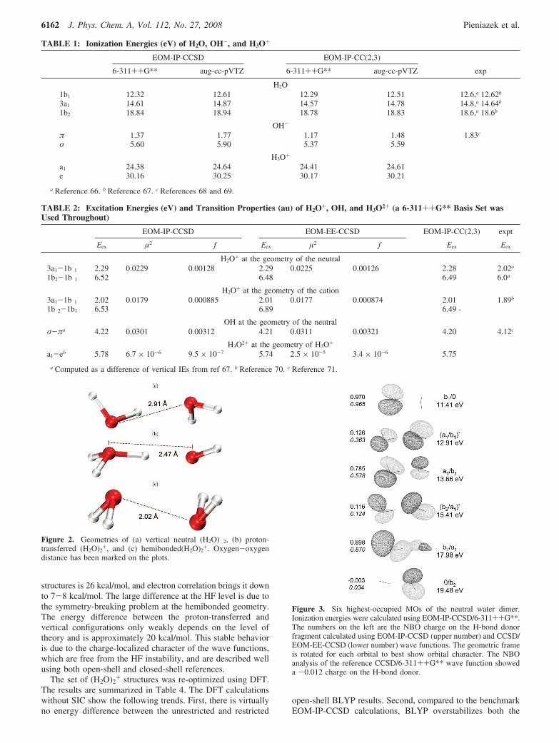
Figure 3. Six highest-occupied MOs of the neutral water dimer.Ionization energies were calculated using EOM-IP-CCSD/6-311++G**.The numbers on the left are the NBO charge on the H-bond donorfragment calculated using EOM-IP-CCSD (upper number) and CCSD/EOM-EE-CCSD (lower number) wave functions. The geometric frameis rotated for each orbital to best show orbital character. The NBOanalysis of the reference CCSD/6-311++G** wave function showeda -0.012 charge on the H-bond donor.
6162 J. Phys. Chem. A, Vol. 112, No. 27, 2008 Pieniazek et al.
vertical and the hemibonded dimer structures by -12 and -13kcal/mol, respectively. The error for the hemibonded structureis comparable in absolute value to that of HF. However, thesign is opposite and, as was shown before,28 the SIE uncorrectedDFT erroneously places the hemibonded minimum below theproton-transferred one. This is a well-known signature of SIEobserved in many systems with charge separation.47,52,53
A simple empirical SIC correction for doublet states basedon removing SI of the unpaired electron implemented forrestricted open-shell BLYP48,49 improves the results significantly.For the recommended choice of the two scaling parameters (0.2and 0), the error in the above relative energies drops to 2-3kcal/mol (compared to EOM-IP-CCSD) and is further reducedto less than 2 kcal/mol upon moving from DZVP to TZVDDbasis set. This is encouraging and justifies considering the useof DFT/BLYP with the present SIC for condensed-phase AIMDcalculations.12 Although it is theoretically more appealing toemploy the same value for both scaling parameters (i.e., 0.2and 0.2, as in other approaches54), this produces less-accurateresults: the correct order of minima is still preserved, but theerror increases to about -4 kcal/mol.
Finally, DFT re-optimization of the proton-transfer andhemibonded structures results in minor geometry and, conse-quently, minor energy changes (Table 4). The geometry of theneutral dimer, which is not a minimum on the cationic surface,collapses to the proton-transfer minimum upon minimization(as for EOM-IP-CCSD).
The use of restricted open-shell formalism with Kohn-ShamDFT deserves an additional comment. As shown by Pople andcoworkers,55 in open-shell systems that have excess R electrons,regions of negative spin density exist. That means that the localdensity of � electrons may be higher than the density of Relectrons. The local excess of � electrons is reproduced bycorrelated calculations, as well as confirmed experimentally. Inthe DFT framework, it can only be reproduced within UKSformalism, simply because the electron density is a sum of the
MO densities, which are the same for both spins in ROKS.However, the SIC applied here is stable only within ROKSformalism because it requires one to identify the unpairedelectron. Density functionals containing non-local operators,such as long-range Hartree-Fock exchange, reduce the SIE ina more fundamental way.56–60 However, symmetric radicalcations are particularly difficult systems, and performance isnot yet fully satisfactory.60
3.2.2. DMO-LCFMO Framework. The dimer spectroscopycan be explained in terms of the individual fragment contribu-tions. The theoretical framework is provided by the DMO-LCFMO theory, which was applied previously to the electronicstructure of the benzene dimer cation15 and is described in theAppendix. It allows one to correlate the dimer and monomertransitions based on the degree of mixing of the FMOs in thedimer. We employ a (H-bond donor orbital)/(H-bond acceptororbital) notation, allowing one to quickly discern the FMOs thatcontribute to a given DMO. If there is no component on thegiven fragment, then the symbol 0 is used. An asterisk signifiesantibonding character of the dimer MO with respect to thefragment’s interaction. The extent to which a given FMOparticipates in a given DMO can be quantified using the NBOanalysis. NBO yields the charges of the fragments, which arerelated to the square of the diabatic wave function defined hereas the charge-localized state. Below we discuss the DMOs atthe vertical and at the proton-transferred geometry.
The orbitals at the vertical configuration are shown in Figure3. The SOMO is the b1 orbital of the H-bond donor moleculeand is called b1/0. It is antisymmetric with respect to the planeof symmetry. The delocalization of this orbital requires mixingwith an antisymmetric orbital of the H-bond acceptor molecule.The only such orbital is 0/b2, which is high in energy, and,therefore, the ground state hole is localized. The two lowerorbitals are linear combinations of the acceptor b1 and the donora1 MOs. The higher energy combination is antibonding withrespect to the monomers and is called (a1/b1)/. The bondingcombination is (a1/b1). Lower in energy, we find the (b2/a1)*and (b2/a1) pair. The antibonding DMO is located mostly onthe acceptor, whereas the donor hosts the bonding component.Finally, the acceptor 0/b2 is the lowest DMO considered here.States corresponding to ionizing the H-bond donor are lowerin energy (the corresponding FMOs are higher in energy), whichcan be rationalized easily in terms of electrostatic interaction.A hole on the H-bond donor fragment is stabilized by thenegatively charged oxygen of the acceptor, whereas a hole onthe acceptor is destabilized by the positively charged proton.
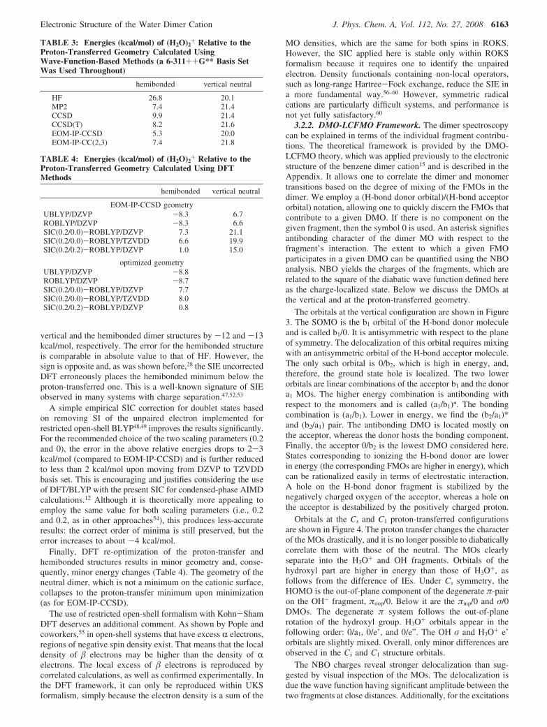
Orbitals at the Cs and C1 proton-transferred configurationsare shown in Figure 4. The proton transfer changes the characterof the MOs drastically, and it is no longer possible to diabaticallycorrelate them with those of the neutral. The MOs clearlyseparate into the H3O+ and OH fragments. Orbitals of thehydroxyl part are higher in energy than those of H3O+, asfollows from the difference of IEs. Under Cs symmetry, theHOMO is the out-of-plane component of the degenerate π-pairon the OH- fragment, πoop/0. Below it are the πinp/0 and σ/0DMOs. The degenerate π system follows the out-of-planerotation of the hydroxyl group. H3O+ orbitals appear in thefollowing order: 0/a1, 0/e’, and 0/e”. The OH σ and H3O+ e’orbitals are slightly mixed. Overall, only minor differences areobserved in the Cs and C1 structure orbitals.
The NBO charges reveal stronger delocalization than sug-gested by visual inspection of the MOs. The delocalization isdue the wave function having significant amplitude between thetwo fragments at close distances. Additionally, for the excitations
TABLE 3: Energies (kcal/mol) of (H2O)2+ Relative to the
Proton-Transferred Geometry Calculated UsingWave-Function-Based Methods (a 6-311++G** Basis SetWas Used Throughout)
hemibonded vertical neutral
HF 26.8 20.1MP2 7.4 21.4CCSD 9.9 21.4CCSD(T) 8.2 21.6EOM-IP-CCSD 5.3 20.0EOM-IP-CC(2,3) 7.4 21.8
TABLE 4: Energies (kcal/mol) of (H2O)2+ Relative to the
Proton-Transferred Geometry Calculated Using DFTMethods
hemibonded vertical neutral
EOM-IP-CCSD geometryUBLYP/DZVP -8.3 6.7ROBLYP/DZVP -8.3 6.6SIC(0.2/0.0)-ROBLYP/DZVP 7.3 21.1SIC(0.2/0.0)-ROBLYP/TZVDD 6.6 19.9SIC(0.2/0.2)-ROBLYP/DZVP 1.0 15.0
optimized geometryUBLYP/DZVP -8.8ROBLYP/DZVP -8.7SIC(0.2/0.0)-ROBLYP/DZVP 7.7SIC(0.2/0.0)-ROBLYP/TZVDD 8.0SIC(0.2/0.2)-ROBLYP/DZVP 0.8
Electronic Structure of the Water Dimer Cation J. Phys. Chem. A, Vol. 112, No. 27, 2008 6163

from H3O+ to OH one might expect to see a -1 charge on thehydroxyl fragment. However, the large positive charge on H3O2+
will polarize the OH- and thus decrease its charge. This isconfirmed by the calculation, revealing -0.771 charge on theOH moiety.
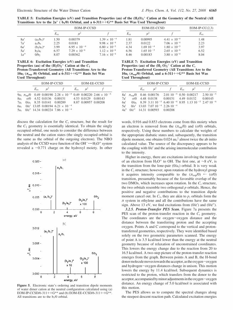
3.2.3. Spectroscopy at the Vertical Configuration. Table 5and Figure 5 present excitation energies and transition propertiesof (H2O)2
+ at the geometry of the neutral species. All excitationsinvolve transfer of an electron to the SOMO, that is, the b1/0orbital. Overall, all theoretical methods are in good agreementin terms of energetics. A state involving excitation to a virtualorbital of the neutral appeared in the EOM-EE-CCSD calcula-tion at 8 eV and was disregarded.
At low energies, up to ca. 2 eV, we find excitations from the(a1/b1)/ and (a1/b1) pair into the SOMO. Their intensity canoriginate from both the intramolecular and intermolecular termsand reflects the partitioning of the DMOs into FMOs, and canbe explained by DMO-LCFMO. Referring to the formalismsummarized in the Appendix, (a1/b1)/ and (a1/b1) are examplesof DMOs in which both the R and � coefficients (i.e., theweights of the FMOs) are significant. The weights of the FMOsare just the square roots of the EOM-IP-CCSD NBO chargesbecause both fragments are neutral in the reference state:
(a1 ⁄ b1)/) 0.355(a1 ⁄ 0)- 0.935(0 ⁄ b1) (3)
(a1 ⁄ b1)) 0.886(a1 ⁄ 0)+ 0.464(0 ⁄ b1) (4)
Neglecting the intermolecular contribution to the intensity,one can evaluate the transition dipole moments for the twotransitions according to eq 7. The corresponding squares are0.0029 and 0.0180 au2. This estimate is in excellent agreementwith the actual EOM-IP-CCSD dimer calculation, which yields
0.0038 and 0.0181 au2, thus supporting the assumption that thesetwo transitions draw their intensity from the H-bond donor a1
component of the DMO and that the intermolecular terms arenegligible. The EOM-IP-CCSD and EOM-EE-CCSD methodspredict different relative intensity of the two bands. AlthoughEOM-EE-CCSD predicts nearly equal intensities, EOM-IP-CCSD suggests a 1:5 intensity ratio in favor of the higher energyband. The origins of this discrepancy can be traced back todifferent partitioning of the a1 component between the (a1/b1)/
and (a1/b1) pair. Our benchmark study61 demonstrated that EOM-IP-CCSD provides a more accurate description of chargedelocalization and more accurate transition properties.
EOM-IP-CCSD predicts weak excitations from the (b2/a1)/
and (b2/a1) pair to be at 4 and 6.5 eV, respectively. EOM-EE-CCSD predicts the transition from the antibonding DMO to lie0.3 eV higher and agrees as to the position of transition fromthe bonding DMO. The antibonding orbital is confined to theH-bond acceptor, while the bonding orbital is confined to thedonor. Conceptually, the character of the transitions is similarto that discussed above. These two excitations correspond tothe symmetry-forbidden b1 r a1 excitation in the monomer.Consequently, the intramolecular term in eq 7 is zero. Orbitalrelaxation and intermolecular terms account for the intensityof these bands, which are still 100 times weaker than the allowedtransitions.
Finally, around 8 eV we find the (b1/0) r (0/b2) transition.It is a pure charge-transfer band (R ) 0), where the hole ismoved from the donor to the acceptor molecule. Its considerableintensity is due to particularly favorable overlap between theinitial and final orbitals. The transition is still one order ofmagnitude weaker than the sum of the (b1/0)r (a1/b1) and (b1/0) r (a1/b1)/ excitations.
3.2.4. Spectroscopic Signatures of Proton Transfer. Excita-tion energies and transition properties at the C1 and Cs proton-transferred geometries are presented in Tables 6 and 7,respectively. The EOM-IP-CCSD data are shown in Figure 6.We were able to obtain EOM-EE-CCSD results only for thethree lowest excitations because the states involving virtualorbitals of the neutral wave function are the next higher inenergy. The lowest-energy transition is at 9.6 eV, well beyondthe energy range potentially probed in a pump-probe experiment.Additionally, the Rydberg character of these states means thatthey are likely to be significantly perturbed in the condensedphase. All of the excitations considered here involve the transferof an electron to the singly occupied (πoop/0) orbital. Becauseof the localized character of the DMOs, the spectrum partitionsinto two parts: transitions within the OH fragment and transitionsfrom the H3O+ to the OH fragment. In the language of theAppendix, the lower-energy part of the spectrum is close to the� ) 0 limit, whereas the higher energy one approaches the R) 0 limit, that is, local and charge-transfer excitations,respectively.
The Cs and C1 configurations differ only slightly in positionsand intensities of the transitions in the low-energy part of thespectrum. The transitions in this region are from the (πinp/0)and (σ/0) orbitals. The former excitation is not present in anisolated OH monomer, as the (πinp/0) - (πoop/0) pair isdegenerate. In the dimer, it splits by 0.5 eV and acquiresoscillator strength. The (πoop/0)r (σ/0) excitation is at 4.5 eV,very close to the monomer value of 4.2 eV; however, itsintensity is less than half. The origin of the intensity decreasecan be investigated using DMO-LCFMO, although its quantita-tive application is complicated by the large extent of chargetransfer occurring in the neutral and in the cation states. We
Figure 4. Six highest-occupied MOs of the neutral water dimer at theCs and C1 proton-transferred configurations. Ionization energies werecalculated using EOM-IP-CCSD/6-311++G**. The NBO charge onthe hydroxyl radical calculated using EOM-IP-CCSD wave functionsis given below. The geometric frame is rotated for each orbital to bestshow orbital character. The NBO analysis of the reference CCSD/6-311++G** wave function showed -0.771 and -0.770 charge on thehydroxyl radical in the Cs and C1 structures, respectively.
6164 J. Phys. Chem. A, Vol. 112, No. 27, 2008 Pieniazek et al.
discuss the calculation for the Cs structure, but the result forthe C1 geometry is essentially identical. To obtain the singlyoccupied orbital, one needs to consider the difference betweenthe neutral and the cation states (the singly occupied orbital isthe same as the orbital of the outgoing electron). The NBOanalysis of the CCSD wave function of the OH- · · ·H3O+ systemrevealed a -0.771 charge on the hydroxyl moiety. In other
words, 0.916 and 0.853 electrons come from this moiety whenan electron is removed from the (πinp/0) and (σ/0) orbitals,respectively. Using these numbers to calculate the weights ofthe appropriate diabatic states and, subsequently, the transitiondipole moment, one obtains 0.024 au,2 almost twice the ab initiocalculated value. The source of the discrepancy appears to bethe coupling with 0/e′ and the arising intermolecular contributionto the intensity.
Higher in energy, there are excitations involving the transferof an electron from H3O+ to OH. The first one, at ∼8 eV, isthe transition from the lone-pair (0/a1) orbital. It is very weakin the Cs structure; however, upon rotation of the hydroxyl groupit acquires intensity comparable to the (πoop/0) r (σ/0)transition, presumably because of the favorable overlap of thetwo DMOs, which increases upon rotation. In the Cs structure,the two orbitals resemble two orthogonal p orbitals. Hence, thepositive and negative contributions to the transition dipolemoment cancel out. In C1, they are akin to pz orbitals form theπ system in ethylene and all the contributions have the samesign. Above 13 eV, we find excitations from (0/e′) and (0/e′′ ).
3.2.5. Proton-Transfer PES Scan. Figure 7a presents thePES scan of the proton-transfer reaction in the Cs geometry.The coordinates are the oxygen-oxygen distance and thedistance between the transferring proton and the acceptingoxygen. Points A and C correspond to the vertical and proton-transferred geometries, respectively. They were identified basedsolely on the two geometric parameters scanned. The energyof point A is 3.5 kcal/mol lower than the energy at the neutralgeometry because of relaxation of unconstrained coordinates.This lowers the energy change due to the reaction from 20 to16.5 kcal/mol. A two-step picture of the proton-transfer reactionemerges from the graph. Between points A and B, the H-bonddonormoleculemovestowardstheacceptor,as theoxygen-oxygenand hydrogen-oxygen distances change in unison. This motionlowers the energy by 11.4 kcal/mol. Subsequent dynamics isrestricted to the proton, which transfers from the donor to theacceptor,accompaniedbyminoradjustmentsintheoxygen-oxygendistance. An energy change of 5.0 kcal/mol is associated withthis motion.
The PES allows us to compute the spectral changes alongthe steepest descent reaction path. Calculated excitation energies
TABLE 5: Excitation Energies (eV) and Transition Properties (au) of the (H2O)2+ Cation at the Geometry of the Neutral (All
Transitions Are to the 2a′′ ( b1/0) Orbital, and a 6-311++G** Basis Set Was Used Throughout)
EOM-IP-CCSD EOM-EE-CCSD EOM-IP-CC(2,3)
Eex µ2 f Eex µ2 f Eex
8a′ (a1/b1)/ 1.50 0.00379 1.39 × 10 -4 1.81 0.00995 4.41 × 10⁻⁴ 1.487a′ a1/b1 2.25 0.0181 9.98 × 10⁻⁴ 2.37 0.0122 7.06 × 10⁻⁴ 2.236a′ (b2/a1)/ 3.99 6.95 × 10 -4 6.80 × 10⁻⁵ 4.34 1.69 10 -4 1.80 × 10⁻⁵ 3.975a′ b2/a1 6.57 7.29 × 10⁻⁶ 1.12 × 10⁻⁶ 6.56 1.65 10 -5 2.65 × 10⁻⁶ 6.521a′′ 0/b2 8.07 0.00362 7.16 × 10⁻⁴ 8.46 0.00183 3.80 × 10⁻⁴ 8.04
Figure 5. Electronic state’s ordering and transition dipole momentsof water dimer cation at the neutral configuration calculated using (a)EOM-IP-CCSD/6-311++G** and (b) EOM-EE-CCSD/6-311++G**.All transitions are to the b1/0 orbital.
TABLE 6: Excitation Energies (eV) and TransitionProperties (au) of the (H2O)2
+ Cation at the C1
Proton-Transferred Geometry (All Transitions Are to the10a1 (πoop /0) Orbital, and a 6-311++G** Basis Set WasUsed Throughout)
EOM-IP-CCSD EOM-EE-CCSD
Eex µ2 f Eex µ2 f
9a1 πinp/0 0.49 0.00190 2.28 × 10 -5 0.49 0.00220 2.66 × 10 -5
8a1 σ/0 4.52 0.0136 0.00151 4.53 0.0129 0.001437a1 0/a1 8.35 0.0141 0.00289 8.87 0.00957 0.002086a1 0/e’ 13.05 0.00194 6.21 × 10 -4
5a1 0/e” 14.34 0.00224 7.86 × 10 -4
TABLE 7: Excitation Energies (eV) and TransitionProperties (au) of the (H2 O)2
+ Cation at the Cs
Proton-Transferred Geometry (All Transitions Are to the10a1 (πoop/0) Orbital, and a 6-311++G** Basis Set WasUsed Throughout)
EOM-IP-CCSD EOM-EE-CCSD
Eex µ2 f Eex µ2 f
8a′ πoop/0 0.46 0.00176 2.01 10 -5 0.50 0.00217 2.50 10 -5
7a′ σ/0 4.48 0.0138 0.00151 4.49 0.0132 0.001456a′ 0/a1 8.39 3.11 10 -5 6.40 10 -6 8.95 1.13 10 -5 2.47 10 -6
5a′ 0/e′ 13.03 7.07 10 -4 2.26 10 -4
4a′′ 0/e′′ 14.31 0.00593 0.00208
Electronic Structure of the Water Dimer Cation J. Phys. Chem. A, Vol. 112, No. 27, 2008 6165

and transition properties are presented in Figure 7b and d. Weemploy the Cs symmetry labels to identify the states in thisdiscussion, as the character of the orbitals changes along thereaction coordinate. All of the transitions involve the transferof an electron to SOMO, the 2a′′ orbital, which is the out-of-plane orbital on the H-bond donor. Its character evolves fromthe H2O+ lone-pair b1 orbital to the π orbital of OH. Ofparticular interest are the 6a′, 7a′, and 8a′ transitions becausethey are within the spectroscopic 1-6 eV window. At point A,two absorption bands appear around 2 eV (8a′ and 7a′). Theyare transitions from the bonding and antibonding combinationsof the donor a1 and acceptor b1 fragment MOs [(a1/b1)/ and(a1/b1)]. The lower-energy transition carries more intensity,which is different from the fully optimized neutral configuration,where the higher energy transition carries more intensity. Thisis due to the slightly different geometry and orbital mixing ofthe H-bond acceptor b1 and donor a1 in the two states. 6a′ and5a′ are bonding and antibonding combinations of donor b2 andacceptor a1. They are at 6.2 and 4.3 eV, respectively. Theintensities of both bands are small. Finally, at 8.5 eV we findthe transition from the 1a′′ (acceptor b2) orbital.
As the reaction proceeds to point B, the intensity of 8a′ and7a′ changes as the bands move apart. In other words, thepartitioning of H-bond donor a1 and acceptor b1 in the (a1/b1)/
and (a1/b1) pair changes. The small-magnitude of change isunderstood easily within the diabatic framework of (a1/0) and(0/b1) states, that is, the states with the charge localized on theH-bond donor and acceptor, respectively. The coupling andseparation of the diabatic states increase at shorter distances.
The two effects largely cancel out, and no drastic changes inintensity are observed. There is a small increase in the intensityof the 6a′ (H-bond acceptor a1) band.
At point B, the proton-transfer step starts taking place andthe character of MOs changes more dramatically. The SOMObecomes the out-of-plane π orbital. The intensity of the 8a′ banddrops significantly with a decrease in energy as it becomes aπ-π transition on the OH fragment. The 7a′ excitation gainsintensity as the energy rises to 4.5 eV and becomes the σ-πexcitation. At the same time, the 6a′ and 5a′ excitations moveto higher energy, becoming CT excitations from H3O+ to OH.The already small intensity of excitation from 6a′ drops furtheras it becomes the apical orbital of H3O+. Both 1a′′ and 5a′transitions move to 12-14 eV, outside the experimental region.
4. Discussion
Three geometries on the ground-state PES of the water dimercation are of prime importance in the photoionization process.The first two are minima: the proton-transferred and thehemibonded structures (Figure 2b and c). The third one is thegeometry of the neutral dimer reached by vertical ionization(Figure 2a). The geometries and relative energetics of thesestructures calculated by correlated electronic structure methodsare in good agreement with each other (Table 3) and previousab initio results.24,25,28 All of these calculation correctly char-acterize the proton-transferred geometry as the global minimumlying 5-10 kcal/mol below the hemibonded local minimum andabout 20 kcal/mol below the structure corresponding to verticalionization of the neutral water dimer. This ordering is repro-duced already at the HF level, which, however, grossly (by about20 kcal/mol) destabilizes the hemibonded structure due to thetendency of HF to artificially localize the MOs in systems withsymmetrically equivalent centers. In contrast, the proton-transferred structure has spin-localized on one fragment andcharge-localized on the other; and in the vertical geometry,corresponding to the neutral water dimer, the spin and chargeare initially localized on one water. The behavior of the DFTmethods is exactly opposite. Because of SIE, DFT/BLYPoverstabilizes structures with delocalized charge and erroneouslypredicts the hemibonded structure to be the global minimum.However, a simple empirical a posteriori SIC48,49 almostcompletely removes this artifact; and the predictions of the SIC-corrected DFT methods are very close to those of MP2. This isgood news for the DFT-based AIMD studies of ionization inliquid water.12 Nonetheless, further benchmark studies includingthe spin localization and cluster dynamics are needed.
For ionization in the condensed phase, the issue of electroniclocalization/delocalization is of interest. We are interested inthe question of whether the charge is localized at one siteimmediately upon ionization or whether it will localize afterbeing initially delocalized over many water molecules. Thislocalization process can be explored only by considering clustersbeyond the dimer. In the dimer, the hole forms on the b1 orbitalof the H-donor fragment immediately upon ionization. Thisinitial localization is due to the fact that the donor water is notacting as an acceptor to any other H bond. The b1 orbital of theH-bond acceptor in fact couples with the a1 orbital of the donor.The neutral water dimer thus represents the most asymmetricarrangement of water molecules. Already in the cyclic trimer,the water molecules become equivalent, which means that uponionization the hole must be initially delocalized. In the bulkphase, each water molecule is likely to serve as a hydrogen-bond acceptor and donor simultaneously, thus more likelydelocalizing the hole. However, in the initial period of delo-
Figure 6. Electronic states ordering and transition dipole moments ofthe water dimer cation at the C1 (a) and Cs symmetry proton-transferredconfigurations calculated using EOM-IP-CCSD/6-311++G**. Alltransitions are to the πoop/0 orbital.
6166 J. Phys. Chem. A, Vol. 112, No. 27, 2008 Pieniazek et al.

calization, which is not yet known, the positive charge willlocalize, thereby starting the proton-transfer reaction. From thispoint onward, the dimer is presumably an adequate model forthe spectroscopy of the condensed-phase proton-transfer process.
In general, small- and medium-sized water clusters withdifferent geometries will provide a natural laboratory toinvestigate the electronic and nuclear dynamics upon ionizationwith varying degrees of initial localization/delocalization of thehole.62
Charge localization is intimately related to the electronicspectroscopy of the system. Using formalism of the Appendix,both the R and � coefficients may be significant (delocalizedcharge), or one can dominate (localized). Thus, excitations willhave a mixed charge transfer and local character. With noknowledge of the degree of charge delocalization, it is impos-sible to say anything about the intensity of mixed bands andreliable ab initio calculations are needed. In the case of dimercation states, one may expect intermolecular contributions tobe less significant than those in their neutral counterparts becauseof the more-compact nature of MOs. This maybe counteractedby the decreased separation manifested in a charged species.Our calculations for the water dimer cation reveal that theintermolecular terms are typically one order of magnitudesmaller than the intramolecular terms for allowed transitions.Note, however, that the interfragment contribution variesexponentially with the distance (separately from R and �) and
may change significantly with relative orientation of the twofragments, thus allowing one to monitor the molecular dynamicsvia intensity and/or position of those bands.
Next, we discuss the nuclear motions along the reactioncoordinate leading from the geometry corresponding to theneutral water dimer to the proton-transfer structure (Figure 7a).The reaction, which is a downhill process without a barrier,proceeds in two steps. The first one involves heavy atommotions; that is, the two water molecules move closer to eachother with the oxygen-oxygen distance decreasing from 2.9 to2.5 A. This step is responsible for the largest part of the energygain (∼12 kcal/mol) along the reaction coordinate. Because itinvolves the motion of heavy atoms, it is relatively slowcompared to the second step, the rapid transfer of the proton,which can happen only when the water oxygens are sufficientlyclose to each other. This process involves a motion of a lightparticle and is, therefore, possibly as fast as a few femtoseconds.Precise time scales are under detailed experimental and theoreti-cal investigation in our labs. The energy gain associated withthe proton hop is smaller, amounting to roughly 5 kcal/mol. Inlarger clusters and in bulk liquid water, this two-step mechanismshould be preserved, although it might be preceded by initialfast charge localization.12 This is exactly the dynamics that wewish to resolve in the condensed phase, with a spectroscopic
Figure 7. (a) The ground-state PES scan for the proton-transfer reaction. The x-axis is the oxygen-oxygen distance, and the y-axis is the distancebetween the transferring proton and the accepting oxygen. At each point, a constrained geometry optimization was conducted. Points A and Ccorrespond to the neutral and proton transferred geometries, respectively. Point B marks the start of the proton transfer. The black line is thesteepest descent path. (b-d) Vertical excitation energies, transition dipole moments, and oscillator strengths along the reaction coordinate. Allcalculations were done using EOM-IP-CCSD/6-311++G**.
Electronic Structure of the Water Dimer Cation J. Phys. Chem. A, Vol. 112, No. 27, 2008 6167

handle on these events being provided by the changing excitationspectrum of the radical species along the proton-transferreaction.
Let us consider how the electronic transitions, their bandpositions, and their intensities evolve as we move along thereaction coordinate. Femtosecond spectroscopy should be ableto monitor the system evolution by recording the changingtransient absorption spectrum. Formally, the H2O+ cation isderived from the OH radical by the addition of a proton. Onemight thus expect the electronic structure and the spectroscopyof the two species to be similar. However, the results from Table2 show that the addition of a proton is not a benign perturbationto the electronic structure. The OH radical has a characteristicabsorption band around 4.2 eV corresponding to the transitionof the bonding σ electron into the nonbonding π hole. Even ifthe proton is brought up along the O-H axis leading to a linearH-O-H+, then the σ to π promotion is pushed up to higherenergy (∼6 eV). More significantly, allowing the structure toadopt the lower-energy bent configuration splits the π orbitalinto b1 and a1 symmetry components. The former π r σtransition becomes dipole-forbidden for the ground-state com-ponent (b1r b2). In C2V, the transition between the two formerlydegenerate b1 and a1 orbitals is dipole-allowed with a transitionenergy of ca. 2.3 eV and 1/3 of the oscillator strength of the OHtransition. Simply put, the 4-eV band disappears and a weaker2.3-eV band appears in its place.
At the vertical geometry, the transitions for the ionized waterdimer are perturbed and include some charge-transfer character;however, the wave functions can still be correlated with thoseof the monomers. Extracting the three points A, B, and C fromFigure 7, we have replotted how the experimental spectrum cantrack the chemical reaction dynamics in Figure 8. At A, thebands at 2 eV (b1/0)r (a1/b1)/ and (b1/0)r (a1/b1) [2a′′ r 8a′and 2a′′ r 7a′] have almost the same oscillator strength as themonomer. The distribution of intensity between the two dependsheavily on the system geometry. The 6-eV monomer-liketransition is now not strictly symmetry-forbidden, particularlythe 4-eV (b2/0) r (b2/a1)/ [2a′′ r 6a′] component with moreCT character. It still is almost two orders of magnitude weakerthan the 2-eV transition. Therefore, the dominant characteristicelectronic absorption of the dimer cation at the Franck-Condongeometry is around 2 eV (620 nm), as for the gas-phasemonomer. Then at B, the (b2/0)r (a1/b1)/ and (b2/0)r (a1/b1)[2a′′ r 8a′ and 2a′′ r 7a′] shift apart, the lower-energy band
carrying more intensity. The 4-eV transition shifts slightly tothe blue. At this point, proton-transfer begins. The lower-energyband becomes a weak π-π excitation, while the 4-eV bandgains intensity to become the σ-π excitation. The shift is aclear fingerprint of the reaction, and the significant changecorresponds to the charge-transfer between the species, that is,from B to C. Overall, the band positions resemble themonomers; however, the intensity pattern and fine structure arestrong functions of the relative geometries in the cluster. Forexample, the comparison of the Cs and C1 geometries of theproton-transferred complexes show a dramatic variation in the0/a1 [6a′] band because of the alignment of the p orbitals onthe two fragments. Although Cs is a saddle point between thetwo equivalent C1 configurations of the product, it is only 0.1kcal/mol above the minima and it allows us to symmetry-labelthe spectroscopic state of the evolving system. However, thetransition to C1 does lead to this large intensity change and oneshould be wary of the role of conformational changes in theband intensities.
We are now ready to discuss the effects of bulk water. Evenif the dimer core is a good representation of the verticallyprepared hole in water, then there is a large range of local neutraldonor-acceptor geometries populated in room-temperaturewater. Although the configuration considered is the lowest-energy cluster, other configurations, particularly with differentorientations of the free hydrogen of the donor with respect tothe acceptor σV plane, should also be considered. Preliminarycalculations have shown that if the O-H group of the H-bonddonor is aligned with one of the acceptor O-H bonds (the donormolecule is rotated by ∼90°), then the transitions with significantcharge-transfer character can be enhanced significantly.62 Inparticular, the band around 4 eV that involves CT (b2/0)f (b2/a1)/ [2a′′ r 7a′] acquires oscillator strength and can becomecomparable to the valence band near 2.3 eV. What if moresolvating waters are included around the ionized core water?Preliminary EOM-IP-CCSD computations on a vertically ion-ized pentamer extracted from ice Ih show that excitations on acentral water give rise to a spectrum similar to that of themonomer with an intense band near 2 eV and little oscillatorstrength at 4 eV. These results will be further quantifiedelsewhere.62
5. Conclusions
The water dimer cation is a prototypical system for the proton-transfer process in the gas and condensed phases. The verticalstructure formed immediately upon ionization is not a stationarypoint on the cation PES, and the system follows the downhillgradient to OH · · ·H3O+. Our study demonstrates that thisprocess can be monitored by femtosecond time-resolved elec-tronic spectroscopy. At the simplest level, the initial spectrumresembles that of H2O+. As the reaction proceeds, band positionsand intensities change. The product of the reaction spectroscopi-cally resembles the free OH radical. A more detailed look athow the electronic spectrum evolves along the proton-transfercoordinate shows that changes in electronic structure are moresubtle. We observed strong coupling between the H-bond donorand acceptor orbitals, which dissolves the monomer states intomore delocalized dimer states. This coupling, which is likelyto be present in the condensed phase as well, will lead tosignificantly delocalized states. Modeling of such states requiresa full quantum treatment of the entire system. Hybrid quantummechanical/molecular mechanical methods are not appropriatefor this situation because the system cannot be partitioned intoa solvent and a chromophore, as, for example, in our study of
Figure 8. Evolution of the electronic spectrum of the water dimercation along the reaction path at points A (red solid line), B (greendotted line), and C (blue dashed line). Points A and C correspond tovertical neutral and proton-transferred geometries. Point B marks thestart of the proton transfer. 0.2 eV full width at half-maximum wasassumed. See Figure 7 for details.
6168 J. Phys. Chem. A, Vol. 112, No. 27, 2008 Pieniazek et al.

electronic spectroscopy of the solvated CN radical.63 AIMD,which is able to directly describe the electronic structure of theentire system, requires a fast electronic structure method, andpresently DFT-based methods are the only viable choice. Wehave found that the energetics and structures of (H2O)2
+ arereproduced reasonably by the ROKS-BLYP method with thesimple SIC correction,48,49 which thus can be employed inAIMD simulations of the bulk.
Appendix
This section outlines the qualitative DMO-LCFMO frame-work for the description of the electronic states of the waterdimer cation. This approach, rooted in the exciton theory,64,65
was developed and applied to the electronic structure of thebenzene dimer cation.15 DMO-LCFMO describes the electronicwave functions of the dimer in terms of the dimer molecularorbitals expressed in the basis of the monomer MOs, whichallows one to correlate properties of the dimer with theproperties of the fragments. For the ionized dimers, one neednot consider the full many-electron wave function of the initialand final states because they can be mapped onto 1-electron-in-2-orbitals ones. The two orbitals are the orbitals involved inthe transition. In the cases considered in this work, the targetorbital is always the same (SOMO of the cation).
An important feature distinguishing the benzene and watersystems is greater charge localization in the ground state of(H2O)2
+. The SOMO is the out-of-plane p orbital of thehydrogen-bond donor, and all of the considered excitation aretransfers of an electron to this orbital. In the 1-electron-in-2-orbitals picture this orbital is vacant, whereas the other one issingly occupied.
Let us introduce a basis of three localized fragment MOs:ωA, νA, λB, where the subscript denotes the fragment. Becauseone of the states in (H2O)2
+ is localized, only a single basisfunction on fragment B is required. A similar approach is usedin molecular electronic structure, where molecular states aredescribed in terms of AOs. Recall, for example, σ(2pz) in O2 orπ(py) and π/(py) in ethylene. In (H2O)2
+, we use the occupiedorbitals of water monomer. We assume that the lower energyMO of the dimer is a delocalized mixture of νA and λB:
|ψ1⟩ )R|νA⟩ + �|λB⟩ (5)
where R and � satisfy the orthonormalization condition. Thehigher-energy DMO is the localized ωA state:
|ψ2⟩ ) |ωA⟩ (6)
This orbital represents the localized SOMO of (H2O)2+. The
transition takes place from |ψ1⟩ to |ψ2⟩ . The transition dipolemoment between states 1 and 2 is
⟨ψ1|µ|ψ2⟩ )R⟨νA|µ|ωA⟩ + �⟨λB|µ|ωA⟩ (7)
The equation shows that both interfragment (⟨λB|µ|ωA⟩) andintrafragment (⟨νA|µ|ωA⟩) terms contribute to the intensity of adimer transition. The weight of each contribution is defined bythe degree of MO mixing, that is, the R and � coefficients. Theirrelative phase determines whether individual contributions addor subtract. Thus, the total intensity of the monomer bands isnot necessarily conserved in the dimer.
Consider first the limit of the ground state being completelylocalized on fragment B (R ) 0):
⟨ψ1|µ|ψ2⟩ ) ⟨λB|µ|ωA⟩ (8)
The transition becomes a pure charge-transfer excitation inwhich the electron moves from B to A. Its intensity may become
strong when the fragments are closer together but will decreaserapidly with the distance because of the exponential decay ofthe fragment wave functions. In the limit of the excited statelocalized on A (� ) 0), we obtain
⟨ψ1|µ|ψ2⟩ ) ⟨νA|µ|ωA⟩ (9)
Thus, the excitation becomes a purely local excitation onfragment A. Within this framework, the electron density onfragment B is not affected. Its intensity is the same as that inthe monomer; that is, a forbidden excitation remains forbiddenand an allowed one remains allowed. However, in a dimer theorbitals and molecular geometries become distorted relative toisolated fragments and forbidden transitions often acquire smallintensities.
Acknowledgment. We thank Dr. C.G. Elles for discussionsand helpful comments. This work was conducted under theauspices of the iOpenShell Center for Computational Studiesof Electronic Structure and Spectroscopy of Open-Shell andElectronically Excited Species (iopenshell.usc.edu) supportedby the National Science Foundation through CRIF:CRF CHE-0625419+0624602+0625237 grant. J.V. acknowledges Prof.M. Sprik and Prof. J. Hutter for valuable discussions andsupport. P.J. acknowledges support from the Czech Ministryof Education (grant LC512) and the Granting Agency of theCzech Republic (grant 202/06/0286). S.E.B. gratefully acknowl-edges the support of the National Science Foundation throughgrant CHE-0617060.
7. Supporting Information Optimized molecular geometries.This material is available free of charge via the Internet at http://pubs.acs.org.
References and Notes
(1) Sancar, A. Biochemistry 1994, 33, 2.(2) Bixon, M.; Jortner, J. J. Phys. Chem. A 2001, 105, 10322.(3) Garrett, B.C.; Dixon, D.A.; Camaioni, D.M.; Chipman, D.M.;
Johnson, M.A.; Jonah, C.D.; Kimmel, G.A.; Miller, J.H.; Rescigno, T.N.;Rossky, P.J.; Xantheas, S.S.; Colson, S.D.; Laufer, A.H.; Ray, D.; Barbara,P.F.; Bartels, D.M.; Becker, K.H.; Bowen, H.; Bradforth, S.E.; Carmichael,I.; Coe, J.V.; Corrales, L.R.; Cowin, J.P.; Dupuis, M.; Eisenthal, K.B.; Franz,J.A.; Gutowski, M.S.; Jordan, K.D.; Kay, B.D.; JA, J.A. LaVerne.; Lymar,S.V.; Madey, T.E.; McCurdy, C.W.; Meisel, D.; Mukamel, S.; Nilsson, A.R.;Orlando, T.M.; Petrik, N.G.; Pimblott, S.M.; Rustad, J.R.; Schenter, G.K.;Singer, S.J.; Tokmakoff, A.; Wang, L.S.; Wittig, C.; Zwier, T.S. Chem.ReV. 2005, 105, 355.
(4) Elles, C.G.; Jailaubekov, A.E.; Crowell, R.A.; Bradforth, S.E.J. Chem. Phys. 2006, 125, 44515.
(5) Elles, C.G.; Shkrob, I.A.; Crowell, R.A.; Bradforth, S.E. J. Chem.Phys. 2007, 126, 64503.
(6) Schnitker, Jurgen.; Rossky, P.J. J. Chem. Phys. 1987, 86, 3471.(7) Boero, M.; Parrinello, M.; Terakura, K.; Ikeshoji, T.; Liew, C.C.
Phys. ReV. Lett. 2003, 90, 226403.(8) Tachikawa, Hiroto. J. Phys. Chem. A 2004, 108, 7853.(9) Tachikawa, H. J. Phys. Chem. A 2002, 106, 6915.
(10) Furuhama, A.; Dupuis, M.; Hirao, K. J. Chem. Phys. 2006, 124,164310.
(11) Novakovskaya, Y.V. Int. J. Quantum Chem. 2007, 107, 2763.(12) VandeVondele, J.; Pieniazek, P.A.; Krylov, A.I.; Bradforth, S.E.;
Jungwirth, P., to be published.(13) Nielsen, S. O.; Michael, B.D.; Hart, E.J. J. Phys. Chem. 1976, 2482,
80.(14) VandeVondele, J.; Sprik, M. Phys. Chem. Chem. Phys. 2005, 7,
1363.(15) Pieniazek, P.A.; Krylov, A.I.; Bradforth, S.E. J. Chem. Phys. 2007,
127, 044317.(16) Ng, C.Y.; Trevor, D.J.; Tiedemann, P.W.; Ceyer, S.T.; Kronebusch,
P.L.; Mahan, B.H.; Lee, Y.T. J. Chem. Phys. 1977, 67, 4235.(17) de Visser, S. P.; de Koning, L.J.; Nibbering, N.M.M. J. Phys. Chem.
1995, 99, 15444.(18) Tomoda, S.; Achiba, Y.; Kimura, K. Chem. Phys. Lett. 1982, 87,
197.
Electronic Structure of the Water Dimer Cation J. Phys. Chem. A, Vol. 112, No. 27, 2008 6169

(19) Moncrieff, D.; Hillier, I.H.; Saunders, V.R. Chem. Phys. Lett. 1982,89, 447.
(20) Sato, K.; Tomoda, S.; Kimura, K. Chem. Phys. Lett. 1983, 95, 579.(21) Curtiss, L.A. Chem. Phys. Lett. 1983, 96, 442.(22) Tomoda, S.; Kimura, K. Chem. Phys. 1983, 82, 215.(23) Curtiss, L.A. Chem. Phys. Lett. 1984, 112, 409.(24) Gill, P.M.W.; Radom, L. J. Am. Chem. Soc. 1988, 110, 4931.(25) Sodupe, M.; Oliva, A.; Bertran, J. J. Am. Chem. Soc. 1994, 116,
8249.(26) Barnett, R.N.; Landman, U. J. Phys. Chem. 1995, 99, 17305.(27) Barnett, R.N.; Landman, U. J. Phys. Chem. A 1997, 107, 2763.(28) Sodupe, M.; Bertran, J.; Rodrguez-Santiago, L.; Baerends, E.J. J.
Phys. Chem. A 1999, 103, 166.(29) Muller, I.B.; Cederbaum, L.S. J. Chem. Phys. 2006, 125, 204305.(30) Lowdin, P.O. ReV. Mod. Phys. 1963, 35, 496.(31) Davidson, E.R.; Borden, W.T. J. Phys. Chem. 1983, 87, 4783.(32) Russ, N.J.; Crawford, T.D.; Tschumper, G.S. J. Chem. Phys. 2005,
120, 7298.(33) Sinha, D.; Mukhopadhyay, D.; Mukherjee, D. Chem. Phys. Lett.
1986, 129, 369.(34) Sinha, D.; Mukhopadhya, D.; Chaudhuri, R.; Mukherjee, D. Chem.
Phys. Lett. 1989, 154, 544.(35) Chaudhuri, R.; Mukhopadhyay, D.; Mukherjee, D. Chem. Phys.
Lett. 1989, 162, 393.(36) Stanton, J.F.; Gauss, J. J. Chem. Phys. 1999, 111, 8785.(37) Sekino, H.; Bartlett, R.J. Int. J. Quantum Chem. Symp. 1984, 18,
255.(38) Koch, H.; Jensen, H.J.Aa.; Jørgensen, P.; Helgaker, T. J. Chem.
Phys. 1990, 93, 3345.(39) Stanton, J.F.; Bartlett, R.J. J. Chem. Phys. 1993, 98, 7029.(40) Glendening, E. D.; Badenhoop, J. K.; Reed, A. E.; Carpenter, J. E.;
Bohmann, J. A.; Morales, C. M.; Weinhold, F. NBO 5.0; TheoreticalChemistry Institute; University of Wisconsin: Madison, WI, 2001.
(41) Shao, Y.; Molnar, L. F.; Jung, Y.; Kussmann, J.; Ochsenfeld, C.;Brown, S.; Gilbert, A. T. B.; Slipchenko, L. V.; Levchenko, S. V.; O’Neil,D. P.; Distasio, R. A., Jr.; Lochan, R. C.; Wang, T.; Beran, G. J. O.; Besley,N. A.; Herbert, J. M.; Lin, C. Y.; Van Voorhis, T.; Chien, S. H.; Sodt, A.;Steele, R. P.; Rassolov, V. A.; Maslen, P.; Korambath, P. P.; Adamson,R. D.; Austin, B.; Baker, J.; Bird, E. F. C.; Daschel, H.; Doerksen, R. J.;Drew, A.; Dunietz, B. D.; Dutoi, A. D.; Furlani, T. R.; Gwaltney, S. R.;Heyden, A.; Hirata, S.; Hsu, C.-P.; Kedziora, G. S.; Khalliulin, R. Z.;Klunziger, P.; Lee, A. M.; Liang, W. Z.; Lotan, I.; Nair, N.; Peters, B.;Proynov, E. I.; Pieniazek, P. A.; Rhee, Y. M.; Ritchie, J.; Rosta, E.; Sherrill,C. D.; Simmonett, A. C.; Subotnik, J. E.; Woodcock, H. L., III; Zhang,W.; Bell, A. T.; Chakraborty, A. K.; Chipman, D. M.; Keil, F. J.; Warshel,A.; Herhe, W. J.; Schaefer, H. F., III; Kong, J.; Krylov, A. I.; Gill, P. M. W.;Head-Gordon, M. Phys. Chem. Chem. Phys. 2006, 8, 3172.
(42) (a) Feller, D. J. Comp. Chem. 1996, 17, 1571. (b) Schuchardt, K. L.;Didier, B. T.; Elsethagen, T.; Sun, L.; Gurumoorthi, V.; Chase, J.; Li, J.;Windus, T. L. Chem. Inf. Model. 2007, 47, 1045.
(43) VandeVondele, J.; Krack, M.; Mohamed, F.; Parrinello, M.;Chassaing, T.; J. Hutter, J. Comp. Phys. Commun. 2005, 167, 103.
(44) Goedecker, S.; Teter, M.; Hutter, J. Phys. Rev. B 1996.(45) Zhang, Y.; Yang, W, J. Chem. Phys. 1998, 109, 2604.(46) Polo, V.; Kraka, E.; Cremer, D. Mol. Phys. 2002, 100, 1771.(47) Lundber, M.; Siegbahn, P. E. M. J. Chem. Phys. 2005, 122, 1.(48) d’Avezac, M.; Calandra, M.; Mauri, F. Phys. ReV. B 2005, 71,
205210.(49) VandeVondele, J.; Sprik, M. Phys. Chem. Chem. Phys. 2005, 7,
1363.(50) Mantz, Y.A.; Gervasio, F.L.; Laino, T.; Parrinello, M. J. Phys.
Chem. A 2007, 111, 105.(51) Cheng, B.-M.; Chew, E. P.; Liu, C.-P.; Bahou, M.; Lee, Y.-P.;
Yung, Y. L.; Gerstell, M. F. Geophys. Res. Lett. 1999, 26, 3657.(52) Bally, T.; Sastry, G.N. J. Phys. Chem. A 1997, 101, 7923.(53) Vanovschi, V.; Krylov, A. I.; Wenthold, P. G. Theor. Chim. Acta
2008, 120, 45.(54) Vydrov, O. A.; Scuseria, G. E. J. Chem. Phys. 2006, 124, 191101.(55) Pople, J. A.; Gill, P. M. W.; Handy, N. C. Int. J. Quantum Chem.
1995, 56, 303.(56) Tawada, Y.; Tsuneda, T.; Yanagisawa, S.; T. Yanai, T.; Hirao, K.K.
J. Chem. Phys. 2004, 120, 8425.(57) Livshits, E.; R. Baer, R. Phys. Chem. Chem. Phys. 2007, 9, 2932.(58) Cohenand, A. J.; Mori-S’anchez, P.; Yang, W. J. Chem. Phys. 2007,
126, 191109.(59) Becke, A. D.; Johnson, E. R. J. Chem. Phys. 2007, 127, 124108.(60) Chai, J.-D.; Head-Gordon, M. J. Chem. Phys. 2008, 128, 084106.(61) Pieniazek, P. A.; Arnstein, S. A.; Bradforth, S. E.; Krylov, A. I.;
Sherrill, C. D. J. Chem. Phys. 2007, 127, 164110.(62) Pieniazek, P. A.; Sundstrom, E. J.; Bradforth, S. E.; Krylov, A. I.
, to be published, 2008.(63) Pieniazek, P. A.; Bradforth, S. E.; Krylov, A. I. J. Phys. Chem. A
2006, 110, 4854.(64) Birks, J. B. Photophysics of Aromatic Molecules; Wiley: New York,
1970.(65) East, A. L. L.; Lim, E. C. J. Chem. Phys. 2000, 113, 8981.(66) Banna, M. S.; McQuaide, B. H.; Malutzki, R.; Schmidt, V. J. Chem.
Phys. 1986, 84.(67) Reutt, J. E.; Wang, L. S.; Lee, Y. T.; Shirley, D. A. J. Chem. Phys.
1986, 85, 6928.(68) Hotop, H.; Patterson, T. A.; Lineberger, W. C. J. Chem. Phys. 1974,
60, 1806.(69) Smith, J. R.; Kim, J. B.; Lineberger, W. C. Phys. ReV. A 1997, 55,
2036.(70) Das, B.; Farley, J. W. J. Chem. Phys. 1991, 95, 8809.(71) Huber, K. P.; Herzberg, G. Constants of Diatomic Molecules (data
prepared by Gallagher, J. W. and Johnson R. D., III); NIST ChemistryWebBook, NIST Standard Reference Database Number 69; Linstrom, P. J.,Mallard, W. G. Eds; National Institute of Standards and Technology:Gaithersburg, MD (http://webbook.nist.gov), July 2001.
JP802140C
6170 J. Phys. Chem. A, Vol. 112, No. 27, 2008 Pieniazek et al.