electronic supplementary information (esi) fileelectronic supplementary information (esi) ... 1hnmr...
TRANSCRIPT
S1
Electronic Supplementary Information (ESI)
A Remote Coordination Booster Enhances Catalytic Efficiency by
Accelerating the Generation of Active Catalyst
Suraj K. Gupta and Joyanta Choudhury*
Organometallics & Smart Materials Laboratory, Department of Chemistry, Indian Institute of
Science Education and Research Bhopal, Bhopal 462 066, India.
E-mail: [email protected]
Electronic Supplementary Material (ESI) for Chemical Communications.This journal is © The Royal Society of Chemistry 2016
S2
Contents
General information S3
Synthesis of ligand precursor and complexes S3
Single crystal X-ray diffraction studies S6
NMR and mass spectra of the complexes S8
UV-Vis spectroscopic studies S19
Electrochemical analysis of the complexes S19
1HNMR studies to check the fate of para‒cymene under oxidative conditions S20
General procedure for the catalysis studies S21
1H NMR spectra of the products obtained by oxidative scission of carbon-carbon
multiple bonds S23
General procedure for studying the kinetic profile of the catalytic reactions with
complex 2 and model complex S29
Oxidation of 4‒methylstyrene under substoichiometric reaction conditions
monitored by mass spectrometry S29
Effect of added para‒cymene and different arenes S31
Para-cymene as a starting material S31
References S32

S3
General information
1H and
13C{
1H} NMR spectra were recorded on Bruker AVANCE III 400 and 500 MHz NMR
spectrometers at room temperature unless mentioned otherwise. Chemical shifts (δ) are
expressed in ppm using the residual proton resonance of the solvent as an internal standard
(CHCl3: δ = 7.26 ppm for 1H spectra, 77.2 ppm for
13C{
1H} spectra; CH3COCH3: δ = 2.05 ppm
for 1H spectra, 29.8 ppm for
13C{1H} spectra); CH3CN: δ = 1.94 ppm for
1H spectra, 118.3 ppm
and 1.3 ppm for 13
C{1H} spectra). All coupling constants (J) are expressed in hertz (Hz) and only
given for 1H-
1H couplings unless mentioned otherwise. The following abbreviations were used to
indicate multiplicity: s (singlet), d (doublet), t (triplet), q (quartet), dd (doublet of doublet), dt
(doublet of triplet), m (multiplet). ESI mass spectroscopy was performed on a Bruker microTOF
QII spectrometer. GCMS analysis was performed on a Agilent 7890A GC/5975C MS system. The UV-Visible absorption studies were carried out on Cary 100 UV-Vis spectrophotometer
using 1.0 cm quartz cuvettes at room temperature. The electrochemical measurements (cyclic
voltammetry, CV and differential pulse voltammogram, DPV) were carried out using a CHI
620E Electrochemical Analyzer at room temperature. Dry solvents and reagents were obtained
from commercial suppliers and used without further purification. RuCl3.xH2O and deuterated
solvents were purchased from Aldrich. [Ru(para-cymene)Cl2]2 was prepared by following a
reported methodS1
. All the alkenes and alkynes are purchased from Aldrich. The products were
previously reported and the identity of the products was verified by GC and GCMS with known
samples. 1H NMR spectroscopy was also used to match the products with the known samples.
Synthesis of ligand precursor and complexes
Scheme S1. Synthesis of ligand precursor
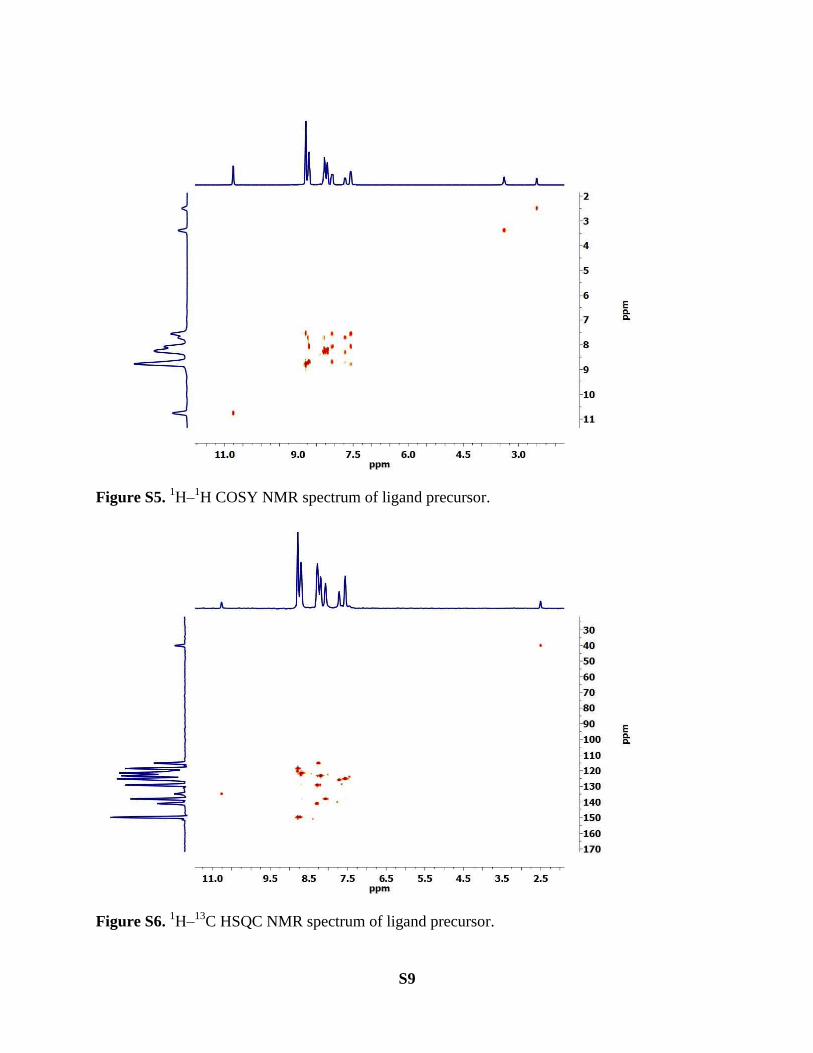
Ligand precursor: 4′-[4-(Imidazol-1-yl)phenyl]- 2,2′:6′,2″-terpyridine (0.75 g, 2.0 mmol) and
2-bromopyridine (1 mL) were mixed in a pressure tube and stirred for 40 h at 140−150 °C in
neat conditions. After that, the reaction mixture was cooled to room temperature and 15 mL of
THF was added to this reaction mixture. The resulting solid was filtered and washed with 30 mL
of THF to give desired product. (Scheme S1). Yield: = 910 mg (85%). 1H NMR (500 MHz,
DMSO‒d6, 300K): δ 10.76 (s, 1H), 8.85 – 8.77 (m, 5H), 8.78 – 8.69 (m, 4H), 8.34 – 8.27 (m,
3H), 8.24 (d, J = 8.2 Hz, 1H), 8.20 (d, J = 8.7 Hz, 2H), 8.12 (ddd, J = 9.2, 6.7, 2.8 Hz, 2H), 7.72
(ddd, J = 7.4, 4.9, 0.9 Hz, 1H), 7.60 (dd, J = 6.6, 4.9 Hz, 2H) ppm. 13
C{1H} NMR (125 MHz,
DMSO‒d6, 300K): δ 155.4, 154.3, 149.3, 149.0, 148.1, 146.3, 140.6, 138.9, 138.2, 135.3, 134.4,

S4
128.8, 125.6, 124.9, 122.9, 122.1, 121.4, 120.1, 118.5, 114.8 ppm. HRMS (ESI, positive ion):
m/z = 453.1862 (calculated for [C29H21N6]+ = 453.1822).
Scheme S2. Synthesis of complex 1.
Complex1
Step I: Ligand precursor (267.0 mg, 0.5 mmol) and [Ru(para-cym)Cl2]2 (154.0 mg, 0.25 mmol)
were mixed in chloroform (8 mL) in a Schlenk tube. The reaction mixture was stirred for 8 h at
room temperature which resulted into dark blue precipitate (Scheme S2). The dark blue
precipitate was filtered and washed with chloroform. This dark blue complex was further used
for the synthesis of complex 1 without any further purification.
Step II: The blue compound formed in step I (142.0 mg, 0.2 mmol) and 2,2':6',2''-terpyridine
(51.0 mg, 0.22 mmol) were mixed in methanol (15 mL) in a Schlenk tube. The reaction mixture
was stirred at 70‒75 ˚C for 36 h. After this, the reaction mixture was allowed to cool down to
room temprature. The reaction mixture was filtered and the solvent was removed in rotavapor.
The solid compound was washed with chloroform to afford the desired compound (Scheme S2).
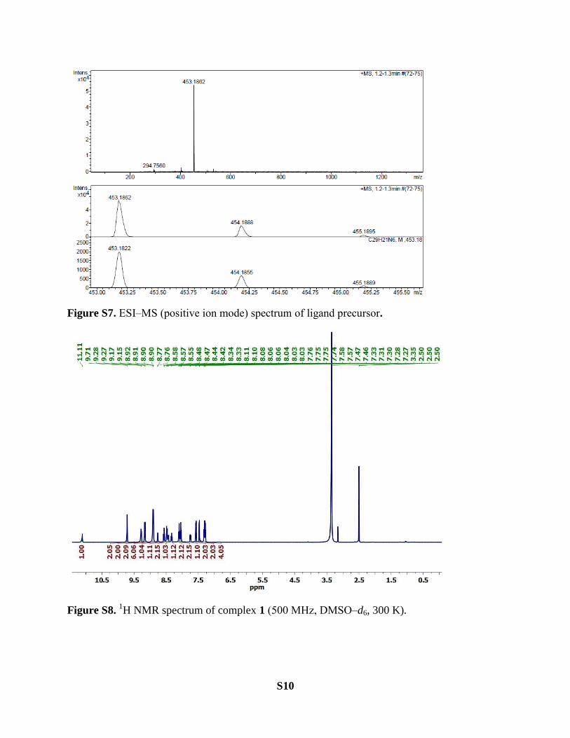
Yield: = 150.0 mg (80%). 1H NMR (500 MHz, DMSO‒d6, 300K): δ 11.11 (s, 1H), 9.71 (s, 2H),
9.28 (d, J = 8.0 Hz, 2H), 9.16 (d, J = 8.2 Hz, 2H), 8.92‒8.90 (m, 6H), 8.76 (d, J = 4.8 Hz, 1H),
8.57 (t, J = 8.1 Hz, 1H), 8.48 (d, J = 8.6 Hz, 2H), 8.43 (d, J = 8.0 Hz, 1H), 8.33 (dd, J = 11.0, 4.6
Hz, 1H), 8.10 (t, J = 7.9 Hz, 2H), 8.07 – 8.02 (m, 2H), 7.75 (dd, J = 7.4, 4.9 Hz, 1H), 7.57 (d, J =
5.6 Hz, 2H), 7.47 (d, J = 5.2 Hz, 2H), 7.33‒7.270 (m, 4H). 13
C{1H} NMR (125 MHz, DMSO‒d6,
300K): δ 158.0, 157.8, 155.3, 154.7, 152.2, 152.1, 149.3, 146.3, 144.8, 140.7, 138.2, 138.1,
136.0, 135.8, 134.5, 129.4, 127.9, 127.7, 125.6, 125.1, 124.7, 124.1, 122.4, 121.9, 121.2, 120.1,
115.0 ppm. HRMS (ESI, positive ion): m/z = 262.7270 (calculated for [C44H32N9Ru]3+
=
262.7273).
S5
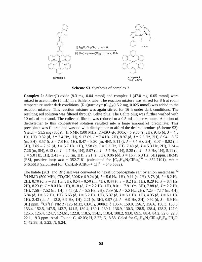
Scheme S3. Synthesis of complex 2.
Complex 2: Silver(I) oxide (9.3 mg, 0.04 mmol) and complex 1 (47.0 mg, 0.05 mmol) were
mixed in acetonitrile (5 mL) in a Schlenk tube. The reaction mixture was stirred for 8 h at room
temperature under dark conditions. [Ru(para-cym)Cl2]2 (15.2 mg, 0.025 mmol) was added to the
reaction mixture. This reaction mixture was again stirred for 16 h under dark conditions. The
resulting red solution was filtered through Celite plug. The Celite plug was further washed with
10 mL of methanol. The collected filtrate was reduced to a 0.5 mL under vacuum. Addition of
diethylether to this concentrated solution resulted into a large amount of precipitate. This
precipitate was filtered and washed with diethylether to afford the desired product (Scheme S3).
Yield: = 51.5 mg (85%). 1H NMR (500 MHz, DMSO‒d6, 300K): δ 9.80 (s, 2H), 9.45 (d, J = 4.5
Hz, 1H), 9.32 (d, J = 7.4 Hz, 1H), 9.17 (d, J = 7.4 Hz, 2H), 8.97 (d, J = 7.5 Hz, 2H), 8.94 – 8.87
(m, 3H), 8.57 (t, J = 7.8 Hz, 1H), 8.47 – 8.30 (m, 4H), 8.11 (t, J = 7.4 Hz, 2H), 8.07 – 8.02 (m,
3H), 7.65 ‒ 7.62 (d, J = 5.7 Hz, 1H), 7.58 (d, J = 5.3 Hz, 2H), 7.48 (d, J = 5.3 Hz, 2H), 7.34 –
7.26 (m, 5H), 6.13 (d, J = 4.7 Hz, 1H), 5.97 (d, J = 5.7 Hz, 1H), 5.35 (d, J = 5.3 Hz, 1H), 5.11 (d,
J = 5.8 Hz, 1H), 2.41 – 2.33 (m, 1H), 2.21 (s, 3H), 0.86 (dd, J = 16.7, 6.8 Hz, 6H) ppm. HRMS
(ESI, positive ion): m/z = 352.7181 (calculated for [C54H45N9ClRu2]3+
= 352.7191), m/z =
546.5618 (calculated for [C54H45N9ClRu2 + Cl]2+
= 546.5632).
The halide (2Cl‒ and Br
‒) salt was converted to hexafluorophosphate salt by anion metathesis.
S2
1H NMR (500 MHz, CD3CN, 300K): δ 9.24 (d, J = 5.6 Hz, 1H), 9.11 (s, 2H), 8.78 (d, J = 8.2 Hz,
2H), 8.70 (d, J = 8.1 Hz, 2H), 8.54 ‒ 8.50 (m, 4H), 8.44 (t, J = 8.2 Hz, 1H), 8.29 (d, J = 8.4 Hz,
2H), 8.23 (t, J = 8.0 Hz, 1H), 8.18 (d, J = 2.2 Hz, 1H), 8.01 – 7.91 (m, 5H), 7.88 (d, J = 2.2 Hz,
1H), 7.56 – 7.52 (m, 1H), 7.45 (d, J = 5.5 Hz, 2H), 7.39 (d, J = 5.5 Hz, 2H), 7.23 ‒ 7.17 (m, 4H),
5.84 (d, J = 6.2 Hz, 1H), 5.65 (d, J = 6.2 Hz, 1H), 5.37 (d, J = 6.1 Hz, 1H), 4.95 (d, J = 6.1 Hz,
1H), 2.43 (dt, J = 13.8, 6.9 Hz, 1H), 2.21 (s, 3H), 0.97 (d, J = 6.9 Hz, 3H), 0.92 (d, J = 6.9 Hz,
3H) ppm. 13
C{1H} NMR (125 MHz, CDCl3, 300K): δ 186.4, 159.0, 156.7, 156.6, 156.3, 153.6,
153.4, 152.5, 147.5, 142.7, 141.5, 139.4, 139.1, 139.1, 136.9, 130.3, 128.5, 128.4, 128.3, 127.0,
125.5, 125.4, 124.7, 124.61, 122.8, 118.5, 114.1, 110.4, 108.2, 93.0, 89.5, 88.4, 84.2, 32.0, 22.8,
22.1, 19.3 ppm. Anal. Found: C, 42.03; H, 3.22; N, 8.58. Calcd for C54H45N9ClRu2P3F18.2H2O:
C, 42.38; H, 3.23; N, 8.24.
S6
Complex 3: This complex was synthesised by following a reported procedure and the synthesis
of the complex was confirmed by matching the NMR chemical shift values with proper
integration ration (Figure S19).S3
. 1H NMR (400 MHz, DMSO‒d6, 300K): 9.33 (d, J = 5.2 Hz,
1H), 8.40 (d, J = 2.1 Hz, 1H), 8.28 – 8.21 (m, 1H), 8.15 (d, J = 8.1 Hz, 1H), 7.80 (d, J = 2.1 Hz,
1H), 7.52 (dd, J = 9.6, 3.5 Hz, 1H), 6.40 (d, J = 5.9 Hz, 1H), 6.36 (d, J = 6.2 Hz, 1H), 6.17 (d, J
= 6.2 Hz, 1H), 5.74 (d, J = 5.9 Hz, 1H), 4.10 (s, 3H), 2.52 – 2.47, 2.41 – 2.30 (m, 1H), 2.12 (s,
3H), 0.84 (t, J = 7.1 Hz, 6H) ppm.
Complex 4: Hexafluorophosphate salt of complex 2 (75.0 mg, 0.05 mmol) and silver triflate
(16.0 mg, 0.06 mmol) were mixed in acetonitrile (5 mL) in a Shlenk tube. The reaction mixture
was refluxed for 60 h under dark conditions. After this, the reaction mixture was allowed to cool
down to room temprature. This reaction mixture was filtered through a Celite plug. The filtrate
was reduced to a minimum volume of 0.5 mL. Addition of 10 mL of diethylether resulted into a
solid product. Crystallization (MeCN/Et2O) of this solid product resulted into the analytically
pure product (Scheme XX, main article). Yield: = 74.0 mg (90 %).1H NMR (500 MHz, CD3CN,
300K): δ 9.09 (s, 2H), 8.94 (d, J = 5.6 Hz, 1H), 8.77 (d, J = 8.2 Hz, 2H), 8.72 (d, J = 8.1 Hz,
2H), 8.53 – 8.49 (m, 4H), 8.43 (t, J = 8.1 Hz, 1H), 8.28 (d, J = 2.1 Hz, 1H), 8.20 (t, J = 8.0 Hz,
1H), 8.01 (d, J = 8.5 Hz, 2H), 7.98 (d, J = 4.0 Hz, 1H), 7.97 – 7.91 (m, 4H), 7.70 (d, J = 2.1 Hz,
1H), 7.56 – 7.51 (m, 1H), 7.44 (d, J = 5.4 Hz, 2H), 7.38 (d, J = 5.5 Hz, 2H), 7.22 ‒ 7.16 (m, 4H),
2.89 (s, 3H), 2.77 (s, 3H), 2.23 (s, 3H), 2.04 (s, 3H) ppm. 13
C{1H} NMR (125 MHz, CD3CN,
300K): δ 190.7, 159.1, 159.0, 156.8, 156.3, 155.7, 154.04, 153.5, 153.4, 147.4, 141.7, 141.6,
139.1, 139.1, 136.9, 130.0, 129.6, 128.6, 128.5, 127.2, 126.1, 125.8, 125.7, 125.5, 124.8, 123.6,
123.4, 122.6, 120.9, 118.7, 113.0, 4.1, 4.1, 1.8 ppm. Anal. Found: C, 39.48; H, 2.87; N, 11.42.
Calcd for C53H43N13Ru2P3F21SO3.CH3CN: C, 39.33; H, 2.76; N, 11.68.
Single crystal X-ray diffraction studies
Hexafluorophosphate salt of complex 1 and complex 4 were crystallized by diffusion of
diethylether into the concentrated acetonitrile solution of the respected complexes. Complex 4
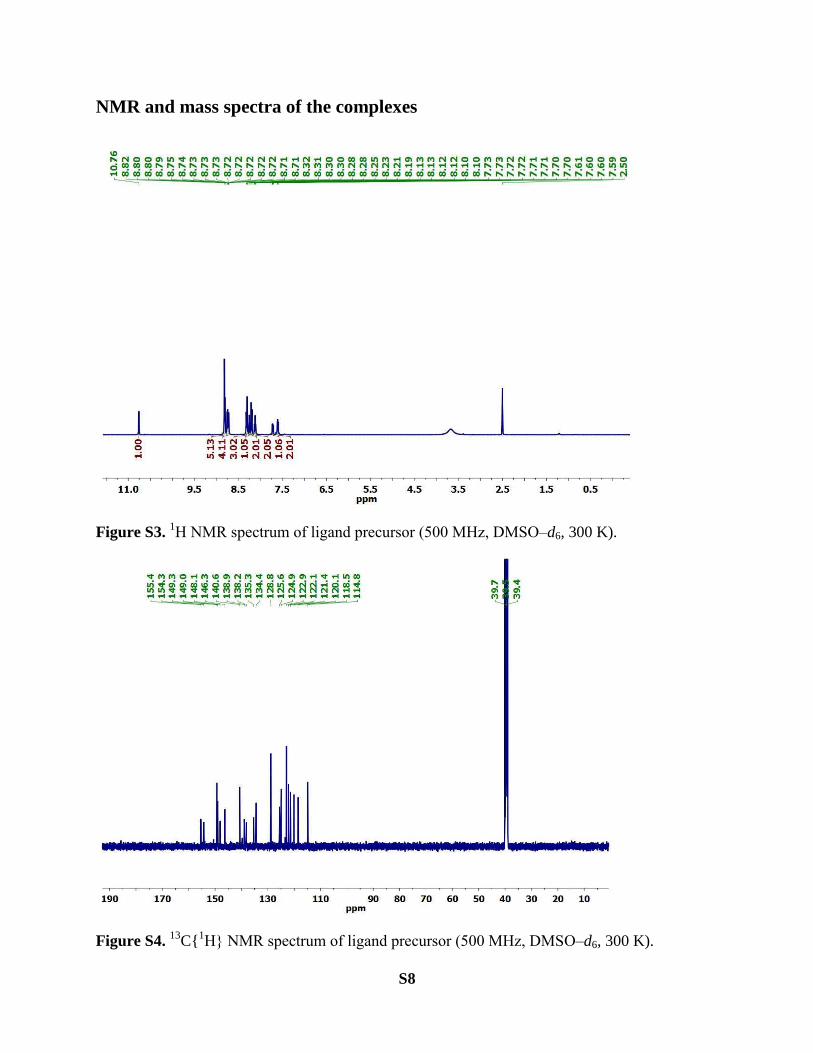
showed distorted octahedral geometries around the ruthenium centre. The CNHC–RuII–Cpyridine
bite angle was 88.9(2)˚ and CNHC–RuII bond length 1.975(6) Å. The Ru–NMeCN bond trans to the
carbene was found to be larger than the other Ru–NMeCN bonds, due to the strong trans influence
of carbene ligand.xx
The triflate anion of complex 4 was found to be highly disordered. Other
bond lengths and bond angles were found to be in good agreement with the reported values for
similar type of complexes in the literature.
S7
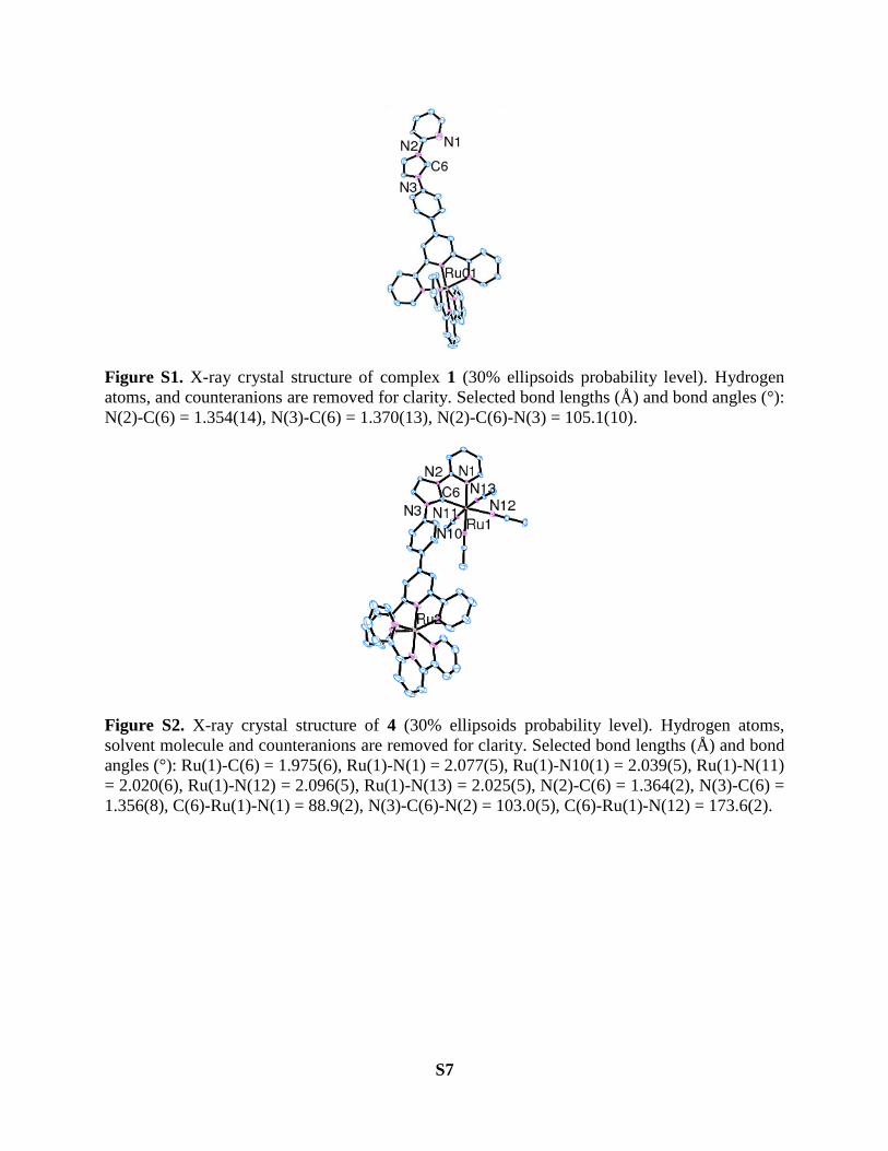
Figure S1. X-ray crystal structure of complex 1 (30% ellipsoids probability level). Hydrogen
atoms, and counteranions are removed for clarity. Selected bond lengths (Å) and bond angles (°):
N(2)-C(6) = 1.354(14), N(3)-C(6) = 1.370(13), N(2)-C(6)-N(3) = 105.1(10).
Figure S2. X-ray crystal structure of 4 (30% ellipsoids probability level). Hydrogen atoms,
solvent molecule and counteranions are removed for clarity. Selected bond lengths (Å) and bond
angles (°): Ru(1)-C(6) = 1.975(6), Ru(1)-N(1) = 2.077(5), Ru(1)-N10(1) = 2.039(5), Ru(1)-N(11)
= 2.020(6), Ru(1)-N(12) = 2.096(5), Ru(1)-N(13) = 2.025(5), N(2)-C(6) = 1.364(2), N(3)-C(6) =
1.356(8), C(6)-Ru(1)-N(1) = 88.9(2), N(3)-C(6)-N(2) = 103.0(5), C(6)-Ru(1)-N(12) = 173.6(2).
S8
NMR and mass spectra of the complexes
Figure S3. 1H NMR spectrum of ligand precursor (500 MHz, DMSO‒d6, 300 K).
Figure S4. 13
C{1H} NMR spectrum of ligand precursor (500 MHz, DMSO‒d6, 300 K).
S9
Figure S5. 1H‒
1H COSY NMR spectrum of ligand precursor.
Figure S6. 1H‒
13C HSQC NMR spectrum of ligand precursor.
S10
Figure S7. ESI‒MS (positive ion mode) spectrum of ligand precursor.
Figure S8. 1H NMR spectrum of complex 1 (500 MHz, DMSO‒d6, 300 K).
S11
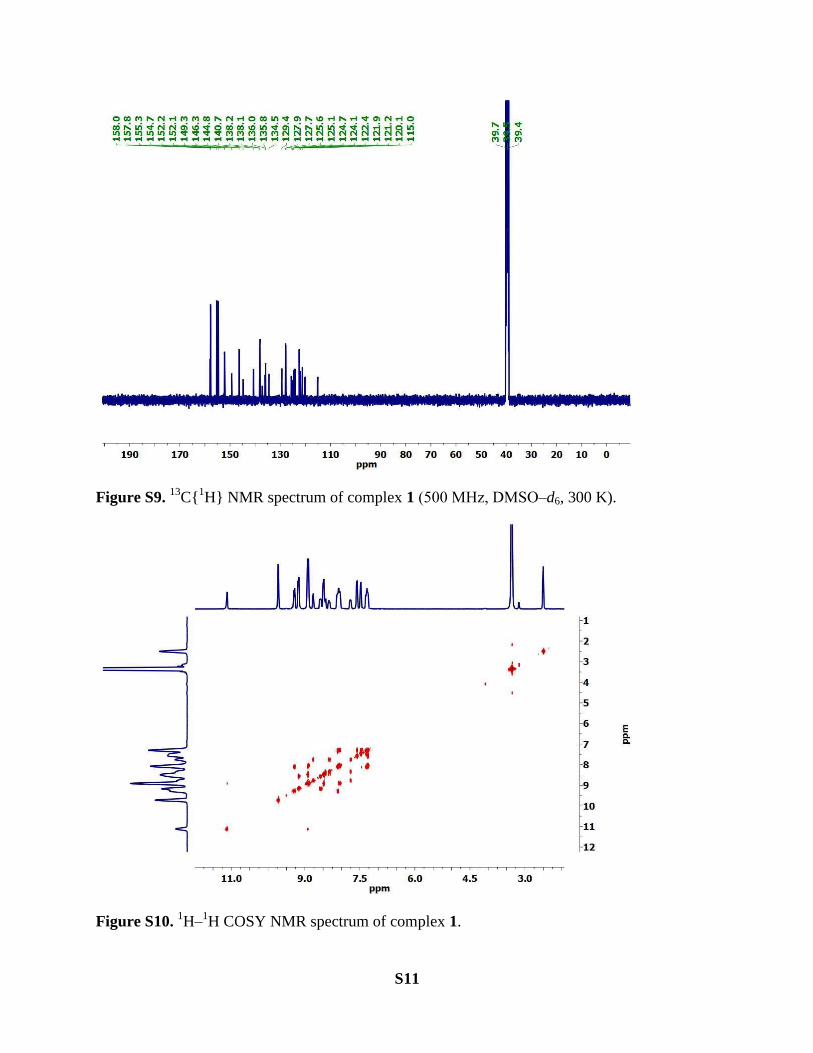
Figure S9. 13
C{1H} NMR spectrum of complex 1 (500 MHz, DMSO‒d6, 300 K).
Figure S10. 1H‒
1H COSY NMR spectrum of complex 1.
S12
Figure S11. 1H‒
13C HSQC NMR spectrum of complex 1.
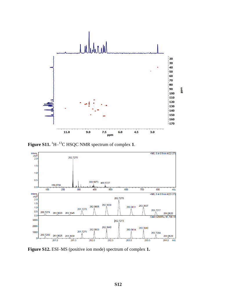
Figure S12. ESI‒MS (positive ion mode) spectrum of complex 1.
S13
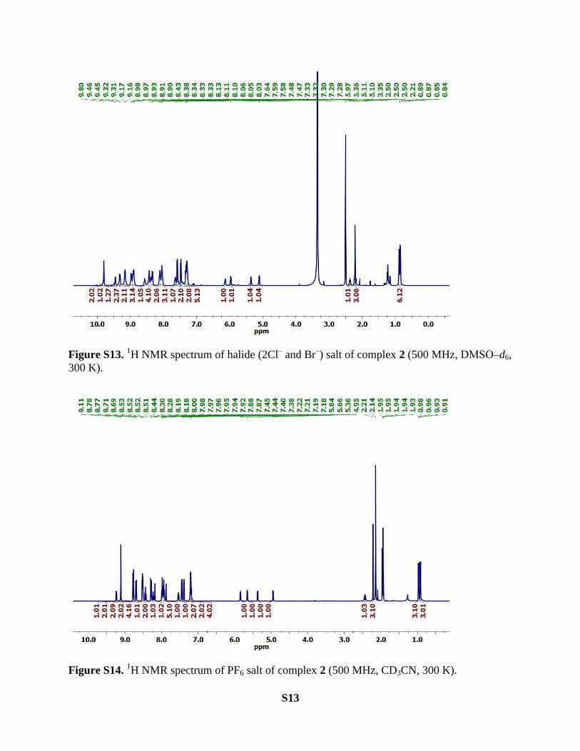
Figure S13. 1H NMR spectrum of halide (2Cl
‒ and Br
‒) salt of complex 2 (500 MHz, DMSO‒d6,
300 K).
Figure S14. 1H NMR spectrum of PF6 salt of complex 2 (500 MHz, CD3CN, 300 K).
S14
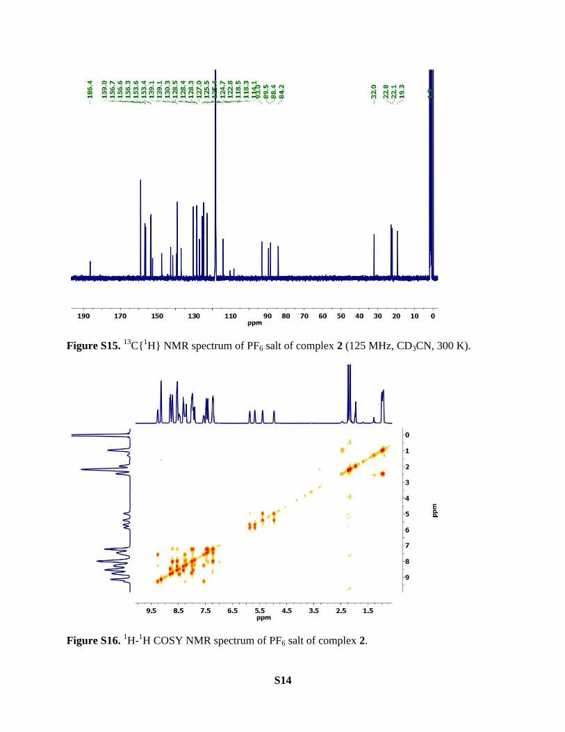
Figure S15. 13
C{1H} NMR spectrum of PF6 salt of complex 2 (125 MHz, CD3CN, 300 K).
Figure S16. 1H-
1H COSY NMR spectrum of PF6 salt of complex 2.

S15
Figure S17. 1H-
13C HSQC NMR spectrum of PF6 salt of complex 2.

S16
Figure S18. ESI-MS (positive ion mode) spectrum of halide (2Cl‒ and Br
‒) salt of complex 2.
Figure S19. 1H NMR spectrum of of complex 3 (400 MHz, DMSO-d6, 300 K).
S17
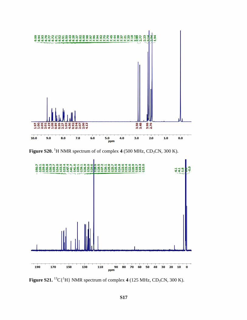
Figure S20. 1H NMR spectrum of of complex 4 (500 MHz, CD3CN, 300 K).
Figure S21. 13
C{1H} NMR spectrum of complex 4 (125 MHz, CD3CN, 300 K).
S18
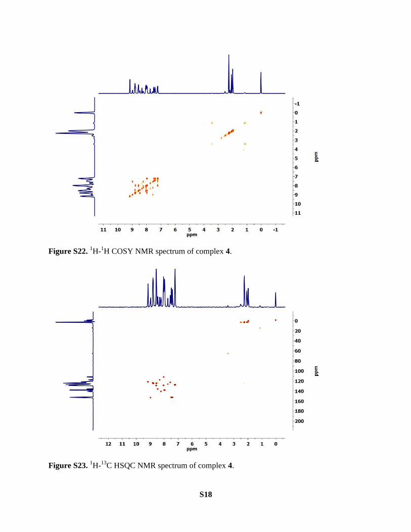
Figure S22. 1H-
1H COSY NMR spectrum of complex 4.
Figure S23. 1H-
13C HSQC NMR spectrum of complex 4.

S19
UV-Vis spectroscopic studies
UV-Vis spectroscopic studies were carried out on a Cary 100 UV-Vis spectrophotometer using
1.0 cm quartz cuvettes at room temperature. Hexafluorophosphate salts of complex 1 and
complex 2 were used for this study. Stock solutions of both the complexes were made in
acetonitrile. The total volume of each sample was kept constant to 2.5 mL.
260 390 520 6500.0
0.4
0.8
1.2
1.6
Ab
so
rba
nc
e
Wavelength (nm)
1
2
A
Figure S24. UV-Vis spectra of complex 1 and complex 2
Electrochemical analysis of the complexes
Electrochemical studied (CV and DPV) were carried out by three electrode configuration.
Working electrode: Pt disk (1 mm diameter); counter electrode: a Pt wire; reference electrode:
saturated calomel electrode, SCE. Hexafluorophosphate salts of complex 1 and complex 2 were
used to avoid any interference arising from halide oxidation processes. [NBu4]PF6 (0.1 M) in
acetonitrile was used as the supporting electrolyte. Ferrocene (E1/2, Fc/Fc+ = 0.37 volts vs. SCE)
was used as an external calibration standard for all the experiments.
S20
0.0 0.4 0.8 1.2 1.6-1
0
1
2
3
Cu
rre
nt
(i,
10
-6 A
)
Potential (V vs SCE)
1
2
B
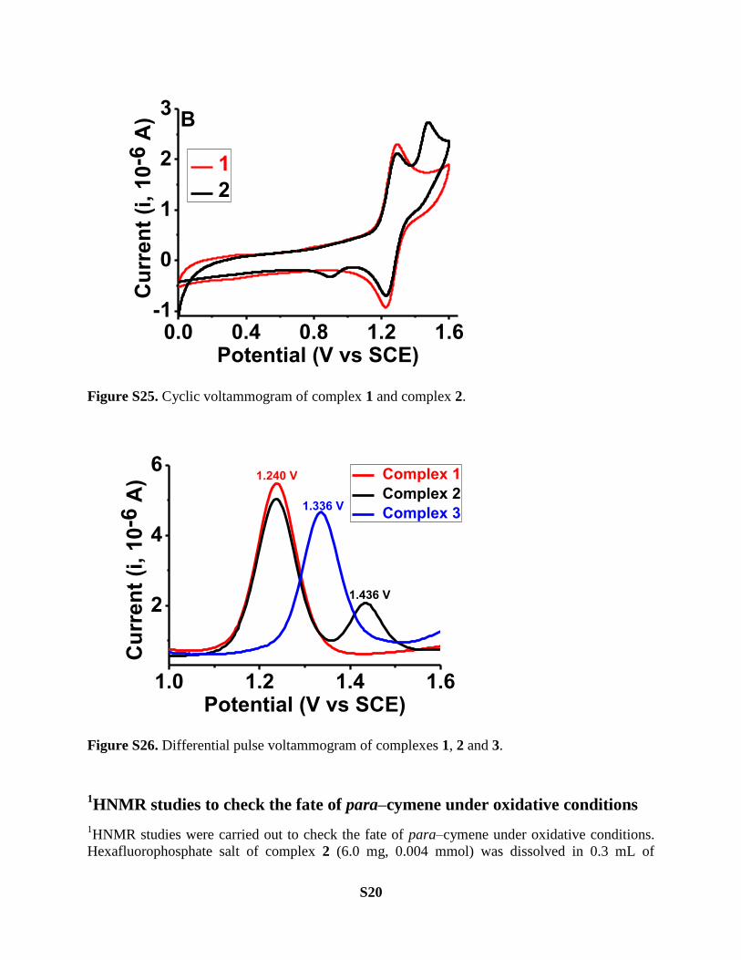
Figure S25. Cyclic voltammogram of complex 1 and complex 2.
1.0 1.2 1.4 1.6
2
4
6
1.436 V
1.240 V
Cu
rre
nt
(i,
10
-6 A
)
Potential (V vs SCE)
Complex 1
Complex 2
Complex 31.336 V
Figure S26. Differential pulse voltammogram of complexes 1, 2 and 3.
1HNMR studies to check the fate of para‒cymene under oxidative conditions
1HNMR studies were carried out to check the fate of para‒cymene under oxidative conditions.
Hexafluorophosphate salt of complex 2 (6.0 mg, 0.004 mmol) was dissolved in 0.3 mL of
S21
acetone‒d6. 10 equivalents of NaIO4 were dissolved in 0.2 mL of D2O and transferred to
acetone‒d6 solution of complex 2 . The fate of para‒cymene was monitored upto 30 min by 1HNMR spectroscopy (Figure 3, main article). Presence of free para‒cymene was confirmed by
comparing the resonance peaks with authentic para‒cymene sample. Similar studies were also
carried out with complex 3 to compare the rate of para‒cymene loss under the similar conditions
(Figure 3, main article).
General procedure for the catalysis studies
Substrate (0.4 mmol) in 1 mL of acetone and catalyst (0.5 mol%, halide salt; 2Cl‒ and Br
‒} of
complex 2 or PF6‒ salt of complex 2 or complex 3 or complex 4) were taken in a round bottom
flask. 2 mL of acetone and 2 mL of H2O were added to it. NaIO4 (213 mg, 1.0 mmol) was
dissolved in 1 mL of H2O and transferred to the reaction mixture. The reaction mixture was
stirred at room temprature for ~15 min‒240 min. After this time, Na2SO3 (2.0 mmol) was added
to the reaction mixture followed by addition of 2 mL of DCM and 3 mL of H2O. The reaction
mixture was further stirred for 10 min. Standard (ethylbenzene or mesitylene or stilbene or
acetophenone) was added as a reference and the reaction mixture was again stirred for 5 min. It
was then transferred to a separating funnel with the help of 3 mL of H2O and 8 mL of DCM. The
organic layer was separated and aqueous layer was again extracted with 5 mL of DCM (two
times). The combined organic layer was washed with 20 mL of brine solution. Products and
unreacted substrates were analyzed by GCMS. The yields of the products were calculated by GC
analyses.
All the products (with yields >20%) were isolated in pure form by silica gel column
chromatography. 1H NMR spectrum of each product was recorded to further confirm their
identity and purity level (see below).
Styrene. Yield of benzaldehyde = 25.5 mg (60%) with complex 2 (halide salt; 2Cl‒ and Br
‒), 4.3
mg (10%) with complex 3 and 17.4 mg (41% ) with complex 4.
1H NMR (400 MHz, CDCl3, 300K): δ 10.03 (s, 1H), 7.92–7.83 (m, 2H), 7.64 (t, J = 7.4 Hz, 1H),
7.54 (t, J = 7.5 Hz, 2H) ppm.
4-Methylstyrene. Yield of 4-methylbenzaldehyde = 29.3 mg (61%) with complex 2 (halide salt;
2Cl‒ and Br
‒), 3.4 mg (7%) with complex 3 and 14.4 mg (30%) with complex 4.
1H NMR (400 MHz, CDCl3, 300K): δ 9.96 (s, 1H), 7.77 (d, J = 8.1 Hz, 2H), 7.33 (d, J = 7.9 Hz,
2H), 2.44 (s, 3H) ppm.
4-Chlorostyrene. Yield of 4-chlorobenzaldehyde = 36.0 mg (64%) with complex 2 (halide salt;
2Cl‒ and Br
‒) and 31.5 mg (56%) with complex 4.
1H NMR (400 MHz, CDCl3, 300K): δ 9.99 (s, 1H), 7.83 (d, J = 8.4 Hz, 2H), 7.52 (d, J = 8.4 Hz,
2H) ppm.
4-Fluorostyrene. Yield of 4-fluorobenzaldehyde = 29.8 mg (60%) with complex 2 (halide salt;
2Cl‒ and Br
‒) and 25.3 mg (51%) with complex 4.
S22
1H NMR (400 MHz, CDCl3, 300K): δ 9.96 (s, 1H), 7.90 (dd, J = 8.6, 5.5 Hz, 2H), 7.20 (t, J = 8.5
Hz, 2H) ppm.
α-Methylstyrene. Yield of acetophenone = 34.6 mg (72%) with complex 2 (halide salt; 2Cl‒ and
Br‒), 10.6 mg (22%) with complex 3 and 13.5 mg (28%) with complex 4.
1H NMR (400 MHz, CDCl3, 300K): δ 8.01–7.90 (m, 2H), 7.56 (t, J = 7.4 Hz, 1H), 7.46 (t, J =
7.6 Hz, 2H), 2.60 (s, 3H) ppm.
4-Vinylanisole. Yield of 4-methoxybenzaldehyde = 23.4 mg (43%) with complex 2 (halide salt;
2Cl‒ and Br
‒), 9.3 mg (17%) with complex 3 and 22.3 mg (41%) with complex 4.
1H NMR (400 MHz, CDCl3, 300K): δ 9.88 (s, 1H), 7.83 (d, J = 8.7 Hz, 2H), 6.99 (d, J = 8.7 Hz,
2H), 3.88 (s, 3H) ppm.
4-Bromostyrene. Yield of 4-bromobenzaldehyde = 44.4 mg (60%) with complex 2 (halide salt;
2Cl‒ and Br
‒) and 47.4 mg (64%) with complex 4.
1H NMR (400 MHz, CDCl3, 300K): δ 9.98 (s, 1H), 7.75 (d, J = 8.3 Hz, 2H), 7.69 (d, J = 8.4 Hz,
2H) ppm.
Trans-stilbene. Yield of benzaldehyde = 56.9 mg (67%) with complex 2 (halide salt; 2Cl‒ and
Br‒), 34.0 mg (40%) with complex 3 and 55.2 mg (65%) with complex 4.
Cis-stilbene. Yield of benzaldehyde = 62 mg (73%) with complex 2 (halide salt; 2Cl‒ and Br
‒),
39.9 mg (47%) with complex 3 and 57.7 mg (68%) with complex 4.
Allylbenzene. Yield of 2-phenylacetaldehyde = 20.7 mg (43%) with complex 2 (halide salt; 2Cl‒
and Br‒) and 24.5 mg (51%) with complex 4.
1H NMR (400 MHz, CDCl3, 300K): δ 9.76 (t, J = 2.4 Hz, 1H), 7.38 (m, 2H), 7.32 (m, 1H), 7.23
(m, 2H), 3.69 (d, J = 2.3 Hz, 2H) ppm.
Trans-α-methylstilbene. Yield of acetophenone = 36.0 mg (75%) with complex 2 (halide salt;
2Cl‒ and Br
‒), 24.0 mg (50%) with complex 3 and 33.6 mg 70% with complex 4.
Diphenylacetylene. Yield of benzil = 50.5 mg (60%) with complex 2 (halide salt; 2Cl‒ and Br
‒),
21.0 mg (25%) with complex 3 and 54.6 mg (65%) with complex 4.
1H NMR (400 MHz, CDCl3, 300K): δ 8.06–7.89 (m, 4H), 7.66 (t, J = 7.4 Hz, 2H), 7.52 (t, J =
7.8 Hz, 4H) ppm.
1-Phenyl1butyne. Yield of 1-Phenylbutane-1,2-dione = 41.5 mg (64%) with complex 2 (halide
salt; 2Cl‒ and Br
‒), 15.6 mg (24%) with complex 3 and 34.4 mg (53%) with complex 4.
1H NMR (400 MHz, CDCl3, 300K): δ 8.04–7.92 (m, 2H), 7.64 (t, J = 7.4 Hz, 1H), 7.50 (t, J =
7.7 Hz, 2H), 2.92 (q, J = 7.3 Hz, 2H), 1.20 (t, J = 7.3 Hz, 3H) ppm.
1-Phenyl1propyne. Yield of 1-Phenylpropane-1,2-dione = 39.7 mg (67%) with complex 2
(halide salt; 2Cl‒ and Br
‒), 11.3 mg (19%) with complex 3 and 30.2 mg (51%) with complex 4.

S23
1H NMR (400 MHz, CDCl3, 300K): δ 8.04–7.97 (m, 2H), 7.65 (t, J = 7.4 Hz, 1H), 7.50 (t, J =
7.8 Hz, 2H), 2.53 (s, 3H) ppm.
1H NMR spectra of the products obtained by oxidative scission of carbon-
carbon multiple bonds.
Figure S27. 1H NMR spectrum of Benzaldehyde (400 MHz, CDCl3, 300 K).
S24
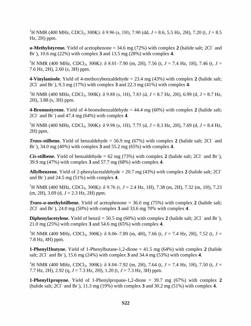
Figure S28. 1H NMR spectrum of 4-Methylbenzaldehyde (400 MHz, CDCl3, 300 K).
Figure S29. 1H NMR spectrum of 4-Chlorobenzaldehyde (400 MHz, CDCl3, 300 K).

S25
Figure S30. 1H NMR spectrum of 4-Fluorobenzaldehyde (400 MHz, CDCl3, 300 K).
Figure S31. 1H NMR spectrum of Acetophenone (400 MHz, CDCl3, 300 K).

S26
Figure S32. 1H NMR spectrum of 4-Methoxybenzaldehyde (400 MHz, CDCl3, 300 K).
Figure S33. 1H NMR spectrum of 4-Bromobenzaldehyde (400 MHz, CDCl3, 300 K).
S27
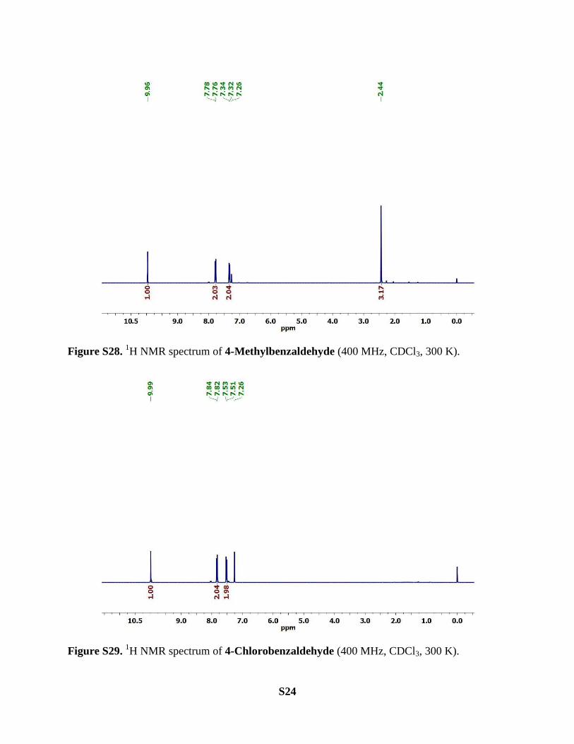
Figure S34. 1H NMR spectrum of 2-Phenylactaldehyde (400 MHz, CDCl3, 300 K).
Figure S35. 1H NMR spectrum of Benzil (400 MHz, CDCl3, 300 K).
S28
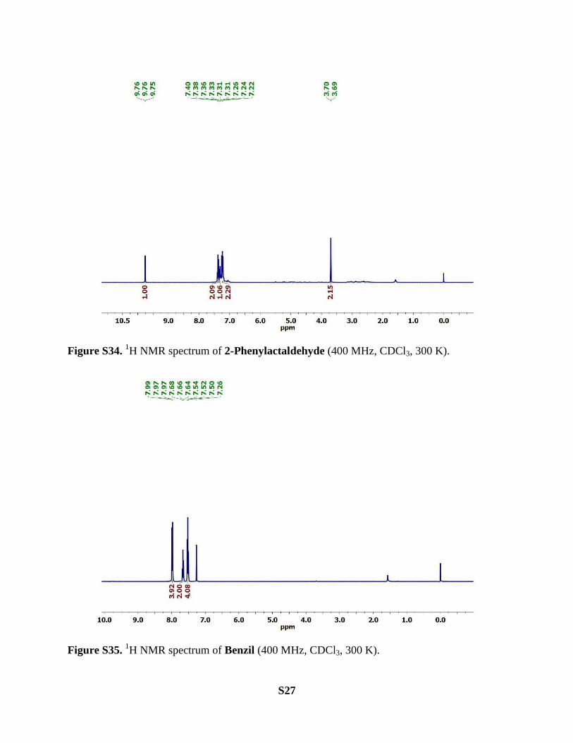
Figure S36. 1H NMR spectrum of 1-Phenylbutane-1,2-dione (400 MHz, CDCl3, 300 K).
Figure S37. 1H NMR spectrum of 1-Phenylpropane-1,2-dione (400 MHz, CDCl3, 300 K).

S29
General procedure for studying the kinetic profile of the catalytic reactions
with complex 2 and complex 3
For these experiments, catalyst (0.5 mol%, hexafluorophosphate salt), 4‒methylstyrene (47.3 mg,
0.4 mmol), mesitylene (used as an internal standard) and NaIO4 (213 mg, 1.0 mmol) were mixed
in the solvent system, acetone-water (6 mL, v/v, 1:1). The reactions were carried out at room
temperature and the yields of the product (4‒methylbenzaldehyde) were determined by GC after
different time intervals upto 60 min. The results were plotted as shown in Figure 4 (main article).
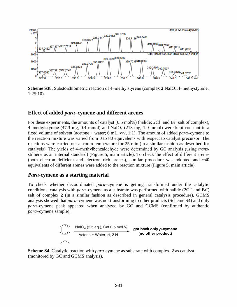
Oxidation of 4‒methylstyrene under substoichiometric reaction conditions
monitored by mass spectrometry
This experiment was carried out to check the specieses present in the reaction mixture during the
catalysis. Catalyst (0.005 mmol, PF6 salt of complex 2), 4‒methylstyrene (0.05 mmol) and NaIO4
(0.125 mmol) were taken in a solvent system (acetone + water; 3 mL, v/v, 1:1) in a round bottom
flask. Reaction was carried out for 30 min at room temprature. After this, GC analysis of the
reaction mixture showed the formation of product (substrate:product, 1:3). ESI-HRMS analysis
of the same reaction mixture showed a number cluster peaks related to different ruthenium
specieses present in the solution during the catalysis (Figure S38).
[C44H31N9Ru2-H]3+
= 296
[C44H31N9Ru2-H + O2]3+
=306
[C44H31N9Ru2(CH3COCH3)(H2O)2Cl]3+
= 339
[C54H45N9Ru2Cl]3+
= 352 (mother complex)
[C54H45N9Ru2ClPF6]2+
= 601 (mother complex)

S30
S31
Scheme S38. Substoichiometric reaction of 4‒methylstyrene (complex 2:NaIO4:4‒methystyrene;
1:25:10).
Effect of added para‒cymene and different arenes
For these experiments, the amounts of catalyst (0.5 mol%) (halide; 2Cl‒ and Br
‒ salt of complex),
4‒methylstyrene (47.3 mg, 0.4 mmol) and NaIO4 (213 mg, 1.0 mmol) were kept constant in a
fixed volume of solvent (acetone + water; 6 mL, v/v, 1:1). The amount of added para‒cymene to
the reaction mixture was varied from 0 to 80 equivalents with respect to catalyst precursor. The
reactions were carried out at room temperature for 25 min (in a similar fashion as described for
catalysis). The yields of 4‒methylbenzaldehyde were determined by GC analysis (using trans-
stilbene as an internal standard) (Figure 5, main article). To check the effect of different arenes
(both electron deficient and electron rich arenes), similar procedure was adopted and ~40
equivalents of different arenes were added to the reaction mixture (Figure 5, main article).
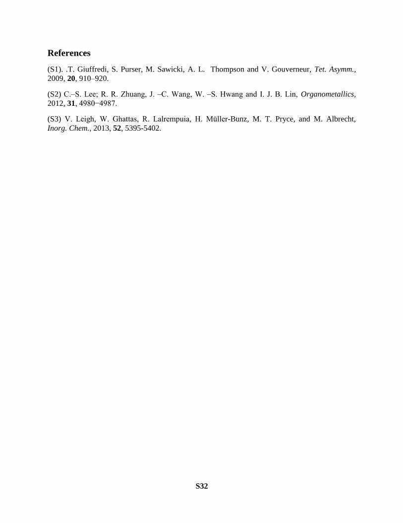
Para-cymene as a starting material
To check whether decoordinated para‒cymene is getting transformed under the catalytic
conditions, catalysis with para‒cymene as a substrate was performed with halide (2Cl‒ and Br
‒)
salt of complex 2 (in a similar fashion as described in general catalysis procedure). GCMS
analysis showed that para‒cymene was not transforming to other products (Scheme S4) and only
para‒cymene peak appeared when analyzed by GC and GCMS (confirmed by authentic
para‒cymene sample).
Scheme S4. Catalytic reaction with para-cymene as substrate with complex‒2 as catalyst
(monitored by GC and GCMS analysis).
S32
References
(S1). .T. Giuffredi, S. Purser, M. Sawicki, A. L. Thompson and V. Gouverneur, Tet. Asymm.,
2009, 20, 910–920.
(S2) C.‒S. Lee; R. R. Zhuang, J. ‒C. Wang, W. ‒S. Hwang and I. J. B. Lin, Organometallics,
2012, 31, 4980−4987.
(S3) . Leigh, W. Ghattas, R. Lalrempuia, H. M ller-Bunz, M. T. Pryce, and M. Albrecht,
Inorg. Chem., 2013, 52, 5395-5402.