electrophoretic separation of tissue-specific alkaline...
TRANSCRIPT

J. clin. Path., 1970, 23, 499-508
Electrophoretic separation of tissue-specific serumalkaline phosphatases
R. CANAPA-ANSON AND D. J. F. ROWEFrom the Departments of Geriatrics and of Chemical Pathology, Whittington Hospital, London
SYNOPSIS Previous electrophoretic methods for the separation of tissue-specific serumalkaline phosphatases have either been unable to separate the liver and bone enzymes or havebeen too involved for routine clinical use. A relatively simple electrophoretic method isdescribed which separates placental, liver, bone, and intestinal alkaline phosphatases in serum.The clinical applications of such a method appear to be mainly in the differential diagnosisof liver and bone disease, especially in complicated hypercalcaemic states where tumourmetastases can affect both bone and liver, in children, and possibly in cirrhosis of the liver.No differences in electrophoretic mobility could be seen between zymograms of different
diseases affecting the same organ. Patients presenting with hepatic cirrhosis all showed amarked serum intestinal alkaline phosphatase zone as well as a liver zone on electrophoresis.An intestinal zone was not present with other types of hepatobiliary disease.The heterogeneity of total serum alkaline phosphatase activity in normal subjects is demon-
strated, alkaline phosphatases of liver and bone, and sometimes of intestine being present innormal serum.
Results obtained in women in the last trimester of pregnancy and in old people are alsodiscussed.
Alkaline phosphatase activity in human serum isderived from liver, bone, intestine, or placenta.In hepatobilary and osteoblastic bone disease theincrease in total serum alkaline phosphataseactivity is due to the release of tissue-specific liverand bone alkaline phosphatase into the serum.In a clinical context the significance of a raisedserum enzyme level is usually obvious, but thepotential value of identifying the tissue or tissuesresponsible for the rise, and of establishing theirrespective contribution to the total increase, haslong been recognized. A method for doing thisand our experience with the method in routineclinical practice is described in the present paper.
Material and Methods
Sera from 186 subjects were analysed. They wereReceived for publication 22 December 1969.
divided on clinical grounds into eight groups asshown in Table I.
Total serum alkaline phosphatase activity wasestimated by a routine adaptation of the methodof Babson (1965) using phenolphthalein mono-phosphate as the substrate. Alkaline phosphataseactivities are expressed in King-Armstrong unitsper 100 ml (K-A u %).The amount of serum used for electrophoresis
varied from 2 to 40 ,ul, depending on the activityof the sample. Alteration of the amount of serumused within this range did not affect the mobilitiesof the various zones of alkaline phosphatase.Primary reference standards were prepared
from tissue homogenates as described by Smith,Lightstone, and Perry (1968). Sera from patientswith established uncomplicated liver and bonedisease, from normal adults, and from normalchildren were used as secondary referencestandards. Our results on patients for diagnosisare compared with the results from the groups of
on 28 June 2018 by guest. Protected by copyright.
http://jcp.bmj.com
/J C
lin Pathol: first published as 10.1136/jcp.23.6.499 on 1 S
eptember 1970. D
ownloaded from

R. Canapa-Anson and D. J. F. Rowe
established liver and bone disease. The groupsof pregnant women and old people are comparedwith the normal adult group. The results fromthe group of sick children are compared withthose obtained from normal children.Using the method described below zones of
alkaline phosphatase activity extracted fromliver, bone, placenta, or gut had similar mobilitiesto the corresponding zones of serum activity.
Numbers in Each Group
16
163014814
2562
185
Group
Patients with oesteoblastic bone disease (Table III)Patient with hepatobiliary disease with hyperbilirubinaemia(Table IV)Normal adults aged 18 to 56 years (Table V)Pregnant women in the last trimester (Table Vt)Normal children aged 2 to 14 years (Table VII)Sick children aged I day to 12 years (Table VIII)Old people aged over 75 years with evidence of osteoporosis(Table IX)Patients for diagnosis (Table X)Cases I to 21, suspected of liver disease, diagnosis supported byelectrophoresisCases 22 to 24, liver disease suspected plus bone diseaseCases 25 to 28, liver disease suspected, also showing an intestinalzone on electrophoresisCases 29 to 36, bone disease suspected, diagnosis supported byelectrophoresisCases 37 to 39, bone disease suspected, diagnosis supported byelectrophoresis which also showed a liver zoneCases 40 to 49, bone disease suspected, diagnosis not supportedby electrophoresisCases 50 to 59, presenting with raised serum AP, liver zoneshown on electrophoresisCase 60, presenting with a raised serum AP, bone zone shownon electrophoresisCases 61 and 62, presenting with raised serum AP, liver andbone zones shown on electrophoresisTotal
Table I Groups ofsubjects studied and the numbers ineach group
: _...........
Fig. 1 Mobility of zones of alkaline phosphataseactivity in relation to other serum protein fractions.
Samples 1, 3, 5, and 7 were stained for alkalinephosphatase (AP) and samples 2, 4, 6, and 8 for totalprotein. (1: liver and intestinal AP; 3: bone AP;5: liver AP; and 7: placental AP.)
This electrophoretic run was prematurely terminatedin order to demonstrate the serum albumin zones.Consequently, differences in electrophoretic mobilitybetween the tissue-specific alkaline phosphatasesshown in this figure are less than those usuallyobtained.
Electrophoresis was carried out on verticalpolyacrylamide gel slabs, essentially as describedby Akroyd (1967). The dimensions of the cellwere 16 x 10 x 0-3 cm.
Forty ml of 8% Cyanogum 41 (acrylamidemonomer plus N N'-methylene-bis-acrylamide,British Drug Houses Ltd) in Tris-citrate buffer,plus 0-2 ml fl-dimethylaminopropionitrile, and0 3 ml 7% ammonium persulphate was pipettedinto the cell, overlaid with distilled water, andallowed to set. In mixing these substances, as faras possible contact with the air was avoided.The composition of the Tris-citrate 'gel' buffer
(pH 8-8) was 1 52 ml 2 M Tris-(hydroxymethyl)-aminomethane plus 0-2 ml 1 M citric acid madeup to 40 ml with distilled water. The electrodecompartment buffer (pH 8'8) was 60 ml 0-5 Mboric acid plus 4-5 ml 2-5 M sodium hydroxidemade up to 1 litre with distilled water.Some improvement in the resolution between
the various alkaline phosphatase zones, especiallywhen assaying sera with normal alkaline phos-phatase levels, could be achieved by adding a
'spacer' gel to the system. This was done bygelling 35 ml 8% Cyanogum 41 as describedabove, removing the excess liquid, and adding5 ml 4% gel solution. This was similarly overlaidwith water and allowed to set.Temporary sample compartments were made
along the upper surface of the gel by insertingsmall pieces of Teflon tubing, approximately5 mm in length, until they sat firmly on the uppersurface of the gel, which was washed several timeswith water and left filled with water. Samples,prestained with bromphenol blue, were pipettedinto the compartments. A 15-mA constant currentwas passed until the samples had just migratedinto the gel. The current was turned off whilethe sample compartments were removed. Whenthis was completed the wick was replaced, andelectrophoresis at 30 mA constant current wascontinued until one and a half hours after thealbumin bands had migrated out of the bottomof the gel. The gel was then removed from thecell and zones of alkaline phosphatase activitywere located by incubating the gel in a solutionof sodium 2-naphthyl phosphate and fast blue BB(F. T. Gurr Ltd) in the dark, at room tempera-ture, at pH 9-7 (Smith et al, 1968) until stainingwas optimal. This time was usually up to twohours. Low activity samples can be incubated intwo changes of 'location' reagent to increase theintensity of staining.To localize zones of alkaline phosphatase
activity to specific serum protein fractionsduplicate samples were run side by side on thesame gel (Fig. 1). After electrophoresis the gelwas sliced, one duplicate being stained withLissamine Light Green and cleared in diluteacetic acid for total protein, and the other foralkaline phosphatase activity as described above.Both strips were then left overnight in diluteacetic acid to attain the same degree of shrinkage
500
z
.,. ... z
'.7
.......
on 28 June 2018 by guest. Protected by copyright.
http://jcp.bmj.com
/J C
lin Pathol: first published as 10.1136/jcp.23.6.499 on 1 S
eptember 1970. D
ownloaded from

Electrophoretic separation of tissue-specific serum alkaline phosphatases
Sample Alkaline Calculated Percentage Activities Visual Assessment ofProportionate Activities ofPercentage AlkalineNo. Phosphatase ofLiver and Bone AP Phosphatase
(K-A u /Y)Liver Bone Liver Bone Liver Bone Liver Bone Liver Bone
1 100 73 27 75 25 75 25 70 30 80 202 110 57 43 60 40 60 40 60 40 65 353 130 40 60 40 60 40 60 45 55 40 604 150 25 75 30 70 35 65 40 60 20 805 150 25 75 30 70 35 65 35 65 20 806 55 25 75 30 70 30 70 40 60 30 707 30 25 75 30 70 30 70 40 60 30 708 15 25 75 30 70 35 65 40 60 40 60
Table II Visual assessments of the proportions of liver and bone AP activity in eight samples
Case Age Sex Diagnosis Serum Serum Total Serum Relative AbsoluteNo. Cakium Inorganic Alkaline Percentage Amounts of
(mg %) Phosphate Phosphatase Proportions Alkaline(mg %) (K-A u%) of Alkaline Phosphatase
Phosphatase (K-A u Y.)
Liver Bone Liver Bone
1 64 F Osteomalacia, secondary adult coeliacdisease 6-9 2-8 32 10 90 3 29
2 84 F Osteomalacia, vegetarian, perniciousanaemia 81 4-4 30 10 90 3 27
3 69 F Osteomalacia, malabsorption 8-8 3-6 40 0 100 None1 404 60 M Osteomalacia 6-9 2-2 31 0 100 None 315 88 M Osteomalacia 8-8 3-7 34 15 85 5 296 83 F Osteomalacia 9-4 2-7 17 20 80 3 147 27 M Hereditary vitamin D-deficient rickets 10 1 1 7 21 20 80 4 178 7 F Hereditary vitamin D-deficient rickets 9 3 1-8 40 0 100 None 409 Hereditary vitamin D-deficient rickets 16 20 80 3 1310 Hereditary vitamin D-deficient rickets 20 0 100 None 2011 14 M Nutritional rickets 9-2 4-2 55 0 100 None 5512 71 M Paget's disease 85 5 95 4 8113 79 M Paget's disease 7-6 3-6 108 0 100 None 10814 85 F Paget's disease 8-1 2-7 45 15 85 7 3815 55 M Paget's disease 9.1 4-2 31 0 100 None 3116 80 F Paget's disease 108 0 100 None 108
Table III Reference table ofpatients with osteoblastic bone diseaseI1n this, and all subsequent tables, where 'none' is entered in the right hand column, this is taken to mean that any activity in the serumfrom the tissue in question was insufficient to show up as a zone on staining.
Case Age Sex DiagnosisNo.
Serum SGOT SGPT Thymol Serum Percentages ActualBilirubin (u/ml) (u/ml) Turbidity Alkaline of Alkaline Amounts of(mg %) (u %) Phosphatase Phosphatase Alkaline
(K-A u %) Phosphatase(K-A u°/.)
Liver Bone Liver Bone
1 62 M Choledocholithiasis 6-12 69 F Choledocholithiasis 2-23 35 M Empyema of gall bladder 6-14 70 M Choledocholithiasis 6-75 78 M Choledocholithiasis 2-06 87 F Choledocholithiasis 10-07 36 F Glandular fever 2-68 22 F Infectious hepatitis 12-89 64 F Infectious hepatitis 17-010 66 M Infectious hepatitis 2-411 75 F Chlorpromazine jaundice 22-612 88 M Obstructive jaundice 1-713 42 M Stab wounds in liver 9-214 70 M Obstructive jaundice 6-415 85 M Obstructive jaundice 4-716 70 F Obstructive jaundice 11-8
700 860 1400 248145 277
49 2139 112 118 9
416 10196 172 1286 127 1
376 61
I70 35 272 53 2
151 1
213752653043522211019
1144041233984
100 0 21 None100 0 37 None100 0 52 None100 0 65 None100 0 30 None90 10 39 4100 0 52 None100 0 22 None100 0 110 None100 0 19 None100 0 114 None100 0 40 None100 0 41 None100 0 23 None100 0 39 None100 0 84 None
Table IV Reference table listing cases with hepatobilary disease and hyperbilirubinaemia
501 on 28 June 2018 by guest. P
rotected by copyright.http://jcp.bm
j.com/
J Clin P
athol: first published as 10.1136/jcp.23.6.499 on 1 Septem
ber 1970. Dow
nloaded from

502 R. Canapa-Anson and D. J. F. Rowe
Results were photographed to provide apermanent record.The relative intensity of electrophoretic zones
of alkaline phosphatase activity was assessedvisually. When more than one zone was presentin any sample the activity of each was expressedas a percentage of the total activity. The validityof this method was tested by showing four labora-tory technicians, unfamiliar with the technique,
Case Age Serum Alkaline Percentages of Alkaline Actual Amounts ofNo. (yr) Phosphatase Phosphatase in Alkaline Phosphatase
(K-A u %) (K-A u%) in
Liver Bone Intestine Liver Bone Intestine
MalesX 19 7 40 60 0 3 4 None2 22 4 15 85 0 0-5 3-5 None3 22 7 75 25 0 5 2 None4 24 6 70 30 0 4 2 None5 24 8 30 60 10 2-5 4 5 16 24 7 40 50 10 2-5 3-5 17 25 6 55 45 0 3 5 2-5 None8 27 6 45 45 10 3 3 0-59 28 5 80 20 0 4 1 None10 28 6 30 60 10 2 350S 511 39 6 50 50 0 3 3 None12 42 6 90 10 0 550S5 NoneFemales13 18 5 40 60 0 2 3 None14 18 5 20 80 0 1 4 None15 19 7 40 50 10 3 35 0-516 19 4 60 40 0 2-5 1-5 None17 19 6 65 35 0 4 2 None18 19 4 40 60 0 1-5 2-5 None19 19 5 50 50 0 2-5 2-5 None20 21 7 50 50 0 3-5 3-5 None21 21 7 55 35 10 4 2 122 22 4 65 35 0 3 1 None23 22 5 80 20 0 4 1 None24 23 6 80 20 0 5 1 None25 23 4 30 50 20 1-5 2 0-526 23 5 20 80 0 1 4 None27 28 5 70 30 0 3-5 1-5 None29 32 8 40 20 40 3 2 330 52 6 70 30 0 4 2 None31 56 7 30 60 10 2 4 1
Table V Reference table of normal adults aged 18 to56 years
Fig. 2 Heterogeneity ofserum alkaline phosphataseactivity in normals.
I Child aged 11 years (AP = 14 K-A uY).2 Female aged 23 years (AP = 5 K-A u %).3 Male aged 24 years (AP = 8 K-A u %).4 Male aged 28 years (AP = 5 K-A u %).5 Female aged 32 years (AP = 8 K-A u %).6 Male aged 42 years (AP = 6 K-A u%Y.).
a gel on which had been run eight sgmples withtotal alkaline phosphatase activities varyingbetween 15 and 150 K-A u%. The samples con-tained different proportions of liver and bonealkaline phosphatase from 25 to 75% of the totalactivity. The subjects were asked to assess theproportions of the two zones in each sample.
Results
The mobilities of the major zones of alkalinephosphatase activity occurring in serum in rela-tion to other serum protein fractions are shownin Figure 1. In 8% polyacrylamide all majorzones of activity run in a region between trans-ferrin and the beta-globulin-gamma-globulincomplex. Placental alkaline phosphatase travels.farthest, followed by that of liver, bone, andintestine in that order. In 5% polyacrylamide themobility of all the serum alkaline phosphatasezones is increased relative to transferrin, and theymigrate in a region in front of, and behind,transferrin (unpublished results).The validity of visual assessment as a measure
of the relative intensities of zones of alkalinephosphatase is shown in Table II. No assessmentvaried by more than 15 % of the calculatedpercentage, and most assessments were accurateto within ± 5 %.
Results obtained from a reference series of 16patients with osteoblastic bone disease are sum-marized in Table III. These patients were suffer-ing from osteomalacia, vitamin D-resistantrickets, nutritional rickets, or Paget's disease.Their total serum alkaline phosphatase activitieswere between 16 and 108 K-A u %, of which theserum bone zone seen after electrophoresis wasnot less than 85 % of the total activity. Theremainder of the activity was of liver type.A summary of the results obtained on a refer-
ence series of 16 patients with hepatobiliarydisease and hyperbilirubinaemia is given inTable IV. All these subjects had a raised serumalkaline phosphatase activity which, in 15 of thecases, was of liver type only. One serum was alsofound to contain a zone of bone alkaline phos-phatase. The level of activity in this zone waswithin the limits for bone activity seen in adultnormals.The results in a reference series of 31 normal
adults are shown in Table V. The total serumalkaline phosphatase activity was between 4 and8 K-A u %. Both liver and bone zones were seenin all these subjects. Variations in the propor-tions of alkaline phosphatase in individual seraappeared to be random with regard to age andsex, and varied between 15 and 90% for liver andbetween 10 and 85% for bone (Fig. 2). Intestinalalkaline phosphatase activity was seen in sixfemale and four male subjects, and in one case(no. 29) accounted for 40% of the total activity.
on 28 June 2018 by guest. Protected by copyright.
http://jcp.bmj.com
/J C
lin Pathol: first published as 10.1136/jcp.23.6.499 on 1 S
eptember 1970. D
ownloaded from

Electrophoretic separation of tissue-specific serum alkaline phosphatases
Further evidence that intestine was the tissue oforigin of this zone was given by the addition ofL-phenylalanine (0005 M) to the incubationsolution after electrophoresis. This resulted inspecific inhibition of the serum intestinal alkalinephosphatase activity (Fishman, Green, andInglis, 1963).The results from 14 women in the last trimester
of pregnancy are summarized in Table VI. Total
Case Serum Alkaline Percentages of Alkaline Actual Amounts of AlkalineNo. Phosphatase Phosphatase in Phosphatase (K-A u%) in
(K-A u %)Placenta Liver Bone Placenta Liver Bone
1 12 0 95 5 None 11 12 8 0 75 25 None 6 23 10 45 35 20 5 3 24 15 60 20 20 9 3 35 18 45 25 30 8 5 56 16 20 80 0 3 13 None7 10 0 80 20 None 8 28 12 30 70 0 4 8 None9 12 33 33 33 4 4 410 13 25 50 25 3 6 311 13 30 10 60 4 1 812 15 60 5 35 9 1 513 12 50 25 25 6 3 314 25 65 10 25 16 3 6
Table VI Pregnant women in the last trimester
Case Age Alkaline Serum Percentages of Alkaline Actual Amounts ofNo. (yr) Phosphatase Phosphatase in Alkaline Phosphatase
(K-A u%Y.) (K-A u%) in
Liver Bone Liver Bone
1 2 17 10 90 2 152 1 1 22 10 90 2 203 11 14 10 90 1 5 12-54 12 17 10 90 2 155 13 24 5 95 1 236 13 19 10 90 2 177 14 15 10 90 2 138 14 17 10 90 2 15
Table VII Reference table of normal children aged 2to 14 years
serum alkaline phosphatase activity was signi-ficantly greater than the levels of activity seen inour adult normals. Placental activity was seenin 11 out of the 14 cases; no intestinal activitycould be demonstrated in any of the sera. Wewere able to detect an increase in serum bonealkaline phosphatase activity over the levels seenin the adult normals in only two cases (nos. 11and 14). Presumably the mother's dietary intakeof calcium and phosphorus is sufficient to supplythe foetal requirements for these chemicals.
Results on a reference series of eight children,all apparently normal, are summarized in TableVII. In all cases the total serum alkaline phos-phatase levels were higher than those seen innormal adults, the increase being due to the highcirculating levels of bone alkaline phosphatasenormally seen in growing children. None of theseeight cases showed less than 90% bone alkalinephosphatase activity on electrophoresis. Theremainder of the activity was invariably of hepatictype and was present in amounts similar to thoseseen in our normal adults.
In a series of 14 sick children aged between1 day and 12 years (Table VIII), the levels oftotal serum alkaline phosphatase activity variedbetween wider limits than those seen in normalchildren. This was due to much greater variationin the serum levels of bone and liver alkalinephosphatase. In the normal children bone activitylay between 12-5 and 23 K-A u% and there wasnever more than 10% liver activity present. Inthe sick children levels of serum bone alkalinephosphatase varied between 4 and 60 K-A u%(10 to 100% of the total serum activity).An intestinal alkaline phosphatase zone was
seen in case 2, amounting to 25% of the totalserum activity. A similar zone of activity was notdemonstrated in any of the other children, eithersick or well. The relevance of this zone is notknown.The results in a series of 25 old people, aged
over 75 years, are summarized in Table IX. Total
Case Age Diagnosis Serum Alkaline Percentages of Actual Amounts ofNo. Phosphatase Alkaline Phosphatase in Alkaline Phosphatase In
(K-A u Y.)Liver Bone Intestine Liver Bone Intestine
1 1 day Metabolic acidosis 60 0 100 0 None 60 None2 1 day Congenital rubella infection 35 0 75 25 None 27 83 5 weeks Jaundice two weeks postpartum 49 0 100 0 None 49 None4 2 years Jaundice, favism 27 0 100 0 None 27 None5 1 day Neonatal hypocalcaemia 33 0 100 0 None 33 None6 1 day Neonatal hypocalcaemia 16 0 100 0 None 16 None7 7 years Acute glomerulonephritis 16 35 65 0 6 10 None8 9 years Polyarthritis 31 80 20 0 25 6 None9 9 years Thalassaemia 12 50 50 0 6 6 None10 10 years Epilepsy 29 25 75 0 7 22 None11 12 years Recurrent rheumatic fever, hepatomegaly 12 50 50 0 6 6 None12 12 years Aspirin overdose 16 0 100 0 None 16 None13 7 years Hepatitis 29 65 35 0 19 10 None14 8 years Fever, rash, hepatomegaly 34 90 10 0 30 4 None
Table VIII Sick children aged I day to 12 years
503 on 28 June 2018 by guest. P
rotected by copyright.http://jcp.bm
j.com/
J Clin P
athol: first published as 10.1136/jcp.23.6.499 on 1 Septem
ber 1970. Dow
nloaded from

R. Canapa-Anson and D. J. F. Rowe
Case Age Sex Additional Diagnoses Serum Serum Serunm Percentages of Actual Amounts ofNo. Calcium Inorganic Alkaline Alkaline Phosphatase in Alkaline Phosphatase
(mg %) Phosphate Phosphatase (K-A u %) in(mg %) (K-A u%)
Liver Bone Liver Bone
1 80 F Kyphosis, pneumonia 9 4 2-9 4 80 20 3 12 75 F Cancer of stomach 8 2 2-0 4 85 15 3 5 0 53 80 F Kyphosis, dementia 8 8 2 7 12 65 35 8 44 93 F Rheumatoid arthritis,
dementia 9-4 4 1 11 70 30 8 35 83 F Kyphosis, hemiplegia 9.1 3 5 8 65 35 5 36 80 F Fractured femur 9 5 3 4 10 65 35 7 37 88 F Osteoarthritis 9.1 3 2 6 85 15 5 18 86 F Pneumonia 8-6 3 8 8 90 10 7 19 84 F Hemiplegia, breast cancer 8 8 3 2 7 80 20 5 5 1.510 78 F 9 1 3 2 7 80 20 5 5 1.511 84 F Pneumonia 9 3 2 5 10 50 50 5 512 83 F Kyphosis, dementia 8 8 3 2 6 100 0 6 None13 84 F Gallstones, dementia 8 1 3.4 5 80 20 4 114 82 F Kyphosis, duodenal ulcer 8 8 2 4 12 60 40 7 515 77 F Osteoarthritis 9.4 3 9 8 80 20 6 216 81 F Gross osteoarthritis 9 4 3.9 9 60 40 5 417 80 F Kyphosis, peripheral
vascular disease 9 4 5.1 7 75 25 5 218 92 F Collapsed thoracic and
lumbar vertebrae 7 8 2 5 5 80 20 4 119 86 F Myelomatosis, li-ver
secondaries 6 100 0 6 None20 78 M Congestive cardiac failure,
uraemia 9 90 10 8 121 85 M Kyphosis, dementia 8 8 3 2 6 100 0 6 None22 75 M Kyphosis, Parkinson's
disease 8 9 2 9 7 100 0 7 None23 78 M Kyphosis, dietary anaemia 8-8 2 7 12 65 35 8 424 86 M Congtstive cardiac failure,
bronchitis 9.4 4 2 9 90 10 8 125 86 M Kyphosis, demmntia 8 4 2 5 10 65 35 6-5 3 5
Table IX Old people with clinical or radiological evidence of osteoporosis aged over 75 years
serum alkaline phosphatase levels were all withinthe usually quoted normal range of 3 to 13K-A u0%. The upper limit of serum alkalinephosphatase activity in these people (12 K-A u %)was greater than that seen in our normal adults.This was always due to an increase in the serum
liver alkaline phosphatase fraction. A bonealkaline phosphatase zone was also seen in themajority of these sera. These patients were selectedbecause all had clinical or radiological evidenceof osteoporosis. No significant difference in
serum bone alkaline phosphatase activity (eitherquantitative or qualitative) could be demonstratedbetween these sera and those from normal adults.
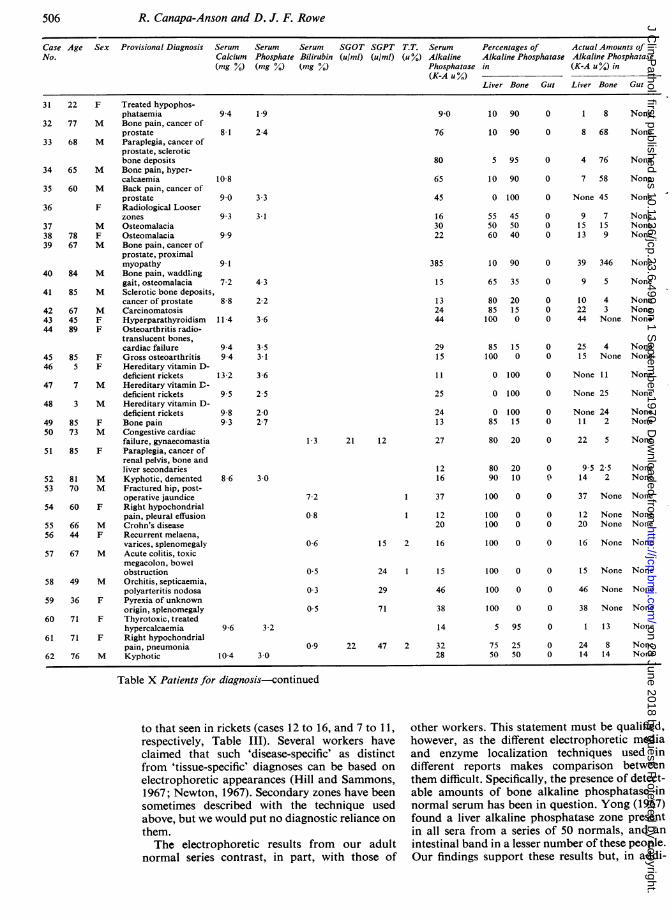
Sixty-three patients for diagnosis are sum-
marized in Table X. The results obtained on
these patients are arranged as listed in Table I,
on the bases of initial diagnosis and electro-phoretic results. Total serum alkaline phosphataseactivities lay between 9 and 385 K-A u %, themajority being above normal limits. Intestinalzones were seen only in cases 25 to 28.
Discussion
Our experience shows that polyacrylamide gelelectrophoresis offers a simple and reliablemeans for separating serum alkaline phosphataseactivity into a number of well defined fractions.The fractions correspond to tissues of origin:
in particular a fraction in liver, bone, intestine,and a placenta can be identified.Our designation of serum alkaline phosphatase
zones as 'liver', 'bone', etc, fractions was basedon two sets of preliminary investigations. First,the bands corresponded in mobility to the mainzones of activity of tissue extracts. These extractswere run both as 'pure' tissue preparations and as
admixtures to normal and abnormal sera.
Second, the bands corresponded to the patternsobserved in series of 'reference' sera, which were
obtained from patients with firmly establisheddiagnoses of advanced but uncomplicated liverand bone disease with high total serum alkalinephosphatase activities, and from normal childrenand adults. Although all the results from the twosets of investigations were in close agreement,they cannot be taken to provide absolute proofof the source of serum enzyme fractions. This isa theoretical limitation applying to all serum
enzyme studies.Confirmatory evidence for the tissues of origin
of specific serum alkaline phosphatase zones was
provided by two other findings. First, as men-
tioned before, the addition of L-phenylalanineto the alkaline phosphatase location reagentinhibited intestinal alkaline phosphatase zones
to a greater extent than other zones. Second, sera
and tissue extracts containing high proportionsof bone alkaline phosphatase demonstrated a
lower resistance to heat denaturation than didsamples containing high proportions of liver,
504 on 28 June 2018 by guest. P
rotected by copyright.http://jcp.bm
j.com/
J Clin P
athol: first published as 10.1136/jcp.23.6.499 on 1 Septem
ber 1970. Dow
nloaded from

Electrophoretic separation of tissue-specific serum alkaline phosphatases
intestinal, or placental alkaline phosphatase Although we are satisfied that the technique(Posen, Neale, and Clubb, 1965). described here clearly separates serum alkalineThe fractions seen after electrophoresis can phosphatase activities derived from different
be estimated semiquantitatively by visual assess- tissues, we have been unable to establish, so far,ment. This method would be unacceptable for patterns characteristic of differenttypesofdisease.estimating total serum alkaline phosphatase For example, the intense liver alkaline phos-activity with any accuracy but this can, of course, phatase zone associated with hepatic carcino-be measured accurately by conventional methods. matous deposits (cases 18 to 21, Table X) didVisual assessment merely compares relative not differ from the pattern associated with gall-intensities of electrophoretic zones. It has proved stone disease (cases 1, 2, 4 to 6, Table IV), andaccurate and reproducible even among untrained the increased bone alkaline phosphatase activityobservers. in Paget's disease was electrophoretically similar
Case Age Sex Provisional Diagnosis Serum Seruim Serum SGOT SGPT T.T. Serum Percentages of Actual Amounts ofNo. Calcium Phosphate Bilirabin (a/mi) (a/ml) (u %) Alkaline Alkaline Phosphatase Alkaline Phosphatase
(mg %) (mg %) (mg %) Phosphatase in (K-A u %A) in(K-A u Y.)
Liver Bone Gut Liver Bone Gut
1 42 M Cholecystitis2 29 M Infectious hepatitis3 79 M Postchlorpromazine
jaundice4 89 F Jaundice of unknown
aetiology5 58 M Jaundice of unknown
aetiology6 76 M Jaundice, cancer of
the pancreas7 64 M Jaundice, cancer of
the pancreas8 39 M Cancer of oesophagus,
palpable liver9 63 M Jaundice, cancer of
the pancreas10 67 F Jaundice, pancreatitis,
palpable liver11 69 M Jaundice, congestive
cardiac failure12 66 F Breast cancer, malignant
nodes in porta hepatis13 80 M Jaundice, cancer of
the bladder14 72 F Hemiplegia, hepato-
megaly15 68 F Acromegaly, fatty
degeneration of liver16 66 M Congestive cardiac
failure, hepatomegaly17 70 F Pneumonia, liver
disease, pyelonephritis18 80 F Breast canccr, hepatic
secondaries19 M Stomach cancer,
hepatomegaly20 60 M Paraplegia, reticulo-
sarcoma, hepaticsecondaries
21 80 F Rectal carcinoma,knobbly liver
22 60 M Paget's disease,infectious hepatitis
23 67 F Paget's disease,infectious hepatitis
24 75 F Breast cancer, ontestosterone, jaundiceopacities compatiblewith gallstones
25 60 F Jaundice, serum hepatitis26 54 M Large, hard liver,
diabetes; acquiredporphyria, cirrhosis
27 37 M Alcoholic cirrhosis28 M Cirrhosis29 86 M Cachexia, cancer of
pancreas, scleroticbone deposits 8-6 6.3
30 3 F Hereditary vitamin D-deficient rickets 9 3 2-7
0-687
0-5
11-4
4.5
20 8
3-2
3-2
2-2
22-0
1*3
6-2
5 6
0-6
03
08
0*5
0*5
1-0
68787
3
95
294
98
103
144
120
40
292
159
78
21
198640 9
17 1
2
118 2
3
82 1
103
71
480
225
105
9
2
25 44 2
177 1
5312
23
32
38
256
65
58
15
14
17
88
42
33
16
16
24
22
44
1-2 115 160 1 33
1.0 3 80
36
0-6 114 170 3 76
100 080 20
100 0
100 0
100 0
100 0
100 0
95 5
100 0
100 0
100 0
100 0
90 10
100 0
100 0
100 0
100 0
100 0
100 0
100 0
100 0
50 50
50 50
0
0
0
0
0
0
0
0
0
0
0
0
0
0
0
0
0
0
0
0
0
0
0
53 None None9 5 2 5 None
23 None None
32 None None
38 None None
256 None None
65 None None
55 3 None
15 None None
14 None None
17 None None
88 None None
38 4 None
33 None None
16 None None
16 None None
24 None None
22 None None
44 None None
33 None None
80 None None
18 18 None
38 38 None
19.0 178 149 2 32 55 45 0 18 14 None8-2 70 122 7 33 80 0 20 26 None 7
0-60O504
156
266
157
135
5 242418
50
31
80 0 2050 0 50
45 10 45
0 100 0
0 100 0
19 None S
12 None 128 2 8
None 50 None
None 31 None
Table X Patients for diagnosis
505
I
on 28 June 2018 by guest. Protected by copyright.
http://jcp.bmj.com
/J C
lin Pathol: first published as 10.1136/jcp.23.6.499 on 1 S
eptember 1970. D
ownloaded from
R. Canapa-Anson and D. J. F. Rowe
Case Age Sex Provisional Diagnosis Serum Serum Serum SGOT SGPT T.T. Serum Percentages of Actual Amounts ofNo. Calcium Phosphate Bilirabin (u/mi) (u/mI) (u%) Alkaline Alkaline Phosphatase Alkaline Phosphatase
(mg %) (mg %) (mg %) Phosphatase in (K-A u%) in(K-A u %) _
Liver Bone Gut Liver Bone Gut
Treated hypophos-phataemiaBone pain, cancer ofprostateParaplegia, cancer ofprostate, scleroticbone depositsBone pain, hyper-calcaemiaBack pain, cancer ofprostateRadiological LooserzonesOsteomalaciaOsteomalaciaBone pain, cancer ofprostate, proximalmyopathyBone pain, waddlinggait, osteomalaciaSclerotic bone deposits,cancer of prostateCarcinomatosisHyperparathyroidismOsteoarthritis radio-translucent bones,cardiac failureGross osteoarthritisHereditary vitamin D-deficient ricketsHereditary vitamin D-deficient ricketsHereditary vitamin D-deficient ricketsBone painCongestive cardiacfailure, gynaecomastiaParaplegia, cancer ofrenal pelvis, bone andliver secondariesKyphotic, dementedFractured hip, post-operative jaundiceRight hypochondrialpain, pleural effusionCrohn's diseaseRecurrent melaena,varices, splenomegalyAcute colitis, toxicmegacolon, bowelobstructionOrchitis, septicaemia,polyarteritis nodosaPyrexia of unknownorigin, splenomegalyThyrotoxic, treatedhypercalcaemiaRight hypochondrialpain, pneumoniaKyphotic
9.4
8 1
10 8
9 0
9.3
9.9
9.1
7 2
8-8
11 4
9.49.4
13 2
9.5
9 89.3
8-6
96
1 9
2-4
90
76
80
65
45
163022
3.33 1
4.32 2
3 6
3.53-1
3 6
2-5
2-027
385
15
132444
2915
11
25
2413
1-3 21 12 27
3 0
7 2
0 8
06
3 2
104 3 0
1216
1 37
1 1220
15 2 16
05 24 1 15
0 3 29 46
0-5 71 38
14
0 9 22 47 2 3228
10 90 0
10 90 0
5
10
0
555060
10
65
8085100
85100
0
0
0
85
80
8090
100
100100
100
100
100
100
5
7550
95
90
100
455040
90
35
20150
150
100
100
10015
20
2010
0
00
0
0
0
0
95
2550
0
0
0
000
0
0
000
0
0
0
0
00
0
0
0
0
00
0
0
0
0
0
00
1 8 None
8 68 None
4 76 None
7 58 None
None 45 None
9 7 None15 15 None13 9 None
39 346 None
9 5 None
10 4 None22 3 None44 None None
25 4 None15 None None
None 11 None
None 25 None
None 24 None11 2 None
22 5 None
9.5 2 5 None14 2 None
37 None None
12 None None20 None None
16 None None
15 None None
46 None None
38 None None
1 13 None
24 8 None14 14 None
Table X Patients for diagnosis-continued
to that seen in rickets (cases 12 to 16, and 7 to 11,respectively, Table III). Several workers haveclaimed that such 'disease-specific' as distinctfrom 'tissue-specific' diagnoses can be based on
electrophoretic appearances (Hill and Sammons,1967; Newton, 1967). Secondary zones have beensometimes described with the technique usedabove, but we would put no diagnostic reliance onthem.The electrophoretic results from our adult
normal series contrast, in part, with those of
other workers. This statement must be qualified,however, as the different electrophoretic mediaand enzyme localization techniques used indifferent reports makes comparison betweenthem difficult. Specifically, the presence of detect-able amounts of bone alkaline phosphatase innormal serum has been in question. Yong (1967)found a liver alkaline phosphatase zone presentin all sera from a series of 50 normals, and an
intestinal band in a lesser number of these people.Our findings support these results but, in addi-
22
77
68
F
M
M
65 M
60 M
F
M78 F67 M
31
32
33
34
35
36
373839
40
41
424344
4546
47
48
4950
51
5253
54
5556
57
58
59
60
61
62
84
85
674589
855
7
3
8573
85
8170
60
6644
67
49
36
71
71
76
M
M
MFF
FF
M
M
FM
F
MM
F
MF
M
M
F
F
F
M
506 on 28 June 2018 by guest. P
rotected by copyright.http://jcp.bm
j.com/
J Clin P
athol: first published as 10.1136/jcp.23.6.499 on 1 Septem
ber 1970. Dow
nloaded from

Electrophoretic separation of tissue-specific serum alkaline phosphatases
tion, a zone of bone alkaline phosphatase activitywas seen in all sera from our normal adult series(Fig. 2).The significance of serum alkaline phosphatase
activities depends, in part, on the age of thesubject. Like earlier workers we find that thetotal serum alkaline phosphatase activity ofchildren is significantly higher than that ofnormal adults (Clark and Beck, 1950). Electro-phoretic fractionation shows that this differenceis due entirely to the higher bone activity inchildhood. Our findings in a somewhat hetero-genous series of sick children suggest that in thisage group fractionation of serum alkaline phos-phatase might prove particularly useful in reveal-ing an abnormally low bone alkaline phos-phatase activity. This appears to be a commonaccompaniment of a variety of acute systemicillnesses, and it may prove a sensitive indicationof arrest of osteoblastic activity and bone growth.Such an index of decreasing osteoblastic activitymight be particularly useful in assessing the effectof steroid treatment on children. A fall in bonealkaline phosphatase activity can be masked bya rise in liver activity if only the total serumactivity is measured (cases 7 to 9, Table VIII).Conversely, a significant rise in liver alkalinephosphatase activity can be missed because of asynchronous fall in bone activity (cases 12 to 14,Table VIII). This fact has been used in the pastto question the usefulness of estimations of serumalkaline phosphatase activity in children (Hobbs,Campbell, and Scheuer, 1968). We believe that,
placental AP _-
liver AP °
bone AP
int,2tinal A.- --0
t
4 5 6 7
Fig. 3 Serum alkaline phosphatase patterns indifferent disease states related to a normal childand to a mixture of tissue extracts.
1 Paget 's disease with infectious hepatitis(AP = 76 K-A u%) in case 23 (Table X).
2 Osteomalacia (AP 31 K-A u%) in case 4(Table III).3 Normal child (AP = 24 K-A u%) in case 5
(Table VII).4 Mixture ofplacental, liver, bone, and intestinal
tissue extracts.5 Cirrhosis of the liver (AP = 24 K-A u%) in
case 26 (Table X).6 Obstructive jaundice (AP = 40 K-A u°/O) in
case 12 (Table IV).7 Late pregnancy (AP = 18 K-A u%) in case 5
(Table VI).
when coupled with electrophoretic fractionation,such studies can, in fact, be of value.At the other extreme of life, we were unable to
confirm reports by earlier workers that oldpeople, and in particular old women, have signi-ficantly higher serum alkaline phosphataseactivities than normal adults (Heino and Jokipii,1962; Klaassen and Siertsema, 1964). Our seriesof old people were all chosen because of theirhaving clinical or radiological evidence ofosteoporosis. We could find no evidence for anychange in serum bone alkaline phosphataseactivity in this group.Between childhood and old age we have found
serum alkaline phosphatase fractionation usefulin dealing with a variety of clinical problems.Not only could it establish the cause of a raisedserum alkaline phosphatase activity when thiswas in doubt but it could also reveal a mixedorigin when this had not been previously sus-pected. For example, case 24 (Table X) was apatient with carcinoma of the breast withjaundice. Abdominal radiographs showed opaci-ties compatible with gallstones. Bony secondarieswere not suspected until the raised serum alkalinephosphatase was shown to be due, in part, to anincrease in the serum bone fraction.A number of patients presented with known
chronic bone disease and a raised serum alkalinephosphatase which had been ascribed to osteo-blastic hyperactivity. Electrophoretic fractiona-tion revealed an abnormal increase in serum liveralkaline phosphatase activity (with or without anincrease in the bone fraction), and further inves-tigation showed the presence of liver disease(cases 36 to 45, Table X). In other cases (forexample, nos. 61 and 62, Table X) previously un-suspected osteoblastic bone involvement wasindicated by the electrophoretic results.
Routine fractionation of alkaline phosphatasehas also raised a number of questions which wehad not previously envisaged and which requirefurther study. Four patients with known orsuspected liver disease (cases 25 to 28, Table X)showed, in addition to a liver band, a markedintestinal band of alkaline phosphatase. Allproved on subsequent investigation to be casesof liver cirrhosis. Since an intestinal band isapparently not a feature of other types of liverdisease, this may prove to be an exceptionalinstance of disease as well as organ specificity.The Lewis blood group and secretor status ofthese patients was not determined.
In agreement with other workers (Gutman,Tyson, and Gutman, 1936) we find that the levelof serum bone alkaline phosphatase activity incases where there are secondary osteolyticcarcinomatous deposits bears little relation tothe degree of bone involvement as assessed radio-logically or clinically.
It is well known that total serum alkalinephosphatase activity is raised in the later stagesof pregnancy and that this increase is due to the
507
:1
on 28 June 2018 by guest. Protected by copyright.
http://jcp.bmj.com
/J C
lin Pathol: first published as 10.1136/jcp.23.6.499 on 1 S
eptember 1970. D
ownloaded from

R. Canapa-Anson and D. J. F. Rowe
appearance of placental alkaline phosphatase inthe serum. This placental component (Fig. 3) canbe readily demonstrated by electrophoresis(Boyer, 1961). Our results support this theoryalthough we could not detect a placental com-ponent in all the cases studied. It has also beenshown that the increase in total serum alkalinephosphatase activity is greater in twin pregnanciesthan in single ones (unpublished results). Theclinical significance of the amount of placentalalkaline phosphatase appearing in the serum as aguide to placental function remains to beexplored.
Instances where the electrophoretic techniquedescribed here is of little use are demonstrated bycases 46 to 48 (Table X). These children hadclinical evidence of bone disease but their totalserum alkaline phosphatase levels were withinnormal limits for their age. The electrophoreticpatterns seen in these cases were indistinguishablefrom those of normal children.
It might be argued that, whatever the intrinsicacademic interest of serum enzymefractionation,the technique is an unnecessary refinement forroutine day-to-day diagnosis. From our exper-ience we would disagree. As a group of diagnosticprocedures, serum enzyme estimations havemany disadvantages compared with older, moreconventional chemical tests. These disadvantagesare offset by two advantages. First, the estima-tions are potentially highly specific. Secondly,they can be extremely sensitive indicators ofdisease activity. Total serum alkaline phosphataseestimations lack both specificity and sensitivity:the results may not only fail to provide therequired information, they may even mislead.This is particularly so when a raised serumalkaline phosphatase level is the only abnormallaboratory finding in a patient complaining ofill defined symptoms-a not uncommon occur-rence. Coupling the total serum alkaline phos-phatase estimation with a sensitive fractionationtechnique makes the estimation a reliable andspecific indicator of disease.
We should like to thank the clinical staff of theWhittington Hospital for allowing us to inves-tigate their patients. We are indebted to Drs T. L.Dormandy and D. E. Sharland for their greathelp and encouragement in the preparation ofthis paper.
References
Akroyd, P. (1967). Acrylamide gel slab electrophoresis in a simpleglass cell for improved resolution and comparison of serumproteins. Analyt. Biochem., 19, 399-410.
Babson, A. L. (1965). Phenolphthalein monophosphate, a newsubstrate for alkaline phosphatase. Clin. Chem., 17, 789.
Boyer, S. H. (1961). Alkaline phosphatase in human sera andplacentae. Science, 134, 1002-1004.
Clark, L. C., and Beck, E. (1950). Plasma 'alkaline' phosphataseactivity. I. Normative data for growing children. J. Pediat.,36, 335-341.
Fishman, W. H., Green, S., and Inglis, N. 1. (1963). L-phenyl-analine: an organ specific, stereospecific inhit itor ofhumanintestinal alkaline phosphatase. Nature (Lond.), 198, 685-686.
Gutman, A. B., Tyson, T. L., and Gutman, E. B. (1936). Serumcalcium, inorganic phosphorus and phosphatase activity inhyperparathyroidism, Paget's disease, multiple myelomaand neoplastic diseases of the bones. Arch. intern. Med.57, 379-413.
Heino, A. E., and Jokipii, S. G. (1962). Serum alkaline phos,phatase levels in the aged. Ann. Med. intern. Fenn., 51,105-109.
Hill, P. G., and Sammons, H. G. (1967). An interpretation of theelevation of serum alkaline phosphatase in disease. J. clin.Path., 20, 654-659.
Hobbs, J. R., Campbell, D. M., and Scheuer, P. J. (1968). Clinicalvalue of serum 5-nucleotidase assay. In 6th InternationalCongress of Clinical Chemistry, Munich 1966, Vol. 2,Clinical Fnzymology, pp. 106-120. Karger, Basle and NewYork.
Klaassen, C. H. L., and Siertsema, L. H. (1964). De invloed vande leeftijd op de alkalische-fosfatasewaarde in het serum.Ned. T. Geneesk., 108, 1433-1436.
Newton, M. A. (1967). The clinical application of alkalinephosphatase electrophoresis. Quart. J. Med., 36, 17-28.
Posen, S., Neale, F. C., and Clubb, J. S. (1965). Heat inactivationin the study of human alkaline phosphatases. Ann. intern.Med., 62, 1234-1243.
Smith, I., Lightstone, P. J., and Perry, J. D. (1968). Separation ofhuman tissue alkaline phosphatases by electrophoresis onacrylamide disc gel. Clin. chim. Acta, 19, 499-505.
Yong, J. M. (1967). Origins of serum alkaline phosphatase. J.clin. Path., 20, 647-653.
508 on 28 June 2018 by guest. P
rotected by copyright.http://jcp.bm
j.com/
J Clin P
athol: first published as 10.1136/jcp.23.6.499 on 1 Septem
ber 1970. Dow
nloaded from